
Preparation and Application of Quaternized Chitosan- and AgNPs-Base Synergistic Antibacterial Hydrogel for Burn Wound Healing

 and
and 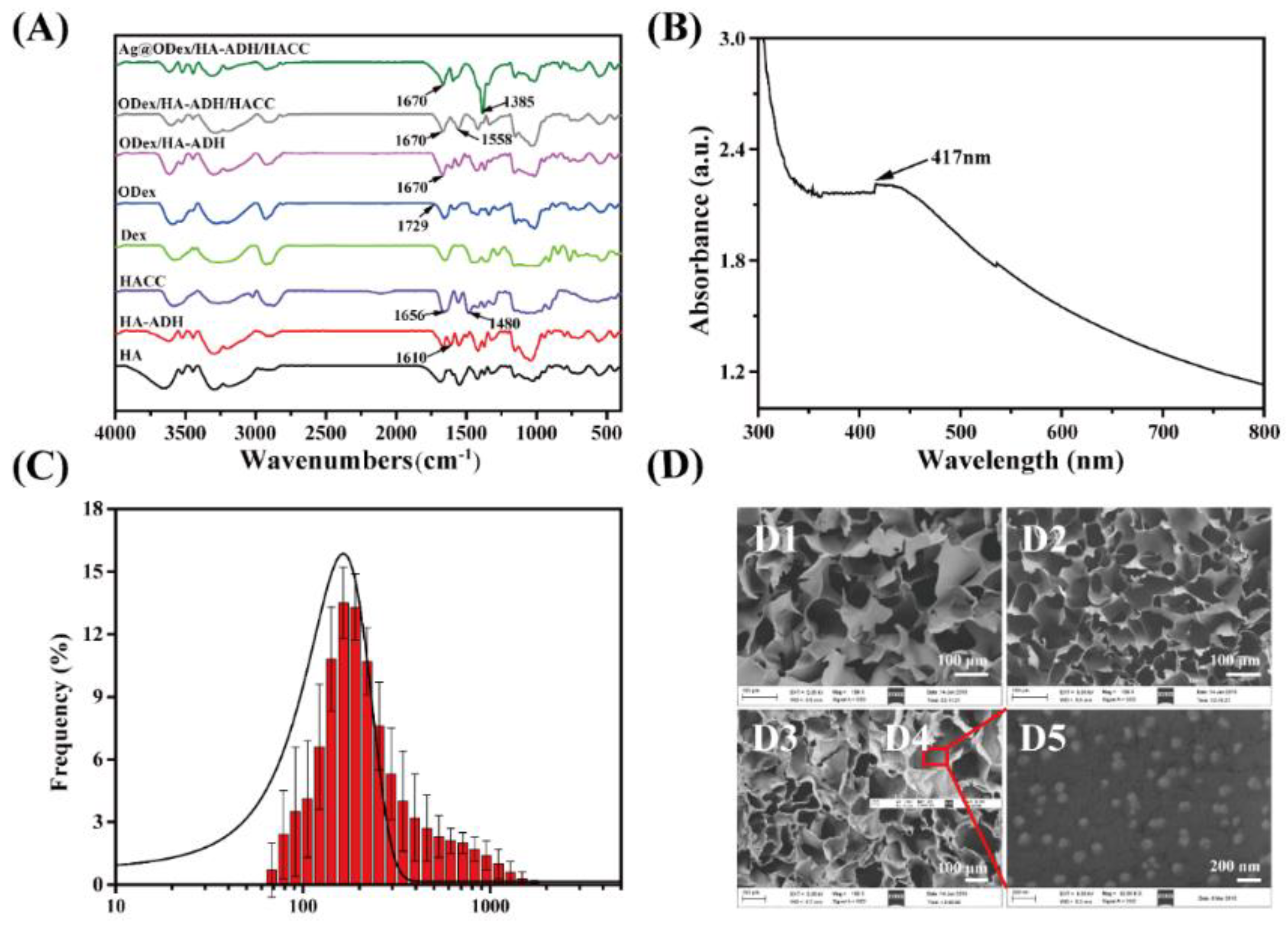
Abstract
:1. Introduction
2. Results and Discussions
2.1. Characterization of the Chemical Construction and Morphology of the Hydrogel
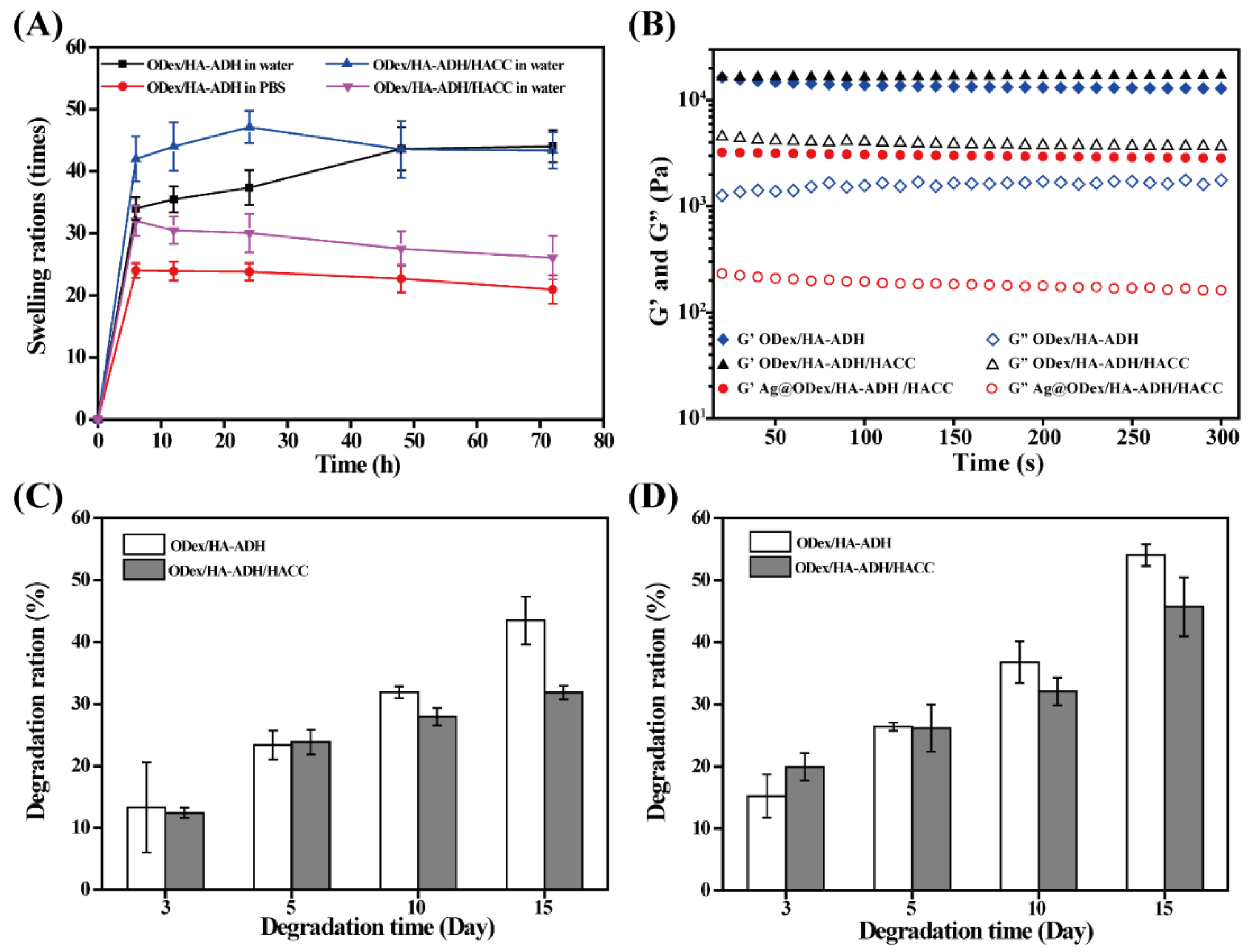
2.2. Swelling, In Vitro Degradation and the Rheology Behaviors of Hydrogels
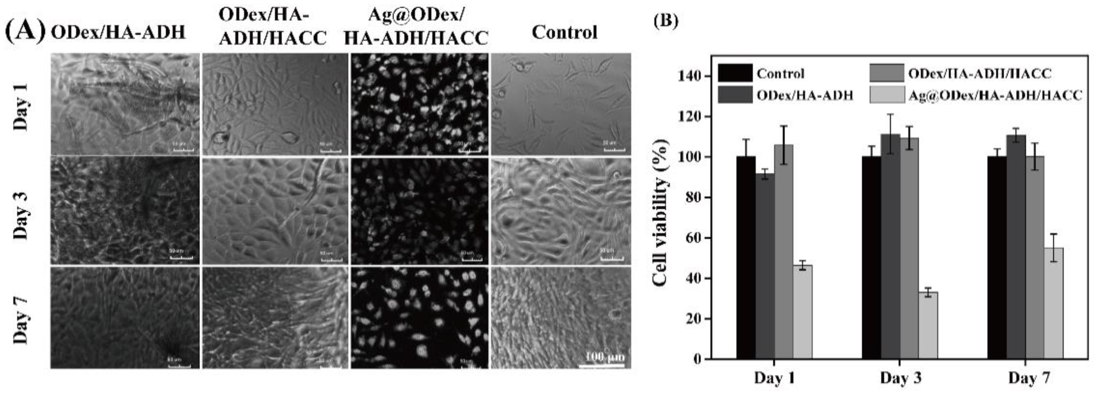
2.3. In Vitro Cytotoxicity of Hydrogel
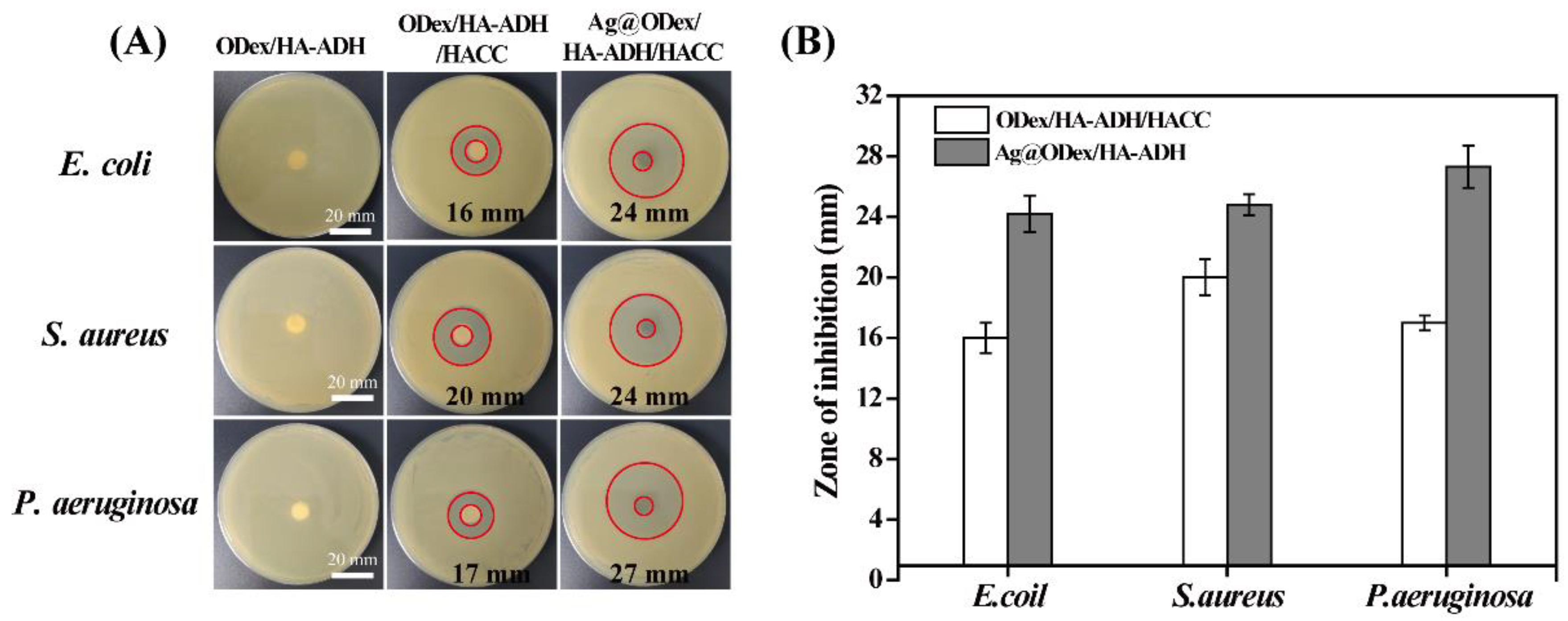
2.4. Antibacterial Activity of Hydrogel In Vitro
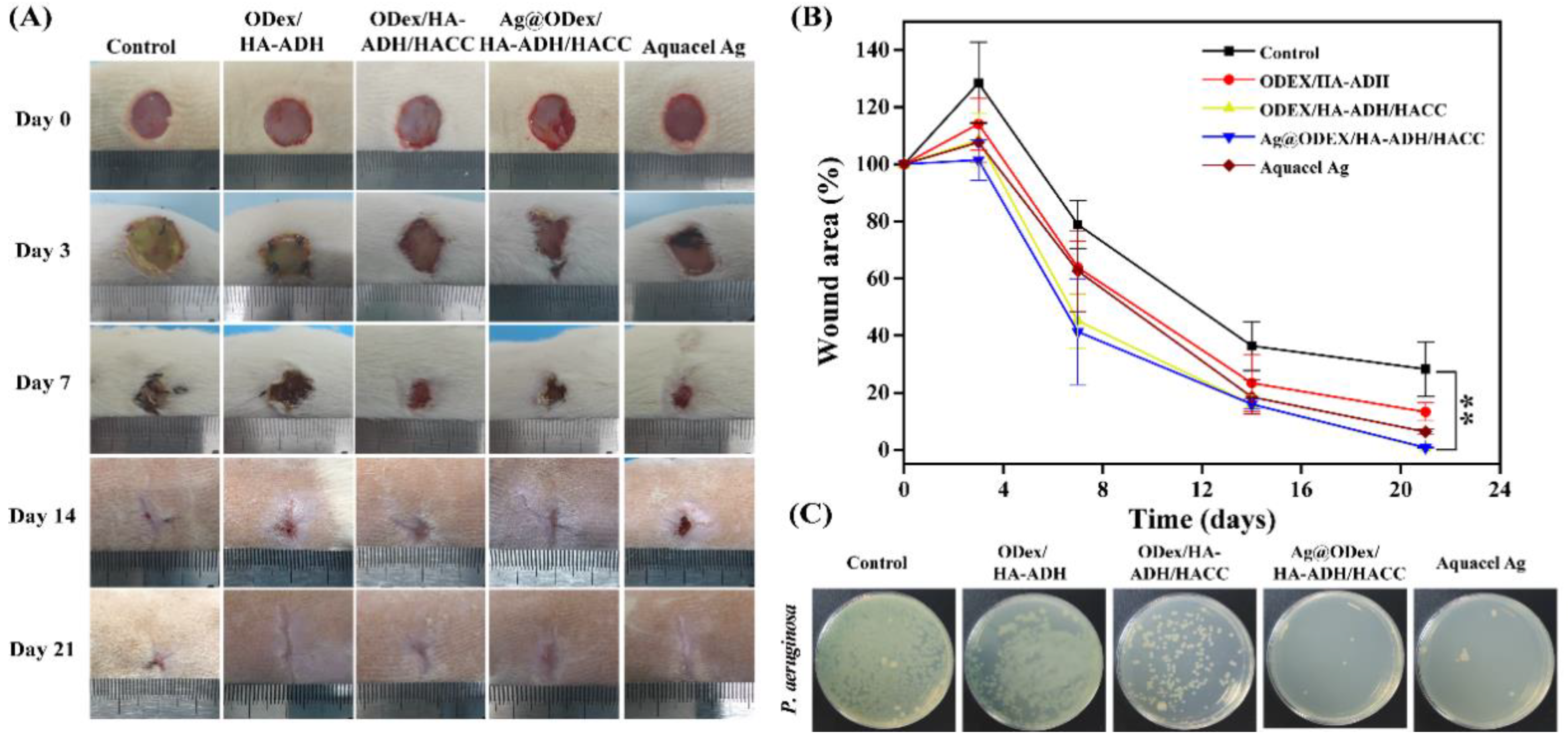
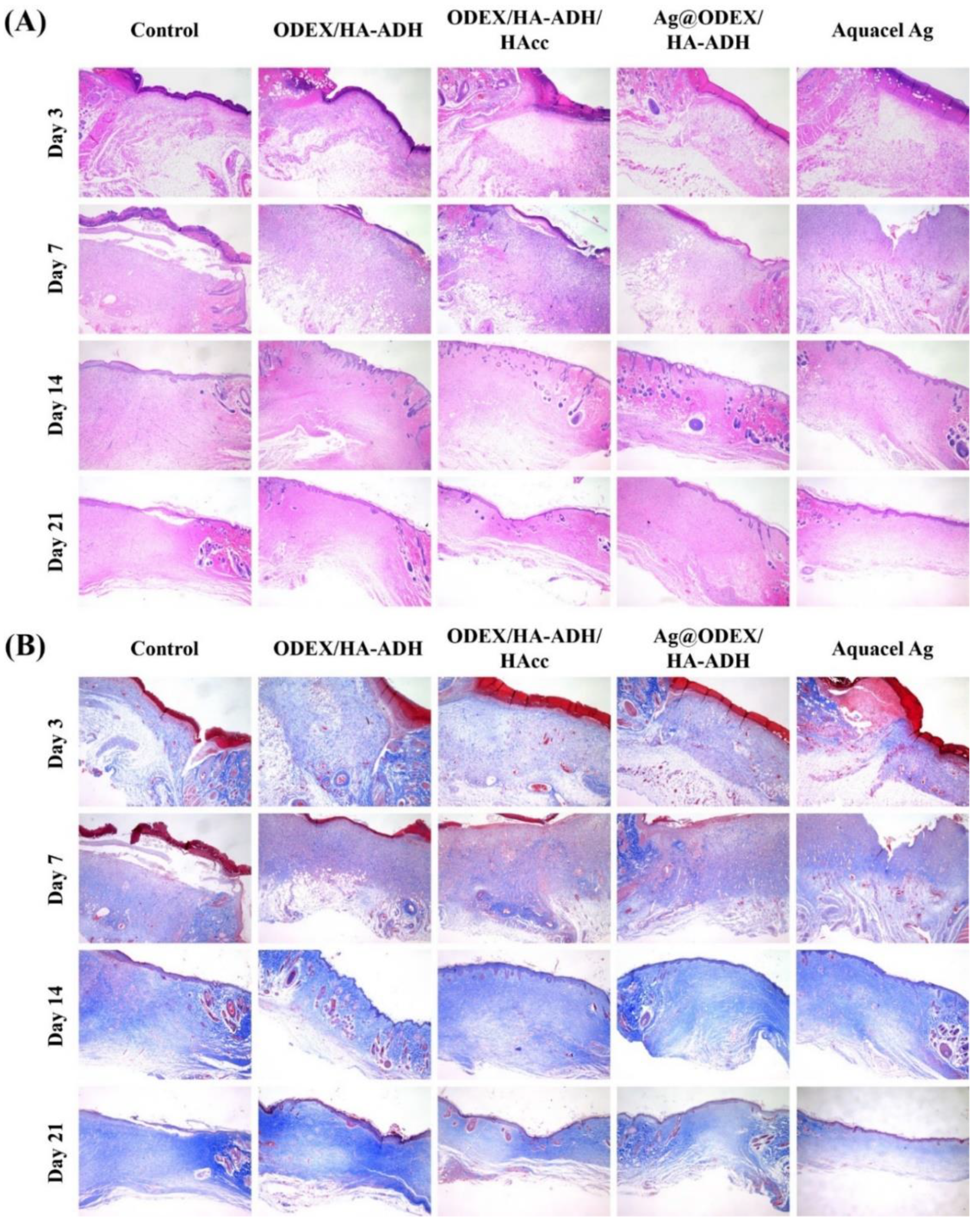
2.5. In Vivo Wound Healing Performance of Hydrogel
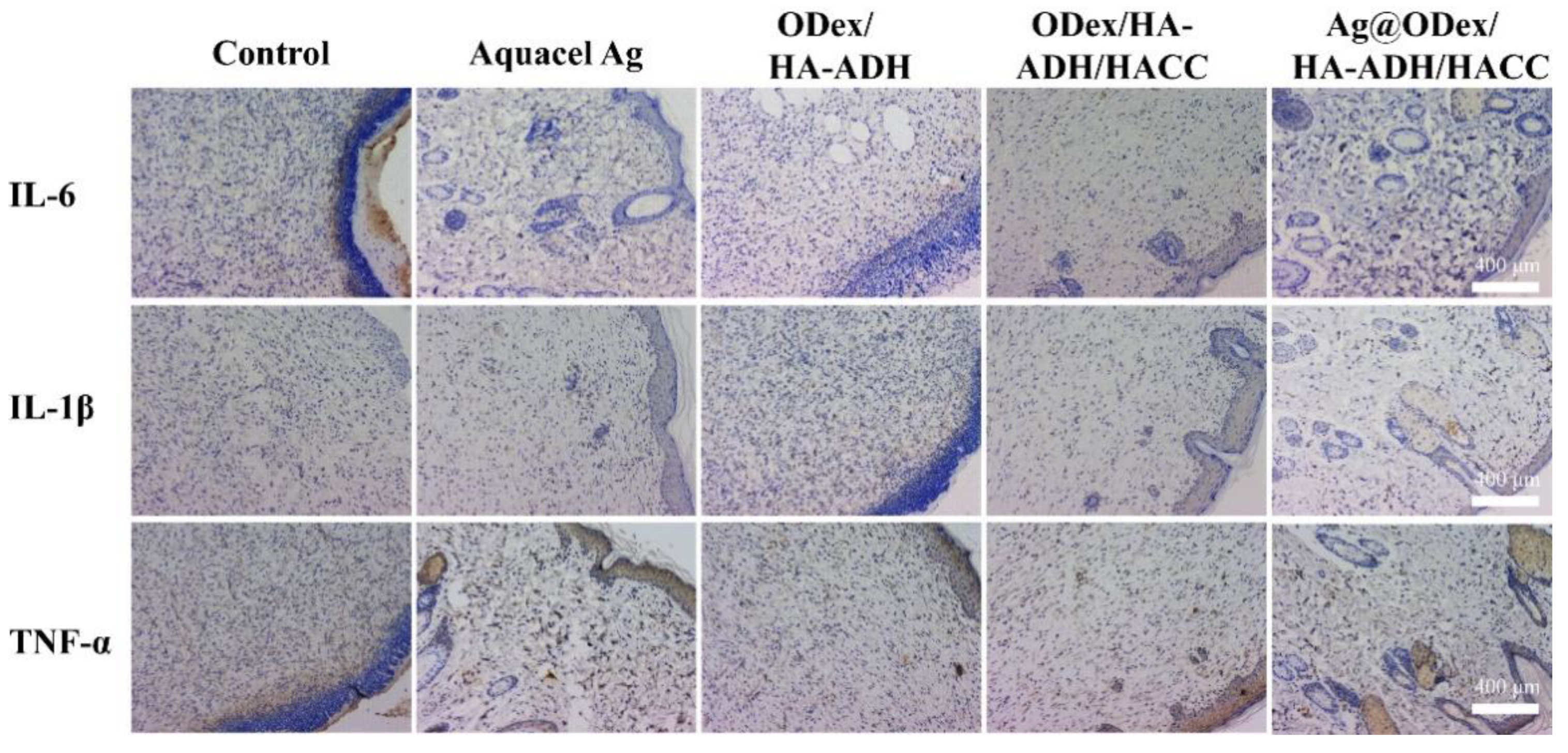
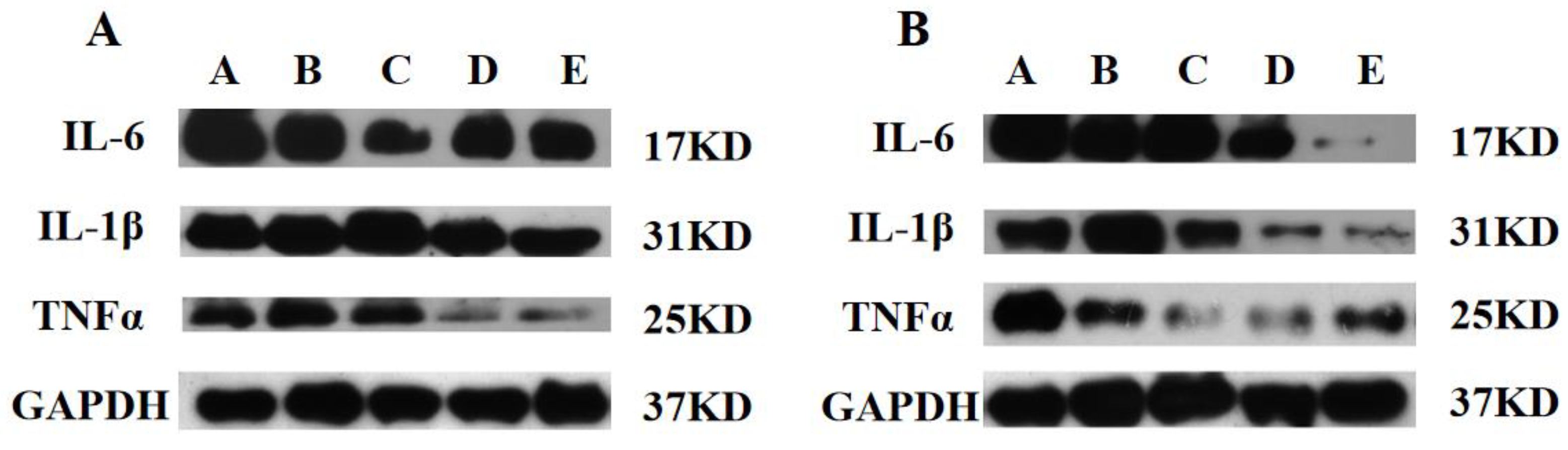
2.6. Immunohistochemistry and Western Blot Analysis
3. Materials and Methods
3.1. Materials and Reagents
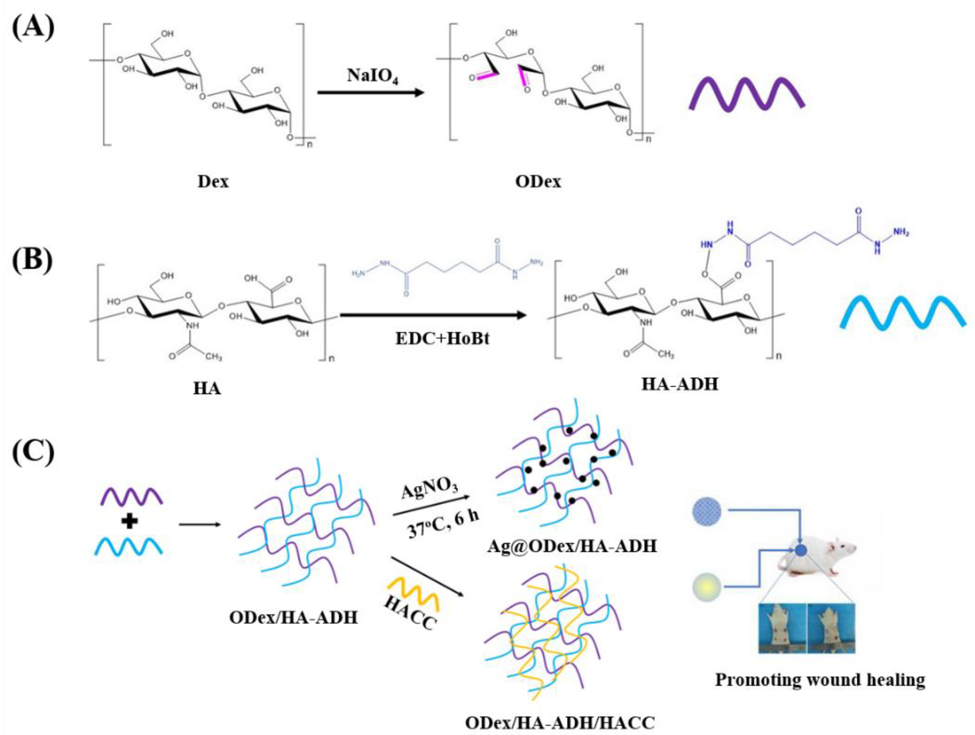
3.2. Synthesis of Oxidized Dextran (ODex)
3.3. Synthesis of HA-ADH
3.4. Preparation of the Hydrogel
3.4.1. Preparation of ODex/HA-ADH Hydrogel
3.4.2. Preparation of ODex/HA-ADH/HACC Hydrogel
3.4.3. Preparation of Ag@ODex/HA-ADH/HACC Hydrogel
3.5. Chemical Structure Characterization of the Hydrogels and AgNPs
3.6. In Vitro Swelling and Degradation Behavior of the Hydrogel
3.6.1. In Vitro Swelling Behavior
3.6.2. In Vitro Degradation Behavior
3.7. Rheological Property of the Hydrogel
3.8. In Vitro Antibacterial Property
3.9. In Vitro Cytotoxicity of the Hydrogel
3.10. In Vivo Wound Healing
3.11. Histological Analysis, Immunohistochemistry, and Western Blot
3.12. Statistical Analysis
4. Conclusions
Supplementary Materials
Author Contributions
Funding
Institutional Review Board Statement
Informed Consent Statement
Data Availability Statement
Acknowledgments
Conflicts of Interest
Sample Availability
References
- Kabashima, K.; Honda, T.; Ginhoux, F.; Egawa, G. The immunological anatomy of the skin. Nat. Rev. Immunol. 2019, 19, 19–30. [Google Scholar] [CrossRef] [PubMed]
- Han, L.; Li, P.F.; Tang, P.F.; Wang, X.; Zhou, T.; Wang, K.F.; Ren, F.Z.; Guo, T.L.; Lu, X. Mussel-inspired cryogels for promoting wound regeneration through photobiostimulation, modulating inflammatory responses and suppressing bacterial invasion. Nanoscale 2019, 11, 15846–15861. [Google Scholar] [CrossRef] [PubMed]
- Parfejevs, V.; Debbache, J.; Shakhova, O.; Schaefer, S.M.; Glausch, M.; Wegner, M.; Suter, U.; Riekstina, U.; Werner, S.; Sommer, L. Injury-activated glial cells promote wound healing of the adult skin in mice. Nat. Commun. 2018, 9, 236. [Google Scholar] [CrossRef]
- Li, J.; Chen, J.; Kirsner, R. Pathophysiology of acute wound healing. Clin. Dermatol. 2007, 25, 9–18. [Google Scholar] [CrossRef]
- Wu, Y.; Huang, S.; Enhe, J.; Fu, X.B. Insights into bone marrow-derived mesenchymal stem cells safety for cutaneous repair and regeneration. Int. Wound J. 2012, 9, 586–594. [Google Scholar] [CrossRef] [PubMed]
- Murakami, K.; Aoki, H.; Nakamura, S.; Nakamura, S.; Takikawa, M.; Hanzawa, M.; Kishimoto, S.; Hattori, H.; Tanaka, Y.; Kiyosawa, T.; et al. Hydrogel blends of chitin/chitosan, fucoidan and alginate as healing-impaired wound dressings. Biomaterials 2010, 31, 83–90. [Google Scholar] [CrossRef]
- Gan, D.L.; Xu, T.; Xing, W.S.; Ge, X.; Fang, L.M.; Wang, K.F.; Ren, F.Z.; Lu, X. Mussel-Inspired Contact-Active Antibacterial Hydrogel with High Cell Affinity, Toughness, and Recoverability. Adv. Funct. Mater. 2019, 29, 1805964. [Google Scholar] [CrossRef] [Green Version]
- Cheng, L.; Cai, Z.W.; Ye, T.J.; Yu, X.H.; Chen, Z.J.; Yan, Y.F.; Qi, J.; Wang, L.; Liu, Z.H.; Cui, W.G.; et al. Injectable Polypeptide-Protein Hydrogels for Promoting Infected Wound Healing. Adv. Funct. Mater. 2020, 30, 2001196. [Google Scholar] [CrossRef]
- Lopez-Silva, T.L.; Leach, D.G.; Azares, A.; Li, I.C.; Woodside, D.G.; Hartgerink, J.D. Chemical functionality of multidomain peptide hydrogels governs early host immune response. Biomaterials 2020, 231, 119667. [Google Scholar] [CrossRef] [PubMed]
- Li, M.; Zhang, Z.Y.; Liang, Y.P.; He, J.H.; Guo, B.L. Multifunctional Tissue-Adhesive Cryogel Wound Dressing for Rapid Nonpressing Surface Hemorrhage and Wound Repair. ACS Appl. Mater. Interfaces 2020, 12, 35856–35872. [Google Scholar] [CrossRef]
- Peng, L.Y.; Chang, L.; Si, M.T.; Lin, J.X.; Wei, Y.; Wang, S.T.; Liu, H.L.; Han, B.; Jiang, L. Hydrogel-Coated Dental Device with Adhesion-Inhibiting and Colony-Suppressing Properties. ACS Appl. Mater. Interfaces 2020, 12, 9718–9725. [Google Scholar] [CrossRef]
- Pan, J.; Zhang, Z.; Zhan, Z.Y.; Xiong, Y.F.; Wang, Y.F.; Cao, K.Y.; Chen, Y.J. In Situ generation of silver nanoparticles and nanocomposite films based on electrodeposition of carboxylated chitosan. Carbohydr. Polym. 2020, 242, 116391. [Google Scholar] [CrossRef] [PubMed]
- Gao, G.; Jiang, Y.W.; Jia, H.R.; Wu, F.G. Near-infrared light-controllable on-demand antibiotics release using thermo-sensitive hydrogel-based drug reservoir for combating bacterial infection. Biomaterials 2019, 188, 83–95. [Google Scholar] [CrossRef]
- Wang, X.Y.; Guo, J.J.; Zhang, Q.; Zhu, S.X.; Liu, L.; Jiang, X.F.; Wei, D.H.; Liu, R.S.; Li, L.D. Gelatin sponge functionalized with gold/silver clusters for antibacterial application. Nanotechnology 2020, 31, 134004. [Google Scholar] [CrossRef] [PubMed]
- Wu, Z.G.; Zhou, W.; Deng, W.J.; Xu, C.L.; Cai, Y.; Wang, X.Y. Antibacterial and Hemostatic Thiol-Modified Chitosan-Immobilized AgNPs Composite Sponges. ACS Appl. Mater. Interfaces 2020, 12, 20307–20320. [Google Scholar] [CrossRef]
- Shi, C.L.; Pu, X.B.; Zheng, G.; Feng, X.L.; Yang, X.; Zhang, B.L.; Zhang, Y.; Yin, Q.S.; Xia, H. An antibacterial and absorbable silk-based fixation material with impressive mechanical properties and biocompatibility. Sci. Rep. 2016, 6, 37418. [Google Scholar] [CrossRef] [PubMed] [Green Version]
- He, X.; Liu, X.Z.; Yang, J.; Du, H.B.; Chai, N.W.; Sha, Z.; Geng, M.R.; Zhou, X.J.; He, C.L. Tannic acid-reinforced methacrylated chitosan/methacrylated silk fibroin hydrogels with multifunctionality for accelerating wound healing. Carbohydr. Polym. 2020, 247, 116689. [Google Scholar] [CrossRef] [PubMed]
- Chen, Y.; Qiu, H.Y.; Dong, M.H.; Cheng, B.; Jin, Y.G.; Tong, Z.R.; Li, P.W.; Li, S.D.; Yang, Z.M. Preparation of hydroxylated lecithin complexed iodine/carboxymethyl chitosan/sodium alginate composite membrane by microwave drying and its applications in infected burn wound treatment. Carbohydr. Polym. 2019, 206, 435–445. [Google Scholar] [CrossRef]
- Mi, X.; Vijayaragavan, K.S.; Heldt, C.L. Virus adsorption of water-stable quaternized chitosan nanofibers. Carbohydr. Res. 2014, 387, 24–29. [Google Scholar] [CrossRef]
- Martin, V.T.; Wang, L.; Zeng, R.; You, D.Q.; Zhang, X.R.; Elodie, W.H.; Yu, B. Carboxymethyl chitosan-zinc coating for prevention of pin tract infection: An animal model. J. Orthop. Surg. 2018, 26, 2309499017749981. [Google Scholar] [CrossRef] [Green Version]
- Cheah, W.Y.; Show, P.L.; Ng, I.S.; Lin, G.Y.; Chiu, C.Y.; Chang, Y.K. Antibacterial activity of quaternized chitosan modified nanofiber membrane. Int. J. Biol. Macromol. 2019, 126, 569–577. [Google Scholar] [CrossRef] [PubMed]
- Hu, D.Y.; Wang, H.X.; Wang, L.J. Physical properties and antibacterial activity of quaternized chitosan/carboxymethyl cellulose blend films. LWT Food Sci. Technol. 2016, 65, 398–405. [Google Scholar] [CrossRef]
- Liu, Q.; Wu, B.L.; Yu, Q.S.; Wang, Y. Immobilization of quaternary ammonium based antibacterial monomer onto dentin substrate by non-thermal atmospheric plasma. Dent. Mater. J. 2019, 38, 821–829. [Google Scholar] [CrossRef] [PubMed] [Green Version]
- Tang, S.H.; Zheng, J. Antibacterial Activity of Silver Nanoparticles: Structural Effects. Adv. Healthc. Mater. 2018, 7, 1701503. [Google Scholar] [CrossRef]
- Gao, C.H.; Cheng, H.; Xu, N.; Li, Y.; Chen, Y.M.F.; Wei, Y.; Gao, B.; Fu, J.J.; Huo, K.F.; Xiong, W. Poly(dopamine) and Ag nanoparticle-loaded TiO2 nanotubes with optimized antibacterial and ROS-scavenging bioactivities. Nanomedicine 2019, 14, 803–818. [Google Scholar] [CrossRef] [PubMed]
- Park, E.J.; Bae, E.; Yi, J.; Kim, Y.; Choi, K.; Lee, S.H.; Yoon, J.; Lee, B.C.; Park, K. Repeated-dose toxicity and inflammatory responses in mice by oral administration of silver nanoparticles. Environ. Toxicol. Pharmacol. 2010, 30, 162–168. [Google Scholar] [CrossRef] [PubMed]
- Jin, F.; Xiang, Q.; Chen, X.; Peng, X.; Xing, X. An environmentally benign dual action antimicrobial: Quaternized chitosan/sodium alga acid multilayer films and silver nanoparticles decorated on magnetic nanoparticles. J. Biomater. Sci. Polym. Ed. 2016, 27, 1447–1461. [Google Scholar] [CrossRef]
- Zhang, Y.; Li, L.M.; Mu, J.F.; Chen, J.C.; Feng, S.Q.; Gao, J.Q. Implantation of a functional TEMPO-hydrogel induces recovery from rat spinal cord transection through promoting nerve regeneration and protecting bladder tissue. Biomater. Sci. 2020, 8, 1695–1701. [Google Scholar] [CrossRef]
- Zhou, B.; Gao, M.; Feng, X.J.; Huang, L.L.; Huang, Q.X.; Kootala, S.; Larsson, T.E.; Zheng, L.; Bowden, T. Carbazate modified dextrans as scavengers for carbonylated proteins. Carbohydr. Polym. 2020, 232, 115802. [Google Scholar] [CrossRef]
- Huang, S.H.; Liu, H.L.; Huang, S.S.; Fu, T.L.; Xue, W.; Guo, R. Dextran methacrylate hydrogel microneedles loaded with doxorubicin and trametinib for continuous transdermal administration of melanoma. Carbohydr. Polym. 2020, 246, 116650. [Google Scholar] [CrossRef]
- Fekri, R.; Salehi, M.; Asadi, A.; Kubicki, M. Synthesis, characterization, anticancer and antibacterial evaluation of Schiff base ligands derived from hydrazone and their transition metal complexes. Inorg. Chim. Acta 2019, 484, 245–254. [Google Scholar] [CrossRef]
- Bulavin, L.; Kutsevol, N.; Chumachenko, V.; Soloviov, D.; Kuklin, A.; Marynin, A. SAXS Combined with UV-vis Spectroscopy and QELS: Accurate Characterization of Silver Sols Synthesized in Polymer Matrices. Nanoscale Res. Lett. 2016, 11, 35. [Google Scholar] [CrossRef] [Green Version]
- Panacek, A.; Kvitek, L.; Prucek, R.; Kolar, M.; Vecerova, R.; Pizurova, N.; Sharma, V.K.; Nevecna, T.; Zboril, R. Silver colloid nanoparticles: Synthesis, characterization, and their antibacterial activity. J. Phys. Chem. B 2006, 110, 16248–16253. [Google Scholar] [CrossRef]
- Huang, S.S.; Liu, H.L.; Liao, K.D.; Hu, Q.Q.; Guo, R.; Deng, K.X. Functionalized GO Nanovehicles with Nitric Oxide Release and Photothermal Activity-Based Hydrogels for Bacteria-Infected Wound Healing. ACS Appl. Mater. Interfaces 2020, 12, 28952–28964. [Google Scholar] [CrossRef] [PubMed]
- Ji, C.D.; Annabi, N.; Khademhosseini, A.; Dehghani, F. Fabrication of porous chitosan scaffolds for soft tissue engineering using dense gas CO2. Acta Biomater. 2011, 7, 1653–1664. [Google Scholar] [CrossRef]
- She, W.C.; Luo, K.; Zhang, C.Y.; Wang, G.; Geng, Y.Y.; Li, L.; He, B.; Gu, Z.W. The potential of self-assembled, pH-responsive nanoparticles of mPEGylated peptide dendron-doxorubicin conjugates for cancer therapy. Biomaterials 2013, 34, 1613–1623. [Google Scholar] [CrossRef]
- Bowler, P.G. Wound pathophysiology, infection and therapeutic options. Ann. Med. 2002, 34, 419–427. [Google Scholar] [CrossRef] [PubMed]
- Zhu, Q.Y.; Jiang, M.; Liu, Q.; Yan, S.N.; Feng, L.B.; Lan, Y.; Shan, G.Q.; Xue, W.; Guo, R. Enhanced healing activity of burn wound infection by a dextran-HA hydrogel enriched with sanguinarine. Biomater. Sci. 2018, 6, 2472–2486. [Google Scholar] [CrossRef] [PubMed]
- Park, H.; Park, K.; Kim, D. Preparation and swelling behavior of chitosan-based superporous hydrogels for gastric retention application. J. Biomed. Mater. Res. A 2006, 76a, 144–150. [Google Scholar] [CrossRef] [PubMed]
- Narayanan, K.B.; Han, S.S. Dual-crosslinked poly(vinyl alcohol)/sodium alginate/silver nanocomposite beads—A promising antimicrobial material. Food Chem. 2017, 234, 103–110. [Google Scholar] [CrossRef]
- Liang, Y.P.; Zhao, X.; Hu, T.L.; Chen, B.J.; Yin, Z.H.; Ma, P.X.; Guo, B.L. Adhesive Hemostatic Conducting Injectable Composite Hydrogels with Sustained Drug Release and Photothermal Antibacterial Activity to Promote Full-Thickness Skin Regeneration During Wound Healing. Small 2019, 15, 1900046. [Google Scholar] [CrossRef]
- Burdick, J.A.; Chung, C.; Jia, X.Q.; Randolph, M.A.; Langer, R. Controlled degradation and mechanical behavior of photopolymerized hyaluronic acid networks. Biomacromolecules 2005, 6, 386–391. [Google Scholar] [CrossRef] [Green Version]
- Zhou, D.; Li, S.Z.; Pei, M.J.; Yang, H.J.; Gu, S.J.; Tao, Y.Z.; Ye, D.Z.; Zhou, Y.S.; Xu, W.L.; Xiao, P. Dopamine-Modified Hyaluronic Acid Hydrogel Adhesives with Fast-Forming and High Tissue Adhesion. ACS Appl. Mater. Interfaces 2020, 12, 18225–18234. [Google Scholar] [CrossRef]
- Huang, W.Q.; Leng, T.; Gao, M.M.; Hu, Q.Q.; Liu, L.S.; Dou, H.J. Scalable dextran-polypyrrole nano-assemblies with photothermal/photoacoustic dual capabilities and enhanced biocompatibility. Carbohydr. Polym. 2020, 241, 116224. [Google Scholar] [CrossRef] [PubMed]
- Huang, Z.Z.; Xu, P.; Chen, G.Q.; Zeng, G.M.; Chen, A.W.; Song, Z.X.; He, K.; Yuan, L.; Li, H.; Hu, L. Silver ion-enhanced particle-specific cytotoxicity of silver nanoparticles and effect on the production of extracellular secretions of Phanerochaete chrysosporium. Chemosphere 2018, 196, 575–584. [Google Scholar] [CrossRef]
- Serra, R.; Grande, R.; Butrico, L.; Rossi, A.; Settimio, U.F.; Caroleo, B.; Amato, B.; Gallelli, L.; de Franciscis, S. Chronic wound infections: The role of Pseudomonas aeruginosa and Staphylococcus aureus. Expert Rev. Anti-Infect. 2015, 13, 605–613. [Google Scholar] [CrossRef]
- Beyth, N.; Yudovin-Farber, I.; Bahir, R.; Domb, A.J.; Weissa, E. Antibacterial activity of dental composites containing quaternary ammonium polyethylenimine nanoparticles against Streptococcus mutans. Biomaterials 2006, 27, 3995–4002. [Google Scholar] [CrossRef]
- Namuiriyachote, N.; Lipipun, V.; Althhatuattananglzul, Y.; Charoonrut, P.; Ritthidej, G.C. Development of polyurethane foam dressing containing silver and asiaticoside for healing of dermal wound. Asian J. Pharm. Sci. 2019, 14, 63–77. [Google Scholar] [CrossRef] [PubMed]
- Nguyen, N.T.P.; Nguyen, L.V.H.; Tran, N.M.P.; Nguyen, D.T.; Nguyen, T.N.T.; Tran, H.A.; Dang, N.N.T.; Vo, T.V.; Nguyen, T.H. The effect of oxidation degree and volume ratio of components on properties and applications of in situ cross-linking hydrogels based on chitosan and hyaluronic acid. Mat. Sci. Eng. C Mater. 2019, 103, 109670. [Google Scholar] [CrossRef]
- Yang, Y.; Xia, T.; Zhi, W.; Wei, L.; Weng, J.; Zhang, C.; Li, X.H. Promotion of skin regeneration in diabetic rats by electrospun core-sheath fibers loaded with basic fibroblast growth factor. Biomaterials 2011, 32, 4243–4254. [Google Scholar] [CrossRef] [PubMed]
- Eming, S.A.; Martin, P.; Tomic-Canic, M. Wound repair and regeneration: Mechanisms, signaling, and translation. Sci. Transl. Med. 2014, 6, 265. [Google Scholar] [CrossRef] [Green Version]
- Chen, W.T.; Zhao, S.Q.; Ita, M.; Li, Y.; Ji, J.J.; Jiang, Y.; Redmond, H.P.; Wang, J.H.; Liu, J.H. An Early Neutrophil Recruitment into the Infectious Site Is Critical for Bacterial Lipoprotein Tolerance-Afforded Protection against Microbial Sepsis. J. Immunol. 2020, 204, 408–417. [Google Scholar] [CrossRef] [PubMed]
- Lin, Z.Q.; Kondo, T.; Ishida, Y.; Takayasu, T.; Mukaida, N. Essential involvement of IL-6 in the skin wound-healing process as evidenced by delayed wound healing in IL-6-deficient mice. J. Leukoc. Biol. 2003, 73, 713–721. [Google Scholar] [CrossRef]
- Edwards, R.; Harding, K.G. Bacteria and wound healing. Curr. Opin. Infect. Dis. 2004, 17, 91–96. [Google Scholar] [CrossRef] [PubMed]
- Deng, F.H.; He, S.Y.; Cui, S.D.; Shi, Y.Q.; Tan, Y.Y.; Li, Z.J.; Huang, C.Y.; Liu, D.L.; Zhi, F.C.; Peng, L. A Molecular Targeted Immunotherapeutic Strategy for Ulcerative Colitis via Dual-targeting Nanoparticles Delivering miR-146b to Intestinal Macrophages. J. Crohns Colitis 2019, 13, 482–494. [Google Scholar] [CrossRef]
- Yan, L.Y.; Qiu, B.B.; Li, T.T.; Wu, D.D.; Zhu, J.H.; Zhao, D.B. Green synthesis of silver nanoparticles from Lonicera japonica leaf extract and their anti-inflammatory and antibacterial effects. Micro. Nano Lett. 2020, 15, 90–95. [Google Scholar] [CrossRef]
- El-Aassar, M.R.; Ibrahim, O.M.; Fouda, M.M.G.; El-Beheri, N.G.; Agwa, M.M. Wound healing of nanofiber comprising Polygalacturonic/Hyaluronic acid embedded silver nanoparticles: In-Vitro and in-Vivo studies. Carbohydr. Polym. 2020, 238, 116175. [Google Scholar] [CrossRef]
- Fehaid, A.; Taniguchi, A. Silver nanoparticles reduce the apoptosis induced by tumor necrosis factor-alpha. Sci. Technol. Adv. Mater. 2018, 19, 526–534. [Google Scholar] [CrossRef]


Publisher’s Note: MDPI stays neutral with regard to jurisdictional claims in published maps and institutional affiliations. |
© 2021 by the authors. Licensee MDPI, Basel, Switzerland. This article is an open access article distributed under the terms and conditions of the Creative Commons Attribution (CC BY) license (https://creativecommons.org/licenses/by/4.0/).
Share and Cite
Chen, X.; Zhang, H.; Yang, X.; Zhang, W.; Jiang, M.; Wen, T.; Wang, J.; Guo, R.; Liu, H. Preparation and Application of Quaternized Chitosan- and AgNPs-Base Synergistic Antibacterial Hydrogel for Burn Wound Healing. Molecules 2021, 26, 4037. https://0-doi-org.brum.beds.ac.uk/10.3390/molecules26134037
Chen X, Zhang H, Yang X, Zhang W, Jiang M, Wen T, Wang J, Guo R, Liu H. Preparation and Application of Quaternized Chitosan- and AgNPs-Base Synergistic Antibacterial Hydrogel for Burn Wound Healing. Molecules. 2021; 26(13):4037. https://0-doi-org.brum.beds.ac.uk/10.3390/molecules26134037
Chicago/Turabian StyleChen, Xushan, Huimin Zhang, Xin Yang, Wuhong Zhang, Ming Jiang, Ting Wen, Jie Wang, Rui Guo, and Hanjiao Liu. 2021. "Preparation and Application of Quaternized Chitosan- and AgNPs-Base Synergistic Antibacterial Hydrogel for Burn Wound Healing" Molecules 26, no. 13: 4037. https://0-doi-org.brum.beds.ac.uk/10.3390/molecules26134037