Article

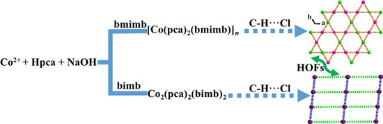
Construction of Two Stable Co(II)-Based Hydrogen-Bonded Organic Frameworks as a Luminescent Probe for Recognition of Fe3+ and Cr2O72− in H2O

1
Guangxi Key Laboratory of Green Chemical Materials and Safety Technology, College of Petroleum and Chemical Engineering, Beibu Gulf University, Qinzhou 535011, China
2
College of International Studies, Beibu Gulf University, Qinzhou 535011, China
3
School of Chemical and Environmental Engineering, Hanshan Normal University, Chaozhou 521041, China
*
Authors to whom correspondence should be addressed.
Submission received: 25 August 2021 / Revised: 21 September 2021 / Accepted: 29 September 2021 / Published: 30 September 2021

Abstract

:
A pair of cobalt(II)-based hydrogen-bonded organic frameworks (HOFs), [Co(pca)2(bmimb)]n (1) and [Co2(pca)4(bimb)2] (2), where Hpca = p-chlorobenzoic acid, bmimb = 1,3-bis((2-methylimidazol-1-yl)methyl)benzene, and bimb = 1,4-bis(imidazol-1-ylmethyl)benzene were hydrothermally synthesized and characterized through infrared spectroscopy (IR), elemental and thermal analysis (EA), power X-ray diffraction (PXRD), and single-crystal X-ray diffraction (SCXRD) analyses. X-ray diffraction structural analysis revealed that 1 has a one-dimensional (1D) infinite chain network through the deprotonated pca monodentate chelation and with a μ2-bmimb bridge Co(II) atom, and 2 is a binuclear Co(II) complex construction with a pair of symmetry-related pca and bimb ligands. For both 1 and 2, each cobalt atom has four coordinated twisted tetrahedral configurations with a N2O2 donor set. Then, 1 and 2 are further extended into three-dimensional (3D) or two-dimensional (2D) hydrogen-bonded organic frameworks through C–H···Cl interactions. Topologically, HOFs 1 and 2 can be simplified as a 4-connected qtz topology with a Schläfli symbol {64·82} and a 4-connected sql topology with a Schläfli symbol {44·62}, respectively. The fluorescent sensing application of 1 was investigated; 1 exhibits high sensitivity recognition for Fe3+ (Ksv: 10970 M−1 and detection limit: 19 μM) and Cr2O72− (Ksv: 12960 M−1 and detection limit: 20 μM). This work provides a feasible detection platform of HOFs for highly sensitive discrimination of Fe3+ and Cr2O72− in aqueous media.

Graphical Abstract

1. Introduction

Recently, a new class of materials named hydrogen-bonded organic frameworks (HOFs) has emerged as an exciting class of compounds stabilized by non-covalent hydrogen-bonding and π−π stacking interactions [1,2,3,4,5]. HOFs have attracted a significant deal of attention especially because of their broad applications, ranging from chemical sensing [6] and bioimaging [7,8] to lighting [9] and 3D optical storage [10,11]. The metal-organic frameworks (MOFs) and covalent-organic frameworks (COFs) are usually connected by coordination bonds or covalent bonds between atoms and show relatively high stabilities [12,13,14,15,16,17,18,19,20], while the HOFs are linked by weak non-covalent interactions, such as hydrogen bonds and π−π stacking interactions, and exhibit relatively low stabilities [21,22,23,24,25]. For HOFs, the solvent guests play important roles in the construction of the supermolecular network system. Once the solvent guests are removed, the supermolecular system is usually broken, and the HOFs collapse. Therefore, it is unsurprising that, although thousands of HOFs have been reported in the literature during the last two decades, examples of HOFs with permanent stable are very rare [26,27].
Hydrogen bonding, particularly within the coordination sphere, is another strategy which may lead to enhancement in the chemical stability at the nodes and therefore, of the framework materials themselves [28,29]. These interactions may act as support and rigidity of the coordination geometry of mononuclear coordination spheres, as well as provide extra thermodynamic and kinetic stabilization to meet the challenge of hydrolytic stability in these materials. However, a small subset of known MOF ligands contains suitable functionality for coordination sphere hydrogen bonding, which can provide new opportunities in ligand design [30,31,32,33,34].
To date, the halogen-containing carboxylic acid ligands, such as p-chlorobenzoic acid (Hpca), have afforded great attention in the preparation of HOFs for their outstanding features of versatile coordination fashions as well as potential hydrogen-bonding donors (C and O) and acceptors (Cl) [35,36,37,38,39,40,41,42,43]. A mononuclear complex can be afforded with the metal ions reaction with Hpca under appropriate conditions. Accordingly, C–H···Cl, C–Cl···π, C–H···π and π···π are excellent candidates for assembling mononuclear complex to HOFs in different ways, ranging from one-dimensional (1D) chains and two-dimensional (2D) sheets to three-dimensional (3D) porous structures [44,45,46,47,48,49,50,51,52]. Of further interest, p-chlorobenzoic acid featuring a chlorine in the 4-positions of the phenyl ring, as a derivative of benzoic acid, remains largely unexplored hitherto in the field of HOFs, compared with the well-studied ligands, benzoic acid.
Based on the above considerations and ongoing our research work in this area [53,54,55,56,57,58,59], we utilized Hpca organic ligands to act as the building blocks. Two Co(II)-based HOFs, [Co(pca)2(bmimb)]n (1) and [Co2(pca)4(bimb)2] (2) were successfully synthesized by hydrothermal methods and characterized by infrared spectrum (IR) (Figure S1), elemental analysis (EA), power X-ray diffraction (PXRD), single-crystal X-ray diffraction (SCXRD) and thermogravimetry (TG) (Scheme 1). In addition, the solid-state luminescence properties of 1 and 2, and the fluorescent detection of cations/anions of 1, were investigated at room temperature in detail.

2. Results and Discussion

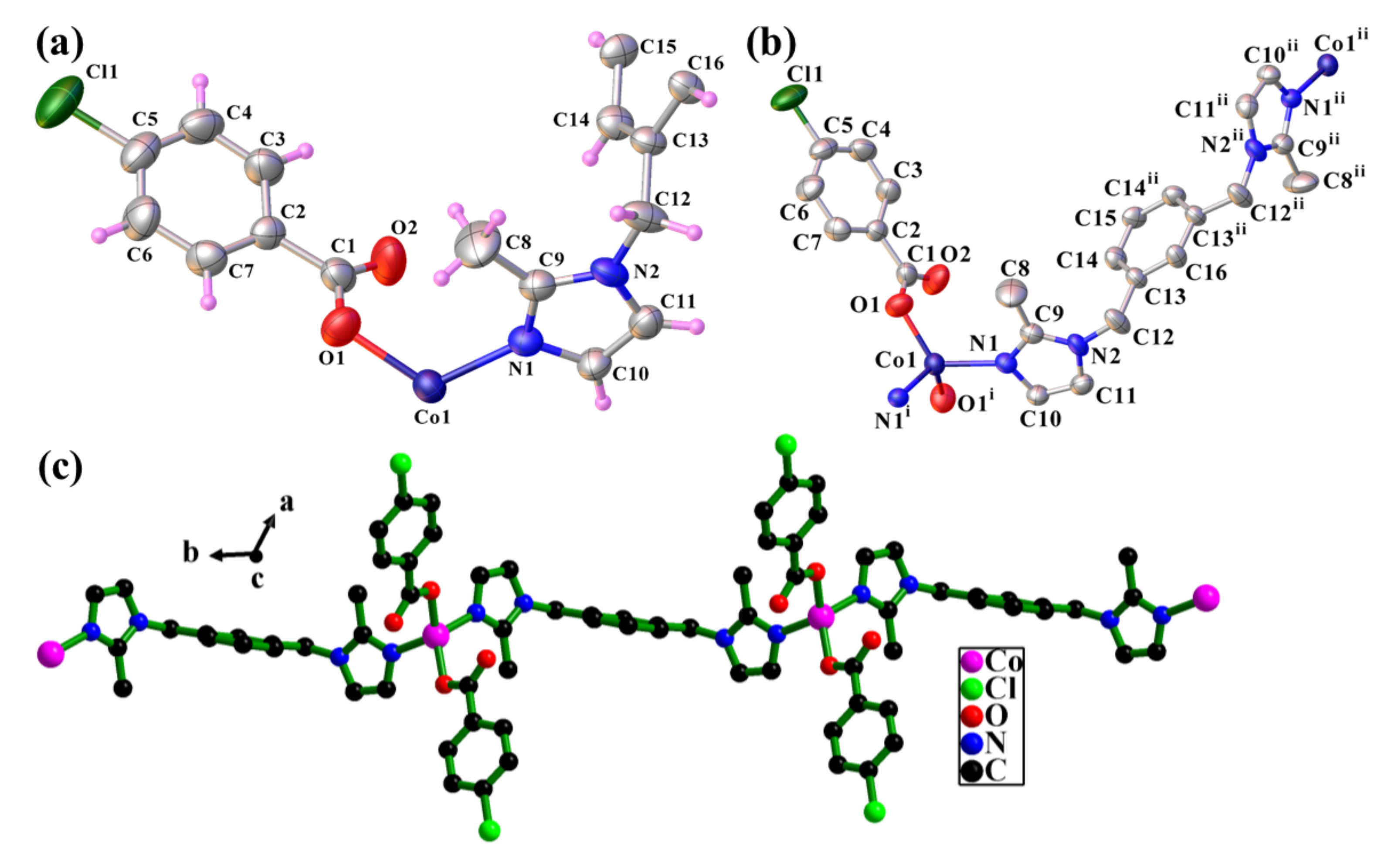
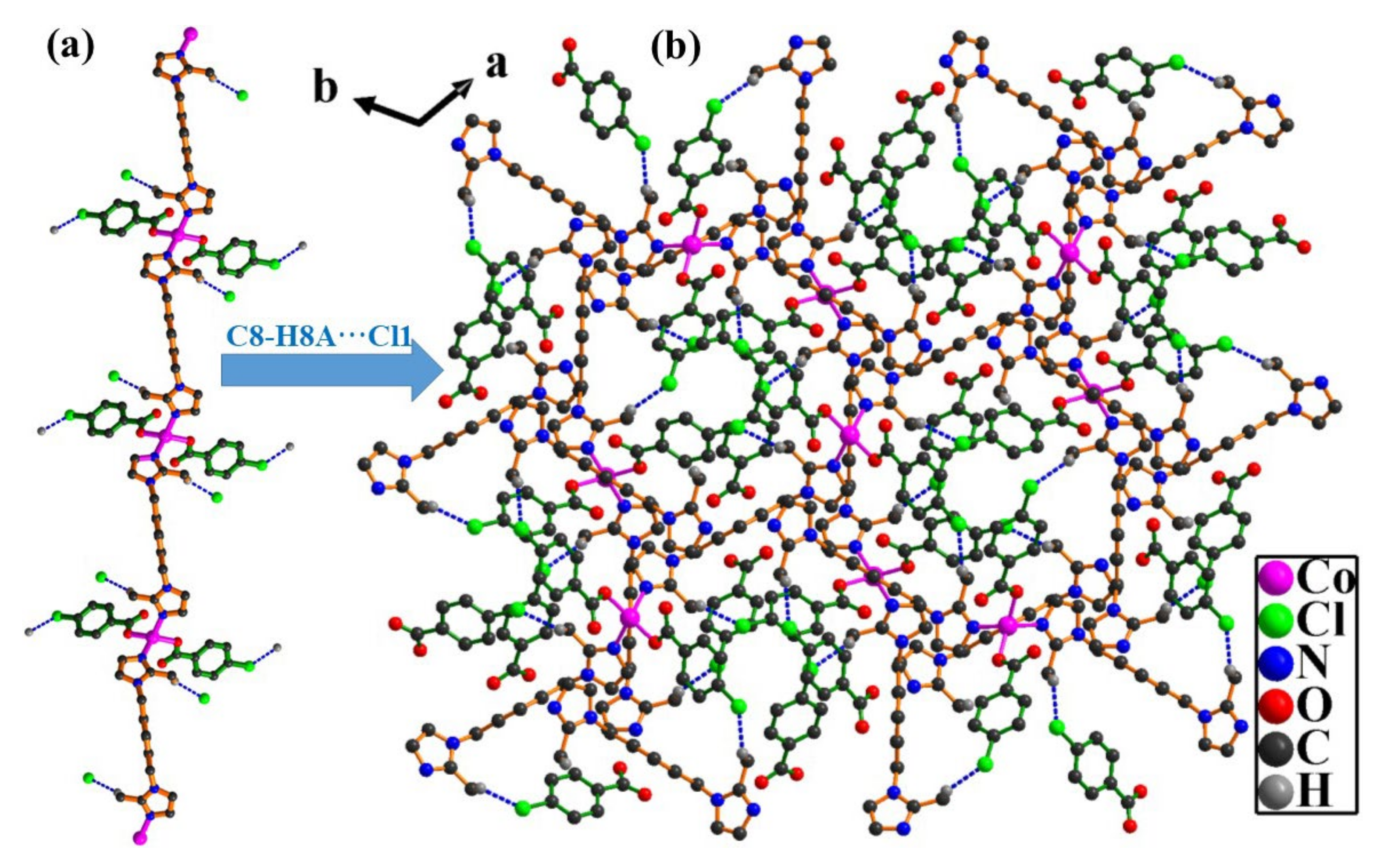
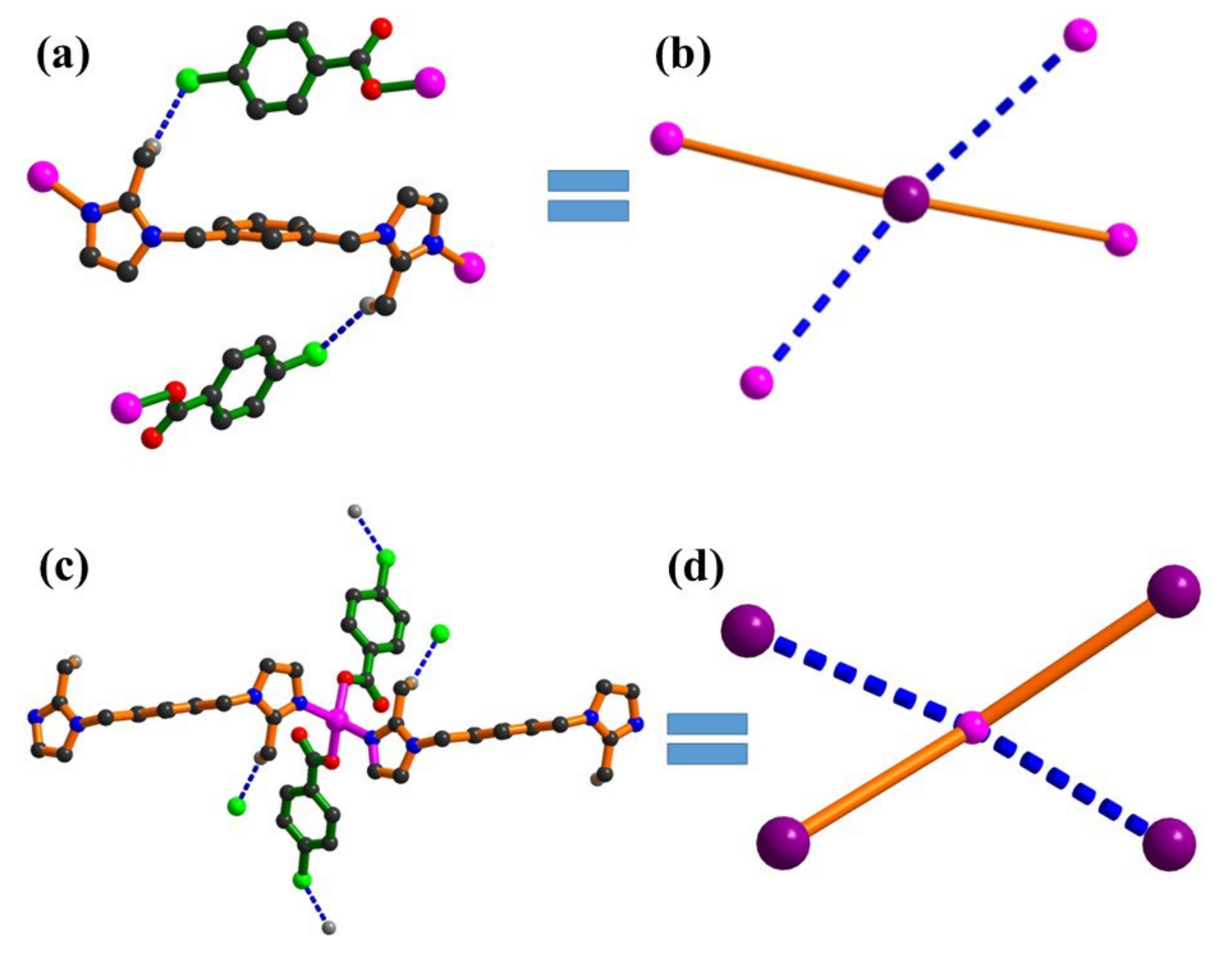
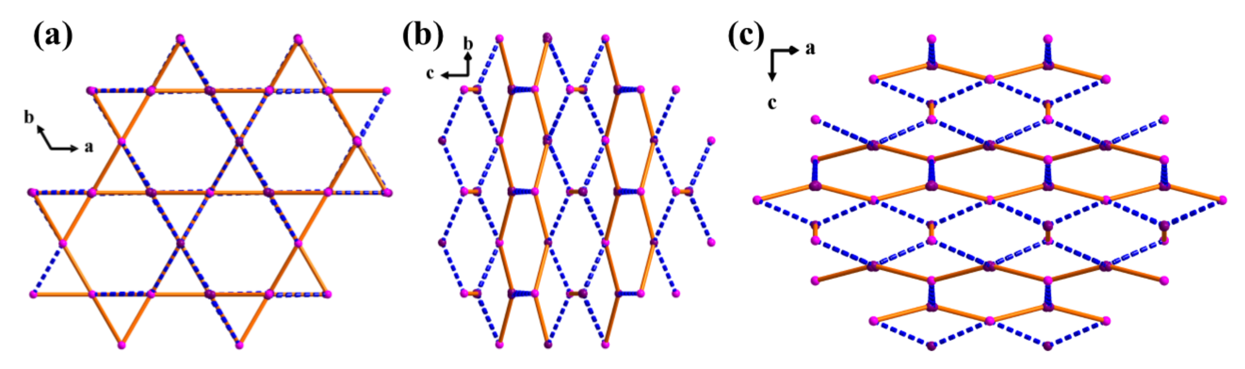
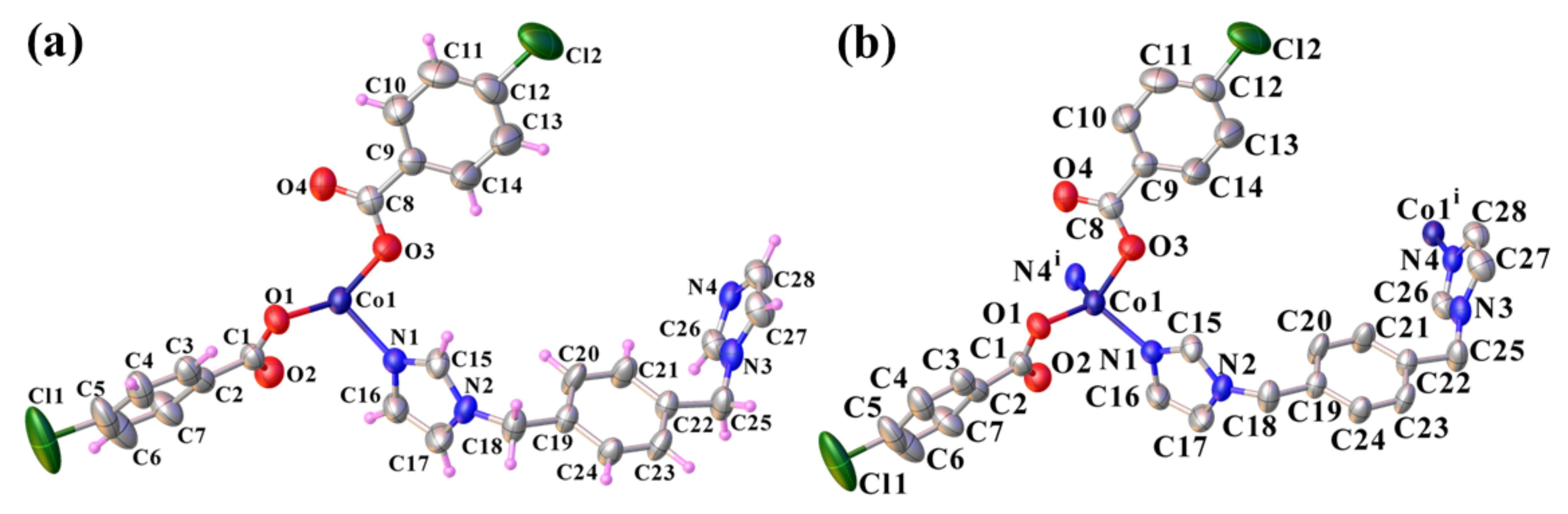
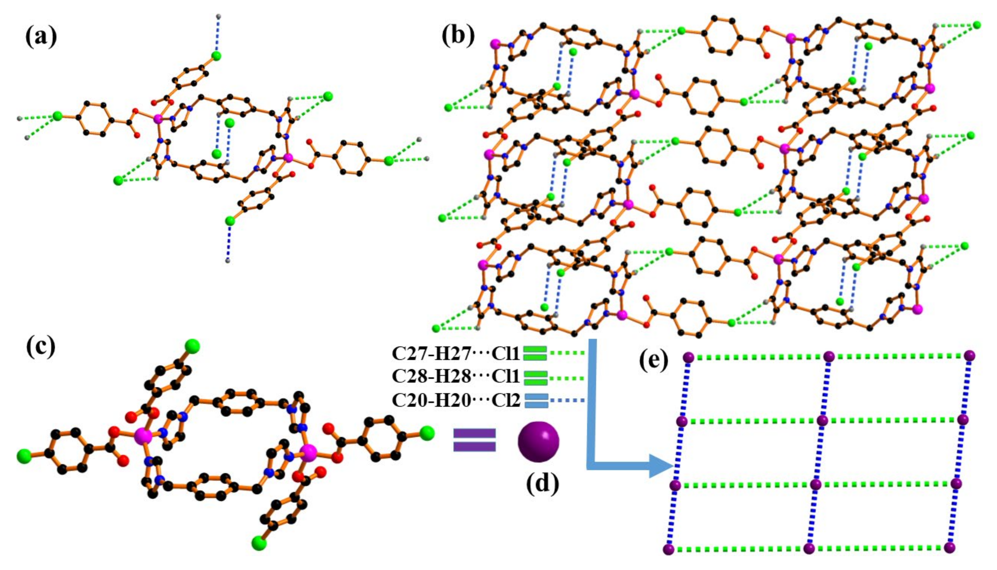
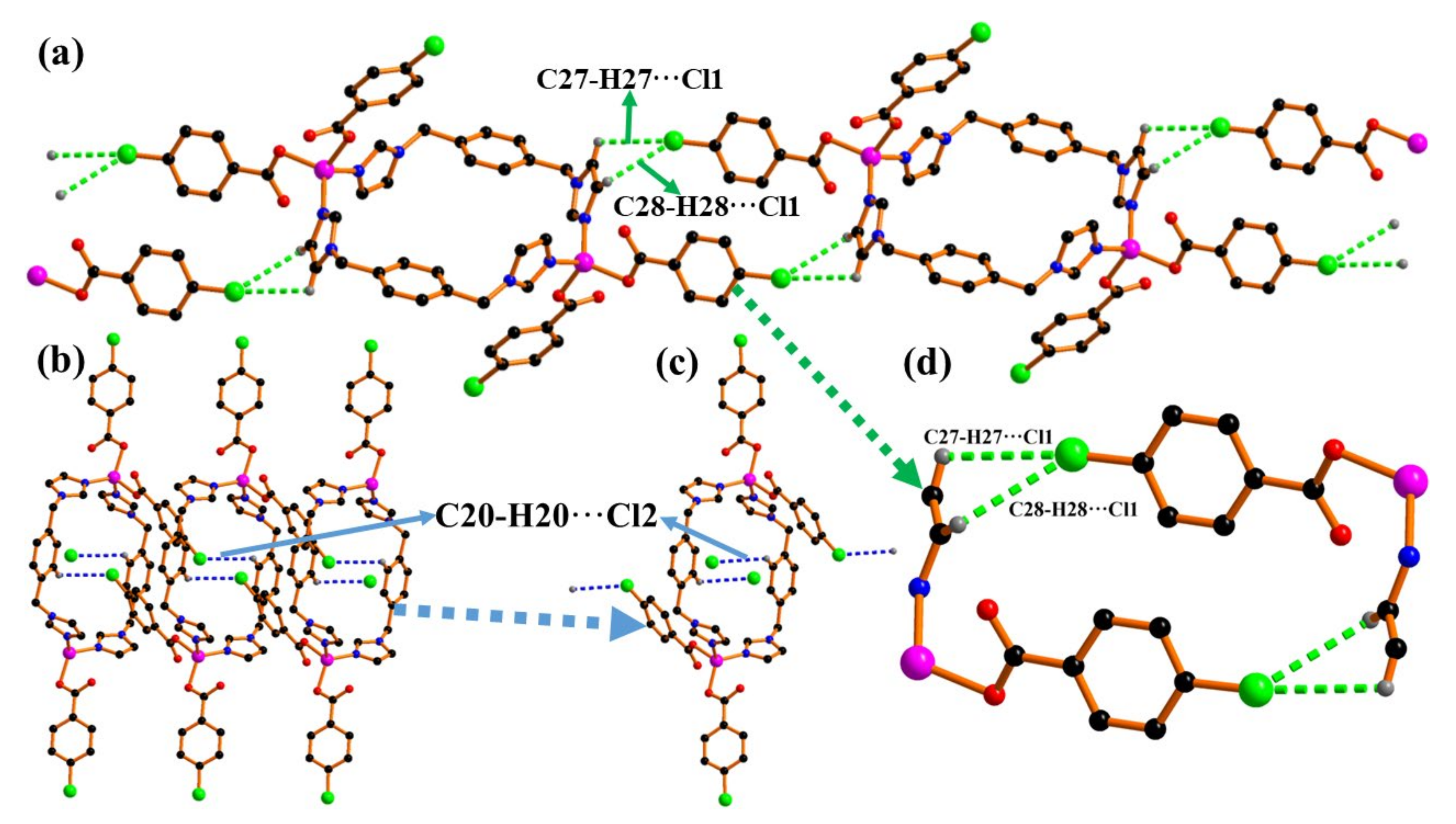
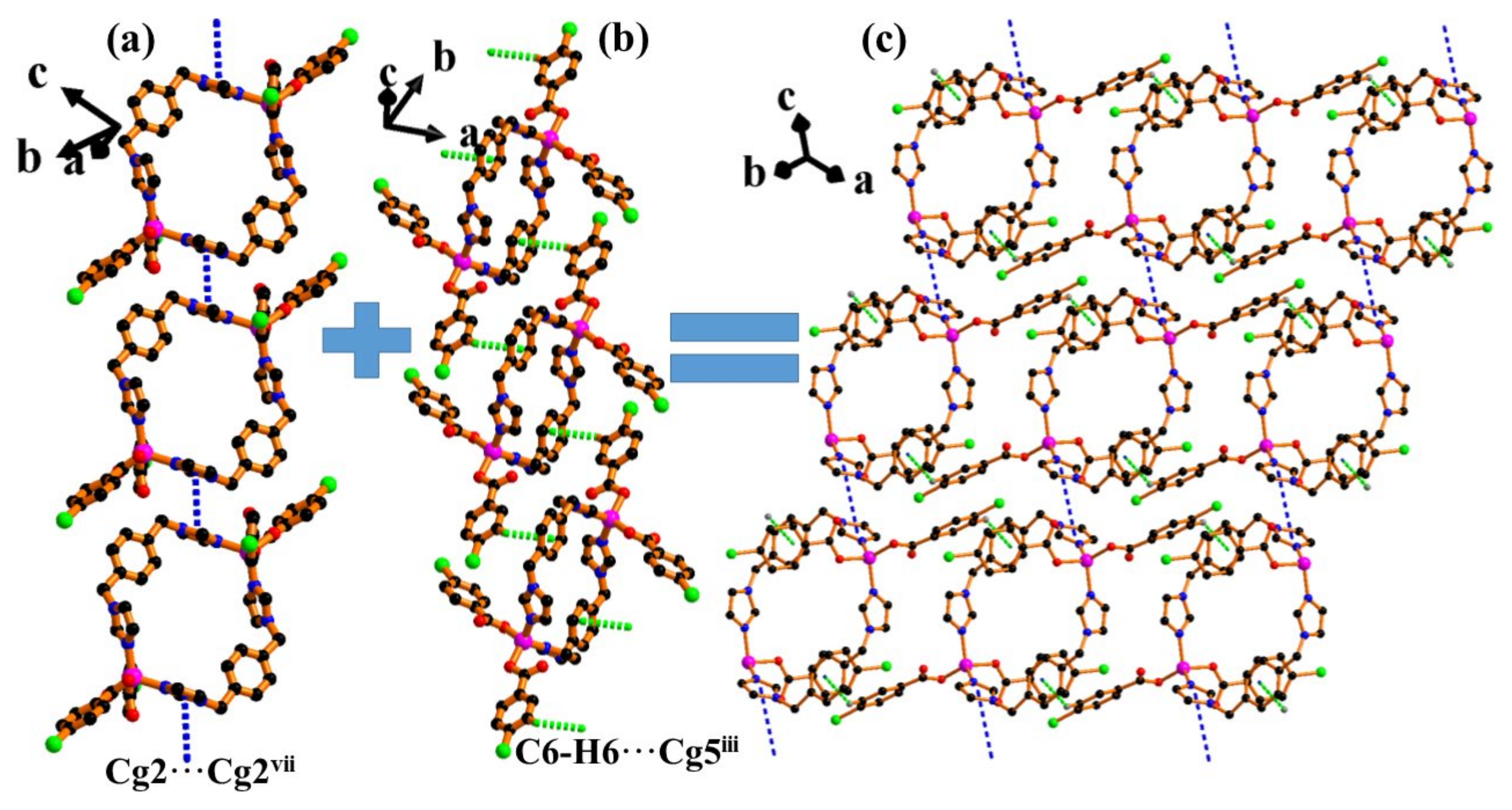
2.1. Description of the Structure of [Co(pca)2(bmimb)]n (1)

X-ray single crystal diffraction revealed that 1 is a one-dimensional coordination polymer and crystallizes in the monoclinic system with space group P21/n (Table S1). The asymmetric unit of 1 is comprised of one Co2+, one deprotonated monodentate chelating ligand pca and half of a neutral auxiliary ligand, bmimb (Figure 1a). Each Co ion is tetra-coordinated with two O atoms (O1 and O1i, symmetry code i seen in Table S2) and two N atoms (N1 and N1i) from two symmetry-related pac and bmimb ligands, resulting in a slightly distorted tetrahedron geometry (Figure 1b). The Co–O bond distance is 1.948(4) Å, and the Co–N distance is 2.048(4) Å (Table S2).
The detailed coordination mode of pca and bmimb ligands is shown in Figure 1c. Notably, the carboxyl group in pca adopts the mondenate type of coordination mode η10, while the bmimb ligand adopts a normal μ2 bridge coordination fashion. Two symmetry-related pca ligands bond to each Co2+ cation by the upside-down fashion through their deprotonated carboxylate O-atom, resulting in substructural blocks {[Co(pca)]2}2+. These neighboring {[Co(pca)]2}2+ are further interconnected through the μ2-bmimb bridge to form a one-dimensional infinite framework with a Co···Co separation of 14.02(4) Å.
There are hydrogen-bonding interactions (calculated by PLATON software) [60] that stabilize its structure and result in the formation of a three-dimensional network (Figure 2 and Table S3). In particular, interchain hydrogen bonds C–H···Cl stabilized the crystal structure of 1; these were formed between the methyl group of bmimb ligand (-CH3, C8), which is the donor, and the chlorine of the pca (Cl1), which acts as the acceptor (C8···Cl1viii = 3.676(7), H8A···Cl1viii = 2.84, C8-H8A···Cl1viii = 147°, symmetry code viii −x+y+1, −x+2, z−1/3). The bond distance between H8A and Cl1 is 2.840 Å; the bond angle of C8–H8A···Cl1 is 147°, which exceeds the preferred minimum hydrogen bond angle of 110°; and the distance between C8 and Cl1 is 3.676(7) Å, which is slightly greater than the sum of the van der Waals radii of C and Cl (3.52 Å) [61]. Therefore, it belongs to a strong C–H···Cl hydrogen bond, compared with the reported literature [62,63,64,65,66,67,68].
Each bmimb links two pca ligands with two inverse centrosymmetric groups -CH3 as a hydrogen-bonded substructural unit ([Co4(pca)2(bmimb)]6+) (Figure 3a), which is further extended by bmimb ligands, resulting in a 3D Co-based hydrogen-bonded organic frameworks. From the topological point of view, each [Co4(pca)2(bmimb)]6+ can be considered to act as a 4-connected node, and each {Co2+} links 2 pca and 2 bmimb molecules, which also can be considered a 4-connected node in 1 (Figure 3). Interestingly, the 3D Co-based hydrogen-bonded organic frameworks can be topologically simplified as a 4-connected 2-nodal qtz net with a Schläfli symbol {64·82} (Figure 4) as analyzed with the TOPOS 4.0 program [69].
Furthermore, between adjacent 1D chains, there exists a C–Cl···π stacking interaction between the symmetry-related pca (C5–Cl5 and C2–C3–C4–C5–C6–C7, Cg2ix, symmetry code: ix 2−y, 1+xy, 1/3+z). The angle C5–Cl5···Cg2ix is 153.1(3)°, with Cl5···Cg2ix and C5···Cg2ix distances of 3.843(4) Å and 5.454(7) Å, respectively. Thus, these hydrogen bonds insert and inter-lock the 1D chain in reversely alternating parallel arrangements, defining 3D HOFs supporting the supramolecular architecture through C–H···O, C–H···N, C–H···Cl and C–Cl···π interactions. The unit cell contains no residual solvent accessible void calculated by PLATON program [60].

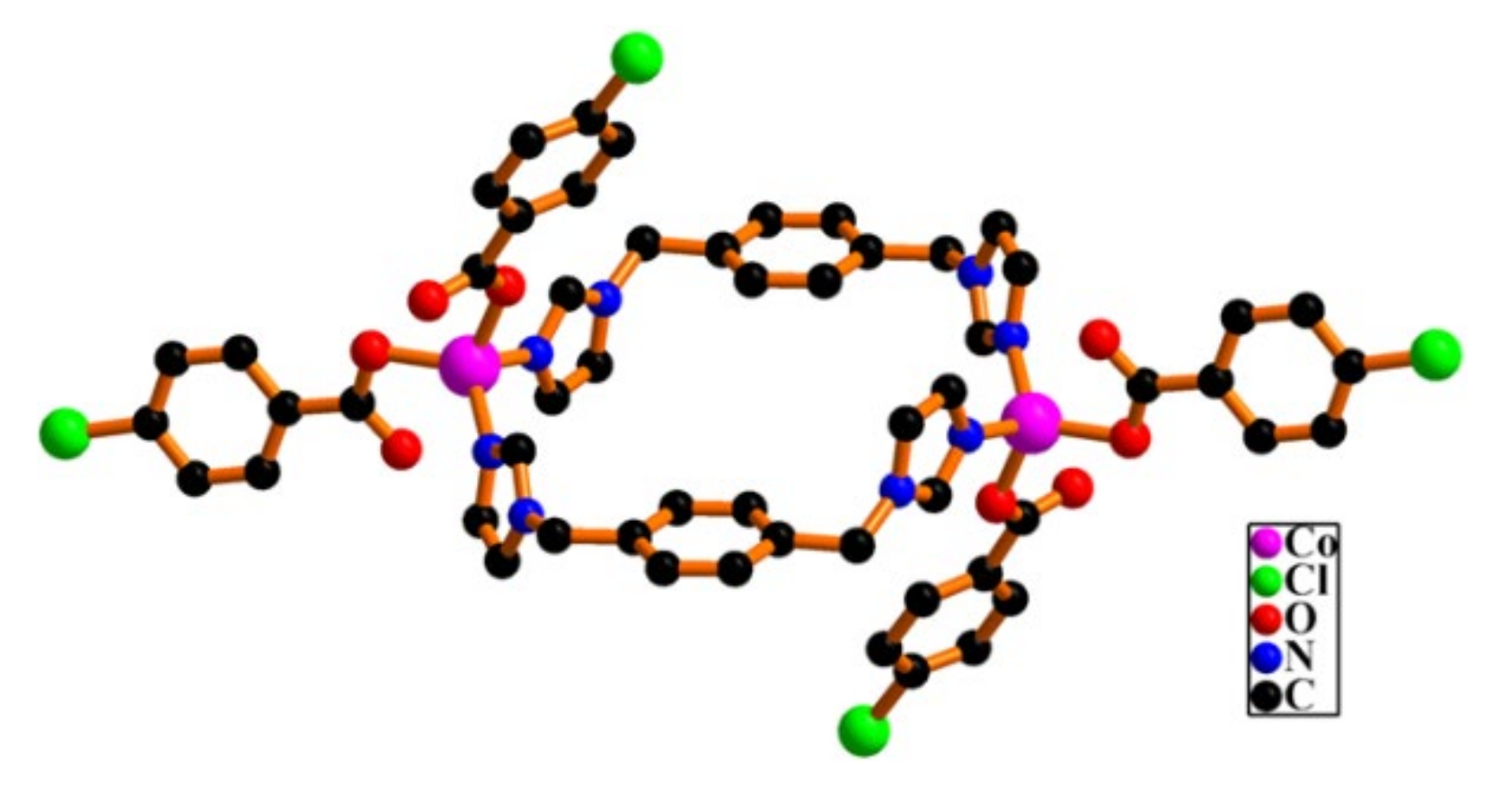
2.2. Description of the Structure of [Co2(pca)4(bimb)2] (2)

Definitely different from 1, X-ray single crystal diffraction indicates that 2 is a centrosymmetric binuclear cobalt complex and crystallizes in the triclinic system with space group P-1 (Table S1). The asymmetric unit of 2 consists of a half-molecule of the formula [Co2(pca)4(bimb)2] with one Co2+, one bimb, and two discrete pca ligands (Figure 5a). The coordination environment of each Co ion (shown in Figure 5b and Figure 6) contains two O atoms (O1 and O3) from two different pac ligands, and two N-atom donors (N1 and N4i, symmetry code i: 1−x, −y, 1−z) from two symmetry-related μ2-bridging bimb, resulting in a slightly distorted tetrahedron geometry. In comparison, 2 has two independent pca ligands, while 1 has one, and 2 has one bimb, while 1 has a half of symmetry-related bridging ligand. It is these different composition and coordination modes that lead to the formation of a completely different structure from compound 1. The Co–O bond distances are 1.961(3) and 1.978(3) Å, and the Co–N distances are 2.013(4) and 2.028(5) Å (Table S2). The two halves of each independent dimer are related by a crystallographic inversion center, which lies at the center of the ring formed by the two Co atoms and the two bimb ligands. The Co1···Co1i separation is 10.85(7) Å, distance that is too long for a metal–metal bond.
In the crystal, intermolecular C–H···Cl hydrogen bonds connected the pca and the bimb ligands together with the phenyl and imidazole carbon atoms of bimb (C20, C27 and C28, donor), and the pca terminal chlorine atoms (Cl1 and Cl2, acceptor) (Figure 7 and Table S3). Interestingly, the C27–H27···Cl1 and C28–H28···Cl1 staggered the [Co2(pca)4(bimb)2] along the a-axis direction, while the C20–H20···Cl2 extended the [Co2(pca)4(bimb)2] along the b-axis, resulting in two mutually perpendicular 1D hydrogen-bonded chains (Figure 8a,b).
The bond distances between H27/H28 and Cl1 are 3.175 Å and 3.125 Å; the bond angles of C27–H27/C28–H28···Cl1 are 104° and 107°, which fall slightly below the preferred minimum hydrogen bond angle of 110°; and the distances between C27/C28 and Cl1 are 3.522(6) Å and 3.511(6) Å, which are almost the same as the sum of the van der Waals radii of C and Cl (3.52 Å) [61]. The bond distance between H20 and Cl2 is 3.308 Å; the bond angle of C20–H20···Cl2 is 122°, which falls the preferred minimum hydrogen bond angle of 110°; and the distance between C20 and Cl2 is 3.884(6) Å, which is greater than the sum of the van der Waals radii of C and Cl (3.52 Å). Therefore, it is a very weak C–H···Cl hydrogen bond, according to the documented data [62,63,64,65,66,67,68].
In the asymmetric unit, each bimb adopts benzene (C20–H20···Cl2) and one of the two unsymmetrical groups imidazole (C27–H27···Cl1, C28–H28···Cl1) involved in hydrogen-bonded C–H···Cl, linking two different pca ligands, which is further grown by symmetry operation, resulting in 2D hydrogen-bonded organic frameworks (HOFs) (Figure 7). If each [Co2(pca)4(bimb)] can be considered to act as a 4-connected node, then each C–H···Cl linking two [Co2(pca)4(bimb)] can be considered a linker in 2 (Figure 8). Topologically, the 2D hydrogen-bonded organic frameworks can be simplified as a 4-connected unodal sql net with a Schläfli symbol {44·62} (Figure 7e and Figure S2) as analyzed with the TOPOS 4.0 program [69].
Furthermore, there exist two types of π-stack interactions, i.e., π···π and C–H···π, between adjacent [Co2(pca)4(bimb)2] units, which are non-negligible forces for stabilizing hydrogen-bonded organic frameworks. There is a π···π stacking interaction between two symmetry-related bimb ligands of imidazole rings (N3-C26-N4-C28-C27, Cg2 and Cg2vii; symmetry code: vii 1−x, −1−y, 2−z) with a Cg2···Cg2vii centroid distance of 3.950(4) Å and with a dihedral angle of 0.0(4)°. These π–π stacking interactions, assembled with the binuclear structure unit into a 1D chain, approximate along the crystallographic c-axis (Figure 9a and Figure S3a). On a similar line, another 1D π-stacked framework along the a-axis direction based on the pca ligand of C–H (C6–H6) and bimb ligand of phenyl ring (C19–C20–C21–C22–C23–C24, Cg5iii; symmetry code: iii −x, 1−y, 1−z) was prepared with a pair of inversion C–H···π interactions (Figure 9b and Figure S3b). The angle of C6-H6···Cg5iii is 135(3)°, and the H6···Cg2ix distance is 2.95 (4) Å. These results form a 2D π-stack supramolecular framework (Figure 9c), which is further stabilized with an intermolecular non-classical hydrogen bond C–H···O. Thus, these hydrogen bonds and π-stacks insert and inter-lock the [Co2(pca)4(bimb)2] blocks in reversely alternating parallel arrangements, defining a 2D Co-based HOFs supporting the supramolecular architecture through C–H···O, C–H···Cl and C–H···π, π···π interactions. PLATON [60] analysis shows that the whole framework is composed of voids of 1.3% that represent 1397.3 Å3 per unit cell volume and 18.9 Å3 total potential solvent area volume.

2.3. Luminescent Properties

2.3.1. Solid State Fluorescence Properties of 1 and 2

Luminescent coordination polymers (CPs) or metal-organic frameworks (MOFs) or hydrogen-bonded organic frameworks (HOFs) have received much attention, attributing the success to their potential applications in photocatalysis, biomedical imaging and fluorescent sensors [6,7,8,9,10,11,12,51,52,53,54,55,56,57,67]. The solid-state fluorescence properties of 1 and 2 were investigated at room temperature. Interestingly, complexes 1 and 2 have approximately the same excitation and emission peaks, except the intensity (Figure S4). The maximum sharp emission peaks center at 423 nm (λex = 376 nm) with the intensity of 753 a.u. for 1, and 231 a.u. for 2, which may be attributed to the π*→n or π*→π transitions. In comparison, the emission intensity of 2 is much lower than 1 by 522 a.u., which may be assigned to different hydrogen-bonded organic frameworks. Therefore, the fluorescent ion recognition test was carried out in water, taking 1 as an example.

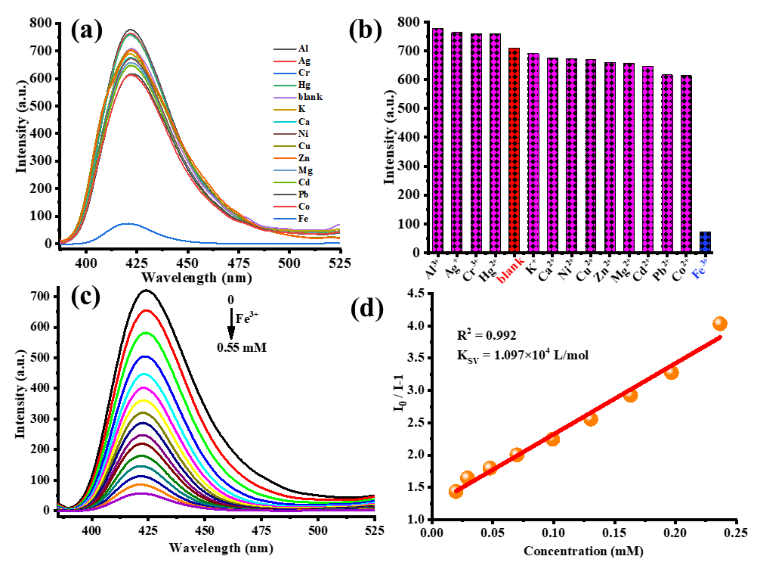
2.3.2. Detection of Fe3+ Ion

As shown in the experimental section, a variety of fluorescence spectral measurements were carried out on the fluorescent response of 1, which interacts with 14 different metal cations. Surprisingly, the luminescence emission intensity of 1 was sharply cut down in the Fe(NO3)3 solution, but slight intensity changes were observed in the other 13 ions (Figure 10a,b). It was found that only slight enlargement or reduction changes occurred when detecting Al3+, Ag+, Cr3+, Hg2+ or Ca2+, Ni2+, Cu2+, Zn2+, Mg2+, while K+ had almost no fluorescence intensity changes. On account of the distinct response of 1 toward Fe3+, the quenching effect of Fe3+ was subsequently explored with a function of Fe(NO3)3 in the concentration range of 0–0.55 mM. As shown in Figure 10c, when the Fe3+ concentration was gradually raised from 0 to 0.55 mM, the fluorescence intensity from 720 a.u. decreased step by step to about 35 a.u., and the quenching efficiency was almost 95% when the concentration of the Fe3+ ions reached 0.55 mM. The well-known Stern-Volmer (SV) equation ((Io/I) = 1 + Ksv[M], Io = fluorescent intensities of 1; I = fluorescent intensities after adding Fe3+; [M] = molar concentration of Fe3+ (mM); Ksv = SV constant (M−1)) can effectively analyze the fluorescent quenching efficiency. As shown in Figure 10d, it is clear that in the concentration range of 0.025–0.25 mM, the relative intensity has a good linear relationship with the Fe3+ concentration (Ksv = 10970 M−1, R2 = 0.992, σ = 0.06938). The detection limit of 1 can be calculated from the standard deviation and slope using the formula 3σ/Ksv (σ: standard error, Ksv: slope). The detection limits of 1 is 19 µM. All of the above results imply that 1 can effectively and selectively sense Fe3+.

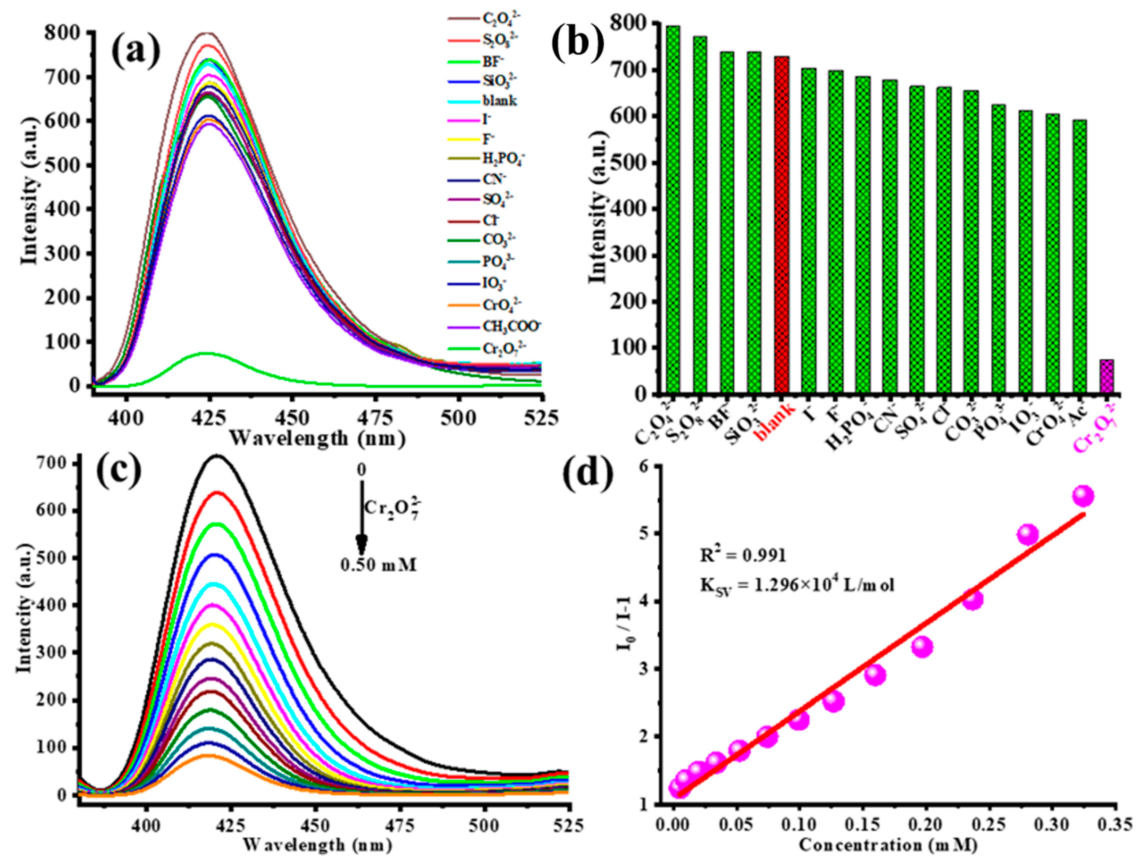
2.3.3. Detection of Cr2O72− Anions

Besides metal cations, in order to improve the use efficiency of 1 as a multifunctional fluorescent probe, the fluorescence recognition anion experiments were performed to screen various potassium salt anions in aqueous solution (Figure 11a,b). Outstandingly, only Cr2O72- afforded a quite obvious luminescent quenching effect, with a quenching percentage of 93.3%, but the quenching percentage of other anions was much lower (less than 15% for Ac) under the same conditions. It was also found that only slight enlargement or reduction changes occurred when detecting C2O42−, S2O82− or I, F, H2PO4, CN, SO42−, Cl, CO32−, PO43−, IO3, CrO42−, Ac, while BF and SiO32− had almost no fluorescence intensity changes. Accordingly, the titration curve clearly indicates that as the Cr2O72− concentration increases from 0 mM to 0.50 mM, and the emission intensity of 1 decreases from about 720 a.u. to 50 a.u. (Figure 11c). As shown in Figure 11d, Ksv, R2 and σ were calculated to be 12960 M−1, 0.991and 0.08656 for 1@Cr2O72-, respectively. The detection limits of 1 for Cr2O72− is calculated to 20 µM based on the formulas of 3σ/Ksv.

2.3.4. Stability and Cyclic Use Test

The powder X-ray diffraction (PXRD) patterns of 1 and 2 are shown in Figure 12a. The experimental peaks are consistent with the theoretical one obtained by single-crystal X-ray data. The stability of 1 was investigated in different pH values of aqueous media varying from 1 to 13. After 24 h of soaking 1 in these solvents, the filtered samples of 1 were dried in air. The experimental PXRD patterns of 1 were also consistent with the theoretical pattern in the pH values from 3 to 11 (Figure 12b) indicating that 1 had relatively good chemical stability. The slight variation in the diffraction intensity may be related to the different crystal orientation or solvent effects. Although the experimental patterns have a few unindexed diffraction lines and some are slightly broadened in comparison to those simulated from the single-crystal model, it can still to be considered that the bulk of the synthesized materials and as-grown crystal are homogeneous, implying its excellent stability. In order to explore the thermal stability of 1 and 2, TG studies were performed in a nitrogen atmosphere at a heating rate of 10 °C min–1 between 50 °C and 800 °C (Figure S5). Up to 300 °C, almost no weight loss was observed on the TG curve, which indicates the absence of an occluded solvent and guest molecules in the samples of 1 and 2. According to TG data, heating above 350 °C leads to several different processes, which lead to the decomposition of the framework of compounds 1 and 2.
Repeatability is an important factor for assessing the applicability of luminescent coordination polymers. Recyclability experiments were conducted to determine the regeneration of 1 to detect Fe3+ and Cr2O72− ions. The used sample of 1 was washed multiple times with alcohol through soaking under stirring at room temperature. After filtration and drying, the regenerated sample 1 was employed for the detection of Fe3+ and Cr2O72− ions with the same method of 1, respectively. It is obvious that the quenching ability of 1 for Fe3+ and Cr2O72− ions, respectively, are mostly unchanged for up to five cycles (Figure 13). All these results indicate that 1 has excellent water stability and reusable value.

3. Materials and Methods

3.1. Chemicals and Reagents

All chemicals and solvents, including the ligands, were purchased from Jinan Henghua Sci. & Tec. Co. Ltd. (Shandong, China) without further purification.

3.2. Apparatus

Powder X-ray diffraction (PXRD) data were collected on a Bruker D8 ADVANCE X-ray diffractometer (Karlsruhe, Germany) with Cu-Kα radiation (λ = 1.5418 Å) at 40 kV and 40 mA with a scanning rate of 6°/min and a step size of 0.02°. The simulation of the PXRD spectra was carried out by the single-crystal data and diffraction-crystal module of the Mercury 2.0 (Hg) program, available free of charge via the internet at http://www.iucr.org (accessed on 25 August 2021). The purity and homogeneity of the bulk products were determined by comparing the simulated and experimental X-ray powder diffraction patterns. The elemental analyses (C, H, N) were performed on a PerkinElmer 240C apparatus. The FT-IR spectra were recorded in the range of 4000–450 cm−1 on a PerkinElmer Frontier spectrometer (Norwalk, CT, USA). Thermogravimetric analyses (TG) were performed under nitrogen with a heating rate of 10 °C min−1, using a PerkinElmer Thermogravimetric Analyzer TGA4000 (Norwalk, CT, USA). Photoluminescence spectra were measured on a Varian Cary Eclipse fluorescence spectrophotometer (Shanghai, China), with a xenon arc lamp as the light source. In the measurements of emission and excitation spectra, the slit width was 10 nm. Point symbol and topological analyses were conducted by using the TOPOS program package (Samara, Russia) [69].
Single-crystal data collections were performed on a Bruker Smart Apex II CCD diffractometer (Tokyo, Japan) with graphite-monochromatized MoKα radiation (λ = 0.71073 Å) at 296(2) K. Using Olex2 [70], the structure was solved with the ShelXT structure solution program using Intrinsic Phasing [71] and refined with the ShelXL refinement package using least squares minimization [72]. All non-hydrogen atoms were refined with anisotropic displacement parameters. Carbon-bound H atoms were placed in calculated positions (dC-H = 0.93 Å for -CH, 0.97 for -CH2, and 0.96 for -CH3) and were included in the refinement in the riding model approximation, with Uiso(H) set to 1.2Ueq(C) for -CH and -CH2, and 1.5Ueq(C) for -CH3. The structures were examined using the Addsym subroutine of PLATON to ensure that no additional symmetry could be applied to the models. Pertinent crystallographic data collection and refinement parameters are collated in Table S1. Selected bond lengths and angles are given in Table S2. Hydrogen-bond geometries are organized in Table S3.
CCDC 2105278 (1) and CCDC 2105279 (2) contain the supplementary crystallographic data for this paper. These data can be obtained free of charge from The Cambridge Crystallographic Data Centre via http://www.ccdc.cam.ac.uk/datarequest/cif (accessed on 25 August 2021).

3.3. Synthesis of [Co(pca)2(bmimb)]n (1)

A mixture of Co(CH3COO)2·4H2O (0.1 mmol), Hpac (0.1 mmol), NaOH (0.1 mmol), bmimb (0.1 mmol),and H2O–C2H5OH (5 mL/5 mL) was added to a 25 mL Teflon-lined stainless steel reactor and heated at 150 °C for 3 days, and then slowly cooled to room temperature. Red block single crystals suitable for X-ray data collection were obtained by filtration. Yield: 45% (based on cobalt acetate tetrahydrate)—Anal. for C30H26Cl2CoN4O4 (636.38): calcd. C 56.57, H 4.09, N 8.80; found C 56.55, H 4.08, N 8.82. Infrared (KBr pellet, cm−1): 3125(w), 3125(w), 1602(s), 1349(s), 1090(m), 858(m), 775(vs), 652(m), 564(m), 516(m).

3.4. Synthesis of [Co2(pca)4(bimb)2] (2)

The synthesis of 2 is similar to that for 1, except for bimb (0.1 mmol) substituted for bmimb (0.1 mmol). Red block single crystals suitable for X-ray data collection were obtained by filtration. Yield: 70% (based on cobalt acetate tetrahydrate)—Anal. for C56H44Cl4Co2N8O8 (1216.65): calcd. C 55.23, H 3.62, N 9.21; found C 55.25, H 3.60, N 9.23. Infrared (KBr pellet, cm−1): 3644(m), 1909(w), 1711(w), 1583(s), 1500(s), 1301(m), 1083(m), 1001(m), 837 (s), 769(s), 667(m), 523(m).

3.5. Comparison of Synthesis and Structure for 1 and 2

Interestingly, the synthesis of 1 and 2 rely on subtle control over various hydrothermal parameters, particularly the starting neutral nitrogen-containing auxiliary ligands. This prompted us to further study this reaction system by the use of different metal ions and investigate its potential to favor the formation of higher dimensionality coordination polymers and/or MOFs. A wide range of experiments was carried out in order to study the impact of the different synthetic parameters (presence/absence or kind of base, metal ratio of the reactants, metal sources, etc.) on the identity and crystallinity of the isolated product.
The reaction mixture of Co(CH3COO)2·4H2O/Hpac/NaOH/bmimb (1:1:1:1) in H2O-C2H5OH (v/v, 1:1) at 150 °C gave a red solution from which the 1D coordination polymer [Co(pca)2(bmimb)]n was subsequently isolated. Following a similar reaction but using a different neutral nitrogen-containing auxiliary ligand source bimb instead of bmimb, the crystals of [Co2(pca)4(bimb)2] (0D) were isolated in good yield. Note that these kinds of cobalt salts do not have any impact on the identity of the isolated compound, but they affect its crystallinity. As a next step, we decided to investigate the impact of the metal ion on the identity of the isolated product by the use of Cu2+/Zn2+/Ni2+/Cd2+ and so on and following a similar synthetic approach to the one that provided access to 1 and 2. No matter the changes and combinations used as various conditions, unfortunately, we could not obtain the target product. In comparison, we had also tried to apply the similar neutral nitrogen-containing auxiliary ligands to react with the combination of Hpca and cobalt salts under various conditions. The same results were obtained as above.
As discussed above, two Co-based HOFs with the homologous organic ligands synthesized under the same reaction conditions show distinct structures and hydrogen bond topologies in their crystal. Using the same metal and organic carboxylic acid ligand, and different neutral auxiliary ligands but with the same synthesis strategy, will likely afford diverse structures. The biggest similarity of 1 and 2 is that they both have intermolecular non-classical hydrogen bonds C–H···Cl, which play an important role in assembling the 0 or 1D structure into the 3D supramolecular framework. Of course, the difference is also obvious between 1 and 2. First, 1 and 2 belong to different crystal systems and space groups. Second, the steric hindrance, rigidity and electronic properties of the bmimb and bimb ligands influence the dimensionality of the reaction product. The steric hindrance and rigidity result in bigger bmimb than bimb, but the electronic properties results in better bmimb and bimb. These factors ultimately cause 1 and 2 to be formed as 1D and 0D structures, respectively, and have different hydrogen-bond framework topologies (3D for 1 and 2D for 2).

3.6. Photoluminescent Sensing Experiments

The luminescence properties of 1 were investigated in the aqueous solution of various analytes at room temperature [73,74]. When measuring the emission spectra, the emission and excitation slit widths were set as 10 nm (similarly hereinafter). For the sensing of cations, 2.0 mg of a ground powder sample of 1 was immersed in 2.0 mL of a 14-cation aqueous solution of M(NO3)x (Mx+ = Al3+, Ag+, Cr3+, Hg2+, K+, Ca2+, Ni2+, Cu2+, Zn2+, Mg2+, Cd2+, Pb2+, Co2+, Zn2+, and Fe3+, 0.0010 mol·L−1). Then, the solid-liquid mixture was ultrasonicated for 30 min to form steady turbid suspension of 1@Mx+ for the fluorescence measurements. The fluorescent intensities of these 1@Mx+ suspensions were immediately recorded at room temperature and compared. For the sensing of 16 anions, the same procedure was used for the 1@Ay- fluorescence measurements (KyA, Ay− = C2O42−, S2O82−, BF, SiO32−, I, F, H2PO4, CN, SO42−, Cl, CO32−, PO43−, IO3, CrO42−, Ac, and Cr2O72−, 0.0010 mol·L−1). The fluorescence intensity of the suspension 1 in water was measured as the blank sample.

3.7. Fluorescence Titration Experiments for Fe3+ and Cr2O72−

The 2.0 mg ground powder sample of 1 was dispersed in 2.0 mL H2O solution of target analyte Fe(NO3)3 with different concentrations (0–0.55 mM). Then, the solid–liquid mixture was ultrasonicated for 30 min to form a steady turbid suspension of 1@Fe3+ for the fluorescence measurements. The fluorescent intensities of these 1@Fe3+ suspensions were immediately recorded at room temperature and compared. The same method was applied to measure the fluorescent spectrum of 1@Cr2O72−, with 1 soaking in different aqueous concentrations of K2Cr2O7 (0–0.50 mM).

3.8. Recyclable Luminescence Experiments

The powder of complex 1 was centrifuged from the suspension and rinsed with distilled water three times. Then, the recovered sample was dried and added to the target analytes again to measure their emission spectra. The same procedure was repeated five times.

4. Conclusions

In summary, two new Co-based hydrogen-bonded organic frameworks derived from the pca and bmimb/bimb ligands were hydrothermally synthesized and characterized by IR spectroscopy, elemental analysis and single-crystal X-ray diffraction. The topological structure analysis revealed that 1 and 2 are 3D or 2D Co-based HOFs with a 4-connected qtz topology with a Schläfli symbol {44·62} or a 4-connected sql topology with a Schläfli symbol {44·62} net. The luminescence investigation indicated that 1 has good fluorescence properties at room temperature and the detection limits of Fe3+ and Cr2O72− can reach 19 µM and 20 µM, respectively. In summary, these existing results may provide a new platform for designing luminous HOFs with multi-functional applications for detecting Fe3+/Cr2O72− in the future.

Supplementary Materials

The following are available online, Figure S1: IR spectra of compounds 1 (a) and 2 (b), Figure S2: The simplified 2D HOFs for 2, Figure S3: (a) The 1D chain constructed by π···π interactions in 2. (b) The 1D chain constructed by C–H···π interactions in 2, Figure S4: The solid state excitation and emission spectra of 1 and 2 at room temperature, Figure S5: TG curves of compounds 1 and 2, Table S1: Crystal structure data for 1 and 2a,b,c, Table S2: Selected bond lengths/Å and bond angles/º for compounds 1 and 2, Table S3. Selected bond lengths (Å) and angles (deg) for 1 and 2 with estimated standard deviations in parentheses.

Author Contributions

Conceptualization, supervision, writing—review and editing, J.-M.L. and M.O.; writing—original draft preparation, Y.-L.Z. and J.-M.L.; synthesis and methodology Q.-Y.W. and J.-M.L.; formal analysis and discussion, J.-M.L., M.O., Q.-Y.W. and Y.-L.Z. All authors have read and agreed to the published version of the manuscript.

Funding

This research was funded by the National Natural Science Foundation of China (22065001), the Natural Science Foundation of Guangxi Province (2018XNSFAA281174), and the Innovative Training Program for Guangxi Province College Students (202011607044 and 202011607074).

Institutional Review Board Statement

Not applicable.

Informed Consent Statement

Not applicable.

Data Availability Statement

The data presented in this study are available in supplementary material.

Conflicts of Interest

The authors declare no conflict of interest.

Sample Availability

Samples of the compounds are not available from the authors.

References

  1. Karmakar, A.; Illathvalappil, R.; Anothumakkool, B.; Sen, A.; Samanta, P.; Desai, A.V.; Kurungot, S.; Ghosh, S.K. Hydrogen-Bonded Organic Frameworks (HOFs): A New Class of Porous Crystalline Proton-Conducting Materials. Angew. Chem. Int. Ed. 2016, 55, 10667–10671. [Google Scholar] [CrossRef] [PubMed]
  2. Cai, S.; Shi, H.; Zhang, Z.; Wang, X.; Ma, H.; Gan, N.; Wu, Q.; Cheng, Z.; Ling, K.; Gu, M.; et al. Hydrogen-Bonded Organic Aromatic Frameworks for Ultralong Phosphorescence by Intralayer π−π Interactions. Angew. Chem. Int. Ed. 2018, 57, 4005–4009. [Google Scholar] [CrossRef] [PubMed]
  3. Luo, J.; Wang, J.W.; Zhang, J.H.; Lai, S.; Zhong, D.C. Hydrogen-bonded organic frameworks: Design, structures and potential applications. CrystEngComm 2018, 20, 5884–5898. [Google Scholar] [CrossRef]
  4. Han, Y.F.; Yuan, Y.X.; Wang, H.B. Porous Hydrogen-Bonded Organic Frameworks. Molecules 2017, 22, 266. [Google Scholar] [CrossRef] [Green Version]
  5. Wang, B.; Lin, R.B.; Zhang, Z.; Xiang, S.; Chen, B. Hydrogen-Bonded Organic Frameworks as a Tunable Platform for Functional Materials. J. Am. Chem. Soc. 2020, 142, 14399–14416. [Google Scholar] [CrossRef] [PubMed]
  6. Dou, Z.; Yu, J.; Cui, Y.; Yang, Y.; Wang, Z.; Yang, D.; Qian, G. Luminescent metal-organic framework films as highly sensitive and fast-response oxygen sensors. J. Am. Chem. Soc. 2014, 136, 5527–5530. [Google Scholar] [CrossRef]
  7. Taylor, K.M.; Rieter, W.J.; Lin, W. Manganese-Based Nanoscale Metal-Organic Frameworks for Magnetic Resonance Imaging. J. Am. Chem. Soc. 2008, 130, 14358–14359. [Google Scholar] [CrossRef]
  8. Taylor-Pashow, K.M.; Della, R.J.; Xie, Z.; Tran, S.; Lin, W. Postsynthetic modifications of iron-carboxylate nanoscale metal–organic frameworks for imaging and drug delivery. J. Am. Chem. Soc. 2009, 131, 14261–14263. [Google Scholar] [CrossRef] [Green Version]
  9. Wang, M.S.; Guo, S.P.; Li, Y.; Cai, L.Z.; Zou, J.-P.; Xu, G.; Zhou, W.-W.; Zheng, F.-K.; Guo, G.-C. A Direct White-Light-Emitting Metal Organic Framework with Tunable Yellow-to-White Photoluminescence by Variation of Excitation Light. J. Am. Chem. Soc. 2009, 131, 13572–13573. [Google Scholar] [CrossRef]
  10. Yu, J.; Cui, Y.; Wu, C.D.; Yang, Y.; Chen, B.; Qian, G. A Two-photon Responsive Metal-Organic Framework. J. Am. Chem. Soc. 2015, 137, 4026–4029. [Google Scholar] [CrossRef]
  11. Deng, J.H.; Luo, J.; Mao, Y.L.; Lai, S.; Gong, Y.N.; Zhong, D.C.; Lu, T.B. π−π stacking interactions: Non-negligible forces for stabilizing porous supramolecular frameworks. Sci. Adv. 2020, 6, eaax9976. [Google Scholar] [CrossRef] [PubMed] [Green Version]
  12. Wang, J.; Yu, M.; Chen, L.; Li, Z.; Li, S.; Jiang, F.; Hong, M. Construction of a Stable Lanthanide Metal-Organic Framework as a Luminescent Probe for Rapid Naked-Eye Recognition of Fe3+ and Acetone. Molecules 2021, 26, 1695. [Google Scholar] [CrossRef]
  13. Wang, P.; Sun, Y.; Li, X.; Wang, L.; Xu, Y.; Li, G. Recent Advances in Metal Organic Frameworks Based Surface Enhanced Raman Scattering Substrates: Synthesis and Applications. Molecules 2021, 26, 209. [Google Scholar] [CrossRef]
  14. Mylonas-Margaritis, I.; Mayans, J.; McArdle, P.; Papatriantafyllopoulou, C. ZnII and CuII-Based Coordination Polymers and Metal Organic Frameworks by the of Use of 2-Pyridyl Oximes and 1,3,5-Benzenetricarboxylic Acid. Molecules 2021, 26, 491. [Google Scholar] [CrossRef]
  15. Trofimova, O.Y.; Maleeva, A.V.; Ershova, I.V.; Cherkasov, A.V.; Fukin, G.K.; Aysin, R.R.; Kovalenko, K.A.; Piskunov, A.V. Heteroleptic LaIII Anilate/Dicarboxylate Based Neutral 3D-Coordination Polymers. Molecules 2021, 26, 2486. [Google Scholar] [CrossRef] [PubMed]
  16. Elliott, R.; Ryan, A.A.; Aggarwal, A.; Zhu, N.; Steuber, F.W.; Senge, M.O.; Schmitt, W. 2D Porphyrinic Metal-Organic Frameworks Featuring Rod-Shaped Secondary Building Units. Molecules 2021, 26, 2955. [Google Scholar] [CrossRef]
  17. Wang, T.T.; Liu, J.Y.; Guo, R.; An, J.D.; Huo, J.Z.; Liu, Y.Y.; Shi, W.; Ding, B. Solvothermal Preparation of a Lanthanide Metal-Organic Framework for Highly Sensitive Discrimination of Nitrofurantoin and L-Tyrosine. Molecules 2021, 26, 3673. [Google Scholar] [CrossRef] [PubMed]
  18. Wang, W.; He, J.; Guo, H.; Dunning, S.G.; Humphrey, S.M.; Jones, R.A. Magnetism and Luminescence of a MOF with Linear Mn3 Nodes Derived from an Emissive Terthiophene-Based Imidazole Linker. Molecules 2021, 26, 4286. [Google Scholar] [CrossRef]
  19. Almáši, M.; Zeleňák, V.; Gyepes, R.; Zauškaa, L.; Bourrellyc, S. A series of four novel alkaline earth metal–organic frameworks constructed of Ca(II), Sr(II), Ba(II) ions and tetrahedral MTB linker: Structural diversity, stability study and low/high-pressure gas adsorption properties. RSC Adv. 2020, 10, 32323–32334. [Google Scholar] [CrossRef]
  20. Garg, A.; Almáši, M.; Paul, D.R.; Poonia, E.; Luthra, J.R.; Sharma, A. Metal-Organic Framework MOF-76 (Nd): Synthesis, Characterization, and Study of Hydrogen Storage and Humidity Sensing. Front. Energy Res. 2021, 8, 604735. [Google Scholar] [CrossRef]
  21. Suzuki, Y.; Tohnai, N.; Saeki, A.; Hisaki, I. Hydrogen-bonded organic frameworks of twisted polycyclic aromatic hydrocarbon. Chem. Commun. 2020, 56, 13369–13372. [Google Scholar] [CrossRef]
  22. Yang, Q.; Wang, Y.; Shang, Y.; Du, J.; Yin, J.; Liu, D.; Kang, Z.; Wang, R.; Sun, D.; Jiang, J. Three Hydrogen-Bonded Organic Frameworks with Water-Induced Single-Crystal-to-Single-Crystal Transformation and High Proton Conductivity. Cryst. Growth Des. 2020, 20, 3456–3465. [Google Scholar] [CrossRef]
  23. Li, Y.L.; Alexandrov, E.V.; Yin, Q.; Li, L.; Fang, Z.B.; Yuan, W.; Proserpio, D.M.; Liu, T.F. Record Complexity in the Polycatenation of Three Porous Hydrogen-Bonded Organic Frameworks with Stepwise Adsorption Behaviors. J. Am. Chem. Soc. 2020, 142, 7218–7224. [Google Scholar] [CrossRef]
  24. Li, P.; Alduhaish, O.; Arman, H.D.; Wang, H.; Alfooty, K.; Chen, B. Solvent Dependent Structures of Hydrogen-Bonded Organic Frameworks of 2,6-Diaminopurine. Cryst. Growth Des. 2014, 14, 3634–3638. [Google Scholar] [CrossRef]
  25. Zhang, J.; Feng, Y.; Staples, R.J.; Zhang, J.; Shreeve, J.M. Taming nitroformate through encapsulation with nitrogen-rich hydrogen-bonded organic frameworks. Nat. Commun. 2021, 12, 2146. [Google Scholar] [CrossRef]
  26. Suzuki, Y.; Gutiérrez, M.; Tanaka, S.; Gomez, E.; Tohnai, N.; Yasuda, N.; Matubayasi, N.; Douhal, A.; Hisaki, I. Construction of isostructural hydrogen-bonded organic frameworks: Limitations and possibilities of pore expansion. Chem. Sci. 2021, 12, 9607–9618. [Google Scholar] [CrossRef] [PubMed]
  27. Luo, X.Z.; Jia, X.J.; Deng, J.H.; Zhong, J.L.; Liu, H.J.; Wang, K.J.; Zhong, D.C. A Microporous Hydrogen-Bonded Organic Framework: Exceptional Stability and Highly Selective Adsorption of Gas and Liquid. J. Am. Chem. Soc. 2013, 135, 11684–11687. [Google Scholar] [CrossRef] [PubMed]
  28. Hawes, C.S. Coordination sphere hydrogen bonding as a structural element in metal–organic Frameworks. Dalton Trans. 2021, 50, 6034–6049. [Google Scholar] [CrossRef]
  29. Tripathy, R.J.; Aneeya, K.; Samantara, A.K.; Behera, J.N. A cobalt metal-organic framework (Co-MOF): A bi-functional electro active material for the oxygen evolution and reduction reaction. Dalton Trans. 2019, 48, 10557–10564. [Google Scholar] [CrossRef]
  30. Duan, J.; Mebs, S.; Laun, K.; Wittkamp, F.; Heberle, J.; Happe, T.; Hofmann, E.; Apfel, U.P.; Winkler, M.; Senger, M.; et al. Geometry of the Catalytic Active Site in [FeFe]-Hydrogenase Is Determined by Hydrogen Bonding and Proton Transfer. ACS Catal. 2019, 9, 9140–9149. [Google Scholar] [CrossRef]
  31. Dahl, E.W.; Szymczak, N.K. Hydrogen Bonds Dictate the Coordination Geometry of Copper: Characterization of a Square-Planar Copper(I) Complex. Angew. Chem. Int. Ed. 2016, 55, 3101–3105. [Google Scholar] [CrossRef] [Green Version]
  32. Hoffert, W.A.; Mock, M.T.; Appel, A.M.; Yang, J.Y. Incorporation of hydrogen-bonding functionalities into the second coordination sphere of iron-based water-oxidation catalysts. Eur. J. Inorg. Chem. 2013, 22, 3846–3857. [Google Scholar] [CrossRef] [Green Version]
  33. Chapovetsky, A.; Welborn, M.; Luna, J.M.; Haiges, R.; Miller , T.F., III; Marinescu, S.C. Pendant Hydrogen-Bond Donors in Cobalt Catalysts Independently Enhance CO2 Reduction. ACS Cent. Sci. 2018, 4, 397–404. [Google Scholar] [CrossRef] [Green Version]
  34. Shanahan, J.P.; Mullis, D.M.; Zeller, M.; Szymczak, N.K. Reductively Stable Hydrogen-Bonding Ligands Featuring Appended CF2–H Units. J. Am. Chem. Soc. 2020, 142, 8809–8817. [Google Scholar] [CrossRef]
  35. Santoro, A.; Bella, G.; Bruno, G.; Neri, G.; Akbari, Z.; Nicolò, F. Cocrystal versus salt, a matter of hydrogen bonds in two benzoic acid crystals. J. Mol. Struct. 2021, 1229, 129801. [Google Scholar] [CrossRef]
  36. Chinthal, C.H.; Kavitha, C.N.; Yathirajan, H.S.; Foro, S.; Rathore, R.S.; Glidewell, C. Fifteen 4-(2-meth-oxy-phen-yl)piperazin-1-ium salts containing organic anions: Supra-molecular assembly in zero, one, two and three dimensions. Acta Crystallogr. Sect. E Cryst. Commun. 2020, 76, 1779–1793. [Google Scholar] [CrossRef] [PubMed]
  37. Chen, Z.Y.; Peng, M.X. Supramolecular Architectures Constructed from Piperazine and Substituted Benzoic Acids. J. Chem. Crystallogr. 2011, 41, 137–142. [Google Scholar] [CrossRef] [Green Version]
  38. Sikorski, A.; Trzybiński, D. The influence of benzoate anion substituents on the crystal packing and hydrogen-bonding network of 9-aminoacridinium salts. Tetrahedron 2011, 67, 2839–2843. [Google Scholar] [CrossRef]
  39. Sasaki, T.; Hisaki, I.; Miyano, T.; Tohnai, N.; Morimoto, K.; Sato, H.; Tsuzuki, S.; Miyata, M. Linkage control between molecular and supramolecular chirality in 21-helical hydrogen-bonded networks using achiral components. Nat. Commun. 2013, 4, 1787. [Google Scholar] [CrossRef] [Green Version]
  40. Kinbara, K.; Hashimoto, Y.; Sukegawa, M.; Nohira, H.; Saigo, K. Crystal Structures of the Salts of Chiral Primary Amines with Achiral Carboxylic Acids:  Recognition of the Commonly-Occurring Supramolecular Assemblies of Hydrogen-Bond Networks and Their Role in the Formation of Conglomerates. J. Am. Chem. Soc. 1996, 118, 3441–3449. [Google Scholar] [CrossRef]
  41. Rani, J.; Grover, V.; Dhamija, S.; Titi, H.M.; Patra, R. Computational insight into the halogen bonded self-assembly of hexa-coordinated metalloporphyrins. Phys. Chem. Chem. Phys. 2020, 22, 11558–11566. [Google Scholar] [CrossRef]
  42. Sugiyama, H.; Uekusa, H. Relationship between crystal structures and photochromic properties of N-salicylideneaminopyridine derivatives. CrystEngComm 2018, 20, 2144–2151. [Google Scholar] [CrossRef]
  43. Kalaj, M.; Carter, K.P.; Cahill, C.L. Isolating Equatorial and Oxo Based Influences on Uranyl Vibrational Spectroscopy in a Family of Hybrid Materials Featuring Halogen Bonding Interactions with Uranyl Oxo Atoms. Eur. J. Inorg. Chem. 2017, 40, 4702–4713. [Google Scholar] [CrossRef]
  44. Wang, Y.; Wang, M.F.; Young, D.J.; Zhu, H.; Hu, F.L.; Mi, Y.; Qin, Z.; Chen, S.L.; Lang, J.P. Tuning the configuration of the flexible metal–alkene-framework affords pure cycloisomers in solid state photodimerization. Chem. Commun. 2021, 57, 1129–1132. [Google Scholar] [CrossRef] [PubMed]
  45. Liu, D.; Li, N.Y.; Lang, J.P. Single-crystal to single-crystal transformation of 1D coordination polymervia photochemical [2+2] cycloaddition reaction. Dalton Trans. 2011, 40, 2170–2172. [Google Scholar] [CrossRef]
  46. Kumar, R.; Singh, U.P. Synthesis, structural, photophysical and thermal properties of some praseodymium(III) complexes. J. Coord. Chem. 2008, 61, 2663–2674. [Google Scholar] [CrossRef]
  47. Qin, Y.Y.; Tang, Y.H.; Kang, Y.; Li, Z.J.; Hu, R.F.; Cheng, J.K.; Wen, Y.H.; Yao, Y. Crystal structures and 31P NMR spectra of two trinuclear molybdenum clusters coordinated by p-chlorobenzoate: Mo3S4(DTP)3(p-ClC6H4COO)(Py)·EtOH and Mo3S4(DTP)3(p-ClC6H4COO)(DMF). J. Mol. Struct. 2004, 707, 235–239. [Google Scholar] [CrossRef]
  48. Wang, N.; Li, B. Synthesis, Crystal Structure and Cytotoxic Property of Bis(4-chlorobenzoato)bis[1-(2-aminoethyl)piperidine]disilver(I). J. Chem. Crystallogr. 2011, 41, 219–222. [Google Scholar] [CrossRef]
  49. Carter, K.P.; Zulatoa, C.H.F.; Cahill, C.L. Exploring supramolecular assembly and luminescent behavior in a series of RE-p-chlorobenzoic acid-1,10-phenanthroline complexes. CrystEngComm 2014, 16, 10189–10202. [Google Scholar] [CrossRef] [Green Version]
  50. Deka, K.; Sarma, R.J.; Baruah, J.B. Nitrogen-oxygen bond formation during oxidative reactions of copper(II)benzoate complexes having 3,5-dimethylpyrazole. Inorg. Chem. Commun. 2006, 9, 931–934. [Google Scholar] [CrossRef]
  51. Sun, D.; Liu, F.J.; Hao, H.J.; Li, Y.H.; Huang, R.B.; Zheng, L.S. Six low-dimensional silver(I) coordination complexes derived from 2-aminobenzonitrile and carboxylates. Inorg. Chimi. Acta 2012, 387, 271–276. [Google Scholar] [CrossRef]
  52. Halaška, J.; Lokaj, J.; Jomová, K.; Růžičková, Z.; Mazúr, M.; Moncol, J. Formation of supramolecular hydrogen-bonding chains and networks from copper (II) halogenobenzoates with N-methylnicotinamide: Supramolecular isomerism. Polyhedron 2020, 175, 114237. [Google Scholar] [CrossRef]
  53. Xu, T.Y.; Nie, H.J.; Li, J.M.; Shi, Z.F. Highly selective sensing of Fe3+/Hg2+ and proton conduction using two fluorescent Zn(II) coordination polymers. Dalton Trans. 2020, 49, 11129–11141. [Google Scholar] [CrossRef]
  54. Xu, T.Y.; Nie, H.J.; Li, J.M.; Shi, Z.F. Luminescent Zn(II)/Cd(II) coordination polymers based on 1-(tetrazol-5-H)-3,5-bis(1-triazole)benzene for sensing Fe3+, Cr2O72, and CrO42 in water. J. Solid State Chem. 2020, 287, 121342. [Google Scholar] [CrossRef]
  55. Xu, T.Y.; Li, J.M.; Han, Y.H.; Wang, A.R.; He, K.H.; Shi, Z.F. A new 3D four-fold interpenetrated dia-like luminescent Zn(II)-based metal–organic framework: The sensitive detection of Fe3+, Cr2O72, and CrO42 in water, and nitrobenzene in ethanol. New J. Chem. 2020, 44, 4011–4022. [Google Scholar] [CrossRef]
  56. Xu, T.Y.; Zhao, Y.L.; Li, J.M.; Shi, Z.F. Co(II) Coordination Polymer Based on Bis(carboxyl) Dibenzylamine and Bis(imidazole) Biphenyl Ligands: Synthesis, Crystal Structure and Detection for Fe3+ Ions in Water. Chinese J. Inorg. Chem. 2020, 36, 1735–1743. [Google Scholar]
  57. Xu, T.Y.; Wang, H.; Li, J.M.; Zhao, Y.L.; Han, Y.H.; Wang, X.L.; He, K.H.; Wang, A.R.; Shi, Z.F. A water-stable luminescent Zn(II) coordination polymer based on 5-sulfosalicylic acid and 1,4-bis(1H-imidazol-1-yl)benzene for highly sensitive and selective sensing of Fe3+ ion. Inorg. Chim. Acta 2019, 493, 72–80. [Google Scholar] [CrossRef]
  58. Li, J.M.; Xu, T.Y.; Zhao, Y.L.; Hu, X.L.; He, K.H. Two 6/10-connected Cu12S6 cluster-based organic frameworks: Crystal structure and proton conduction. Dalton Trans. 2021, 50, 7484–7495. [Google Scholar] [CrossRef]
  59. Zhao, Y.L.; Li, J.L. Synthesis, Crystal and Hirshfeld Surface Structure of a 1D Ni(II) Coordination Polymer: Luminescent Detection for Fe3+ in H2O. ChemistrySelect 2021, 6, 4135–4142. [Google Scholar]
  60. Spek, A.L. Structure validation in chemical crystallography. Acta Crystallogr. Sect. D Biol. Crystallogr. 2009, 65, 148–155. [Google Scholar] [CrossRef]
  61. Arunan, E.; Desiraju, G.R.; Klein, R.A.; Sadlej, J.; Scheiner, S.; Alkorta, I.; Clary, D.C.; Crabtree, R.H.; Dannenberg, J.J.; Hobza, P.; et al. Definition of the hydrogen bond (IUPAC Recommendations 2011). Pure Appl. Chem. 2011, 83, 1637–1641. [Google Scholar] [CrossRef]
  62. Liu, M.; Yin, C.; Chen, P.; Zhang, M.; Parkin, S.; Zhou, P.; Li, T.; Yu, F.; Long, S. sp2 C-H···Cl hydrogen bond in the conformational polymorphism of 4-chloro-phenylanthranilic acid. CrystEngComm 2017, 19, 4345–4354. [Google Scholar] [CrossRef]
  63. Aakerö, C.B.; Evans, T.A.; Seddon, K.R.; Pálinkó, I. The C-H···Cl hydrogen bond: Does it exist? New J. Chem. 1999, 23, 145–152. [Google Scholar] [CrossRef] [Green Version]
  64. Mahmoudkhani, A.H.; Langerb, V.; Casar, B.M. Two-dimensional network of C-H···Cl-Co hydrogen bonds in the structure of 1,1,4,4-tetraisopropyl-piperazinium tetrachlorocobaltate(II). Acta Cryst. 2001, E57, m393–m395. [Google Scholar]
  65. Jiang, G.; Bai, J.; Xing, H.; Li, Y.; You, X. A Tetrahedral Water Tetramer in a Zeolite-like Metal-Organic Framework Constructed from {(H3O)2[Fe6O{(OCH2)3CCH3}4Cl6].4H2O}. Cryst. Growth Des. 2006, 6, 1264–1266. [Google Scholar] [CrossRef]
  66. Bryantsev, V.S.; Hay, B.P. Are C-H Groups Significant Hydrogen Bonding Sites in Anion Receptors? Benzene Complexes with Cl, NO3, and ClO4. J. Am. Chem. Soc. 2005, 127, 8282–8283. [Google Scholar] [CrossRef] [PubMed]
  67. Tresca, B.W.; Zakharov, L.N.; Carroll, C.N.; Johnson, D.W.; Haley, M.M. Aryl C-H···Cl hydrogen bonding in a fluorescent anion sensor. Chem. Commun. 2013, 49, 7240–7242. [Google Scholar] [CrossRef] [PubMed] [Green Version]
  68. Feng, G.; Gou, Q.; Evangelisti, L.; Vallejo-López, M.; Lesarri, A.; Cocineroc, E.J.; Caminati, W. Competition between weak hydrogen bonds: C-H···Cl is preferred to C-H···F in CH2ClF-H2CO, as revealed by rotational spectroscopy. Phys. Chem. Chem. Phys. 2014, 16, 12261–12265. [Google Scholar] [CrossRef] [PubMed]
  69. Blatov, V.A.; Shevchenko, A.P.; Proserpio, D.M. Applied Topological Analysis of Crystal Structures with the Program Package ToposPro. Cryst. Growth Des. 2014, 14, 3576–3586. [Google Scholar] [CrossRef]
  70. Dolomanov, O.V.; Bourhis, L.J.; Gildea, R.J.; Howard, J.A.K.; Puschmann, H. OLEX2: A complete structure solution, refinement and analysis program. J. Appl. Cryst. 2009, 42, 339–341. [Google Scholar] [CrossRef]
  71. Sheldrick, G.M. SHELXT–Integrated space-group and crystal-structure determination. Acta Cryst. A 2015, 71, 3–8. [Google Scholar] [CrossRef] [PubMed] [Green Version]
  72. Sheldrick, G.M. Crystal structure refinement with SHELXL. Acta Crystallogr. Sect. C Struct. Chem. 2015, 71, 3–8. [Google Scholar] [CrossRef] [PubMed]
  73. Wang, X.-Q.; Feng, D.-D.; Tang, J.; Zhao, Y.-D.; Li, J.; Yang, J.; Kim, C.K.; Su, F. A Water-Stable Zinc(II)-Organic Framework as a Multi-responsive Luminescent Sensor for Toxic Heavy Metal Cations, Oxyanions and Organochlorine Pesticides in Aqueous Solution. Dalton Trans. 2019, 48, 16776–16785. [Google Scholar] [CrossRef] [PubMed]
  74. Sun, J.; Zhou, T.; Pan, D.; Zhang, X.; Wang, Y.; Shi, Y.-C.; Yu, H. Synthesis, structure, and photoluminescence properties of coordination polymers of 4,4′,4″,4‴-tetrakiscarboxyphenylsilane and 3,5-bis(1′,2′,4′-triazol-1′-yl)pyridine. CrystEngComm 2020, 22, 534–545. [Google Scholar] [CrossRef]
Scheme 1. The route for synthesis of 1 and 2.
Molecules 26 05955 sch001
Figure 1. (a) The asymmetric unit of 1. (b) The coordination environment for Co2+ in 1. (c) The 1D chain framework in 1.
Molecules 26 05955 g001
Figure 2. (a) The enlarged C8–H8A···Cl1 interactions in the 1D chain framework for 1. (b) View of the 3D Co-based hydrogen-bonded framework assembled with C–H···Cl interactions in 1 from c direction.
Molecules 26 05955 g002
Figure 3. (a) The enlarged C8–H8A···Cl1 interactions in 1. (b) The [Co4(bmimb)(pca)2]6+ unit simplified as a four connected node (violet ball represents the core of the bmimb ligand, and pink ball represents Co2+; solid lines represent bmimb ligands, and dash lines represent C–H···Cl hydrogen bonds). (c) The Co(pca)2(bmimb)2 unit linked by four symmetry-related C8–H8A···Cl1 interactions in 1. (d) The Co(pca)2(bmimb)2 unit simplified as a four connected node (violet ball represents the core of the bmimb ligand, and pink ball represents Co2+; solid lines represent bmimb ligands, and dash lines represent C–H···Cl hydrogen bonds).
Molecules 26 05955 g003
Figure 4. (ac): View of the simplified 3D Co-based hydrogen-bonded framework of 1 from c, a and b directions (violet ball represents the core of bmimb ligand, and pink ball represents Co2+; solid lines represent bmimb ligands, and dash lines represent C–H···Cl hydrogen bonds).
Molecules 26 05955 g004
Figure 5. (a) The asymmetric unit of 2. (b) The coordination environment for Co2+ in 2.
Molecules 26 05955 g005
Figure 6. The [Co2(pca)4(bimb)2] structure unit.
Molecules 26 05955 g006
Figure 7. (a) The C–H···Cl interactions in 2 (bright green dash lines represent C20–H20···Cl2 hydrogen bonds, and blue dash lines represent C27–H27···Cl1 and C28–H28···Cl1 hydrogen bonds). (b) The 2D hydrogen-bonded organic framework constructed by C–H···Cl interactions in 2. (c) The binuclear cobalt structure of 2. (d) The C–H···Cl interactions simplified as a linker, and the [Co2(pca)4(bimb)2] simplified as a node. (e) The simplified Co-based HOFs in 2.
Molecules 26 05955 g007
Figure 8. (a) The 1D chain constructed by C27–H27/C28–H28···Cl1 interactions in 2. (b) The 1D chain constructed by a pair of C20–H20···Cl2 interactions in 2. (c) The enlarged C20–H20···Cl2 interactions in 2. (d) The enlarged C27–H27/C28–H28···Cl1 interactions in 2.
Molecules 26 05955 g008
Figure 9. (a) The 1D chain constructed by π···π interactions in 2 (blue dash lines represent π···π interactions). (b) The 1D chain constructed by C–H···π interactions in 2 (bright green dash lines represent C–H···π interactions). (c) The 2D framework assembled with π···π and C–H···π interactions in 2 (blue dash lines represent π···π interactions, and bright green dash lines represent C–H···π interactions).
Molecules 26 05955 g009
Figure 10. (a) Photoluminescence spectra of 1 in water and aqueous solutions containing different metal ions (1 × 10⁻³ M) when excited at 376 nm. (b) The photoluminescence intensities of 1 in aqueous solutions with various inorganic cations (1 × 10⁻³ M) when excited at 376 nm. (c) The emission spectra for 1 in aqueous solution of different Fe3+ concentrations. (d) The linear relationships for 1 in aqueous solution of different Fe3+ concentrations.
Molecules 26 05955 g010
Figure 11. (a) Photoluminescence spectra of 1 in water and aqueous solutions containing different anions (1 × 10⁻³ M) when excited at 376 nm. (b) The photoluminescence intensities of 1 in aqueous solutions with various anions (1 × 10⁻³ M) when excited at 376 nm. (c) The emission spectra for 1 in aqueous solution of different Cr2O72− concentrations. (d) The linear relationships for 1 in aqueous solution of different Cr2O72− concentrations.
Molecules 26 05955 g011
Figure 12. (a) PXRD profiles of the as-synthesized 1 and 2, and the simulated result as reference. (b) PXRD profiles of the as-synthesized 1 and the simulated result as reference as well as luminescence sensing analytes, and 1 after dispersed pH = 1–13 aqueous solution.
Molecules 26 05955 g012
Figure 13. Photoluminescence intensity of 1 in five recyclable experiments for the Fe3+ and Cr2O72− in aqueous solution (the red bar shows the initial fluorescence intensity; the green bar shows the intensity upon addition of distilled water of 1 mM Fe3+ ion; and the blue bar shows the intensity upon addition of distilled water of 1mM Cr2O72− ion).
Molecules 26 05955 g013
Publisher’s Note: MDPI stays neutral with regard to jurisdictional claims in published maps and institutional affiliations.

