1. Introduction
Phenanthrenes, a small group of aromatic secondary metabolites, have recently gained considerable attention due to their structural diversity and promising pharmacological properties. To date, various phenanthrene derivatives have been described from plant species belonging to the Annonaceae, Aristolochiaceae, Cannabaceae, Combretaceae, Euphorbiaceae, Dioscoreaceae, Lauraceae, Malpighiaceae, Orchidaceae, Stemonaceae, and Juncaceae families [
1]. Phenanthrenes are reported to possess a wide range of biological activities including pronounced cytotoxic, antiproliferative, and apoptosis induction effects [
2,
3].
Juncus maritimus Lam. is a perennial halophyte herb native to coastal salt marshes regularly flooded with seawater. In the Algerian, Moroccan, and Tunisian folk medicines, preparations of the plant have long been used as analgesic, antiseptic, and anti-inflammatory remedies to treat various ailments, such as infections of the urinary and reproductive systems, injuries, wounds, and skin diseases [
4]. The rhizomes of
J. maritimus are also recommended for insomnia [
5]. However, only a few studies investigated the phytochemical constituents of this plant. In one study, the dichloromethane partition of methanol extract obtained from the rhizomes of
J. maritimus exerted strong antiviral activity. Bioactivity-guided fractionation led to the identification of the known phenanthrene, dehydrojuncusol, as a novel inhibitor of hepatitis C (HCV) replication [
6]. Dehydrojuncusol interfered with the function of nonstructural protein NS5A, an essential component of the viral life cycle targeted by many antiviral agents in the treatment of HCV [
7]. A recent article described the isolation of the known effusol from
J. maritimus, which showed significant in vitro antifungal activity against the common wheat pathogen
Zymoseptoria tritici [
8]. The dichloromethane leaf extract of the plant displayed enhanced free radical scavenging activity in a ferric reducing antioxidant power assay [
9]. These results clearly demonstrate that
J. maritimus is worthy of further phytochemical analysis.
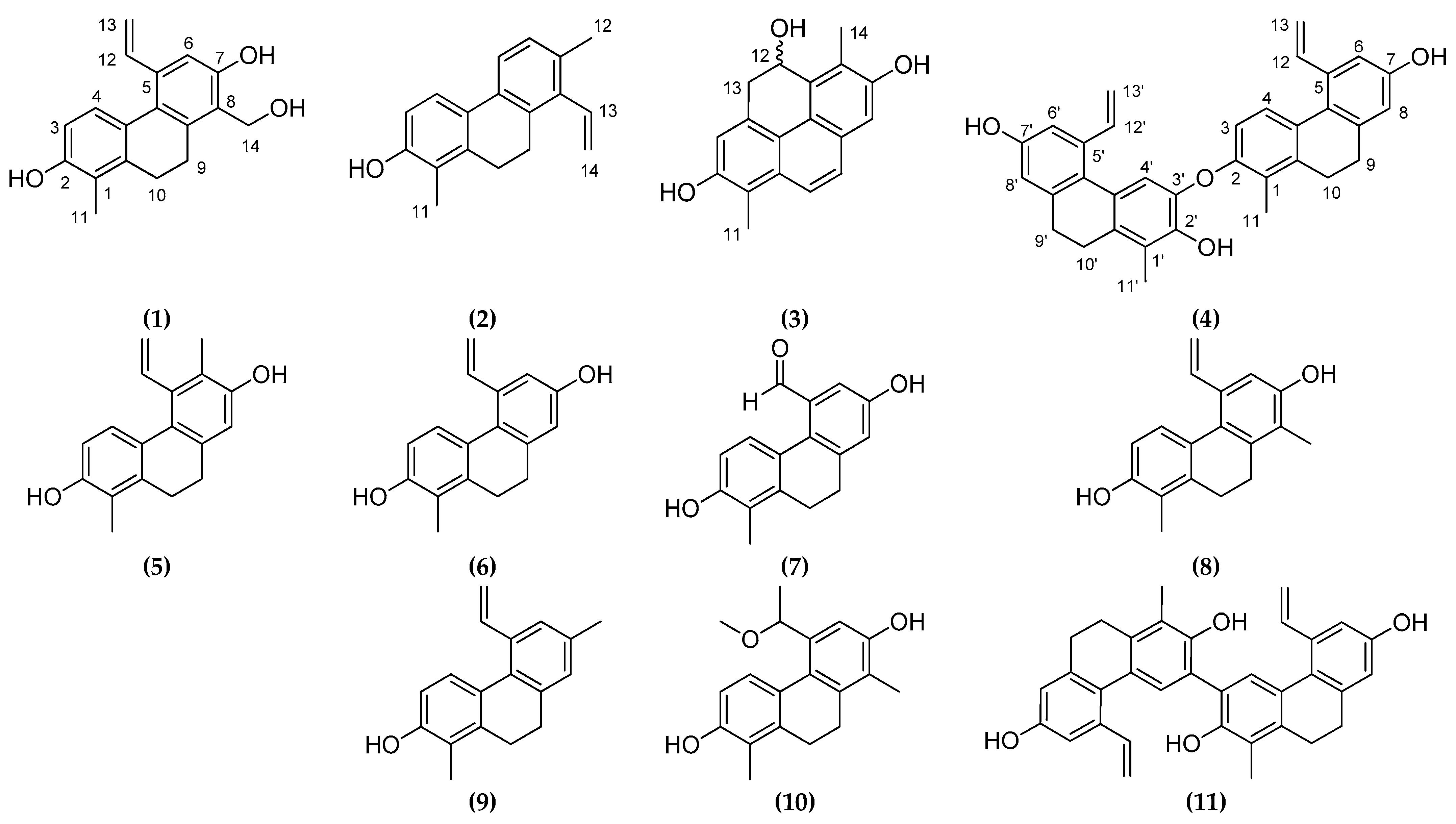
As part of our ongoing research program, we describe here the isolation and structure determination of four new [maritins A–D (1–4)] and seven known (5–11) phenanthrenes from the methanol extract of J. maritimus. The antiproliferative activity of the isolated phenanthrenes was investigated on seven human cancer cell lines (HeLa, HTM-26, T-47D, A2780, A2780cis, MCF-7, KCR) and one normal cell line (MRC-5).
2. Results
Dried aerial parts of
J. maritimus were ground and extracted with MeOH at room temperature. After concentration, the extract was dissolved in 50% aqueous MeOH, and solvent–solvent partition was performed with
n-hexane, chloroform (CHCl
3), and ethyl acetate (EtOAc). The CHCl
3 phase was separated and purified with a combination of different chromatographic methods (column chromatography (CC), vacuum liquid chromatography (VLC), medium pressure liquid chromatography (MPLC), preparative thin-layer chromatography (TLC), and HPLC) to afford 11 compounds (
Figure 1).
The structure determination was carried out by extensive spectroscopic analysis using 1D (1H- and 13CJMOD) and 2D (1H-1H COSY, HSQC, HMBC, and NOESY) NMR and HRMS spectroscopy and comparison of the spectral data with published literature values.
Compound
1 (maritin A) was isolated as a yellow amorphous solid. Its HRESIMS provided the molecular formula C
18H
18O
3 through the presence of a peak at
m/z 281.1183 [M − H]
− (calcd. for C
18H
17O
3, 281.1178). The
1H-NMR spectrum displayed signals of two
ortho-coupled aromatic methines (
δH 7.13 d and 6.63 d,
J = 8.4 Hz), an aromatic proton singlet (
δH 6.92), two methylenes (
δH 2.76 m and 2.68 m, each 2H), an oxymethylene (
δH 4.79 s, 2H), a methyl group (
δH 2.21 s, 3H), and a vinyl moiety (
δH 6.90 dd,
J = 17.4 and 10.9 Hz;
δH 5.65 dd,
J = 17.4 and 1.2 Hz;
δH 5.18 dd,
J = 10.9 and 1.2 Hz) (
Table 1,
Figure S1). The 18 carbon resonances observed in the
13C-JMOD NMR spectrum, including two oxygen-bearing sp
2 carbons at
δC 155.1 and 155.3, were attributable to a pentasubstituted phenanthrene derivative.
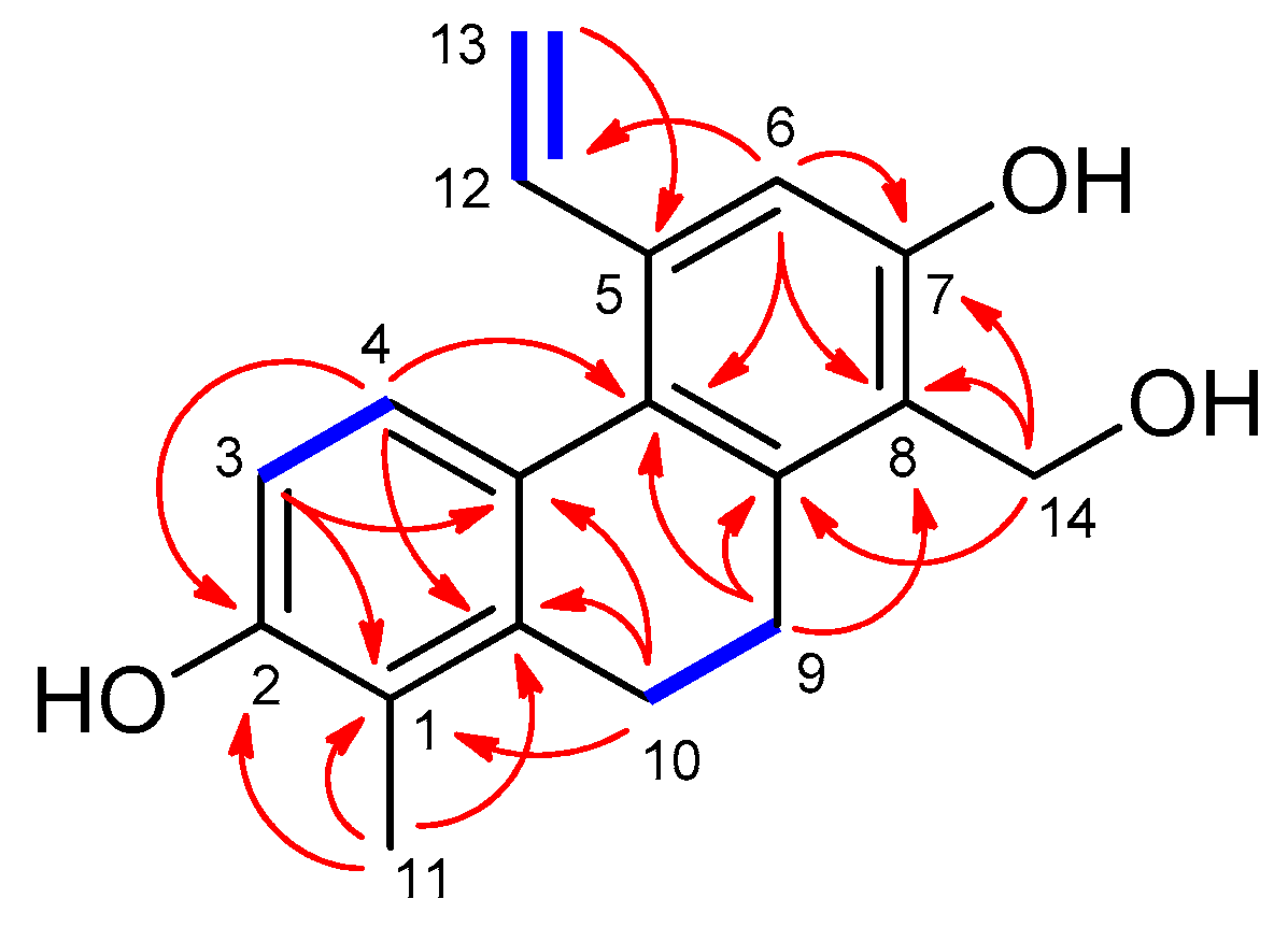
The
1H-
1H COSY correlations defined three sequences of correlated protons, namely, –CH
2–CH
2– (H
2-9–H
2-10), –CH=CH
2 (H-12–H-13a and H-13b), and –CH=CH– (H-3–H-4) fragments (
Figure 2). The structure of compound
1 was assembled with the aid of an HMBC experiment. Heteronuclear long-range correlations of H-3 and H
2-10 with C-4a (
δC 127.2), H-4, H-6, and H
2-9 with C-5a (
δC 128.3), H
2-9, H
2-10, and H
2-14 with C-8a (
δC 141.2), as well as of H-4, H
2-9, H
2-10, and H
3-11 with C-1a (
δC 140.1) established a 9,10-dihydrophenanthrene skeleton. HMBC correlations from H-3, H-4, and H
3-11 to C-2 (
δC 155.1), and from H-6 and H
2-14 to C-7 (
δC 155.3) suggested that compound
1 contains two hydroxy groups at the positions of C-2 and C-7. The location of the H
3-11 methyl group at C-1 was dictated by its HMBC correlations with C-1, C-1a, and C-2. The two- and three-bond correlations between H
2-14 (
δH 4.79), C-7, C-8 (
δC 124.4), and C-8a demonstrated that the freely rotating hydroxymethyl substituent is attached to C-8. The location of the vinyl moiety at C-5 (
δC 136.6) was confirmed by the H-6/C-12 and H-13/C-5 HMBC correlations. The NOE cross-peaks between H-4/H-12, H-13a/H-6, H
2-9/H
2-14, and H
2-10/H
3-11 were consistent with the proposed structure of
1, as shown in
Figure 2.
Compound
2 (maritin B) was obtained as a white amorphous solid. Its molecular formula was deduced to be C
18H
18O based on the protonated molecule in the HRESIMS at
m/z [M + H]
+ 251.1429 (calcd. for C
18H
19O, 251.1430). The
1H-NMR spectrum contained signals of two pairs of
ortho-coupled aromatic protons (
δH 7.47 d and 6.73 d,
J = 8.2 Hz; 7.49 d and 7.11 d,
J = 8.4 Hz), two methylenes (
δH 2.88 m and 2.74 m, each 2H), a vinyl substituent (
δH 6.77 dd,
J = 17.9 and 11.4 Hz;
δH 5.59 dd,
J = 11.4 and 2.0 Hz;
δH 5.22 dd,
J = 17.9 and 2.0 Hz), and two methyl groups (
δH 2.32 s and 2.24 s, each 3H) (
Table 1). The HMBC correlations from H
3-11 (
δH 2.24) to C-1 (
δC 120.9), C-1a (
δC 137.7), and C-2 (
δC 153.3), and further correlations between H-3 (
δH 6.73), H-4 (
δH 7.47), and C-2 showed that a methyl and a hydroxy group are situated on the adjacent carbons C-1 and C-2, respectively. The locations of another methyl (
δH 2.32) and a vinyl substituent at C-7 and C-8, respectively, were apparent from the HMBC correlations H
3-12/C-6, H
3-12/C-7, H
3-12/C-8, H-6/C-8, H
2-9/C-8, and H-14/C-8. Further heteronuclear correlations were detected between H-3, H-5 (
δH 7.49), H
2-10 (
δH 2.74), and C-4a (
δC 128.4), H-4, H-6 (
δH 7.11), H
2-9 (
δH 2.88), and C-5a (
δC 133.3), and from H-5, H
2-9, and H
2-10 to C-8a (
δC 134.1). The NOE cross-peaks H-6/H
3-12, H
3-12/H-13, H
2-9/H-14b, and H
2-10/H
3-11 supported the proposed structure of compound
2.
Separation of the plant extract yielded compound 3 (maritin C) as an orange amorphous solid. According to a peak of the deprotonated molecule at m/z 279.1027 [M − H]− in the HRESIMS data, the molecular formula C18H16O3 (calcd. for C18H15O3, 279.1021) was assigned to 3. The 1H-NMR spectrum exhibited two aromatic methines coupled with each other (δH 7.79 and 7.56 d, J = 9.2 Hz), two aromatic singlets (δH 7.17 and 7.02), two methyl groups (δH 2.50 s and 2.49 s, each 3H), and signals of an oxymethine (δH 5.45, br s) and a saturated methylene (δH 3.38 and 3.29, each 1H). The 1H-1H COSY spectrum afforded two structural elements, the aforementioned –CH=CH– (δH 7.79 and 7.56) and a –CH(OR)–CH2– fragment (δH 5.45, 3.38, and 3.29). The proton signals at δH 7.02 (H-3) and δH 2.49 (H3-11) gave HMBC correlations with a downfield shifted, nonprotonated carbon displayed at δC 153.4, while the aromatic singlet at δH 7.17 (H-8) and the methyl group at δH 2.50 (H3-14) gave HMBC correlations to a carbon resonating at δC 155.1. Thus, it was deduced that this phenanthrene bears hydroxy groups at C-2 and C-7. The two methyls were placed onto C-1 and C-6 on the basis of the corresponding H3-11/C-1, H3-11/C-1a, H3-11/C-2, H3-14/C-5, H3-14/C-6, and H3-14/C-7 HMBC correlations. Further long-range correlations from H-9 (δH 7.56) to C-1a, C-5a, and C-8, as well as from H-10 (δH 7.79) to C-1, C-4a, and C-8a established a phenanthrene skeleton with an aromatic ring B. Considering the HMBC cross-peaks of H-13a (δH 3.38) with C-3 (δC 117.0), C-4 (δC 130.9), C-4a (δC 122.5), and C-5 (δC 134.6), it was clear that a vinyl group was incorporated into an oxygen-substituted cyclohexadiene ring. From a biosynthetic point of view, compound 3 was likely formed from a dehydrojuncusol precursor through the modification of its vinylic double bond, followed by a ring closure between C-4 and C-13. The depicted structure of maritin C was corroborated by NOE cross-peaks between H-3/H-13a and b, H-12/H3-14, H-8/H-9, and H-10/H3-11. The specific optical rotation of 3 was recorded as zero, therefore, it was isolated as a racemic mixture.
Compound
4 (maritin D) has the molecular formula C
34H
30O
4 compatible with its protonated molecule at
m/z 503.2203 [M + H]
+ (calcd. for C
34H
31O
4, 503.2222) in the HRESIMS data. The 34 carbon signals displayed in the
13C-JMOD NMR spectrum suggested that compound
4 is a phenanthrene dimer (
Figure S19). The
1H-NMR spectrum, combined with homonuclear
1H-
1H COSY correlations, showed the presence of two vinyl groups (H-12–H
2-13:
δH 6.96 dd,
J = 17.4 and 10.9 Hz;
δH 5.67 d,
J = 17.4 Hz;
δH 5.23 d,
J = 10.9 Hz; H-12′–H
2-13′:
δH 6.64 dd,
J = 17.3 and 11.4 Hz;
δH 5.33 dd,
J = 17.3 and 0.9 Hz;
δH 4.78 d,
J = 11.4 Hz), a –CH=CH– (H-3–H-4:
δH 7.38 and 6.83 d,
J = 8.4 Hz) and two –CH
2–CH
2– structural portions (H
2-9–H
2-10:
δH 2.68 m and 2.78 m, each 2H; H
2-9′–H
2-10′:
δH 2.63 m and 2.64 m, each 2H), and two methyls (H
3-11:
δH 2.28 s; H
3-11′:
δH 2.30 s, each 3H) in
4 (
Table 2).
Two pairs of
meta-coupled aromatic protons (H-6 and H-8:
δH 6.88 d and 6.69 d,
J = 2.2 Hz; H-6′ and H-8′:
δH 6.68 br s and 6.61 br s) were also identified via weaker
4JH-H (
W-type) COSY cross-peaks and three-bond HMBC correlations between the corresponding methine groups. Further analysis of the HMBC correlations unambiguously determined that
4 is comprised of two monomers of a known 9,10-dihydrophenanthrene, effusol, which was also isolated as an individual compound (
6) from the plant. Taking into account the HMBC correlations from H-4′ and H
3-11′ to C-2′ (
δC 144.6), it was concluded that oxygen atoms are connected to both of the vicinal carbons C-2′ and C-3′ (
δC 156.6). Although no HMBC correlations were observed between the monomers, NOE cross-peaks H-4′/H
3-11 and H-13′b/H-12 indicated the close proximity of these protons, and consequently implied that the monomers must be attached through an ether bond between C-2/C-2′ or C-2/C-3′. In order to determine the exact structure, energy-minimized structures were generated for each of the hypothetical compounds by using the MM2 force field method. A minimum energy conformation (
Figure 3) provided by molecular dynamics calculations was in good agreement with the aforementioned NOE correlations and suggested that the ether bond was formed between C-2/C-3′. The proposed structure was further confirmed by the significantly shielded nature of H-4′ and vinyl resonances H-12′–H
2-13′ compared to H-4 and H-12–H
2-13 of the other monomer (
Table 2).
This phenomenon was likely caused by the anisotropic effect of aromatic ring A since H-4′ and the vinyl moiety H-12′–H
2-13′ are located in the shielding cone of ring A. In case of the presence of a C-2/C-2′ linkage, H-4′ and H-12′–H
2-13′ would be located too far from ring A and, therefore, their chemical shifts would be less affected by the aromatic ring current effects. Considering the above findings, the structure of
4 was formulated as depicted in
Figure 1.
Besides the new compounds maritins A–D (
1–
4), seven known phenanthrenes, namely, juncusol (
5) [
10], effusol (
6) [
10], 2,7-dihydroxy-1-methyl-5-aldehyde-9,10-dihydrophenanthrene (
7) [
11], 2,7-dihydroxy-1,8-dimethyl-5-vinyl-9,10-dihydrophenanthrene (
8) [
10], juncunol (
9) [
10], jinflexin A (
10) [
12], and effususin A (
11) [
13], were also isolated from
J. maritimus. Their structures were identified by 1D and 2D NMR spectroscopy, and by comparison of the
1H and
13C NMR chemical shift values with literature data. All compounds but effusol (
7) are described for the first time from
J. maritimus. Moreover, the
1H and
13C NMR assignments of jinflexin A in methanol-
d4 were not reported previously.
The obtained phenanthrenes
1–
11 were tested for their antiproliferative activity against seven human tumor cell lines (HeLa, HTM-26, T-47D, A2780, A2780cis, MCF-7, KCR) and one normal human fetal lung fibroblast (MRC-5) cell line using the 3-(4,5-dimethylthiazol-2-yl)-2,5-diphenyltetrazolium bromide (MTT) assay with cisplatin as a positive control (
Table 3). Among the tested compounds, dimeric phenanthrenes (
4 and
11) built up by effusol monomers showed substantial antiproliferative activity against all cell lines investigated. The highest activities were detected on T-47D ductal carcinoma cells (IC
50 9.1 μM for
4 and 6.2 μM for
11, respectively) for both compounds. No significant differences were observed between the effects of dimers on different cell lines.
T-47D cells were the most sensitive to the phenanthrenes, but four compounds (3, 4, 10, and 11) displayed remarkable antiproliferative activity (IC50 ˂ 20 µM) against A2780cis and MCF-7 cells, too. Maritin B (2), juncusol (5), and effusol (6) were effective only against the HeLa cells (with respective IC50 values of 11.0, 0.5, and 2.3 μM). Maritin A (1) and compound 8 differ from each other in their substitution at the position of C-8. Based on the experimental data, it seems that replacement of the C-8 methyl group in 8 to a hydroxymethyl moiety present in 1 leads to a drastic increase in the antiproliferative activity. Effusol (6) and compound 7 also have similar chemical structures, the difference between them involves the presence of a C-5 vinyl group in 6 and a formyl moiety at the same position in 7. No significant differences were observed upon comparison of the activities of these phenanthrenes except for the HeLa cell line. Interestingly, effusol (6) containing a C-7 hydroxy function displayed much stronger antiproliferative activity on several tumor cell lines than juncunol (9), which has a C-7 methyl substituent. This finding suggests that the presence of a polar hydroxy substituent on C-7 is probably more favorable for the antiproliferative effects than its methyl counterpart. Compounds 7–9 were weakly effective on all cell lines tested. Although the structure of 8 closely resembles that of jinflexin A (10) (the two phenanthrenes contain a vinyl and a methoxyethyl moiety at C-5, respectively), substantial differences were found in their antiproliferative profiles: compound 8 had no effects on any of the cell lines investigated, while 10 exerted activity against all seven tumor cell lines. The presence of a methoxyethyl moiety instead of a vinyl group at C-5 appears to enhance the antiproliferative activity of phenanthrenes. Unfortunately, the tested phenanthrenes showed no selectivity except for compound 1 that exhibited a less antiproliferative effect on MRC-5 normal lung fibroblasts compared to the other cancer cell lines. The other compounds considerably inhibited the proliferation of MRC-5 cells, too.
3. Materials and Methods
3.1. General Experimental Procedures
Optical rotation was determined in CHCl3 at ambient temperature, using a Perkin-Elmer 341 polarimeter (PerkinElmer, MA, USA). NMR spectra were recorded in CDCl3, methanol-d4, and dimethyl sulfoxide-d6 on a Bruker Avance DRX 500 spectrometer (Bruker, MA, USA) at 500 MHz (1H) and 125 MHz (13C-JMOD). The signals of the deuterated solvents were taken as references. The chemical shift values (δ) were given in ppm and coupling constants (J) are expressed in Hz. The high-resolution MS spectra were acquired on a Thermo Scientific Q-Exactive Plus Orbitrap mass spectrometer (Thermo Fisher Scientific, MA USA) equipped with ESI ion source in positive ionization mode. The resolution was over 1 ppm. The data were acquired and processed with MassLynx software (Waters, MA, USA). All solvents used for chromatographic separations and purification steps were analytical or HPLC grade (VWR Ltd., Szeged, Hungary).
For vacuum liquid chromatography (VLC), silica gel (silica gel GF254, 15 μm, Merck) and reversed-phase silica (LiChroprep RP-18, 40-63 μm, Merck) were used. Medium-pressure liquid chromatography (MPLC) was performed by a Combi Flash Rf+ Lumen instrument (Teledyne ISCO, NE, USA) on a reversed-phase RediSep Rf HP Gold (50 g) column. Preparative thin-layer chromatography (prep. TLC) was performed on silica gel plates (TLC silica gel 60 F254, Merck, Darmstadt, Germany), and on reversed-phase silica gel plates (TLC silica gel 60 RP-18 F254S, Merck). Sephadex LH-20 (25–100 μm, Sigma-Aldrich, Budapest, Hungary) was used for gel filtration. HPLC was carried out on a Waters Millipore instrument with UV detection at 254 nm over normal- (Kinetex Luna Silica, 3 μm, 150 × 4.6 mm, Phenomenex Inc., CA, USA) and reversed-phase (Kinetex 5 μm C18, 150 × 4.6 mm and LiChrospher RP-18, 5 μm, 250 × 4 mm) columns.
3.2. Plant Material
Juncus maritimus Lam. (whole plants, 2.2 kg) was collected in June 2018, near Vir (coordinates: 44°31′80.74” N; 15°05′72.00” E) (Croatia), and identified by one of the authors, László Bakacsy (Department of Plant Biology, University of Szeged, Szeged, Hungary). A voucher specimen (No. 884) has been deposited at the Herbarium of the Department of Pharmacognosy, University of Szeged, Szeged, Hungary.
3.3. Extraction and Isolation
The plant material (aerial part) was air-dried (2.2 kg) at room temperature. Thereafter, it was ground and percolated with 40 L methanol at room temperature. After evaporation, the extract was dissolved in 50% aqueous methanol, and repetitive solvent-–solvent partition was performed with 6 × 0.5 L n-hexane, 10 × 0.5 L chloroform, and 5 × 0.5 L EtOAc. The concentrated chloroform-soluble fraction (32 g) was separated by vacuum liquid chromatography (VLC) on silica gel with a gradient system of cyclohexane-–EtOAc-–MeOH [from 98:2:0 to 1:1:1 (1500 mL/eluent); volume of collected fractions was 150 mL]. This separation yielded 14 fractions (A-N). The fractions were combined according to their TLC patterns.
All major fractions were purified by Sephadex LH-20 gel chromatography using CH2Cl2-–MeOH (1:1) as eluent. Fraction B/2 was separated by normal-phase HPLC under gradient conditions, using cyclohexane–EtOAc (19:1 to 9:1 in 10 min and 9:1 to 65:35 in 1 min; flow rate 1.5 mL/min) as mobile phase to obtain compounds 5 (tR = 8.3 min, 1.2 mg) and 2 (tR = 10.4 min, 2.9 mg). Purification of fractions D/4 and D/5 by preparative TLC afforded compounds 6 (4.3 mg) and 7 (3.4 mg).
Fraction E was chromatographed by reversed-phase MPLC using MeOH–H2O (from 8:2 to 1:0). Subfraction E/1 was then further purified by reversed-phase HPLC under gradient conditions, using MeOH–H2O (from 45:55 to 82:18 in 10 min; flow rate 1.2 mL/min) as mobile phase to yield compound 10 (tR = 5.6 min, 2.4 mg). Subfraction E/2 was separated by preparative TLC on silica gel using cyclohexane–EtOAc–EtOH (20:10:1) as solvent system to yield compounds 9 (3.5 mg) and 3 (4.5 mg).
Fractions H/3 and I/4 were combined (HI/3-4) because of their similar chemical composition and were purified by reversed-phase MPLC using a stepwise gradient solvent system composed of MeOH–H2O (from 8:2 to 1:0). Subfraction HI/3-4/1 was separated by preparative TLC on silica gel using cyclohexane–EtOAc–EtOH (20:10:1) as eluent to isolate compound 8 (10.4 mg). HI/3-4/1/2 was purified by reversed-phase HPLC under gradient conditions, using MeOH–H2O (from 45:55 to 82:18 in 10 min; flow rate 1.2 mL/min) as mobile phase, and compound 1 (tR = 9.0 min, 5.6 mg) was isolated. Subfraction HI/3-4/7 was further fractionated by normal-phase HPLC under gradient conditions, using cyclohexane–EtOAc (from 80:20 to 65:35 in 12 min; flow rate 1.7 mL/min) as mobile phase, to afford compound 11 (tR = 13.2 min, 2.3 mg). Subfraction HI/3-4/9 was separated by preparative TLC on silica gel using an isocratic cyclohexane–EtOAc–EtOH (60:30:3) eluent, and then HI/3-4/9/3 was purified by reversed-phase HPLC under gradient conditions, using acetonitrile–H2O (from 56:44 to 70:30 in 10 min; flow rate 1.2 mL/min) as mobile phase to yield compound 4 (tR = 7.5 min, 2.0 mg).
Maritin A (1)
Yellow amorphous solid; for
1H- and
13C-JMOD NMR (in methanol-
d4) data, see
Table 1; HRESIMS
m/z 281.1183 [M − H]
− (calcd. for C
18H
17O
3, 281.1178).
Maritin B (2)
White amorphous solid; for
1H- and
13C-JMOD NMR (in CDCl
3) data, see
Table 1; HRESIMS
m/z [M + H]
+ 251.1430 (calcd. for C
18H
19O, 251.1430).
Maritin C (3)
Orange amorphous solid; [α]
25D 0 (
c 0.1, MeOH); for
1H-and
13C-JMOD NMR (in methanol-
d4) data, see
Table 1; HRESIMS
m/z 279.1027 [M − H]
− C
18H
16O
3 (calcd. for C
18H
15O
3, 279.1021).
Maritin D (4)
Yellow amorphous solid; for
1H- and
13C-JMOD NMR (in methanol-
d4) data, see
Table 2; HRESIMS
m/z [M + H]
+ 503.2203 (calcd. for C
34H
31O
4, 503.2222).
Jinflexin A (10):
1H-NMR (500 MHz, methanol-d4): δH 6.67 (1H, d, J = 8.3 Hz; H-3), 6.87 (1H, d, J = 8.3 Hz; H-4), 6.86 (1H, s; H-6), 2.87 (1H, m; H-9a), 2.37 (1H, m; H-9b), 2.88 (1H, m; H-10a), 2.38 (1H, m; H-10b), 2.22 (3H, s; H3-11), 4.85 (1H, overlaps with residual H2O signal; H-12), 1.57 (3H, d, J = 6.2 Hz; H3-13), 2.19 (3H, s; H3-14), 2.92 (3H, s; 12-OCH3). 13C NMR (125 MHz, methanol-d4): δC 121.8 (C-1), 140.5 (C-1a), 154.9 (C-2), 112.4 (C-3), 127.5 (C-4), 127.4 (C-4a), 129.3 (C-5a), 138.4 (C-5), 111.5 (C-6), 155.1 (C-7), 121.4 (C-8), 139.8 (C-8a), 27.6 (C-9), 26.7 (C-10), 11.8 (C-11), 76.5 (C-12), 23.4 (C-13), 11.8 (C-14), 55.7 (12-OCH3).
3.4. Antiproliferative Assay
3.4.1. Cell Lines
Breast cancer cell line MCF-7 (ATCC®® HTB-22) and the drug-resistant subline of the human breast cancer MCF-7 (ECACC 86012803; KCR) were purchased from LGC Promochem (Teddington, UK). Both cell lines were cultured in Eagle’s Minimal Essential Medium (EMEM, containing 4.5 g/L glucose) supplemented with a non-essential amino acid mixture, a selection of vitamins, and 10% heat-inactivated fetal bovine serum. In every third passage, 0.56 µg/mL doxorubicin was added to the medium in order to maintain the ABCB1 (P-glycoprotein) expression in KCR cells. A2780 human ovarian cancer cell line (ECACC European Collection of Authentical Cell Culture, Sigma Cat. no. 93112519) and the cisplatin-resistant human ovarian cancer cell line A2780cis (ECACC European Collection of Authentical Cell Culture, Sigma Cat. no. 93112517) were purchased from Merck KGaA (Darmstadt, Germany). The human ovarian cancer cell lines were cultured in RPMI 1640 medium supplemented with 10% heat-inactivated fetal bovine serum. The RPMI 1640 medium of the cisplatin-resistant cell line A2780 was supplemented with 1 µM cisplatin. HeLa (ATCC® CCL-2™) human cervix carcinoma, HTB-26 breast adenocarcinoma, T-47D (ATCC® HTB-133™) ductal carcinoma, and MRC-5 human embryonal lung fibroblast cell lines (ATCC® CCL-171) were purchased from LGC Promochem (Teddington, UK). The cells were cultured in Eagle’s Minimal Essential Medium (EMEM, containing 4.5 g/L glucose) supplemented with a non-essential amino acid mixture, a selection of vitamins, and 10% heat-inactivated fetal bovine serum. HTB-26 cell line was cultured in RPMI 1640 medium supplemented with 10% heat-inactivated fetal bovine serum. T-47D cells were cultured in RPMI 1640 medium supplemented with 10% heat-inactivated fetal bovine serum, 2 mM L-glutamine, 1 mM Na-pyruvate, and 100 mM Hepes. All of the cells were incubated at 37 °C, in a 5% CO2, 95% air atmosphere.
3.4.2. Antiproliferative Assay
The antiproliferative effect of the compounds was determined on the human breast (MCF-7, KCR, T-47D, HTB-26), cervical (HeLa), and ovarian (A2780, A2780cis) cancer cells, and on MRC-5 (human embryonic lung fibroblast) cell line. The adherent cells were cultured in 96-well flat-bottomed microtiter plates, using EMEM supplemented with 10% heat-inactivated fetal bovine serum or RPMI 1640 supplemented with 10% heat-inactivated fetal bovine serum, respectively. The density of the cells was adjusted to 6 × 10
3 cells in 100 μL per well, the cells were seeded for 24 h at 37 °C, 5% CO
2, then the medium was removed from the plates and fresh medium (100 μL per well) was added to the cells. The effects of increasing concentrations of compounds on cell proliferation were tested in 96-well flat-bottomed microtiter plates. The compounds were diluted in the appropriate medium, the dilutions of compounds were performed in separate plates and then added to the cells. The starting concentration of the compounds was 100 μM, and two-fold serial dilution was performed (concentration range: 100-0.19 μM). The culture plates were incubated at 37 °C for 72 h; at the end of the incubation period, 20 μL of MTT (thiazolyl blue tetrazolium bromide, Sigma) solution (from a stock solution of 5 mg/mL) was added to each well. After incubation at 37 °C for 4 h, 100 μL of sodium dodecyl sulfate (SDS) (Sigma) solution (10% in 0.01 M HCI) were added to each well and the plates were further incubated at 37 °C overnight. Cell growth was determined by measuring the optical density (OD) at 540/630 nm with Multiscan EX ELISA reader (Thermo Labsystems, Cheshire, WA, USA). Mean IC
50 values were obtained by best fitting the dose-dependent inhibition curves in GraphPadPrism5 program (GraphPad Software version 5.00 for Windows, San Diego, CA, USA) from four parallel experiments for each cell line. Results are expressed in terms of IC
50, defined as the inhibitory dose that reduces the proliferation of the cells exposed to the tested compounds by 50% [
14].


 ,
, 


