
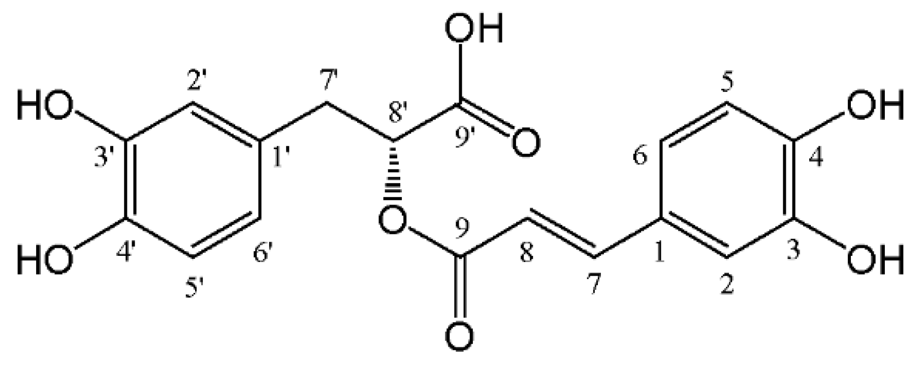
A Comprehensive Review of Rosmarinic Acid: From Phytochemistry to Pharmacology and Its New Insight

 ,
,
Abstract
:1. Introduction
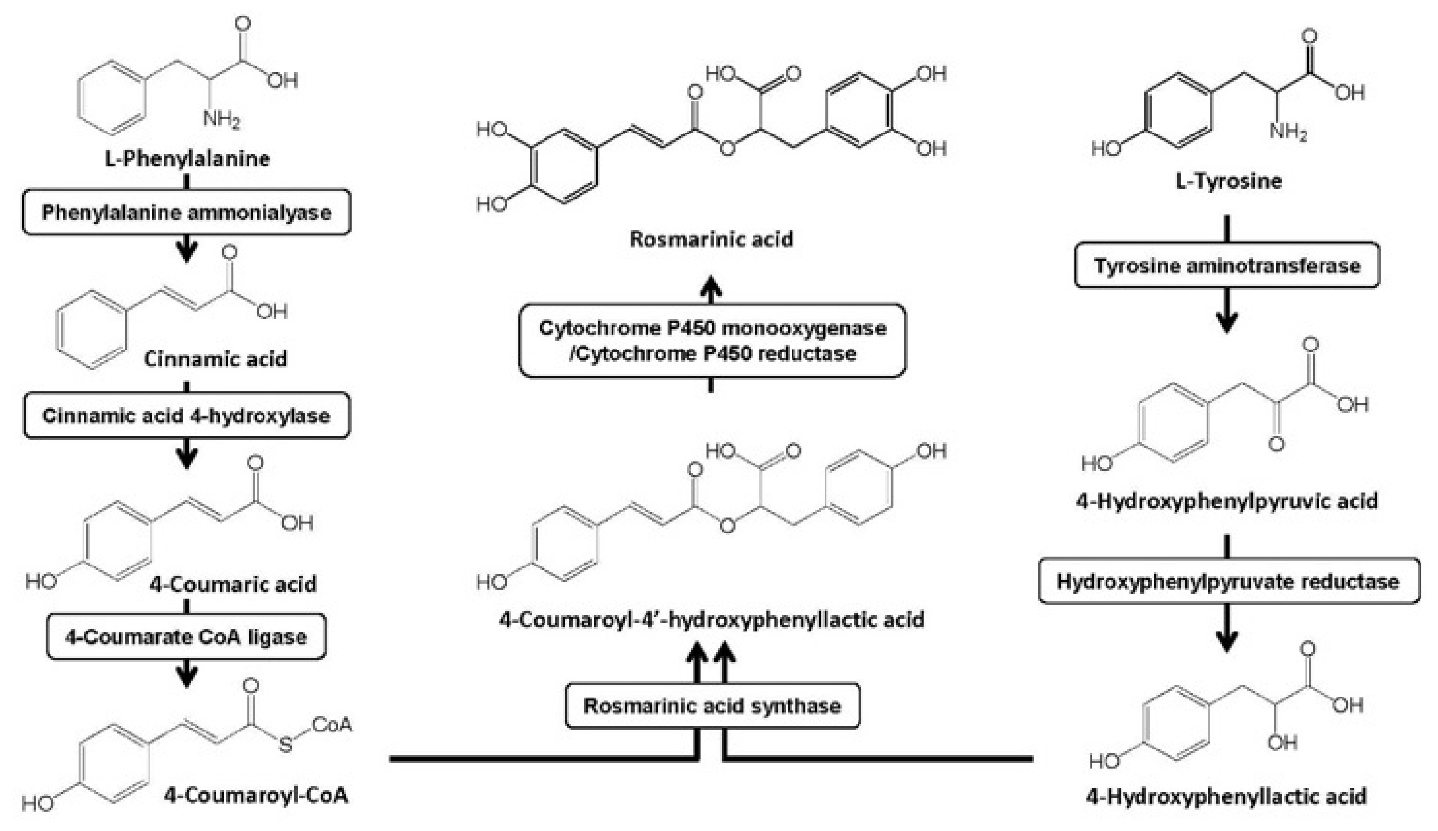
2. Sources and Biosynthesis in the Plants
3. Extraction from Plants
4. Natural Derivatives in Plants
5. Analytical Technique
6. Pharmacology
6.1. Anti-Inflammation
6.2. Anti-Oxidation
6.3. Anti-Diabetes
6.4. Anti-Tumor
6.5. Anti-Virus
6.6. Neuroprotection
6.7. Hepatoprotection
6.8. Other Activities
7. Clinical Studies
8. Applications in Food Science
9. Pharmacokinetics
10. Future Perspectives
11. Conclusions
Supplementary Materials
Author Contributions
Funding
Informed Consent Statement
Conflicts of Interest
References
- Scarpati, M.L.; Oriente, G. Isolamento e costituzione dell’acido rosmarinico (dal rosmarinus off.). Ric. Sci. 1958, 28, 2329–2333. [Google Scholar]
- Elufioye, T.O.; Habtemariam, S. Hepatoprotective effects of rosmarinic acid: Insight into its mechanisms of action. Biomed. Pharmacother. 2019, 112, 108600. [Google Scholar] [CrossRef] [PubMed]
- Fachel, F.N.S.; Schuh, R.S.; Veras, K.S.; Bassani, V.L.; Koester, L.S.; Henriques, A.T.; Braganhol, E.; Teixeira, H.F. An overview of the neuroprotective potential of rosmarinic acid and its association with nanotechnology-based delivery systems: A novel approach to treating neurodegenerative disorders. Neurochem. Int. 2019, 122, 47–58. [Google Scholar] [CrossRef] [PubMed]
- Hitl, M.; Kladar, N.; Gavaric, N.; Bozin, B. Rosmarinic acid-human pharmacokinetics and health benefits. Planta Med. 2021, 87, 273–282. [Google Scholar] [CrossRef] [PubMed]
- Luo, C.X.; Zou, L.; Sun, H.J.; Peng, J.Y.; Gao, C.; Bao, L.C.; Ji, R.P.; Jin, Y.; Sun, S.Y. A review of the anti-inflammatory effects of rosmarinic acid on inflammatory diseases. Front. Pharmacol. 2020, 11, 153. [Google Scholar] [CrossRef]
- Nadeem, M.; Imran, M.; Gondal, T.A.; Imran, A.; Shahbaz, M.; Amir, R.M.; Sajid, M.W.; Qaisrani, T.B.; Atif, M.; Hussain, G.; et al. Therapeutic potential of rosmarinic acid: A comprehensive review. Appl. Sci. 2019, 9, 3139. [Google Scholar] [CrossRef] [Green Version]
- Ngo, Y.L.; Lau, C.H.; Chua, L.S. Review on rosmarinic acid extraction, fractionation and its anti-diabetic potential. Food Chem. Toxicol. 2018, 121, 687–700. [Google Scholar] [CrossRef]
- Rahbardar, M.G.; Hosseinzadeh, H. Effects of rosmarinic acid on nervous system disorders: An updated review. Naunyn-Schmiedeberg’s Arch. Pharmacol. 2020, 393, 1779–1795. [Google Scholar] [CrossRef]
- Swamy, M.K.; Sinniah, U.R.; Ghasemzadeh, A. Anticancer potential of rosmarinic acid and its improved production through biotechnological interventions and functional genomics. Appl. Microbiol. Biotechnol. 2018, 102, 7775–7793. [Google Scholar] [CrossRef]
- European and Mediterranean Plant Protection Organization. Available online: http://gd.eppo.int (accessed on 10 May 2022).
- Akhtar, M.S.; Hossain, M.A.; Said, S.A. Isolation and characterization of antimicrobial compound from the stem-bark of the traditionally used medicinal plant Adenium obesum. J. Tradit. Complement. Med. 2016, 7, 296–300. [Google Scholar] [CrossRef] [Green Version]
- Tufa, T.; Damianakos, H.; Zengin, G.; Graikou, K.; Chinou, I. Antioxidant and enzyme inhibitory activities of disodium rabdosiin isolated from Alkanna sfikasiana Tan, Vold and Strid. S. Afr. J. Bot. 2019, 120, 157–162. [Google Scholar] [CrossRef]
- Kuruuzum-Uz, A.; Suleyman, H.; Cadirci, E.; Guvenalp, Z.; Demirezer, L.O. Investigation on anti-Inflammatory and antiulcer activities of Anchusa azurea extracts and their major constituent rosmarinic acid. Z. Nat. C 2012, 67, 360–366. [Google Scholar]
- Li, M.H.; Chang, W.; Lu, W.J.; Ma, G.Z. Chemical constituents of Anchusa italica Retz. and protective effects on hypoxia/reoxygenation induced oxidative stress injury in rat primary cardiomyocyte. Northwest Pharm. J. 2020, 35, 335–340. [Google Scholar]
- Braca, A.; Bader, A.; Siciliano, T.; Morelli, L.; De Tommasi, N. New pyrrolizidine alkaloids and glycosides from Anchusa strigosa. Planta Med. 2003, 69, 835–841. [Google Scholar]
- Takeda, R.; Hasegawa, J.; Shinozaki, M. The first isolation of lignans, megacerotonic acid and anthocerotonic acid, from non-vascular plants, Anthocerotae (hornworts). Tetrahedron Lett. 1990, 31, 4159–4162. [Google Scholar] [CrossRef]
- Lasure, A.; Vanpoel, B.; Pieters, L.; Claeys, M.; Gupta, M.; Vandenberghe, D.; Vlietinck, A.J. Complement-inhibiting properties of Apeiba tibourbou. Planta Med. 1994, 60, 276–277. [Google Scholar] [CrossRef]
- Olivier, D.K.; van Wyk, B.E.; van Heerden, F.R. The chemotaxonomic and medicinal significance of phenolic acids in Arctopus and Alepidea (Apiaceae subfamily Saniculoideae). Biochem. Syst. Ecol. 2008, 36, 724–729. [Google Scholar] [CrossRef]
- Yuzbasioglu, M.; Kuruuzum-Uz, A.; Guvenalp, Z.; Simon, A.; Toth, G.; Harput, U.S.; Kazaz, C.; Bilgili, B.; Duman, H.; Saracoglu, I.; et al. Cytotoxic compounds from endemic Arnebia purpurea. Nat. Prod. Commun. 2015, 10, 595–596. [Google Scholar] [CrossRef] [Green Version]
- Argoti, J.C.; Linares-Palomino, P.J.; Salido, S.; Ramirez, B.; Insuasty, B.; Altarejos, J. On-line activity screening for radical scavengers from Baccharis chilco. Chem. Biodivers. 2013, 10, 189–197. [Google Scholar] [CrossRef]
- Badem, M.; Sener, S.O.; Kanbolat, S.; Korkmaz, N.; Yildirmis, S.; Ozgen, U.; Aliyazicioglu, R.; Salva, E.; Kaban, K.; Kandemir, A.; et al. Evaluation of biological activities of Barbarea integrifolia and isolation of a new glucosinolate derivated compound. Z. Nat. C 2021, 76, 375–382. [Google Scholar] [CrossRef]
- Scognamiglio, M.; Buommino, E.; Coretti, L.; Graziani, V.; Russo, R.; Caputo, P.; Donnarumma, G.; D’Abrosca, B.; Fiorentino, A. Phytochemical investigation and antimicrobial assessment of Bellis sylvestris leaves. Phytochem. Lett. 2016, 17, 6–13. [Google Scholar] [CrossRef]
- Andrade, J.M.D.; Passos, C.D.; Rubio, M.A.K.; Mendonca, J.N.; Lopes, N.P.; Henriques, A.T. Combining in vitro and in silico approaches to evaluate the multifunctional profile of rosmarinic acid from Blechnum brasiliense on targets related to neurodegeneration. Chem. Biol. Interact. 2016, 254, 135–145. [Google Scholar] [CrossRef] [PubMed]
- Zhang, J.; Wang, Z.W.; Mi, Q. Phenolic compounds from Canna edulis Ker residue and their antioxidant activity. LWT-Food Sci. Technol. 2011, 44, 2091–2096. [Google Scholar] [CrossRef]
- Ly, T.N.; Shimoyamada, M.; Yamauchi, R. Isolation and characterization of rosmarinic acid oligomers in Celastrus hindsii Benth leaves and their antioxidative activity. J. Agric. Food Chem. 2006, 54, 3786–3793. [Google Scholar] [CrossRef] [PubMed]
- Yoshida, M.; Fuchigami, M.; Nagao, T.; Okabe, H.; Matsunaga, K.; Takata, J.; Karube, Y.; Tsuchihashi, R.; Kinjo, J.; Mihashi, K.; et al. Antiproliferative constituents from Umbelliferae plants VII. Active triterpenes and rosmarinic acid from Centella asiatica. Biol. Pharm. Bull. 2005, 28, 173–175. [Google Scholar] [CrossRef] [PubMed] [Green Version]
- Chen, F.Y.; Zou, Y.; Chen, J.; Huang, W.M.; Bian, Y.T.; Luo, Y.M. Studies on chemical constituents of Chloranthus fortune. Chin. Tradit. Herb. Drugs 2020, 51, 1485–1490. [Google Scholar]
- Ma, X.H.; Huang, M.; Deng, S.H.; Yang, J.; Ke, R.F.; Song, P.; Yang, X.Z. Chemical constituents and bioactivity of Chloranthus multistachys Pei. J. Yunnan Univ. 2017, 39, 124–129. [Google Scholar]
- Sun, Z.C.; Zheng, Q.X.; Ma, G.X.; Zhang, X.P.; Yuan, J.Q.; Wu, H.F.; Liu, H.L.; Yang, J.S.; Xu, X.D. Four new phenolic acids from Clerodendranthus spicatus. Phytochem. Lett. 2014, 8, 16–21. [Google Scholar] [CrossRef]
- Tezuka, Y.; Stampoulis, P.; Banskota, A.H.; Awale, S.; Tran, K.Q.; Saiki, I.; Kadota, S. Constituents of the Vietnamese medicinal plant Orthosiphon stamineus. Chem. Pharm. Bull. 2000, 48, 1711–1719. [Google Scholar] [CrossRef] [Green Version]
- Murata, T.; Sasaki, K.; Sato, K.; Yoshizaki, F.; Yamada, H.; Mutoh, H.; Umehara, K.; Miyase, T.; Warashina, T.; Aoshima, H.; et al. Matrix metalloproteinase-2 inhibitors from Clinopodium chinense var. parviflorum. J. Nat. Prod. 2009, 72, 1379–1384. [Google Scholar] [CrossRef]
- Saltos, M.B.V.; Puente, B.F.N.; Malafronte, N.; Braca, A. Phenolic compounds from Clinopodium tomentosum (Kunth) Govaerts (Lamiaceae). J. Brazil. Chem. Soc. 2014, 25, 2121–2124. [Google Scholar]
- Wei, X.M.; Cheng, J.K.; Cheng, D.L.; Gao, L.M. Chemical constituents from Clinopodium urticifolium. J. Chin. Chem. Soc. Taip. 2004, 51, 1043–1049. [Google Scholar] [CrossRef]
- Kumaran, A.; Karunakaran, R.J. Activity-guided isolation and identification of free radical-scavenging components from an aqueous extract of Coleus aromaticus. Food Chem. 2007, 100, 356–361. [Google Scholar] [CrossRef]
- Pan, L.L.; Zhao, Q.; Liu, H.Y. Chemical contituents of Coleus forskohlii. J. Yunnan Univ. Chin. Tradit. Med. 2012, 35, 11–13, 45. [Google Scholar]
- Tewtrakul, S.; Miyashiro, H.; Nakamura, N.; Hattori, M.; Kawahata, T.; Otake, T.; Yoshinaga, T.; Fujiwara, T.; Supavita, T.; Yuenyongsawad, S.; et al. HIV-1 integrase inhibitory substances from Coleus parvifolius. Phytother. Res. 2003, 17, 232–239. [Google Scholar] [CrossRef]
- Li, H.M.; Hwang, S.H.; Kang, B.G.; Hong, J.S.; Lim, S.S. Inhibitory effects of Colocasia esculenta (L.) Schott constituents on aldose reductase. Molecules 2014, 19, 13212–13224. [Google Scholar] [CrossRef] [Green Version]
- Fouseki, M.M.; Damianakos, H.; Karikas, G.A.; Roussakis, C.; Gupta, M.P.; Chinou, I. Chemical constituents from Cordia alliodora and C. colloccoca (Boraginaceae) and their biological activities. Fitoterapia 2016, 115, 9–14. [Google Scholar] [CrossRef]
- Marini, G.; Graikou, K.; Zengin, G.; Karikas, G.A.; Gupta, M.P.; Chinou, I. Phytochemical analysis and biological evaluation of three selected Cordia species from Panama. Ind. Crop. Prod. 2018, 120, 84–89. [Google Scholar] [CrossRef]
- Owis, A.I.; Abo-Youssef, A.M.; Osman, A.H. Leaves of Cordia boissieri A. DC. as a potential source of bioactive secondary metabolites for protection against metabolic syndrome-induced in rats. Z. Nat. C 2017, 72, 107–118. [Google Scholar] [CrossRef]
- Fatima, M.; Siddiqui, B.S.; Begum, S. New neolignan glucoside and new biphenyl ether lignan from the fruits of Cordia latifolia. Chem. Nat. Compd. 2017, 53, 432–435. [Google Scholar] [CrossRef]
- Giles-Rivas, D.; Estrada-Soto, S.; Aguilar-Guadarrama, A.B.; Almanza-Perez, J.; Garcia-Jimenez, S.; Colin-Lozano, B.; Navarrete-Vazquez, G.; Villalobos-Molina, R. Antidiabetic effect of Cordia morelosana, chemical and pharmacological studies. J. Ethnopharmacol. 2020, 251, 112543. [Google Scholar] [CrossRef] [PubMed]
- Al-Musayeib, N.; Perveen, S.; Fatima, I.; Nasir, M.; Hussain, A. Antioxidant, anti-glycation and anti-Inflammatory activities of phenolic constituents from Cordia sinensis. Molecules 2011, 16, 10214–10226. [Google Scholar] [CrossRef] [PubMed] [Green Version]
- Ticli, F.K.; Hage, L.I.S.; Cambraia, R.S.; Pereira, P.S.; Magro, A.J.; Fontes, M.R.M.; Stabeli, R.G.; Giglio, J.R.; Franca, S.C.; Soares, A.M.; et al. Rosmarinic acid, a new snake venom phospholipase A2 inhibitor from Cordia verbenacea (Boraginaceae): Antiserum action potentiation and molecular interaction. Toxicon 2005, 46, 318–327. [Google Scholar] [CrossRef] [PubMed]
- Damianakos, H.; Jeziorek, M.; Syklowska-Baranek, K.; Buchwald, W.; Pietrosiuk, A.; Chinou, I. Pyrrolizidine alkaloids from Cynoglossum columnae Ten. (Boraginaceae). Phytochem. Lett. 2016, 15, 234–237. [Google Scholar] [CrossRef]
- Sabrin, M.S.; Selenge, E.; Takeda, Y.; Batkhuu, J.; Ogawa, H.; Jamsransuren, D.; Suganuma, K.; Murata, T. Isolation and evaluation of virucidal activities of flavanone glycosides and rosmarinic acid derivatives from Dracocephalum spp. against feline calicivirus. Phytochemistry 2021, 191, 112896. [Google Scholar] [CrossRef] [PubMed]
- Shi, Q.Q.; Dang, J.; Wen, H.X.; Yuan, X.; Tao, Y.D.; Wang, Q.L. Anti-hepatitis, antioxidant activities and bioactive compounds of Dracocephalum heterophyllum extracts. Bot. Stud. 2016, 57, 16. [Google Scholar] [CrossRef] [PubMed] [Green Version]
- Olennikov, D.N.; Chirikova, N.K.; Okhlopkova, Z.M.; Zulfugarov, I.S. Chemical composition and antioxidant activity of Tánara Ótó (Dracocephalum palmatum Stephan), a medicinal plant used by the North-Yakutian Nomads. Molecules 2013, 18, 14105–14121. [Google Scholar] [CrossRef] [Green Version]
- Zuo, M.Y.; Yang, C.; Tian, Q.; Luo, Y.; Yang, C.; Zeng, L.; Li, G.P. Chemical constituents of Dracocephalum tanguticum Maxim of genus Dracocephalum. J. Yunnan Univ. Natl. 2015, 24, 101–103. [Google Scholar]
- Le, T.T.; Kang, T.K.; Do, H.T.; Nghiem, T.D.; Lee, W.B.; Jung, S.H. Protection against oxidative stress-induced retinal cell death by compounds isolated from Ehretia asperula. Nat. Prod. Commun. 2022, 16, 1934578X211067986. [Google Scholar] [CrossRef]
- Iqbal, K.; Nawaz, S.A.; Malik, A.; Riaz, N.; Mukhtar, N.; Mohammad, P.; Choudhary, M.I. Isolation and lipoxygenase-inhibition studies of phenolic constituents from Ehretia obtusifolia. Chem. Biodivers. 2005, 2, 104–111. [Google Scholar] [CrossRef]
- Simpol, L.R.; Otsuka, H.; Ohtani, K.; Kasai, R.; Yamasaki, K. Nitrile glucosides and rosmarinic acid, the histamine inhibitor from Ehretia-philippinensis. Phytochemistry 1994, 36, 91–95. [Google Scholar] [CrossRef]
- Li, L.; Peng, Y.; Xu, L.J.; Li, M.H.; Xiao, P.G. Flavonoid glycosides and phenolic acids from Ehretia thyrsiflora. Biochem. Syst. Ecol. 2008, 36, 915–918. [Google Scholar] [CrossRef]
- Zhong, J.D.; Feng, F.; Li, H.M.; Li, H.Z.; Li, R.T. Chemical constituents from Elsholtiza bodinieri Vaniot. J. Kunming Univ. Sci. Technol. 2013, 38, 75–79, 100. [Google Scholar]
- Li, H.; Nakashima, T.; Tanaka, T.; Zhang, Y.J.; Yang, C.R.; Kouno, I. Two new maltol glycosides and cyanogenic glycosides from Elsholtzia rugulosa Hemsl. J. Nat. Med. 2008, 62, 75–78. [Google Scholar] [CrossRef] [PubMed] [Green Version]
- Peng, H.Y.; Xing, Y.; Gao, L.L.; Zhang, L.; Zhang, G.L. Simultaneous separation of apigenin, luteolin and rosmarinic acid from the aerial parts of the copper-tolerant plant Elsholtzia splendens. Environ. Sci. Pollut. Res. 2014, 21, 8124–8132. [Google Scholar] [CrossRef]
- Devkota, H.P.; Tsushiro, K.; Watanabe, T. Bioactive phenolic compounds from the flowers of Farfugium japonicum (L.) Kitam. var. giganteum (Siebold et Zucc.) Kitam. (Asteraceae). Nat. Prod. Res. 2021. [Google Scholar] [CrossRef]
- Parejo, I.; Viladomat, F.; Bastida, J.; Schmeda-Hirschmann, G.; Burillo, J.; Codina, C. Bioguided isolation and identification of the nonvolatile antioxidant compounds from fennel (Foeniculum vulgare Mill.) waste. J. Agric. Food Chem. 2004, 52, 1890–1897. [Google Scholar] [CrossRef]
- Hawas, U.W.; Gamal-Eldeen, A.M.; El-Desouky, S.K.; Kim, Y.K.; Huefner, A.; Saf, R. Induction of caspase-8 and death receptors by a new dammarane skeleton from the dried fruits of Forsythia koreana. Z. Nat. C 2013, 68, 29–38. [Google Scholar]
- Shahat, A.A.; Hidayathulla, S.; Khan, A.A.; Alanazi, A.M.; Al Meanazel, O.T.; Alqahtani, A.S.; Alsaid, M.S.; Hussein, A.A. Phytochemical profiling, antioxidant and anticancer activities of Gastrocotyle hispida growing in Saudi Arabia. Acta Trop. 2019, 191, 243–247. [Google Scholar] [CrossRef]
- Yu, Z.B.; Wu, X.; Ye, Y.H.; Zhou, Y.W. Chemical constituents of Glechoma longituba. Nat. Prod. Res. Dev. 2008, 20, 262–264. [Google Scholar]
- Aquino, R.; Ciavatta, M.L.; De Simone, F.; Pizza, C. A flavanone glycoside from Hamelia patens. Phytochemistry 1990, 29, 2358–2360. [Google Scholar] [CrossRef]
- Trute, A.; Nahrstedt, A. Identification and quantitative analysis of phenolic compounds from the dry extract of Hedera helix. Planta Med. 1997, 63, 177–179. [Google Scholar] [CrossRef] [PubMed]
- Jin, X.Q.; Pang, S.Q. Studies on chemical constituents and their anti-tumor activities in roots of Helicteres angustifolia. Anhui Med. Pharmaceut. J. 2016, 20, 34–37. [Google Scholar]
- Tra, N.T.; Ha, N.T.T.; Cham, B.T.; Anh, L.T.T.; Yen, L.T.H.; Giang, B.L.; Anh, D.T.T.; Tuyen, N.V.; Kiem, P.V. A new benzofuran derivative from the stems of Helicteres hirsuta. Nat. Prod. Commun. 2019, 14, 1934578X19858814. [Google Scholar] [CrossRef] [Green Version]
- Satake, T.; Kamiya, K.; Saiki, Y.; Hama, T.; Fujimoto, U.; Kitanaka, S.; Kimura, Y.; Uzawa, J.; Endang, H.; Umar, M. Studies on the constituents of fruits of Helicteres isora L. Chem. Pharm. Bull. 1999, 47, 1444–1447. [Google Scholar] [CrossRef] [Green Version]
- De Lucena, H.F.S.; Madeiro, S.A.L.; Siqueira, C.D.; Barbosa, J.M.; Agra, M.D.; da Silva, M.S.; Tavares, J.F. Hypenol, a new lignan from Hypenia salzmannii. Helv. Chim. Acta 2013, 96, 1121–1125. [Google Scholar] [CrossRef]
- Abedini, A.; Roumy, V.; Mahieux, S.; Biabiany, M.; Standaert-Vitse, A.; Riviere, C.; Sahpaz, S.; Bailleul, F.; Neut, C.; Hennebelle, T. Rosmarinic acid and its methyl ester as antimicrobial components of the hydromethanolic extract of Hyptis atrorubens Poit. (Lamiaceae). Evid. Based Complement. Altern. Med. 2013, 2013, 604536. [Google Scholar] [CrossRef] [Green Version]
- Almtorp, G.T.; Hazell, A.C.; Torssell, K.B.G. A lignan and pyrone and other constituents from Hyptis-capitata. Phytochemistry 1991, 30, 2753–2756. [Google Scholar] [CrossRef]
- Falcao, R.A.; do Nascimento, P.L.A.; de Souza, S.A.; da Silva, T.M.G.; de Queiroz, A.C.; da Matta, C.B.B.; Moreira, M.S.A.; Camara, C.A.; Silva, T.M.S. Antileishmanial phenylpropanoids from the leaves of Hyptis pectinata (L.) Poit. Evid. Based Complement. Altern. Med. 2013, 2013, 460613. [Google Scholar] [CrossRef] [Green Version]
- Tang, G.Q.; Liu, X.L.; Gong, X.; Lin, X.J.; Lai, X.D.; Wang, D.; Ji, S.G. Studies on the chemical compositions of Hyptis suaveolens (L.) Poit. J. Serb. Chem. Soc. 2019, 84, 245–252. [Google Scholar] [CrossRef] [Green Version]
- Kuhnt, M.; Rimpler, H.; Heinrich, M. Lignans and other compounds from the Mixe-Indian medicinal plant Hyptis-verticillata. Phytochemistry 1994, 36, 485–489. [Google Scholar] [CrossRef]
- Furukawa, M.; Makino, M.; Ohkoshi, E.; Uchiyama, T.; Fujimoto, Y. Terpenoids and phenethyl glucosides from Hyssopus cuspidatus (Labiatae). Phytochemistry 2011, 72, 2244–2252. [Google Scholar] [CrossRef] [PubMed]
- Arif, Z.; Khan, S.; Farheen, S.; Kazmi, M.H.; Fatima, I.; Malik, A.; Ali, M.S.; Inamullah, F.; Afaq, S.; Shaikh, S.A.; et al. Turpesteryl ester, a new antibacterial steroid from Ipomoea turpethum. Chem. Nat. Compd. 2020, 56, 270–273. [Google Scholar] [CrossRef]
- Niu, X.M.; Li, S.H.; Na, Z.; Mei, S.X.; Zhao, Q.S.; Sun, H.D. Studies on chemical constituents of Isodon eriocalyx var. laxiflora. Chin. Tradit. Herb. Drugs 2003, 34, 300–303. [Google Scholar]
- Li, L.J.; Yu, L.J.; Wu, Z.Z.; Liu, X. Chemical constituents in ethyl acetate extract from Rabdosia flexicaulis. Chin. Tradit. Herb. Drugs 2015, 46, 339–343. [Google Scholar]
- Zhou, W.T.; Xie, H.H.; Xu, X.Y.; Liang, Y.G.; Wei, X.Y. Phenolic constituents from Isodon lophanthoides var. graciliflorus and their antioxidant and antibacterial activities. J. Funct. Foods 2014, 6, 492–498. [Google Scholar]
- Kuang, Y.H.; Lin, Q.; Liang, S.; Yao, X.H.; Wang, Z.M.; Li, C.Y. Water-soluble chemical constituents from Rabdosia lophanthoides. Chin. J. Exp. Tradit. Med. Formulae 2014, 20, 110–112. [Google Scholar]
- Huang, H.; Chao, Q.R.; Tan, R.X.; Sun, H.D.; Wang, D.C.; Ma, J.; Zhao, S.X. A new rosmarinic acid derivative from Isodon oresbius. Planta Med. 1999, 65, 92–93. [Google Scholar] [CrossRef] [Green Version]
- Zheng, X.K.; Li, Q.; Feng, W.S. Studies on chemical constituents of phenolic acids in Rabdosia rubescens. Chin. Pharm. J. 2004, 39, 335–336. [Google Scholar]
- Khan, S.; Taning, C.N.T.; Bonneure, E.; Mangelinckx, S.; Smagghe, G.; Ahmad, R.; Fatima, N.; Asif, M.; Shah, M.M. Bioactivity-guided isolation of rosmarinic acid as the principle bioactive compound from the butanol extract of Isodon rugosus against the pea aphid, Acyrthosiphon pisum. PLoS ONE 2019, 14, e0215048. [Google Scholar] [CrossRef] [Green Version]
- Jiang, B.; Hou, A.J.; Li, M.L.; Li, S.H.; Han, Q.B.; Wang, S.J.; Lin, Z.W.; Sun, H.D. Cytotoxic ent-kaurane diterpenoids from Isodon sculponeata. Planta Med. 2002, 68, 921–925. [Google Scholar] [CrossRef] [PubMed]
- Murata, T.; Miyase, T.; Yoshizaki, F. Hyaluronidase inhibitors from Keiskea japonica. Chem. Pharm. Bull. 2012, 60, 121–128. [Google Scholar] [CrossRef] [PubMed] [Green Version]
- Dehaghi, N.K.; Gohari, A.R.; Sadat-Ebrahimi, S.S.; Badi, H.N.; Amanzadeh, Y. Phytochemistry and antioxidant activity of Lallemantia iberica aerial parts. Res. J. Pharmacogn. 2016, 3, 27–34. [Google Scholar]
- Yadikar, N.; Bobakulov, K.; Li, G.; Aisa, H.A. Seven new phenolic compounds from Lavandula angustifolia. Phytochem. Lett. 2018, 23, 149–154. [Google Scholar] [CrossRef]
- Parejo, I.; Caprai, E.; Bastida, J.B.; Viladomat, F.; Jauregui, O.; Codina, C. Investigation of Lepechinia graveolens for its antioxidant activity and phenolic composition. J. Ethnopharmacol. 2004, 94, 175–184. [Google Scholar] [CrossRef]
- Crespo, M.I.; Chaban, M.F.; Lanza, P.A.; Joray, M.B.; Palacios, S.M.; Vera, D.M.A.; Carpinella, M.C. Inhibitory effects of compounds isolated from Lepechinia meyenii on tyrosinase. Food Chem. Toxicol. 2019, 125, 383–391. [Google Scholar] [CrossRef]
- Esteves, P.F.; Kuster, R.M.; Barbi, N.D.; Menezes, F.D. Chemical composition and cytotoxic activity of Lepechinia speciosa (St. Hill) Epling. Lat. Am. J. Pharm. 2010, 29, 38–44. [Google Scholar]
- Revoltella, S.; Baraldo, G.; Waltenberger, B.; Schwaiger, S.; Kofler, P.; Moesslacher, J.; Huber-Seidel, A.; Pagitz, K.; Kohl, R.; Jansen-Duerr, P.; et al. Identification of the NADPH oxidase 4 inhibiting principle of Lycopus europaeus. Molecules 2018, 23, 653. [Google Scholar] [CrossRef] [Green Version]
- Woo, E.R.; Piao, M.S. Antioxidative constituents from Lycopus lucidus. Arch. Pharm. Res. 2004, 27, 173–176. [Google Scholar] [CrossRef]
- Neamah, S.I.; Sarhan, I.A.; Al-Shaye’a, O.N. Extraction and evaluation of the anti-inflammatory activity of six compounds of Marrubium vulgare L. Biosci. Res. 2018, 15, 2393–2400. [Google Scholar]
- Murata, T.; Miyase, T.; Yoshizaki, F. Hyaluronidase inhibitory rosmarinic acid derivatives from Meehania urticifolia. Chem. Pharm. Bull. 2011, 59, 88–95. [Google Scholar] [CrossRef] [PubMed] [Green Version]
- Tagashira, M.; Ohtake, Y. A new antioxidative 1,3-benzodioxole from Melissa officinalis. Planta Med. 1998, 64, 555–558. [Google Scholar] [CrossRef]
- Ji, Z.Y.; Yang, Y.X.; Zhuang, F.F.; Yan, F.L.; Wang, C.H. Chemical constituents from Melissa officinalis leaves. J. Chin. Med. Mater. 2015, 38, 510–513. [Google Scholar]
- Aksit, H.; Celik, S.M.; Sen, O.; Erenler, R.; Demirtas, I.; Telci, I.; Elmastas, M. Complete isolation and characterization of polar portion of Mentha dumetorum water extract. Rec. Nat. Prod. 2014, 8, 277–280. [Google Scholar]
- She, G.M.; Xu, C.; Liu, B.; Shi, R.B. Polyphenolic Acids from mint (the aerial of Mentha haplocalyx Briq.) with DPPH radical scavenging activity. J. Food Sci. 2010, 75, C359–C362. [Google Scholar] [CrossRef] [PubMed]
- Guvenalp, Z.; Ozbek, H.; Karadayi, M.; Gulluce, M.; Kuruuzum-Uz, A.; Salih, B.; Demirezer, O. Two antigenotoxic chalcone glycosides from Mentha longifolia subsp longifolia. Pharm. Biol. 2015, 53, 888–896. [Google Scholar] [CrossRef] [PubMed]
- Fecka, I.; Kowalczyk, A.; Cisowski, W. Optimization of the separation of flavonoid glycosides and rosmarinic acid from Mentha piperita on HPTLC plates. JPC-J. Planar Chromatogr. 2004, 17, 22–25. [Google Scholar] [CrossRef]
- Inoue, T.; Sugimoto, Y.; Masuda, H.; Kamei, C. Antiallergic effect of flavonoid glycosides obtained from Mentha piperita L. Biol. Pharm. Bull. 2002, 25, 256–259. [Google Scholar] [CrossRef] [Green Version]
- Zheng, H.J.; Gao, H.Y.; Chen, G.T.; Yang, X.K.; Wu, B.; Wu, L.J. Chemical constituents of the active parts of Mentha spicata L. (Ⅱ). J. Shenyang Pharmaceut. Univ. 2006, 23, 212–215, 255. [Google Scholar]
- Wang, F.; Xiang, R.Y.; Lin, C.Z.; Zhu, C.C. Chemical constituents from Mesona chinensis. J. Chin. Med. Mater. 2017, 40, 2839–2843. [Google Scholar]
- Akkol, E.K.; Dereli, F.T.G.; Ilhan, M. Assessment of antidepressant effect of the aerial parts of Micromeria myrtifolia Boiss. & Hohen on mice. Molecules 2019, 24, 1869. [Google Scholar] [CrossRef] [Green Version]
- Liang, C.Q.; Zhou, X.L.; Wang, P.C.; Tan, X.D.; Luo, Q.; Chen, X.; Pan, Z.H. Chemical constituents from stems and leaves of Microsorium fortune. J. Chin. Med. Mater. 2017, 40, 2089–2092. [Google Scholar]
- De Tommasi, N.; De Simone, F.; De Feo, V.; Pizza, C. Phenylpropanoid glycosides and rosmarinic acid from Momordica balsamina. Planta Med. 1991, 57, 201. [Google Scholar] [CrossRef] [PubMed]
- Goldansaz, S.M.; Festa, C.; Pagano, E.; De Marino, S.; Finamore, C.; Parisi, O.A.; Borrelli, F.; Sonboli, A.; D’Auria, M.V. Phytochemical and biological studies of Nepeta asterotricha Rech. f. (Lamiaceae): Isolation of nepetamoside. Molecules 2019, 24, 1684. [Google Scholar] [CrossRef] [PubMed] [Green Version]
- Takeda, Y.; Ooiso, Y.; Masuda, T.; Honda, G.; Otsuka, H.; Sezik, E.; Yesilada, E. Iridoid and eugenol glycosides from Nepeta cadmea. Phytochemistry 1999, 49, 787–791. [Google Scholar] [CrossRef]
- Rabee, M.; Andersen, Ø.M.; Fossen, T.; Enerstvedt, K.H.; Abu Ali, H.; Rayyan, S. Acylated flavone O-glucuronides from the aerial parts of Nepeta curviflora. Molecules 2020, 25, 3782. [Google Scholar] [CrossRef] [PubMed]
- Ruiz-Vargas, J.A.; Morales-Ferra, D.L.; Ramirez-Avila, G.; Zamilpa, A.; Negrete-Leon, E.; Acevedo-Fernandez, J.J.; Pena-Rodriguez, L.M. α-Glucosidase inhibitory activity and in vivo antihyperglycemic effect of secondary metabolites from the leaf infusion of Ocimum campechianum mill. J. Ethnopharmacol. 2019, 243, 112081. [Google Scholar] [CrossRef]
- Kelm, M.A.; Nair, M.G.; Strasburg, G.M.; DeWitt, D.L. Antioxidant and cyclooxygenase inhibitory phenolic compounds from Ocimum sanctum Linn. Phytomedicine 2000, 7, 7–13. [Google Scholar] [CrossRef]
- Chatzopoulou, A.; Karioti, A.; Gousiadou, C.; Vivancos, V.L.; Kyriazopoulos, P.; Golegou, S.; Skaltsa, H. Depsides and other polar constituents from Origanum dictamnus L. and their in vitro antimicrobial activity in clinical strains. J. Agric. Food Chem. 2010, 58, 6064–6068. [Google Scholar] [CrossRef]
- Basli, A.; Delaunay, J.C.; Pedrot, E.; Bernillon, S.; Madani, K.; Monti, J.P.; Merillon, J.M.; Chibane, M.; Richard, T. New cyclolignans from Origanum glandulosum active against beta-amyloid aggregation. Rec. Nat. Prod. 2014, 8, 208–216. [Google Scholar]
- Erenler, R.; Sen, O.; Aksit, H.; Demirtas, I.; Yaglioglu, A.S.; Elmastas, M.; Telci, I. Isolation and identification of chemical constituents from Origanum majorana and investigation of antiproliferative and antioxidant activities. J. Sci. Food Agr. 2016, 96, 822–836. [Google Scholar] [CrossRef] [PubMed]
- Elmastas, M.; Celik, S.M.; Genc, N.; Aksit, H.; Erenler, R.; Gulcin, I. Antioxidant activity of an anatolian herbal tea Origanum minutiflorum: Isolation and characterization of its secondary metabolites. Int. J. Food Prop. 2018, 21, 374–384. [Google Scholar] [CrossRef] [Green Version]
- Erenler, R.; Meral, B.; Sen, O.; Elmastas, M.; Aydin, A.; Eminagaoglu, O.; Topcu, G. Bioassay-guided isolation, identification of compounds from Origanum rotundifolium and investigation of their antiproliferative and antioxidant activities. Pharm. Biol. 2017, 55, 1646–1653. [Google Scholar] [CrossRef] [PubMed] [Green Version]
- Koukoulitsa, C.; Karioti, A.; Bergonzi, M.C.; Pescitelli, G.; Di Bari, L.; Skaltsa, H. Polar constituents from the aerial parts of Origanum vulgare L. ssp hirtum growing wild in Greece. J. Agric. Food Chem. 2006, 54, 5388–5392. [Google Scholar] [CrossRef] [PubMed]
- Lee, K.H.; Yang, M.C.; Kim, K.H.; Kwon, H.C.; Choi, S.U.; Lee, K.R. A new phenolic amide from the roots of Paris verticillata. Molecules 2008, 13, 41–45. [Google Scholar] [CrossRef] [Green Version]
- Lim, H.J.; Woo, K.W.; Lee, K.R.; Lee, S.K.; Kim, H.P. Inhibition of proinflammatory cytokine generation in lung inflammation by the leaves of Perilla frutescens and its constituents. Biomol. Ther. 2014, 22, 62–67. [Google Scholar] [CrossRef] [Green Version]
- Ha, T.J.; Lee, J.H.; Lee, M.H.; Lee, B.W.; Kwon, H.S.; Park, C.H.; Shim, K.B.; Kim, H.T.; Baek, I.Y.; Jang, D.S. Isolation and identification of phenolic compounds from the seeds of Perilla frutescens (L.) and their inhibitory activities against alpha-glucosidase and aldose reductase. Food Chem. 2012, 135, 1397–1403. [Google Scholar] [CrossRef]
- Gu, L.H.; Wu, T.; Wang, Z.T. TLC bioautography-guided isolation of antioxidants from fruit of Perilla frutescens var. acuta. LWT-Food Sci. Technol. 2009, 42, 131–136. [Google Scholar] [CrossRef]
- Senol, F.S.; Slusarczyk, S.; Matkowski, A.; Perez-Garrido, A.; Giron-Rodriguez, F.; Ceron-Carrasco, J.P.; den-Haan, H.; Pena-Garcia, J.; Perez-Sanchez, H.; Domaradzki, K.; et al. Selective in vitro and in silico butyrylcholinesterase inhibitory activity of diterpenes and rosmarinic acid isolated from Perovskia atriplicifolia Benth. and Salvia glutinosa L. Phytochemistry 2017, 133, 33–44. [Google Scholar] [CrossRef]
- Kubínová, R.; Švajdlenka, E.; Schneiderová, K.; Hanáková, Z.; Dall’ Acqua, S.; Farsa, O. Polyphenols and diterpenoids from Plectranthus forsteri ‘Marginatu’. Biochem. Syst. Ecol. 2013, 49, 39–42. [Google Scholar] [CrossRef]
- Ji, H.S.; Li, H.; Mo, E.J.; Kim, U.H.; Kim, Y.H.; Park, H.Y.; Jeong, T.S. Low-density lipoprotein-antioxidant flavonoids and a phenolic ester from Plectranthus hadiensis var. tomentosus. Appl. Biol. Chem. 2019, 62, 58. [Google Scholar] [CrossRef]
- Kubínová, R.; Pořízková, R.; Navrátilová, A.; Farsa, O.; Hanáková, Z.; Bačinská, A.; Čížek, A.; Valentová, M. Antimicrobial and enzyme inhibitory activities of the constituents of Plectranthus madagascariensis (Pers.) Benth. J. Enzym. Inhib. Med. Chem. 2014, 29, 749–752. [Google Scholar] [CrossRef] [PubMed]
- Kubínová, R.; Gazdová, M.; Hanáková, Z.; Jurkaninová, S.; Dall’ Acqua, S.; Cvačka, J.; Humpa, O. New diterpenoid glucoside and flavonoids from Plectranthus scutellarioides (L.) R. Br. S. Afr. J. Bot. 2019, 120, 286–290. [Google Scholar] [CrossRef]
- Hu, H.B.; Wang, G.W.; Liu, J.X.; Cao, H.; Zheng, X.D. Studies on phenolic compounds from Polygomun aviculane. China J. Chin. Mater. Med. 2006, 31, 740–742. [Google Scholar]
- Wang, Z.J.; Zhao, Y.Y.; Wang, B.; Al, T.M.; Chen, Y.Y. Depsides from Prunella vulgaris. Chin. Chem. Lett. 2000, 11, 997–1000. [Google Scholar]
- Kim, H.I.; Quan, F.S.; Kim, J.E.; Lee, N.R.; Kim, H.J.; Jo, S.J.; Lee, C.M.; Jang, D.S.; Inn, K.S. Inhibition of estrogen signaling through depletion of estrogen receptor alpha by ursolic acid and betulinic acid from Prunella vulgaris var. lilacina. Biochem. Biophys. Res. Commun. 2014, 451, 282–287. [Google Scholar] [CrossRef]
- Lee, I.K.; Kim, D.H.; Lee, S.Y.; Kim, K.R.; Choi, S.U.; Hong, J.K.; Lee, J.H.; Park, Y.H.; Lee, K.R. Triterpenoic acids of Prunella vulgaris var. lilacina and their cytotoxic activities in vitro. Arch. Pharm. Res. 2008, 31, 1578–1583. [Google Scholar]
- Chanu, M.B.; Labala, R.K.; Sheikh, Y.; Borah, J.C.; Ghosh, S.K.; Sahoo, D.; Singh, O.J.; Shakya, A.; Thongam, B. Bioassay guided isolation of alpha-glucosidase inhibitory compound, in vivo postprandial anti hyperglycemia and docking study of the isolated compound from the leaves of the methanolic extract of Quercus serrata. Biosci. Biotech. Res. Commun. 2018, 11, 647–657. [Google Scholar] [CrossRef]
- Hyun, H.B.; Shrestha, S.; Boo, K.H.; Cho, S.K. Evaluation of antioxidant potential of ethyl acetate fraction of Rosmarinus officinalis L. and its major components. J. Korean. Soc. Appl. Biol. Chem. 2015, 58, 715–722. [Google Scholar] [CrossRef]
- Bai, N.S.; He, K.; Roller, M.; Lai, C.S.; Shao, X.; Pan, M.H.; Ho, C.T. Flavonoids and phenolic compounds from Rosmarinus officinalis. J. Agric. Food Chem. 2010, 58, 5363–5367. [Google Scholar] [CrossRef]
- Koysu, P.; Genc, N.; Elmastas, M.; Aksit, H.; Erenler, R. Isolation, identification of secondary metabolites from Salvia absconditiflora and evaluation of their antioxidative properties. Nat. Prod. Res. 2019, 33, 3592–3595. [Google Scholar] [CrossRef] [PubMed]
- Qu, G.W.; Yue, X.D.; An, F.S.; Dai, S.J.; Li, G.S.; Li, B.F. Chemical constituents contained in Salvia castanea. China J. Chin. Mater. Med. 2012, 37, 1985–1989. [Google Scholar]
- Zhang, H.J.; Li, L.N. Salvianolic Acid I: A new depside from Salvia cavalerici. Planta Med. 1994, 60, 70–72. [Google Scholar] [CrossRef] [PubMed]
- Ertas, A.; Cakirca, H.; Yener, I.; Akdeniz, M.; Firat, M.; Topcu, G.; Kolak, U. Bioguided isolation of secondary metabolites from Salvia cerino-pruinosa Rech. f. var. cerino-pruinosa. Rec. Nat. Prod. 2021, 15, 585–592. [Google Scholar]
- Gao, J.F.; Ding, L.; Zhang, P.; Liu, J.X. Chemical constituents of Salvia chinensis. China J. Chin. Mater. Med. 2013, 38, 1556–1559. [Google Scholar]
- Qian, T.X.; Li, L.N. Isosalvianolic acid-C, a depside possessing a dibenzooxepin skeleton. Phytochemistry 1992, 31, 1068–1070. [Google Scholar]
- Tezuka, Y.; Kasimu, R.; Li, J.X.; Basnet, P.; Tanaka, K.; Namba, T.; Kadota, S. Constituents of roots of Salvia deserta Schang (Xinjiang-Danshen). Chem. Pharm. Bull. 1998, 46, 107–112. [Google Scholar] [CrossRef] [Green Version]
- Wang, X.L.; Rena, K.; Zaoranmu, N.; Du, N.S. Studies on the chemical constituents of the flowers of Salvia deserta Schang. J. Xinjiang Med. Univ. 2003, 26, 583–585. [Google Scholar]
- Ai, C.B.; Deng, Q.H.; Song, W.Z.; Li, L.N. Salvianolic acidJ, a depside from Salvia-flava. Phytochemistry 1994, 37, 907–908. [Google Scholar] [CrossRef]
- Kang, J.; Tang, Y.B.; Liu, Q.; Guo, N.; Zhang, J.; Xiao, Z.Y.; Chen, R.Y.; Shen, Z.F. Isolation, modification, and aldose reductase inhibitory activity of rosmarinic acid derivatives from the roots of Salvia grandifolia. Fitoterapia 2016, 112, 197–204. [Google Scholar] [CrossRef]
- Xia, G.H.; Bi, D.W.; Li, H.Z.; Wang, L.Q. Triterpenes and phenolic acids from roots of Salvia kiaometiensis. Chin. Tradit. Herb. Drugs 2019, 50, 1043–1048. [Google Scholar]
- Gohari, A.R.; Saeidnia, S.; Malmir, M.; Hadjiakhoondi, A.; Ajani, Y. Flavones and rosmarinic acid from Salvia limbata. Nat. Prod. Res. 2010, 24, 1902–1906. [Google Scholar] [CrossRef] [PubMed]
- Shi, G.Y.; Guo, Q.M.; Zhou, F.Q. Study on chemical constituents of leaves of Salvia miltiorrhiza Bge. J. Shanxi Univ. 2015, 38, 692–695. [Google Scholar]
- Tung, N.H.; Hung, L.Q.; Oanh, H.V.; Huong, D.T.L.; Thuong, P.T.; Long, D.D.; Hai, N.T. Bioactive phenolic compounds from the roots of Danshen (Salvia miltiorrhiza). Nat. Prod. Commun. 2018, 13, 1305–1307. [Google Scholar] [CrossRef] [Green Version]
- Lu, Y.R.; Foo, L.Y. Rosmarinic acid derivatives from Salvia officinalis. Phytochemistry 1999, 51, 91–94. [Google Scholar] [CrossRef]
- Cioffi, G.; Bader, A.; Malafronte, A.; Dal Piaz, F.; De Tommasi, N. Secondary metabolites from the aerial parts of Salvia palaestina Bentham. Phytochemistry 2008, 69, 1005–1012. [Google Scholar] [CrossRef]
- Nugroho, A.; Kim, M.H.; Choi, J.; Baek, N.I.; Park, H.J. In vivo sedative and gastroprotective activities of Salvia plebeia extract and its composition of polyphenols. Arch. Pharm. Res. 2012, 35, 1403–1411. [Google Scholar] [CrossRef]
- Gong, X.; Yang, S.S. Isolation, identification and antioxidant properties of flavonoids from Salvia plebeia. Chin. Wild Plant. Resour. 2013, 32, 24–27. [Google Scholar]
- Yang, Y.; Bing, Z.; Sun, L.N.; Wu, Z.J.; Chen, W.S. Chemical constituents of Salvia przewalskii Maxim. Asian J. Chem. 2013, 25, 1747–1748. [Google Scholar]
- Wu, Z.J.; Ouyang, M.A.; Yang, C.R. Polyphenolic constituents of Salvia przewalskii. Acta Bot. Yunnanica 1999, 21, 512–516. [Google Scholar]
- Wu, Z.J.; Ouyang, M.A.; Yang, C.R. Polyphenolic constituents of Salvia sonchifolia. Acta Bot. Yunnanica 1999, 21, 393–398. [Google Scholar]
- Moharram, F.A.; Marzouk, M.S.; El-Shenawy, S.M.; Gaara, A.H.; El Kady, W.M. Polyphenolic profile and biological activity of Salvia splendens leaves. J. Pharm. Pharmacol. 2012, 64, 1678–1687. [Google Scholar] [CrossRef] [PubMed]
- Çulhaoğlu, B.; Hatipoğlu, S.D.; Dönmez, A.A.; TopÇu, G. Antioxidant and anticholinesterase activities of lupane triterpenoids and other constituents of Salvia trichoclada. Med. Chem. Res. 2015, 24, 3831–3837. [Google Scholar] [CrossRef]
- Rungsimakan, S.; Rowan, M.G. Terpenoids, flavonoids and caffeic acid derivatives from Salvia viridis L. cvar. Blue Jeans. Phytochemistry 2014, 108, 177–188. [Google Scholar] [CrossRef] [PubMed]
- Zhang, Z.F.; Chen, H.S.; Li, J.R.; Jiang, J.D.; Li, Z.R. Studies on polyphenolic chemical constituents from root of Salvia yunnansis. China J. Chin. Mater. Med. 2007, 32, 1886–1890. [Google Scholar]
- Arda, N.; Goren, N.; Kuru, A.; Pengsuparp, T.; Pezzuto, J.M.; Qiu, S.X.; Cordell, G.A. Saniculoside N from Sanicula europaea L. J. Nat. Prod. 1997, 60, 1170–1173. [Google Scholar] [CrossRef]
- Zhou, L.Y.; Liu, H.Y.; Xie, B.B.; Liu, Z.H.; Chen, C.X. Two new glycosides from Sanicula lamelligera. Z. Nat. B 2006, 61, 607–610. [Google Scholar] [CrossRef]
- Huang, M.J.; Li, Y.L.; Zeng, G.Y.; Yuan, W.M.; Tan, J.B.; Tan, G.S.; Zhou, Y.J. Chemical constituents of Sarcandra glabra. Cent. South Pharm. 2007, 5, 459–461. [Google Scholar]
- Moghadam, S.E.; Ebrahimi, S.N.; Gafner, F.; Ochola, J.B.; Marubu, R.M.; Lwande, W.; Haller, B.F.; Salehi, P.; Hamburger, M. Metabolite profiling for caffeic acid oligomers in Satureja biflora. Ind. Crop. Prod. 2015, 76, 892–899. [Google Scholar] [CrossRef]
- Lee, I.K.; Kim, M.A.; Lee, S.Y.; Hong, J.K.; Lee, J.H.; Lee, K.R. Phytochemical constituents of Schizonepeta tenuifolia Briquet. Nat. Prod. Sci. 2008, 14, 100–106. [Google Scholar] [CrossRef] [Green Version]
- Deveci, E.; Tel-Cayan, G.; Duru, M.E.; Ozturk, M. Phytochemical contents, antioxidant effects, and inhibitory activities of key enzymes associated with Alzheimer’s disease, ulcer, and skin disorders of Sideritis albiflora and Sideritis leptoclada. J. Food Biochem. 2019, 43, e13078. [Google Scholar] [CrossRef] [PubMed]
- Garcia, J.M.; Prieto, L.J.; Guevara, A.; Malagon, D.; Osorio, C. Chemical studies of yellow tamarillo (Solanum betaceum Cav.) fruit flavor by using a molecular sensory approach. Molecules 2016, 21, 1729. [Google Scholar] [CrossRef] [PubMed]
- Taiwo, B.J.; Obuotor, E.M.; Onawunmi, G.O.; Ogundaini, A.O. Radical scavenging compounds from the aerial parts of Solenostemon monostachys Briq (Lamiaceae). Afr. J. Tradit. Complement. Altern. Med. 2015, 12, 140–144. [Google Scholar] [CrossRef] [Green Version]
- Trifan, A.; Skalicka-Wozniak, K.; Granica, S.; Czerwinska, M.E.; Kruk, A.; Marcourt, L.; Wolfender, J.L.; Wolfram, E.; Esslinger, N.; Grubelnik, A.; et al. Syrnphytum officinale L.: Liquid-liquid chromatography isolation of caffeic acid oligomers and evaluation of their influence on pro-inflammatory cytokine release in LPS-stimulated neutrophils. J. Ethnopharmacol. 2020, 262, 113169. [Google Scholar] [CrossRef] [PubMed]
- Boonyarikpunchai, W.; Sukrong, S.; Towiwat, P. Antinociceptive and anti-inflammatory effects of rosmarinic acid isolated from Thunbergia laurifolia Lindl. Pharmacol. Biochem. Behav. 2014, 124, 67–73. [Google Scholar] [CrossRef]
- Dall’ Acqua, S.; Peron, G.; Ferrari, S.; Gandin, V.; Bramucci, M.; Quassinti, L.; Martonfi, P.; Maggi, F. Phytochemical investigations and antiproliferative secondary metabolites from Thymus alternans growing in Slovakia. Pharm. Biol. 2017, 55, 1162–1170. [Google Scholar] [CrossRef] [Green Version]
- Khouya, T.; Ramchoun, M.; Amrani, S.; Harnafi, H.; Rouis, M.; Couchie, D.; Simmet, T.; Alem, C. Anti-inflammatory and anticoagulant effects of polyphenol-rich extracts from Thymus atlanticus: An in vitro and in vivo study. J. Ethnopharmacol. 2020, 252, 112475. [Google Scholar] [CrossRef]
- Erenler, R.; Sen, O.; Yildiz, I.; Aydin, A. Antiproliferative Activities of chemical constituents isolated from Thymus praecox subsp grossheimii (Ronniger) Jalas. Rec. Nat. Prod. 2016, 10, 766–770. [Google Scholar]
- Sevindik, H.G.; Ozgen, U.; Atila, A.; Er, H.O.; Kazaz, C. Phtytochemical studies and quantitative HPLC analysis of rosmarinic acid and luteolin 5-O-β-D-glucopyranoside on Thymus praecox subsp grossheimii var. grossheimii. Chem. Pharm. Bull. 2015, 63, 720–725. [Google Scholar] [CrossRef] [Green Version]
- Lee, I.C.; Bae, J.S.; Kim, T.; Kwon, O.J.; Kim, T.H. Polyphenolic constituents from the aerial parts of Thymus quinquecostatus var. japonica collected on Ulleung island. J. Korean Soc. Appl. Biol. Chem. 2011, 54, 811–816. [Google Scholar] [CrossRef]
- Aziz, S.; Irshad, M.; Habib-ur-Rehman. Isolation of a new antibacterial polyphenol from Thymus serpyllum. Chem. Nat. Compd. 2014, 49, 1023–1027. [Google Scholar] [CrossRef]
- Kontogiorgis, C.; Ntella, M.; Mpompou, L.; Karallaki, F.; Athanasios, P.; Hadjipavlou-Litina, D.; Lazari, D. Study of the antioxidant activity of Thymus sibthorpii Bentham (Lamiaceae). J. Enzym. Inhib. Med. Chem. 2016, 31, 154–159. [Google Scholar] [CrossRef] [PubMed] [Green Version]
- Ozgen, U.; Mavi, A.; Terzi, Z.; Kazaz, C.; Asci, A.; Kaya, Y.; Secen, H. Relationship between chemical structure and antioxidant activity of luteolin and its glycosides isolated from Thymus sipyleus subsp sipyleus var. sipyleus. Rec. Nat. Prod. 2011, 5, 12–21. [Google Scholar]
- Engelbertz, J.; Lechtenberg, M.; Studt, L.; Hensel, A.; Verspohl, E.J. Bioassay-guided fractionation of a thymol-deprived hydrophilic thyme extract and its antispasmodic effect. J. Ethnopharmacol. 2012, 141, 848–853. [Google Scholar] [CrossRef] [PubMed]
- Lin, Y.L.; Chang, Y.Y.; Kuo, Y.H.; Shiao, M.S. Anti-lipid-peroxidative principles from Tournefortia sarmentosa. J. Nat. Prod. 2002, 65, 745–747. [Google Scholar] [CrossRef]
- Guo, X.J.; Ding, X.; Dong, Y.C.; Xu, G.H. Chemical constituents of Veronica sibirica L. Pennell. J. Med. Sci. Yanbian Univ. 2018, 41, 14–23. [Google Scholar]
- Li, G.Z.; Meng, Q.Y.; Wang, L.J.; Luo, B.; Ge, Z.H.; Liu, W.J. Chemical constituents from Ziziphora clinopodioides. Chin. Tradit. Herbal Drugs 2015, 46, 2534–2539. [Google Scholar]
- Wang, J.Y.; Pan, X.R.; Han, Y.; Guo, D.S.; Guo, Q.Q.; Li, R.G. Rosmarinic acid from eelgrass shows nematicidal and antibacterial activities against pine wood nematode and its carrying bacteria. Mar. Drugs 2012, 10, 2729–2740. [Google Scholar] [CrossRef] [Green Version]
- Achamlale, S.; Rezzonico, B.; Grignon-Dubois, M. Rosmarinic acid from beach waste: Isolation and HPLC quantification in Zostera detritus from Arcachon lagoon. Food Chem. 2009, 113, 878–883. [Google Scholar] [CrossRef]
- De Eknamkul, W.; Ellis, B.E. Tyrosine aminotransferase: The entrypoint enzyme of the tyrosine-derived pathway in rosmarinic acid biosynthesis. Phytochemistry 1987, 26, 1941–1946. [Google Scholar] [CrossRef]
- Petersen, M.; Häusler, E.; Karwatzki, B.; Meinhard, J. Proposed biosynthetic pathway for rosmarinic acid in cell cultures of Coleus blumei Benth. Planta 1993, 189, 10–14. [Google Scholar] [CrossRef]
- Petersen, M. Cytochrome P450-Dependent hydroxylation in the biosynthesis of rosmarinic acid in Coleus. Phytochemistry 1997, 45, 1165–1172. [Google Scholar] [CrossRef]
- Razzaque, A.; Ellis, B.E. Rosmarinic acid production in Coleus cell cultures. Planta 1977, 137, 287–291. [Google Scholar] [CrossRef] [PubMed]
- Berger, A.; Meinhard, J.; Petersen, M. Rosmarinic acid synthase is a new member of the superfamily of BAHD acyltransferases. Planta 2006, 224, 1503–1510. [Google Scholar] [CrossRef]
- Yang, J.D.; Ma, J.Y.; Li, Q.; Zhu, T.T.; Ji, Q.; Zhang, L. Bioinformatics analysis of rosmarinic acid synthase based on genome of Salvia miltiorrhiza. Genom. Appl. Biol. 2017, 36, 1611–1622. [Google Scholar]
- Sha, X.X.; Su, S.L.; Shen, F.; Jiang, S.; Yan, H.; Guo, S.; Qian, D.W.; Duan, J.A. Distribution of salvianolic acids in aerial parts of Salvia miltiorrhiza during different growing periods and accumulation dynamic analysis. Chin. Tradit. Herbal Drugs 2015, 46, 3414–3419. [Google Scholar]
- Oliveira, G.D.A.R.; de Oliveira, A.E.; da Conceição, E.C.; Leles, M.I.G. Multiresponse optimization of an extraction procedure of carnosol and rosmarinic and carnosic acids from rosemary. Food Chem. 2016, 211, 465–473. [Google Scholar] [CrossRef]
- Hernández-Hernández, E.; Ponce-Alquicira, E.; Jaramillo-Flores, M.E.; Guerrero Legarreta, I. Antioxidant effect rosemary (Rosmarinus officinalis L.) and oregano (Origanum vulgare L.) extracts on TBARS and colour of model raw pork batters. Meat Sci. 2009, 81, 410–417. [Google Scholar] [CrossRef]
- Dastmalchi, K.; Damien Dorman, H.J.; Laakso, I.; Hiltunen, R. Chemical composition and antioxidative activity of Moldavian balm (Dracocephalum moldavica L.) extracts. LWT-Food Sci. Technol. 2007, 40, 1655–1663. [Google Scholar] [CrossRef]
- Shekarchi, M.; Hajimehdipoor, H.; Saeidnia, S.; Gohari, A.R.; Hamedani, M.P. Comparative study of rosmarinic acid content in some plants of Labiatae family. Pharmacogn. Mag. 2012, 8, 37–41. [Google Scholar]
- Chatterjee, A.; Tandon, S.; Ahmad, A. Comparative extraction and downstream processing techniques for quantitative analysis of rosmarinic acid in Rosmarinus officinalis. Asian J. Chem. 2014, 26, 4313–4318. [Google Scholar] [CrossRef]
- Miron, T.L.; Herrero, M.; Ibáñez, E. Enrichment of antioxidant compounds from lemon balm (Melissa officinalis) by pressurized liquid extraction and enzyme-assisted extraction. J. Chromatogr. A 2013, 1288, 1–9. [Google Scholar] [CrossRef] [PubMed] [Green Version]
- Sánchez-Camargo, A.P.; Mendiola, J.A.; Valdés, A.; Castro-Puyana, M.; García-Cañas, V.; Cifuentes, A.; Herrero, M.; Ibáñez, E. Supercritical antisolvent fractionation of rosemary extracts obtained by pressurized liquid extraction to enhance their antiproliferative activity. J. Supercrit. Fluids 2016, 107, 581–589. [Google Scholar] [CrossRef]
- Sik, B.; Hanczne, E.L.; Kapcsandi, V.; Ajtony, Z. Conventional and nonconventional extraction techniques for optimal extraction processes of rosmarinic acid from six Lamiaceae plants as determined by HPLC-DAD measurement. J. Pharm. Biomed. 2020, 184, 113173. [Google Scholar] [CrossRef] [PubMed]
- Zu, G.; Zhang, R.R.; Yang, L.; Ma, C.H.; Zu, Y.G.; Wang, W.J.; Zhao, C.J. Ultrasound-assisted extraction of carnosic acid and rosmarinic acid using ionic liquid solution from Rosmarinus officinalis. Int. J. Mol. Sci. 2012, 13, 11027–11043. [Google Scholar] [CrossRef] [Green Version]
- Liu, T.T.; Sui, X.Y.; Zhang, R.R.; Yang, L.; Zu, Y.G.; Zhang, L.; Zhang, Y.; Zhang, Z.H. Application of ionic liquids based microwave-assisted simultaneous extraction of carnosic acid, rosmarinic acid and essential oil from Rosmarinus officinalis. J. Chromatogr. A 2011, 1218, 8480–8489. [Google Scholar] [CrossRef]
- Lim, S.H.; Nam, K.H.; Kim, K.; Yi, S.A.; Lee, J.; Han, J.W. Rosmarinic acid methyl ester regulates ovarian cancer cell migration and reverses cisplatin resistance by inhibiting the expression of Forkhead Box M1. Pharmaceuticals 2020, 13, 302. [Google Scholar] [CrossRef]
- Liu, R.X.; Heiss, E.H.; Waltenberger, B.; Blažević, T.; Schachner, D.; Jiang, B.H.; Krystof, V.; Liu, W.H.; Schwaiger, S.; Peña-Rodríguez, L.M.; et al. Constituents of mediterranean spices counteracting vascular smooth muscle cell proliferation: Identification and characterization of rosmarinic acid methyl ester as a novel inhibitor. Mol. Nutr. Food Res. 2018, 62, 1700860. [Google Scholar] [CrossRef]
- Panya, A.; Laguerre, M.; Bayrasy, C.; Lecomte, J.; Villeneuve, P.; McClements, D.J.; Decker, E.A. An investigation of the versatile antioxidant mechanisms of action of rosmarinate alkyl esters in oil-in-water emulsions. J. Agric. Food Chem. 2012, 60, 2692–2700. [Google Scholar] [CrossRef]
- Suriyarak, S.; Gibis, M.; Schmidt, H.; Velleneuve, P.; Weiss, J. Antimicrobial mechanism and activity of dodecyl rosmarinate against Staphylococcus carnosus LTH1502 as influenced by addition of salt and change in pH. J. Food Protect. 2014, 77, 444–452. [Google Scholar] [CrossRef]
- Thammason, H.; Khetkam, P.; Pabuprapap, W.; Suksamrarn, A.; Kunthalert, D. Ethyl rosmarinate inhibits lipopolysaccharide-induced nitric oxide and prostaglandin E-2 production in alveolar macrophages. Eur. J. Pharmacol. 2018, 824, 17–23. [Google Scholar] [CrossRef] [PubMed]
- Zhu, F.X.; Xu, Z.M.; Yonekura, L.; Yang, R.H.; Tamura, H. Antiallergic activity of rosmarinic acid esters is modulated by hydrophobicity, and bulkiness of alkylside chain. Biosci. Biotechnol. Biochem. 2015, 79, 1178–1182. [Google Scholar] [CrossRef] [PubMed]
- Liu, J.X.; Zhang, Y.; Hu, Q.P.; Li, J.Q.; Liu, Y.T.; Wu, Q.G.; Wu, J.G.; Lai, X.P.; Zhang, Z.D.; Li, X.; et al. Anti-inflammatory effects of rosmarinic acid-4-O-β-D-glucoside in reducing acute lung injury in mice infected with influenza virus. Antivir. Res. 2017, 144, 34–43. [Google Scholar] [CrossRef] [PubMed]
- Lim, C.; Kim, C.H.; Lim, S.H.; Cho, S. Salvianolic acid B attenuated ischemia/reperfusion-induced brain injury in mice by inhibiting reactive oxygen species-mediated inflammation. Rec. Nat. Prod. 2021, 15, 25–34. [Google Scholar] [CrossRef]
- Tao, X.M.; Li, D.; Zhang, C.; Wen, G.H.; Wu, C.; Xu, Y.Y.; Kan, Y.; Lu, W.P.; Ding, H.Y.; Yang, Y. Salvianolic acid B protects against acute and chronic liver injury by inhibiting Smad2C/L phosphorylation. Exp. Ther. Med. 2021, 21, 34. [Google Scholar] [CrossRef] [PubMed]
- Zhang, Y.F.; Feng, X.T.; Du, M.; Ding, J.; Liu, P. Salvianolic acid B attenuates the inflammatory response in atherosclerosis by regulating MAPKs/NF-kappa B signaling pathways in LDLR-/- mice and RAW264.7 cells. Int. J. Immunopathol. Pharmacol. 2021, 36, 03946320221079468. [Google Scholar] [CrossRef]
- Katary, M.A.; Abdelsayed, R.; Alhashim, A.; Abdelhasib, M.; Elmarakby, A.A. Salvianolic acid B slows the progression of breast cancer cell growth via enhancement of apoptosis and reduction of oxidative stress, inflammation, and angiogenesis. Int. J. Mol. Sci. 2019, 20, 5653. [Google Scholar] [CrossRef] [Green Version]
- Huang, M.A.; Wang, P.J.; Xu, S.Y.; Xu, W.; Xu, W.; Chu, K.D.; Lu, J.J. Biological activities of salvianolic acid B from Salvia miltiorrhiza on type 2 diabetes induced by high-fat diet and streptozotocin. Pharm. Biol. 2015, 53, 1058–1065. [Google Scholar] [CrossRef] [Green Version]
- Bilgin, M.; Elhussein, E.A.A.; Ozyurek, M.; Guclu, K.; Sahin, S. Optimizing the extraction of polyphenols from Sideritis montana L. using response surface methodology. J. Pharmaceut. Biomed. 2018, 158, 137–143. [Google Scholar] [CrossRef]
- Duletić-Laušević, S.; Aradski, A.A.; Kolarević, S.; Vuković-Gačić, B.; Oalđe, M.; Živković, J.; Šavikin, K.; Marin, P.D. Antineurodegenerative, antioxidant and antibacterial activities and phenolic components of Origanum majorana L. (Lamiaceae) extracts of different origin. J. Appl. Bot. Food Qual. 2018, 91, 126–134. [Google Scholar]
- Piatczak, E.; Owczarek, A.; Lisiecki, P.; Gonciarz, W.; Kozlowska, W.; Szemraj, M.; Chmiela, M.; Kiss, A.K.; Olszewska, M.A.; Grzegorczyk-Karolak, I. Identification and quantification of phenolic compounds in Salvia cadmica Boiss. and their biological potential. Ind. Crops Prod. 2021, 160, 113113. [Google Scholar] [CrossRef]
- Li, P.H.; Liu, A.L.; Li, Y.H.; Yuan, B.; Xiao, W.J.; Liu, Z.H.; Zhang, S.; Lin, H.Y. Development and validation of an analytical method based on HPLC-ELSD for the simultaneous determination of rosmarinic acid, carnosol, carnosic acid, oleanolic acid and ursolic acid in rosemary. Molecules 2019, 24, 323. [Google Scholar] [CrossRef] [PubMed] [Green Version]
- Li, X.C.; Yu, C.; Sun, W.K.; Liu, G.Y.; Jia, J.Y.; Wang, Y.P. Simultaneous determination of magnesium lithospermate B, rosmarinic acid, and lithospermic acid in beagle dog serum by liquid chromatography/tandem mass spectrometry. Rapid Commun. Mass Spectrom. 2004, 18, 2878–2882. [Google Scholar] [CrossRef] [PubMed]
- Li, S.; Xie, X.M.; Li, D.X.; Yu, Z.G.; Tong, L.; Zhao, Y.L. Simultaneous determination and tissue distribution studies of four phenolic acids in rat tissue by UFLC-MS/MS after intravenous administration of salvianolic acid for injection. Biomed. Chromatogr. 2018, 32, e4128. [Google Scholar] [CrossRef]
- Liu, Z.; Wu, X.Q.; Si, Z.Y.; Kong, D.S.; Yang, D.W.; Zhou, F.Q.; Wang, Z.J. Simultaneous determination of nine constituents by validated UFLC-MS/MS in the plasma of cough variant asthma rats and its application to pharmacokinetic study after oral administration of Huanglong cough oral liquid. J. Pharm. Biomed. 2021, 193, 113726. [Google Scholar] [CrossRef]
- Qu, C.; Xu, D.Q.; Yue, S.J.; Shen, L.F.; Zhou, G.S.; Chen, Y.Y.; Wang, X.P.; Bai, J.Q.; Liu, F.; Tang, Y.P.; et al. Pharmacodynamics and pharmacokinetics of Danshen in isoproterenol-induced acute myocardial ischemic injury combined with Honghua. J. Ethnopharmacol. 2020, 247, 112284. [Google Scholar] [CrossRef]
- Baskan, S.; Oztekin, N.; Erim, F.B. Determination of carnosic acid and rosmarinic acid in sage by capillary electrophoresis. Food Chem. 2007, 101, 1748–1752. [Google Scholar] [CrossRef]
- Adimcilar, V.; Kalaycioglu, Z.; Aydogdu, N.; Dirmenci, T.; Kahraman, A.; Erim, F.B. Rosmarinic and carnosic acid contents and correlated antioxidant and antidiabetic activities of 14 Salvia species from Anatolia. J. Pharm. Biomed. 2019, 175, 112763. [Google Scholar] [CrossRef]
- Tang, Z.X.; Zeng, Y.K.; Zhou, Y.; He, P.G.; Fang, Y.Z.; Zang, S.L. Determination of active ingredients of Origanum Vulgare L. and its medicinal preparations by capillary electrophoresis with electrochemical detection. Anal. Lett. 2006, 39, 2861–2875. [Google Scholar] [CrossRef]
- Acosta, G.; Arce, S.; Martinez, L.D.; Llabot, J.; Gomez, M.R. Monitoring of phenolic compounds for the quality control of Melissa officinalis products by capillary electrophoresis. Phytochem. Anal. 2012, 23, 177–183. [Google Scholar] [CrossRef]
- Cao, J.L.; Wei, J.C.; Tian, K.; Su, H.X.; Wan, J.B.; Li, P. Simultaneous determination of seven phenolic acids in three Salvia species by capillary zone electrophoresis with β-cyclodextrin as modifier. J. Sep. Sci. 2014, 37, 3738–3744. [Google Scholar] [CrossRef] [PubMed]
- Cheung, H.Y.; Zhang, Q.F. Enhanced analysis of triterpenes, flavonoids and phenolic compounds in Prunella vulgaris L. by capillary zone electrophoresis with the addition of running buffer modifiers. J. Chromatogr. A 2008, 1213, 231–238. [Google Scholar] [CrossRef] [PubMed]
- Gao, J.; Deng, G.H.; Chen, S.Y.; Wang, H.; Qin, Y.L. Determination of rosmarinic acid in Salvia miltiorrhiza and Compound Salvia tablets by micellar electrokiectic capillary chromatography. Lishizhen Med. Mater. Med. Res. 2012, 23, 1095–1096. [Google Scholar]
- Kirchler, C.G.; Pezzei, C.K.; Bec, K.B.; Henn, R.; Ishigaki, M.; Ozaki, Y.; Huck, C.W. Critical evaluation of NIR and ATR-IR spectroscopic quantifications of rosmarinic acid in Rosmarini folium supported by quantum chemical calculations. Planta Med. 2017, 83, 1076–1084. [Google Scholar] [CrossRef] [PubMed] [Green Version]
- Pezzei, C.K.; Schönbichler, S.A.; Hussain, S.; Kirchler, C.G.; Huck-Pezzei, V.A.; Popp, M.; Krolitzek, J.; Bonn, G.K.; Huck, C. Near-infrared and mid-infrared spectroscopic techniques for a fast and nondestructive quality control of Thymi herba. Planta Med. 2018, 84, 420–427. [Google Scholar] [CrossRef] [PubMed] [Green Version]
- Saltas, D.; Pappas, C.S.; Daferera, D.; Tarantilis, P.A.; Polissiou, M.G. Direct determination of rosmarinic acid in Lamiaceae herbs using diffuse reflectance infrared fourier transform spectroscopy (DRIFTS) and chemometrics. J. Agric. Food Chem. 2013, 61, 3235–3241. [Google Scholar] [CrossRef]
- Hu, Z.N.; Huang, L.J.; Chen, W.P. The inhibitory effects of rosmarinic acid on catabolism induced by IL-1β in rat chondrocyte. Acta Biochim. Pol. 2018, 65, 535–538. [Google Scholar] [CrossRef]
- Youn, J.; Lee, K.H.; Won, J.; Huh, S.J.; Yun, H.S.; Cho, W.G.; Paik, D.J. Beneficial effects of rosmarinic acid on suppression of collagen induced arthritis. J. Rheumatol. 2003, 30, 1203–1207. [Google Scholar]
- Jang, A.H.; Kim, T.H.; Kim, G.D.; Kim, J.E.; Kim, H.J.; Kim, S.S.; Jin, Y.H.; Park, Y.S.; Park, C.S. Rosmarinic acid attenuates 2,4-dinitrofluorobenzene-induced atopic dermatitis in NC/Nga mice. Int. Immunopharmacol. 2011, 11, 1271–1277. [Google Scholar] [CrossRef]
- Jin, B.R.; Chung, K.S.; Cheon, S.Y.; Lee, M.; Hwang, S.; Noh Hwang, S.; Rhee, K.J.; An, H.J. Rosmarinic acid suppresses colonic inflammation in dextran sulphate sodium (DSS)-induced mice via dual inhibition of NF-κB and STAT3 activation. Sci. Rep. 2017, 7, 46252. [Google Scholar] [CrossRef]
- Mai, P.; Chen, C.; Xiao, X.H.; Ma, X.; Shi, Y.P.; Miao, G.Y.; Zhang, L.P. Rosmarinic acid protects against ulcerative colitis by regulating macrophage polarization depending on heme oxygenase-1 in mice. Eur. J. Inflamm. 2020, 18, 2058739220959916. [Google Scholar] [CrossRef]
- Jiang, K.F.; Ma, X.F.; Guo, S.; Zhang, T.; Zhao, G.; Wu, H.C.; Wang, X.Y.; Deng, G.Z. Anti-inflammatory effects of rosmarinic acid in lipopolysaccharide-induced mastitis in mice. Inflammation 2018, 41, 437–448. [Google Scholar] [CrossRef] [PubMed]
- Fan, Y.T.; Yin, G.J.; Xiao, W.Q.; Qiu, L.; Yu, G.; Hu, Y.L.; Xing, M.; Wu, D.Q.; Cang, X.F.; Wan, R.; et al. Rosmarinic acid attenuates sodium taurocholate-induced acute pancreatitis in rats by inhibiting nuclear factor-κB activation. Am. J. Chin. Med. 2015, 43, 1117–1135. [Google Scholar] [CrossRef] [PubMed]
- Chu, X.; Ci, X.X.; He, J.K.; Jiang, L.X.; Wei, M.M.; Cao, Q.J.; Guan, M.F.; Xie, X.X.; Deng, X.M. Effects of a natural prolyl oligopeptidase inhibitor, rosmarinic acid, on lipopolysaccharide induced acute lung injury in mice. Molecules 2012, 17, 3586–3598. [Google Scholar] [CrossRef] [PubMed] [Green Version]
- Fasolo, J.M.M.A.; Vizuete, A.F.K.; Rico, E.P.; Rambo, R.B.S.; Toson, N.S.B.; Santos, E.; de Oliveira, D.L.; Goncalves, C.A.S.; Schapoval, E.E.S.; Heriques, A.T. Anti-inflammatory effect of rosmarinic acid isolated from Blechnum brasiliense in adult zebrafish brain. Comp. Biochem. Phys. C 2021, 239, 108874. [Google Scholar] [CrossRef]
- Van Dyke, T.E.; Braswell, L.; Offenbacher, S. Inhibition of gingivitis by topical application of ebselen and rosmarinic acid. Agents Actions 1986, 19, 376–377. [Google Scholar] [CrossRef]
- Wang, Y.Y.; Meng, J.; Men, L.; An, B.R.; Jin, X.X.; He, W.J.; Lu, S.C.; Li, N. Rosmarinic acid protects mice from concanavalin A-induced hepatic injury through AMPK signaling. Biol. Pharm. Bull. 2020, 43, 1749–1759. [Google Scholar] [CrossRef]
- Oh, H.A.; Park, C.S.; Ahn, H.J.; Park, Y.S.; Kim, H.M. Effect of Perilla frutescens var. acuta Kudo and rosmarinic acid on allergic inflammatory reactions. Exp. Biol. Med. 2011, 236, 99–106. [Google Scholar] [CrossRef]
- Liang, Z.M.; Wu, L.Q.; Deng, X.; Liang, Q.L.; Xu, Y.F.; Deng, R.H.; Lv, L.; Ji, M.; Hao, Z.H.; He, J.K. The antioxidant rosmarinic acid ameliorates oxidative lung damage in experimental allergic asthma via modulation of NADPH oxidases and antioxidant enzymes. Inflammation 2020, 43, 1902–1912. [Google Scholar] [CrossRef]
- Lin, C.X.; Xiao, J.; Xi, Y.; Zhang, X.Y.; Zhong, Q.Q.; Zheng, H.J.; Cao, Y.; Chen, Y.J. Rosmarinic acid improved antioxidant properties and healthspan via the IIS and MAPK pathways in Caenorhabditis elegans. Biofactors 2019, 45, 774–787. [Google Scholar] [CrossRef]
- Khalaf, A.A.; Ibrahim, E.I.; Ibrahim, M.A.; Tohamy, A.F.; Aboseada, M.A.; Hassan, H.M.; Zaki, A.R. Rosmarinic acid attenuates chromium-induced hepatic and renal oxidative damage and DNA damage in rats. J. Biochem. Mol. Toxic. 2020, 34, e22579. [Google Scholar] [CrossRef] [PubMed]
- Cai, X.; Yang, F.; Zhu, L.H.; Xia, Y.; Wu, Q.Y.; Xue, H.Q.; Lu, Y.H. Rosmarinic acid, the main effective constituent of Orthosiphon stamineus, inhibits intestinal epithelial apoptosis via regulation of the Nrf2 pathway in mice. Molecules 2019, 24, 3027. [Google Scholar] [CrossRef] [PubMed] [Green Version]
- Vlavcheski, F.; Naimi, M.; Murphy, B.; Hudlicky, T.; Tsiani, E. Rosmarinic acid, a rosemary extract polyphenol, increases skeletal muscle cell glucose uptake and activates AMPK. Molecules 2017, 22, 1669. [Google Scholar] [CrossRef] [PubMed] [Green Version]
- Runtuwene, J.; Cheng, K.C.; Asakawa, A.; Amitani, H.; Amitani, M.; Morinaga, A.; Inui, A. Rosmarinic acid ameliorates hyperglycemia and insulin sensitivity in diabetic rats, potentially by modulating the expression of PEPCK and GLUT4. Drug Des. Dev. Ther. 2016, 10, 2193–2202. [Google Scholar]
- Jayanthy, G.; Subramanian, S. RA abrogates hepatic gluconeogenesis and insulin resistance by enhancing IRS-1 and AMPK signalling in experimental type 2 diabetes. RSC Adv. 2015, 5, 44053–44067. [Google Scholar] [CrossRef]
- Azevedo, M.F.; Lima, C.F.; Fernandes-Ferreira, M.; Almeida, M.J.; Wilson, J.M.; Pereira-Wilson, C. Rosmarinic acid, major phenolic constituent of Greek sage herbal tea, modulates rat intestinal SGLT1 levels with effects on blood glucose. Mol. Nutr. Food Res. 2011, 55, S15–S25. [Google Scholar] [CrossRef] [Green Version]
- Li, H.; Zhang, Y.F.; Chen, H.H.; Huang, E.; Zhuang, H.L.; Li, D.; Ni, F. Rosmarinic acid inhibits stem-like breast cancer through Hedgehog and Bcl-2/Bax signaling pathways. Pharmacogn. Mag. 2019, 15, 600–606. [Google Scholar] [CrossRef]
- Cao, W.; Hu, C.; Wu, L.L.; Xu, L.B.; Jiang, W.Z. Rosmarinic acid inhibits inflammation and angiogenesis of hepatocellular carcinoma by suppression of NF-κB signaling in H22 tumor-bearing mice. J. Pharmacol. Sci. 2016, 132, 131–137. [Google Scholar] [CrossRef] [Green Version]
- Venkatachalam, K.; Gunasekaran, S.; Jesudoss, V.A.S.; Namasivayam, N. The effect of rosmarinic acid on 1,2-dimethylhydrazine induced colon carcinogenesis. Exp. Toxicol. Pathol. 2013, 65, 409–418. [Google Scholar] [CrossRef]
- Karthikkumar, V.; Sivagami, G.; Vinothkumar, R.; Rajkumar, D.; Nalini, N. Modulatory efficacy of rosmarinic acid on premalignant lesions and antioxidant status in 1,2-dimethylhydrazine induced rat colon carcinogenesis. Environ. Toxicol. Pharmacol. 2012, 34, 949–958. [Google Scholar] [CrossRef]
- Sharmila, R.; Manoharan, S. Anti-tumor activity of rosmarinic acid in 7,12-dimethylbenz(a)anthracene (DMBA) induced skin carcinogenesis in Swiss albino mice. Indian J. Exp. Biol. 2012, 50, 187–194. [Google Scholar] [PubMed]
- Canturk, Z.; Dikmen, M.; Artagan, O.; Ozarda, M.G.; Ozturk, N. Cytotoxic effects of resveratrol, rutin and rosmarinic acid on ARH-77 human (multiple myeloma) cell line. Nat. Prod. Commun. 2016, 11, 1441–1444. [Google Scholar] [CrossRef] [PubMed] [Green Version]
- Jang, Y.G.; Hwang, K.A.; Choi, K.C. Rosmarinic acid, a component of rosemary tea, Induced the cell cycle arrest and apoptosis through modulation of HDAC2 expression in prostate cancer cell lines. Nutrients 2018, 10, 1784. [Google Scholar] [CrossRef] [PubMed] [Green Version]
- Jin, B.X.; Liu, J.N.; Gao, D.N.; Xu, Y.; He, L.; Zang, Y.J.; Li, N.; Lin, D.D. Detailed studies on the anticancer action of rosmarinic acid in human Hep-G2 liver carcinoma cells: Evaluating its effects on cellular apoptosis, caspase activation and suppression of cell migration and invasion. J. BUON 2020, 25, 1383–1389. [Google Scholar] [PubMed]
- Han, Y.H.; Kee, J.Y.; Hong, S.H. Rosmarinic acid activates AMPK to inhibit metastasis of colorectal cancer. Front. Pharmacol. 2018, 9, 68. [Google Scholar] [CrossRef] [PubMed]
- Chen, S.G.; Leu, Y.L.; Cheng, M.L.; Ting, S.C.; Liu, C.C.; Wang, S.D.; Yang, C.H.; Hung, C.Y.; Sakurai, H.; Chen, K.H.; et al. Anti-enterovirus 71 activities of Melissa officinalis extract and its biologically active constituent rosmarinic acid. Sci. Rep. 2017, 7, 12264. [Google Scholar] [CrossRef] [PubMed] [Green Version]
- Lin, W.Y.; Yu, Y.J.; Jinn, T.R. Evaluation of the virucidal effects of rosmarinic acid against enterovirus 71 infection via in vitro and in vivo study. Virol. J. 2019, 16, 94. [Google Scholar] [CrossRef]
- Hsieh, C.F.; Jheng, J.R.; Lin, G.H.; Chen, Y.L.; Ho, J.Y.; Liu, C.J.; Hsu, K.Y.; Chen, Y.S.; Chan, Y.K.F.; Yu, H.M.; et al. Rosmarinic acid exhibits broad anti-enterovirus A71 activity by inhibiting the interaction between the five-fold axis of capsid VP1 and cognate sulfated receptors. Emerg. Microbes. Infect. 2020, 9, 1194–1205. [Google Scholar] [CrossRef]
- Swarup, V.; Ghosh, J.; Ghosh, S.; Saxena, A.; Basu, A. Antiviral and anti-inflammatory effects of rosmarinic acid in an experimental murine model of Japanese encephalitis. Antimicrob. Agents Chemother. 2007, 51, 3367–3370. [Google Scholar] [CrossRef] [Green Version]
- Tsukamoto, Y.; Ikeda, S.; Uwai, K.; Taguchi, R.; Chayama, K.; Sakaguchi, T.; Narita, R.; Yao, W.L.; Takeuchi, F.; Otakaki, Y.; et al. Rosmarinic acid is a novel inhibitor for Hepatitis B virus replication targeting viral epsilon RNA-polymerase interaction. PLoS ONE 2018, 13, e0197664. [Google Scholar] [CrossRef] [Green Version]
- Mahalapbutr, P.; Sangkhawasi, M.; Kammarabutr, J.; Chamni, S.; Rungrotmongkol, T. Rosmarinic acid as a potent influenza neuraminidase inhibitor: In vitro and in silico study. Curr. Top. Med. Chem. 2020, 20, 2046–2055. [Google Scholar] [CrossRef] [PubMed]
- Ren, P.; Jiang, H.; Li, R.G.; Wang, J.; Song, N.; Xu, H.M.; Xie, J.X. Rosmarinic acid inhibits 6-OHDA-induced neurotoxicity by anti-oxidation in MES23.5 cells. J. Mol. Neurosci. 2009, 39, 220–225. [Google Scholar] [CrossRef] [PubMed]
- Wang, J.Y.; Xu, H.M.; Jiang, H.; Du, X.X.; Sun, P.; Xie, J.X. Neurorescue effect of rosmarinic acid on 6-hydroxydopamine-lesioned nigral dopamine neurons in rat model of Parkinson’s disease. J. Mol. Neurosci. 2012, 47, 113–119. [Google Scholar] [CrossRef] [PubMed]
- Gok, D.K.; Hidisoglu, E.; Ocak, G.A.; Er, H.; Acun, A.D.; Yargicoglu, P. Protective role of rosmarinic acid on amyloid beta 42-induced echoic memory decline: Implication of oxidative stress and cholinergic impairment. Neurochem. Int. 2018, 118, 1–13. [Google Scholar]
- Khamse, S.; Sadr, S.S.; Roghani, M.; Hasanzadeh, G.; Mohammadian, M. Rosmarinic acid exerts a neuroprotective effect in the kainate rat model of temporal lobe epilepsy: Underlying mechanisms. Pharm. Biol. 2015, 53, 1818–1825. [Google Scholar] [CrossRef] [PubMed]
- Fonteles, A.A.; de Souza, C.M.; de sousa, J.C.N.; Menezes, A.P.F.; do Carmo, M.R.S.; Fernandes, F.D.P.; de Araújo, P.R.; de Andrade, G.M. Rosmarinic acid prevents against memory deficits in ischemic mice. Behav. Brain Res. 2016, 297, 91–103. [Google Scholar] [CrossRef]
- Ma, Z.J.; Lu, Y.B.; Yang, F.G.; Li, S.P.; He, X.G.; Gao, Y.C.; Zhang, G.Z.; Ren, E.H.; Wang, Y.G.; Kang, X.W. Rosmarinic acid exerts a neuroprotective effect on spinal cord injury by suppressing oxidative stress and inflammation via modulating the Nrf2/HO-1 and TLR4/NF-κ B pathways. Toxicol. Appl. Pharmacol. 2020, 397, 115014. [Google Scholar] [CrossRef]
- Li, G.S.; Jiang, W.L.; Tian, J.W.; Qu, G.W.; Zhu, H.B.; Fu, F.H. In vitro and in vivo antifibrotic effects of rosmarinic acid on experimental liver fibrosis. Phytomedicine 2010, 17, 282–288. [Google Scholar] [CrossRef]
- Domitrovic, R.; Skoda, M.; Marchesi, V.V.; Cvijanovic, O.; Pugel, E.P.; Stefan, M.B. Rosmarinic acid ameliorates acute liver damage and fibrogenesis in carbon tetrachloride-intoxicated mice. Food Chem. Toxicol. 2013, 51, 370–378. [Google Scholar] [CrossRef]
- Lin, S.Y.; Wang, Y.Y.; Chen, W.Y.; Liao, S.L.; Chou, S.T.; Yang, C.P.; Chen, C.J. Hepatoprotective activities of rosmarinic acid against extrahepatic cholestasis in rats. Food Chem. Toxicol. 2017, 108, 214–223. [Google Scholar] [CrossRef]
- Lou, K.H.; Yang, M.; Duan, E.D.; Zhao, J.H.; Yu, C.; Zhang, R.P.; Zhang, L.C.; Zhang, M.; Xiao, Z.C.; Hu, W.Y.; et al. Rosmarinic acid stimulates liver regeneration through the mTOR pathway. Phytomedicine 2016, 23, 1574–1582. [Google Scholar] [CrossRef] [PubMed]
- Komeili-Movahhed, T.; Bassirian, M.; Changizi, Z.; Moslehi, A. SIRT1/NF κB pathway mediates anti-inflammatory and anti-apoptotic effects of rosmarinic acid on in a mouse model of nonalcoholic steatohepatitis (NASH). J. Recept. Signal Transduct. 2021. [Google Scholar] [CrossRef] [PubMed]
- Yao, Y.; Xu, Y.L.; Wang, Y. Protective roles and mechanisms of rosmarinic acid in cyclophosphamide-induced premature ovarian failure. J. Biochem. Mol. Toxicol. 2020, 34, e22591. [Google Scholar] [CrossRef] [PubMed]
- Zhang, T.T.; Ma, S.S.; Liu, C.; Hu, K.; Xu, M.; Wang, R.S. Rosmarinic acid prevents radiation-induced pulmonary fibrosis through attenuation of ROS/MYPT1/TGFβ1 signaling via miR-19b-3p. Dose-Response 2020, 18, 155932582096841. [Google Scholar] [CrossRef] [PubMed]
- Ji, R.P.; Sun, H.J.; Peng, J.Y.; Ma, X.D.; Bao, L.C.; Fu, Y.F.; Zhang, X.X.; Luo, C.X.; Gao, C.; Jin, Y.; et al. Rosmarinic acid exerts an antagonistic effect on vascular calcification by regulating the Nrf2 signaling pathway. Free Radic. Res. 2019, 53, 187–197. [Google Scholar] [CrossRef] [PubMed]
- Lee, J.S.; Jung, E.S.; Koh, J.; Kim, Y.S.; Park, D. Effect of rosmarinic acid on atopic dermatitis. J. Dermatol. 2008, 35, 768–771. [Google Scholar] [CrossRef]
- Osakabe, N.; Takano, H.; Sanbongi, C.; Yasuda, A.; Yanagisawa, R.; Inoue, K.; Yoshikawa, T. Anti-inflammatory and anti-allergic effect of rosmarinic acid (RA); inhibition of seasonal allergic rhinoconjunctivitis (SAR) and its mechanism. Biofactors 2004, 21, 127–131. [Google Scholar] [CrossRef]
- Wang, Y.; Liu, F.X.; Wang, W.Q.; Su, J.Y.; Xue, G.; Pi, Q. Application of rosemary acid in sea buckthorn fruit wine. China Brew. 2018, 37, 121–123. [Google Scholar]
- Li, Y.; Zhang, X.J.; Ma, L.; Guo, T.; Yu, Y.; Dai, H.J.; Zhou, H.Y.; Zhang, Y.H. Application of rabbit skin gelatin/rosmarinic acid composite film in pork quality preservation during cold storage. Food Sci. 2019, 40, 281–287. [Google Scholar]
- Li, N.; Mei, J.; Shen, Y.; Xie, J. Quality improvement of half-smooth tongue sole (Cynoglossus Semilaevis) fillets by chitosan coatings containing rosmarinic acid during storage. CYTA-J. Food 2018, 16, 1018–1029. [Google Scholar] [CrossRef] [Green Version]
- Guo, S.B.; Xu, L.L.; Jiang, L.J.; Wang, F.; Wang, Z.J.; Zhang, J.Y.; Liu, B. Profiling and identification of in vivo metabolism of rosmarinic acid in rats. China J. Chin. Mater. Med. 2019, 44, 4704–4712. [Google Scholar]
- Su, J.; Jia, F.Y.; Lu, J.J.; Chen, W.X.; Sun, H.; Liu, T.; Wu, X. Characterization of the metabolites of rosmarinic acid in human liver microsomes using liquid chromatography combined with electrospray ionization tandem mass spectrometry. Biomed. Chromatogr. 2020, 34, e4806. [Google Scholar] [CrossRef] [PubMed]
- Wang, J.X.; Li, G.Y.; Rui, T.Q.; Kang, A.; Li, G.C.; Fu, T.M.; Li, J.S.; Di, L.Q.; Cai, B.C. Pharmacokinetics of rosmarinic acid in rats by LC-MS/MS: Absolute bioavailability and dose proportionality. RSC Adv. 2017, 7, 9057–9063. [Google Scholar] [CrossRef] [Green Version]
- Min, J.B.; Chen, H.; Gong, Z.P.; Liu, X.; Wu, T.; Li, W.R.; Fang, J.S.; Huang, T.L.; Zhang, Y.F.; Zhao, W.; et al. Pharmacokinetic and pharmacodynamic properties of rosmarinic acid in rat cholestatic liver injury. Molecules 2018, 23, 2287. [Google Scholar] [CrossRef] [PubMed] [Green Version]
- Lu, P.; Xing, Y.; Xue, Z.F.; Ma, Z.; Zhang, B.; Peng, H.; Zhou, Q.; Liu, H.F.; Liu, Z.D.; Li, J.W. Pharmacokinetics of salvianolic acid B, rosmarinic acid and Danshensu in rat after pulmonary administration of Salvia miltiorrhiza polyphenolic acid solution. Biomed. Chromatogr. 2019, 33, e4561. [Google Scholar] [CrossRef]
- Yang, Y.M.; Ying, S.; Li, T.; Zhen, J.; Chen, D.M.; Wang, J.M. A sensitive LC-MS/MS-based bioanalytical method for quantification of salviaflaside and rosmarinic acid in rat plasma and its application in a pharmacokinetic study. Biomed. Chromatogr. 2018, 32, e4259. [Google Scholar] [CrossRef]
- Zhang, L.; Xu, H.Y.; Zhan, L.B. Pharmacokinetic assessments of liquiritin, protocatechuic aldehyde and rosmarinic acid in rat plasma by UPLC-MS-MS after administration of ZibuPiyin Recipe. J. Chromatogr. Sci. 2018, 56, 139–146. [Google Scholar] [CrossRef]
- Ouyang, H.Z.; He, J. Simultaneous determination of nine constituents of Xuebijing Injection in rat plasma and their pharmacokinetics by LC-MS/MS. China J. Chin. Mater. Med. 2018, 43, 3553–3561. [Google Scholar]
- Ozgun, G.S.; Ozgun, E. The cytotoxic concentration of rosmarinic acid increases MG132-induced cytotoxicity, proteasome inhibition, autophagy, cellular stresses, and apoptosis in HepG2 cells. Hum. Exp. Toxicol. 2019, 39, 514–523. [Google Scholar] [CrossRef]
- Ghaffari, H.; Venkataramana, M.; Ghassam, B.J.; Nayaka, S.C.; Nataraju, A.; Geetha, N.P.; Prakash, H.S. Rosmarinic acid mediated neuroprotective effects against H2O2-induced neuronal cell damage in N2A cells. Life Sci. 2014, 113, 7–13. [Google Scholar] [CrossRef]
- Costa, P.; Sarmento, B.; Gonçalves, S.; Romano, A. Protective effects of Lavandula viridis L’Hér extracts and rosmarinic acid against H2O2-induced oxidative damage in A172 human astrocyte cell line. Ind. Crops Prod. 2013, 50, 361–365. [Google Scholar] [CrossRef]
- Baba, S.; Osakabe, N.; Natsume, M.; Yasuda, A.; Muto, Y.; Hiyoshi, K.; Takano, H.; Yoshikawa, T.; Terao, J. Absorption, metabolism, degradation and urinary excretion of rosmarinic acid after intake of Perilla frutescens extract in humans. Eur. J. Nutr. 2005, 44, 1–9. [Google Scholar] [CrossRef] [PubMed]
- Noguchi-Shinohara, M.; Ono, K.; Hamaguchi, T.; Iwasa, K.; Nagai, T.; Kobayashi, S.; Nakamura, H.; Yamada, M. Pharmacokinetics, safety and tolerability of Melissa officinalis extract which contained rosmarinic acid in healthy individuals: A randomized controlled trial. PLoS ONE 2015, 10, e0126422. [Google Scholar] [CrossRef] [PubMed]
- Li, H.Z.; Ren, Z.Q.; Reddy, N.V.; Hou, T.Y.; Zhang, Z.J. In silico evaluation of antimicrobial, antihyaluronidase and bioavailability parameters of rosmarinic acid in Perilla frutescens leaf extracts. SN Appl. Sci. 2020, 2, 1547. [Google Scholar] [CrossRef]
- Baranauskaite, J.; Duman, G.; Corapcioglu, G.; Baranauskas, A.; Taralp, A.; Ivanauskas, L.; Bernatoniene, J. Liposomal incorporation to improve dissolution and stability of rosmarinic acid and carvacrol extracted from oregano (O. onites L.). Biomed. Res. Int. 2018, 2018, 6147315. [Google Scholar] [CrossRef] [PubMed] [Green Version]
- Madureira, A.R.; Campos, D.A.; Oliveira, A.; Sarmento, B.; Pintado, M.M.; Gomes, A.M. Insights into the protective role of solid lipid nanoparticles on rosmarinic acid bioactivity during exposure to simulated gastrointestinal conditions. Colloids Surf. B Biointerfaces 2016, 139, 277–284. [Google Scholar] [CrossRef]


| No. | Plant | Family | Part | Reference |
|---|---|---|---|---|
| 1 | Adenium obesum | Apocynaceae | Stem Barks | [11] |
| 2 | Alkanna sfikasiana Tan, Vold and Strid | Boraginaceae | Roots | [12] |
| 3 | Anchusa azurea Miller var. azurea | Boraginaceae | Roots | [13] |
| 4 | Anchusa italica Retz. | Boraginaceae | - | [14] |
| 5 | Anchusa strigosa Banks et Sol | Boraginaceae | Roots | [15] |
| 6 | Anthoceros punctatus | Anthocerotaceae | - | [16] |
| 7 | Apeiba tibourbou Aubl. | Tiliaceae | Leaves | [17] |
| 8 | Arctopus monacanthus | Apiaceae | Roots | [18] |
| 9 | Arnebia purpurea S. Erik & H. Sumbul | Boraginaceae | Roots | [19] |
| 10 | Baccharis chilco Kunth | Asteraceae | Aerial parts | [20] |
| 11 | Barbarea integrifolia | Brassicaceae | Aerial parts | [21] |
| 12 | Bellis sylvestris | Asteraceae | Leaves | [22] |
| 13 | Blechnum brasiliense | Blechnaceae | Leaves | [23] |
| 14 | Canna edulis Ker | Cannceae | Rhizomes | [24] |
| 15 | Celastrus hindsii Benth | Celastraceae | Leaves | [25] |
| 16 | Centella asiatica | Apiaceae | Aerial parts | [26] |
| 17 | Chloranthus fortune (A. Gray) Solms-Laub | Chloranthaceae | Whole plants | [27] |
| 18 | Chloranthus multistachys Pei | Chloranthaceae | - | [28] |
| 19 | Clerodendranthus spicatus (Thunb.) C.Y. Wu | Lamiaceae | Whole plants | [29] |
| Aerial parts | [30] | |||
| 20 | Clinopodium chinense var. parviflorum | Lamiaceae | Aerial parts | [31] |
| 21 | Clinopodium tomentosum (Kunth) Govaerts | Lamiaceae | Aerial parts | [32] |
| 22 | Clinopodium urticifolium | Lamiaceae | Whole plants | [33] |
| 23 | Coleus aromaticus Benth. | Lamiaceae | Leaves | [34] |
| 24 | Coleus forskohlii (Willd) Briq. | Lamiaceae | Whole plants | [35] |
| 25 | Coleus parvifolius Benth. | Lamiaceae | Aerial parts | [36] |
| 26 | Colocasia esculenta (L.) Schott | Araceae | Leaves | [37] |
| 27 | Cordia alliodora | Boraginaceae | Root barks | [38] |
| 28 | Cordia bicolor | Boraginaceae | Leaves | [39] |
| 29 | Cordia boissieri A. DC. | Boraginaceae | Leaves | [40] |
| 30 | Cordia dentata | Boraginaceae | Leaves | [39] |
| 31 | Cordia latifolia Roxb. | Boraginaceae | Fruits | [41] |
| 32 | Cordia megalantha | Boraginaceae | Leaves | [39] |
| 33 | Cordia morelosana Standley | Boraginaceae | Aerial parts | [42] |
| 34 | Cordia sinensis | Boraginaceae | Whole plants | [43] |
| 35 | Cordia verbenacea | Boraginaceae | Leaves | [44] |
| 36 | Cynoglossum columnae Ten. | Boraginaceae | Roots | [45] |
| 37 | Dracocephalum fruticulosum Steph. Ex Willd. | Lamiaceae | Aerial parts | [46] |
| 38 | Dracocephalum heterophyllum | Lamiaceae | Whole plants | [47] |
| 39 | Dracocephalum nutans L. | Lamiaceae | Aerial parts | [46] |
| 40 | Dracocephalum palmatum Stephan | Lamiaceae | Aerial parts | [48] |
| 41 | Dracocephalum tanguticum Maxim. | Lamiaceae | Whole plants | [49] |
| 42 | Ehretia asperula | Boraginaceae | Leaves | [50] |
| 43 | Ehretia obtusifolia | Boraginaceae | Whole plants | [51] |
| 44 | Ehretia philippinensis | Boraginaceae | Barks | [52] |
| 45 | Ehretia thyrsiflora | Boraginaceae | Leaves | [53] |
| 46 | Elsholtiza bodinieri Vaniot | Lamiaceae | Whole plants | [54] |
| 47 | Elsholtzia rugulosa Hemsl. | Lamiaceae | Aerial parts | [55] |
| 48 | Elsholtzia splendens Nakai | Lamiaceae | Flowers and leaves | [56] |
| 49 | Farfugium japonicum (L.) Kitam. Var. giganteum (Siebold et Zucc.) Kitam. | Asteraceae | Flowers | [57] |
| 50 | Foeniculum vulgare Mill. | Apiaceae | Aerial parts | [58] |
| 51 | Forsythia koreana Nakai | Oleaceae | Fruits | [59] |
| 52 | Gastrocotyle hispida | Boraginaceae | Aerial parts | [60] |
| 53 | Glechoma longituba | Lamiaceae | Whole plants | [61] |
| 54 | Hamelia patens Jacq. | Rubiaceae | Aerial parts | [62] |
| 55 | Hedera helix L. | Araliaceae | - | [63] |
| 56 | Helicteres angustifolia Linn. | Sterculiaceae | Roots | [64] |
| 57 | Helicteres hirsuta Lour | Sterculiaceae | Stems | [65] |
| 58 | Helicteres isora L. | Sterculiaceae | Fruits | [66] |
| 59 | Hypenia salzmannii (Benth.) Harley | Lamiaceae | Leaves | [67] |
| 60 | Hyptis atrorubens Poit. | Lamiaceae | Leaves and stems | [68] |
| 61 | Hyptis capitata Jacq. | Lamiaceae | Aerial parts | [69] |
| 62 | Hyptis pectinata (L.) Poit | Lamiaceae | Leaves | [70] |
| 63 | Hyptis suaveolens (L.) Poit | Lamiaceae | Aerial parts | [71] |
| 64 | Hyptis verticillata Jacq. | Lamiaceae | Aerial parts | [72] |
| 65 | Hyssopus cuspidatus | Lamiaceae | Whole plants | [73] |
| 66 | Ipomoea turpethum (L.) R.Br. | Convolvulaceae | Whole plants | [74] |
| 67 | Isodon eriocalyx (Dunn) Hara var. laxiflora C. Y. Wu et H. W. Li | Lamiaceae | Leaves | [75] |
| 68 | Isodon flexicaulis C. Y. Wu et H. W. Li | Lamiaceae | Aerial parts | [76] |
| 69 | Isodon lophanthoides var. graciliflorus | Lamiaceae | Aerial parts | [77] |
| Leaves | [78] | |||
| 70 | Isodon oresbius (W. W. Smith) Kudo | Lamiaceae | Aerial parts | [79] |
| 71 | Isodon rubescens (Hemsl.) Hara | Lamiaceae | - | [80] |
| 72 | Isodon rugosus (Wall. Ex Benth.) Codd | Lamiaceae | Aerial parts | [81] |
| 73 | Isodon sculponeata (Vaniot) Hara. | Lamiaceae | Leaves | [82] |
| 74 | Keiskea japonica Miq. | Lamiaceae | Aerial parts | [83] |
| 75 | Lallemantia iberica (Bieb.) Fisch & C.A. Mey | Lamiaceae | Aerial parts | [84] |
| 76 | Lavandula angustifolia Mill. | Lamiaceae | Aerial parts | [85] |
| 77 | Lepechinia graveolens (Reg.) Epling. | Lamiaceae | - | [86] |
| 78 | Lepechinia meyenii (Walp.) Epling | Lamiaceae | - | [87] |
| 79 | Lepechinia speciosa (St. Hill) Epling | Lamiaceae | - | [88] |
| 80 | Lycopus europaeus L. | Lamiaceae | Whole plants | [89] |
| 81 | Lycopus lucidus Turcz. | Lamiaceae | Aerial parts | [90] |
| 82 | Marrubium vulgare L. | Lamiaceae | Leaves | [91] |
| 83 | Meehania urticifolia (Miq.) Makino | Lamiaceae | Whole plants | [92] |
| 84 | Melissa officinalis L. | Lamiaceae | Aerial parts | [93] |
| Leaves | [94] | |||
| 85 | Mentha dumetorum | Lamiaceae | Aerial parts | [95] |
| 86 | Mentha haplocalyx Briq. | Lamiaceae | Aerial parts | [96] |
| 87 | Mentha longifolia (L.) Hudson subsp. longifolia | Lamiaceae | Aerial parts | [97] |
| 88 | Mentha piperita L. | Lamiaceae | Leaves | [98] |
| Aerial parts | [99] | |||
| 89 | Mentha spicata L. | Lamiaceae | Whole plants | [100] |
| 90 | Mesona chinensis Benth. | Lamiaceae | Whole plants | [101] |
| 91 | Micromeria myrtifolia Boiss. & Hohen | Lamiaceae | Aerial parts | [102] |
| 92 | Microsorum fortunei (T. Moore) Ching | Polypodiaceae | Leaves and stems | [103] |
| 93 | Momordica balsamina | Cucurbitaceae | Aerial parts | [104] |
| 94 | Nepeta asterotricha Rech. F. | Lamiaceae | Aerial parts | [105] |
| 95 | Nepeta cadmea Boiss. | Lamiaceae | Aerial parts | [106] |
| 96 | Nepeta curviflora Boiss. | Lamiaceae | Aerial parts | [107] |
| 97 | Ocimum campechianum Mill. | Lamiaceae | Leaves | [108] |
| 98 | Ocimum sanctum Linn. | Lamiaceae | Leaves and stems | [109] |
| 99 | Origanum dictamnus L. | Lamiaceae | Aerial parts | [110] |
| 100 | Origanum glandulosum Desf | Lamiaceae | Aerial parts | [111] |
| 101 | Origanum majorana L. | Lamiaceae | Aerial parts | [112] |
| 102 | Origanum minutiflorum | Lamiaceae | Aerial parts | [113] |
| 103 | Origanum rotundifolium Boiss. | Lamiaceae | Aerial parts | [114] |
| 104 | Origanum vulgare L. ssp. Hirtum | Lamiaceae | Aerial parts | [115] |
| 105 | Paris veriticillata Bieb. | Liliaceae | Roots | [116] |
| 106 | Perilla frutescens (L.) Britton var. acuta Kudo | Lamiaceae | Leaves | [117] |
| Seeds | [118] | |||
| 107 | Perilla frutescens var. acuta | Lamiaceae | Fruits | [119] |
| 108 | Perovskia atriplicifolia Benth. | Lamiaceae | Roots | [120] |
| 109 | Plectranthus forsteri ‘Marginatus’ | Lamiaceae | Aerial parts | [121] |
| 110 | Plectranthus hadiensis var. tomentosus | Lamiaceae | Aerial parts | [122] |
| 111 | Plectranthus madagascariensis (Pers.) Benth | Lamiaceae | Aerial parts | [123] |
| 112 | Plectranthus scutellarioides (L.) R. Br. | Lamiaceae | Aerial parts | [124] |
| 113 | Polygomun aviculane | Polygonaceae | Aerial parts | [125] |
| 114 | Prunella vulgaris L. | Lamiaceae | Spikes | [126] |
| 115 | Prunella vulgaris var. lilacina | Lamiaceae | Spikes | [127] |
| Aerial parts | [128] | |||
| 116 | Quercus serrata Murray | Fagaceae | Leaves | [129] |
| 117 | Rosmarinus officinalis L. | Lamiaceae | Sprigs | [130] |
| Leaves | [131] | |||
| 118 | Salvia absconditiflora Greuter & Burdet | Lamiaceae | Aerial parts | [132] |
| 119 | Salvia castanea Diels f. tomentosa Stib. | Lamiaceae | Rhizomes | [133] |
| 120 | Salvia cavaleriei Levi. | Lamiaceae | Whole plants | [134] |
| 121 | Salvia cerino-pruinosa Rech. F. var. cerino-pruinosa | Lamiaceae | Aerial parts | [135] |
| 122 | Salvia chinensis Benth. | Lamiaceae | Aerial parts | [136] |
| Whole plants | [137] | |||
| 123 | Salvia deserta Schang | Lamiaceae | Roots | [138] |
| Flowers | [139] | |||
| 124 | Salvia flava Forrest | Lamiaceae | Whole plants | [140] |
| 125 | Salvia grandifolia W. W. Smith | Lamiaceae | Roots | [141] |
| 126 | Salvia kiaometiensis Lévl. | Lamiaceae | Roots | [142] |
| 127 | Salvia limbata C.A. Meyer | Lamiaceae | Aerial parts | [143] |
| 128 | Salvia miltiorrhiza Bunge | Lamiaceae | Leaves | [144] |
| Roots | [145] | |||
| 129 | Salvia officinalis | Lamiaceae | - | [146] |
| 130 | Salvia palaestina Bentham | Lamiaceae | Aerial parts | [147] |
| 131 | Salvia plebeia R. Br. | Lamiaceae | Leaves | [148] |
| Whole plants | [149] | |||
| 132 | Salvia przewalskii Maxim | Lamiaceae | Roots and rhizomes | [150] |
| Roots | [151] | |||
| 133 | Salvia sonchifolia C.Y. Wu | Lamiaceae | Roots | [152] |
| 134 | Salvia splendens Sellow ex Roem & Schult | Lamiaceae | Leaves | [153] |
| 135 | Salvia trichoclada Bentham | Lamiaceae | Whole plants | [154] |
| 136 | Salvia viridis L. cvar. Blue Jeans | Lamiaceae | Aerial parts | [155] |
| 137 | Salvia yunaansis | Lamiaceae | Roots | [156] |
| 138 | Sanicula europaea L. | Apiaceae | Aerial parts | [157] |
| 139 | Sanicula lamelligera Hance | Apiaceae | Whole plants | [158] |
| 140 | Sarcandra glabra (Thunb.) Nakai. | Chloranthaceae | Whole plants | [159] |
| 141 | Satureja biflora | Lamiaceae | Aerial parts | [160] |
| 142 | Schizonepeta tenuifolia Briquet | Lamiaceae | Aerial parts | [161] |
| 143 | Sideritis albiflora | Lamiaceae | Aerial parts | [162] |
| 144 | Sideritis leptoclada | Lamiaceae | Aerial parts | [162] |
| 145 | Solanum betaceum Cav. | Solanaceae | Fruits | [163] |
| 146 | Solenostemon monostachys Briq | Lamiaceae | Aerial parts | [164] |
| 147 | Symphytum officinale L. | Boraginaceae | Roots | [165] |
| 148 | Thunbergia laurifolia Lindl | Acanthaceae | Leaves | [166] |
| 149 | Thymus alternans Klokov | Lamiaceae | Aerial parts | [167] |
| 150 | Thymus atlanticus (Ball) Roussine | Lamiaceae | Leaves | [168] |
| 151 | Thymus praecox subsp grossheimii (Ronniger) Jalas | Lamiaceae | Aerial parts | [169] |
| 152 | Thymus praecox subsp grossheimii (Ronniger) Jalas var. grossheimii | Lamiaceae | Aerial parts | [170] |
| 153 | Thymus quinquecostatus var. japonica | Lamiaceae | Aerial parts | [171] |
| 154 | Thymus serpyllum | Lamiaceae | Whole plants | [172] |
| 155 | Thymus sibthorpii Bentham | Lamiaceae | Aerial parts | [173] |
| 156 | Thymus sipyleus subsp. Sipyleus var. sipyleus | Lamiaceae | Aerial parts | [174] |
| 157 | Thymus vulgaris L. | Lamiaceae | Leaves | [175] |
| 158 | Tournefortia sarmentosa Lam. | Boraginaceae | Stems | [176] |
| 159 | Veronica sibirica L.Pennell | Scrophulariaceae | Rhizomes | [177] |
| 160 | Ziziphora clinopodioides Lam. | Lamiaceae | Aerial parts | [178] |
| 161 | Zostera marina | Potamogetonaceae | Leaves | [179] |
| 162 | Zostera noltii | Potamogetonaceae | Leaves | [180] |
| No. | Drug | Animal | Administration Mode | Pharmacokinetic Characters | Reference |
|---|---|---|---|---|---|
| 1 | RA | Normal rats | Intragastrical administration, 12.5 mg/kg | AUC(0→∞) = 866.51 ng/mL·h, Tmax = 0.139 h, Cmax = 215.21 ng/mL, CL = 15.00 L/(h·kg) | [284] |
| Intragastrical administration, 25 mg/kg | AUC(0→∞) = 1308.62 ng/mL·h, Tmax = 0.181 h, Cmax = 361.57 ng/mL, CL = 19.20 L/(h·kg) | ||||
| Intragastrical administration, 50 mg/kg | AUC(0→∞) = 1866.58 ng/mL·h, Tmax = 0.306 h, Cmax = 790.96 ng/mL, CL = 27.60 L/(h·kg) | ||||
| Intravenous administration, 0.625 mg/kg | AUC(0→∞) = 2556.14 ng/mL·h, Cmax = 6166.89 ng/mL, CL = 6.00 L/(h·kg) | ||||
| 2 | RA | Cholestatic liver injured rats | Intragastrical administration, 100 mg/kg | AUC(0→∞) = 23.984 mg/mL·h, Tmax = 0.988 h, Cmax = 2.876 mg/mL, CL = 4.169 L/(h·kg) | [285] |
| Normal rats | AUC(0→∞) = 20.500 mg/mL·h, Tmax = 0.704 h, Cmax = 2.542 mg/mL, CL = 4.876 L/(h·kg) | ||||
| 3 | Salvia miltiorrhiza polyphenolic acid solution | Normal rats | Pulmonary administration, 10 mg/kg | AUC(0→∞) = 200.01 ng/mL·h, Tmax = 0.07 h, Cmax = 370.78 ng/mL, CL = 0.05 L/(h·kg) | [286] |
| Intravenous administration, 10 mg/kg | AUC(0→∞) = 209.34 ng/mL·h, Tmax = 0.03 h, Cmax = 1344.10 ng/mL, CL = 0.05 L/(h·kg) | ||||
| 4 | Prunella vulgaris extract | Normal rats | Intragastrical administration, 10 mL/kg (1.25 mg/mL for RA) | AUC(0→∞) = 737.7 ng/mL·h, Tmax = 1.5 h, Cmax = 120.8 ng/mL, CL = 21.0 L/(h·kg) | [287] |
| 5 | ZibuPiyin Recipe | Normal rats | Intragastrical administration, 3.951 g/kg (0.03 mg/g for RA) | AUC(0→∞) = 3099.4 μg/mL·h, Tmax = 1.7 h, Cmax = 222.7 ng/mL | [288] |
| 6 | Xuebijing Injection | Normal rats | Intravenous administration, 6 mL/kg (12.56 μg/mL for RA) | AUC(0→∞) = 4.10 ng/mL·h, Tmax = 0.08 h, Cmax = 173.19 ng/mL | [289] |
Publisher’s Note: MDPI stays neutral with regard to jurisdictional claims in published maps and institutional affiliations. |
© 2022 by the authors. Licensee MDPI, Basel, Switzerland. This article is an open access article distributed under the terms and conditions of the Creative Commons Attribution (CC BY) license (https://creativecommons.org/licenses/by/4.0/).
Share and Cite
Guan, H.; Luo, W.; Bao, B.; Cao, Y.; Cheng, F.; Yu, S.; Fan, Q.; Zhang, L.; Wu, Q.; Shan, M. A Comprehensive Review of Rosmarinic Acid: From Phytochemistry to Pharmacology and Its New Insight. Molecules 2022, 27, 3292. https://0-doi-org.brum.beds.ac.uk/10.3390/molecules27103292
Guan H, Luo W, Bao B, Cao Y, Cheng F, Yu S, Fan Q, Zhang L, Wu Q, Shan M. A Comprehensive Review of Rosmarinic Acid: From Phytochemistry to Pharmacology and Its New Insight. Molecules. 2022; 27(10):3292. https://0-doi-org.brum.beds.ac.uk/10.3390/molecules27103292
Chicago/Turabian StyleGuan, Huaquan, Wenbin Luo, Beihua Bao, Yudan Cao, Fangfang Cheng, Sheng Yu, Qiaoling Fan, Li Zhang, Qinan Wu, and Mingqiu Shan. 2022. "A Comprehensive Review of Rosmarinic Acid: From Phytochemistry to Pharmacology and Its New Insight" Molecules 27, no. 10: 3292. https://0-doi-org.brum.beds.ac.uk/10.3390/molecules27103292