Article

Mass Defect Filtering-Oriented Identification of Resin Glycosides from Root of Convolvulus scammonia Based on Quadrupole-Orbitrap Mass Spectrometer

1
State Key Laboratory Basis of Xinjiang Indigenous Medicinal Plants Resource Utilization, Xinjiang Technical Institute of Physics and Chemistry, Chinese Academy of Sciences, Urumqi 830011, China
2
University of Chinese Academy of Sciences, Beijing 100049, China
*
Authors to whom correspondence should be addressed.
Submission received: 3 May 2022 / Revised: 28 May 2022 / Accepted: 3 June 2022 / Published: 6 June 2022

Abstract

:
This work aimed to develop and evaluate a post-acquisition data processing strategy, referred to as a mass defect filter (MDF), for rapid target the resin glycosides in root of Convolvulus scammonia by setting mass rang and mass defect range from high-resolution MS data. The full-scan mass data were acquired by high-performance liquid chromatography coupled with Q Exactive Plus hybrid quadrupole-orbitrap mass spectrometer that featured high resolution, mass accuracy, and sensitivity. To screen resin glycosides, three parent filter m/z 871, m/z 853, and m/z 869 combined with diagnostic fragment ions (DFIs) approach were applied to remove the interference from complex herbal extract. The targeted components were characterized based on detailed fragment ions. Using this approach, 80 targeted components, including 22 glycosidic acids and 58 resin glycosides were tentatively identified. The present results suggested that the proposed MDF strategy would be adaptable to the analysis of complex system in relevant filed.

Graphical Abstract

1. Introduction

Convolvulus scammonia belongs to morning glory family (Convolvulaceae) and its extractive scammony, known commonly as resin glycosides which are unique secondary metabolites in natural. These active principles are responsible for the drastic purgative action and Convolvulaceous plants used in traditional medicine throughout the world, especially Convolvulus scammonia [1].
A new interest in resin glycosides caused by the discovery of novel biological activities such as cytotoxicity toward cancer cells [1,2], anti-bacterial [1], anti-viral [3,4,5], anti-inflammatory [6], and multidrug resistance modulatory [7,8].
Resin glycosides are composed of differently acylated oligosaccharides glycosidically linked to hydroxylated fatty acids which are usually linked back to the sugar chain to form macro lactone rings of various sizes [1,9]. Even though phytochemical investigations on Convolvulus scammonia were initiated in the last nineteenth century, scammonic acid A together with scammonins I–VIII have been discovered [10,11,12], the structure of more active compounds still remain unrevealed. Due to the pivotal role in preventing and curing disease, the structural features of resin glycosides in Convolvulus scammonia should be investigated further. However, the structural complexity, high molecular weight, and lack of references render characterization and identification of resin glycosides challenging.
A hybrid quadrupole-orbitrap mass spectrometer (Q-Orbitrap) is currently a preferred platform for chemical elucidation [13,14,15,16,17], combined with data mining strategies, such as mass defect filter (MDF) [18,19], neutral loss filter (NLF), and diagnostic ion filter (DIF) which have exhibited superiority in characterization of the interested components.
To sensitively characterize the resin glycosides in Convolvulus scammonia, we present a strategy that enable targeted components characterization established on a mass defect filter (MDF). The core of this strategy is setting the mass tolerance and mass defect tolerance based on high-resolution mass spectrometry data and then excluding ions that fall out of the expected range to obtain the mass spectrum data set for the target compounds [20,21].
By this strategy, 80 targeted components were tentatively characterized, including 22 glycosidic acids and 58 resin glycosides, among which minor components are screened and characterized for the first time in Convolvulus scammonia. The unprecedented one m/z 1149 is another case of resin glycoside with a hydroxy C17 fatty acid as its aglycone [22].

2. Results and Discussion

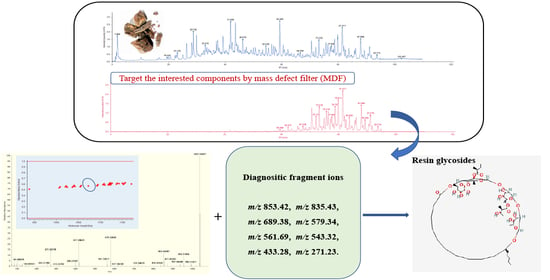
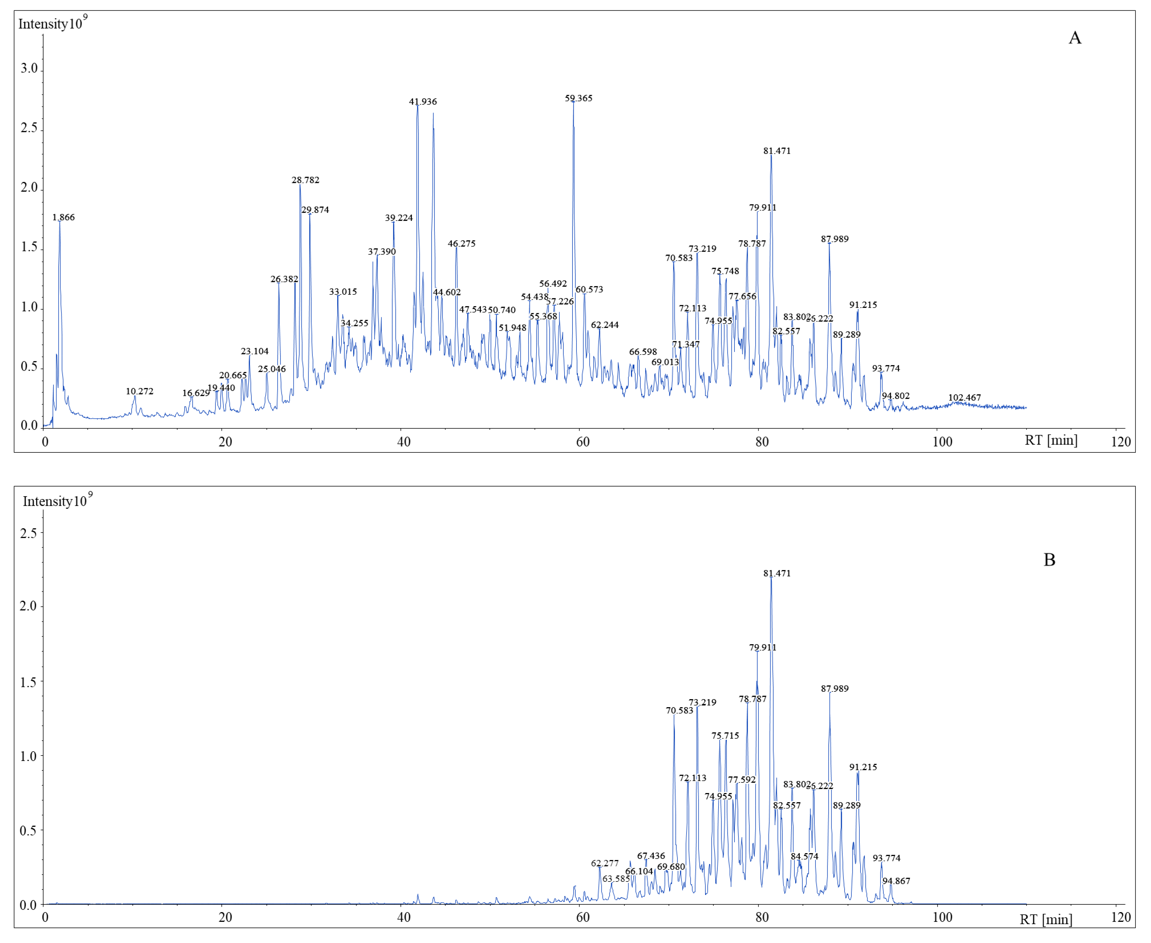
2.1. DFIs Determinations and Fragmentation Patterns Analysis

DFIs was originally proposed from the fact that the components contained in TCMs could usually be structurally classified into several subfamilies and components of the same family usually contained the same carbon skeleton or substructures, from which the same fragment ions could be defined as diagnostic fragment ions (DFIs). Then, these well-defined DFIs were used to screen and identify the analogues. Herein we applied such a strategy to analysis with predefined diagnostic fragment ions [23].
Turpethic acids C and Turpethoside B were used as the references compounds to deduce the fragmentation of resin glycosides and glycosidic acids. In the MS/MS spectrum, the observed neutral loss (NL) masses and diagnostic fragment ions (DFIs) in support of the characterization of glycosidic acids and resin glycosides are offered in Table 1.
Turpethic acids C (Figure 1), a glycosidic acid with deprotonated ion [M − H] at m/z 1047, neutral loss of rhamnopyranosyl (146 Da, Rha) generate fragment ion m/z 901, loss of glucopyranosyl (162 Da, Glc) made ion m/z 885, m/z 739 [M − H − 146 − 162], m/z 593 [739 − C6H10O4] combined with m/z 447 [593 − C6H10O4] were checked. Characteristic ion m/z 285 [447 − C6H10O5], which was 14 Da higher than jalapinolic acid [(S)-11-hydroxyhexadecanoic acid] indicated this resin glycoside having a hydroxylated C17 fatty acid as its aglycone.
Turpethoside B exhibited [M − H] at m/z 1265 (Figure 2), fragmentation ion at m/z 1163 was attributed to loss of 2-methylbutyric acid (102 Da, Mba) from m/z 1265 [M − H − C5H10O2], successively loss of 2-methylbutyric acid and tiglic acid (100 Da, Tga) to produce m/z 1063 [1265 − C5H8O2 − C5H10O2], m/z 1061 [1265 − C5H10O2 − C5H10O2], m/z 1019 [1265 − C11H18O6 (O-methylbutyrylglucose unit)] m/z 979 [1265 + H2O − C5H10O2 − C5H10O2 − C5H8O2], m/z 961 [1265 − C5H10O2 − C5H10O2 − C5H8O2] together with diagnostic fragment ion m/z 835 [1265 + H2O − C11H18O6 − C5H8O2 − C5H10O2], m/z 579 [835 − 146 − (146 − 2 × H2O)], m/z 561 [835 − 146 − (146 − H2O)], m/z 543 [835 − 146 − 146], m/z 433 [579 − 146], m/z 417 [579 − 162] as well as m/z 271 [417 − 146] were observed.

2.2. Construction of MDF Model

According to resin glycosides in Convolvulus scammonia were reported. Jalapinolic acid [(S)-11-hydroxyhexadecanoic acid] is the most frequently aglycone for the macrocyclic lipooligosaccharide core. Therefore, the parent drug filter was based on the jalapinolic acid, m/z 871 (C40H71O20) picked as filter for glycosidic acids, m/z 853 (C40H69O19) and m/z 869(C40H69O20) for resin glycosides. Transformations such as 2-methylbutyric acid (Mba), tiglic acid (Tga), isobutyric acid (Iba), (2R,3R)-3-hydroxy-2-methylbutyric (Nia), 3-hydroxy-methylenebutyric (Hma) were supplied to MDF workflow. Using the mass defect values calculated above and the exact mass values calculated from the elemental composition input (Table 2), mass tolerance 112 Da and mass defect tolerance 0.058 Da were set to define the rectangular mass defect filters (Table 3). The use of the MDF to filtrate targeted compounds from Convolvulus scammonia are given in Figure 3.
The generated chromatograms are displayed in Figure 4. As shown in the chromatogram, after filtration by MDF technique, the noise level was lower than the original chromatogram and ions which the mass defect were not within the filter ranges, were excluded, the targeted components became predominant ions in the TIC. Meanwhile, the sensitivity of minor constituents such as m/z 1170.60286 at 59.46 min was largely improved.

2.3. Identified Glycosidic Acids

Glycosidic acids exhibited deprotonated ions as the base peak were observed; neutral loss of monosaccharide units including deoxyhexose, hexose and short organic acid moieties such as tiglic acid, 2-methylbutyric acid, (2R,3R)-3-hydroxy-2-methylbutyric were the main fragment patterns; aglycone ions m/z 257, m/z 271, m/z 285 indicated the presence of the hydroxylated fatty acid with chain lengths of C15, C16, and C17, respectively. A total of 22 glycosidic acids were detected and tentatively characterized in root of Convolvulus scammonia, in which 21 compounds were based on scammonic acid A parent filter, Their ESI-MSn information is shown in Table 4. Notable ion at m/z 1170 was founded that scammonic acid B as its parent drug. Diagnostic fragment ion at m/z 271 was the common aglycone, suggesting the presence of jalapinolic acid.
Peak 1, 5, 8 all gave high intensity of deprotonated ion at m/z 1053,the HCD-MS2 of which exhibited produced ions at m/z 953 [M − H − 100(3hydroxy-2-methylbutyric acid residue)], m/z 871 [M − H − 100(3-hydroxy-2-methylbutyric acid residue) − 82(tiglic acid residue)], which indicated the parent drug filter was scammonia acid A, diagnostic fragment ions m/z 853 [1053 + H2O − 118 − 100], m/z 835 were generated due to successively loss of 3-hydroxy-2-methylbutyric acid and tiglic acid from m/z 1053, furthermore, successive elimination of deoxyhexose moiety (146 Da), hexose moiety (162 Da) to generate m/z 725 [871 − 146], m/z 579 [725 − 146 − 146], m/z 417 [579 − 162] coupled with m/z 271 [M − H − 100 − 82 − 146 − 146 − 162 − 146] supported the existence of scammonic acid A. It was conclusion that peak 1, 5, 8 were isomer of 3-hydroxy-2-methylbutyric acid + tiglic acid + scammonic acid A.
Peak 2, 6 had the same [M − H] ion at m/z 1055, which was tentatively identified as isomer of 3-hydroxy-2-methylbutyric acid + 2-methylbutyric acid + scammonic acid A. The typical fragment ions m/z 955 [M − H − 100], m/z 871 [M − H − 100 − 84(2-methylbutyric acid residue)], m/z 853 [M − H + H2O − 118 − 102] represented successively loss of 3-hydroxy-2-methylbutyric acid residue (100 Da), 2-methylbutyric acid residue (84 Da) from deprotonated ion, respectively. It’s also gave ions at m/z 579 (loss of 2 mol deoxyhexose moiety, 146 Da), m/z 417 (loss of hexose moiety, 162 Da) and m/z 271 in MS/MS spectrum by cleavage of glycosidic bond on m/z 871(scammonic acid A).
Peak 3 was eluted at tR 59.405 min with precursor ion at m/z 1169, its diagnostic ions m/z 887 of scammonic acid B can distinguished itself from scammonic acid A (m/z 871). Fragment ions at m/z 969 [1169 − 100(3-hydroxy-2-methylbutyric acid residue) − 100], m/z 951 [1169 − 100 − 82(tiglic acid residue)], m/z 887 [969 − 82] were attributed to loss of C5H6O residue (tiglic acid residue, 82 Da) from m/z 969, m/z 851 [951 − 100] were also detected. Characteristic ions m/z 579 were observed for elimination of deoxyhexose moiety 146 Da and hexose moiety 162 Da from scammonic acid B (m/z 887), in addition, m/z 417 [579 − 162] combined with m/z 271 [417 − 146] supported that oligoglycosides conjunction type in scammonic acid B was deoxyhexose–hexose–deoxyhexose–hexose. According to information mentioned above, peak 3 was tentatively characterized as 3-hydroxy-2-methylbutyric acid + 3-hydroxy-2-methylbutyric acid + tiglic acid + scammonic acid B.
Peak 4 had high intensity ion at m/z 1071 with elemental composition C50H87O24, m/z 971 indicated the transformation was 3-hydroxy-2-methylbutyric acid residue [M − H − 100], scammonic acid A as parent drug template was supported from diagnostic ions m/z 871 [971 − 100], m/z 853 [1071 + H2O −118 − 118], m/z 725 [871 − 146(deoxyhexose moiety)], m/z 579, m/z 561 and m/z 417. It supposed that peak 4 was 2 mol 3-hydroxy-2-methylbutyric acid substituted on scammonic acid A.
Peak 7 gave deprotonated ion at m/z 1041 eluted at retention time of 65.234, fragmentation ions at m/z 941 which was attributed to loss of 3-hydroxy-2-methylbutyric acid residue (100 Da) from m/z 1041, m/z 923 [1041 − 118]. Typical ions at m/z 871 [M − H − 100(3-hydroxy-2-methylbutyric acid resiue) − 70(isobutyric acid residue)], m/z 853 [1041 + H2O − 118 − 88], m/z 835 [1041 − 118 − 88] as evidence for the presence of scammonic acid A. In addition, m/z 579, m/z 417 as well as m/z 271 supported that. The tentative identification given to peak 7 was 3-hydroxy-2-methylbutyric acid + isobutyric acid + scammonic acid A.
Peak 9, 10, 11, 12, 13 were tentatively characterized as isomer of 3-hydroxy-2-methylbutyric acid + 3-hydroxy-2-methylbutyric acid + tiglic acid + scammonic acid A according to elemental composition C55H94O25 (MW, 1154 Da). Fragmentation ions at m/z 1053 [M − H − 100], m/z 953 [M − H − 100 − 100] demonstrated that the existence of 3-hydroxy-2-methylbutyric acid, successive loss of tiglic acid to give fragmentation ion at m/z 871 which was diagnostic ion of scammonic acid A. furthermore, m/z 579, m/z 417, m/z 217 were founded in MS/MS spectrum.
Peak 14, 16 as well as 19 displayed the same deprotonated ion at m/z 1123, which was 100 Da higher than m/z 1023, it indicated that the presence of 3-hydroxy-2-methylbutyric acid. In addition to this, there were similarity fragmentation ions with m/z 1023 [M − H − 100(3-hydroxy-2-methylbutyric acid residue)], such as m/z 941, m/z 871, m/z 579, m/z 417, m/z 271. Above all, we inferred that ion at m/z 1123 was based on parent filter scammonic acid A and substituted by 3-hydroxy-2-methylbutyric acid, tiglic acid as well as isobutyric acid.
Peak 15 showed [M − H] ions at m/z 1023 with the elemental composition of C49H84O22. In the MS/MS spectrum, m/z 941 indicated that loss of substitutes tiglic acid residue (82 Da) from [M − H], and then successively loss of isobutyric acid residue (70 Da) to produce ions at m/z 871, fragmentation ions m/z 725, m/z 579 suggested successive loss of rhamnose monohydrate from m/z 871, neutral loss of glucose (162 Da) to produce ion at m/z 417. Diagnostic ion m/z 271 supplied the evidence for the existence of jalapinolic acid aglycone. We inferred from fragmentation ions that peak 15 was scammonic acid A combined with 1 mol each of tiglic acid and isobutyric acid.
Peak 17 and 22 exhibited [M − H] ion at m/z 1137. Prominent ion at m/z 871 [1137 − 82(tiglic acid residue) − 100(3-hydroxy-2-methylbutyric acid residue − 84(2-methylbutyric acid residue)] provided evidence for the existence of scammonic acid A. Fragmentation ions at m/z 1055, m/z 955, m/z 937 [1137 + H2O − 100 − 118], m/z 835 [1137 + H2O − 100 − 118 − 102] were checked. Breakage of glycosidic bond was the main fragmentation pathway of scammonic acid A, from which m/z 579 [871 − 146 − 146], m/z 417 [871 − 146 − 146 − 162] and m/z 217 [871 − 146 − 146 − 162 − 146] were generated. We presumed that peak 17 and 22 were isomer of scammonic acid A + tiglic acid + 3-hydroxy-2-methylbutyric acid + 2-methylbutyric acid.
Peak 18, the [M − H] m/z 1037 with formula C50H86O22 eluted at 74.561 min, in analysis of its MS/MS spectrum, the [M − H − 82(tiglic acid residue)] ion at m/z 955, m/z 935 [M − H − 102(2-methylbutyric acid)], [M − H − 82 − 84(2-methylbutyric acid resiude)] ion at m/z 871 and characteristic ions of scammonic acid A were checked. Compared with peak 18, 23, peak 19 was 100 Da lower, it illustrated that there was absence of 3-hydroxy-2-methylbutyric acid. Therefore, we inferred that peak 18 was based on scammonic acid A and substituted by tiglic acid and 2-methylbutyric acid.
Peak 20 showed [M − H] at m/z 1039, fragmentation ions at m/z 955, m/z 871 demonstrated that 2 mol of 2-methylbutyric acid substituted on scammonic acid A, m/z 579, m/z 417 combined with m/z 271 as notable markers for scammonic acid A were checked in MS/MS spectrum. Peak 20 was tentatively characterized as scammonic acid A + 2-methylbutyric acid + 2-methylbutyric acid.
Peak 21 with high intensity at m/z 1125 was eluted at 78.93min. Study of the MS/MS spectrum of glycosidic acids, loss of organic acid and cleavage of glycosidic bond were main fragmentation pathway, ions at m/z 1025 [M − H − 100], m/z 941 [M − H − 100 − 84] as well as m/z 871 [M − H − 100 − 84 − 70] were generated according to successively loss of 3-hydroxy-2-methylbutyric acid, 2-methylbutyric acid and isobutyric acid from deprotonated ion m/z 1125. Diagnostic ions of scammonia acid A at m/z 579, m/z 417, m/z 271 were observed. We presumed that peak 21 was scammonic acid A + 3-hydroxy-2-methylbutyric acid + 2-methylbutyric acid and isobutyric acid.

2.4. Identified Resin Glycosides

Resin glycosides in Convolvulus scammonia L share the common structure of a macrolactone composed by one acylated glycosidic acid. Through the post-data mining strategy MDF, totally 58 resin glycosides were checked, in which 56 resin glycosides were established on parent filter m/z 853 except ion at m/z 1151 and m/z 1149.
Peak was eluted at 85.271min with elemental composition C56H94O20. The precursor ion [M − H] m/z 1149 firstly elimination of C2H4O (44 Da), fragmentation ion m/z 1049 was attributed to loss of 100(3-hydroxy-2-methylbutyric acid residue) from precursor ion. Ion at m/z 949 [1049 − 100] indicated there was existence of 2 mol 3-hydroxy-2-methylbutyric acid in the structure. Diagnostic fragment ion m/z 867 [1049 − 100 − 82(tiglic acid residue)] coupled with noteworthy ions m/z 849, m/z 593 [867 − 146 − (146 − H2O)], m/z 575 [867 − 146 − 146], m/z 431 [593 − 162], m/z 413 [575 − 162] as well as m/z 285 [431 − 146] were checked. These ions showed the evidence for the existence of C17 fatty acid aglycone. According to the detailed fragmentation ion information and fragment rules, we inferred that compound with structural of m/z 867 + 2 mol 3-hydroxy-2-methylbutyric acid + tiglic acid.
Peak with elemental composition C55H92O25 (m/z 1151) was eluted at 71.689 min. In MS/MS spectrum [M + HCOO] ion at m/z 1197 was observed, its deprotonated ion at m/z 1151 successively loss of 3-hydroxy-2-methylbutyric acid (118 Da) to generate ion at m/z 1051 [M − H − 100(3-hydroxy-2-methylbutyric acid residue)] and m/z 951 [M − H − 100]−, respectively. Fragmentation ion m/z 1033 [1151 − 118], 1007 [1151 − 144(hexose unit − H2O)], m/z 933 [1151 + H2O − 118 − 118] coupled with m/z 907 [1151 − C11H16O6(O-tigloylhexose unit)] and m/z 905 [1151 − C11H18O6(O-3-hydroxy-2-methylbutyrylpentose unit)] were checked. Diagnostic ion at m/z 869 [M − H − 100 − 100 − 82(tiglic acid residue)] demonstrated the sugar units were hexose–pentose–pentose–hexose which linked to jalapinolic acid to form macrolactone ring, it also illustrated the existence of tiglic acid. According to the fragmentation ions, we inferred this compound was based on parent drug filter m/z 869 and substituted by 2 mol 3-hydroxy-2-methylbutyric acid and 1 mol tiglic acid.
Peaks listed below were all based on parent template m/z 853, totally 56 peaks were tentatively characterized, the detail of ESI-MSn information is shown in Table 5. Analysis of the MS/MS spectrum, the proposed fragmentation patterns of resin glycosides are as follows:
  • adduct ion [M + HCOO], [M + Cl] combined with [M − H] were observed, it is different from glycosidic acids;
  • loss of short organic acid such as 2-methylbutyric acid, tiglic acid, isobutyric acid, (2R,3R)-3-hydroxy-2-methylbutyric, 3-hydroxy-2-methylenebutyric were common characteristics;
  • breakage of glycosidic linkage is prone to loss of 162 Da, 146 Da;
  • loss of C2H4O (44 Da) was observed when (2R,3R)-3-hydroxy-2-methylbutyric substituted on resin glycosides
  • parent drug filter ion at m/z 853 with lower intensity was obtained and identical ions at m/z 579, m/z 561, m/z 543, m/z 399, m/z 417 combined with m/z 271 were discovered.
Peak 1′ displayed precursor ion at m/z 937 and adduct ion [M + HCOO] at m/z 983, [M + Cl] at m/z 973 were found. Diagnostic ion at m/z 853 [M − H − 84(2-methylbutyric acid residue)], m/z 835 [937 − 102] were discovered in MS/MS spectrum, from which we inferred that there was 2-methylbutyric acid substituted on parent drug filter m/z 853. Therefore, Peak 1′ was tentatively identified as m/z 853 + 2-methylbutyric acid.
Peak 2′, 5′, 6′, 8′, 11′, 13′, 22′ all exhibited [M + HCOO] at m/z 1081 with high intensity and [M + Cl] at m/z 1071 in MS spectrum, analysis of MS/MS spectrum, [M − H] at m/z 1035 (C50H83O22) gave high abundance, fragmentation ion at m/z 991 [M − H − 44], m/z 953 [1035 − 82(tiglic acid residue)] and m/z 935 [1035 − 100(3-hydroxy-2-methylbutyric)] with highest intensity appeared. Diagnostic ion m/z 853 [935 − 82(tiglic aid residue)] with ion m/z 835 [1035 + H2O − 118 − 100] served as evidence for parent drug. Based on fragmentation rules, peak 2′, 5′, 6′, 8′, 11′, 13′, 22′ were characterized as isomer of m/z 853 + 3-hydroxy-2-methylbutyric + tiglic aid.
Peak 3′ showed [M + HCOO] at m/z 1069 and [M − H] at m/z 1023 in MS/MS spectrum. Ion at m/z 979 indicated loss of 44 Da (C2H4O) from deprotonated ion, m/z 935 through loss of isobutyric acid from m/z 1023, m/z 923 [M − H − 100] demonstrated the presence of (2R,3R)-3-hydroxy-2-methylbutyric, noteworthy ion at m/z 835 [M − H − 100 − 88] as parent drug filter observed. A tentative identification of Peak 3′ was m/z 853 + (2R,3R)-3-hydroxy-2-methylbutyric + isobutyric acid.
Peak 4′, 20′, 25′ all gave [M − H] at m/z 1123 was 100 Da higher than m/z 1023, which had the same fragmentation pattern with m/z 1023, namely loss of organic acid and breakage of glycosidic linkage. From fragmentation ions m/z 1079, m/z 1035, m/z 1023 as well as m/z 835, we presumed the structure was m/z 853 + 2 mol 3-hydroxy-2-methylbutyric + isobutyric acid.
Peak 7′, 10′, 16′, 18′ all exhibited [M + HCOO] at m/z 1083 and [M + Cl] at m/z 1073 in MS spectrum, analysis of MS/MS spectrum [M − H] at m/z 1037 combined with m/z 993 [M − H − 44], m/z 937 [M − H − 100(3-hydroxy-2-methylbutyric residue)] as well as markable fragmentation ion at m/z 853 [937 − 84], m/z 579, m/z 561, m/z 417, m/z 217 were checked. Peak 7′, 10′, 16′, 18′ were tentatively identified as isomer of m/z 853 + 3-hydroxy-2-methylbutyric + 2-methylbutyric acid.
Peak 9′, 14′ showed [M + HCOO] at m/z 1099 with high abundance. In analysis of MS/MS spectrum, [M − H] at m/z 1053 with elemental composition C50H85O23 was prone to loss of 44 Da to form ion at m/z 1009, ions at m/z 909 and m/z 891 were formed from [M − H − 44 − 100(3-hydroxy-2-methylbutyric residue)], [M − H − 44 − 118], respectively. Fragmentation ions at m/z 953 [M − H − 100], m/z 853 [M − H − 100 − 100], m/z 835 [1053 + H2O − 118 − 118] both of these fragmentation ions illustrated the existence of 2 mol 3-hydroxy-2-methylbutyric. According to the fragment rules, peak 9′ and 14′ were tentatively characterized as isomer of m/z 853 + 2 mol 3-hydroxy-2-methylbutyric.
Peak 12′, 15′, 17′, 19′, 21′, 23′, 26′, 28′, 41′ were tentatively identified as isomer of m/z 853 + 2 mol 3-hydroxy-2-methylbutyric + tiglic acid according to fragmentation rule of resin glycosides. The [M + HCOO] ion at m/z 1181 with high intensity and [M + Cl] ion at m/z 1171 were observed in MS spectrum, in analysis of its MS/MS spectrum, the noteworthy ions m/z 1091 and m/z 1047 were due to successively loss of C2H4O (44 Da) from precursor ion m/z 1135, ion at m/z 1035 was attributed to loss of 100 Da (3-hydroxy-2-methylbutyric residue) from [M − H], m/z 991 [1091 − 100], m/z 935 from [M − H − 100 − 100]. Characteristic ion m/z 853 [M − H − 100 − 100 − 82] was checked, it illustrated the existence of tiglic acid, diagnostic ions of at m/z 579, m/z 561, m/z 417 combined with m/z 217 supported the parent drug filter was m/z 853.
Peak 24′, 29′, 32′, 34′ showed [M − H] at m/z 1137, successively loss of 44 Da from m/z 1137 to generate m/z 1093 and m/z 1049 with high abundance, according to fragmentation pattern of m/z 1135 we inferred there were 2 mol 3-hydroxy-2-methylbutyric substituted on resin glycosides. Ion at m/z 1037 [1137 − 100(3-hydroxy-2-methylbutyric residue], m/z 991 [1093 − 2-methylbutyric acid], m/z 853 [1037 − 100 − 84], m/z 579, m/z 561, m/z 417 as well as m/z 271 were discovered. Therefore, peak 24′, 29′, 32′, 34′ were tentatively characterized as m/z 853 + 2 mol 3-hydroxy-2-methylbutyric + 2-methylbutyric acid.
Peak 27′, 30′, 36′, 42′, 46′, 54′ exhibited high intensity [M + HCOO] at ion m/z 1151 and [M + Cl] at ion m/z 1141 also checked in MS spectrum. [M − H] at ion m/z 1105 loss of 44 Da to make fragmentation ion at m/z 1061, ion at m/z 1005 from deprotonated ion m/z 1105 dropped 3-hydroxy-2-methylbutyric residue (100 Da), m/z 973 [1061 − 88(isobutyric acid)], m/z 961 [1061 − C5H8O2(tiglic acid)], m/z 917 [1005 − isobutyric acid], m/z 835 [917 − (tiglic acid − H2O)] were found. Cleavage of glycoside bond was prone to produce diagnostic ions m/z 579, m/z 561, m/z 417 and m/z 271. We presumed that the structure of peak 27′, 30′, 36′, 42′, 46′, 54′ was 3-hydroxy-2-methylbutyric, tiglic acid combined with isobutyric acid substituted on parent drug m/z 853.
Peak 31′, 48′, 51′ displayed [M + HCOO] at m/z 1153 and [M + Cl] at m/z 1143. In the MS/MS spectrum [M − H] at m/z 1107 with elemental composition C54H91O23 was observed, m/z 1005 was attributed to loss of 2-methylbutyric acid from 1107, m/z 989 [M − H − 118] and m/z 905 [M − H − 102 − 100] illustrated the presence of 3-hydroxy-2-methylbutyric. Cleavage of glycoside bond and loss of hexose moiety, pentose moiety as noteworthy fragment pathway was checked, the corresponding fragment ions at m/z 579 [835 − 146 − (146 − 2 × H2O)], m/z 561 [835 − 146 − (146 − H2O)], m/z 543 [835 − 146 − 146], m/z 399 [561 − 162], m/z 417 [579 − 162] combined with m/z 271 [417 − 146] were discovered. Peak 31′, 48′, 51′ were tentatively characterized as isomer of m/z 853 + 2-methylbutyric acid + 3-hydroxy-2-methylbutyric + isobutyric acid based on fragment rules.
Peak 33′, 38′ gave [M + HCOO] at m/z 1053 and [M − H] at m/z 1007 which was 100 Da lower than m/z 1107, it presumed absence of 3-hydroxy-2-methylbutyric. Fragment ion at m/z 923 [M − H − 84], m/z 905 [M − H − 102] suggested the existence of 2-methylbutyric acid, ion at m/z 919 [M − H − 88] coupled with m/z 835 [M − H − 102 − 70] demonstrated the presence of isobutyric acid. The diagnostic ions m/z 579, m/z 561, m/z 417, m/z 399 as well as m/z 271 supported the breakage of glycosides bond from m/z 853. Therefore, the tentative identification given to peak 33′, 38′ was isomer of m/z 853 + 2-methylbutyric acid+ isobutyric acid.
Peak 35′, 40′ were tentatively characterized as isomer of m/z 853 + 2-methylbutyric acid + tiglic acid based on proposed fragment rules of resin glycosides. A high abundance of [M + HCOO] ion at m/z 1065 coupled with [M + Cl] at m/z 1054 were discovered. In analysis of MS/MS spectrum, the precursor ion at m/z 1019 gave high intensity, ion at m/z 937 [M − H − 82], m/z 919 [M − H − 100] served as evidence for the existence of tiglic acid. Ion at m/z 853 [M − H − 82 − 84] demonstrated the presence of 2-methylbutyric acid.
Peak 37′, 49′, 52′, 56′ showed [M − H] at m/z 1119, a difference of 100 Da between m/z 1019 and m/z 1119 suggested 3-hydroxy-2-methylbutyric substituted on resin glycoside, ion at m/z 1001 [M − H − 118] and m/z 1019 [M − H − 3-hydroxy-2-methylbutyric residue] supported that speculation. Ions at m/z 937 [1019 − 82], 917 [937 − 102] coupled with m/z 835 were observed in MS/MS spectrum. According to fragment rules and information listed above, the tentative identification given to peak 37′, 49′, 52′, 56′ were isomer of m/z 853 + tiglic acid + 2-methylbutyric acid + 3-hydroxy-2-methylbutyric.
Peak 39′, 45′ exhibited [M + HCOO] at m/z 1139 in MS spectrum, [M − H] at m/z 1093 loss of C2H4O (44 Da) to form m/z 1049, product ions at m/z 1005 [M − H − 88], m/z 905 [M − H − 88 − 100], m/z 835 [M − H − 88 − 100 − 70] were checked in MS/MS spectrum, we inferred peak 39′, 45′ were isomer of m/z 853 + 3-hydroxy-2-methylbutyric + 2 mol isobutyric acid.
Peak 43′, 47′ showed [M − H] at m/z 1021, adduct ion [M + HCOO] at m/z 1067, ion at m/z 937 [M − H − (102 − H2O)] due to loss of 2-methylbutyric acid from deprotonated ion, m/z 919 [M − H − 102] with high abundance were obtained. Characteristic ion m/z 853 was attribute to successively loss of 2-methylbutyric acid residue from m/z 1021, m/z 561, m/z 417, m/z 399 combined with m/z 271 were observed. Therefore, peak 43′, 47′ were tentatively characterized as isomer of m/z 853 + 2-methylbutyric acid+2-methylbutyric acid.
Peak 44′, 50′ exhibited deprotonated ion at m/z 1117, [M + HCOO] ion at m/z 1163 was observed in MS spectrum. In MS/MS a predominant productive ion m/z 1017 [M − H − (118 − H2O)], coupled with m/z 999 [M − H − 118], m/z 917 [M − H − 118 − (100 − H2O)] and m/z 935 [M − H − 100 − 82]. Ion at m/z 835 combined with m/z 561, m/z 417, m/z 399, m/z 271 from the cleavage of glycoside bond were checked. We thus inferred the structure of peak 44′ and 50′ was 2 mol tiglic acid + 3-hydroxy-2-methylbutyric substituted on parent drug m/z 853.
Peak 53′, 55′ were eluted at 91.147 min and 89.303 min, respectively. For structural elucidation, [M − H] at m/z 1119 and its adduct ion [M + HCOO] at m/z 1165, [M + Cl] at m/z 1155 were found in MS spectrum. In analysis of its MS/MS spectrum, ion at m/z 1019 [M − H − C5H8O2], m/z 1001 [M − H − 118] indicated the existence of 3-hydroxy-2-methylbutyric, m/z 919 [M − H − 118 − 82(100 − H2O)], m/z 835 [M − H − 118 − 82 − 84(102 − H2O)] with high abundance were generated. According to proposed fragment pattern of resin glycoside, we gave tentative characterization to m/z 1119 was m/z 853 + 3-hydroxy-2-methylbutyric + tiglic acid + 2-methylbutyric acid.

3. Materials and Methods

3.1. Chemicals and Reagents

Acetonitrile, methanol and formic acid (MS grade) were purchased from Fisher Scientific (Fair Lawn, NJ, USA). Deionized water was obtained from Watson’s (Hong Kong, China).

3.2. References

References Turpethic acids C and Turpethoside B were provided by Professor Xiaoyi Wei, Key Laboratory of Plant Resources Conservation and Sustainable Utilization, South China Botanical Garden, Chinese Academy of Sciences, Guangzhou, China.

3.3. Sample Preparation

The extract of Convolvulus scammonia L. was provided by Xinjiang Uygur Medicine Limited Liability Company (Urumqi, China). The sample (1.0 g) was dispersed in 10 mL 100% methanol, and extraction was performed on a water bath at 25 °C assisted with ultrasound for 40 min. The extract was centrifuged at 14,000 rpm for 7 min, leading to the supernatant used as the test solution.

3.4. HPLC Condition

Chromatographic separation was performed on Ultimate 3000 RSLC system (Dionex, Sunnyvale, CA, USA). An ACQ-UITY UPLC® HSS T3 column (100 mm × 2.1 mm i.d., 1.8 mm, Waters, MA, USA) maintained at 40 °C was used. The mobile phase consisted of acetonitrile (A) and 0.1% formic acid–water (B) with a gradient elution of 5% B at 0–1 min, 5–9% B at 1–4 min, 9–18% B at 4–22 min, 18–88% B at 22–92 min, 88–95% B at 92–92.1 min and 95% B at 92.1–110 min. A flow rate of 0.15 mL/min was set and injection volume was 1.00 μL.

3.5. Mass Spectrometric Conditions

High-resolution MS data were recorded on a Q ExactiveTM PLUS hybrid Quadrupole-Orbitrap Mass Spectrometer equipped with a heated ESI source (Thermo Fisher Scientific, Waltham, MA, USA) operating in the negative mode. The HESI parameters were set as follows: spray voltage, −2.8 kV; Capillary temperature 320 °C; sheath gas pressure, 35 arb; aux gas pressure, 10 arb; spare gas pressure, 0 arb; probe heater temperature, 350 °C; s-lens RF, 55 V. The Orbitrap analyzer scanned over m/z 100–1500 at a resolution of 70,000 in full scan MS1, and dynamic mass ranges in MS2 with resolution set at 13,500. AGC target values for MS1 and MS2 were 5e6 and 1e5, respectively. Maximum injection time (IT) for MS1 was defined as 100 ms and 50 ms for MS2.
MS2 experiments were performed by high collision induced dissociation (HCD) to improve the sensitivity and offer more fragments information for structural elucidation at normalized collision energy (NCE) 10/30/50 V, with isolation window at 4.0 m/z. Dynamic exclusion allows each precursor ion to be selected 1 time before being excluded with 10 s duration. The HRMS full scan and HCD MS2 data were viewed and processed by Xcalibur 4.2. (Thermo Fisher Scientific, Waltham, MA, USA).

3.6. Mass Defect Filter Approach

Resin glycosides analogues usually shared with the similar core structure; characteristic compounds were generated via various substituents. Therefore, with increasing the number of these substituted groups, the decimal mass and integer mass of the molecular weight would show the linear or fixed relationship in exploring the interested precursor ions. In other words, the elemental compositions of these compounds would produce regular changes with the increase of their molecular weights. Thus, their molecular ions and the isotope distributions can be dynamically presented by preferred mathematical method [23,24,25,26].
The MDF strategy relies on the decimal mass shift to remove the interferences and to pick out the target ions for structural elucidation. Core substructure and different substituent combination were the two essential parts to realize the MDF approach. In the present study, Molecular Weight 872 Da (C40H72O20), MW 854 Da (C40H70O19), MW 870 Da (C40H70O20) were picked as parent drug filter, mass defect type was standard mass defect (Standard Mass Defect = exact mass − nominal mass). Elemental composition prediction of the detected components was based on the following settings: elements in use, C 0–70, H 0–110, O 0–30; mass tolerance, <5 ppm; adduct species [M + HCOO], [M − H]. The High-resolution MS data were processed by MDF using Compound Discovery 3.2 (Thermo Scientific, Waltham, MA, USA).

4. Conclusions

In the present study, a sensitive and effective strategy for rapid identification of the characteristic structural analogues resin glycosides in Convolvulus scammonia extract has been developed by using MDF coupled to DPIs analysis on a hybrid Q-Orbitrap mass spectrometer. Compared with the chemical constituents reported on Convolvulus scammonia [10,11,12], this work enriched our knowledge about resin glycosides of Convolvulus scammonia, not only the number of consititues, but also the variety of structural. A total of 80 components were identified, among which 79 indicated hydroxy C16 fatty acid as main aglycone that was in accordance with phytochemistry study on Convolvulus scammonia. In addition, aglycone with hydroxy C17 fatty acid was discovered in Convolvulus scammonia for the first time. Furthermore, ion m/z 1151 and m/z 1169 had deoxyhexose–hexose–deoxyhexose–hexose oligoglycosides conjunction type were checked due to the minor components can be exposed for the signal-noise ratio improved by MDF approach. For DPIs analysis, the reference compounds turpethoside B in which the carboxyl group of (S)-12-hydroxy-pentadecanoic acid was linked to C-2 of Rha to form a macrolactone ring, but the carboxyl group of jalapinolic acid was linked to C-3 of Rha to form a macrolactone ring as common appeared in Convolvulus scammonia, therefore, the abundance of dignositic fragment ions was a little bit different. In a word, DPIs analysis can provide a criterion to classify the target constituents detected into certain chemical families.
Mass defect filtering-oriented identification of resin glycosides is definitely an effective strategy, there were more MDF methods, including multiple MDF, raster MDF, five-point screening MDF and polygonal MDF, developed for more accurate precursor ions filtered and different types of plant metabolites can be discriminated by sorts of MDF methods.

Author Contributions

Conceptualization, Q.Y., C.L. and H.A.A.; methodology, R.A., G.K. and C.L.; software, R.A. and C.L.; validation, Q.Y., R.A. and C.L.; formal analysis, Q.Y., R.A. and C.L.; investigation, Q.Y., R.A., G.K. and C.L.; resources, Q.Y., X.X. and H.A.A.; data curation, Q.Y., C.L. and X.X.; writing—original draft preparation, Q.Y. and C.L.; writing—review and editing, H.A.A., C.L. and X.X.; visualization, C.L. and X.X.; supervision, H.A.A. and X.X.; project administration, R.A.; funding acquisition, H.A.A. All authors have read and agreed to the published version of the manuscript.

Funding

This research was funded by the National Key R&D Program of China, grant number 2020YFE020560.

Institutional Review Board Statement

Not applicable.

Informed Consent Statement

Not applicable.

Data Availability Statement

Not applicable.

Conflicts of Interest

The authors declare no conflict of interest.

References

  1. Pereda-Miranda, R.; Rosas-Ramírez, D.; Castañeda-Gómez, J. Resin glycosides from the morning glory family. Prog. Chem. Org. Nat. Prod. 2010, 92, 77–147. [Google Scholar]
  2. Fan, B.; Lu, Y.; Yang, M.; Li, J.; Chen, G. Evolvulins I and II, resin glycosides with a trihydroxy aglycone unit from Evolvulus alsinoides. Org. Lett. 2019, 21, 6548–6551. [Google Scholar] [CrossRef] [PubMed]
  3. Ono, M.; Takigawa, A.; Kanemaru, Y.; Kawakami, G.; Kabata, K.; Okawa, M.; Kinjo, J.; Yokomizo, K.; Yoshimitsu, H.; Nohara, T. Calysolins V–IX, resin glycosides from Calystegia soldanella and their antiviral activity toward herpes. Chem. Pharm. Bull. 2014, 62, 97–105. [Google Scholar] [CrossRef] [Green Version]
  4. Ono, M. Resin glycosides from Convolvulaceae plants. J. Nat. Med. 2017, 71, 591–604. [Google Scholar] [CrossRef]
  5. Ono, M.; Kanemaru, Y.; Yasuda, S.; Okawa, M.; Kinjo, J.; Miyashita, H.; Yokomizo, K.; Yoshimitsu, H.; Nohara, T. A new resin glycoside from Calystegia soldanella and its antiviral activity towards herpes. Nat. Prod. Res. 2017, 31, 2660–2664. [Google Scholar] [CrossRef] [PubMed]
  6. Yoshikawa, K.; Yagi, C.; Hama, H.; Tanaka, M.; Arihara, S.; Hashimoto, T. Ipomotaosides A–D, resin glycosides from the aerial parts of Ipomoea batatas and their inhibitory activity against COX-1 and COX-2. J. Nat. Prod. 2016, 73, 1763–1766. [Google Scholar] [CrossRef]
  7. Figueroa-González, G.; Jacobo-Herrera, N.; Zentella-Dehesa, A.; Pereda-Miranda, R. Reversal of multidrug resistance by morning glory resin glycosides in human breast cancer cells. J. Nat. Prod. 2012, 75, 93–97. [Google Scholar] [CrossRef] [PubMed]
  8. Corona-Castaneda, B.; Rosas-Ramírez, D.; Castañeda-Gómez, J.; Aparicio-Cuevas, M.; Fragoso-Serrano, M.; Figueroa-Gonzalez, G.; Pereda-Miranda, R. Resin glycosides from Ipomoea wolcottiana as modulators of the multidrug resistance phenotype in vitro. Phytochemistry 2016, 123, 48–57. [Google Scholar] [CrossRef]
  9. Eich, E. Solanaceae and Convolvulaceae: Secondary Metabolites; Springer: Berlin/Heidelberg, Germany, 2008; pp. 525–582. [Google Scholar]
  10. Naoki, N.; Hirokuni, K.; Toshio, K.; Kazumoto, M. Scammonins I and II, the resin glycosides of radix scammoniae from Convolvulus Scammonia. Phytochemistry 1990, 29, 3565–3569. [Google Scholar]
  11. Hirokuni, K.; Naoki, N.; Toshio, K.; Kazumoto, M. Scammonin III–VI, resin glycosides of Convolvulus Scammonia. Phytochemistry 1991, 30, 957–963. [Google Scholar]
  12. Naoki, N.; Hirokuni, K.; Toshio, K.; Kazumoto, M. Scammonins VII and VIII, two resin glycosides from Convolvulus Scammonia. Phytochemistry 1992, 31, 2761–2766. [Google Scholar]
  13. Jia, S.L.; Du, Z.F.; Song, C.W.; Jin, S.N.; Zhang, Y.; Feng, Y.L.; Xiong, C.M.; Jiang, H.L. Identification and characterization of curcuminoids in turmeric using ultra-high performance liquid chromatography-quadrupole time of flight tandem mass spectrometry. J. Chromatogr. A 2017, 1521, 110–122. [Google Scholar] [CrossRef]
  14. Hao, Y.M.; Huo, J.H.; Wang, T.; Sun, G.D.; Wang, W.M. Chemical profiling of Coptis rootlet and screening of its bioactive compounds in inhibiting Staphylococcus aureus by UPLC-Q-TOF/MS. J. Pharm. Biomed. Anal. 2020, 180, 113089. [Google Scholar] [CrossRef]
  15. Ahad, H.; Jin, H.L.; Liu, Y.F.; Wang, J.X.; Sun, G.Y.; Liang, X.M.; Aisa, H.A. Chemical profiling of spermidines in goji berry by strong cation exchange solid-phase extraction (SCX-SPE) combined with ultrahigh-performance liq- uid chromatography-quadrupole time-of-flight mass spectrometry (UPLC-Q-TOF/MS/MS). J. Chromatogr. B 2020, 1137, 121923. [Google Scholar] [CrossRef]
  16. Liu, Z.H.; Wang, Y.Q.; Mei, X.D.; Wang, F.; Yang, X.Z.; Li, X.D.; Jiang, F.; Zhang, J.Y. Comprehensive analysis of the chemical constituents in sulfur-fu-migated Lonicerae Japonicae Flos using UHPLC-LTQ-Orbitrap mass spectrometry. Chin. J. Nat. Med. 2020, 18, 148–160. [Google Scholar] [PubMed]
  17. Cai, W.; Li, K.L.; Xiong, P.; Gong, K.Y.; Zhu, L.; Yang, J.B.; Wu, W.H. A systematic strategy for rapid identification of chlorogenic acids derivatives in Duhaldea nervosa using UHPLC-Q-Exactive Orbitrap mass spectrometry. Arab. J. Chem. 2020, 13, 3751–3761. [Google Scholar] [CrossRef]
  18. Qiao, S.; Shi, X.W.; Shi, R.; Liu, M.; Liu, T.; Zhang, K.R.; Wang, Q.; Yao, M.C.; Zhang, L.T. Identification of urinary metabolites of imperatorin with a single run on an LC/Triple TOF system based on multiple mass defect filter data acquisition and multiple data mining techniques. Anal. Bioanal. Chem. 2013, 405, 6721–6738. [Google Scholar] [CrossRef] [PubMed]
  19. Zhu, M.S.; Ma, L.; Zhang, H.Y.; Humphreys, W.G. Detection and structural characterization of glutathione-trapped reactive metabolites using liquid chromatography-high-resolution mass Spectrometry and mass defect filtering. Anal. Chem. 2007, 79, 8333–8341. [Google Scholar] [CrossRef]
  20. Liu, M.Y.; Zhao, S.H.; Wang, Z.Q.; Wang, Y.F.; Liu, T.; Li, S.; Wang, C.C.; Wang, H.T.; Tu, P.F. Identification of metabolites of deoxyschizandrin in rats by UPLC-Q-TOF-MS/MS based on multiple mass defect filter data acquisition and multiple data processing techniques. J. Chromatogr. B 2014, 949, 115–126. [Google Scholar] [CrossRef] [PubMed]
  21. An, Y.L.; Wei, W.L.; Li, H.J.; Li, Z.W.; Yao, C.L.; Qu, H.; Yao, S.; Huang, Y.; Zhang, J.Q.; Bi, Q.R.; et al. An enhanced strategy integrating offline superimposed two-dimensional separation with mass defect filter and diagnostic ion filter: Comprehensive characterization of steroid alkaloids in Fritillariae Pallidiflorae Bulbus as a case study. J. Chromatogr. A 2021, 1643, 462029. [Google Scholar] [CrossRef]
  22. Ding, W.B.; Jiang, Z.H.; Wu, P.; Xu, L.X.; Wei, X.Y. Resin glycosides from the aerial parts of Operculina turpethum. Phytochemistry 2012, 81, 165–174. [Google Scholar] [CrossRef]
  23. Xie, T.; Liang, Y.; Hao, H.J.; Xie, A.L.; Gong, P.; Dai, C.; Liu, L.; Kang, A.; Zheng, X.; Wang, G. Rapid identification of ophiopogonins and ophiopogonones in Ophiopogon japonicus extract with a practical technique of mass defect filtering based on high resolution mass spectrometry. J. Chromatogr. A 2012, 1227, 234–244. [Google Scholar] [CrossRef]
  24. Mortishire-Smith, R.J.; Castro-Perez, J.M.; Yu, K.; Shockcor, J.P.; Goshawk, J.; Hartshorn, M.J.; Hill, A. Generic dealkylation: A tool for increasing the hit-rate of metabolite rationalization, and automatic customization of mass defect filters. Rapid Commun. Mass Spectrom. 2009, 23, 939–948. [Google Scholar] [CrossRef]
  25. Claesen, J.; Dittwald, P.; Burzykowski, T.; Valkenborg, D. An efficient method to calculate the aggregated isotopic distribution and exact center-masses. J. Am. Soc. Mass Spectrom. 2012, 23, 753–763. [Google Scholar] [CrossRef] [Green Version]
  26. Zhang, H.; Zhang, D.; Ray, K.; Zhu, M. Mass defect filter technique and its applications to drug metabolite identification by high-resolution mass spectrometry. J. Mass Spectrom. 2009, 44, 999–1016. [Google Scholar] [CrossRef] [PubMed]
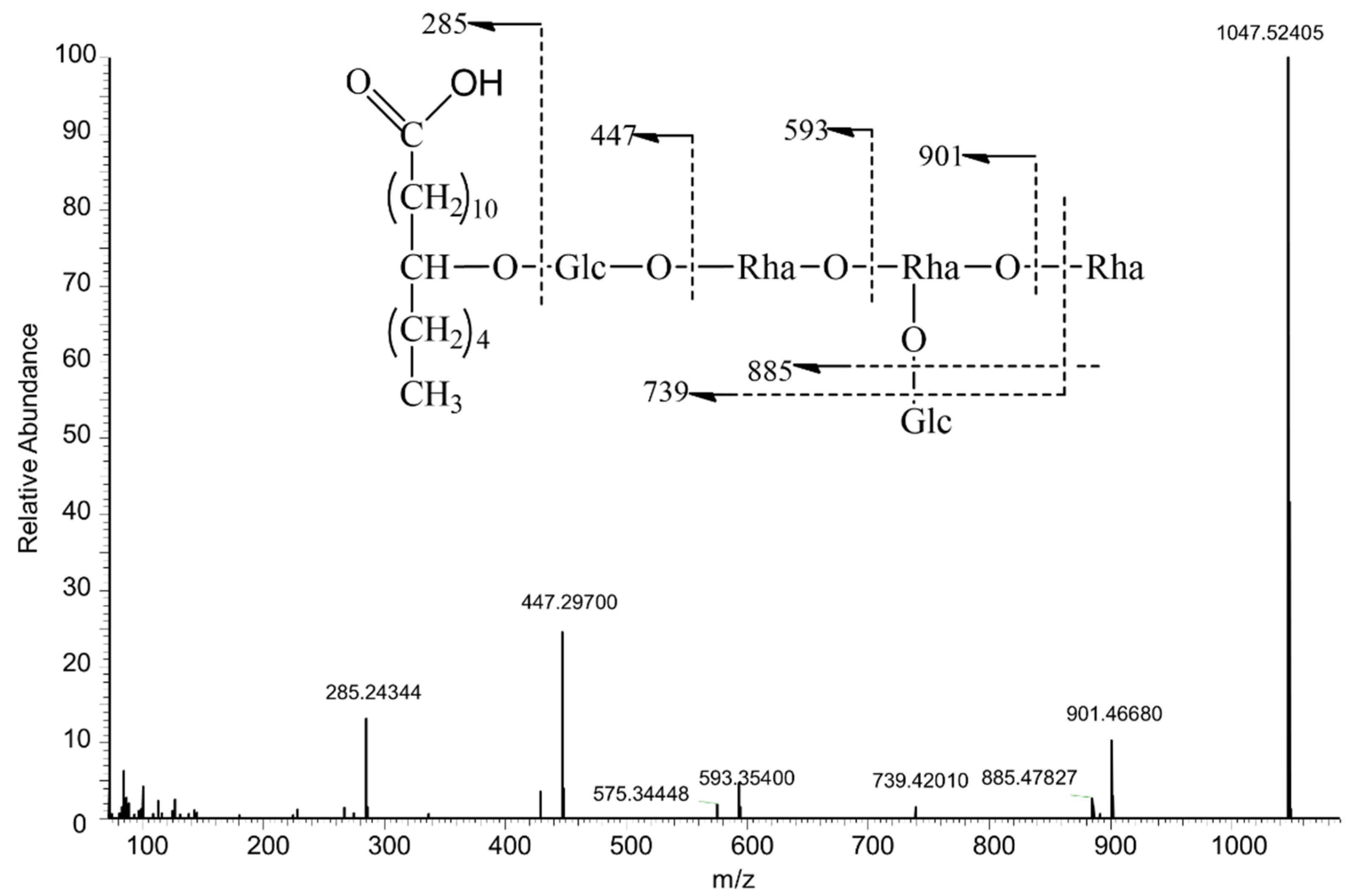
Figure 1. Proposed fragmentation pathway of Turpethic acids C.
Molecules 27 03638 g001
Figure 2. Proposed fragmentation pathway of Turpethoside B.
Molecules 27 03638 g002
Figure 3. Targeted compounds screened from Convolvulus scammonia by MDF.
Molecules 27 03638 g003
Figure 4. The TIC chromatogram of Convolvulus scammonia without filtration (A) and with MDF filtration (B).
Molecules 27 03638 g004
Table 1. A summary of key NL and DFIs for structure elucidation.
ReferencesPrecursor Ion
[M − H]
Adduct Ions
[M + Cl], [M + HCOO]
Diagnostic Fragment Ions (m/z)Neutral Loss
(Da)
Turpethoside Bm/z 1265.65344[M + Cl] m/z 1301.62927m/z 853.42, m/z 835.43,HCOOH: 46.01
m/z 689.38, m/z 579.34,H2O: 18.01
[M + HCOO] m/z 1311.65771m/z 561.69, m/z 543.32,Tiglic acid: 100.05
m/z 433.28, m/z 271.23.2-Methylbutyric acid: 102.08
Tupethic acid Cm/z 1047.52417m/z 593.35, m/z 574.45,Rha: 146.06
m/z 447.30, m/z 429.29,Glc: 162.05, H2O: 18.01
m/z 285.24, m/z 267.46.
Table 2. Transformations on resin glycosides.
SubstitutesMass Change, DaMass Defect Shift, mDa
+2-methylbutyric acid+C5H8O, 84.056956.9
+tiglic acid+C5H6O, 82.041341.3
+isobutyric acid+C4H6O, 70.041341.3
+3-hydroxy-2-methylenebutyric+C4H4O, 68.025725.7
+3-hydroxy-2-methylbutyric+C5H8O2, 100.051951.9
+Methyl+CH3, 14.0156515.7
Table 3. Mass-range (Da)-mass defect(mDa) for known resin glycosides from Convolvulus scammonia.
FiltersMass Change,
Da
Mass Defect Shift, mDa
m/z 853, C40H69O19+isobutyric acidC44H75O20 (923)0.485
+tiglic acidC45H75O20 (935)0.485
+2-methylbutyric acidC45H77O19 (937)0.500
+isobutyric acid, tiglic acidC49H81O21 (1005)0.526
+tiglic acid, tiglic acidC50H81O21 (1017)0.526
+2-methylbutyric acid, tiglic acid.C50H83O21 (1019)0.542
m/z 869, C40H69O20+2-methylbutyric acid, tiglic acidC50H83O22 (1035)0.537
m/z 871, C40H71O20+CH3, 2-methylbutyric acid, tiglic acidC51H87O22 (1051)0.568
Table 4. Summary of the mass spectral data of characterized glycosidic acids in ESI negative ion mode.
No.Rt
min
FormulaΔMass
ppm
Parent
Compounds
m/z
TransformationsComposition ChangeMSn
158.424C50H86O231.628713-hydroxy-2-methylbutyric+C10H14O3MS [M − H] 1053.55042
Tiglic acidMS2 971, 953, 935, 871, 853, 835, 725, 707, 579, 561, 417, 399, 271
259.374C50H88O230.668712-methylbutyric acid+C10H16O3MS [M − H] 1055.56519
3-hydroxy-2-methylbutyric acidMS2 971, 955, 953, 937,871, 853, 835, 725, 707, 579, 561, 417, 399, 271
359.504C55H94O26–0.418873-hydroxy-2-methylbutyric+C15H22O6MS [M − H] 1169.59558
3-hydroxy-2-methylbutyricMS2 969, 951, 887, 851,579, 561, 417, 399, 271
Tiglic acid
460.543C50H88O240.848713-hydroxy-2-methylbutyric acid+C10H16O4MS [M − H] 1071.55969
3-hydroxy-2-methylbutyric acidMS2 953, 909, 871, 853, 835, 725, 707, 579, 561, 417, 399, 271
562.294C50H86O230.698713-hydroxy-2-methylbutyric+C10H14O3MS [M − H] 1053.54944
Tiglic acidMS2 971,953, 935, 871, 853, 835, 725, 707, 579, 561, 417, 399, 271
663.494C50H88O231.138712-methylbutyric acid+C10H16O3MS [M − H] 1055.56555
3-hydroxy-2-methylbutyric acidMS2 971, 955, 953, 937,871, 853, 835, 725, 707, 579, 561, 417, 399, 271
765.234C49H86O231.75871isobutyric acid+C9H14O3MS [M − H] 1041.05454
3-hydroxy-2-methylbutyric acidMS2 941, 923, 871, 853, 835, 725, 707,579, 561, 417, 399, 271
865.571C50H86O231.048713-hydroxy-2-methylbutyric+C10H14O3MS [M − H] 1053.54980
Tiglic acidMS2 971, 953, 935, 871, 853, 835, 725, 707, 579, 561, 417, 399, 271
965.721C55H94O250.58871Tiglic acid+C15H22O5MS [M − H] 1153.60181
3-hydroxy-2-methylbutyric acidMS2 1053,953, 871, 853, 835, 725, 707, 579, 561, 417, 399, 271
3-hydroxy-2-methylbutyric acid
1065.966C55H94O250.68871Tiglic acid+C15H22O5MS [M − H] 1153.60205
3-hydroxy-2-methylbutyric acidMS2 1053, 953, 871, 853, 835, 725, 707, 579, 561, 417, 399, 271
3-hydroxy-2-methylbutyric acid
1167.468C55H94O250.47871Tiglic acid+C15H22O5MS [M − H] 1153.60168
3-hydroxy-2-methylbutyric acidMS2 1053, 953, 871, 853, 835, 725, 707, 579, 561, 417, 399, 271
3-hydroxy-2-methylbutyric acid
1268.081C55H94O251.10871Tiglic acid+C15H22O5MS [M − H] 1153.60242
3-hydroxy-2-methylbutyric acidMS2 1053, 953, 871, 853, 835, 725, 707, 579, 561, 417, 399, 271
3-hydroxy-2-methylbutyric acid
1370.587C55H94O250.58871Tiglic acid+C15H22O5MS [M − H] 1153.60181
3-hydroxy-2-methylbutyric acidMS2 1053, 953, 871, 853, 835, 725, 707, 579, 561, 417, 399, 271
3-hydroxy-2-methylbutyric acid
1471.012C54H92O241.30871Tiglic acid+C14H20O4MS [M − H] 1123.59126
Isobutyric acidMS2 1023, 941, 871, 853, 835, 725, 707, 579, 561, 417, 399, 271
3-hydroxy-2-methylbutyric acid
1571.260C49H84O221.31871Tiglic acid+C9H12O2MS [M − H] 1023.53931
Isobutyric acidMS2 941, 871, 853, 725, 579, 561, 417, 399, 271
1671.331C54H92O240.98871Tiglic acid+C14H20O4MS [M − H] 1123.59167
Isobutyric acidMS2 1023, 941, 871, 853, 835, 725, 707, 579, 561, 417, 399, 271
3-hydroxy-2-methylbutyric acid
1774.490C55H94O240.94871Tiglic acid+C15H22O4MS [M − H] 1137.60730
2-methylbutyric acidMS2 1055, 955, 871, 853, 835, 725, 707, 579, 561, 417, 399, 271
3-hydroxy-2-methylbutyric acid
1874.561C50H86O220.048712-methylbutyric acid+C10H14O2MS [M − H] 1037.55518
Tiglic acidMS2 955, 937, 935, 871, 853, 835, 725, 707, 579, 561, 417, 399, 271
1976.437C54H92O240.20871Tiglic acid+C14H20O4MS [M − H] 1123.59082
Isobutyric acidMS2 1023, 941, 871, 853, 835, 725, 707, 579, 561, 417, 399, 271
3-hydroxy-2-methylbutyric acid
2076.632C50H88O221.428712-methylbutyric acid+C10H16O2MS [M − H] 1039.57056
2-methylbutyric acidMS2 955, 871, 853, 835, 725, 707, 579, 561, 417, 399, 271
2178.369C54H94O240.95871isobutyric acid+C14H22O4MS [M − H] 1125.60742
2-methylbutyric acidMS2 1025, 941, 871, 853, 835, 725, 707, 579, 561, 417, 399, 271
3-hydroxy-2-methylbutyric acid
2279.754C55H94O240.84871Tiglic acid+C15H22O4MS [M − H] 1137.60718
2-methylbutyric acidMS2 1055, 955, 871, 853, 835, 725, 707, 579, 561, 417, 399, 271
3-hydroxy-2-methylbutyric acid
Table 5. Summary of the mass spectral data of characterized resin glycosides in ESI negative ion mode.
No.Rt
min
FormulaΔMass
ppm
Parent
Compounds
m/z
TransformationsComposition ChangeMSn
m/z
1′67.577C45H78O201.518532-methylbutyric acid+C5H8OMS [M + FA − H] 983.50909
[M − H] 937.50262
MS2 853, 835, 579, 561, 417, 399, 271
2′68.468C50H84O221.17853Tiglic acid+C10H14O3MS [M + FA − H] 1081.54431
3-hydroxy-2-methylbutyric acid[M − H] 1035.54016
MS2 991, 953, 935, 853, 835, 717, 679, 661, 643, 579, 561, 417, 399, 271
3′70.902C49H84O221.24853Isobutyric acid+C9H14O3MS [M + FA − H] 1069.54492
3-hydroxy-2-methylbutyric acid[M − H] 1023.53955
MS2 935, 923, 835, 773, 671, 663, 579, 561, 417, 399, 271
4′71.865C54H92O241.52853Isobutyric acid+C14H22O5MS [M + FA − H] 1169.59766
3-hydroxy-2-methylbutyric acid[M − H] 1123.59155
3-hydroxy-2-methylbutyric acidMS2 1079, 1035, 1023, 935, 835, 661, 635, 579, 561, 417, 399, 271
5′72.129C50H84O220.65853Tiglic acid+C10H14O3MS [M + FA − H] 1081.54419
3-hydroxy-2-methylbutyric acid[M − H] 1035.53955
MS2 991, 953, 935, 853, 835, 717, 679, 661, 643, 579, 561, 417, 399, 271
6′73.185C50H84O220.67853Tiglic acid+C10H14O3MS [M + FA − H] 1081.54431
3-hydroxy-2-methylbutyric acid[M − H] 1035.53943
MS2 991, 953, 935, 853, 835, 717, 679, 661, 643, 579, 561, 417, 399, 271
7′73.657C50H86O220.668532-methylbutyric acid+C10H16O3MS [M + FA − H] 1083.55994
3-hydroxy-2-methylbutyric acid[M − H] 1037.55225
MS2 1019, 993, 937, 853, 835, 707, 663, 579, 561, 417, 399, 271
8′73.905C50H84O221.13853Tiglic acid+C10H14O3MS [M + FA − H] 1081.54480
3-hydroxy-2-methylbutyric acid[M − H] 1035.54114
MS2 991, 953, 935, 853, 835, 717, 679, 661, 643, 579, 561, 417, 399, 271
9′74.610C50H86O23-0.318533-hydroxy-2-methylbutyric acid+C10H16O4MS [M + FA − H] 1099.55291
3-hydroxy-2-methylbutyric acid[M − H] 1053.54980
MS2 1009, 909, 891, 853, 835, 737, 661, 579, 561, 417, 399, 271
10′74.845C50H86O22-0.618532-methylbutyric acid+C10H16O3MS [M + FA − H] 1083.55847
3-hydroxy-2-methylbutyric acid[M − H] 1037.55383
MS2 1019, 993, 935, 853, 835, 707, 663, 579, 561, 417, 399, 271
11′74.977C50H84O220.76853Tiglic acid+C10H14O3MS [M + FA − H] 1081.54431
3-hydroxy-2-methylbutyric acid[M − H] 1035.53748
MS2 991, 953, 935, 853, 835, 717, 679, 661, 579, 561, 417, 399, 271
12′75.092C55H92O240.65853Tiglic acid+C15H22O5MS [M + FA − H] 1181.59656
3-hydroxy-2-methylbutyric acid[M − H] 1135.59160
3-hydroxy-2-methylbutyric acidMS2 1091, 1047, 1035, 991, 935, 853, 835, 661, 635, 579, 561, 417, 399, 271
13′75.702C50H84O220.50853Tiglic acid+C10H14O3MS [M + FA − H] 1081.54419
3-hydroxy-2-methylbutyric acid[M − H] 1035.53918
MS2 991, 953, 935, 853, 835, 717, 679, 661, 643, 579, 561, 417, 399, 271
14′76.552C50H86O230.878533-hydroxy-2-methylbutyric acid+C10H16O4MS [M + FA − H] 1099.55505
3-hydroxy-2-methylbutyric acid[M − H] 1053.54810
MS2 1009, 909, 891, 853, 835, 737, 661, 579, 561, 417, 399, 271
15′76.776C55H92O241.46853Tiglic acid+C15H22O5MS [M + FA − H] 1181.59766
3-hydroxy-2-methylbutyric acid[M − H] 1135.59009
3-hydroxy-2-methylbutyric acidMS2 1091, 1047, 1035, 991, 935, 853, 835, 661, 635, 579, 561, 417, 399, 271
16′77.177C50H86O220.428532-methylbutyric acid+C10H16O3MS [M + FA − H] 1083.55969
3-hydroxy-2-methylbutyric acid[M − H] 1037.55469
MS2 1019, 993, 935, 853, 835, 707, 663, 579, 561, 417, 399, 271
17′77.521C55H92O241.08853Tiglic acid+C15H22O5MS [M + FA − H] 1181.59729
3-hydroxy-2-methylbutyric acid[M − H] 1135.59583
3-hydroxy-2-methylbutyric acidMS2 1091, 1047, 1035, 991, 935, 853, 835, 661, 635, 579, 561, 417, 399, 271
18′78.166C50H86O221.128532-methylbutyric acid+C10H16O3MS [M + FA − H] 1083.56042
3-hydroxy-2-methylbutyric acid[M − H] 1037.55444
MS2 1019, 993, 935, 853, 835, 707, 663, 579, 561, 417, 399, 271
19′78.756C55H92O240.92853Tiglic acid+C15H22O5MS [M + FA − H] 1181.59717
3-hydroxy-2-methylbutyric acid[M − H] 1135.58752
3-hydroxy-2-methylbutyric acidMS2 1091, 1047, 1035, 991, 935, 853, 835, 661, 635, 579, 561, 417, 399, 271
20′79.008C54H92O241.27853Isobutyric acid+C14H22O5MS [M + FA − H] 1169.59766
3-hydroxy-2-methylbutyric acid[M − H] 1123.59155
3-hydroxy-2-methylbutyric acidMS2 1079, 1035, 1023, 935, 835, 661, 635, 579, 561, 417, 399, 271
21′79.452C55H92O241.17853Tiglic acid+C15H22O5MS [M + FA − H] 1181.59729
3-hydroxy-2-methylbutyric acid[M − H] 1135.58594
3-hydroxy-2-methylbutyric acidMS2 1091, 1047, 1035, 991, 935, 853, 835, 661, 635, 579, 561, 417, 399, 271
22′79.881C50H84O220.94853Tiglic acid+C10H14O3MS [M + FA − H] 1081.54468
3-hydroxy-2-methylbutyric acid[M − H] 1035.53992
MS2 991, 953, 935, 853, 835, 717, 679, 661, 643, 579, 561, 417, 399, 271
23′79.903C55H92O241.03853Tiglic acid+C15H22O5MS [M + FA − H] 1181.59729
3-hydroxy-2-methylbutyric acid[M − H] 1135.59265
3-hydroxy-2-methylbutyric acidMS2 1091, 1047, 1035, 991, 935, 853, 835, 661, 635, 579, 561, 417, 399, 271
24′80.623C55H94O240.808532-methylbutyric acid+C15H24O5MS [M + FA − H] 1183.61243
3-hydroxy-2-methylbutyric acid[M − H] 1137.60852
3-hydroxy-2-methylbutyric acidMS2 1093, 1049, 1037, 991, 853, 661, 635, 579, 561, 417, 399, 271
25′80.774C54H92O241.35853Isobutyric acid+C14H22O5MS [M + FA − H] 1169.59766
3-hydroxy-2-methylbutyric acid[M − H] 1123.59155
3-hydroxy-2-methylbutyric acidMS2 1079, 1035, 1023, 935, 835, 661, 635, 579, 561, 417, 399, 271
26′81.148C55H92O241.22853Tiglic acid+C15H22O5MS [M + FA − H] 1181.59705
3-hydroxy-2-methylbutyric acid[M − H] 1135.59314
3-hydroxy-2-methylbutyric acidMS2 1091, 1047, 1035, 991, 935, 853, 835, 661, 635, 579, 561, 417, 399, 271
27′81.305C54H90O231.198532-methylbutyric acid+C14H20O4MS [M + FA − H] 1151.58691
3-hydroxy-2-methylbutyric acid[M − H] 1105.58093
3-hydroxy-2-methylenebutyric acidMS2 1061, 1005, 973, 961, 917, 835, 661, 635, 579, 561, 417, 399, 271
28′81.463C55H92O240.94853Tiglic acid+C15H22O5MS [M + FA − H] 1181.59741
3-hydroxy-2-methylbutyric acid[M − H] 1135.59253
3-hydroxy-2-methylbutyric acidMS2 1091, 1047, 1035, 991, 935, 853, 835, 661, 635, 579, 561, 417, 399, 271
29′82.021C55H94O240.728532-methylbutyric acid+C15H24O5MS [M + FA − H] 1183.61243
3-hydroxy-2-methylbutyric acid[M − H] 1137.60681
3-hydroxy-2-methylbutyric acidMS2 1093, 1049, 1037, 991, 853, 661, 635, 579, 561, 417, 399, 271
30′82.154C54H90O231.178532-methylbutyric acid+C14H20O4MS [M + FA − H] 1151.58679
3-hydroxy-2-methylbutyric acid[M − H] 1105.58411
3-hydroxy-2-methylenebutyric acidMS2 1061, 1005, 961, 835, 661, 635, 579, 561, 417, 399, 271
31′83.169C54H92O230.92853Isobutyric acid+C14H22O4MS [M + FA − H] 1153.60205
2-methylbutyric acid[M − H] 1107.59619
3-hydroxy-2-methylbutyric acidMS2 1063, 989, 905, 961, 835, 661, 635, 579, 561, 417, 399, 271
32′83.256C55H94O240.878532-methylbutyric acid+C15H24O5MS [M + FA − H] 1183.61267
3-hydroxy-2-methylbutyric acid[M − H] 1137.60510
3-hydroxy-2-methylbutyric acidMS2 1093, 1049, 1037, 991, 853, 661, 635, 579, 561, 417, 399, 271
33′83.331C49H84O211.908532-methylbutyric acid+C9H12O2MS [M + FA − H] 1053.55078
Isobutyric acid[M − H] 1007.54419
MS2 923, 905, 919, 835, 773, 671, 663, 579, 561, 417, 399, 271
34′83.788C55H94O240.668532-methylbutyric acid+C15H24O5MS [M + FA − H] 1183.61230
3-hydroxy-2-methylbutyric acid[M − H] 1137.60889
3-hydroxy-2-methylbutyric acidMS2 1093, 1049, 1037, 991, 853, 661, 635, 579, 561, 417, 399, 271
35′84.032C50H84O211.748532-methylbutyric acid+C10H14O2MS [M + FA − H] 1065.55054
Tiglic acid[M − H] 1019.54510
MS2 937, 919, 835, 661, 643, 579, 561, 417, 399, 271
36′84.590C54H90O231.20853Tiglic acid+C14H20O4MS [M + FA − H] 1151.58679
isobutyric acid[M − H] 1105.58105
3-hydroxy-2-methylenebutyric acidMS2 1061, 1005, 961, 835, 661, 635, 579, 561, 417, 399, 271
37′84.800C55H92O231.01853Tiglic acid+C15H22O4MS [M + FA − H] 1165.60217
2-methylbutyric acid[M − H] 1119.59570
3-hydroxy-2-methylbutyric acidMS2 1075, 1019, 1001, 937, 917, 835, 661, 635, 579, 561, 417, 399, 271
38′84.863C49H84O211.668532-methylbutyric acid+C9H12O2MS [M + FA − H] 1053.55042
Isobutyric acid[M − H] 1007.54437
MS2 923, 905, 919, 835, 773, 671, 663, 579, 561, 417, 399, 271
39′84.632C53H90O230.63853Isobutyric acid+C13H20O4MS [M + FA − H] 1139.58594
Isobutyric acid[M − H] 1093.57764
3-hydroxy-2-methylbutyric acidMS2 1049, 1005, 905, 835, 749, 661, 579, 561, 417, 399, 271
40′85.717C50H84O210.488532-methylbutyric acid+C10H14O2MS [M + FA − H] 1065.54919
Tiglic acid[M − H] 1019.54413
MS2 937, 919, 835, 661, 643, 579, 561, 417, 399, 271
41′85.850C55H92O240.40853Tiglic acid+C15H22O5MS [M + FA − H] 1181.59656
3-hydroxy-2-methylbutyric acid[M − H] 1135.59082
3-hydroxy-2-methylbutyric acidMS2 1091, 1047, 1035, 991, 935, 853, 835, 661, 635, 579, 561, 417, 399, 271
42′86.207C54H90O231.20853Tiglic acid+C14H20O4MS [M + FA − H] 1151.58618
isobutyric acid[M − H] 1105.58044
3-hydroxy-2-methylenebutyric acidMS2 1061, 1005, 961, 835, 661, 635, 579, 561, 417, 399, 271
43′86.989C50H86O211.618532-methylbutyric acid+C10H16O2MS [M + FA − H] 1067.56604
2-methylbutyric acid[M − H] 1021.55975
MS2 937, 919, 853, 835, 663, 579, 561, 417, 399, 271
44′87.502C55H90O230.41853Tiglic acid+C15H20O4MS [M + FA − H] 1163.58655
Tiglic acid[M − H] 1117.58057
3-hydroxy-2-methylbutyric acidMS2 1099, 1073, 1017, 999, 937, 935, 917, 835, 661, 635, 579, 561, 417, 399, 271
45′87.684C53H90O230.55853Isobutyric acid+C13H20O4MS [M + FA − H] 1139.5863
Isobutyric acid[M − H] 1093.57959
3-hydroxy-2-methylbutyric acidMS2 1049, 1005, 905, 835, 749, 661, 579, 561, 417, 399, 271
46′87.994C54H90O230.56853Tiglic acid+C14H20O4MS [M + FA − H] 1151.58618
isobutyric acid[M − H] 1105.58044
3-hydroxy-2-methylenebutyric acidMS2 1061, 1005, 961, 835, 661, 635, 579, 561, 417, 399, 271
47′88.589C50H86O211.068532-methylbutyric acid+C10H16O2MS [M + FA − H] 1067.56567
2-methylbutyric acid[M − H] 1021.55957
MS2 937, 919, 853, 835, 663, 579, 561, 417, 399, 271
48′88.674C54H92O230.58853Isobutyric acid+C14H22O4MS [M + FA − H] 1153.60168
2-methylbutyric acid[M − H] 1107.59656
3-hydroxy-2-methylbutyric acidMS2 1063, 989, 905, 961, 835, 661, 635, 579, 561, 417, 399, 271
49′89.303C55H92O230.29853Tiglic acid+C15H22O4MS [M + FA − H] 1165.60132
2-methylbutyric acid[M − H] 1119.59595
3-hydroxy-2-methylbutyric acidMS2 1075, 1037, 1001, 853, 835, 661, 635, 579, 561, 417, 399, 271
50′89.328C55H90O231.44853Tiglic acid+C15H20O4MS [M + FA − H] 1163.58691
Tiglic acid[M − H] 1117.58142
3-hydroxy-2-methylbutyric acidMS2 1099, 1073, 1017, 937, 935, 891, 853, 835, 661, 635, 579, 561, 417, 399, 271
51′90.649C54H92O230.81853Isobutyric acid+C14H22O4MS [M + FA − H] 1153.60193
2-methylbutyric acid[M − H] 1107.59668
3-hydroxy-2-methylbutyric acidMS2 1063, 1007, 1005, 961, 835, 661, 635, 579, 561, 417, 399, 271
52′91.147C55H92O230.91853Tiglic acid+C15H22O4MS [M + FA − H] 1165.60229
2-methylbutyric acid[M − H] 1119.59680
3-hydroxy-2-methylbutyric acidMS2 1075, 1037, 1001, 853, 835, 661, 635, 579, 561, 417, 399, 271
53′91.797C55H94O230.598532-methylbutyric acid+C15H24O4MS [M + FA − H] 1167.61743
2-methylbutyric acid[M − H] 1121.61243
3-hydroxy-2-methylbutyric acidMS2 1077, 1021, 1019, 919, 853, 835, 661, 635, 579, 561, 417, 399, 271
54′91.855C54H90O231.398532-methylbutyric acid+C14H20O4MS [M + FA − H] 1151.58716
3-hydroxy-2-methylbutyric acid[M − H] 1105.58069
3-hydroxy-2-methylenebutyric acidMS2 1061, 1005, 961, 835, 661, 635, 579, 561, 417, 399, 271
55′93.802C55H94O230.668532-methylbutyric acid+C15H24O4MS [M + FA − H] 1167.61743
2-methylbutyric acid[M − H] 1121.61145
3-hydroxy-2-methylbutyric acidMS2 1077, 1021, 1019, 919, 853, 835, 661, 635, 579, 561, 417, 399, 271
56′94.849C55H92O231.09853Tiglic acid+C15H22O4MS [M + FA − H] 1165.60242
2-methylbutyric acid[M − H] 1119.59680
3-hydroxy-2-methylbutyric acidMS2 1075, 1037, 1001, 853, 835, 661, 635, 579, 561, 417, 399, 271
Publisher’s Note: MDPI stays neutral with regard to jurisdictional claims in published maps and institutional affiliations.

