Article

Novel Sterically Crowded and Conformationally Constrained α-Aminophosphonates with a Near-Neutral pKa as Highly Accurate 31P NMR pH Probes. Application to Subtle pH Gradients Determination in Dictyostelium discoideum Cells

1
Aix Marseille Univ, CNRS, ICR, UMR 7273, SMBSO, 13397 Marseille, France
2
Yelen Analytics, 10 Boulevard Tempête, 13820 Ensuès-la-Redonne, France
3
Aix Marseille Univ, CNRS, ICR, UMR 7273, CT, 13397 Marseille, France
*
Author to whom correspondence should be addressed.
Submission received: 21 May 2022 / Revised: 10 July 2022 / Accepted: 12 July 2022 / Published: 14 July 2022
(This article belongs to the Special Issue NMR Spectroscopy in Drug Discovery Research)

Abstract

:
In order to discover new 31P NMR markers for probing subtle pH changes (<0.2 pH unit) in biological environments, fifteen new conformationally constrained or sterically hindered α-aminophosphonates derived from diethyl(2-methylpyrrolidin-2-yl)phosphonate were synthesized and tested for their pH reporting and cytotoxic properties in vitro. All compounds showed near-neutral pKas (ranging 6.28–6.97), chemical shifts not overlapping those of phosphorus metabolites, and spectroscopic sensitivities (i.e., chemical shifts variation Δδab between the acidic and basic forms) ranging from 9.2–10.7 ppm, being fourfold larger than conventional endogenous markers such as inorganic phosphate. X-ray crystallographic studies combined with predictive empirical relationships and ab initio calculations addressed the inductive and stereochemical effects of substituents linked to the protonated amine function. Satisfactory correlations were established between pKas and both the 2D structure and pyramidalization at phosphorus, showing that steric crowding around the phosphorus is crucial for modulating Δδab. Finally, the hit 31P NMR pH probe 1b bearing an unsubstituted 1,3,2-dioxaphosphorinane ring, which is moderately lipophilic, nontoxic on A549 and NHLF cells, and showing pKa = 6.45 with Δδab = 10.64 ppm, allowed the first clear-cut evidence of trans-sarcolemmal pH gradients in normoxic Dictyostelium discoideum cells with an accuracy of <0.05 pH units.

1. Introduction

Intracellular (cytosolic) pH (pHi) is a fundamental physiological parameter whose changes affect nearly all metabolic and growth/proliferation cellular processes, and can also occur in response to exogenous agents. Maintaining pH homeostasis in the cytosol, extracellular milieu (pHe) and subcellular compartments (such as lysosomes, mitochondria, and endosomal vesicles) is crucial for all living organisms. Among the mechanisms of pHi regulation, the Na+/H+ antiporter represents a major pathway for the exit of protons from cells during acidification and the entry of Na+ from the extracellular medium to balance the net charge movement [1]. Indeed, pHi and pHe are in a dynamic steady state and, in normal mammalian cells, pHi is close to neutrality (~7.2) while pHe is slightly alkaline (ranging 7.3–7.5) [1]. Therefore, experimentally assessing pH regulation in pathological situations requires both pHi and pHe to be monitored simultaneously and accurately.
Since Moon and Richard’s first report [2], 31P nuclear magnetic resonance (NMR) has become a powerful and non-invasive technique for determining pHi on the basis of the variation of pH, with the chemical shift of endogenous inorganic phosphate (PO43−; Pi) [3,4,5,6,7,8]. This technique has been applied to a variety of biological systems, e.g., for monitoring bioenergetics and/or the metabolism of various organs [9,10,11,12,13,14,15]. 31P NMR is also routinely used in the study of food chemistry, quality, and safety control [16,17] or for monitoring the production of everyday products [18], to name but a few. Despite Pi being ubiquitous in cells, its use as an ‘universal’ 31P NMR pH sensor in biological studies has, however, many drawbacks that may impair spectroscopic detection. First, given that the absolute value of the chemical shift difference between the protonated acidic (δa) and unprotonated basic (δb) forms of Piδab = |δaδb|) is only ~2.6 ppm, with a pKa ~6.75 (for its second acidity [19]), it cannot sense pHi/pHe differences of less than 0.3 pH units [2]. Moreover, the fluctuations of Pi concentration during metabolism and its low availability in the extracellular space and many organelles considerably hinder direct access to local pH measurements using this probe [20]. Ideally, the structural requirements for improved 31P NMR pH markers would both involve (i) increasing Δδab as to get pH titration curves with the steepest slope around the pKa value and (ii) modulating the pKa value(s) to fine tune extracellular/intracellular pH domains of interest.
In order to minimize the possible cytotoxic effects, the first synthetic attempts towards these above conditions focused on modifications around the Pi structure to get alkylated phosphonic acids RP (O)(OH)2 (Figure 1A), which were found to exhibit slightly increased Δδab values and pKas of 7.0 ± 0.5 [21]. To illustrate, the methylphosphonate pKa = 7.36, with Δδab = 3.88 ppm at 20 °C in a Krebs–Henseleit (KH) buffer is currently used in organ perfusion experiments [19]. Further targets were α-, β- and γ-substituted linear aminophosphonic acids (Figure 1B) whose Δδab values, however, did not exceed 3.4 ppm [22], but showed relatively weak toxicity and had lower pKas suitable for probing the acidic domains in cells, tumors, and perfused organs [23,24,25,26]. Since in all these above compounds, protonation always occurs at the phosphorus first, it was then speculated that scaffolds bearing a non-ionizable P-atom (e.g., a phosphonate) and a protonable α-amino group (typically, a secondary amine) would offer considerable advantages in terms of Δδab and pKa modulation due to different inductive and steric effects. This approach led to the development of two families of uncharged pH probes, including cyclic pyrrolidine-based α-aminophosphonates (Figure 1C) and linear polysubstituded α-aminophosphonates (Figure 1D) [19,27,28,29,30,31]. For all these compounds, it was found that protonation, which gives the ammonium salt as the acidic species, resulted in lower pKas and gave Δδab values up to fourfold greater than that of Pi or phosphonic acids. Thus, the reported pH titration curves for the α-aminophosphonate having the structure shown in Figure 1D, with {R1 = R2 = R3 = Me; R4 = Et; R5 = Me; R6 = H} afforded Δδab = 10.3 ppm and pKa = 7.01 in KH buffer [28]. Later, the successful mitochondrial targeting of selected compounds shown in Figure 1D was achieved by grafting a triphenylphosphonium cation (Figure 1E), and most of the resulting structures retained the good pH-probing performance of their parent compounds [32,33]. To illustrate, using a nontoxic micromolar concentration of the triphenylphosphonium bromide having the structure shown in Figure 1E (right part) with {R1 = R2 = H; R3 = R4 = Me; n = 7}, for which Δδab = 10.3 ppm and pKa = 6.99, allowed the first 31P NMR assessment of acidic and cytosolic mitochondrial compartments of the green alga Chlamydomonas reinhardtii [33].
The knowledge of small trans-sarcolemmal pH gradients is one of the most important pieces of dynamic information to be obtained using very sensitive pH probes, especially when the external Pi peak is hardly or not detectable [34]. This was addressed in normoxic perfused rat hearts either by 31P NMR with the cell impermeable phenylphosphonic acid (Figure 1A; R = Ph) [34] or by 19F NMR using a fluorinated pyridine derivative [35], and in these early studies, reachable trans-sarcolemmal gradients were found to be 0.24 and 0.38 pH units, respectively. Based on their significantly larger Δδabs when compared to, e.g., phenylphosphonic acid (having pKa = 7.00 and Δδab = 2.10 ppm; Figure 1A [21]), many newer 31P NMR pH probes displayed in Figure 1C,D could be much better alternatives in probing more subtle pH changes (<0.2 pH units) occurring in acidotic cells. Actually, such an improvement was obtained using diethyl(2-methylpyrrolidin-2-yl)phosphonate 1a (DEPMPH, having pKa = 7.01 and Δδab = 9.77 ppm [19]; see Figure 1 inset) during ischemia of the rat isolated heart or liver [27]. However, despite the given advantage to probe the intra, extra and acidic regions simultaneously, 1a was found to be less accurate than Pi for cytosolic pH determination [27]. The line width broadening at pH ≈ pKa impairs any accurate pH determination [29], therefore excluding probes having a pKa close to the pH region of interest. Moreover, the strictly neutral pKa of 1a was a drawback for assessing accurately the pH variations in acidic compartments [27]. It can therefore be inferred that, besides a Δδab value > 10 ppm, a better differentiation between the intracellular vs. extracellular NMR peaks would demand shifting of the pKa of the probe downwards to, e.g., 6.2–6.8.
Previously a semi-empirical linear model was established for predicting the pKa of 1a and derivatives based on their 2D geometries and the additive inductive effect of substituents in β position of the nitrogen (e.g., R1–R5 and the phosphonyl group in Figure 1D) [19]. In the present study the DEPMPH pyrrolidine scaffold was used to elaborate novel derivatives in which the P-bound alkoxy groups were either constrained as a 2-oxo-1,3,2-dioxaphospharinane ring (CyDEPMPHs family 1bf; Figure 1F) or induced steric crowding around the P-atom (crowded family 1gp; Figure 1G). Compounds were investigated for their 31P NMR relaxation properties and pH reporting behavior in vitro. The collected experimental Δδab values and pKas were found to be consistent inputs to consolidate the empirical relationships of [19]. Additional density functional theory (DFT) calculations were performed on the [C–P–O] pyramidalization angle of compounds 1ap, pooled with a large series of structurally related compounds. The results, together with structural complements using X-ray structures of those crystallized CyDEPMPHs, established a relationship between pyramidalization around P and Δδab. From the above parameters, experimentally determined cytotoxicities, and calculated lipophilicities, the hit compound 1b was selected and its ability to simultaneously probe the intra, extra and acidic regions was checked in normoxic Dictyostelium discoideum cells. For the first time, a trans-membrane pH gradient with an estimated precision of < 0.05 pH units was found in full accordance with the pH values given by the cytosolic and external Pi peaks, suggesting that 1b could be a useful tool in a pathological situation where Pi peaks fall below the NMR detection limit.

2. Results and Discussion

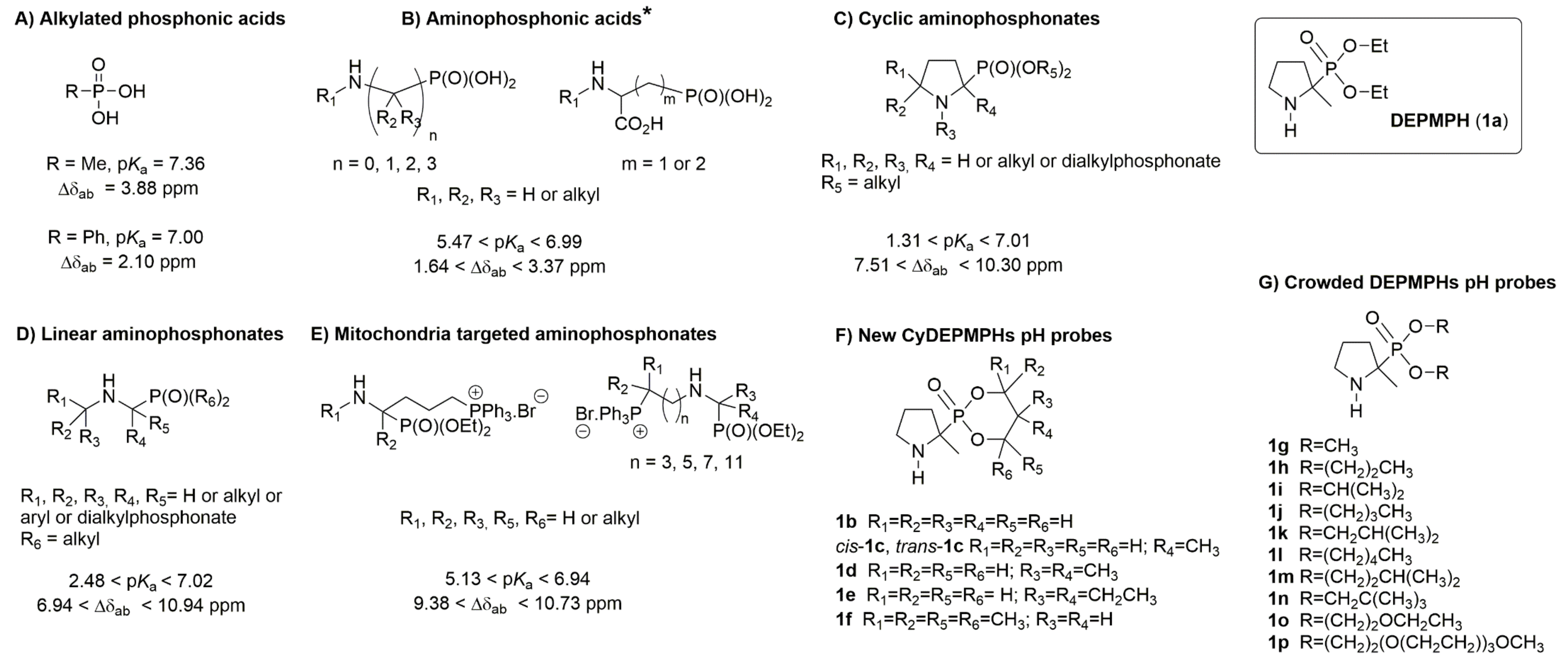
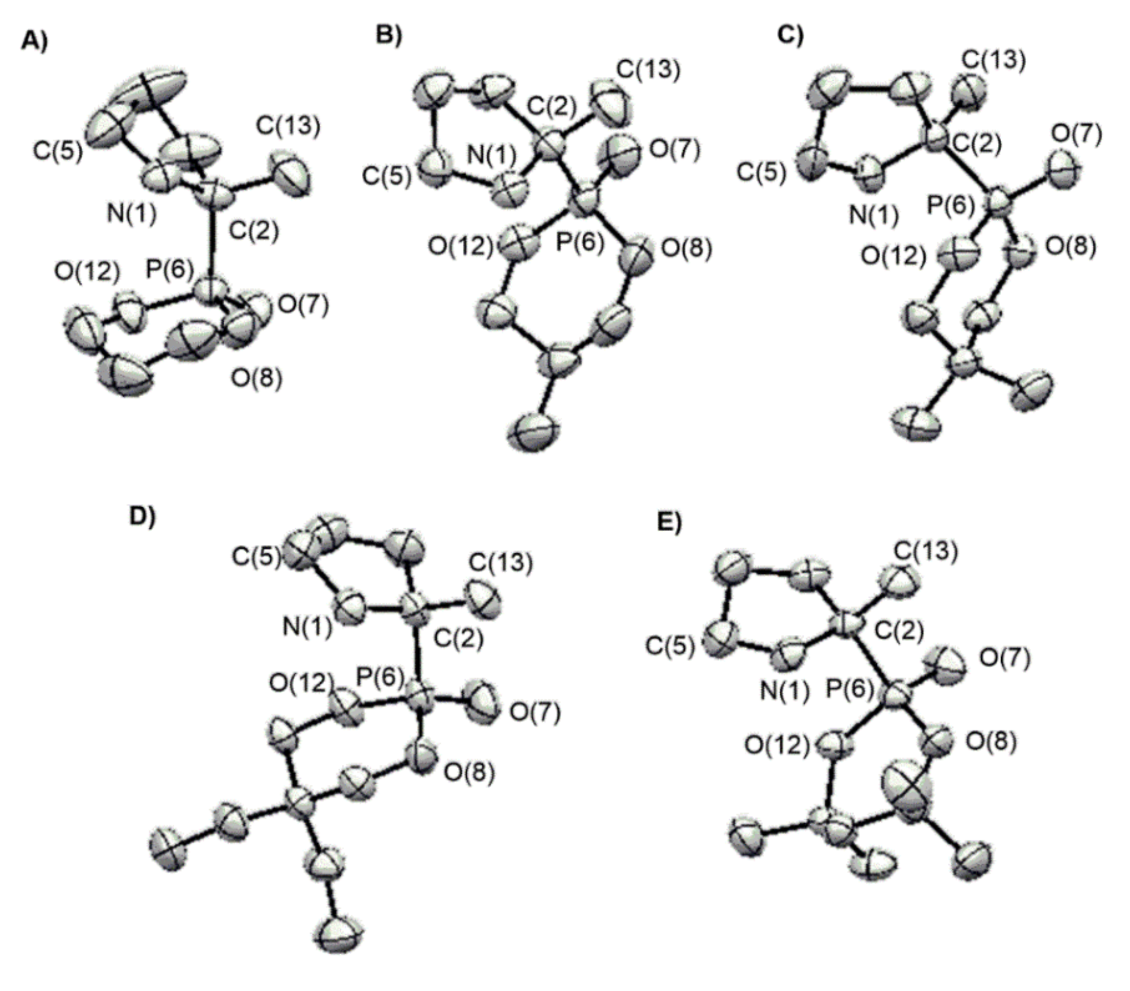
2.1. Chemistry and X-ray Crystallography

Aminophosphonates 1ap were synthesized according to the general sequence outlined in Scheme 1, involving the reaction of the corresponding dialkyl H-phosphonate 2ap with 2-methyl-1-pyrroline. Starting compounds dialkyl H-phosphonates 2a, 2g, 2i, and 2j are commercially available, while the other precursors 2bf, h, kp were synthesized according to [36] with slight modifications. Briefly, H-phosphonic bis(dimethylamide) was prepared in situ by the addition of one equivalent of water to hexamethylphosphorous triamide (HMPT) in tetrahydrofuran (THF), and then alcohol was added to give the corresponding compounds 2bf, h, kp. Reactions were monitored directly by 31P NMR by following the disappearance of HMPT peak (δ = 122.24 ppm) followed by the formation of bis(dimethylamino)phosphoric acid peak (δ = 24.0 ppm), then by the disappearance of this latter and the formation of the corresponding dialkyl H-phosphonate peak. Dialkyl (2-methylpyrrolidin-2-yl) phosphonates 1ap were obtained by the nucleophilic addition of dialkyl H-phosphonates 2ap onto 2-methyl-1-pyrroline in toluene [37] in good yields (49–73%).
Single crystals suitable for X-ray diffraction analysis were obtained for the five compounds 1b, trans-1c, and 1df by recrystallization from a dichloromethane:tert-butylmethylether (TBME) mixture (Figure 2). The pyrroline rings were found to adopt puckered conformations which can be either the E + 4 (ϕ = 72°) envelope conformation (1b), the T1 (ϕ = 18°) twist conformation (trans-1c) or an intermediate situation between the T1 and E + 4 conformations (1df) according to the classification of [38]. The C–P bond is pseudo-axial in compounds 1e and 1f and pseudo-equatorial in compounds 1b, trans-1c, and 1d (Table 1), as referred to by the sign of the C(5)–N(1)–C(2)–C(3) dihedral angle in nitrones analogues of CyDEPMPHs [39].

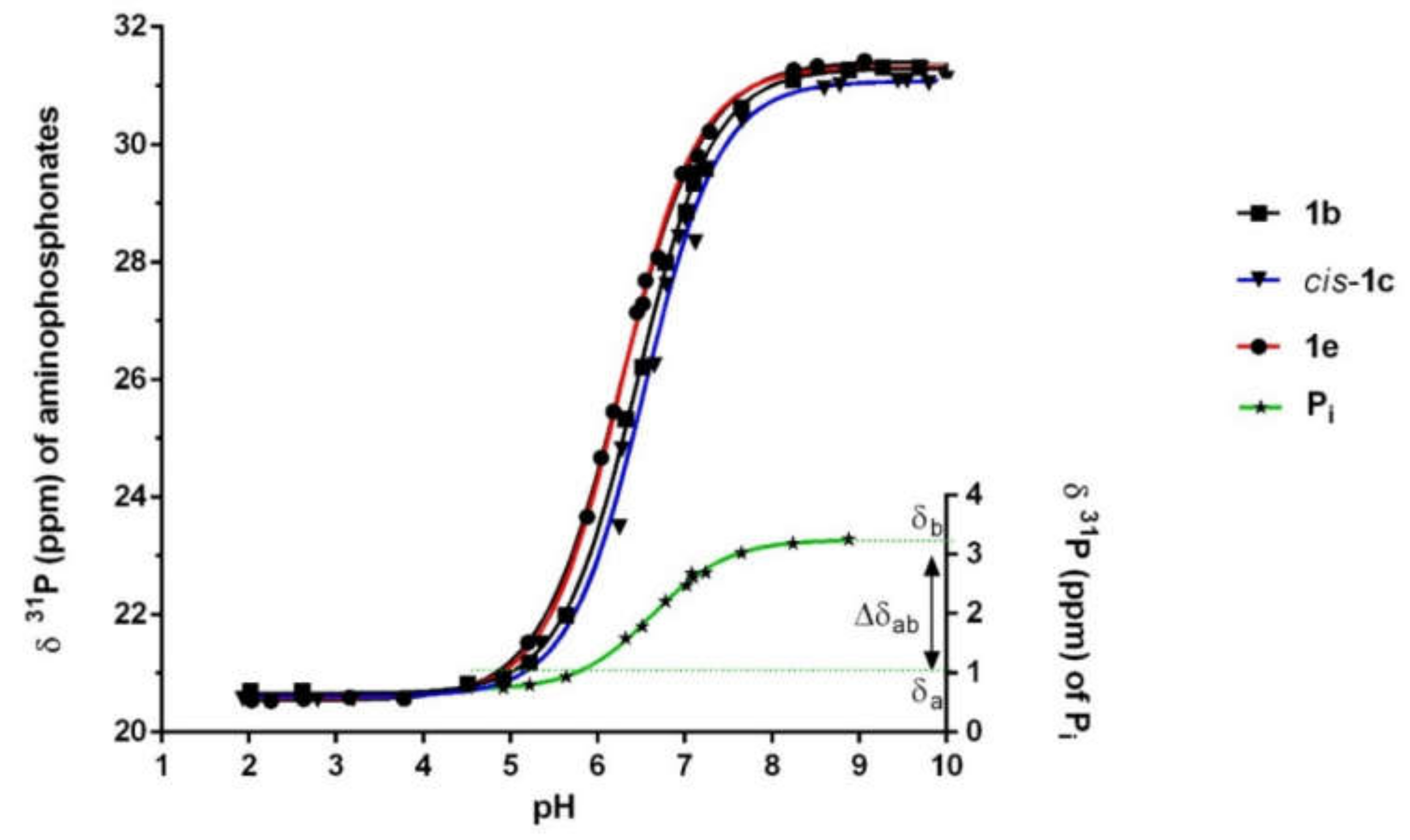
2.2. pH Dependent 31P NMR Properties of New α-Aminophosphonates

The pH dependence of the 31P NMR chemical shift of 1ap was investigated at 22 °C in KH buffer (pH 7.35). Monophasic acid–base titration curves reflecting protonation on nitrogen were obtained for all the derivatives (see examples in Figure 3) and their fitting to the Henderson–Hasselbalch equation (see Equation (2) in Section 3) allowed the calculation of pKas, the 31P NMR parameters (δa, δb, and Δδab), and the spin lattice relaxation time (T1) (Table 2).
All the new derivatives fulfilled both the desired conditions for more accurate pH probing (see Introduction), exhibiting near-neutral pKas (ranging 6.28–6.97) and very high Δδab values. Indeed, the average 31P NMR sensitivities obtained here could compete with those of the best linear aminophosphonates available so far (Table S1). Interestingly, the compounds having no constrained phosphorus bound alkoxy groups such as 1a and almost all elements from the crowded family showed slightly more basic pKas and lower 31P sensitivities when compared to compounds from the CyDEPMPHs family, for which Δδab > 10 ppm. The only exception was compound 1f, which retained a high sensitivity despite Δδab = 9.35 ppm and a pKa value in the range of that of the crowded DEPMPHs (Table 2).
In a biological milieu where NMR peaks are usually broad, any added pH probe should exhibit protonated and non-protonated forms in fast exchange on the NMR timescale to provide a single sharp peak for each cytosolic/intracellular compartment. This implies that the T1 time of the probe should be as short as possible in order for the best resolved signals to be recorded within the timescale of expected variations of biological parameters, such as pH [28]. All new pH probes demonstrated T1 values similar to those found in other related aminophosphonates [28,30]. Expectedly, molecular motion restriction in the conformationally constrained CyDEPMPHs 1bf versus the long-chain linear phosphonates 1lp from the crowded series generally resulted in slightly increased T1 values (Table 2). It is worth noting that probe exchanges between cell compartments are slow on T1 and chemical shift NMR timescales, while diffusion kinetics may exhibit strong variations for linear versus cyclic aminophosphonates (e.g., being 10 min for 1a and 60 min for 2-aminoethylphosphonate (Figure 1B with R1–R3 = H and n = 2) [29]). Given that pHi variations in vivo may occur in minutes (reported value were of ~10 min following acidosis [40] and 15–20 min following alkalinization [41]), T1 does not therefore appear to be a decisive parameter to discriminate between probes having similar chemical structures.

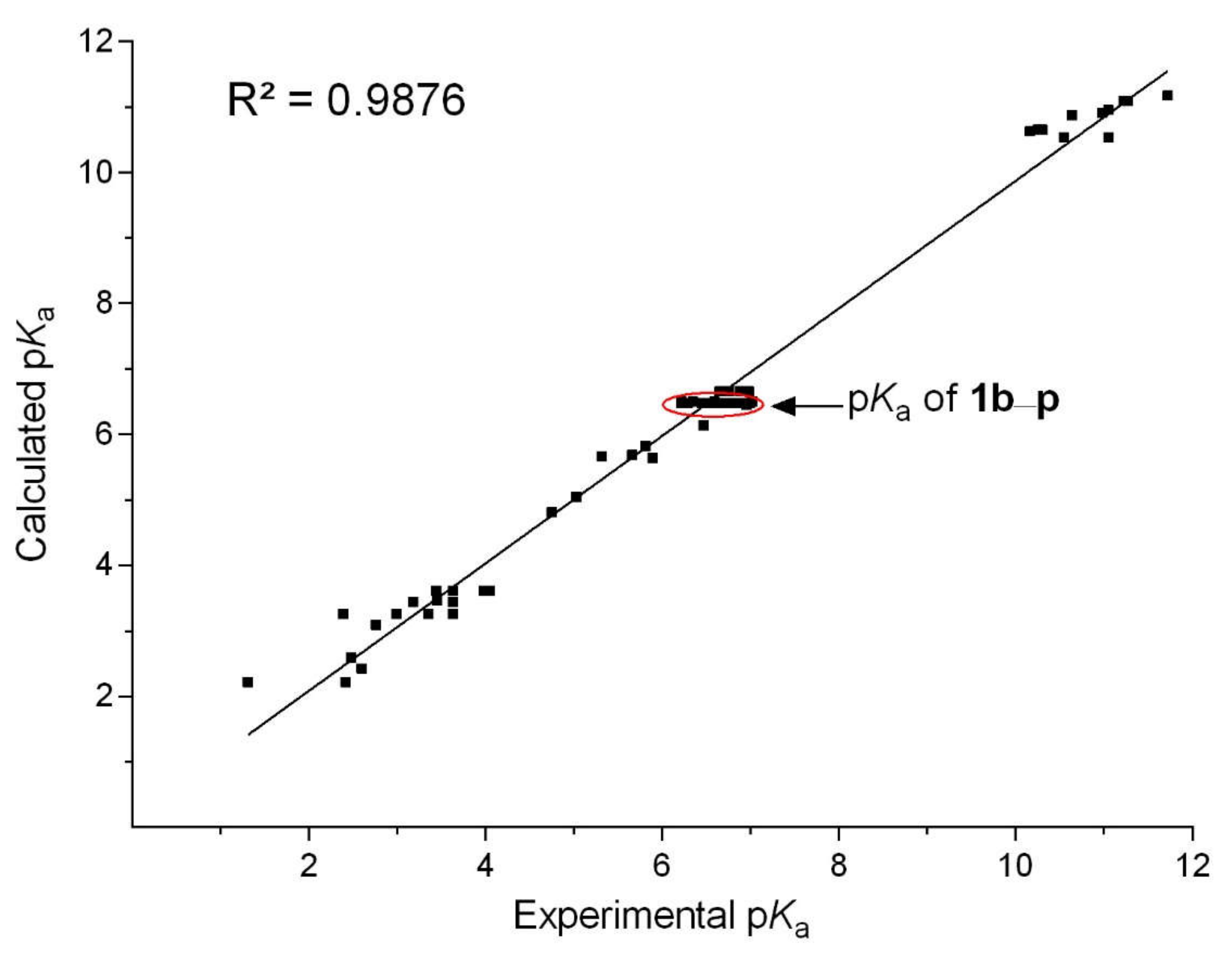
2.3. pKa Modeling as a Function of Substituents

Predicting pKa values by molecular modeling is of paramount importance in the design of improved 31P NMR pH markers. In this regard, a pertinent approach is to consider the pH-dependent variations of electron density around the protonation site; that is, pKa should depend linearly on the sum of individual inductive and stereochemical contributions of substituents. This has been applied successfully, first to a series of alkylphosphonic acids as depicted in Figure 1A [42], second to the cyclic and linear α-aminophosphonates developed in the early 2000s (see Figure 1C,D). In these studies [19,28], pKas were computed using the semi-empirical linear model of Equation (1):
pKa = a0 + ∑ai × ni
where, for a given structurally related family of compounds, a0 is the pKa of the common parent chemical structure and parameters ni (with maximum Σni = 6) and ai (in pH units, with 1 < ai < 10) represent the number of each different kind of substituents of type i having the same structure and/or position relative to the nitrogen protonation site and the weight of its electronic effect relative to the pKa value a0, respectively.
Starting from the general dialkylamino structure with up to seven surrounding substituents, a new refinement, including compounds 1ap, of the previous ai database collected from earlier studies afforded the updated Table S2 (see general structure in heading), where the selected reference structure bears a cycloalkyl substituent. The extended ai database encompassed a total of 59 aminophosphonates, comprising 1a and the new analogues 1bp, twenty-two linear (LAP-122) and ten cyclic (CAP-110) aminophosphonates, five aminophosphophonic acid derivatives (APA-15), and six alkylamines (Tables S1 and S2) studied previously [19,22,28,29,30,31,39,43,44]. As shown in Figure 4, updating the ai values still resulted in a good fit (R2 = 0.9876) between experimental and calculated pKas.
In the computational pKas model of Equation (1), all compounds 1ap share the same substitution pattern, i.e., one H atom on the nitrogen, two C(5)–H, three alkyls on C(1) and C(5), and one C(1)–Phosphonate. Having the same {n1n10} set of {1, 0, 2, 3, 0, 0, 0, 0, 1, 0}, compounds 1ap obviously yielded the same calculated pKa = 6.475, while an experimental range of ΔpKa = 0.75 was found (Table 2). When compared to this common pKa value, the experimental pKas of the more substituted compounds 1f and 1i were found to be strongly underestimated by 0.315 and 0.495 units, respectively (Table 2), suggesting that steric factors should be accounted for in the pKas predictive model.

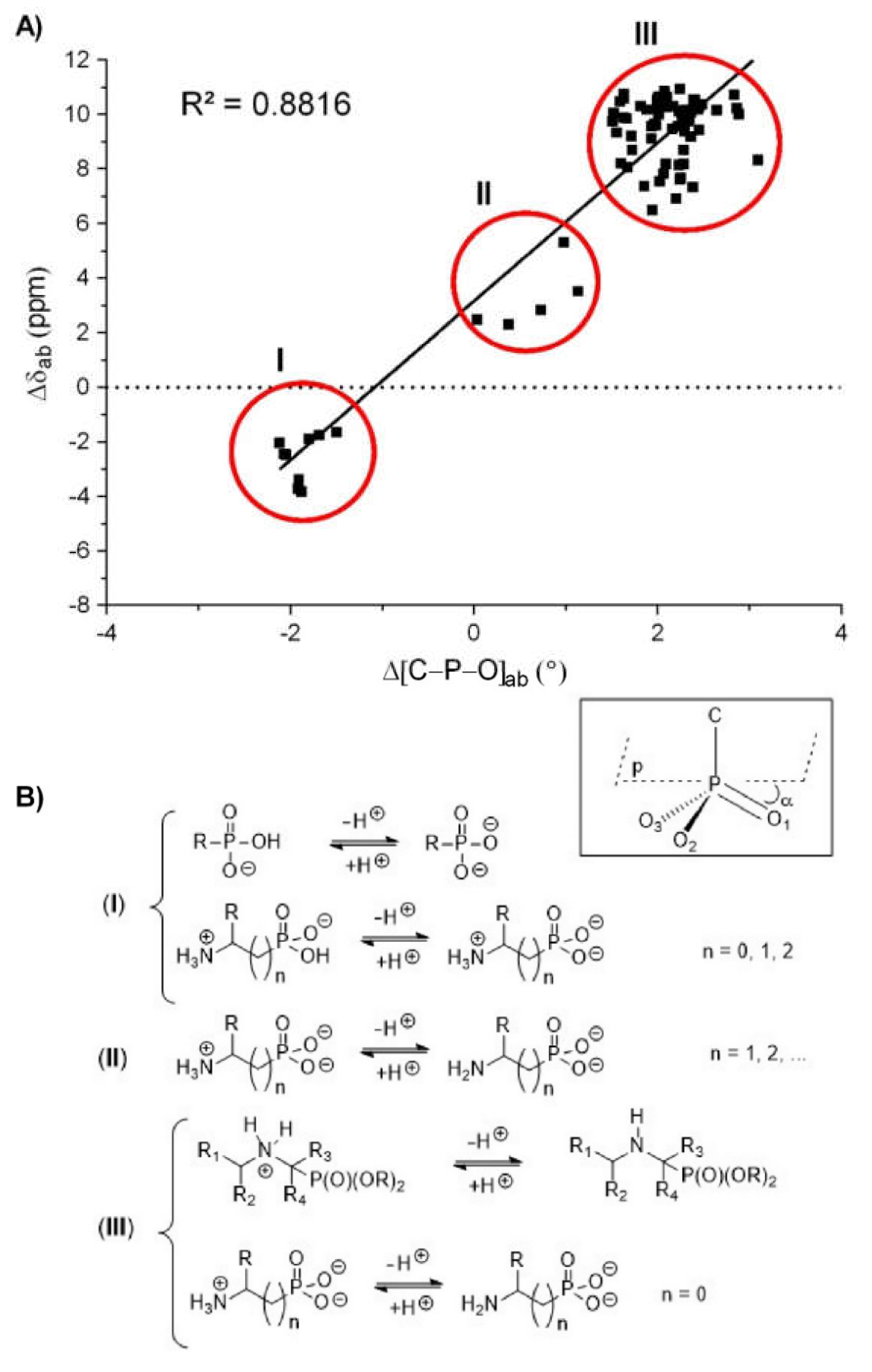
2.4. [C–P–O] Pyramidalization Angle Calculations

Previously, electrostatic interactions such as internal +NH2····(O)P hydrogen bonding have been proposed to explain the shielding of the NMR peaks occurring upon protonation of α-aminophosphonates (i.e., δb > δa), an effect that would decrease the pyramidalization of the angle [C–P–O] of the tetrahedral phosphorylated moiety [19]. In view of the lack of accuracy of the predictive model of Equation (1) for the most sterically hindered new pH probes, it was investigated whether additional conformationally derived effects may be implicated.
DFT-B3LYP calculations using the Gaussian16 package were carried out for optimizing [C–P–O] angle variations between acidic and basic forms (Δ[C–P–O]ab) for 1ap, pooled with the previously studied compounds LAP-134, CAP-110, and APA-17 (Table S1) [19,22,28,29,30,31,32,39,43,45]. For this large set of compounds (corresponding to 78 pairs of acidic and basic chemical shifts) a moderately good linear correlation (y = 2.907 x + 3.150; R2 = 0.8816) was obtained between Δ[C–P–O]ab and Δδab (Figure 5A), confirming that pyramidalization at phosphorus should have a notable impact on the 31P NMR sensitivity of aminophosphonates. For simplification, the [C–P–O] pyramidalization angle α here was defined as the average of the three angles between P–O1–3 bonds and the p-plane orthogonal to the P–C bond (Figure 5B, inset); that is, α decreases as the pyramid is more flattened. Negative Δ[C–P–O]ab variations associated to Δδab < 0 (i.e., δa > δb) were found for APA-16 aminophosphonic derivatives (Table S1) in the pKa2 region of the second acidity (Figure 5A, region I, and Figure 5B, reactions I). A second group of molecules (Figure 5A, region II, and Figure 5B, reactions II) showed small changes in the pyramidalization angle (0 < Δ[C–P–O]ab < 1.13°), associated with low 31P NMR sensitivities (2.3 < Δδab < 5.3 ppm). These latter structures bear at least two carbons between the N and P atoms of the phosphonic derivatives (LAP-23, APA-2,57; Table S1).
As anticipated behind the design of compounds 1bp, Figure 5A (region III) and Figure 5B (reactions III) show that those molecules having the largest Δ[C–P–O]ab (ranging 1.51–3.60°) also exhibited the greater Δδabs (6.48 < Δδab < 10.94 ppm). Together with 1ap, region III compounds in Figure 5A also included the derivatives APA-1, LAP-122,2434, and CAP-110 (Table S1); that is, in all these molecules, the environment around P is moderately to strongly sterically crowded. Of interest, among the CyDEPMPHs, the apparently less constrained 1b (Figure 2A), which bears a less bulky unsubstituted 1,3,2-dioxaphosphorinane ring, demonstrated the highest protonation-induced Δ[C–P–O]ab value of 2.10°.

2.5. In Vitro Cytotoxicity Studies

The cytotoxic activities of compounds 1ap in A549 human lung carcinoma cells and in normal human lung fibroblasts (NHLF) were evaluated in 0.2% DMSO-supplemented Dulbecco’s modified Eagle’s medium (DMEM) and fibroblast basal medium (FBM), respectively. The methods used included the tetrazolium dye MTT reduction, the fluorometric microculture cytotoxicity (FMCA), and the intracellular ATP assays. The MTT and FMCA assays are standard colorimetric determinations of cell viability during in vitro drug treatment while the ATP assay is an index of metabolic activity.
The IC50 values following 48 h incubation at 37 °C of the cells with varying concentrations of test compounds are reported in Table 3, together with the predicted lipophilicities based on AlogP calculations. When the three above-cited endpoints were found to be affected by treatment with a test compound, the release of cytosolic lactate dehydrogenase (LDH) was assessed to check for cell necrosis.
In the supernatant of untreated A549 and NHLF cells, the baseline LDH value was found to be 10.2 ± 1.5 and 11.3 ± 1.9 UI/mg protein, respectively. When compared to the total LDH activity measured after 100% lysis of the cells, which was 690 ± 47 and 701 ± 34 UI/mg protein for A549 and NHLF, respectively, the baseline released LDH vs. total cellular LDH in untreated cells was found to be as low as 1–2%.
After 48 h incubation of the cells, no significant dispersion among assays was found with compounds 1be of the CyDEPMPHs family (p > 0.05 by one-way ANOVA), suggesting those aminophosphonates induced cell death by altering metabolic activity, enzymatic and mitochondrial functions rather than by causing membrane damage and/or cell necrosis. The more lipophilic 1d and 1e were found to be slightly more toxic (Table 3).
Regarding the three viability assays, the more lipophilic compound of the CyDEPMPHs family, 1f, was associated with the strongest global decrease of IC50 values, which was of 42–50% (p < 0.05) as compared to 1b, and reached 50–60% (p < 0.05) as compared to 1a. In parallel, the application of 1f resulted in a significant LDH leakage (>45% of total baseline LDH content; p < 0.01 vs. untreated cells and 1a, 1b) showing it additionally caused cell necrosis.
In the crowded family of pH probes, the decrease in viability globally paralleled the increase of lipophilicity, except for 1o and 1p. Hence, IC50 values for 1jp decreased by 60–80% (p < 0.05) as compared to the hydrophilic 1g in the MTT and ATP assays, while an even greater impact was found for these lipophilic compounds in the FMCA assay (i.e., a 80–90% decrease; p < 0.05 vs. 1g), suggesting they induced membrane damage at concentrations > 10 mM (Table 3). Consistently, application of 1jp to both types of cells resulted in a strong LDH leakage at IC50 values (60–80% of total baseline intracellular content).
Altogether, the cytotoxicity data of Table 3 demonstrated that most of the new pH probes, i.e., 1be and 1gi, could be added safely in cell medium at a concentration window (up to 1–20 mM) compatible with NMR applications. Due to its innocuity and optimal 31P NMR titration parameters (pKa = 6.45; Δδab = 10.64 ppm), compound 1b was considered ideal for probing small pH gradients, since it can be used at 4 mM, a concentration at which >98% cell viability was preserved.

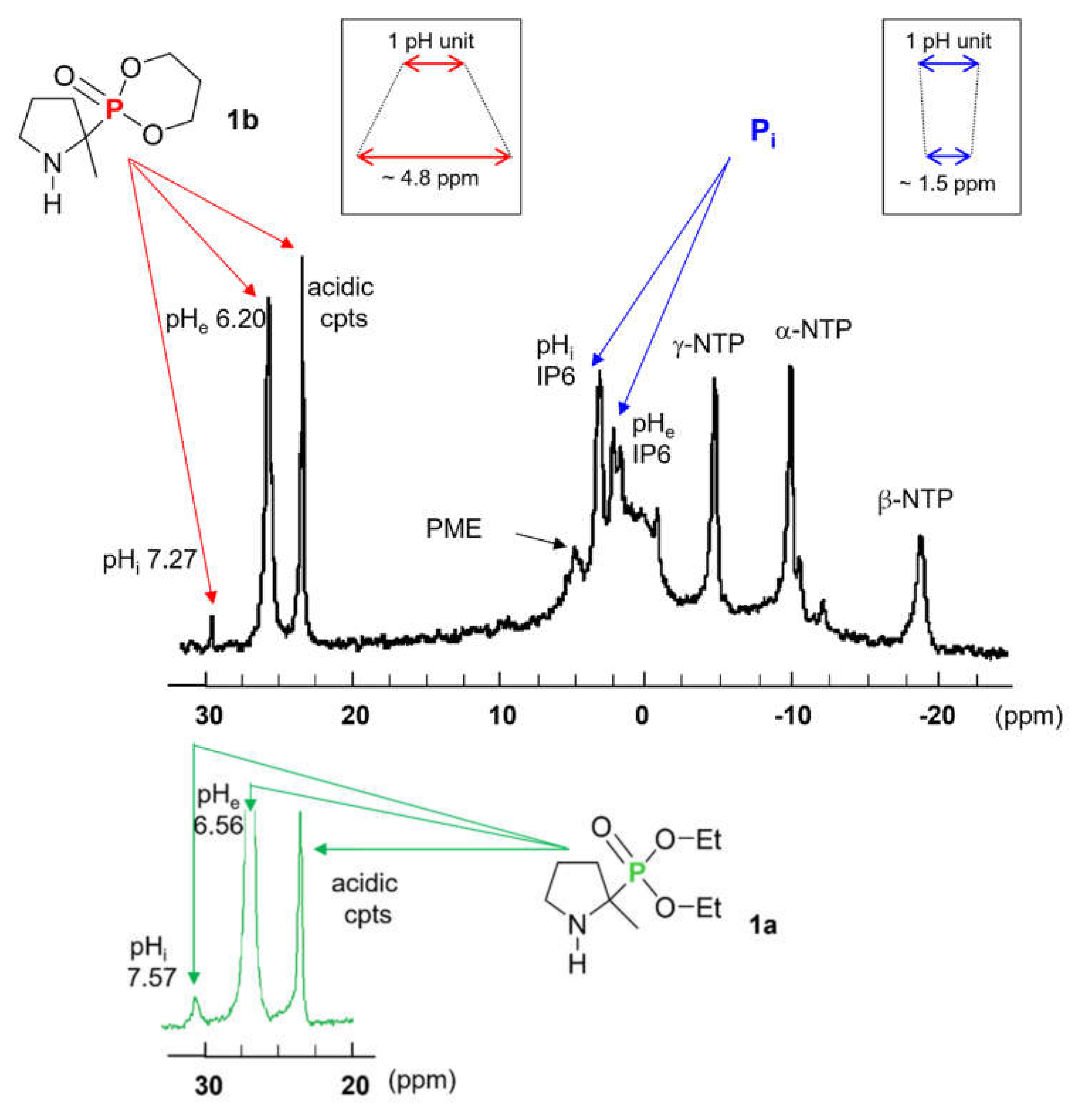
2.6. Application of the 31P NMR pH Probes 1b vs. 1a in Dictyostelium discoideum Amoebae

Dictyostelium discoideum, a widely used cell model for investigating acidic organelles, has been found to be particularly suitable to monitor by 31P NMR the kinetics of anoxia- and reoxygenation-induced pH variations in various endosomal acidic vesicles [23]. In the search for improved resonance tools for assessing trans-sarcolemmal pH gradients, i.e., pHi/pHe differences, D. discoideum was also found to be a reliable model, since in this case endogenous and external 31P NMR Pi peaks are easily distinguishable [21] and may allow a direct comparison with the expected resonances arising from the probe, provided it could significantly accumulate within the cytosolic and external compartments. Such properties were not reported in other models, including the rat heart [46] and liver [47] due, among other reasons, to the low and/or varying concentrations of Pi, the large line widths of cellular 31P NMR peaks, and the low pHi/pHe difference < 0.5 pH units.
Figure 6 shows representative 31P NMR spectra obtained during incubation at 20 °C of normoxic amoebae in the presence of 4 mM of 1a (lower trace, deshielded region) or 1b (upper trace). In the more deshielded spectral region (20–30 ppm) of the experiment with 1b, prominent resonances were apparent, likely arising from the probe internalized in cells and located in three distinct intracellular compartments. From the calibration curve of 1b (Figure 3), the corresponding pH values were calculated as ~5.6 (for the more acidic compartment at 22.6 ppm) and 6.20 and 7.27 for the lowfield peaks at 25.4 and 29.9 ppm, respectively. These two latter compartments probed by 1b likely correspond to the extracellular and cytosolic environments, in full accordance with the pH values determined by the two Pi peaks, pHi ~7.24 (cytosol) and pHe ~6.32 (extracellular milieu).
When 1a was used instead of 1b, acidic compartments were similarly probed at 23.6 ppm (pH~5.7) while the cytosolic and extracellular regions exhibited broader lines and higher apparent pH values of pHi 7.57 (resonance at 30.5 ppm) and pHe ~6.56 (broad resonance at ~26.5 ppm), respectively. This showed that 1a accumulated mainly in the external space vs. the cytosol and confirmed the previous observations [29] of (i) a line broadening at the resonance corresponding to the cytosolic pH, and (ii) a slight alkalinization of the cytosolic and extracellular compartments because of the higher pKa of the probe, which behaved as a weak base.
The innocuity of 1b (4 mM) in the system was confirmed by its lack of effect on the endogenous intracellular phosphorylated compounds including nucleotides, inositol hexakisphosphate and phosphomonoesters, as compared to control spectra (data not shown). As already reported with other series of cyclic and linear α-aminophosphonates [29], 1b was internalized in amoebae in less than 5 min (data not shown) both in the cytosol, extracellular region and acidic vesicles. To explain the rapid rate of diffusion and intracellular distribution observed for 1b and other aminophosphonates [27,29], it was supposed that these compounds are membrane permeable under their unprotonated amine form, and can accumulate by passive diffusion [29]. This general property confers a great advantage to α-aminophosphonates as compared to the commonly used alkylated phosphonic acids derivatives, which were shown to internalize slowly, at least during 90 min, within the cells, involving energy requiring processes such as continuous H+ pumping by vacuolar H+–ATPase, leading to NTP depletion [48].
To stress on the specific 31P NMR pH reporting properties, the biocompatible 1b showed a ~3.2 better potential accuracy than Pi (Figure 6). This may allow us to reliably address pH differences as low as 0.05 pH units, corresponding to narrow peaks separated by ~0.25 ppm. As anticipated in the design of the novel pH probes, this property results from their improved Δδab values, giving a steepest slope around pKas close to that of Pi.

3. Materials and Methods

3.1. Chemistry

All reagents and solvents were of the highest grade available from Sigma-Aldrich, Fluka, SDS, or Acros, and were used without further purification, except for 2-methyl-1-pyrroline (from Acros Organics, Halluin, France), which was distilled under reduced pressure (20 mm Hg, bp: 50 °C) before use. Diethyl H-phosphonate (2a), dimethyl H-phosphonate (2g), di(isopropyl) H-phosphonate (2i), and di-n-butyl H-phosphonate (2j) were from Acros. The dialkyl H-phosphonates 2bf and the aminophosphonates 1bf belonging to the CyDEPMPHs family were synthesized and purified as previously described [39]. DEPMPH (1a) [19], and its analogue 1i [49], and dialkyl H-phosphonates 2h and 2n, and aminophosphonates 1g, 1h, and 1n [29] were synthesized and purified as previously described. Reactions were followed by thin layer chromatography (TLC) on Merck-Kieselgel 60 F254 precoated silica gel plates, and the spots were visualized by staining with phosphomolybdic acid. NMR spectra (1H NMR at 300 MHz, 13C NMR at 75.5 MHz and 31P NMR at 121.5 MHz) were recorded on a Bruker AVL 300 spectrometer. Chemical shifts (δ) are expressed in ppm (parts per million) relative to internal tetramethylsilane (1H and 13C) or external 85% H3PO4 (31P), and coupling constants J are given in Hz. The abbreviations s, d, t, m and q refer to singlet, doublet, triplet, multiplet and quartet signals, respectively. Melting points were determined using a B-540 Buchi apparatus and are uncorrected. Elemental analyses were performed using a Thermo Finnigan EA-1112 analyzer and were within 0.2% of theoretical values. Column chromatography was performed on Merck silica gel 60 (230~400 mesh).

3.1.1. Synthesis of Dialkyl H-Phosphonates 2km, 2o, and 2p

Water (1.1 g, 61 mmol) was added to HMPT (10 g, 61 mmol) in refluxing anhydrous THF (20 mL) under argon atmosphere and the mixture was stirred at reflux for 2 h. Then the corresponding alcohol (122 mmol) was added and the mixture was refluxed for another 2 h. Afterwards, the mixture was concentrated under reduced pressure to give the corresponding dialkyl H-phosphonate 2km, 2o, and 2p in high yields (95–98%) and a purity of >95% determined by 31P NMR. 31P NMR (121.5 MHz, CDCl3) δ 2k, 8.42; 2l, 8.05; 2m, 7.80; 2o, 9.39; 2p, 9.53.

3.1.2. General Procedure for Synthesis of Compounds 1jm, 1o, and 1p

A mixture of 2-methyl-1-pyrroline (1.1 eq) and the corresponding dialkyl H-phosphonate (1 eq) was stirred at room temperature until completion. TLC or 31P NMR were used to follow the reaction. The mixture was poured into water (aminophosphonate final concentration, ~1.8 M) and slowly acidified to pH 3 with concentrated HCl. The aqueous layer was extracted with TBME (3 × volume of water), basified with NaOH pellets to pH 9–10, and extracted with CH2Cl2 (4 × volumes of water). The combined organic phases were dried over MgSO4, filtered and concentrated under reduced pressure. The residue was purified by SiO2 column chromatography with a CH2Cl2:EtOH mixture as eluent to yield the expected aminophosphonates.
Di-n-butyl (2-methylpyrrolidin-2-yl)phosphonate (1j). Title compound was obtained by stirring 2-methyl-1-pyrroline (10 g, 120 mmol) with commercially available di-n-butyl H-phosphonate (21.1 g, 109 mmol) for 10 days. Flash chromatography using a CH2Cl2:EtOH, 90:10, v:v as eluent yielded 1j as a yellow oil (22.3 g, 67%); 1H NMR (300 MHz, CDCl3) δ 4.06–3.97 (m, 4H, 2 × O-CH2), 3.04–2.88 (m, 2H, 5-CH2), 2.22–2.10 (m, 1H, 3-C(H)H), 1.83–1.70 (m, 4H, NH, 3-C(H)H, 4-CH2), 1.64–1.51 (m, 5H, 2 × O-CH2-CH2 and 3-C(H)H), 1.37–1.28 (q, J = 6.0 Hz, 4H, 2 × CH2-CH3), 1.27 (d, J = 15.0 Hz, 3H, 2-C(CH3)), 0.87 (t, J = 6.0 Hz, 6H, O-(CH2)3-CH3); 13C NMR (75.5 MHz, CDCl3) δ 66.0 (d, J = 8.2 Hz, O-CH2), 65.8 (d, J = 8.2 Hz, O-CH2), 59.2 (d, J = 163.8 Hz, 2-C), 47.0 (d, J = 7.5 Hz, 5-C), 34.3 (d, J = 3.0 Hz, 3-C), 32.7 (d, J = 2.5 Hz, O-CH2-CH2), 32.6 (d, J = 2.5 Hz, O-CH2-CH2), 25.6 (d, J = 4.5 Hz, 4-C), 24.2 (d, J = 6.0 Hz, 2-C(CH3)), 18.7 (2C, CH2-CH3), 13.5 (2 × O-(CH2)2-CH3); 31P NMR (121.5 MHz, CDCl3) δ 31.03. Elemental analysis calcd (%) for C13H28NO3P: C 56.30, H 10.18, N 5.05; found: C 54.72, H 10.10, N 5.01.
Diisobutyl (2-methylpyrrolidin-2-yl)phosphonate (1k). Title compound was obtained by stirring 2-methyl-1-pyrroline (5.5 g, 66 mmol) with commercially available diisobutyl H-phosphonate 2k (11.8 g, 60 mmol) for 7 days. Flash chromatography using a CH2Cl2:EtOH, 90:10, v:v as eluent yielded 1k as a yellow oil (11.8 g, 70%); 1H NMR (300 MHz, CDCl3) δ 3.54–3.43 (m, 4H, 2 × O-CH2), 2.73–2.57 (m, 2H, 5-CH2), 1.95–1.88 (m, 1H, 3-C(H)H), 1.63–1.24 (m, 6H, NH, 3-C(H)H, 4-CH2, 2 × CH(CH3)2), 0.98 (d, J = 15.0 Hz, 3H, 2-C(CH3)), 0.60–0.57 (d, J = 6.0 Hz, 12H, 2 × CH(CH3)2); 13C NMR (75.5 MHz, CDCl3) δ 71.4 (d, J = 7.5 Hz, O-CH2), 71.1 (d, J = 7.5 Hz, O-CH2), 58.8 (d, J = 165.3 Hz, 2-C), 46.2 (d, J = 7.5 Hz, 5-C), 32.9 (d, J = 2.5 Hz, 3-C), 28.4 (2 × CH(CH3)2), 24.5 (d, J = 4.5 Hz, 4-C), 23.5 (d, J = 6.0 Hz, 2-C(CH3)), 17.8 (2 × CH(CH3)2); 31P NMR (121.5 MHz, CDCl3) δ 31.17. Elemental analysis calcd (%) for C13H28NO3P: C 56.30, H 10.18, N 5.05; found: C 55.06, H 10.56, N 4.96.
Dipentyl (2-methylpyrrolidin-2-yl)phosphonate (1l). Title compound was obtained by stirring 2-methyl-1-pyrroline (2.5 g, 30 mmol) with dipentyl H-phosphonate 2l (6 g, 27.3 mmol) for 7 days. Flash chromatography using a CH2Cl2:EtOH, 90:10, v:v as eluent yielded 1l as a yellow oil (4.2 g, 49%); 1H NMR (300 MHz, CDCl3) δ 4.07–3.99 (m, 4H, 2 × O-CH2), 3.07–2.91 (m, 2H, 5-CH2), 2.22–2.18 (m, 1H, 3-C(H)H), 1.90–1.52 (m, 8H, 2 × OCH2CH2, NH, 3-C(H)H, 4-CH2), 1.30 (m, 8H, 2 × CH2CH2CH3), 1.30 (d, J = 15.0 Hz, 3H, 2-C(CH3)), 0.86 (t, J = 9.0 Hz, 6H, 2 × CH2CH3); 13C NMR (75.5 MHz, CDCl3) δ 66.4 (d, J = 7.5 Hz, O-CH2), 66.1 (d, J = 7.5 Hz, O-CH2), 59.6 (d, J = 164.6 Hz, 2-C), 47.1 (d, J = 7.5 Hz, 5-C), 34.6 (d, J = 2.5 Hz, 3-C), 30.4 (d, J = 4.5 Hz, 4-C), 27.6 (2C, CH2CH2CH3), 25.7 (d, J = 4.5 Hz, 2C, OCH2CH2), 24.3 (d, J = 6.8 Hz, 2-C(CH3)), 22.2 (2 × CH2CH3), 13.9 (2C, CH2CH3); 31P NMR (121.5 MHz, CDCl3) δ 30.95. Elemental analysis calcd (%) for C15H32NO3P: C 58.99, H 10.56, N 4.59; found: C 56.10, H 10.35, N 3.90.
Diisopentyl (2-methylpyrrolidin-2-yl)phosphonate (1m). Title compound was obtained by stirring 2-methyl-1-pyrroline (4.4 g, 53 mmol) with diisopentyl H-phosphonate 2m (10.8 g, 48.2 mmol) for 7 days. Flash chromatography using a CH2Cl2:EtOH, 90:10, v:v as eluent yielded 1m as a yellow oil (4.2 g, 49%); 1H NMR (300 MHz, CDCl3) δ 4.07–3.99 (m, 4H, 2 ×O-CH2), 3.07–2.91 (m, 2H, 5-CH2), 2.22–2.18 (m, 1H, 3-C(H)H), 1.90–1.52 (m, 8H, 2 × OCH2CH2, NH, 3-C(H)H, 4-CH2), 1.30 (m, 8H, 2 × CH2CH2CH3), 1.30 (d, J = 15.0 Hz, 3H, 2-C(CH3)), 0.86 (t, J = 9.0 Hz, 6H, 2 × CH2CH3); 13C NMR (75.5 MHz, CDCl3) δ 66.4 (d, J = 7.5 Hz, O-CH2), 66.1 (d, J = 7.5 Hz, O-CH2), 59.6 (d, J = 164.6 Hz, 2-C), 47.1 (d, J = 7.5 Hz, 5-C), 34.6 (d, J = 2.5 Hz, 3-C), 30.4 (d, J = 4.5 Hz, 4-C), 27.6 (2C, CH2CH2CH3), 25.7 (d, J = 4.5 Hz, 2C, OCH2CH2), 24.3 (d, J = 6.8 Hz, 2-C(CH3)), 22.2 (2 × CH2CH3), 13.9 (2C, CH2CH3); 31P NMR (121.5 MHz, CDCl3) δ 30.95. Elemental analysis calcd (%) for C15H32NO3P: C 58.99, H 10.56, N 4.59; found: C 56.10, H 10.35, N 3.90.
Di(2-ethoxyethyl)(2-methylpyrrolidin-2-yl)phosphonate (1o). Title compound was obtained by stirring 2-methyl-1-pyrroline (2.5 g, 30 mmol) with diethoxyethyl H-phosphonate 2o (6.2 g, 27.3 mmol) for 1 day. Flash chromatography using a CH2Cl2:EtOH, 96:4, v:v as eluent yielded 1o as a yellow oil (6.1 g, 73%); 1H NMR (300 MHz, CDCl3) δ 4.23–4.11 (m, 4H, 2 × P-O-CH2), 3.66–3.62 (t, J = 6.0 Hz, 4H, 2 × CH2OCH2CH3), 3.57–3.50 (q, J = 6.0 Hz, 4H, 2 × CH2CH3), 3.02–2.90 (m, 2H, 5-CH2), 2.25–2.17 (m, 1H, 3-C(H)H), 1.90–1.53 (m, 4H, 3-C(H)H, 4-CH2, NH), 1.31 (d, J = 15.0 Hz, 2-C(CH3)), 1.13 (t, J = 9.0 Hz, 6H, 2 × CH2CH3); 13C NMR (75.5 MHz, CDCl3) δ 69.7 (d, J = 5.3 Hz, 2 × CH2OCH2CH3), 66.5 (2 × CH2CH3), 65.2 (d, J = 7.5 Hz, P-O-CH2), 65.3 (d, J = 7.5 Hz, P-O-CH2), 59.7 (d, J = 163.8 Hz, 2-C), 46.9 (d, J = 6.8 Hz, 5-C), 34.6 (d, J = 3.8 Hz, 3-C), 25.6 (d, J = 3.8 Hz, 4-C), 24.2 (d, J = 6.8 Hz, 2-C(CH3)), 15.1 (2C, CH2CH3); 31P NMR (121.5 MHz, CDCl3) δ 31.65. Elemental analysis calcd (%) for C13H28NO3P: C 50.48, H 9.12, N 4.53; found: C 49.16, H 9.03, N 4.39.
Di(2,5,8,11-tetraoxatridecan-13-yl)(2-methylpyrrolidin-2-yl)phosphonate (1p). Title compound was obtained by stirring 2-methyl-1-pyrroline (0.6 g, 7.5 mmol) with di(2,5,8,11-tetraoxatridecan-13-yl) H-phosphonate 2p (3.2 g, 6.8 mmol) for 1 day. Flash chromatography using a CH2Cl2:EtOH, 96:4, v:v as eluent yielded 1q as a yellow oil (2.7 g, 71%); 1H NMR (300 MHz, CDCl3) δ 4.30–4.12 (m, 4H, 2 × POCH2), 3.71–3.65 (m, 4H, 2 × OCH2), 3.65–3.58 (m, 20H, 10 × OCH2), 3.55–3.49 (m, 4H, 2 × OCH2), 3.33 (6H, 2 × OCH3), 3.10–2.91 (m, 2H, 5-CH2), 2.28–2.17 (m, 1H, 3-C(H)H), 1.90–1.52 (m, 4H, 3-C(H)H, 4-H, NH), 1.32 (d, J = 15.0 Hz, 2-C(CH3)); 13C NMR (75.5 MHz, CDCl3) δ 72.7 (OCH2), 71.8 (OCH2), 70.5 (OCH2), 70.4 (OCH2), 70.3 (OCH2), 70.1 (OCH2), 65.3 (d, J = 7.5 Hz, POCH2), 65.2 (d, J = 7.5 Hz, POCH2), 59.7 (d, J = 164.6 Hz, 2-C), 58.9 (2C, OCH3), 46.9 (d, J = 7.5 Hz, 5-C), 34.5 (d, J = 2.3 Hz 3-C), 25.5 (d, J = 4.5 Hz, 4-C), 23.0 (d, J = 6.8 Hz, 2-C(CH3)); 31P NMR (121.5 MHz, CDCl3) δ 29.52. Elemental analysis calcd (%) for C23H48NO11P: C 50.63, H 8.87, N 2.57; found: C 47.67, H 8.84, N 2.37.

3.2. X-ray Crystallography

CyDEPMPHs were dissolved in the minimum of CH2Cl2, recrystallized from TBME, and dried in a vacuum to give single crystals suitable for X-ray diffraction studies. Intensities were collected at 293 K on a Nonius Kappa CCD diffractometer (Bruker) using graphite-monochromated Mo radiation (λ = 0.71073 Å).
Crystal data for 1b: C8H16NO3P M = 205.20, monoclinic space group P-21 c, Hall group -P 2ybc, a = 12.3308(2), b = 13.7792(2), c = 15.2243(2) Å, β = 126.3761(7)°, V = 2082.69(5) Å3, Z = 4.
Crystal data for trans-1c: C9H18NO3P M = 219.21, monoclinic space group P-21 c, Hall group -P 2ybc, a = 6.6716(1), b = 12.6291(2), c = 13.6892(3) Å, β = 92.9872(8)°, V = 1151.83(4) Å3, Z = 4.
Crystal data for 1d: C10H20NO3P M = 233.24, monoclinic space group P-21 c, Hall group -P 2ybc, a = 10.5502(5), b = 11.1665(4), c = 11.4207(2) Å, V = 1214.42(9) Å3, Z = 4.
Crystal data for 1e: C12H24NO3P M = 261.29, orthorhombic space group Pbca, Hall group -P 2ac 2ab, a = 13.3114(2), b = 10.4734(2), c = 20.0050(2) Å, V = 2789.01(9) Å3, Z = 8.
Crystal data for 1f: C12H24NO3P M = 261.29, triclinic space group P-1, Hall group -P 1, a = 6.5802(5), b = 7.9200(9), c = 14.228(1) Å, V = 699.37(11) Å3, Z = 2.
CCDC 794184, 794158, 794208, 794102, 793,993 contains the supplementary crystallographic data of 1b, trans-1c, 1df, respectively. These data are provided free of charge by The Cambridge Crystallographic Data Centre, 12 Union Road, Cambridge, UK (fax: +44 1223 336033; e-mail: [email protected]).

3.3. P NMR pH-Titration of Aminophosphonates 1a-p

All titrations were carried out at 22 °C in modified KH medium (pH 7.35), consisting of (in mM): KH2PO4, 1.2; MgSO4, 1.2; NaCl, 118.5; KCl, 4.8; NaHCO3, 25, and EDTA, 0.55 dissolved in doubly distilled deionized water. Spectra were acquired (32 scans) at 22 °C on a Bruker AMX 400 spectrometer with a 9.4 Tesla wide-bore magnet at a phosphorus frequency of 161.98 MHz. Samples were placed in 10-mm tubes. 2H2O in a small capillary was used as a lock signal for the spectrometer. Chemical shifts are given in ppm with respect to 85% external H3PO4 at 0 ppm and were plotted against pH to fit the Henderson–Hasselbach Equation (2) for NMR using nonlinear regression (Prism, GraphPad Software Inc., San-Diego, CA, USA):
p H = p K a + log δ δ a δ b δ
where δ is the experimental 31P chemical shift and the δa and δb correspond to the limiting chemical shift values of protonated and unprotonated amines forms, respectively. Calculated titration data (pKa, δa, δb and their difference (Δδab)) are expressed as mean ± SD.

3.4. Computational Methods for [C–P–O] Angle Calculations

[C–P–O] angle calculations were performed for both acidic and basic forms of the aminophosphonates. To improve the accuracy of the calculations for compounds 1ap, compounds LAP-134, CAP-110, APA-15 (Table S1), and 6 amines (i.e., dimethylamine, diethylamine, diisopropylamine, piperidine, pyrrolidine, tetramethylpiperidine) were included to the pooled molecules. The angle calculations were performed using Gaussian16 software [50] after geometry optimization at B3LYP/6-31G(d) level of theory. To obtain the most stable conformation of each molecule before DFT optimization, a simulated annealing calculation at the AM1 level with the Ampac 11 [51] software package was performed. The optimizations were achieved with a maximum time of 12 h. A check for the absence of imaginary frequencies and a gradient close to zero was performed. All calculations were performed in water solvent with the Polarizable Continuum Model (PCM) [52]. Data (in °) are expressed as the differences in [C–P–O] angles between the acidic and basic structures (Δ[C–P–O]ab).

3.5. Cell Culture, Cytotoxicity Assays, and pH Assessment in Amoebae

DMEM, GlutaMax™, and phosphate-buffered saline (PBS) were obtained from Gibco Life Technologies Inc. (Thermo Fisher Scientific, Illkirch, France). FBM and growth factors (FGM; Clonetics FGM-2 Bullet Kit) were from Lonza (Amboise, France). A549 (ATCC CCL-185; LGC Standards, Molsheim, France) and NHLF (Lonza) cells were routinely maintained in [DMEM + GlutaMax] and [FBM + FGM], respectively, as described previously [53,54]. After reaching 90% confluence, cells were harvested for subculture. Cells were trypsinized, seeded in 96-well microplates (density, 2.5 × 10⁴ cells/well) and incubated at 37 °C in a humidified atmosphere with 5% CO2 to reach ~80% confluence in the appropriate medium. The medium was renewed and cells were exposed for 48 h to the tested compounds (1–1000 µg/mL) or DMSO (0.2%) added medium (control). Afterwards, cells were washed twice with PBS 1X (+/+) for cytotoxicity analysis. Supernatant medium samples were kept to evaluate cytosolic LDH release using a commercial kit (Biolabo, Maisy, France). To estimate the total LDH content, a control measurement was performed for each set of experiments by treating cells with 1% Triton X-100 to induce a total LDH release in the supernatant and induce a 100% loss of viability.
The FMCA and MTT assays were carried out as described [55]. Intracellular ATP content was assayed using a luciferin–luciferase reagent (Biofax A®; Yelen Analytics, Marseille, France; http://www.yelen-analytics.com (accessed on 10 January 2021)) according to [55]. IC50 values, defined as the concentration of test compounds resulting in 50% cell viability after 48 h, were calculated from concentration–response curves (PrismSoftware) and are expressed as mean ± SD (standard deviation relative to the mean from 3–10 independent experiments).

3.6. Dictyostelium Discoideum Cells Cultures and 31P NMR

D. discoideum amoebae, axenic strain (ATCC 24397), were cultured aerobically as previously described [21,29]. Briefly, cells were harvested in their exponential growth phase, washed with ice-cold 20 mM 2-morpholinoethanesulfonic acid sodium salt (MES-Na) buffer and then suspended (3 × 10⁸ cells/mL) at 20 °C under O2 bubbling in MES-Na (20 mM), 6% 2H2O and 5 µL of Antifoam 289 (Sigma-Aldrich, Saint Quentin Fallavier, France) to a final volume of 20 mL (pH 6.5). Afterwards, 1a or 1b were added (4 mM final concentration) to the cell medium and incubation was prolonged for up to 30 min at 20 °C.
Incubation samples were then placed in 25-mm NMR tubes and an 2H2O sample, placed in a small capillary, was used as a lock signal for the NMR spectrometer. 31P NMR spectra were routinely recorded during cell incubation at 20 °C on a Bruker AMX 400 spectrometer with a 9.4 T wide-bore magnet at a phosphorus frequency of 161.98 MHz. Chemical shifts are given in ppm with respect to 85% external H3PO4 at 0 ppm and to 50 mM methylene diphosphonate (pH 8.9), placed in a capillary and used as an additional standard at 16.4 ppm. Spectral acquisitions were carried out using a 60° (10 µs) pulse width, a 0.28 s acquisition time, a 0.72 s repetition delay, and gated Waltz proton decoupling [21]. Data were stored during 120 min as 300-scan (5 min) or 900-scan (15 min) blocks. Gaussian line broadening (GB = 0.05, LB = –5 Hz) was applied prior to Fourier transformation. The extracellular and intracellular distribution of the probe was determined by the respective areas of the corresponding resonances.

3.7. Statistics

Titration and biological values are expressed as mean ± SD. Differences were analyzed using a one-way analysis of variance (ANOVA) followed by a posteriori Newman–Keuls test. Intergroup differences were considered to be significant at p < 0.05.

4. Conclusions

A novel series of fifteen α-aminophosphonates have been synthesized and screened for their 31P NMR properties in the probing of subtle pH changes (as low as 0.05 pH units) occurring in normal or acidotic cells. The new compounds, obtained in only two or three synthetic steps, showed the following enhanced properties: (i) chemical shifts in the 18–36 ppm range distinct from those of phosphorus metabolites; (ii) near-neutral pKas (6.3–7.0); (iii) a high NMR sensitivity (Δδab ranging 9.2–10.6 ppm) that can be modulated by adjusting the substituents and steric effects around the phosphorus atom; (iv) no cytotoxic effect for most of them in the concentration range used for biological NMR applications (up to 20 mM); (v) the ability to penetrate the compartments of interest (i.e., intra- and extracellular media); (vi) protonated and non-protonated forms in fast exchange on the NMR timescale, thus providing a single signal for each compartment. The hit compound 1b was applied successfully to accurately measure a trans-sarcolemmal pH gradient in D. discoideum. It is believed this aminophosphonate may be widely used to study the proton exchange dynamics between cellular compartments.

Supplementary Materials

The following are available online at https://0-www-mdpi-com.brum.beds.ac.uk/article/10.3390/molecules27144506/s1, Table S1: pKa and Δδab values in KH medium reported in the literature for various aminophosphosphorylated compounds; Table S2: Substituents types and Increments ai for pKa calculation using Equation (1).

Author Contributions

Conceptualization, S.P.; methodology, E.R., B.B., D.S. and S.P.; validation, M.C. (Marcel Culcasi), S.P. and S.T.-L.; formal analysis, M.C. (Marcel Culcasi), S.P. and S.T.-L.; investigation, C.D., E.R., M.C. (Mathieu Cassien) and B.B.; data curation, D.S., B.B, M.C. (Marcel Culcasi), E.R., M.C. (Mathieu Cassien), S.P. and S.T.-L.; writing—original draft preparation, C.D. and E.R.; writing—review and editing, M.C. (Marcel Culcasi), S.P. and S.T.-L.; supervision, M.C. (Marcel Culcasi), S.P. and S.T.-L.; project administration, S.P.; funding acquisition, M.C. (Marcel Culcasi), S.P. and S.T.-L. All authors have read and agreed to the published version of the manuscript.

Funding

This work was funded by the MNERT (CNRS-UMR 7273, Institut de Chimie Radicalaire) grants program to C.D. Authors thank Yelen Analytics (Marseille, France) for financial support in cell culture studies. Part of this study (chemistry, NMR titrations, biological NMR) was supported by fundings of the Agence Nationale de la Recherche, France (ANR ROS Signal—N° ANR-09-BLAN-005-03). Other part of this study (pKa modeling and CPO angle calculations) was supported by fundings of the Agence Nationale de la Recherche, France (ANR JCJC MitoDiaPM—N° ANR-17-CE34-0006-01).

Institutional Review Board Statement

Not applicable.

Informed Consent Statement

Not applicable.

Data Availability Statement

The data presented in this study are available on request from the authors.

Acknowledgments

Authors thank G. Gosset for her very valuable contribution to synthesis and titration experiments, M. Giorgi for his kind help in collection of crystallographic data and M. Satre (UMR 5092 CNRS CEA and University Joseph Fournier, Grenoble, France) for expert assistance in NMR experiments on Dictyostelium discoideum amoebae.

Conflicts of Interest

The authors declare no conflict of interest.

Sample Availability

Samples of the compounds are not available from the authors.

References

  1. Casey, J.R.; Grinstein, S.; Orlowski, J. Sensors and regulators of intracellular pH. Nat. Rev. Mol. Cell Biol. 2010, 11, 50–61. [Google Scholar] [CrossRef] [PubMed]
  2. Moon, R.B.; Richards, J.H. Determination of intracellular pH by 31P magnetic resonance. J. Biol. Chem. 1973, 248, 7276–7278. [Google Scholar] [CrossRef]
  3. Cohen, S.M.; Ogawa, S.; Rottenberg, H.; Glynn, P.; Yamane, T.; Brown, T.R.; Shulman, R.G. 31P nuclear magnetic resonance studies of isolated rat liver cells. Nature 1978, 273, 554–556. [Google Scholar] [CrossRef]
  4. Barton, J.K.; den Hollander, J.A.; Lee, T.M.; MacLaughlin, A.; Shulman, R.G. Measurement of the internal pH of yeast spores by 31P nuclear magnetic resonance. Proc. Natl. Acad. Sci. USA 1980, 77, 2470–2473. [Google Scholar] [CrossRef] [Green Version]
  5. Adam, W.R.; Koretsky, A.P.; Weiner, M.W. 31P-NMR in vivo measurement of renal intracellular pH: Effects of acidosis and K+ depletion in rats. Am. J. Physiol. 1986, 251, F904–F910. [Google Scholar] [CrossRef]
  6. Pietri, S.; Bernard, M.; Cozzone, P.J. Hydrodynamic and energetic aspects of exogenous free fatty acid perfusion in the isolated rat heart during high flow ischemia and reoxygenation: A 31P magnetic resonance study. Cardiovasc. Res. 1991, 25, 398–406. [Google Scholar] [CrossRef]
  7. Durand, T.; Gallis, J.L.; Masson, S.; Cozzone, P.J.; Canioni, P. pH regulation in perfused rat liver: Respective role of Na(+)-H+ exchanger and Na(+)-HCO3 cotransport. Am. J. Physiol. 1993, 265, G43–G50. [Google Scholar] [CrossRef]
  8. Khramtsov, V.V. Biological imaging and spectroscopy of pH. Curr. Org. Chem. 2005, 9, 909–923. [Google Scholar] [CrossRef]
  9. Sapega, A.A.; Sokolow, D.P.; Graham, T.J.; Chance, B. Phosphorus nuclear magnetic resonance: A non-invasive technique for the study of muscle bioenergetics during exercise. Med. Sci. Sports Exerc. 1987, 19, 410–420. [Google Scholar] [CrossRef]
  10. Roden, M. Non-invasive studies of glycogen metabolism in human skeletal muscle using nuclear magnetic resonance spectroscopy. Curr. Opin. Clin. Nutr. Metab. Care 2001, 4, 261–266. [Google Scholar] [CrossRef]
  11. Foxall, P.J.; Nicholson, J.K. Nuclear magnetic resonance spectroscopy: A non-invasive probe of kidney metabolism and function. Exp. Nephrol. 1998, 6, 409–414. [Google Scholar] [CrossRef] [PubMed]
  12. Chapman, J.D. Measurement of tumor hypoxia by invasive and non-invasive procedures: A review of recent clinical studies. Radiother. Oncol. 1991, 20, 13–19. [Google Scholar] [CrossRef]
  13. Mancuso, A.; Zhu, A.; Beardsley, N.J.; Gliskson, J.D.; Wehrli, S.; Pickup, S. Artificial tumor model suitable for monitoring 31P and 13C NMR spectroscopic changes during chemotherapy-induced apoptosis in human glioma cells. Magn. Reson. Med. 2005, 54, 67–78. [Google Scholar] [CrossRef] [PubMed]
  14. Street, J.C.; Mahmood, U.; Ballon, D.; Alfieri, A.A.; Koutcher, J.A. 13C and 31P NMR investigation of effect of 6-aminonicotimnamide on metabolism of RIF-1 tumor cells in vitro. J. Biol. Chem. 1996, 271, 4114–4119. [Google Scholar] [CrossRef] [Green Version]
  15. Bubnovskaya, L.; Mikhailenko, V.; Kovelskaya, A.; Osinsky, S. Bioenergetic status and hypoxia in Lewis lung carcinoma assessed by 31P NMR spectroscopy: Correlation with tumor progression. Exp. Oncol. 2007, 29, 207–211. [Google Scholar]
  16. Fan, K.; Zhang, M. Recent developments in the food quality detected by non-invasive nuclear magnetic resonance technology. Crit. Rev. Food Sci. Nutr. 2019, 59, 2202–2213. [Google Scholar] [CrossRef]
  17. Hatzakis, E. Nuclear magnetic resonance (NMR) spectroscopy in food sciences: A comprehensive review. Compr. Rev. Food Sci. Food Saf. 2019, 18, 189–220. [Google Scholar] [CrossRef] [Green Version]
  18. Cheng, C.H.; Balsandorj, Z.; Hao, Z.; Pan, L. High-precision measurement of pH in the full toothpaste using NMR chemical shift. J. Magn. Reson. 2020, 317, 106771. [Google Scholar] [CrossRef]
  19. Pietri, S.; Miollan, M.; Martel, S.; Le Moigne, F.; Blaive, B.; Culcasi, M. α- and β-Phosphorylated amines and pyrrolidines, a new class of low toxic highly sensitive 31P NMR pH indicators. J. Biol. Chem. 2000, 275, 19505–19512. [Google Scholar] [CrossRef] [Green Version]
  20. Lundberg, P.; Harmsen, E.; Ho, C.; Vogel, H.J. Nuclear magnetic resonance studies of cellular metabolism. Anal. Biochem. 1990, 191, 193–222. [Google Scholar] [CrossRef]
  21. Satre, M.; Martin, J.B.; Klein, G. Methyl phosphonate as a 31P-NMR probe for intracellular pH measurements in Dictyostelium amoebae. Biochimie 1989, 71, 941–948. [Google Scholar] [CrossRef]
  22. Robitaille, P.M.L.; Robitaille, P.A.; Brown, G.G., Jr.; Brown, G.G. An analysis of the pH-dependent chemical-shift behavior of phosphorus-containing metabolites. J. Magn. Reson. 1991, 92, 73–84. [Google Scholar] [CrossRef]
  23. Brénot, F.; Aubry, L.; Martin, J.B.; Satre, M.; Klein, G. Kinetics of endosomal acidification in Dictyostelium discoideum amoebae. 31P-NMR evidence for a very acidic early endosomal compartment. Biochimie 1992, 74, 883–895. [Google Scholar] [CrossRef]
  24. Raghunand, N.; Altbach, M.I.; van Sluis, R.; Baggett, B.; Taylor, C.W.; Bhujwalla, Z.M.; Gillies, R.J. Plasmalemmal pH-gradients in drug-sensitive and drug-resistant MCF-7 human breast carcinoma xenografts measured by 31P magnetic resonance spectroscopy. Biochem. Pharmacol. 1999, 57, 309–312. [Google Scholar] [CrossRef]
  25. Lutz, N.W.; Le Fur, Y.; Chiche, J.; Pouysségur, J.; Cozzone, P.J. Quantitative in vivo characterization of intracellular and extracellular pH profiles in heterogeneous tumors: A novel method enabling multiparametric pH analysis. Cancer Res. 2013, 73, 4616–4628. [Google Scholar] [CrossRef] [Green Version]
  26. Vidal, G.; Thiaudiere, E.; Canioni, P.; Gallis, J.L. Aminomethylphosphonate and 2-aminoethylphosphonate as 31P-NMR pH markers for extracellular and cytosolic spaces in the isolated perfused rat liver. NMR Biomed. 2000, 13, 289–2996. [Google Scholar] [CrossRef]
  27. Pietri, S.; Martel, S.; Culcasi, M.; Delmas-Beauvieux, M.C.; Canioni, P.; Gallis, J.L. Use of diethyl(2-methylpyrrolidin-2-yl)phosphonate as a highly sensitive extra- and intracellular 31P NMR pH indicator in isolated organs. J. Biol. Chem. 2001, 276, 1750–1758. [Google Scholar] [CrossRef] [Green Version]
  28. Martel, S.; Clément, J.L.; Muller, A.; Culcasi, M.; Pietri, S. Synthesis and 31P NMR characterization of new low toxic highly sensitive pH probes designed for in vivo acidic pH studies. Bioorg. Med. Chem. 2002, 10, 1451–1458. [Google Scholar] [CrossRef]
  29. Gosset, G.; Satre, M.; Blaive, B.; Clément, J.L.; Martin, J.B.; Culcasi, M.; Pietri, S. Investigation of subcellular acidic compartments using α-aminophosphonate 31P nuclear magnetic resonance probes. Anal. Biochem. 2008, 380, 184–194. [Google Scholar] [CrossRef]
  30. Gosset, G.; Martel, S.; Clément, J.L.; Blaive, B.; Olive, G.; Culcasi, M.; Rosas, R.; Thévand, A.; Pietri, S. Nouveaux marqueurs de pH utilisables en RMN du 31P. Détermination de la relaxation longitudinale en fonction de la structure chimique, de la température, du pH et du milieu biologique. CR Chim. 2008, 11, 541–552. [Google Scholar] [CrossRef]
  31. Thétiot-Laurent, S.; Gosset, G.; Clément, J.-L.; Cassien, M.; Mercier, A.; Siri, D.; Gaudel-Siri, A.; Rockenbauer, A.; Culcasi, M.; Pietri, S. New amino-acid based β-phosphorylated nitroxides for probing acidic pH in biological systems by EPR spectroscopy. ChemBioChem 2017, 18, 300–315. [Google Scholar] [CrossRef] [PubMed] [Green Version]
  32. Culcasi, M.; Casano, G.; Lucchesi, C.; Mercier, A.; Clément, J.L.; Pique, V.; Michelet, L.; Krieger-Liszkay, A.; Robin, M.; Pietri, S. Synthesis and biological characterization of new aminophosphonates for mitochondrial pH determination by 31P NMR spectroscopy. J. Med. Chem. 2013, 56, 2487–2499. [Google Scholar] [CrossRef] [PubMed]
  33. Culcasi, M.; Thétiot-Laurent, S.; Atteia, A.; Pietri, S. Mitochondrial, acidic, and cytosolic pHs determination by 31P NMR spectroscopy: Design of new sensitive targeted pH probes. In Mitochondrial Medicine: Methods in Molecular Biology; Weissig, V., Edeas, M., Eds.; Humana Press: New York, NY, USA, 2015; Volume 1265, pp. 135–147. [Google Scholar] [CrossRef]
  34. Clarke, K.; Stewart, L.C.; Neubauer, S.; Balshi, J.A.; Smith, T.W.; Ingwall, J.S.; Nédélec, J.F.; Humphrey, S.M.; Kléber, A.G.; Springer, C.S., Jr. Extracellular volume and transsarcolemmal proton movement during ischemia and reperfusion: A 31P NMR spectroscopic study of the isovolumic rat heart. NMR Biomed. 1993, 6, 278–286. [Google Scholar] [CrossRef] [PubMed]
  35. Hunjan, S.; Mason, R.P.; Mehta, V.D.; Kulkarni, P.V.; Aravind, S.; Arora, V.; Antich, P.P. Simultaneous intracellular and extracellular pH measurement by 19F NMR of 6-fluoropyridoxol. Magn. Reson. Med. 1998, 39, 551–556. [Google Scholar] [CrossRef] [PubMed]
  36. Page, P.; Mazières, M.R.; Bellan, J.; Sanchez, M.; Chadret, B. A simple and convenient synthesis of 2-phosphonomethyl pyridines. Phosphorus Sulfur Silicon Relat. Elem. 1992, 70, 205–210. [Google Scholar] [CrossRef]
  37. Ządło-Dobrowolska, A.; Kłossowski, S.; Koszelewski, D.; Paprocki, D.; Ostaszewski, R. EnzymaticUgi Reaction with Amines and CyclicImines. Chem. Eur. J. 2016, 22, 16684–16689. [Google Scholar] [CrossRef]
  38. Pfafferott, G.; Oberhammer, H.; Boggs, J.E.; Caminati, W. Geometric structure and pseudorotational potential of pyrrolidine. An ab initio and electron diffraction study. J. Am. Chem. Soc. 1985, 107, 2305–2309. [Google Scholar] [CrossRef]
  39. Gosset, G.; Clément, J.L.; Culcasi, M.; Rockenbauer, A.; Pietri, S. CyDEPMPOs: A class of stable cyclic DEPMPO derivatives with improved properties as mechanistics markers of stereoselective hydroxyl radical adduct formation in biological systems. Biorg. Med. Chem. 2011, 19, 2218–2230. [Google Scholar] [CrossRef]
  40. Huck, V.; Niemeyer, A.; Goerge, T.; Schnaeker, E.M.; Ossig, R.; Rogge, P.; Schneider, M.F.; Oberleithner, H.; Schneider, S.W. Delay of acute intracellular pH recovery after acidosis decreases endothelial cell activation. J. Cell Physiol. 2007, 211, 399–409. [Google Scholar] [CrossRef]
  41. Simchowitz, L.; Davis, A.O. Intracellular pH recovery from alkalinization. Characterization of chloride and bicarbonate transport by the anion exchange system of human neutrophils. J. Gen. Physiol. 1990, 96, 1037–1059. [Google Scholar] [CrossRef] [Green Version]
  42. Ohta, K. Prediction of pKa values of alkylphosphonic acids. Bull. Chem. Soc. Jpn. 1992, 65, 2543–2545. [Google Scholar] [CrossRef]
  43. Lemercier, C. Nitroxydes β-Phosphorés et n-Alcoxyamines Dérivées en Polymérisation Radicalaire Contrôlée: Syntheses, Etudes Physico-Chimiques, Mécanismes. Ph.D. Thesis, Université d’Aix-Marseille I, Marseille, France, 2000. [Google Scholar]
  44. Jencks, W.P.; Regenstein, J. Ionization constants of acids and bases. In Handbook of Biochemistry and Molecular Biology, 4th ed.; Lundblad, R.L., MacDonald, F.M., Eds.; CRC Press: Boca Raton, FL, USA, 2010; pp. 595–635. ISBN 978-0-8493-9168-2. [Google Scholar]
  45. Gosset, G. Nouvelles Sondes Phosphorées Adaptées à la Mesure du pH et du Stress Oxydant par RMN du 31P et par RPE en Milieu Cellulaire. Ph.D. Thesis, Université d’Aix-Marseille I, Marseille, France, 2009. [Google Scholar]
  46. Maurelli, E.; Culcasi, M.; Delmas-Beauvieux, M.C.; Miollan, M.; Gallis, J.L.; Tron, T.; Pietri, S. New perspectives on the cardioprotective phosphonate effect of the spin trap 5-(diethoxyphosphoryl)-5-methyl-1-pyrroline N-oxide: An hemodynamic and 31P NMR study in rat hearts. Free Radic. Biol. Med. 1999, 27, 34–41. [Google Scholar] [CrossRef]
  47. Delmas-Beauvieux, M.C.; Pietri, S.; Culcasi, M.; Leducq, N.; Valeins, H.; Liebgott, T.; Diolez, P.; Canioni, P.; Gallis, J.-L. Use of spin-traps during warm ischemia-reperfusion in rat liver: Comparative effect on energetic metabolism studied using 31P nuclear magnetic resonance. MAGMA 1997, 5, 45–52. [Google Scholar] [CrossRef] [PubMed]
  48. Davies, L.; Farrar, N.A.; Satre, M.; Dottin, R.P.; Gross, J.D. Vacuolar H(+)–ATPase and weak base action in Dictyostelium. Mol. Microbiol. 1996, 22, 119–126. [Google Scholar] [CrossRef]
  49. Chalier, F.; Tordo, P. 5-Diisopropoxyphosphoryl-5-methyl-1-pyrroline N-oxide, DIPPMPO, a crystalline analog of the nitrone DEPMPO: Synthesis and spin trapping properties. J. Chem. Soc. Perkin Trans. 2002, 2, 2110–2117. [Google Scholar] [CrossRef]
  50. Frisch, M.J.; Trucks, G.W.; Schlegel, H.B.; Scuseria, G.E.; Robb, M.A.; Cheeseman, J.R.; Scalmani, G.; Barone, V.; Petersson, G.A.; Nakatsuji, H.; et al. Gaussian 16, Revision A.03; Gaussian Inc.: Wallingford, CT, USA, 2016. [Google Scholar]
  51. AMPAC 11, 1992–2017 Semichem, Inc. 12456 W 62nd Terrace—Suite D, Shawnee, KS 66216. Available online: http://www.semichem.com/ (accessed on 15 September 2019).
  52. Tomasi, J.; Mennucci, B.; Cammi, R. Quantum mechanical continuum solvation models. Chem. Rev. 2005, 105, 2999–3093. [Google Scholar] [CrossRef]
  53. Cassien, M.; Petrocchi, C.; Thétiot-Laurent, S.; Robin, M.; Ricquebourg, E.; Kandouli, C.; Asteian, A.; Rockenbauer, A.; Mercier, A.; Culcasi, M.; et al. On the vasoprotective mechanisms underlying novel β-phosphorylated nitrones: Focus on free radical characterization, scavenging and NO-donation in a biological model of oxidative stress. Eur. J. Med. Chem. 2016, 119, 197–217. [Google Scholar] [CrossRef] [Green Version]
  54. Cassien, M.; Mercier, A.; Thétiot-Laurent, S.; Culcasi, M.; Ricquebourg, E.; Asteian, A.; Herbette, G.; Bianchini, J.-P.; Raharivelomanana, P.; Pietri, S. Improving the antioxidant properties of Calophyllum inophyllum seed oil from French Polynesia: Development and biological applications of resinous ethanol-soluble extracts. Antioxidants 2021, 30, 199. [Google Scholar] [CrossRef]
  55. Kandouli, C.; Cassien, M.; Mercier, A.; Delehedde, C.; Ricquebourg, E.; Stocker, P.; Mekaouche, M.; Leulmi, Z.; Mechakra, A.; Thétiot-Laurent, S.; et al. Antidiabetic, antioxidant and anti-inflammatory properties of water and n-butanol soluble extracts from Saharian Anvillea radiata in high-fat-diet fed mice. J. Ethnopharmacol. 2017, 207, 251–267. [Google Scholar] [CrossRef]
Figure 1. (AE) Families of phosphorylated derivatives used as exogenous 31P NMR pH probes. (F) Conformationally constrained CyDEPMPHs pH probes (current study). (G) Sterically crowded DEPMPH-derived pH probes (current study). Inset: Structure of DEPMPH (1a). * pKa and Δδab values of the second OH of phosphonic group.
Molecules 27 04506 g001
Scheme 1. General synthesis of aminophosphonates 1ap. Reagents and conditions: (i) 1 equiv H2O, 3 h, 70 °C, THF; (ii) 1 equiv diol (series bf), 6 h, 70 °C, THF or 2 equiv alcohol (series h,kp), 3 h, 70 °C, THF; (iii) 1.1 equiv 2-methyl-1-pyrroline, 2 h–10 d, rt, toluene.
Molecules 27 04506 sch001
Figure 2. ORTEP drawings of CyDEPMPHs (A) 1b; (B) trans-1c; (C) 1d; (D) 1e; (E) 1f showing the labeling of atoms (see also Table 1 heading) and their displacement ellipsoids at the 40% probability level. For clarity, H-atoms have been omitted.
Molecules 27 04506 g002
Figure 3. pH Dependence of the 31P chemical shift of CyDEPMPHs 1b, cis-1c, and 1e compared to Pi. Titrations were performed at 22 °C in a modified KH buffer (pH 7.35) and each curve was obtained by the nonlinear fitting of 3–10 independent experiments. Outlined: limiting acidic (δa) and basic (δb) chemical shifts, and their difference (Δδab) for Pi.
Molecules 27 04506 g003
Figure 4. Correlation between experimental and calculated pKa values for a pooled database of 59 α-aminophosphonates using the updated linear predictive model of Equation (1) (see also Table S2). The red circle indicates the pKa domain of new compounds 1bp.
Molecules 27 04506 g004
Figure 5. (A) Plot of 31P NMR sensitivity against ab initio calculated pyramidalization angles at phosphorus between the acidic and basic forms of a series of phosphorylated pH probes. Circled regions I to III visualize increasing pH probing properties. (B) Proton exchange reactions on (I) the O atom of phosphonic or aminophosphonic acids, (II) the N atom of aminophosphonic compounds with at least two carbons between the N and P atoms and (III) the N atom of aminophosphonates or aminophosphonic acids with only one carbon between the N and P atoms. Inset: Pyramidalization angle α.
Molecules 27 04506 g005
Figure 6. 31P NMR spectra from D. discoideum cells (3 × 10⁸ cells/mL) aerobically incubated at 20 °C in the presence of 4 mM of 1b (upper trace) or 1a (green trace, only deshielded region shown). The signals correspond to a 15-min accumulation (900 scans) starting 15 min after addition of the probe. Using the corresponding titration curves allowed for the simultaneous calculation of all pH values, i.e., intracellular (pHi), extracellular (pHe), and that of acidic compartments (cpts). Cytosolic and extracellular inorganic phosphate (Pi) pH values (pHi ~7.24 and pHe ~6.32) were drawn from the 1b titration curve (Figure 3). Shielded resonances in the upper trace are from phosphomonoesters (PME); inositol hexaphosphate (IP6); γ-, α-, and β-nucleoside triphosphates (NTPs) [21,29]. Chemical shifts are given in ppm with respect to 85% external H3PO4 at 0 ppm and to 50 mM methylene diphosphonate (pH 8.9) placed in a capillary and used as an additional standard at 16.4 ppm. Insets: a schematic representation for comparison between 1b and Pi pH probing accuracies around their pKa values.
Molecules 27 04506 g006
Table 1. General Structure, Atoms Numbering and Selected X-Ray Crystallographic Data of CyDEPMPHs a.
Molecules 27 04506 i001
1btrans-1c1d1e1f
Bond length (Å)
N(1)–H0.941(0)1.058(0)1.053(0)1.004(0)0.990(0)
N(1)–C(2)1.481(2)1.482(3)1.485(3)1.482(3)1.486(3)
N(1)–C(5)1.463(4)1.452(3)1.478(3)1.480(3)1.461(3)
C(2)–P(6)1.810(2)1.813(2)1.814(2)1.826(2)1.815(2)
P(6)–O(7)1.466(2)1.465(2)1.462(2)1.466(2)1.465(2)
P(6)–O(8)1.570(2)1.579(2)1.573(1)1.578(2)1.575(1)
P(6)–O(12)1.576(2)1.571(1)1.579(1)1.581(2)1.561(2)
Distance length (Å)
H–O(7)4.559(1)4.560(2)4.589(0)4.644(1)4.658(1)
Bond angle (°)
C(5)–N(1)–C(2)108.1(2)108.3(2)106.4(2)109.0(2)106.7(2)
N(1)–C(2)–C(3)105.7(2)105.2(2)106.7(1)105.0(2)106.1(2)
N(1)–C(2)–P(6)107.9(1)108.9(1)108.6(1)108.6(1)110.0(2)
N(1)–C(2)–C(13)111.7(2)111.6(2)110.5(1)111.7(2)110.6(2)
C(2)–P(6)–O(7)112.9(1)112.0(1)112.3(9)113.1(1)111.5(1)
C(2)–P(6)–O(12)107.5(9)107.1(9)107.4(8)107.7(9)109.1(1)
C(2)–P(6)–O(8)106.9(9)108.2(1)1080(8)107.7(9)105.1(9)
C(13)–C(2)–P(6)109.4(1)110.0(2)108.8(1)108.4(1)108.7(2)
Dihedral angle (°)
C(5)–N(1)–C(2)–C(3)−0.5(2)−1.0(2)−9.0(2)0.4(2)10.9(2)
C(5)–N(1)–C(2)–P(6)117.7(2)−117.9(2)109.3(2)−117.8(2)106.7(2)
N(1)–C(2)–C(3)–C(4)5.6(0)24.1(2)−12.3(2)−20.2(2)12.9(2)
H– N(1)–C(2)–P(6)−122.6(2)127.9(0)−132.6(1)138.8(1)119.7(1)
N(1)–C(2)–P(6)–O(7)−174.1(1)175.8(1)−175.8(1)177.2(1)170.8(1)
N(1)–C(2)–P(6)–O(12)−92.2(0)53.2(2)−51.6(1)53.9(1)46.9(2)
N(1)–C(2)–P(6)–O(8)7.87(1)−60.4(2)61.8(1)−58.9(1)–66.5(2)
C(13)–C(2)–P(6)–O(7)64.2(2)−61.5(2)63.9(1)−61.3(2)–68.0(2)
a Values in parentheses are estimated standard deviations.
Table 2. 31P NMR Parameters and pKa Values a in KH Medium at 22 °C.
CompoundspKaδa (ppm)δb (ppm)Δδab (ppm)T1 (s)
Pi6.72 ± 0.110.81 ± 0.023.32 ± 0.032.51 ± 0.0310.50
1a7.01 ± 00223.08 ± 0.0632.85 ± 0.069.77 ± 0.115.40
CyDEPMPHs family
1b6.45 ± 0.0120.65 ± 0.0531.29 ± 0.0410.64 ± 0.096.00
trans-1c6.45 ± 0.0219.07 ± 0.1129.95 ± 0.0810.58 ± 0.195.11
cis-1c6.54 ± 0.0320.60 ± 0.1631.08 ± 0.1210.48 ± 0.284.77
1d6.22 ± 0.0119.63 ± 0.0830.33 ± 0.0610.70 ± 0.144.51
1e6.28 ± 0.0120.58 ± 0.0531.32 ± 0.0510.74 ± 0.104.45
1f6.79 ± 0.0118.34 ± 0.0327.69 ± 0.039.35 ± 0.064.06
Crowded family
1g6.70 ± 0.0126.29 ± 0.0535.89 ± 0.049.60 ± 0.097.96
1h6.89 ± 0.0123.85 ± 0.0433.39 ± 0.049.55 ± 0.085.06
1i6.97 ± 0.0221.44 ± 0.1031.30 ± 0.109.86 ± 0.204.70
1j6.88 ± 0.0123.85 ± 0.0633.45 ± 0.059.60 ± 0.114.27
1k6.78 ± 0.0423.69 ± 0.2033.03 ± 0.189.34 ± 0.384.16
1l6.71 ± 0.0123.78 ± 0.0733.35 ± 0.079.57 ± 0.143.90
1m6.76 ± 0.0223.93 ± 0.1033.37 ± 0.109.44 ± 0.203.62
1n6.71 ± 0.0123.57 ± 0.0632.76 ± 0.069.19 ± 0.123.91
1o6.62 ± 0.0124.09 ± 0.0733.88 ± 0.069.79 ± 0.133.75
1p6.63 ± 0.0124.10 ± 0.0933.84 ± 0.079.74 ± 0.162.64
a Values are means ± SD (n = 3–10); δa and δb, limiting chemical shifts of the protonated and unprotonated form, respectively; Δδab = δaδb.
Table 3. Cytotoxicity of α-Aminophosphonates Against A549 Cells and Normal Human Lung Fibroblasts (NHLF) a, and their Calculated Lipophilicities.
CompoundsIC50 (mM) bAlogP c
FMCAMTTATP
A549NHLFA549NHLFA549NHLF
1a122 ± 8112 ± 7118 ± 11110 ± 8119 ± 7109 ± 101.04
CyDEPMPHs family
1b95± 889 ± 592 ± 879 ± 685 ± 970 ± 70.26
trans-1c80 ± 572 ± 776 ± 570 ± 369 ± 762 ± 60.54
cis-1c77 ± 570 ± 578 ± 771 ± 967 ± 561 ± 50.54
1d74 ± 969 ± 662 ± 5 *58 ± 8 *63 ± 5 *56 ± 80.77
1e70 ± 7 *67 ± 8 *65 ± 6 *67 ± 7 *62 ± 5 *55 ± 5 *1.51
1f44 ± 6 *,†35 ± 7 *,†52 ± 9 *,†45 ± 4 *,†53 ± 4 *,†47 ± 6 *,†1.73
Crowded family
1g118 ± 9104 ± 6111 ± 397 ± 4107 ± 992 ± 90.25
1h101 ± 495 ± 797 ± 489 ± 298 ± 587 ± 51.65
1i103 ± 398 ± 395 ± 490 ± 396 ± 489 ± 61.46
1j18 ± 7 *16 ± 7 *30 ± 8 *21 ± 7 *31 ± 7 *20 ± 8 *2.31 §
1k17 ± 6 *14 ± 6 *39 ± 5 *29 ± 8 *29 ± 7 *14 ± 8 *2.20 §
1l11 ± 8 *7 ± 6 *21 ± 7 *14 ± 5 *23 ± 7 *12 ± 8 *3.31 §
1m10 ± 4 *8 ± 7 *15 ± 4 *11 ± 2 *14 ± 3 *13 ± 4 *3.03 §
1n12 ± 3 *9 ± 4 *16 ± 2 *10 ± 2 *17 ± 7 *13 ± 6 *2.92 §
1o11 ± 5 *7 ± 4 *14 ± 3 *11 ± 2 *16 ± 3 *12 ± 4 *0.90 §
1p13 ± 3 *10 ± 4 *16 ± 2 *11 ± 2 *20 ± 4 *17 ± 4 *0.25 §
a Cells seeded at 2.5 × 10⁴ cells/well in DMEM (A549) or FBM (NHLF) until confluence were then incubated for 48 h at 37 °C with test compounds (0.01–100 mM) in culture medium + 0.2% DMSO (taken as vehicle). Data are means ± SD of 3–10 independent experiments made in triplicate for at least four concentrations. b IC50 defined as the concentration of compound resulting in 50% loss of cell viability after 48 h and calculated from concentration–response curves. c Calculated using the ALOGPS 2.1 software (available at Virtual Computational Chemistry Laboratory: www.vcclab.org/lab/alogps/ accessed on 25 July 2021). Statistics: one-way-ANOVA followed by the Newman–Keuls test: * p < 0.05 vs. 1b, trans-1c, and cis-1c; p < 0.05 vs. 1d and 1e; § p < 0.05 vs. 1g, 1h, and 1i.
Publisher’s Note: MDPI stays neutral with regard to jurisdictional claims in published maps and institutional affiliations.

