
Characterization, Antiplasmodial and Cytotoxic Activities of Green Synthesized Iron Oxide Nanoparticles Using Nephrolepis exaltata Aqueous Extract

 , ,
, , 

Abstract
:1. Introduction
2. Materials and Methods
2.1. Plant Collection and Extraction
2.2. Green Synthesis of FeO NPs
2.3. Characterization of FeO NPs
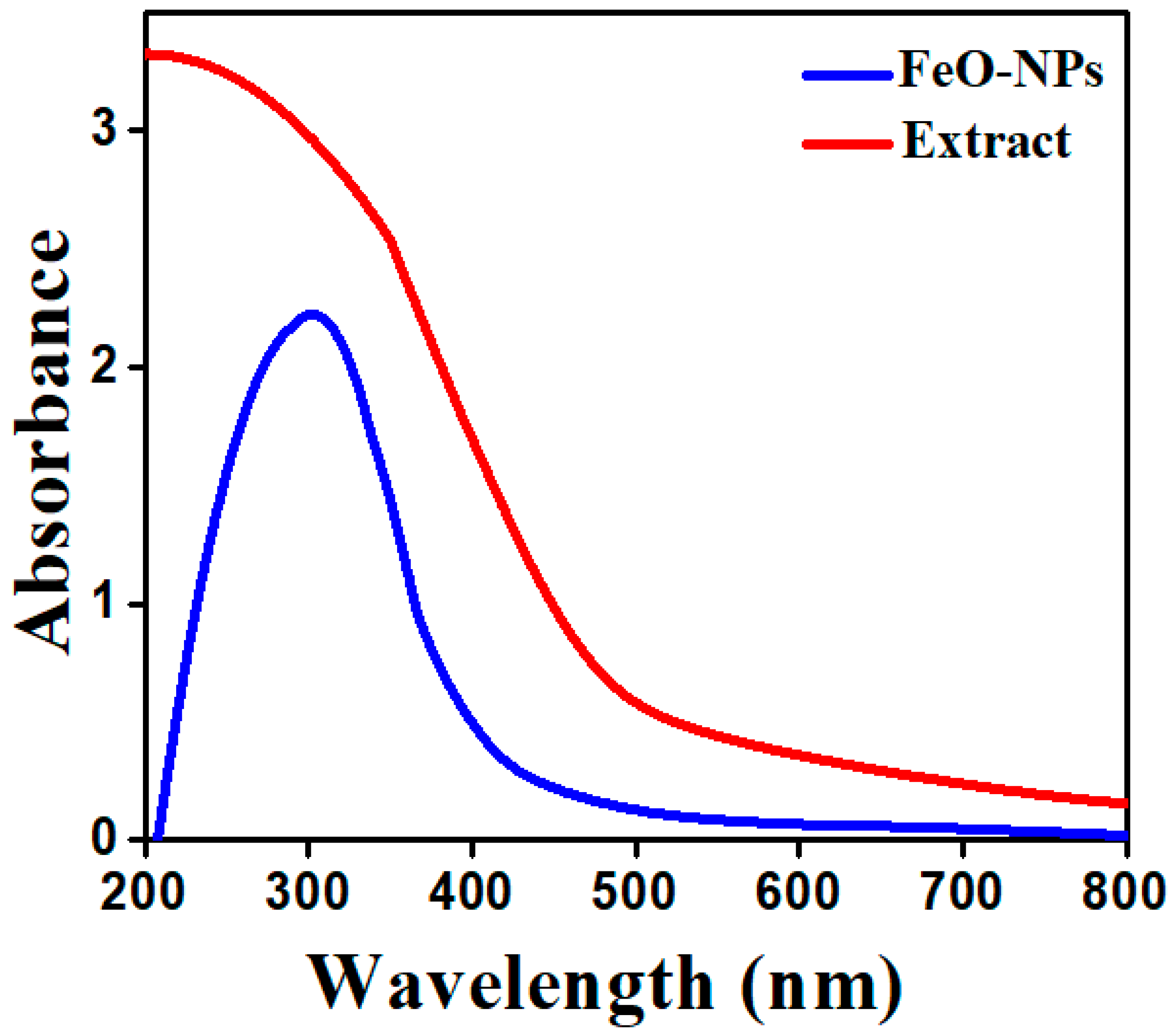
2.3.1. UV-Vis Spectroscopy
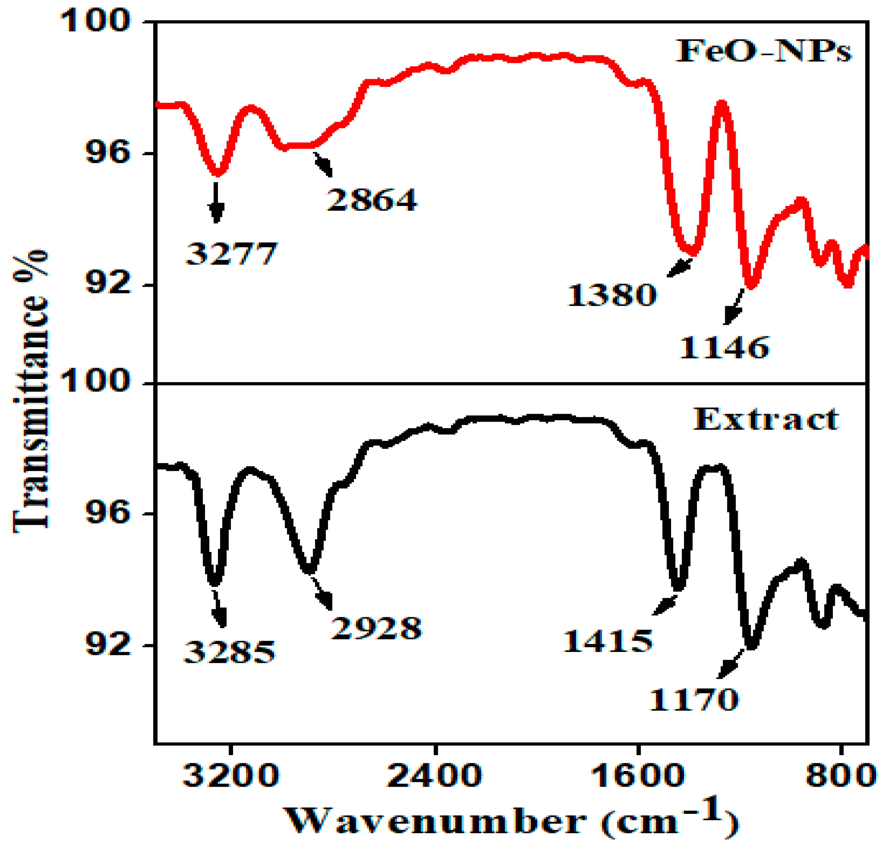
2.3.2. FT-IR Analysis
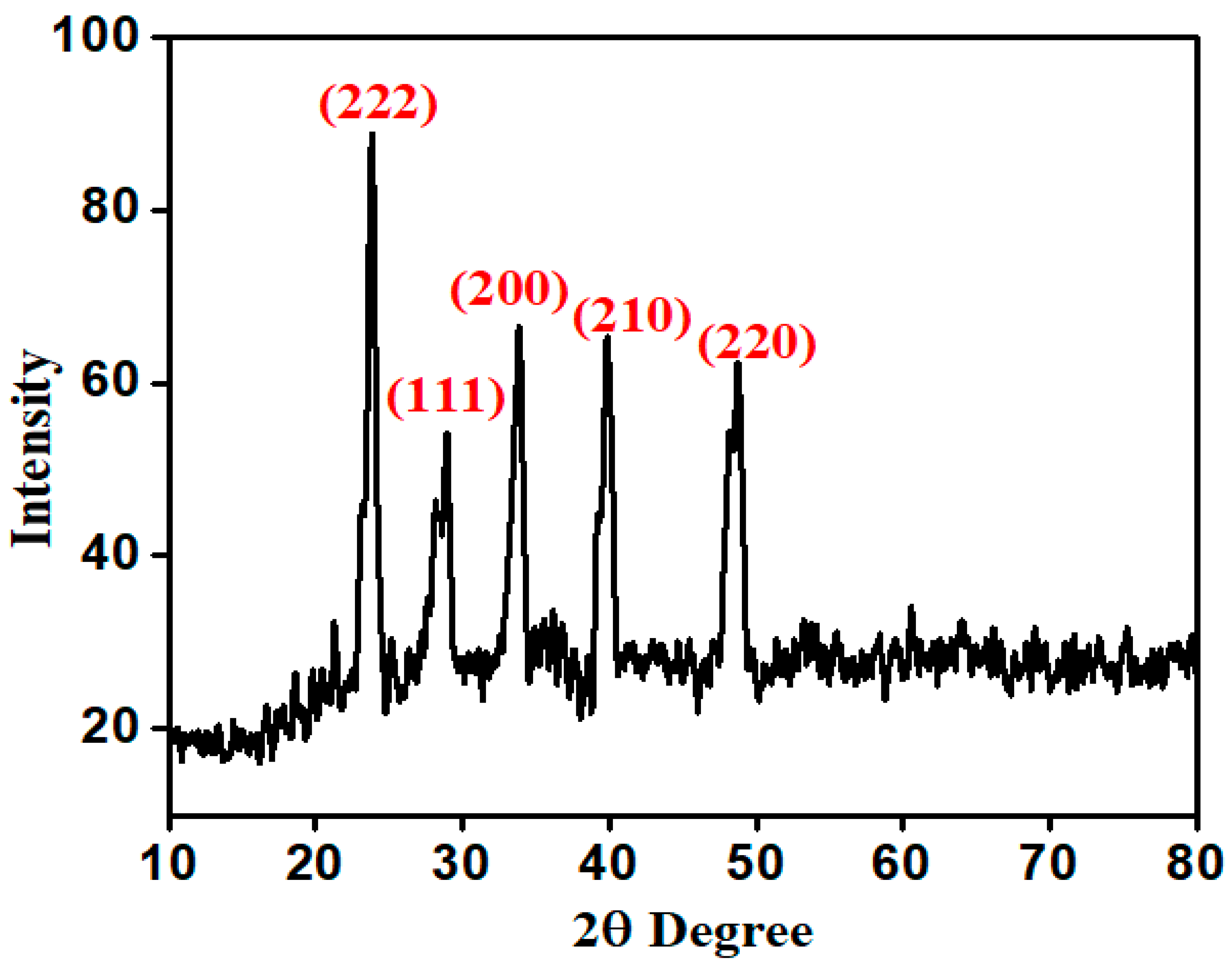
2.3.3. XRD Analysis
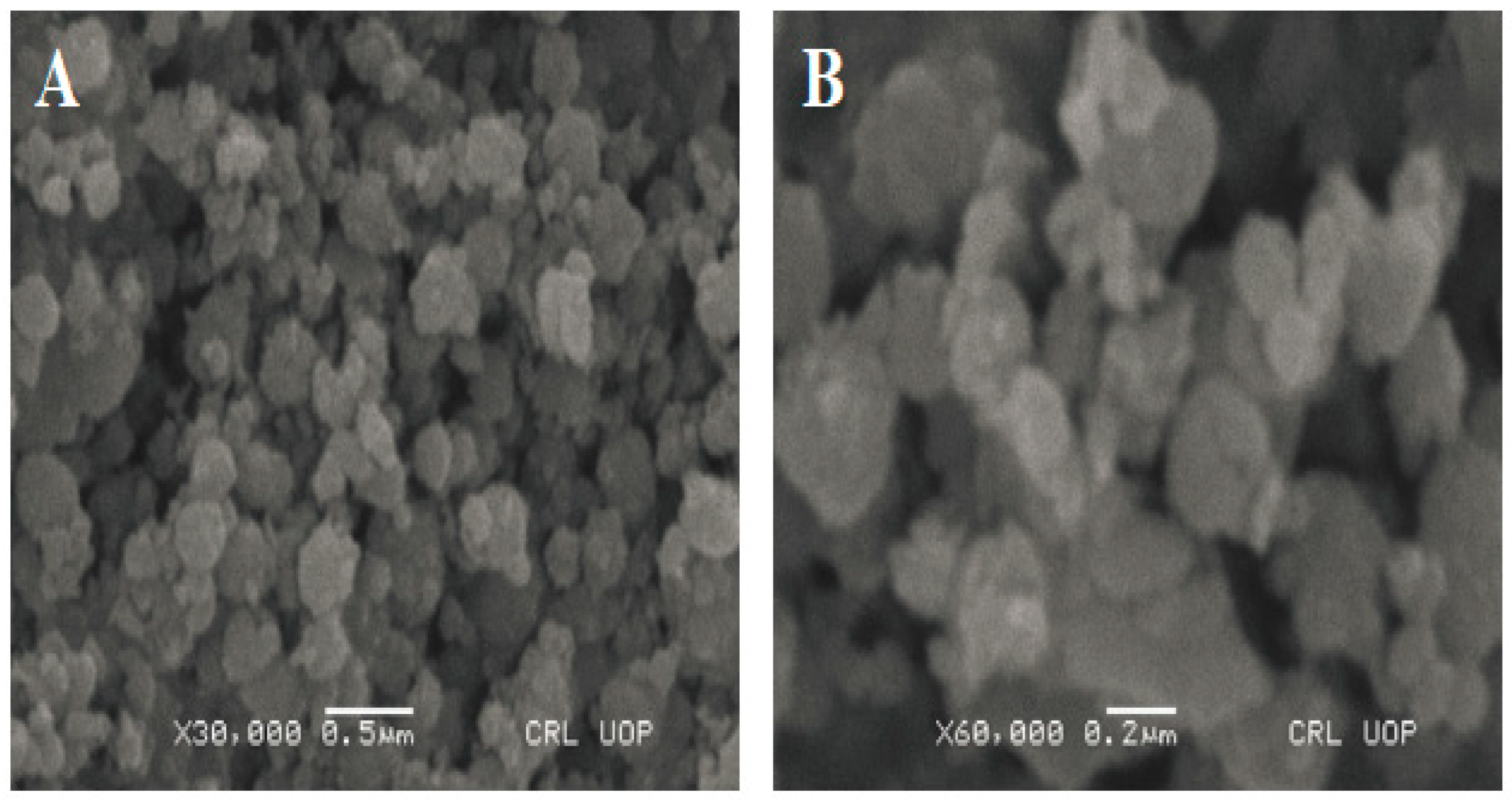
2.3.4. SEM Analysis
2.3.5. Energy Dispersive X-ray (EDX)
2.4. Antiplasmodial Activity
2.4.1. Sample Collection
2.4.2. Parasite Staining and Visualization
2.4.3. In Vitro Cultivation of Parasites
2.4.4. Drug Dilutions
2.4.5. Antiplasmodial Activity
2.5. Cytotoxic Activity of FeO NPs
Cytotoxic Evaluation
3. Results and Discussion
3.1. UV-Visible Analysis of FeO NPs
3.2. FT-IR Analysis of FeO NPs
3.3. XRD Pattern of FeO NPs
3.4. SEM Analysis of FeO NPs
3.5. EDX Analysis of FeO NPs
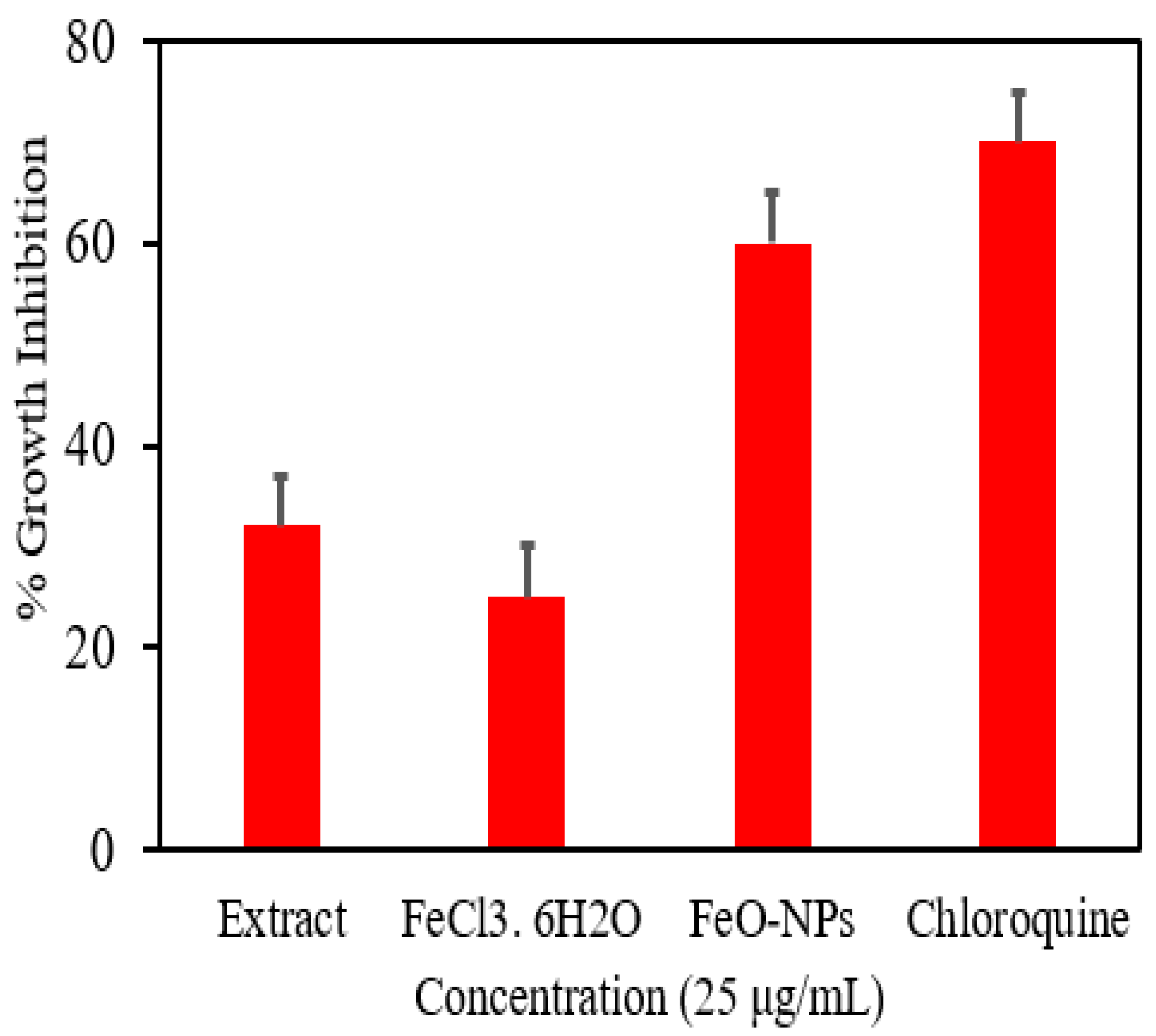
3.6. Antiplasmodial Evaluation
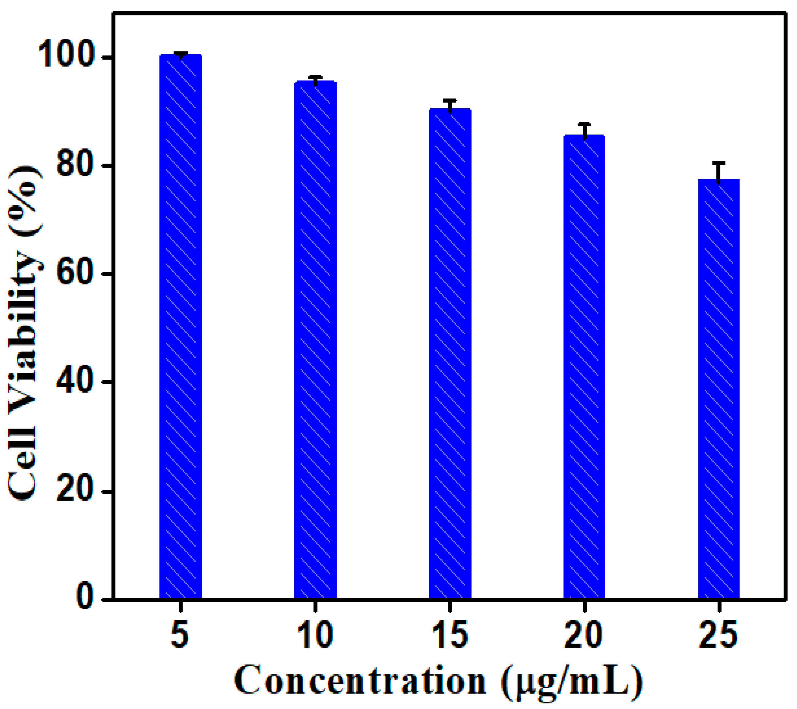
3.7. Biocompatibility Assessment
Cytotoxic Activity
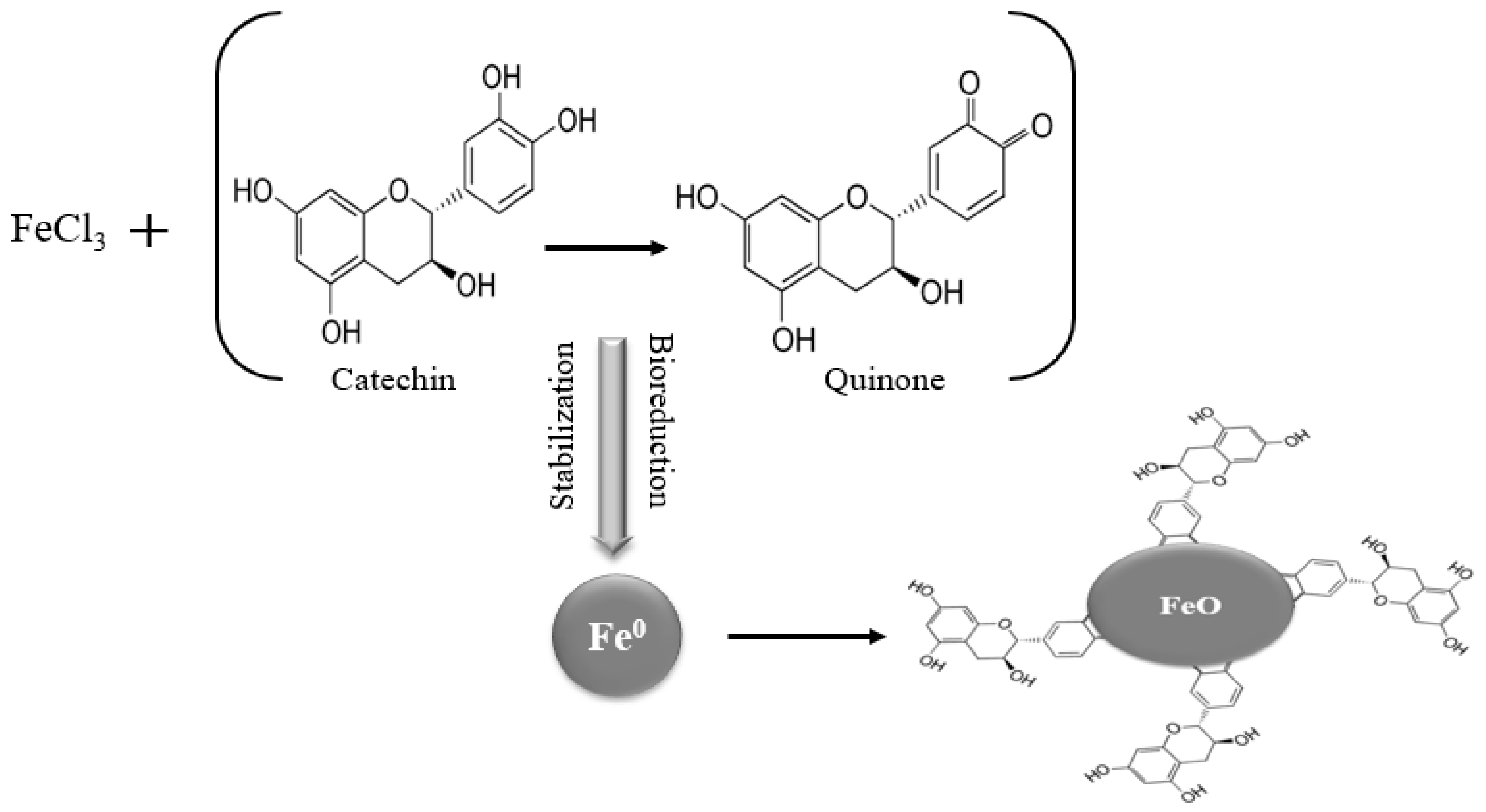
3.8. Proposed Mechanism for FeO NPs
4. Conclusions
Author Contributions
Funding
Institutional Review Board Statement
Informed Consent Statement
Data Availability Statement
Conflicts of Interest
References
- Fozia, F.; Ahmad, N.; Buoharee, Z.A.; Ahmad, I.; Aslam, M.; Wahab, A.; Ullah, R.; Ahmad, S.; Alotaibi, A.; Tariq, A. Characterization and Evaluation of Antimicrobial Potential of Trigonella incise (Linn) Mediated Biosynthesized Silver Nanoparticles. Molecules 2022, 27, 4618. [Google Scholar] [CrossRef] [PubMed]
- Faisal, S.; Abdullah; Rizwan, M.; Ullah, R.; Alotaibi, A.; Khattak, A.; Bibi, N.; Idrees, M. Paraclostridium benzoelyticum Bacterium-Mediated Zinc Oxide Nanoparticles and Their In Vivo Multiple Biological Applications. Oxid. Med. Cell. Longev. 2022, 2022, 5994033. [Google Scholar] [CrossRef] [PubMed]
- Ahmad, N.; Jabeen, M.; Haq, Z.U.; Ahmad, I.; Wahab, A.; Islam, Z.U.; Ullah, R.; Bari, A.; Abdel-Daim, M.M.; El-Demerdash, F.M.; et al. Green fabrication of silver nanoparticles using Euphorbia serpens Kunth aqueous extract, their characterization, and investigation of its in vitro antioxidative, antimicrobial, insecticidal, and cytotoxic activities. BioMed Res. Int. 2022, 2022, 5562849. [Google Scholar] [CrossRef] [PubMed]
- Aslam, M.; Fozia, F.; Gul, A.; Ahmad, I.; Ullah, R.; Bari, A.; Mothana, R.A.; Hussain, H. Phyto-extract-mediated synthesis of silver nanoparticles using aqueous extract of Sanvitalia procumbens, and characterization, optimization and photocatalytic degradation of azo dyes Orange G and Direct Blue-15. Molecules 2021, 26, 6144. [Google Scholar] [CrossRef] [PubMed]
- Gul, A.; Shaheen, A.; Ahmad, I.; Khattak, B.; Ahmad, M.; Ullah, R.; Bari, A.; Ali, S.S.; Alobaid, A.; Asmari, M.M.; et al. Green synthesis, characterization, enzyme inhibition, antimicrobial potential, and cytotoxic activity of plant mediated silver nanoparticle using Ricinus communis leaf and root extracts. Biomolecules 2021, 11, 206. [Google Scholar] [CrossRef]
- Iftikhar, M.; Zahoor, M.; Naz, S.; Nazir, N.; Batiha, G.E.-S.; Ullah, R.; Bari, A.; Hanif, M.; Mahmood, H.M. Green Synthesis of Silver Nanoparticles Using Grewia optiva Leaf Aqueous Extract and Isolated Compounds as Reducing Agent and Their Biological Activities. J. Nanomater. 2020, 2020, 8949674. [Google Scholar] [CrossRef]
- Bashir, A.K.H.; Furqan, C.M.; Bharuth-Ram, K.; Kaviyarasu, K.; Tchokonte, M.B.T.; Maaza, M. Structural, optical and Mossbauer investigation on the biosynthesized α-Fe2O3: Study on different precursors. Phys. E Low-Dimens. Syst. Nanostructures 2019, 111, 152–157. [Google Scholar] [CrossRef]
- Nguyen, N.H.; Padil, V.V.T.; Slaveykova, V.I.; Cernik, M.; Sevcu, A. Green synthesis of metal and metal oxide nanoparticles and their effect on the unicellular alga Chlamydomonas reinhardtii. Nanoscale Res. Lett. 2018, 13, 159. [Google Scholar] [CrossRef]
- Ali, A.; Zafar, M.Z.H.; Haq, I.u.; Phull, A.R.; Ali, J.S.; Hussain, A. Synthesis, characterization, applications, and challenges of iron oxide nanoparticles. Nanotechnol. Sci. Appl. 2016, 9, 49. [Google Scholar] [CrossRef] [Green Version]
- Song, C.; Sun, W.; Xiao, Y.; Shi, X. Ultrasmall iron oxide nanoparticles: Synthesis, surface modification, assembly, and biomedical applications. Drug Discov. Today 2019, 24, 835–844. [Google Scholar] [CrossRef]
- Dadfar, S.M.; Roemhild, K.; Drude, N.I.; Stillfried, S.; Knuchel, R.; Kiessling, F.; Lammers, T. Iron oxide nanoparticles: Diagnostic, therapeutic and theranostic applications. Adv. Drug Deliv. Rev. 2019, 138, 302–325. [Google Scholar] [CrossRef] [PubMed]
- El-Kassas, H.Y.; Aly-Eldeen, M.A.; Gharib, S.M. Green synthesis of iron oxide (Fe3O4) nanoparticles using two selected brown seaweeds: Characterization and application for lead bioremediation. Acta Oceanol. Sin. 2016, 35, 89–98. [Google Scholar] [CrossRef]
- Vangijzegem, T.; Stanicki, D.; Laurent, S. Magnetic iron oxide nanoparticles for drug delivery: Applications and characteristics. Expert Opin. Drug Deliv. 2019, 16, 69–78. [Google Scholar] [CrossRef] [PubMed]
- Wu, W.; He, Q.; Jiang, C. Magnetic iron oxide nanoparticles: Synthesis and surface functionalization strategies. Nanoscale Res. Lett. 2008, 3, 397–415. [Google Scholar] [CrossRef] [PubMed] [Green Version]
- Orsini, N.J.; Babic-Stojic, B.; Spasojevic, V.; Calatayud, M.P.; Cvjeticanin, N.; Goya, G.F. Magnetic and power absorption measurements on iron oxide nanoparticles synthesized by thermal decomposition of Fe (acac)3. J. Magn. Magn. Mater. 2018, 449, 286–296. [Google Scholar] [CrossRef]
- Patino-Ruiz, D.; Sanchez-Botero, L.; Hinestroza, J.; Herrera, A. Modification of Cotton Fibers with Magnetite and Magnetic Core-Shell Mesoporous Silica Nanoparticles. Phys. Status Solidi (a) 2018, 215, 1800266. [Google Scholar] [CrossRef]
- Atchudan, R.; Edison, T.N.J.I.; Perumal, S.; Ranjithkumar, D.; Lee, Y.R. Direct growth of iron oxide nanoparticles filled multi-walled carbon nanotube via chemical vapour deposition method as high-performance supercapacitors. Int. J. Hydrog. Energy 2019, 44, 2349–2360. [Google Scholar] [CrossRef]
- Vasantharaj, S.; Sathiyavimal, S.; Senthilkumar, P.; LewisOscar, F.; Pugazhendhi, A. Biosynthesis of iron oxide nanoparticles using leaf extract of Ruellia tuberosa antimicrobial properties and their applications in photocatalytic degradation. J. Photochem. Photobiol. B Biol. 2019, 192, 74–82. [Google Scholar] [CrossRef]
- Mirza, A.U.; Kareem, A.; Nami, S.A.; Khan, M.S.; Rehman, S.; Bhat, S.A.; Mohammad, A.; Nishat, N. Biogenic synthesis of iron oxide nanoparticles using Agrewia optiva and Prunus persica phyto species: Characterization, antibacterial and antioxidant activity. J. Photochem. Photobiol. B Biol. 2018, 185, 262–274. [Google Scholar] [CrossRef]
- Whitty, J.; Ansah, E. Malaria control stalls in high incidence areas. Bio. Med. J. 2019, 7, 365. [Google Scholar] [CrossRef] [Green Version]
- Somsak, V.; Borkaew, P.; Klubsri, C.; Dondee, K.; Bootprom, P.; Saiphet, B. Antimalarial properties of aqueous crude extracts of Gynostemma pentaphyllum and Moringa oleifera leaves in combination with artesunate in Plasmodium berghei-infected mice. J. Trop. Med. 2016, 2016, 6. [Google Scholar] [CrossRef] [PubMed] [Green Version]
- Adebayo, J.O.; Krettli, A.U. Potential antimalarials from Nigerian plants: A review. J. Ethnopharmacol. 2011, 133, 289–302. [Google Scholar] [CrossRef] [PubMed] [Green Version]
- Prasetyo, H.; Sulistyo, U.; Sadhana, I.P.; Amarwati, S.; Istiadi, H. The blocking effects of Nephrolepis exaltata on the sinonasal pathogenesis. In Proceedings of the 10th Asia Pasific IAP Congress, Bali, Indonesia, 24–27 April 2017; Volume 10, pp. 24–27. [Google Scholar]
- El-Tantawy, M.E.; Afifi, M.S.; Shams, M.M. Chemical composition, antimicrobial and cytotoxic activities of volatile constituents from the subterranean organs of Nephrolepis cordifolia (L.) C. Presl and Nephrolepis exaltata (L.) family Nephrolepidaceae grown in Egypt. Can. J. Pure Appl. Sci. 2015, 9, 3365–3370. [Google Scholar]
- Sharma, D.K.; Dave, R.S.; Shah, K.R. Phytochemical screening and characterization of volatile compounds by gas chromatography-mass spectrometry from “Nephrolepis exaltata”. Asian J. Pharm. Clin. Res. 2021, 14, 93–98. [Google Scholar] [CrossRef]
- Kanagasubbulakshmi, S.; Kadirvelu, K. Green synthesis of iron oxide nanoparticles using Lagenaria siceraria and evaluation of its antimicrobial activity. Def. Life Sci. J. 2017, 2, 422–427. [Google Scholar] [CrossRef]
- Panneerselvam, C.; Murugan, K.; Amerasan, D. Biosynthesis of silver nanoparticles using plant extract and its anti-plasmodial property. Adv. Mater. Res. 2015, 1086, 11–30. [Google Scholar] [CrossRef]
- Bagavan, A.; Rahuman, A.A.; Kamaraj, C.; Kaushik, N.K.; Mohanakrishnan, D.; Sahal, D. Sahal, Antiplasmodial activity of botanical extracts against Plasmodium falciparum. Parasitol. Res. 2011, 108, 1099–1109. [Google Scholar] [CrossRef]
- Sankar, V.; Cheeran, V.; Ganesh, M.R.; Sivakumar, B. Synthesis, Antibacterial and Anticancer Activity of 1, 2-Substituted 2, 3-Dihydro-1H-Benzo [4, 5] Imidazo [1, 2] [1, 3, 2] Diazaphosphol-1-Oxides. Pharm. Chem. J. 2020, 54, 827–833. [Google Scholar] [CrossRef]
- Mahdavi, M.; Namvar, F.; Ahmad, M.B.; Mohamad, R. Green biosynthesis and characterization of magnetic iron oxide (Fe3O4) nanoparticles using seaweed (Sargassum muticum) aqueous extract. Molecules 2013, 18, 5954–5964. [Google Scholar] [CrossRef]
- Yang, Y.; Yan, H. A green and facile approach for synthesis of magnetite nanoparticles with tunable sizes and morphologies. Mater. Lett. 2012, 73, 129–132. [Google Scholar] [CrossRef]
- Bishnoi, S.; Kumar, A.; Selvaraj, R. Facile synthesis of magnetic iron oxide nanoparticles using inedible Cynometra Ramiflora fruit extract waste and their photocatalytic degradation of methylene blue dye. Mater. Res. Bull. 2018, 97, 121–127. [Google Scholar] [CrossRef]
- Mandel, K.; Kolb, C.; Straber, M.; Dembski, S.; Sext, G. Size controlled iron oxide nano octahedra obtained via sonochemistry and natural ageing. Colloids Surf. A Physicochem. Eng. Asp. 2014, 457, 27–32. [Google Scholar] [CrossRef]
- Arokiyaraj, S.; Saravanan, M.; Prakash, N.U.; Arasu, M.V.; Vijayakumar, B.; Vincent, S. enhanced antibacterial activity of iron oxide magnetic nanoparticles treated with Argemone mexicana L. leaf extract: An in vitro study. Mater. Res. Bull. 2013, 48, 3323–3327. [Google Scholar] [CrossRef]
- Abid, M.A.; Kadhim, D.A. Novel comparison of iron oxide nanoparticle preparation by mixing iron chloride with henna leaf extract with and without applied pulsed laser ablation for methylene blue degradation. J. Environ. Chem. Eng. 2020, 8, 104138. [Google Scholar] [CrossRef]
- Prabu, H.J.; Johnson, I. Plant-mediated biosynthesis and characterization of silver nanoparticles by leaf extracts of Tragia involucrata, Cymbopogon citronella, Solanum verbascifolium and Tylophora ovata. Karbala Int. J. Mod. Sci. 2015, 1, 237–246. [Google Scholar] [CrossRef] [Green Version]
- Bibi, I.; Nazar, N.; Ata, S.; Sultan, M.; Ali, A.; Abbas, A.; Jilani, K.; Kamal, S.; Sarim, F.M.; Khan, M.I.; et al. Green synthesis of iron oxide nanoparticles using pomegranate seeds extract and photocatalytic activity evaluation for the degradation of textile dye. J. Mater. Res. Technol. 2019, 8, 6115–6124. [Google Scholar] [CrossRef]
- Ponarulselvam, S.; Panneerselvam, C.; Murugan, K.; Aarthi, N.; Kalimuthu, K.; Thangamani, S. Synthesis of silver nanoparticles using leaves of Catharanthus roseus Linn. G. Don and their antiplasmodial activities. Asian Pac. J. Trop. Biomed. 2012, 2, 574–580. [Google Scholar] [CrossRef] [Green Version]
- Sengul, A.B.; Asmatulu, E. Toxicity of metal and metal oxide nanoparticles: A review. Environ. Chem. Lett. 2020, 18, 1659–1683. [Google Scholar] [CrossRef]
- Bendale, Y.; Bendale, V.; Paul, S. Evaluation of cytotoxic activity of platinum nanoparticles against normal and cancer cells and its anticancer potential through induction of apoptosis. Integr. Med. Res. 2017, 6, 141–148. [Google Scholar] [CrossRef]
- Lopez-Badillo, C.M.; Hernandez-Gonzalez, M.; Hernandez-Centeno, F.; Olivas-Armendariz, I.; Rodriguez-Gonzalez, C.A.; Muzquiz-Ramos, E.M.; Lopez-Cuevas, J.; la Pena, H.Y.L.-D. Antibacterial activity and in vitro cytotoxicity studies of Ag-doped CaO nanoparticles. Mater. Lett. 2021, 283, 128741. [Google Scholar] [CrossRef]
- Zangeneh, A.; Zangeneh, M.M.; Moradi, R. Ethnomedicinal plant-extract-assisted green synthesis of iron nanoparticles using Allium saralicum extract, and their antioxidant, cytotoxicity, antibacterial, antifungal and cutaneous wound-healing activities. Appl. Organomet. Chem. 2020, 34, 5247. [Google Scholar] [CrossRef]
- Xiao, L.; Mertens, M.; Wortmann, L.; Kremer, S.; Valldor, M.; Lammers, T.; Kiessling, F.; Mathur, S. Enhanced in vitro and in vivo cellular imaging with green tea coated water-soluble iron oxide nanocrystals. ACS Appl. Mater. Interfaces 2015, 7, 6530–6540. [Google Scholar] [CrossRef] [PubMed]


| S. No | Test Samples | Test Sample Concentration (μg/mL) | Parasite Inhibitory Concentration (%) |
|---|---|---|---|
| 1 | FeCl3·6H2O | 25 | 23 ± 0.3 |
| 2 | Plant extract N. exaltata | 25 | 35 ± 0.6 |
| 3 | FeO NPs | 25 | 62 ± 1.3 |
| 4 | Control | 25 | 70 ± 0.32 |
Publisher’s Note: MDPI stays neutral with regard to jurisdictional claims in published maps and institutional affiliations. |
© 2022 by the authors. Licensee MDPI, Basel, Switzerland. This article is an open access article distributed under the terms and conditions of the Creative Commons Attribution (CC BY) license (https://creativecommons.org/licenses/by/4.0/).
Share and Cite
Nadeem, F.; Fozia, F.; Aslam, M.; Ahmad, I.; Ahmad, S.; Ullah, R.; Almutairi, M.H.; Aleya, L.; Abdel-Daim, M.M. Characterization, Antiplasmodial and Cytotoxic Activities of Green Synthesized Iron Oxide Nanoparticles Using Nephrolepis exaltata Aqueous Extract. Molecules 2022, 27, 4931. https://0-doi-org.brum.beds.ac.uk/10.3390/molecules27154931
Nadeem F, Fozia F, Aslam M, Ahmad I, Ahmad S, Ullah R, Almutairi MH, Aleya L, Abdel-Daim MM. Characterization, Antiplasmodial and Cytotoxic Activities of Green Synthesized Iron Oxide Nanoparticles Using Nephrolepis exaltata Aqueous Extract. Molecules. 2022; 27(15):4931. https://0-doi-org.brum.beds.ac.uk/10.3390/molecules27154931
Chicago/Turabian StyleNadeem, Faisal, Fozia Fozia, Madeeha Aslam, Ijaz Ahmad, Shakeel Ahmad, Riaz Ullah, Mikhlid H. Almutairi, Lotfi Aleya, and Mohamed M. Abdel-Daim. 2022. "Characterization, Antiplasmodial and Cytotoxic Activities of Green Synthesized Iron Oxide Nanoparticles Using Nephrolepis exaltata Aqueous Extract" Molecules 27, no. 15: 4931. https://0-doi-org.brum.beds.ac.uk/10.3390/molecules27154931