
In Vitro and Human Pilot Studies of Different Topical Formulations Containing Rosa Species for the Treatment of Psoriasis
 , ,
, ,  ,
, 
 , , ,
, , ,  , , and
Abstract
:
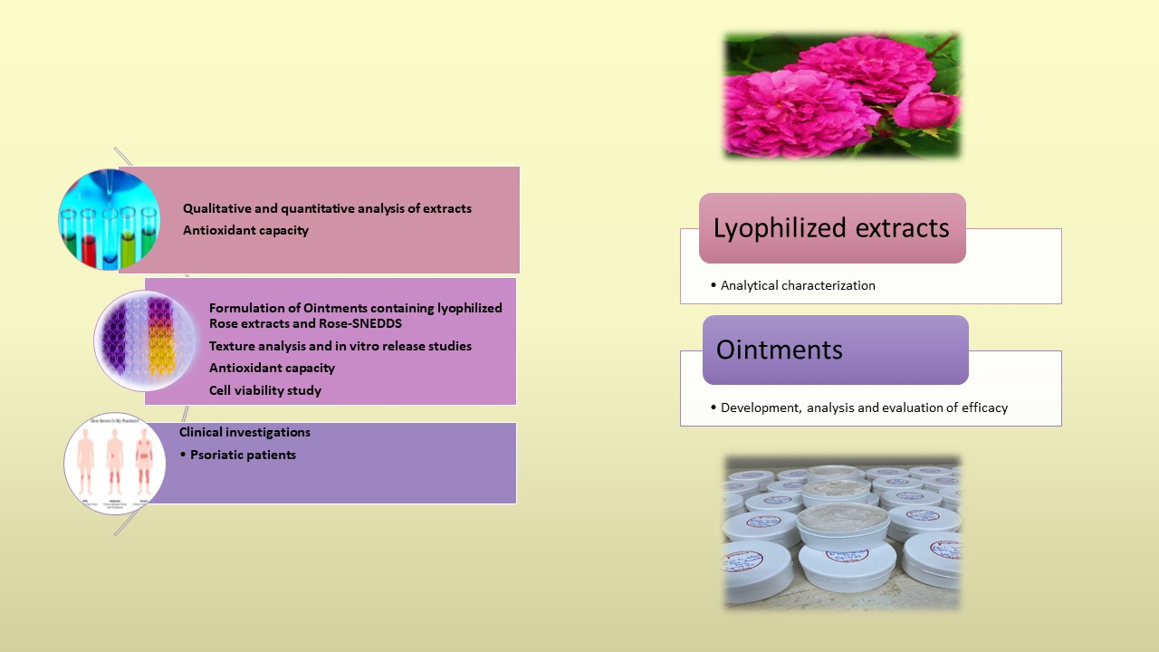
1. Introduction
2. Results
2.1. Determination of the Components and the Antioxidant Capacity of the Extracts
2.2. Investigation of Self-Nano-Emulsifying Drug Delivery System
2.3. Ointment Formulation
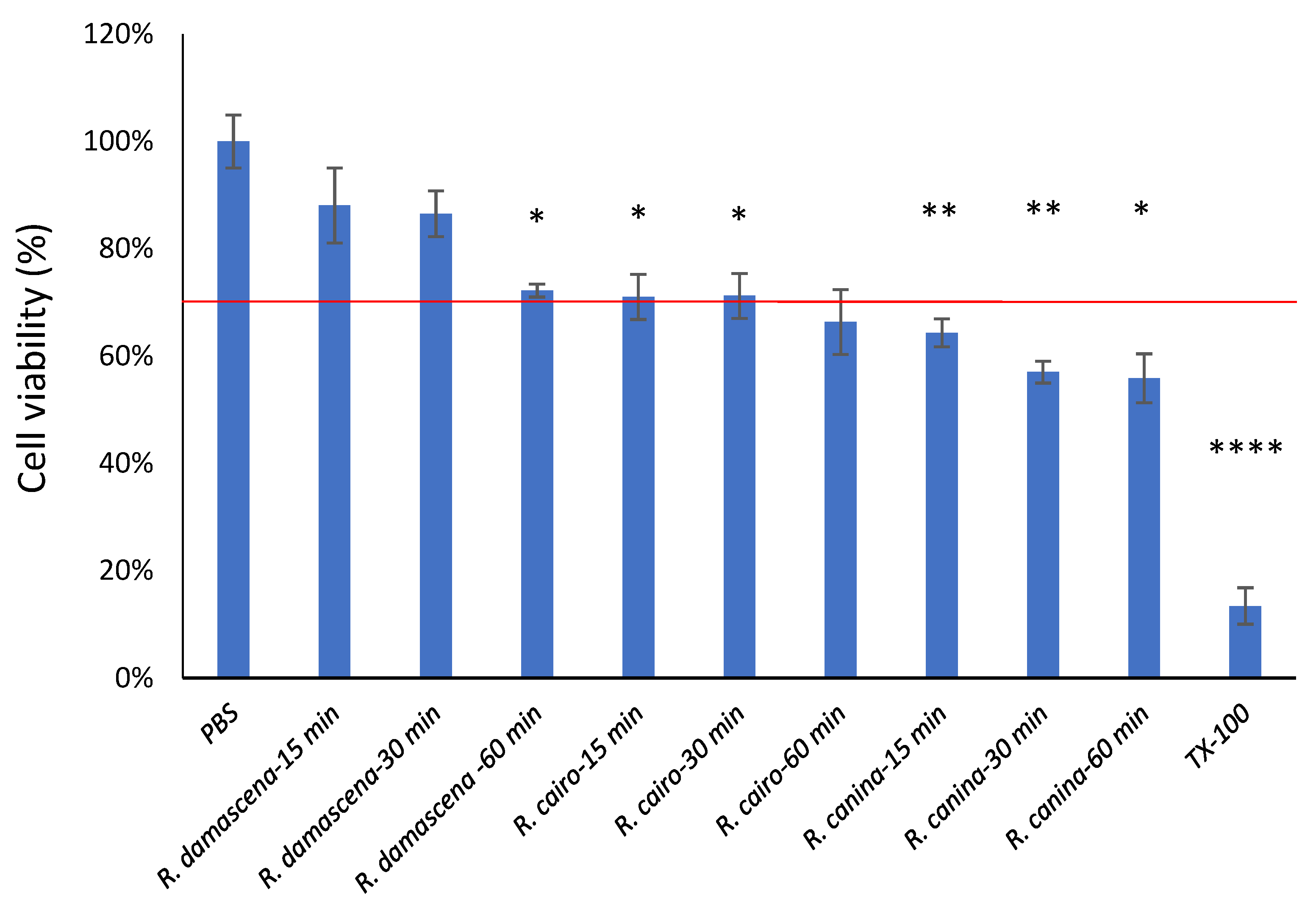
2.4. Cell Viability Study (MTT Assay)
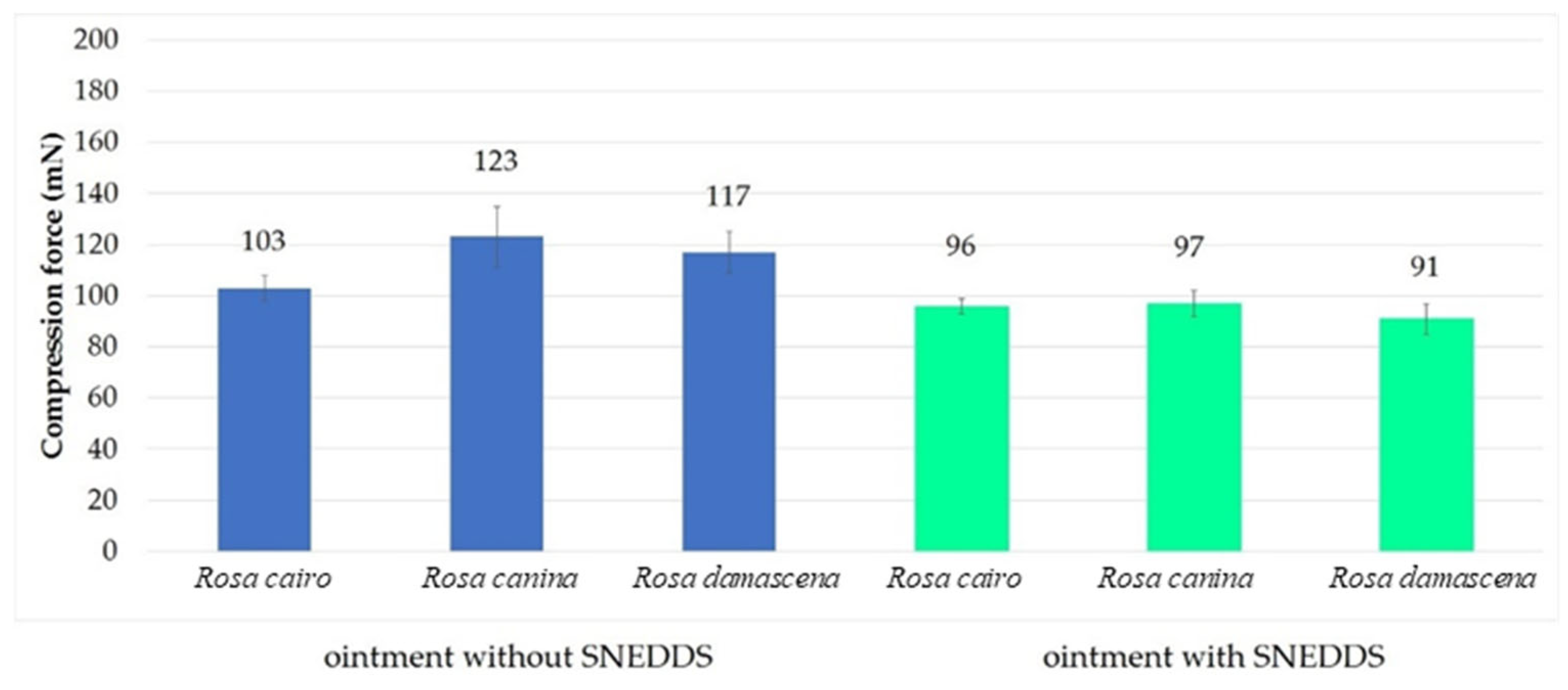
2.5. Texture Analysis
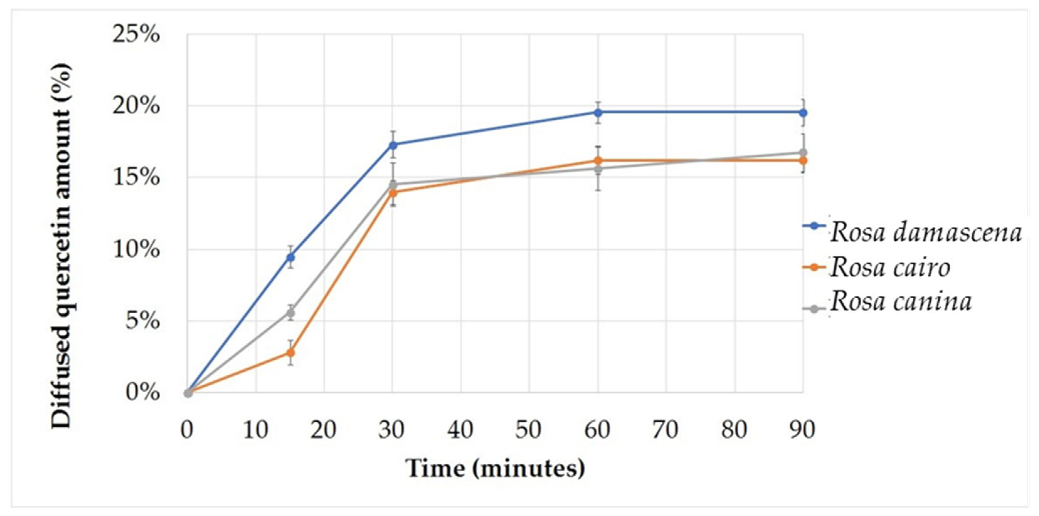
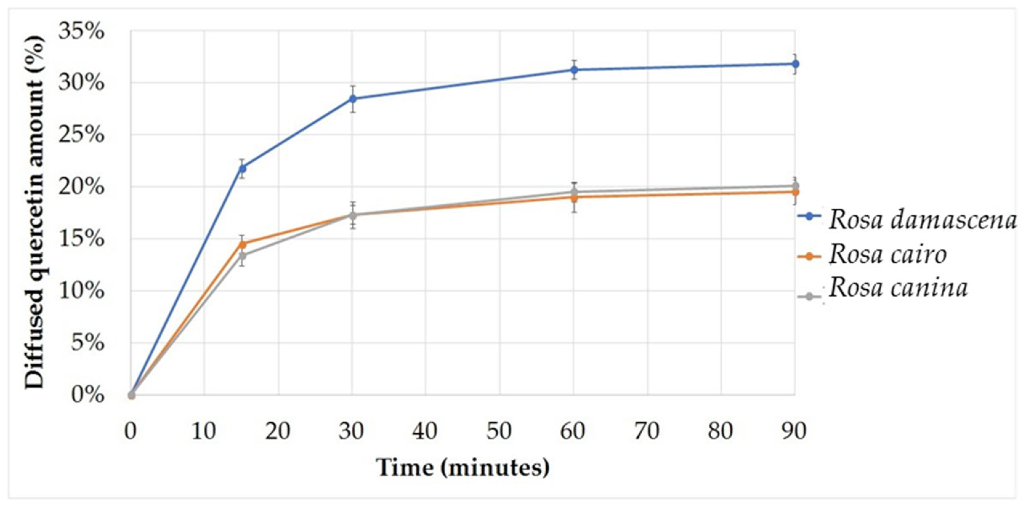
2.6. In Vitro Release Studies
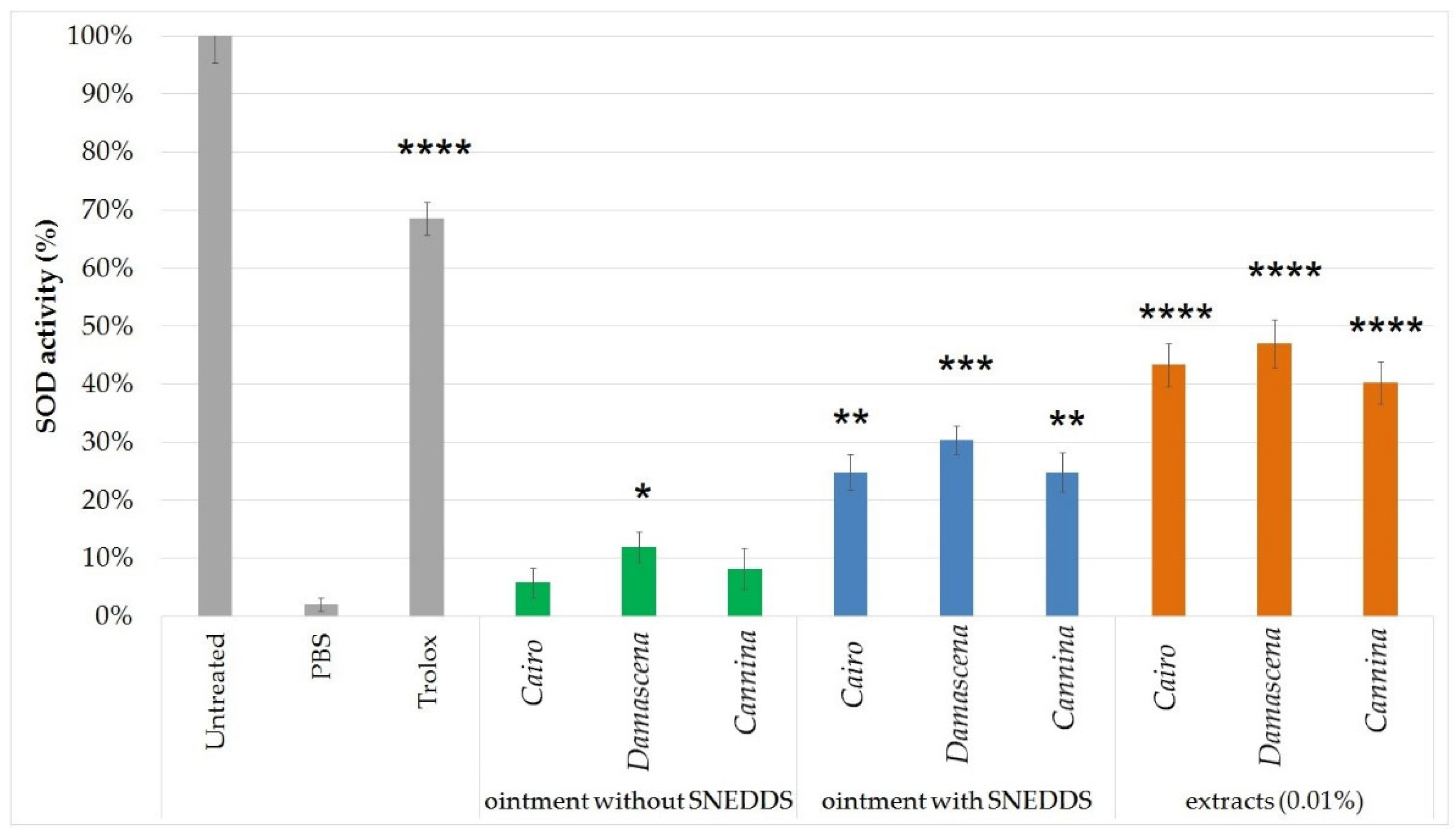
2.7. Superoxide Dismutase Activity of the Topical Formulations
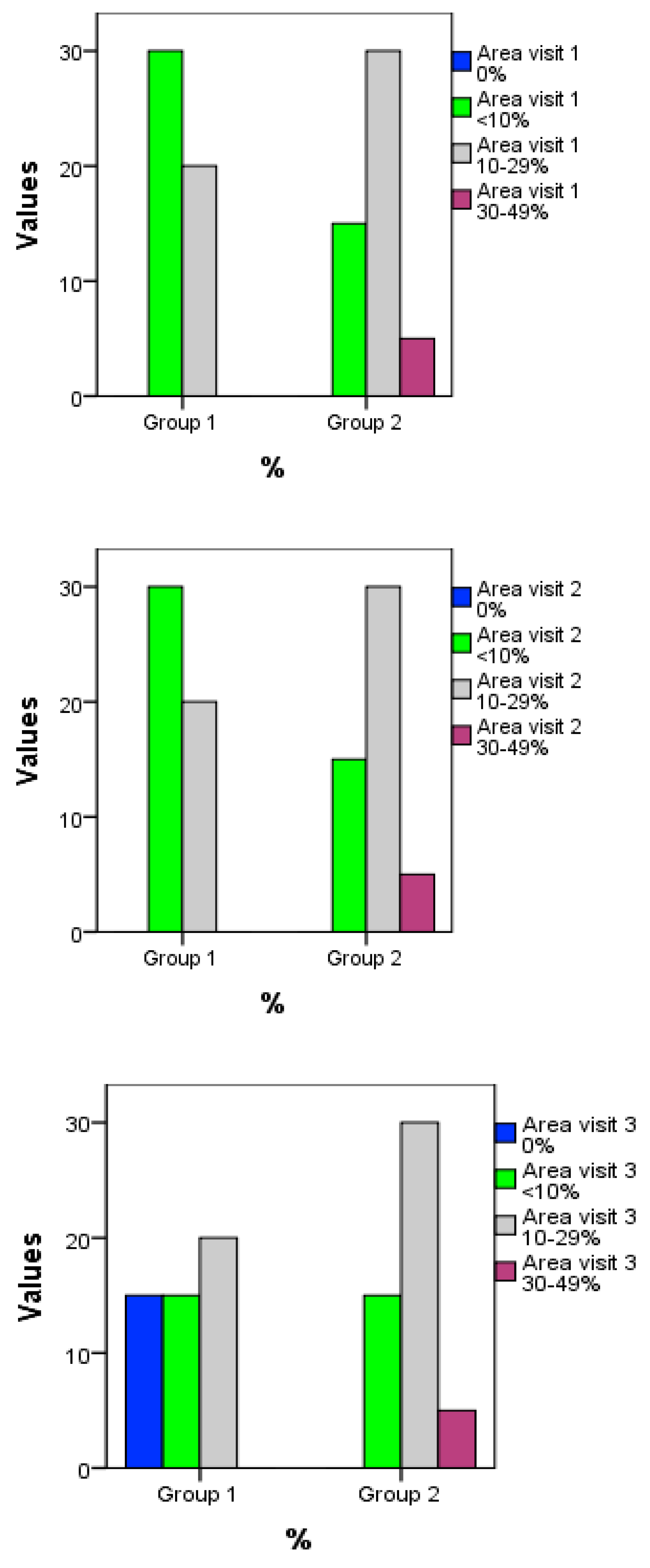
2.8. Clinical Investigation
3. Discussion
4. Materials and Methods
4.1. Preparation and Characterization of Dry Extract
4.2. Formulation and Investigation of Self-Nanoemulsifying Drug Delivery System
4.3. Formulation of Ointments Containing Lyophilized Rose Extracts and Rose-SNEDDS
4.4. Texture Analysis
4.5. In Vitro Release Studies
4.6. Superoxide Dismutase (SOD) Assay
4.7. Cell Viability Study (MTT Assay)
4.8. Clinical Study
5. Conclusions
Author Contributions
Funding
Institutional Review Board Statement
Informed Consent Statement
Data Availability Statement
Conflicts of Interest
References
- Cragg, G.M.; Newman, D.J. Natural Products: A Continuing Source of Novel Drug Leads. Biochim. Biophys. Acta—Gen. Subj. 2013, 1830, 3670–3695. [Google Scholar] [CrossRef] [PubMed]
- Schieber, A.; Mihalev, K.; Berardini, N.; Mollov, P.; Carle, R. Flavonol Glycosides from Distilled Petals of Rosa Damascena Mill. Z. Nat. C 2005, 60, 379–384. [Google Scholar] [CrossRef] [PubMed]
- Cai, Y.-Z.; Xing, J.; Sun, M.; Zhan, Z.-Q.; Corke, H. Phenolic Antioxidants (Hydrolyzable Tannins, Flavonols, and Anthocyanins) Identified by LC-ESI-MS and MALDI-QIT-TOF MS from Rosa Chinensis Flowers. J. Agric. Food Chem. 2005, 53, 9940–9948. [Google Scholar] [CrossRef] [PubMed]
- Boskabady, M.H.; Shafei, M.N.; Saberi, Z.; Amini, S. Pharmacological Effects of Rosa Damascena. Iran. J. Basic Med. Sci. 2011, 14, 295–307. [Google Scholar]
- Shirazi, M.; Mohebitabar, S.; Bioos, S.; Yekaninejad, M.S.; Rahimi, R.; Shahpiri, Z.; Malekshahi, F.; Nejatbakhsh, F. The Effect of Topical Rosa Damascena (Rose) Oil on Pregnancy-Related Low Back Pain. J. Evid. Based. Complement. Altern. Med. 2017, 22, 120–126. [Google Scholar] [CrossRef]
- Kumar, N.; Bhandari, P.; Singh, B.; Bari, S.S. Antioxidant Activity and Ultra-Performance LC-Electrospray Ionization-Quadrupole Time-of-Flight Mass Spectrometry for Phenolics-Based Fingerprinting of Rose Species: Rosa Damascena, Rosa Bourboniana and Rosa Brunonii. Food Chem. Toxicol. 2009, 47, 361–367. [Google Scholar] [CrossRef]
- Tofighi, Z.; Molazem, M.; Doostdar, B.; Taban, P.; Shahverdi, A.R.; Samadi, N.; Yassa, N. Antimicrobial Activities of Three Medicinal Plants and Investigation of Flavonoids of Tripleurospermum Disciforme. Iran. J. Pharm. Res. IJPR 2015, 14, 225–231. [Google Scholar]
- Ashrafzadeh, F.; Rakhshandeh, H.; Mahmodi, E.; Ashrafzadeh, F.; Rakhshandeh, H.; Mahmodi, E. Rosa Damascena Oil: An Adjunctive Therapy for Pediatric Refractory Seizures. Iran. J. Child Neurol. 2007, 1, 13–17. [Google Scholar]
- Hosseini, M. Effects of Different Extracts of Eugenia Caryophyllata on Pentylenetetrazole-Induced Seizures in Mice. J. Chin. Integr. Med. 2012, 10, 1476–1481. [Google Scholar] [CrossRef]
- Biswas, N.R.; Gupta, S.K.; Das, G.K.; Kumar, N.; Mongre, P.K.; Haldar, D.; Beri, S. Evaluation of Ophthacare Eye Drops? A Herbal Formulation in the Management of Various Ophthalmic Disorders. Phyther. Res. 2001, 15, 618–620. [Google Scholar] [CrossRef]
- Nestle, F.O.; Kaplan, D.H.; Barker, J. Psoriasis. N. Engl. J. Med. 2009, 361, 496–509. [Google Scholar] [CrossRef] [PubMed]
- Rendon, A.; Schäkel, K. Psoriasis Pathogenesis and Treatment. Int. J. Mol. Sci. 2019, 20, 1475. [Google Scholar] [CrossRef] [PubMed]
- Boehncke, W.H.; Schön, M.P. Psoriasis. Lancet 2015, 386, 983–994. [Google Scholar] [CrossRef]
- Man, M.-Q.; Ye, L.; Hu, L.; Jeong, S.; Elias, P.M.; Lv, C. Improvements in Epidermal Function Prevent Relapse of Psoriasis: A Self-controlled Study. Clin. Exp. Dermatol. 2019, 44, 654–657. [Google Scholar] [CrossRef]
- Herman, A.; Herman, A. Topically Used Herbal Products for the Treatment of Psoriasis—Mechanism of Action, Drug Delivery, Clinical Studies. Planta Med. 2016, 82, 1447–1455. [Google Scholar] [CrossRef]
- Bungau, S.; Vesa, C.M.; Abid, A.; Behl, T.; Tit, D.M.; Purza, A.L.; Pasca, B.; Todan, L.M.; Endres, L. Withaferin A—A Promising Phytochemical Compound with Multiple Results in Dermatological Diseases. Molecules 2021, 26, 2407. [Google Scholar] [CrossRef]
- Gavra, D.I.; Marian, E.; Pallag, A.; Vicaș, L.G.; Lucaciu, R.L.; Micle, O.; Ionescu, C.; Bacskay, I.; Hangan, A.C.; Sevastre, B.; et al. Phytochemical Screening and Biological Activity of Ethanolic Extract of Rosa X Damascena Mill. Cultivated in the Western Region of Romania. Farmacia 2022, 70, 248–257. [Google Scholar] [CrossRef]
- Gutteridge, J.M.C.; Halliwell, B. Free Radicals and Antioxidants in the Year 2000: A Historical Look to the Future. Ann. N. Y. Acad. Sci. 2006, 899, 136–147. [Google Scholar] [CrossRef]
- Kazaz, S.; Baydar, H.; Erbas, S. Variations in Chemical Compositions of Rosa Damascena Mill. and Rosa canina L. Fruits. Czech J. Food Sci. 2009, 27, 178–184. [Google Scholar] [CrossRef]
- Fetni, S.; Bertella, N.; Ouahab, A.; Martinez Zapater, J.M.; De Pascual-Teresa Fernandez, S. Composition and Biological Activity of the Algerian Plant Rosa canina L. by HPLC-UV-MS. Arab. J. Chem. 2020, 13, 1105–1119. [Google Scholar] [CrossRef]
- Kerasioti, E.; Apostolou, A.; Kafantaris, I.; Chronis, K.; Kokka, E.; Dimitriadou, C.; Tzanetou, E.N.; Priftis, A.; Koulocheri, S.D.; Haroutounian, S.A.; et al. Polyphenolic Composition of Rosa canina, Rosa Sempervivens and Pyrocantha Coccinea Extracts and Assessment of Their Antioxidant Activity in Human Endothelial Cells. Antioxidants 2019, 8, 92. [Google Scholar] [CrossRef] [PubMed]
- Ayati, Z.; Amiri, M.S.; Ramezani, M.; Delshad, E.; Sahebkar, A.; Emami, S.A. Phytochemistry, Traditional Uses and Pharmacological Profile of Rose Hip: A Review. Curr. Pharm. Des. 2019, 24, 4101–4124. [Google Scholar] [CrossRef] [PubMed]
- Schieber, A.; Berardini, N.; Carle, R. Identification of Flavonol and Xanthone Glycosides from Mango (Mangifera indica L. Cv. “Tommy Atkins”) Peels by High-Performance Liquid Chromatography-Electrospray Ionization Mass Spectrometry. J. Agric. Food Chem. 2003, 51, 5006–5011. [Google Scholar] [CrossRef] [PubMed]
- Guo, X.-D.; Zhang, D.-Y.; Gao, X.-J.; Parry, J.; Liu, K.; Liu, B.-L.; Wang, M. Quercetin and Quercetin-3-O-Glucuronide Are Equally Effective in Ameliorating Endothelial Insulin Resistance through Inhibition of Reactive Oxygen Species-Associated Inflammation. Mol. Nutr. Food Res. 2013, 57, 1037–1045. [Google Scholar] [CrossRef]
- Fujii, T.; Saito, M. Inhibitory Effect of Quercetin Isolated from Rose Hip (Rosa canina L.) against Melanogenesis by Mouse Melanoma Cells. Biosci. Biotechnol. Biochem. 2009, 73, 1989–1993. [Google Scholar] [CrossRef]
- Taneva, I.; Petkova, N.; Dimov, I.; Ivanov, I.; Denev, P. Characterization of Rose Hip (Rosa canina L.) Fruits Extracts and Evaluation of Their in Vitro Antioxidant Activity. J. Pharmacogn. Phytochem. 2016, 5, 35–38. [Google Scholar]
- Gavra, D.I.; Pallag, A.; Marian, E.; Vicaș, L.J.T. A Comparative Study of the Amount of Polyphenols, Flavonoids and Antioxidant Capacity of Rosae Caninae Flos versus Cynosbati Fructus. Ann. Univ. Oradea Fascicle Environ. Prot. 2018, 31, 23–30. [Google Scholar]
- Glevitzky, I.; Dumitrel, G.A.; Glevitzky, M.; Pasca, B.; Otrisal, P.; Bungau, S.; Cioca, G.; Pantis, C.; Popa, M. Statistical Analysis of the Relationship between Antioxidant Activity and the Structure of Flavonoid Compounds. Rev. Chim. 2019, 70, 3103–3107. [Google Scholar] [CrossRef]
- Mileva, M.; Kusovaski, V.; Krastev, D.; Dobreva, A.; Galabov, A. Chemical Composition, In Vitro Antiradical and Antimicrobial Activities of Bulgarian Rosa Alba L. Essential Oil against Some Oral Pathogens. Int. J. Curr. Microbiol. App. Sci. 2014, 3, 11–20. [Google Scholar]
- Dehghan Kashani, A.; Rasooli, I.; Rezaee, M.B.; Owlia, P. Antioxidative Properties and Toxicity of White Rose Extract. Iran. J. Toxicol. 2011, 5, 415–425. [Google Scholar]
- Tofighi, Z.; Alipour, F.; Yassa, N.; Hadjiakhoondi, A.; Hadavinia, H.; Goodarzy, S.; Golestani, R. Chemical Composition and Antioxidant Activity of Otostegia Persica Essential Oil from Iran. Int. J. Essent. Oil Ther. 2009, 3, 45–48. [Google Scholar]
- Achuthan, C.R.; Babu, B.H.; Padikkala, J. Antioxidant and Hepatoprotective Effects of Rosa Damascena. Pharm. Biol. 2003, 41, 357–361. [Google Scholar] [CrossRef]
- Nazari-Vanani, R.; Azarpira, N.; Heli, H. Development of Self-Nanoemulsifying Drugdelivery Systems for Oil Extracts of Citrus Aurantium L. Blossoms and Rose Damascena and Evaluation of Anticancer Properties. J. Drug Deliv. Sci. Technol. 2018, 47, 330–336. [Google Scholar] [CrossRef]
- Nazari-Vanani, R.; Azarpira, N.; Heli, H.; Karimian, K.; Sattarahmady, N. A Novel Self-Nanoemulsifying Formulation for Sunitinib: Evaluation of Anticancer Efficacy. Colloids Surf. B Biointerfaces 2017, 160, 65–72. [Google Scholar] [CrossRef] [PubMed]
- Nazari-Vanani, R.; Moezi, L.; Heli, H. In Vivo Evaluation of a Self-Nanoemulsifying Drug Delivery System for Curcumin. Biomed. Pharmacother. 2017, 88, 715–720. [Google Scholar] [CrossRef] [PubMed]
- Neslihan Gursoy, R.; Benita, S. Self-Emulsifying Drug Delivery Systems (SEDDS) for Improved Oral Delivery of Lipophilic Drugs. Biomed. Pharmacother. 2004, 58, 173–182. [Google Scholar] [CrossRef]
- Rebello, S.; Asok, A.K.; Mundayoor, S.; Jisha, M.S. Surfactants: Toxicity, Remediation and Green Surfactants. Environ. Chem. Lett. 2014, 12, 275–287. [Google Scholar] [CrossRef]
- Shafiq, S.; Shakeel, F.; Talegaonkar, S.; Ahmad, F.J.; Khar, R.K.; Ali, M. Development and Bioavailability Assessment of Ramipril Nanoemulsion Formulation. Eur. J. Pharm. Biopharm. 2007, 66, 227–243. [Google Scholar] [CrossRef]
- Smail, S.S.; Ghareeb, M.M.; Omer, H.K.; Al-Kinani, A.A.; Alany, R.G. Studies on Surfactants, Cosurfactants, and Oils for Prospective Use in Formulation of Ketorolac Tromethamine Ophthalmic Nanoemulsions. Pharmaceutics 2021, 13, 467. [Google Scholar] [CrossRef]
- Luu, M.T.; Mercier, M.; Thau, P. Sucrose Ester Multilamellar Emulsifiers for Skin Moisturization. Cosmet. Toilet. 2010, 125, 48–50. [Google Scholar]
- Klang, V.; Matsko, N.; Raupach, K.; El-Hagin, N.; Valenta, C. Development of Sucrose Stearate-Based Nanoemulsions and Optimisation through γ-Cyclodextrin. Eur. J. Pharm. Biopharm. 2011, 79, 58–67. [Google Scholar] [CrossRef] [PubMed]
- Szűts, A.; Szabó-Révész, P. Sucrose Esters as Natural Surfactants in Drug Delivery Systems—A Mini-Review. Int. J. Pharm. 2012, 433, 1–9. [Google Scholar] [CrossRef] [PubMed]
- Grela, E.; Kozłowska, J.; Grabowiecka, A. Current Methodology of MTT Assay in Bacteria—A Review. Acta Histochem. 2018, 120, 303–311. [Google Scholar] [CrossRef]
- Zamiri-Akhlaghi, A.; Rakhshandeh, H.; Tayarani-Najaran, Z.; Mousavi, S.H. Study of cytotoxic properties of Rosa damascena extract in human cervix carcinoma cell line. Phytomed. Receiv. 2011, 1, 75. [Google Scholar]
- Aydın Acar, Ç.; Pehlivanoğlu, S. Biosynthesis of Silver Nanoparticles Using Rosa canina Extract and Its Anti-Cancer and Anti-Metastatic Activity on Human Colon Adenocarcinoma Cell Line HT29. Mehmet Akif Ersoy Üniv. Sağlık Bilim. Enst. Derg. 2019, 7, 124–131. [Google Scholar] [CrossRef]
- Pratiwi, L.; Sari, R.; Apridamayanti, P. Self-Nanoemulsifying Drug Delivery System (SNEDDS) with Enhanched Solubilization of Ethanol Extract from Mangosteen Peels (Garcinia Mangostana, L.) for Treatment of Topical Gangrene Foot: Design and Optimization. Int. J. Drug Deliv. Technol. 2017, 7, 314–319. [Google Scholar] [CrossRef]
- Villar, A.M.S.; Naveros, B.C.; Campmany, A.C.C.; Trenchs, M.A.; Rocabert, C.B.; Bellowa, L.H. Design and Optimization of Self-Nanoemulsifying Drug Delivery Systems (SNEDDS) for Enhanced Dissolution of Gemfibrozil. Int. J. Pharm. 2012, 431, 161–175. [Google Scholar] [CrossRef]
- Alvi, I.A.; Madan, J.; Kaushik, D.; Sardana, S.; Pandey, R.S.; Ali, A. Comparative Study of Transfersomes, Liposomes, and Niosomes for Topical Delivery of 5-Fluorouracil to Skin Cancer Cells. Anticancer Drugs 2011, 22, 774–782. [Google Scholar] [CrossRef]
- Prusty, A.K.; Sahu, S.K. Biodegradable Nanoparticles—A Novel Approach for Oral Administration of Biological Products. Int. J. Pharm. Sci. Nanotechnol. 2009, 2, 503–508. [Google Scholar] [CrossRef]
- Fehér, P.; Ujhelyi, Z.; Váradi, J.; Fenyvesi, F.; Róka, E.; Juhász, B.; Varga, B.; Bombicz, M.; Priksz, D.; Bácskay, I.; et al. Efficacy of Pre- and Post-Treatment by Topical Formulations Containing Dissolved and Suspended Silybum Marianum against UVB-Induced Oxidative Stress in Guinea Pig and on HaCaT Keratinocytes. Molecules 2016, 21, 1269. [Google Scholar] [CrossRef] [Green Version]
- Masuda, H.; Hironaka, S.; Matsui, Y.; Hirooka, S.; Hirai, M.; Hirata, Y.; Akao, M.; Kumagai, H. Comparative Study of the Antioxidative Activity of Culinary Herbs and Spices, and Hepatoprotective Effects of Three Selected Lamiaceae Plants on Carbon Tetrachloride-Induced Oxidative Stress in Rats. Food Sci. Technol. Res. 2015, 21, 407–418. [Google Scholar] [CrossRef]
- Yamauchi, S.; Sugahara, T.; Nakashima, Y.; Okada, A.; Akiyama, K.; Kishida, T.; Maruyama, M.; Masuda, T. Radical and Superoxide Scavenging Activities of Matairesinol and Oxidized Matairesinol. Biosci. Biotechnol. Biochem. 2006, 70, 1934–1940. [Google Scholar] [CrossRef] [PubMed]
- Galli, F.; Piroddi, M.; Annetti, C.; Aisa, C.; Floridi, E.; Floridi, A. Oxidative stress and reactive oxygen species. In Cardiovascular Disorders in Hemodialysis; KARGER: Basel, Switzerland, 2005; pp. 240–260. [Google Scholar]
- Zarrinkalam, E.; Ranjbar, K.; Salehi, I.; Kheiripour, N.; Komaki, A. Resistance Training and Hawthorn Extract Ameliorate Cognitive Deficits in Streptozotocin-Induced Diabetic Rats. Biomed. Pharmacother. 2018, 97, 503–510. [Google Scholar] [CrossRef]
- Nikolova, G.; Karamalakova, Y.; Kovacheva, N.; Stanev, S.; Zheleva, A.; Gadjeva, V. Protective Effect of Two Essential Oils Isolated from Rosa Damascena Mill. and Lavandula Angustifolia Mill, and Two Classic Antioxidants against L-Dopa Oxidative Toxicity Induced in Healthy Mice. Regul. Toxicol. Pharmacol. 2016, 81, 1–7. [Google Scholar] [CrossRef]
- Gulliver, W.P.; Donsky, H.J. A Report on Three Recent Clinical Trials Using Mahonia Aquifolium 10% Topical Cream and a Review of the Worldwide Clinical Experience With Mahonia Aquifolium for the Treatment of Plaque Psoriasis. Am. J. Ther. 2005, 12, 398–406. [Google Scholar] [CrossRef] [PubMed]
- Choonhakarn, C.; Busaracome, P.; Sripanidkulchai, B.; Sarakarn, P. A Prospective, Randomized Clinical Trial Comparing Topical Aloe Vera with 0.1% Triamcinolone Acetonide in Mild to Moderate Plaque Psoriasis. J. Eur. Acad. Dermatol. Venereol. 2010, 24, 168–172. [Google Scholar] [CrossRef]
- Gelmini, F.; Beretta, G.; Anselmi, C.; Centini, M.; Magni, P.; Ruscica, M.; Cavalchini, A.; Maffei Facino, R. GC–MS Profiling of the Phytochemical Constituents of the Oleoresin from Copaifera Langsdorffii Desf. and a Preliminary in Vivo Evaluation of Its Antipsoriatic Effect. Int. J. Pharm. 2013, 440, 170–178. [Google Scholar] [CrossRef]
- Stücker, M.; Memmel, U.; Hoffmann, M.; Hartung, J.; Altmeyer, P. Vitamin B12 Cream Containing Avocado Oil in the Therapy of Plaque Psoriasis. Dermatology 2001, 203, 141–147. [Google Scholar] [CrossRef]
- Lin, Y.-K.; Wong, W.-R.; Chang, Y.-C.; Chang, C.-J.; Tsay, P.-K.; Chang, S.-C.; Pang, J.-H.S. The Efficacy and Safety of Topically Applied Indigo Naturalis Ointment in Patients with Plaque-Type Psoriasis. Dermatology 2007, 214, 155–161. [Google Scholar] [CrossRef]
- Sarafian, G.; Afshar, M.; Mansouri, P.; Asgarpanah, J.; Raoufinejad, K.; Rajabi, M. Topical Turmeric Microemulgel in the Management of Plaque Psoriasis; A Clinical Evaluation. Iran. J. Pharm. Res. IJPR 2015, 14, 865–876. [Google Scholar]
- Najafizadeh, P.; Hashemian, F.; Mansouri, P.; Farshi, S.; Surmaghi, M.S.; Chalangari, R. The Evaluation of the Clinical Effect of Topical St Johns Wort (Hypericum perforatum L.) in Plaque Type Psoriasis Vulgaris: A Pilot Study. Australas. J. Dermatol. 2012, 53, 131–135. [Google Scholar] [CrossRef] [PubMed]
- Lin, Y.-K.; See, L.-C.; Huang, Y.-H.; Chang, Y.-C.; Tsou, T.-C.; Lin, T.-Y.; Lin, N.-L. Efficacy and Safety of Indigo Naturalis Extract in Oil (Lindioil) in Treating Nail Psoriasis: A Randomized, Observer-Blind, Vehicle-Controlled Trial. Phytomedicine 2014, 21, 1015–1020. [Google Scholar] [CrossRef] [PubMed]
- Feldman, S.R.; Krueger, G.G. Psoriasis Assessment Tools in Clinical Trials. Ann. Rheum. Dis. 2005, 64 (Suppl. S2), ii65–ii68, discussion ii69–ii73. [Google Scholar] [CrossRef]
- Endres, L.; Tit, D.M.; Bungau, S.; Pascalau, N.A.; Maghiar Țodan, L.; Bimbo-Szuhai, E.; Iancu, G.M.; Negrut, N. Incidence and Clinical Implications of Autoimmune Thyroiditis in the Development of Acne in Young Patients. Diagnostics 2021, 11, 794. [Google Scholar] [CrossRef] [PubMed]
- Basra, M.K.A.; Hussain, S. Application of the Dermatology Life Quality Index in Clinical Trials of Biologics for Psoriasis. Chin. J. Integr. Med. 2012, 18, 179–185. [Google Scholar] [CrossRef]
- Krueger, G.; Koo, J.; Lebwohl, M.; Menter, A.; Stern, R.S.; Rolstad, T. The Impact of Psoriasis on Quality of Life: Results of a 1998 National Psoriasis Foundation Patient-Membership Survey. Arch. Dermatol. 2001, 137, 280–284. [Google Scholar]
- Bernstein, S.; Donsky, H.; Gulliver, W.; Hamilton, D.; Nobel, S.; Norman, R. Treatment of Mild to Moderate Psoriasis with Relieva, a Mahonia Aquifolium Extract—A Double-Blind, Placebo-Controlled Study. Am. J. Ther. 2006, 13, 121–126. [Google Scholar] [CrossRef]
- Suresh, K. Novel Topical Drug Carriers as a Tool for Treatment of Psoriasis: Progress and Advances. Afr. J. Pharm. Pharmacol. 2013, 7, 138–147. [Google Scholar] [CrossRef]
- Pető, Á.; Kósa, D.; Haimhoffer, Á.; Nemes, D.; Fehér, P.; Ujhelyi, Z.; Vecsernyés, M.; Váradi, J.; Fenyvesi, F.; Frum, A.; et al. Topical Dosage Formulation of Lyophilized Philadelphus Coronarius L. Leaf and Flower: Antimicrobial, Antioxidant and Anti-Inflammatory Assessment of the Plant. Molecules 2022, 27, 2652. [Google Scholar] [CrossRef]
- Kalantari, A.; Kósa, D.; Nemes, D.; Ujhelyi, Z.; Fehér, P.; Vecsernyés, M.; Váradi, J.; Fenyvesi, F.; Kuki, Á.; Gonda, S.; et al. Self-Nanoemulsifying Drug Delivery Systems Containing Plantago Lanceolata—An Assessment of Their Antioxidant and Antiinflammatory Effects. Molecules 2017, 22, 1773. [Google Scholar] [CrossRef]
- Józsa, L.; Ujhelyi, Z.; Vasvári, G.; Sinka, D.; Nemes, D.; Fenyvesi, F.; Váradi, J.; Vecsernyés, M.; Szabó, J.; Kalló, G.; et al. Formulation of Creams Containing Spirulina Platensis Powder with Different Nonionic Surfactants for the Treatment of Acne Vulgaris. Molecules 2020, 25, 4856. [Google Scholar] [CrossRef] [PubMed]
- Jurca, T.; Józsa, L.; Suciu, R.; Pallag, A.; Marian, E.; Bácskay, I.; Mureșan, M.; Stan, R.L.; Cevei, M.; Cioară, F.; et al. Formulation of Topical Dosage Forms Containing Synthetic and Natural Anti-Inflammatory Agents for the Treatment of Rheumatoid Arthritis. Molecules 2020, 26, 24. [Google Scholar] [CrossRef] [PubMed]
- Pető, Á.; Kósa, D.; Haimhoffer, Á.; Fehér, P.; Ujhelyi, Z.; Sinka, D.; Fenyvesi, F.; Váradi, J.; Vecsernyés, M.; Gyöngyösi, A.; et al. Nicotinic Amidoxime Derivate BGP-15, Topical Dosage Formulation and Anti-Inflammatory Effect. Pharmaceutics 2021, 13, 2037. [Google Scholar] [CrossRef] [PubMed]
- Kósa, D.; Pető, Á.; Fenyvesi, F.; Váradi, J.; Vecsernyés, M.; Gonda, S.; Vasas, G.; Fehér, P.; Bácskay, I.; Ujhelyi, Z. Formulation of Novel Liquid Crystal (LC) Formulations with Skin-Permeation-Enhancing Abilities of Plantago Lanceolata (PL) Extract and Their Assessment on HaCaT Cells. Molecules 2021, 26, 1023. [Google Scholar] [CrossRef] [PubMed]
- Nemes, D.; Kovács, R.; Nagy, F.; Tóth, Z.; Herczegh, P.; Borbás, A.; Kelemen, V.; Pfliegler, W.P.; Rebenku, I.; Hajdu, P.B.; et al. Comparative Biocompatibility and Antimicrobial Studies of Sorbic Acid Derivates. Eur. J. Pharm. Sci. 2020, 143, 105162. [Google Scholar] [CrossRef]


| Extract (Alcoholic 10%) | DPPH (%) | FRAP (μM TE/100 g DW) | CUPRAC (μM TE/100 mL) |
|---|---|---|---|
| Rosa canina | 60.03 ± 0.01 | 844.24 ± 0.03 | 2077.67 ± 0.04 |
| Rosa cairo | 61.97 ± 0.03 | 850.12 ± 0.02 | 2055 ± 0.05 |
| Composition | Droplet Size (nm) | Zeta Potential (mV) |
|---|---|---|
| SNEDDS–Rose damascena | 91.75 ± 4.12 | −33.1 ± 0.21 |
| SNEDDS–Rose cairo | 127.12 ± 3.22 | −31.5 ± 0.18 |
| SNEDDS–Rose canina | 131.35 ± 5.21 | −31.8 ± 0.24 |
| Composition | Release Rate k 102 (µg/cm2× √min) ± S.D. | Diffusion Coefficient (after 90 min) D 105 (cm2/min) ± S.D. |
|---|---|---|
| R. damascena | 6.139 ± 0.05 | 0.146 ± 0.003 |
| R. cairo | 5.597 ± 0.09 | 0.156 ± 0.005 |
| R. canina | 5.894 ± 0.10 | 0.212 ± 0.011 |
| R. damascena in SNEDDS | 10.199 ± 1.32 ** | 0.213 ± 0.008 ** |
| R. cairo in SNEDDS | 6.436 ± 0.45 * | 0.225 ± 0.006 |
| R. canina in SNEDDS | 6.622 ± 0.32 * | 0.564 ± 0.012 ** |
| Paired Variables | Group 1 | Group 2 | ||||||
|---|---|---|---|---|---|---|---|---|
| Mean | SD | t | p | Mean | SD | t | p | |
| Area_1—Area_3 | 0.30000 | 0.48305 | 1.964 | 0.081 | 0.30000 | 0.48305 | 1.964 | 0.081 |
| Area_2—Area_3 | 0.30000 | 0.48305 | 1.964 | 0.081 | 0.30000 | 0.48305 | 1.964 | 0.081 |
| Erythema_1—Erythema_2 | 0.60000 | 0.69921 | 2.714 | 0.024 | 0.10000 | 0.31623 | 1.000 | 0.343 |
| Erythema_1—Erythema_3 | 1.90000 | 0.56765 | 10.585 | 0.001 | 0.70000 | 0.48305 | 4.583 | 0.001 |
| Erythema_2—Erythema_3 | 1.30000 | 0.48305 | 8.510 | 0.001 | 0.60000 | 0.51640 | 3.674 | 0.005 |
| Induration_1—Induration_2 | 0.80000 | 0.42164 | 6.000 | 0.001 | 0.20000 | 0.42164 | 1.500 | 0.168 |
| Induration_1—Induration_3 | 1.70000 | 0.82327 | 6.530 | 0.001 | 0.80000 | 0.63246 | 4.000 | 0.003 |
| Induration_2—Induration_3 | 0.90000 | 0.56765 | 5.014 | 0.001 | 0.60000 | 0.51640 | 3.674 | 0.005 |
| Desquamation_1—Desquamation_2 | 0.80000 | 0.63246 | 4.000 | 0.003 | 0.70000 | 0.48305 | 4.583 | 0.001 |
| Desquamation_1—Desquamation_3 | 2.00000 | 0.66667 | 9.487 | 0.001 | 1.60000 | 0.51640 | 9.798 | 0.001 |
| Desquamation_2—Desquamation_3 | 1.20000 | 0.63246 | 6.000 | 0.001 | 0.90000 | 0.56765 | 5.014 | 0.001 |
| PASI_Score_1—PASI_Score_2 | 1.12000 | 0.66800 | 5.302 | 0.001 | 0.54000 | 0.29889 | 5.713 | 0.001 |
| PASI_Score_1—PASI_Score_3 | 2.84000 | 1.11076 | 8.085 | 0.001 | 1.70000 | 0.71336 | 7.536 | 0.001 |
| PASI_Score_2—PASI_Score_3 | 1.72000 | 0.68767 | 7.909 | 0.001 | 1.16000 | 0.57966 | 6.328 | 0.001 |
| DLQI_Score_1—DLQI_Score_2 | 0.10000 | 0.31623 | 1.000 | 0.343 | 0.40000 | 0.51640 | 2.449 | 0.037 |
| DLQI_Score_1—DLQI_Score_3 | 2.10000 | 0.56765 | 11.699 | 0.001 | 1.00000 | 0.47140 | 6.708 | 0.001 |
| DLQI_Score_2—DLQI_Score_3 | 2.00000 | 0.66667 | 9.487 | 0.001 | 0.60000 | 0.51640 | 3.674 | 0.005 |
| Correlations | |||||||
|---|---|---|---|---|---|---|---|
| Pearson Correlation | Area | Erythema | Induration | Desquamation | PASI Score | DLQI Score | |
| Area | r | 1 | 0.556 * | 0.549 * | 0.373 | 0.052 | 0.445 * |
| p | 0.011 | 0.012 | 0.105 | 0.828 | 0.049 | ||
| Erythema | r | 0.556 * | 1 | 0.579 ** | 0.448 * | 0.473 * | 0.493 * |
| p | 0.011 | 0.007 | 0.048 | 0.035 | 0.027 | ||
| Induration | r | 0.549 * | 0.579 ** | 1 | 0.704 ** | 0.564 ** | 0.346 |
| p | 0.012 | 0.007 | 0.001 | 0.010 | 0.135 | ||
| Desquamation | r | 0.373 | 0.448 * | 0.704 ** | 1 | 0.529 * | 0.248 |
| p | 0.105 | 0.048 | 0.001 | 0.017 | 0.292 | ||
| PASI Score | r | 0.052 | 0.473 * | 0.564 ** | 0.529 * | 1 | 0.502 * |
| p | 0.828 | 0.035 | 0.010 | 0.017 | 0.024 | ||
| DLQI Score | r | 0.445 * | 0.493 * | 0.346 | 0.248 | 0.502 * | 1 |
| p | 0.049 | 0.027 | 0.135 | 0.292 | 0.024 | ||
| N | 20 | ||||||
| SNEDDS | Rose Extract (Rosa cairo/Rosa canina/Rosa damascena) | IPM | Cremophor RH 40 | Transcutol HP |
|---|---|---|---|---|
| Rose-SNEDDS | 5 g | 15 g | 40 g | 40 g |
| A | B | C | D | E | F | |
|---|---|---|---|---|---|---|
| SP70 | + | + | + | + | + | + |
| Cetostearyl Alcohol | + | + | + | + | + | + |
| Stearic acid | + | + | + | + | + | + |
| R. cairo | + | - | - | - | - | - |
| R. canina | - | + | - | - | - | - |
| R. damascena | - | - | + | - | - | - |
| R. cairo SNEDDS | - | - | - | + | - | - |
| R. canina SNEDDS | - | - | - | - | + | - |
| R. damascena SNEDDS | - | - | - | - | - | + |
| Isopropyl Myristate | + | + | + | + | + | + |
| Propylene Glycol | + | + | + | + | + | + |
| Distilled Water | + | + | + | + | + | + |
Publisher’s Note: MDPI stays neutral with regard to jurisdictional claims in published maps and institutional affiliations. |
© 2022 by the authors. Licensee MDPI, Basel, Switzerland. This article is an open access article distributed under the terms and conditions of the Creative Commons Attribution (CC BY) license (https://creativecommons.org/licenses/by/4.0/).
Share and Cite
Gavra, D.I.; Endres, L.; Pető, Á.; Józsa, L.; Fehér, P.; Ujhelyi, Z.; Pallag, A.; Marian, E.; Vicas, L.G.; Ghitea, T.C.; et al. In Vitro and Human Pilot Studies of Different Topical Formulations Containing Rosa Species for the Treatment of Psoriasis. Molecules 2022, 27, 5499. https://0-doi-org.brum.beds.ac.uk/10.3390/molecules27175499
Gavra DI, Endres L, Pető Á, Józsa L, Fehér P, Ujhelyi Z, Pallag A, Marian E, Vicas LG, Ghitea TC, et al. In Vitro and Human Pilot Studies of Different Topical Formulations Containing Rosa Species for the Treatment of Psoriasis. Molecules. 2022; 27(17):5499. https://0-doi-org.brum.beds.ac.uk/10.3390/molecules27175499
Chicago/Turabian StyleGavra, Diana Ioana, Laura Endres, Ágota Pető, Liza Józsa, Pálma Fehér, Zoltán Ujhelyi, Annamária Pallag, Eleonora Marian, Laura Gratiela Vicas, Timea Claudia Ghitea, and et al. 2022. "In Vitro and Human Pilot Studies of Different Topical Formulations Containing Rosa Species for the Treatment of Psoriasis" Molecules 27, no. 17: 5499. https://0-doi-org.brum.beds.ac.uk/10.3390/molecules27175499