
Antibacterial Effects of Flavonoids and Their Structure-Activity Relationship Study: A Comparative Interpretation

 ,
,  , , , , and
Abstract
:1. Introduction
2. Literature Sources and Search Strategy
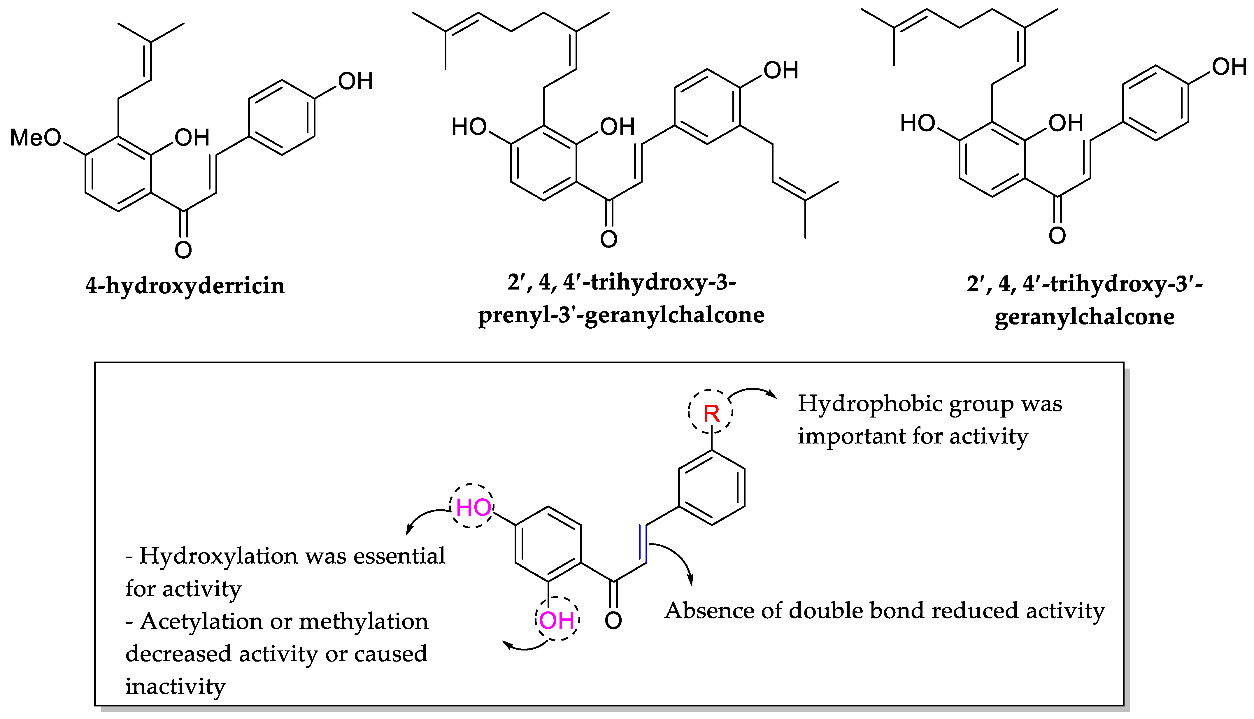
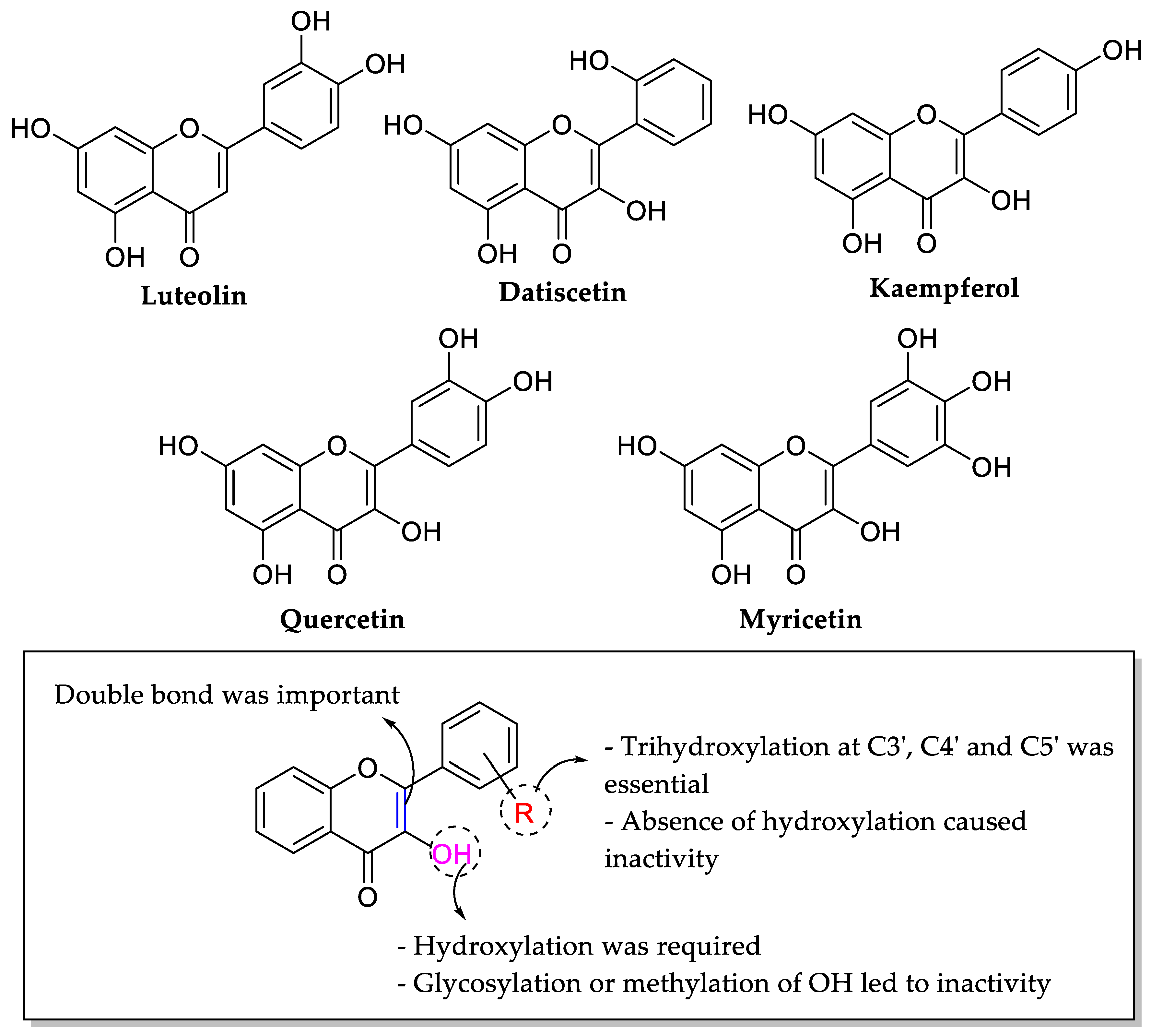
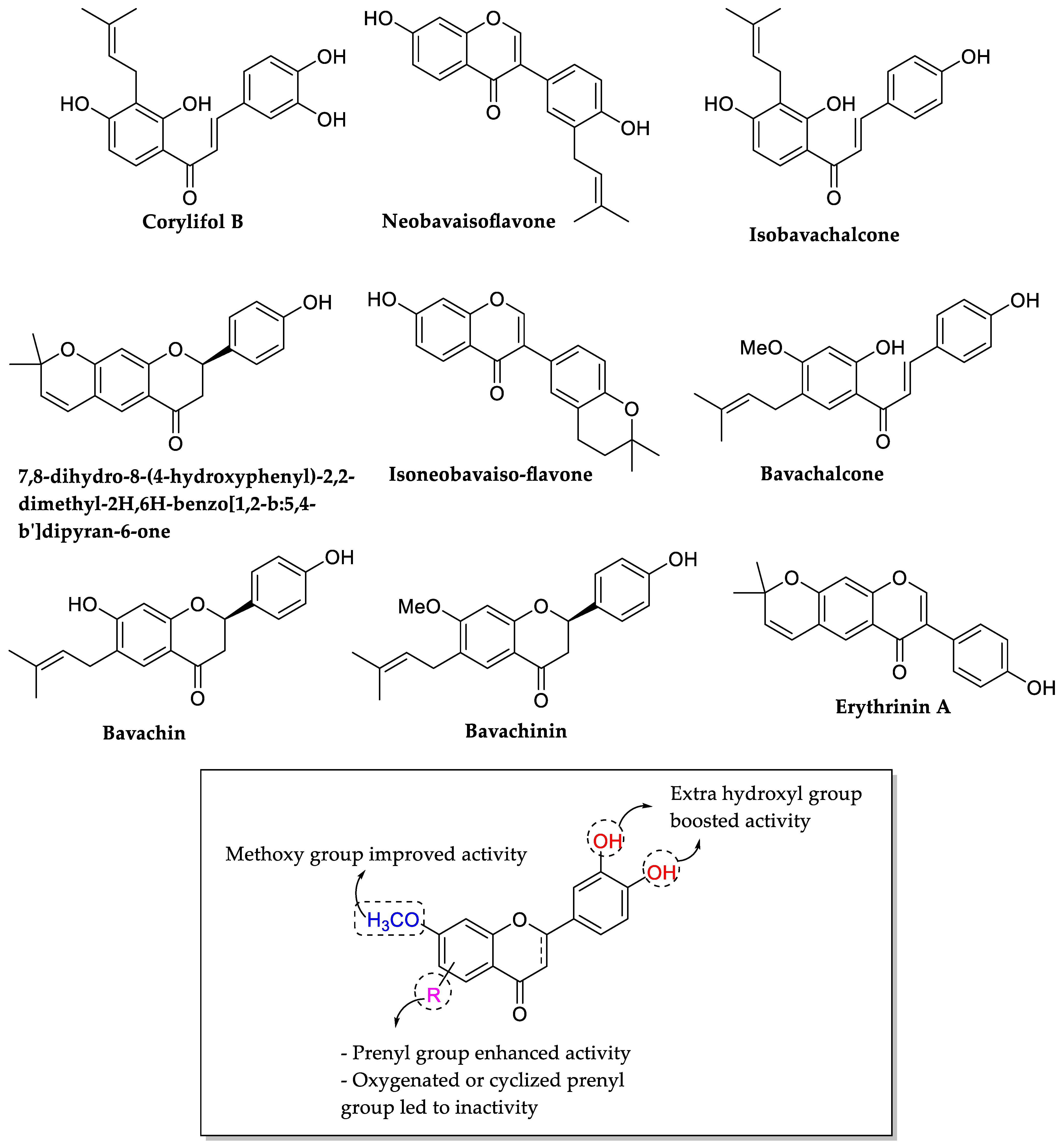
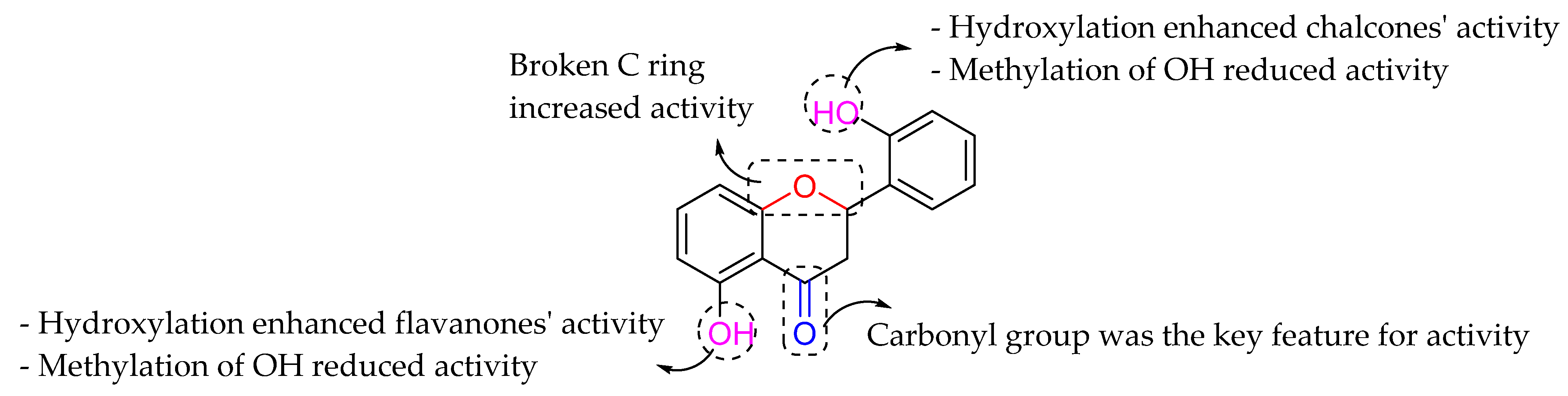
3. Discussion
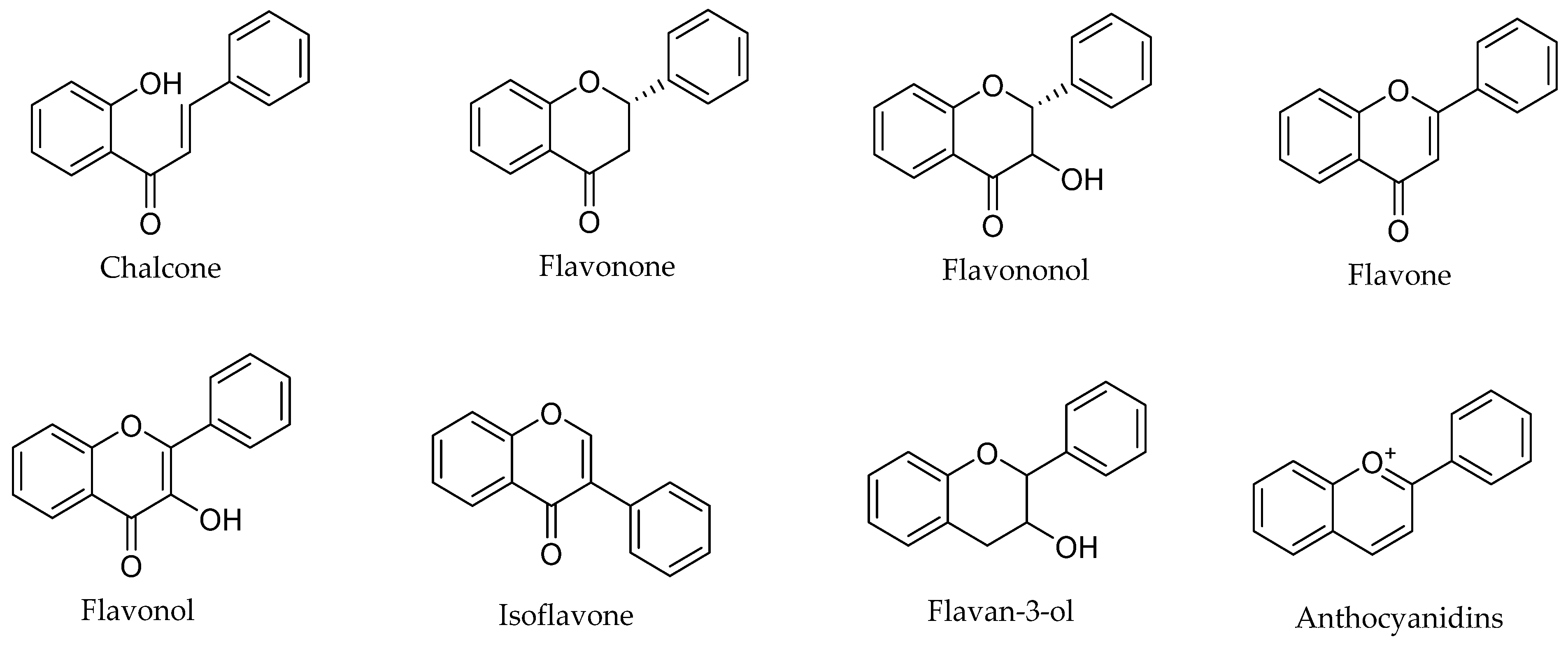
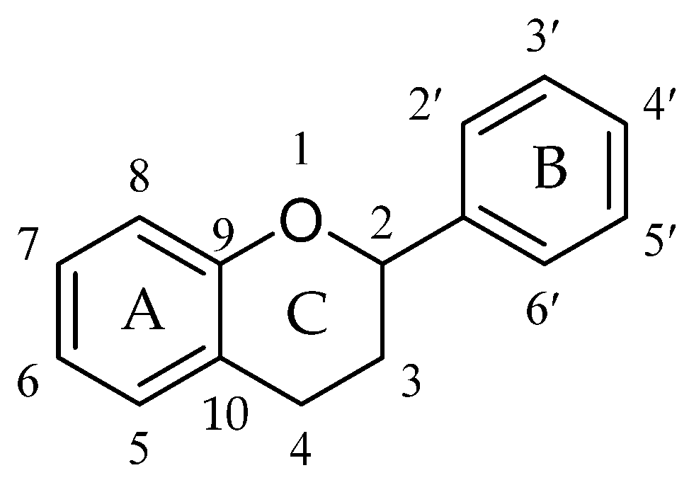
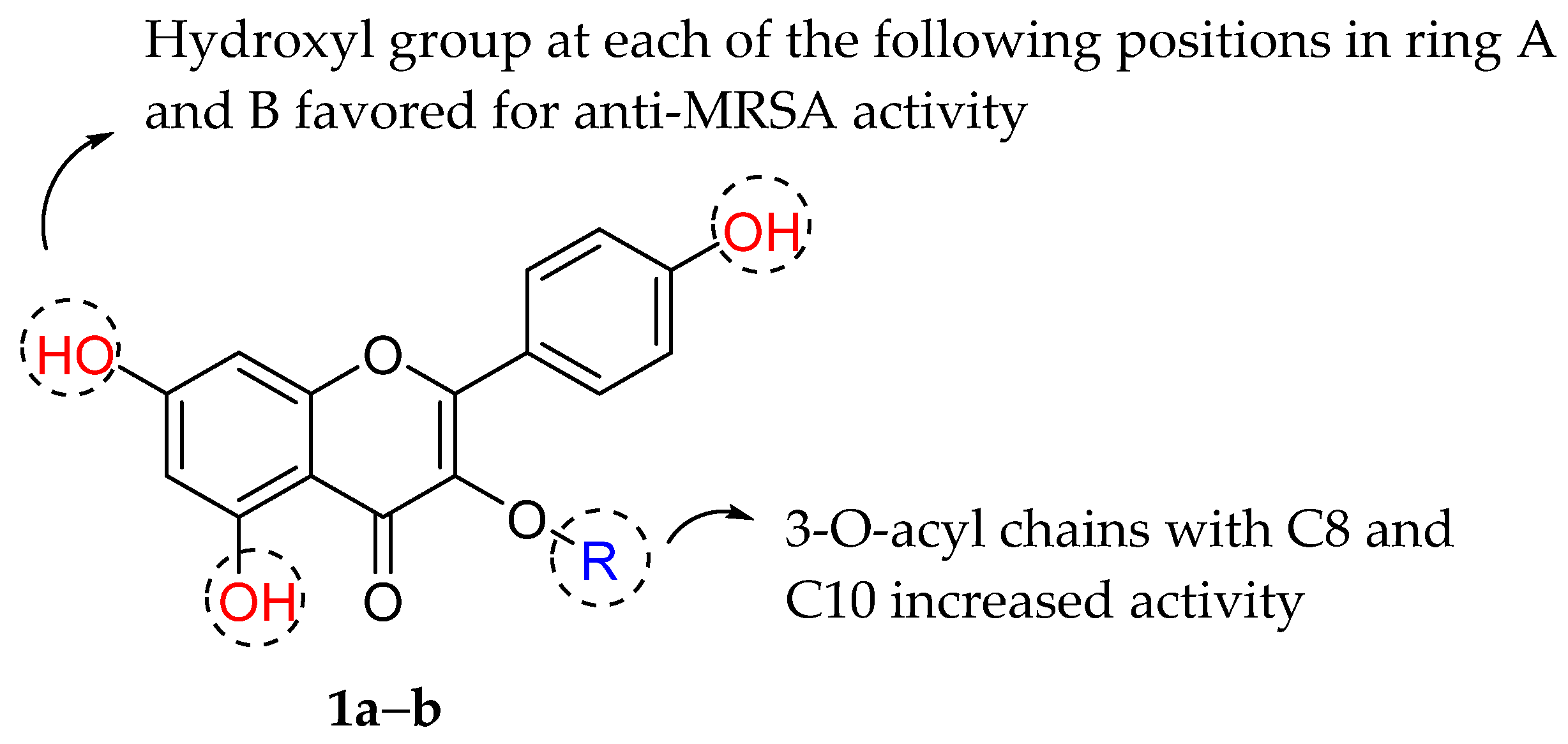
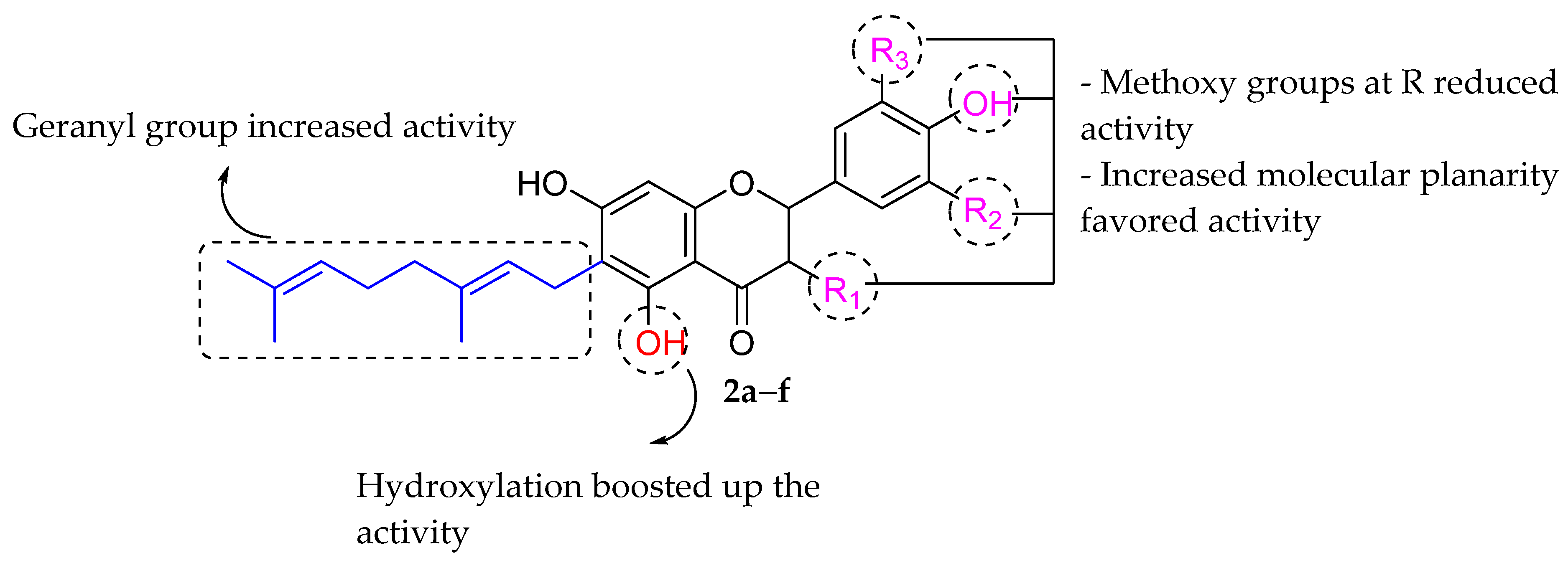
Anti-Bacterial Activity of Flavonoids
4. Conclusions
Author Contributions
Funding
Acknowledgments
Conflicts of Interest
References
- Nathan, C.; Cars, O. Antibiotic Resistance—Problems, Progress, and Prospects. N. Engl. J. Med. 2014, 371, 1761–1763. [Google Scholar] [CrossRef] [PubMed]
- Andersson, D.I.; Balaban, N.Q.; Baquero, F.; Courvalin, P.; Glaser, P.; Gophna, U.; Kishony, R.; Molin, S.; Tønjum, T. Antibiotic resistance: Turning evolutionary principles into clinical reality. FEMS Microbiol. Rev. 2020, 44, 171–188. [Google Scholar] [CrossRef] [PubMed]
- Ferri, M.; Ranucci, E.; Romagnoli, P.; Giaccone, V. Antimicrobial resistance: A global emerging threat to public health systems. Crit. Rev. Food Sci. Nutr. 2017, 57, 2857–2876. [Google Scholar] [CrossRef] [PubMed]
- Ahmad, M.; Khan, A.U. Global economic impact of antibiotic resistance: A review. J. Glob. Antimicrob. Resist. 2019, 19, 313–316. [Google Scholar] [CrossRef]
- Choudhury, R.; Panda, S.; Singh, D.V. Emergence and dissemination of antibiotic resistance: A global problem. Indian J. Med. Microbiol. 2012, 30, 384–390. [Google Scholar] [CrossRef] [PubMed]
- Prestinaci, F.; Pezzotti, P.; Pantosti, A. Antimicrobial resistance: A global multifaceted phenomenon. Pathog. Glob. Health 2015, 109, 309–318. [Google Scholar] [CrossRef] [Green Version]
- Michael, C.A.; Dominey-Howes, D.; Labbate, M. The Antimicrobial Resistance Crisis: Causes, Consequences, and Management. Front. Public Health 2014, 2, 145. [Google Scholar] [CrossRef]
- Centers for Disease Control and Prevention (CDC). Antibiotic. Antimicrobial Resistance. Available online: https://www.cdc.gov/drugresistance/index.html (accessed on 24 August 2021).
- World Health Organization. Antibiotic Resistance. 2020. Available online: https://www.who.int/news-room/fact-sheets/detail/antibiotic-resistance (accessed on 17 August 2021).
- Peterson, E.; Kaur, P. Antibiotic Resistance Mechanisms in Bacteria: Relationships between Resistance Determinants of Antibiotic Producers, Environmental Bacteria, and Clinical Pathogens. Front. Microbiol. 2018, 9, 2928. [Google Scholar] [CrossRef]
- Munita, J.M.; Arias, C.A. Mechanisms of Antibiotic Resistance. Microbiol. Spectr. 2016, 4, 464–472. [Google Scholar] [CrossRef] [Green Version]
- Sekyere, J.O.; Asante, J. Emerging mechanisms of antimicrobial resistance in bacteria and fungi: Advances in the era of genomics. Futur. Microbiol. 2018, 13, 241–262. [Google Scholar] [CrossRef]
- Kakoullis, L.; Papachristodoulou, E.; Chra, P.; Panos, G. Mechanisms of Antibiotic Resistance in Important Gram-Positive and Gram-Negative Pathogens and Novel Antibiotic Solutions. Antibiotics 2021, 10, 415. [Google Scholar] [CrossRef] [PubMed]
- Kapoor, G.; Saigal, S.; Elongavan, A. Action and resistance mechanisms of antibiotics: A guide for clinicians. J. Anaesthesiol. Clin. Pharmacol. 2017, 33, 300–305. [Google Scholar] [CrossRef] [PubMed]
- Centers for Disease Control and Prevention (CDC). Pneumococcal Disease and Antibiotic Resistance. Available online: https://www.cdc.gov/pneumococcal/drug-resistance.html (accessed on 1 October 2021).
- Chisti, M.J.; Harris, J.B.; Carroll, R.W.; Shahunja, K.M.; Shahid, A.S.M.S.B.; Moschovis, P.P.; Schenkel, S.R.; Rahman, A.S.M.M.H.; Shahrin, L.; Faruk, T.; et al. Antibiotic-Resistant Bacteremia in Young Children Hospitalized with Pneumonia in Bangladesh Is Associated with a High Mortality Rate. Open Forum Infect. Dis. 2021, 8, ofab260. [Google Scholar] [CrossRef] [PubMed]
- Peyrani, P.; Mandell, L.; Torres, A.; Tillotson, G. The burden of community-acquired bacterial pneumonia in the era of antibiotic resistance. Expert Rev. Respir. Med. 2019, 13, 139–152. [Google Scholar] [CrossRef] [PubMed]
- Solmaz, I.; Kalın, B.S. Assessment of antibiotic resistance of infectious agents in patients with pneumonia in tertiary critical care unit and effect on clinical outcomes. Int. J. Clin. Pract. 2021, 75, e13872. [Google Scholar] [CrossRef]
- Chen, H.-H.; Stringer, A.; Eguale, T.; Rao, G.G.; Ozawa, S. Impact of Antibiotic Resistance on Treatment of Pneumococcal Disease in Ethiopia: An Agent-Based Modeling Simulation. Am. J. Trop. Med. Hyg. 2019, 101, 1042–1053. [Google Scholar] [CrossRef]
- Kurz, S.G.; Furin, J.J.; Bark, C.M. Drug Resistant Tuberculosis: Challenges and Progress. Infect. Dis. Clin. N. Am. 2016, 30, 509–522. [Google Scholar] [CrossRef] [Green Version]
- Nguyen, L. Antibiotic resistance mechanisms in M. tuberculosis: An update. Arch. Toxicol. 2016, 90, 1585–1604. [Google Scholar] [CrossRef] [Green Version]
- Zine, S.; Patankar, S.A.; Raopati, S.S. Rise of Antibiotic Resistance in Tuberculosis. Res. J. Pharm. Technol. 2018, 11, 3201. [Google Scholar] [CrossRef]
- Gygli, S.M.; Borrell, S.; Trauner, A.; Gagneux, S. Antimicrobial resistance in Mycobacterium tuberculosis: Mechanistic and evolutionary perspectives. FEMS Microbiol. Rev. 2017, 41, 354–373. [Google Scholar] [CrossRef] [Green Version]
- Turnidge, J. Impact of antibiotic resistance on the treatment of sepsis. Scand. J. Infect. Dis. 2003, 35, 677–682. [Google Scholar] [CrossRef] [PubMed]
- Nagarjuna, D.; Mittal, G.; Dhanda, R.S.; Gaind, R.; Yadav, M. Alarming levels of antimicrobial resistance among sepsis patients admitted to ICU in a tertiary care hospital in India—A case control retrospective study. Antimicrob. Resist. Infect. Control 2018, 7, 150. [Google Scholar] [CrossRef] [PubMed] [Green Version]
- Rhee, C.; Kadri, S.S.; Dekker, J.P.; Danner, R.L.; Chen, H.-C.; Fram, D.; Zhang, F.; Wang, R.; Klompas, M. For the CDC Prevention Epicenters Program. Prevalence of Antibiotic-Resistant Pathogens in Culture-Proven Sepsis and Outcomes Associated with Inadequate and Broad-Spectrum Empiric Antibiotic Use. JAMA Netw. Open 2020, 3, e202899. [Google Scholar] [CrossRef] [PubMed]
- Pop-Vicas, A.; Opal, S.M. The clinical impact of multidrug-resistant gram-negative bacilli in the management of septic shock. Virulence 2013, 5, 206–212. [Google Scholar] [CrossRef] [Green Version]
- Patel, A.L.; Chaudhry, U.; Sachdev, D.; Sachdeva, P.N.; Bala, M.; Saluja, D. An insight into the drug resistance profile & mechanism of drug resistance in Neisseria gonorrhoeae. Indian J. Med. Res. 2011, 134, 419–431. [Google Scholar]
- Tapsall, J.W. Antibiotic Resistance in Neisseria gonorrhoeae. Clin. Infect. Dis. 2005, 41 (Suppl. S4), S263–S268. [Google Scholar] [CrossRef] [Green Version]
- Unemo, M.; Shafer, W.M. Antibiotic resistance in Neisseria gonorrhoeae: Origin, evolution, and lessons learned for the future. Ann. N. Y. Acad. Sci. 2011, 1230, E19–E28. [Google Scholar] [CrossRef]
- Abraham, S.; Poehlmann, C.; Spornraft-Ragaller, P. Gonorrhea: Data on antibiotic resistance and accompanying infections at the University Hospital Dresden over a 10-year time period. J. Dtsch. Dermatol. Ges. 2012, 11, 241–249. [Google Scholar] [CrossRef]
- Fletcher, S.; Dronavalli, M.; Alexander, K.; Ghosh, S.; Boonwaat, L.; Thomas, J.; Robinson, A.; Patel, Z.; Forssman, B.; Pal, N.; et al. Trends in Antimicrobial Resistance Patterns in Neisseria gonorrhoeae in Australia and New Zealand: A Meta-analysis and Systematic Review. Antibiotics 2019, 8, 191. [Google Scholar] [CrossRef] [Green Version]
- Bodie, M.; Gale-Rowe, M.; Alexandre, S.; Auguste, U.; Tomas, K.; Martin, I. Multidrug resistant gonorrhea: Addressing the rising rates of gonorrhea and drug-resistant gonorrhea: There is no time like the present. Can. Commun. Dis. Rep. 2019, 45, 54. [Google Scholar] [CrossRef]
- Wi, T.; Lahra, M.M.; Ndowa, F.; Bala, M.; Dillon, J.-A.R.; Ramon-Pardo, P.; Eremin, S.R.; Bolan, G.; Unemo, M. Antimicrobial resistance in Neisseria gonorrhoeae: Global surveillance and a call for international collaborative action. PLoS Med. 2017, 14, e1002344. [Google Scholar] [CrossRef] [PubMed] [Green Version]
- Odeyemi, O.A.; Sani, N.A. Antibiotic resistance and burden of foodborne diseases in developing countries. Future Sci. OA 2016, 2, FSO139. [Google Scholar] [CrossRef] [PubMed] [Green Version]
- Hashempour-Baltork, F.; Hosseini, H.; Shojaee-Aliabadi, S.; Torbati, M.; Alizadeh, A.M.; Alizadeh, M. Drug Resistance and the Prevention Strategies in Food Borne Bacteria: An Update Review. Adv. Pharm. Bull. 2019, 9, 335–347. [Google Scholar] [CrossRef] [PubMed] [Green Version]
- Caniça, M.; Manageiro, V.; Abriouel, H.; Moran-Gilad, J.; Franz, C.M. Antibiotic resistance in foodborne bacteria. Trends Food Sci. Technol. 2019, 84, 41–44. [Google Scholar] [CrossRef]
- Medalla, F.; Gu, W.; Friedman, C.R.; Judd, M.; Folster, J.; Griffin, P.M.; Hoekstra, R.M. Increased Incidence of Antimicrobial-Resistant Nontyphoidal Salmonella Infections, United States, 2004–2016. Emerg. Infect. Dis. 2021, 27, 1662–1672. [Google Scholar] [CrossRef]
- Angulo, F.J.; Mølbak, K. Human Health Consequences of Antimicrobial Drug—Resistant Salmonella and Other Foodborne Pathogens. Clin. Infect. Dis. 2005, 41, 1613–1620. [Google Scholar] [CrossRef]
- Friedman, N.; Temkin, E.; Carmeli, Y. The negative impact of antibiotic resistance. Clin. Microbiol. Infect. 2016, 22, 416–422. [Google Scholar] [CrossRef]
- Eliopoulos, G.M.; Cosgrove, S.E.; Carmeli, Y. The Impact of Antimicrobial Resistance on Health and Economic Outcomes. Clin. Infect. Dis. 2003, 36, 1433–1437. [Google Scholar] [CrossRef]
- Cosgrove, S.E. The Relationship between Antimicrobial Resistance and Patient Outcomes: Mortality, Length of Hospital Stay, and Health Care Costs. Clin. Infect. Dis. 2006, 42, S82–S89. [Google Scholar] [CrossRef] [Green Version]
- Mulvey, M.R.; Simor, A.E. Antimicrobial resistance in hospitals: How concerned should we be? Can. Med. Assoc. J. 2009, 180, 408–415. [Google Scholar] [CrossRef] [Green Version]
- Panche, A.N.; Diwan, A.D.; Chandra, S.R. Flavonoids: An overview. J. Nutr. Sci. 2016, 5, e47. [Google Scholar] [CrossRef] [PubMed] [Green Version]
- Wang, T.; Li, Q.; Bi, K. Bioactive flavonoids in medicinal plants: Structure, activity and biological fate. Asian J. Pharm. Sci. 2018, 13, 12–23. [Google Scholar] [CrossRef] [PubMed]
- Kumar, S.; Pandey, A.K. Chemistry and Biological Activities of Flavonoids: An Overview. Sci. World J. 2013, 2013, 162750. [Google Scholar] [CrossRef] [PubMed] [Green Version]
- Jucá, M.M.; Filho, F.M.S.C.; de Almeida, J.C.; da Silva Mesquita, D.; de Moraes Barriga, J.R.; Dias, K.C.F.; Barbosa, T.M.; Vasconcelos, L.C.; Leal, L.K.A.M.; Ribeiro, J.E., Jr.; et al. Flavonoids: Biological activities and therapeutic potential. Nat. Prod. Res. 2020, 3, 692–705. [Google Scholar] [CrossRef] [PubMed]
- Chen, X.; Mukwaya, E.; Wong, M.S.; Zhang, Y. A systematic review on biological activities of prenylated flavonoids. Pharm. Biol. 2014, 52, 655–660. [Google Scholar] [CrossRef] [PubMed]
- Fang, W.-Y.; Ravindar, L.; Rakesh, K.; Manukumar, H.; Shantharam, C.; Alharbi, N.S.; Qin, H.-L. Synthetic approaches and pharmaceutical applications of chloro-containing molecules for drug discovery: A critical review. Eur. J. Med. Chem. 2019, 173, 117–153. [Google Scholar] [CrossRef] [PubMed]
- Karak, P. Biological Activities of Flavonoids: An Overview. Int. J. Pharm. Sci. Res. 2019, 10, 1567–1574. [Google Scholar]
- Heim, K.E.; Tagliaferro, A.R.; Bobilya, D.J. Flavonoid antioxidants: Chemistry, metabolism and structure-activity relationships. J. Nutr. Biochem. 2002, 13, 572–584. [Google Scholar] [CrossRef]
- Dias, M.C.; Pinto, D.C.G.A.; Silva, A.M.S. Plant Flavonoids: Chemical Characteristics and Biological Activity. Molecules 2021, 26, 5377. [Google Scholar] [CrossRef]
- Cosme, P.; Rodríguez, A.B.; Espino, J.; Garrido, M. Plant Phenolics: Bioavailability as a Key Determinant of Their Potential Health-Promoting Applications. Antioxidants 2020, 9, 1263. [Google Scholar] [CrossRef]
- Croft, K.D. The chemistry and biological effects of flavonoids and phenolic acids. Ann. N. Y. Acad. Sci. 1998, 854, 435–442. [Google Scholar] [CrossRef]
- Halbwirth, H. The Creation and Physiological Relevance of Divergent Hydroxylation Patterns in the Flavonoid Pathway. Int. J. Mol. Sci. 2010, 11, 595–621. [Google Scholar] [CrossRef] [PubMed]
- Lago, J.H.G.; Toledo-Arruda, A.; Mernak, M.; Barrosa, K.H.; Martins, M.A.; Tibério, I.F.L.C.; Prado, C.M. Structure-Activity Association of Flavonoids in Lung Diseases. Molecules 2014, 19, 3570–3595. [Google Scholar] [CrossRef] [PubMed] [Green Version]
- Alseekh, S.; de Souza, L.P.; Benina, M.; Fernie, A.R. The style and substance of plant flavonoid decoration; towards defining both structure and function. Phytochemistry 2020, 174, 112347. [Google Scholar] [CrossRef] [PubMed]
- Xie, Y.; Yang, W.; Tang, F.; Chen, X.; Ren, L. Antibacterial Activities of Flavonoids: Structure-Activity Relationship and Mechanism. Curr. Med. Chem. 2014, 22, 132–149. [Google Scholar] [CrossRef] [PubMed]
- Cushnie, T.P.T.; Lamb, A.J. Antimicrobial activity of flavonoids. Int. J. Antimicrob. Agents 2005, 26, 343–356. [Google Scholar] [CrossRef] [PubMed]
- Górniak, I.; Bartoszewski, R.; Króliczewski, J. Comprehensive review of antimicrobial activities of plant flavonoids. Phytochem. Rev. 2019, 18, 241–272. [Google Scholar] [CrossRef] [Green Version]
- Biharee, A.; Sharma, A.; Kumar, A.; Jaitak, V. Fitoterapia Antimicrobial flavonoids as a potential substitute for overcoming antimicrobial resistance. Fitoterapia 2020, 146, 104720. [Google Scholar] [CrossRef]
- Wu, T.; Zang, X.; He, M.; Pan, S.; Xu, X. Structure–Activity Relationship of Flavonoids on Their Anti-Escherichia coli Activity and Inhibition of DNA Gyrase. J. Agric. Food Chem. 2013, 61, 8185–8190. [Google Scholar] [CrossRef]
- Donadio, G.; Mensitieri, F.; Santoro, V.; Parisi, V.; Bellone, M.; De Tommasi, N.; Izzo, V.; Piaz, F.D. Interactions with Microbial Proteins Driving the Antibacterial Activity of Flavonoids. Pharmaceutics 2021, 13, 660. [Google Scholar] [CrossRef]
- Xu, H.-X.; Lee, S.F. Activity of plant flavonoids against antibiotic-resistant bacteria. Phytother. Res. 2001, 15, 39–43. [Google Scholar] [CrossRef]
- Song, M.; Liu, Y.; Li, T.; Liu, X.; Hao, Z.; Ding, S.; Panichayupakaranant, P.; Zhu, K.; Shen, J. Plant Natural Flavonoids Against Multidrug Resistant Pathogens. Adv. Sci. 2021, 8, 2100749. [Google Scholar] [CrossRef] [PubMed]
- Ruddock, P.S.; Charland, M.; Ramirez, S.; López, A.; Towers, G.H.N.; Arnason, J.T.; Liao, M.; Dillon, J.-A.R. Antimicrobial Activity of Flavonoids from Piper lanceaefolium and Other Colombian Medicinal Plants Against Antibiotic Susceptible and Resistant Strains of Neisseria gonorrhoeae. Sex. Transm. Dis. 2011, 38, 82–88. [Google Scholar] [CrossRef] [PubMed]
- Abreu, A.C.; Serra, S.C.; Borges, A.; Saavedra, M.J.; Mcbain, A.J.; Salgado, A.J.; Simões, M. Combinatorial Activity of Flavonoids with Antibiotics Against Drug-Resistant Staphylococcus aureus. Microb. Drug Resist. 2015, 21, 600–609. [Google Scholar] [CrossRef] [PubMed]
- Tsuchiya, H.; Sato, M.; Miyazaki, T.; Fujiwara, S.; Tanigaki, S.; Ohyama, M.; Tanaka, T.; Iinuma, M. Comparative study on the antibacterial activity of phytochemical flavanones against methicillin-resistant Staphylococcus aureus. J. Ethnopharmacol. 1996, 50, 27–34. [Google Scholar] [CrossRef]
- Stapleton, P.D.; Shah, S.; Hamilton-Miller, J.M.T.; Hara, Y.; Nagaoka, Y.; Kumagai, A.; Uesato, S.; Taylor, P.W. Anti-Staphylococcus aureus activity and oxacillin resistance modulating capacity of 3-O-acyl-catechins. Int. J. Antimicrob. Agents 2004, 24, 374–380. [Google Scholar] [CrossRef]
- Otsuka, N.; Liu, M.-H.; Shiota, S.; Ogawa, W.; Kuroda, T.; Hatano, T.; Tsuchiya, T. Anti-methicillin Resistant Staphylococcus aureus (MRSA) Compounds Isolated from Laurus nobilis. Biol. Pharm. Bull. 2008, 31, 1794–1797. [Google Scholar] [CrossRef] [Green Version]
- Šmejkal, K.; Chudík, S.; Klouček, P.; Marek, R.; Cvačka, J.; Urbanová, M.; Julínek, O.; Kokoška, L.; Šlapetová, T.; Holubová, P.; et al. Antibacterial C-Geranylflavonoids from Paulownia tomentosa Fruits. J. Nat. Prod. 2008, 71, 706–709. [Google Scholar] [CrossRef]
- Yi, Z.; Yu, Y.; Liang, Y.; Zeng, B. In vitro antioxidant and antimicrobial activities of the extract of Pericarpium Citri Reticulatae of a new Citrus cultivar and its main flavonoids. LWT 2008, 41, 597–603. [Google Scholar] [CrossRef]
- Wu, T.; He, M.; Zang, X.; Zhou, Y.; Qiu, T.; Pan, S.; Xu, X. A structure–activity relationship study of flavonoids as inhibitors of E. coli by membrane interaction effect. Biochim. Biophys. Acta Biomembr. 2013, 1828, 2751–2756. [Google Scholar] [CrossRef] [Green Version]
- Yadav, A.K.; Thakur, J.; Prakash, O.; Khan, F.; Saikia, D.; Gupta, M.M. Screening of flavonoids for antitubercular activity and their structure–activity relationships. Med. Chem. Res. 2013, 22, 2706–2716. [Google Scholar] [CrossRef]
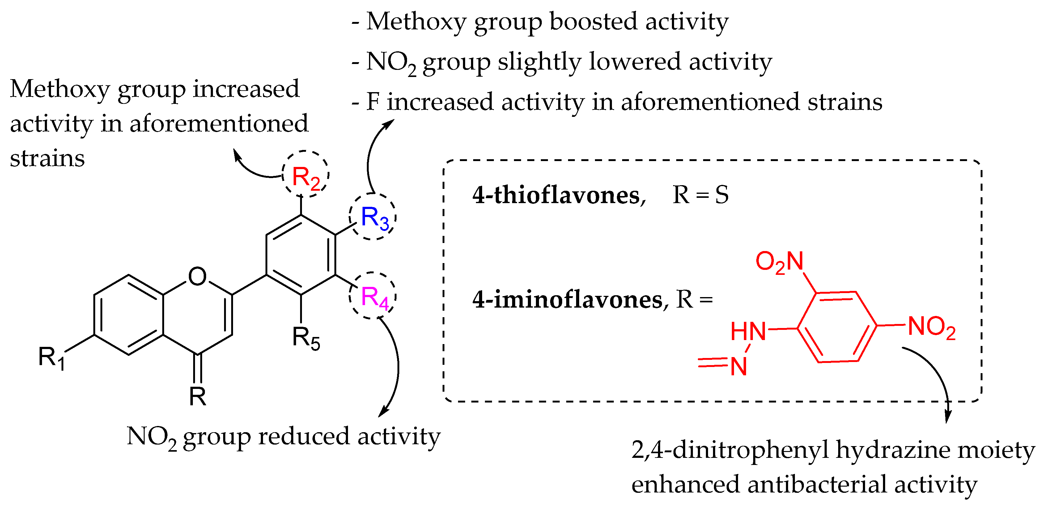
- Ullah Mughal, E.; Ayaz, M.; Hussain, Z.; Hasan, A.; Sadiq, A.; Riaz, M.; Malik, A.; Hussain, S.; Choudhary, M.I. Synthesis and antibacterial activity of substituted flavones, 4-thioflavones and 4-iminoflavones. Bioorg. Med. Chem. 2006, 14, 4704–4711. [Google Scholar] [CrossRef] [PubMed]
- Echeverría, J.; Opazo, J.; Mendoza, L.; Urzúa, A.; Wilkens, M. Structure-Activity and Lipophilicity Relationships of Selected Antibacterial Natural Flavones and Flavanones of Chilean Flora. Molecules 2017, 22, 608. [Google Scholar] [CrossRef] [PubMed]
- Ávila, H.P.; Smânia, E.D.F.A.; Monache, F.D.; Smânia, A. Structure–activity relationship of antibacterial chalcones. Bioorg. Med. Chem. 2008, 16, 9790–9794. [Google Scholar] [CrossRef]
- Yin, S.; Fan, C.-Q.; Wang, Y.; Dong, L.; Yue, J.-M. Antibacterial prenylflavone derivatives from Psoralea corylifolia, and their structure–activity relationship study. Bioorg. Med. Chem. 2004, 12, 4387–4392. [Google Scholar] [CrossRef]
- Alcaráz, L.; Blanco, S.; Puig, O.; Tomás, F.; Ferretti, F. Antibacterial Activity of Flavonoids Against Methicillin-resistant Staphylococcus aureus strains. J. Theor. Biol. 2000, 205, 231–240. [Google Scholar] [CrossRef]
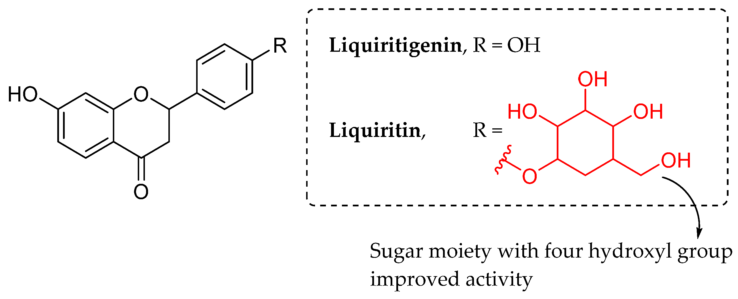
- Xie, Y.; Chen, J.; Xiao, A.; Liu, L. Antibacterial Activity of Polyphenols: Structure-Activity Relationship and Influence of Hyperglycemic Condition. Molecules 2017, 22, 1913. [Google Scholar] [CrossRef] [Green Version]
- Hummelova, J.; Rondevaldova, J.; Balastikova, A.; Lapcik, O.; Kokoska, L. The relationship between structure and in vitro antibacterial activity of selected isoflavones and their metabolites with special focus on antistaphylococcal effect of demethyltexasin. Lett. Appl. Microbiol. 2015, 60, 242–247. [Google Scholar] [CrossRef]
- Feng, L.; Maddox, M.M.; Alam, Z.; Tsutsumi, L.S.; Narula, G.; Bruhn, D.F.; Wu, X.; Sandhaus, S.; Lee, R.B.; Simmons, C.J.; et al. Synthesis, Structure–Activity Relationship Studies, and Antibacterial Evaluation of 4-Chromanones and Chalcones, as Well as Olympicin A and Derivatives. J. Med. Chem. 2014, 57, 8398–8420. [Google Scholar] [CrossRef]
- Simard, F.; Gauthier, C.; Legault, J.; Lavoie, S.; Mshvildadze, V.; Pichette, A. Structure elucidation of anti-methicillin resistant Staphylococcus aureus (MRSA) flavonoids from balsam poplar buds. Bioorg. Med. Chem. 2016, 24, 4188–4198. [Google Scholar] [CrossRef]
- Omosa, L.K.; Midiwo, J.O.; Mbaveng, A.T.; Tankeo, S.B.; Seukep, J.A.; Voukeng, I.K.; Dzotam, J.K.; Isemeki, J.; Derese, S.; Omolle, R.A.; et al. Antibacterial activities and structure-activity relationships of a panel of 48 compounds from Kenyan plants against multidrug resistant phenotypes. SpringerPlus 2016, 5, 901. [Google Scholar] [CrossRef] [PubMed] [Green Version]
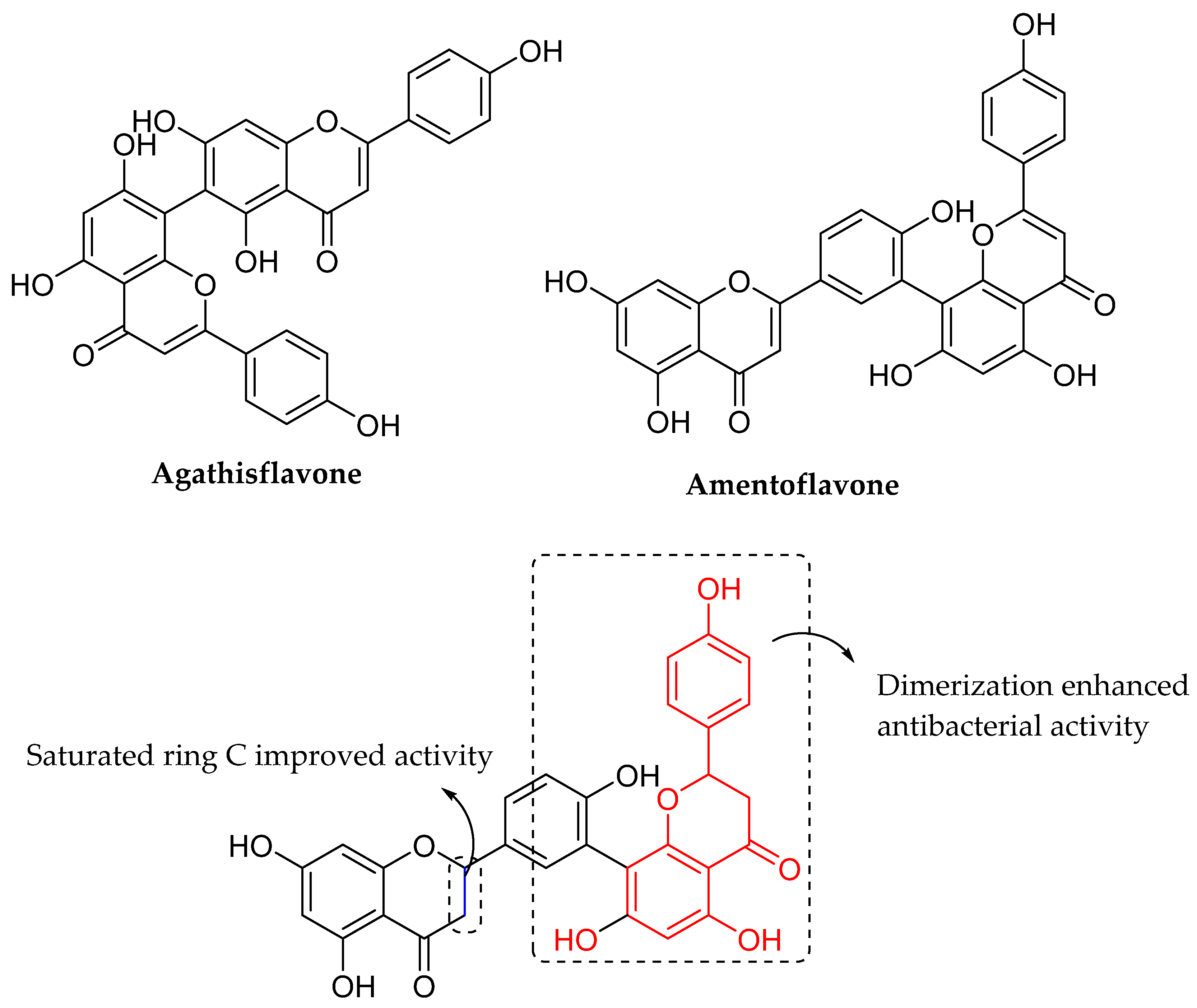
- Linden, M.; Brinckmann, C.; Feuereisen, M.M.; Schieber, A. Effects of structural differences on the antibacterial activity of biflavonoids from fruits of the Brazilian peppertree (Schinus terebinthifolius Raddi). Food Res. Int. 2020, 133, 109134. [Google Scholar] [CrossRef] [PubMed]
- Kong, W.; Zhao, Y.; Xing, X.; Ma, X.; Sun, X.; Yang, M.; Xiao, X. Antibacterial evaluation of flavonoid compounds against E. coli by microcalorimetry and chemometrics. Appl. Microbiol. Biotechnol. 2015, 99, 6049–6058. [Google Scholar] [CrossRef] [PubMed]
- Maxam, A.M.; Gilbert, W. Sequencing end-labeled DNA with base-specific chemical cleavages. Methods Enzymol. 1980, 65, 99–560. [Google Scholar] [CrossRef]
- Bahrin, L.G.; Apostu, M.O.; Birsa, L.M.; Stefan, M. The antibacterial properties of sulfur containing flavonoids. Bioorg. Med. Chem. Lett. 2014, 24, 2315–2318. [Google Scholar] [CrossRef] [PubMed]
- Bahrin, L.G.; Hopf, H.; Jones, P.G.; Sarbu, L.G.; Babii, C.; Mihai, A.C.; Stefan, M.; Birsa, L.M. Antibacterial structure–activity relationship studies of several tricyclic sulfur-containing flavonoids. Beilstein J. Org. Chem. 2016, 12, 1065–1071. [Google Scholar] [CrossRef] [Green Version]
- Kagechika, H.; Kawachi, E.; Hashimoto, Y.; Himi, T.; Shudo, K. Retinobenzoic Acids. 1. Structure-Activity Relationships of Aromatic Amides with Retinoidal Activity. J. Med. Chem. 1988, 31, 2182–2192. [Google Scholar] [CrossRef]
- Ngaini, Z.; Mortadza, N.A. Synthesis of halogenated azo-aspirin analogues from natural product derivatives as the potential antibacterial agents. Nat. Prod. Res. 2018, 33, 3507–3514. [Google Scholar] [CrossRef]
- Farooq, S.; Ngaini, Z.; Mortadza, N.A. Microwave-assisted Synthesis and Molecular Docking Study of Heteroaromatic Chalcone Derivatives as Potential Antibacterial Agents. Bull. Korean Chem. Soc. 2020, 41, 918–924. [Google Scholar] [CrossRef]
- Qin, H.-L.; Zhang, Z.-W.; Lekkala, R.; Alsulami, H.; Rakesh, K. Chalcone hybrids as privileged scaffolds in antimalarial drug discovery: A key review. Eur. J. Med. Chem. 2020, 193, 112215. [Google Scholar] [CrossRef]
- Narwal, S.; Kumar, S.; Verma, P.K. Synthesis and biological activity of new chalcone scaffolds as prospective antimicrobial agents. Res. Chem. Intermed. 2021, 47, 1625–1641. [Google Scholar] [CrossRef]
- Sadgrove, N.J.; Oliveira, T.B.; Khumalo, G.P.; Van Vuuren, S.F.; Van Wyk, B.-E. Antimicrobial Isoflavones and Derivatives from Erythrina (Fabaceae): Structure Activity Perspective (Sar & Qsar) on Experimental and Mined Values Against Staphylococcus aureus. Antibiotics 2020, 9, 223. [Google Scholar] [CrossRef]
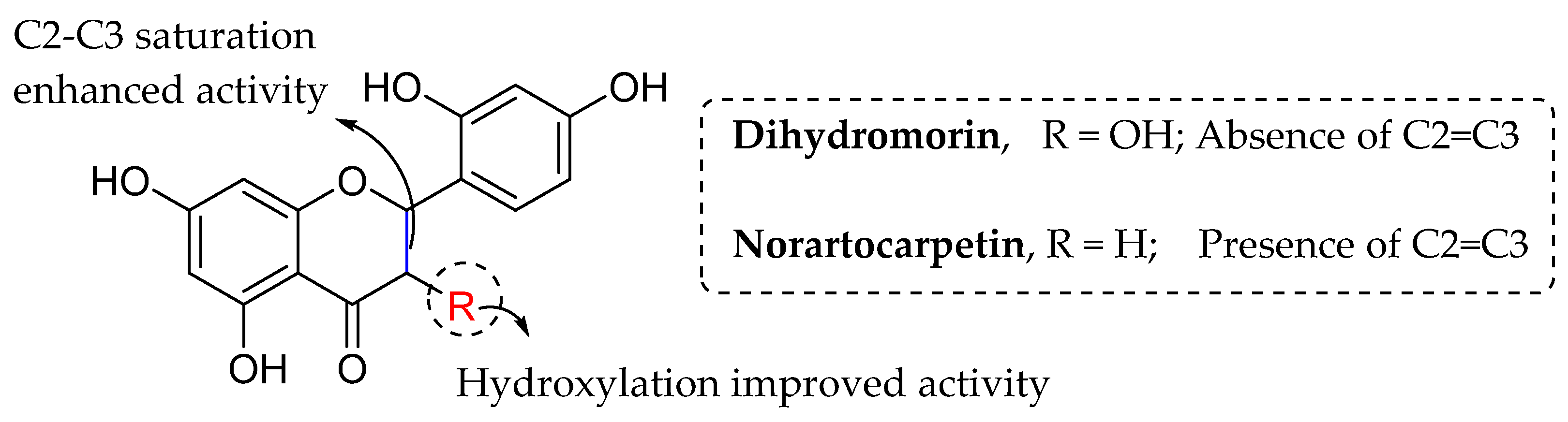
- Septama, A.; Jantan, I.; Panichayupakaranant, P.; Aluwi, M.; Rahmi, E. Immunosuppressive and antibacterial activities of dihydromorin and norartocarpetin isolated from Artocarpus heterophyllus heartwoods. Asian Pac. J. Trop. Biomed. 2020, 10, 361–368. [Google Scholar] [CrossRef]
- Nielsen, S.F.; Boesen, T.; Larsen, M.; Schønning, K.; Kromann, H. Antibacterial chalcones—Bioisosteric replacement of the 4′-hydroxy group. Bioorg. Med. Chem. 2004, 12, 3047–3054. [Google Scholar] [CrossRef]
- Ansari, F.L.; Baseer, M.; Iftikhar, F.; Kulsoom, S.; Ullah, A.; Nazir, S.; Shaukat, A.; Haq, I.-U.; Mirza, B. Microwave assisted synthesis, antibacterial activity against Bordetella bronchiseptica of a library of 3′-hydroxyaryl and heteroaryl chalcones and molecular descriptors-based SAR. Arkivoc 2009, 2009, 318–332. [Google Scholar] [CrossRef] [Green Version]
- Yokozawa, T.; Dong, E.; Kawai, Y.; Gemba, M.; Shimizu, M. Protective effects of some flavonoids on the renal cellular membrane. Exp. Toxicol. Pathol. 1999, 51, 9–14. [Google Scholar] [CrossRef]
- Woźnicka, E.; Kuźniar, A.; Nowak, D.; Nykiel, E.; Kopacz, M.; Gruszecka, J.; Golec, K. Comparative study on the antibacterial activity of some flavonoids and their sulfonic derivatives. Acta Pol. Pharm. Drug Res. 2013, 70, 567–571. [Google Scholar]
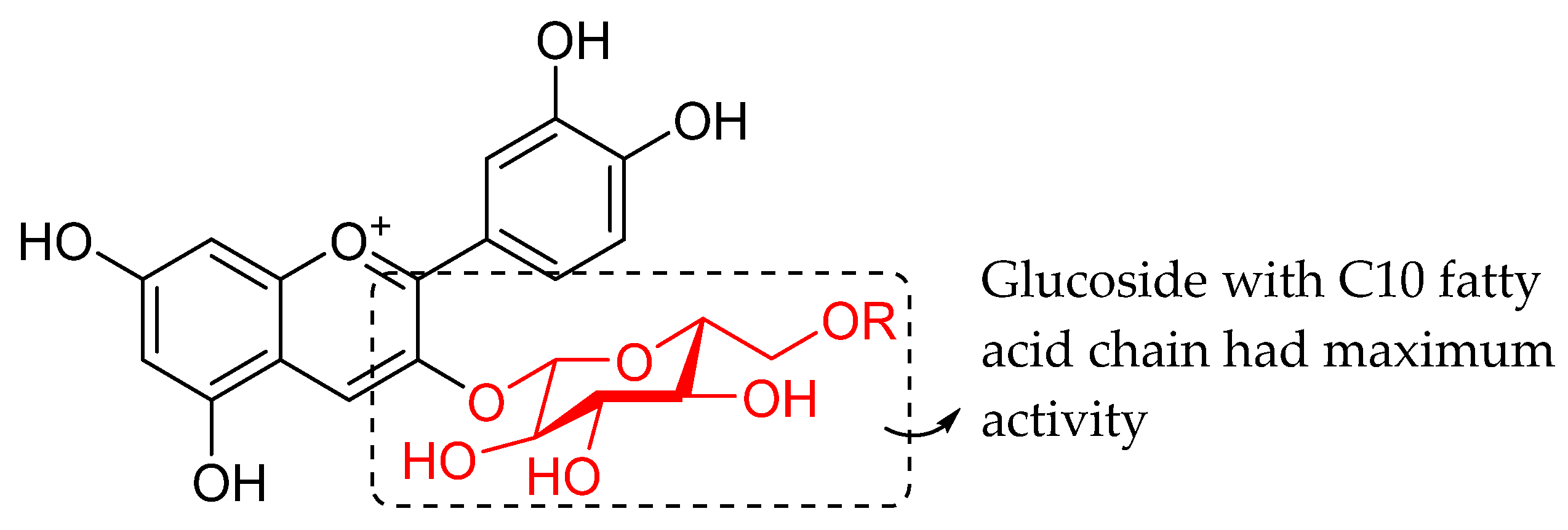
- Oliveira, H.; Correia, P.; Bessa, L.; Guimarães, M.; Gameiro, P.; Freitas, V.; Mateus, N.; Cruz, L.; Fernandes, I. Cyanidin-3-Glucoside Lipophilic Conjugates for Topical Application: Tuning the Antimicrobial Activities with Fatty Acid Chain Length. Processes 2021, 9, 340. [Google Scholar] [CrossRef]
- Shoaib, M.; Shah, S.W.A.; Ali, N.; Umar, N.; Shah, I.; Ullah, S.; Tahir, M.N. Synthesis, crystal studies and biological evaluation of flavone derivatives. Pak. J. Pharm. Sci. 2020, 33, 11–20. [Google Scholar]
- Sato, M.; Tanaka, H.; Tani, N.; Nagayama, M.; Yamaguchi, R. Different antibacterial actions of isoflavones isolated from Erythrina poeppigiana against methicillin-resistant Staphylococcus aureus. Lett. Appl. Microbiol. 2006, 43, 243–248. [Google Scholar] [CrossRef]
- Fang, Y.; Lu, Y.; Zang, X.; Wu, T.; Qi, X.; Pan, S.; Xu, X. 3D-QSAR and docking studies of flavonoids as potent Escherichia coli inhibitors. Sci. Rep. 2016, 6, 23634. [Google Scholar] [CrossRef]
- Alfonso, A.A.; Nicolás, G.; María, P.F.; Joaquín, A.; Antonio, G.; Sofía, S.; Elena, O.M. Synthesis and evaluation of antimicrobial and antibiofilm properties of a-type procyanidin analogues against resistant bacteria in food. J. Agric. Food Chem. 2018, 66, 2151–2158. [Google Scholar]
- Ortega-Vidal, J.; Cobo, A.; Ortega-Morente, E.; Gálvez, A.; Martínez-Bailén, M.; Salido, S.; Altarejos, J. Antimicrobial activity of phenolics isolated from the pruning wood residue of European plum (Prunus domestica L.). Ind. Crops Prod. 2022, 176, 114296. [Google Scholar] [CrossRef]


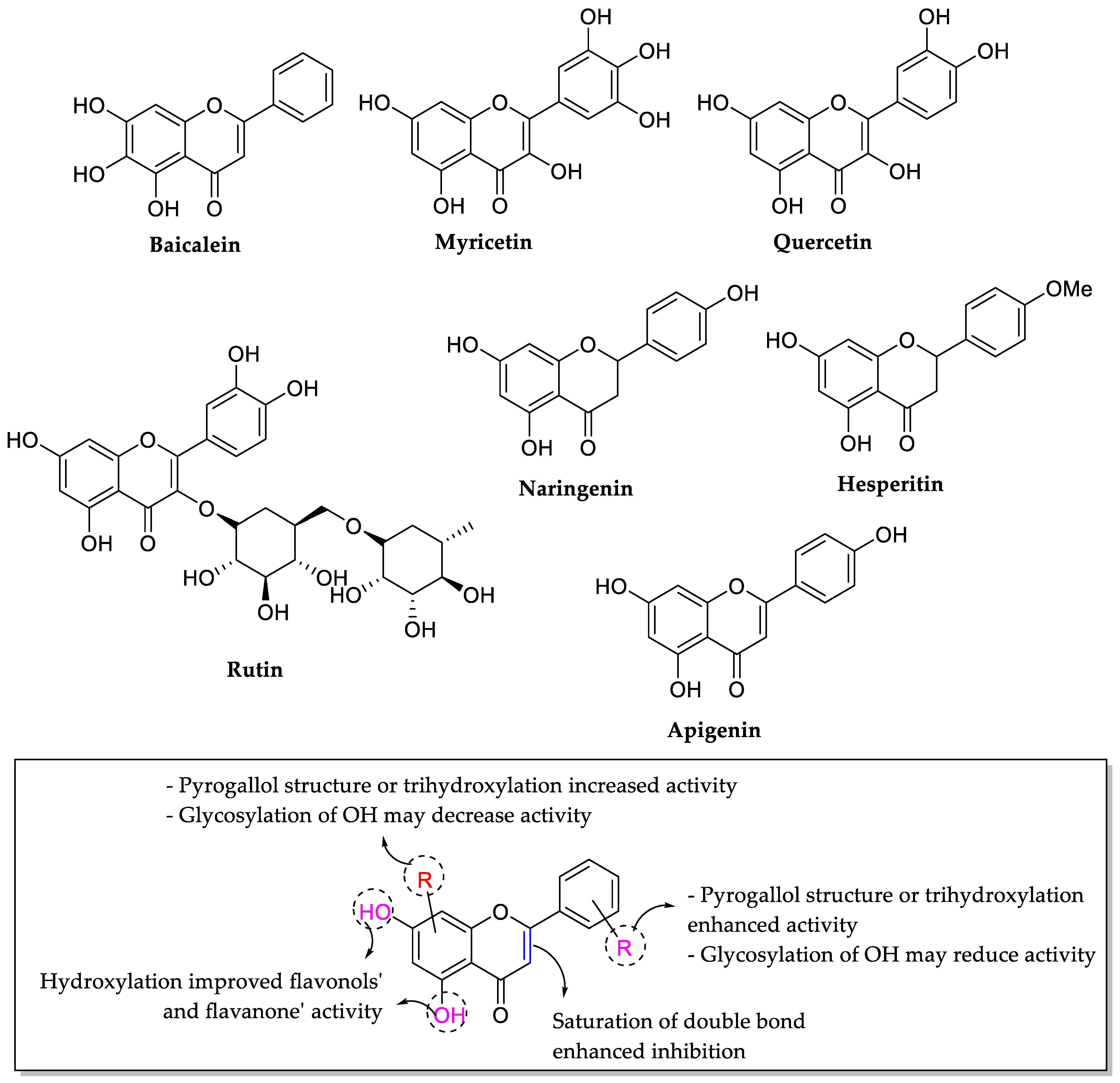
| Com. No. | R | Anti-MRSA Activity (MIC: μg/mL) | ||||||
|---|---|---|---|---|---|---|---|---|
| MRSA Strains | ||||||||
| OM481 | OM505 | OM584 | OM623 | * COL | N315 | 209P | ||
| * 1a | α-L-(2‴,4‴-di-E-p-coumaroyl)-rhamnoside | 1 | 1 | 2 | 2 | 1 | 1 | 0.5 |
| * 1b | α-L-(2″-Z-p-coumaroyl-4‴-E-p-coumaroyl)-rhamnoside | 1 | 2 | 2 | 2 | 1 | 1 | 0.5 |
| Oxacillin | - | 512 | 128 | 256 | 256 | 512 | 8 | 0.13 |
| Ciprofloxacin | - | 8 | 1 | 16 | 8 | <0.13 | 0.25 | <0.13 |
| Norfloxacin | - | 128 | 8 | 64 | 64 | 1 | 2 | 0.5 |
| Erythromycin | - | >1024 | >128 | >1023 | >128 | >0.12 | >1024 | 2 |
| Tetracycline | - | 4 | 0.25 | 128 | 64 | 128 | 0.13 | 0.13 |
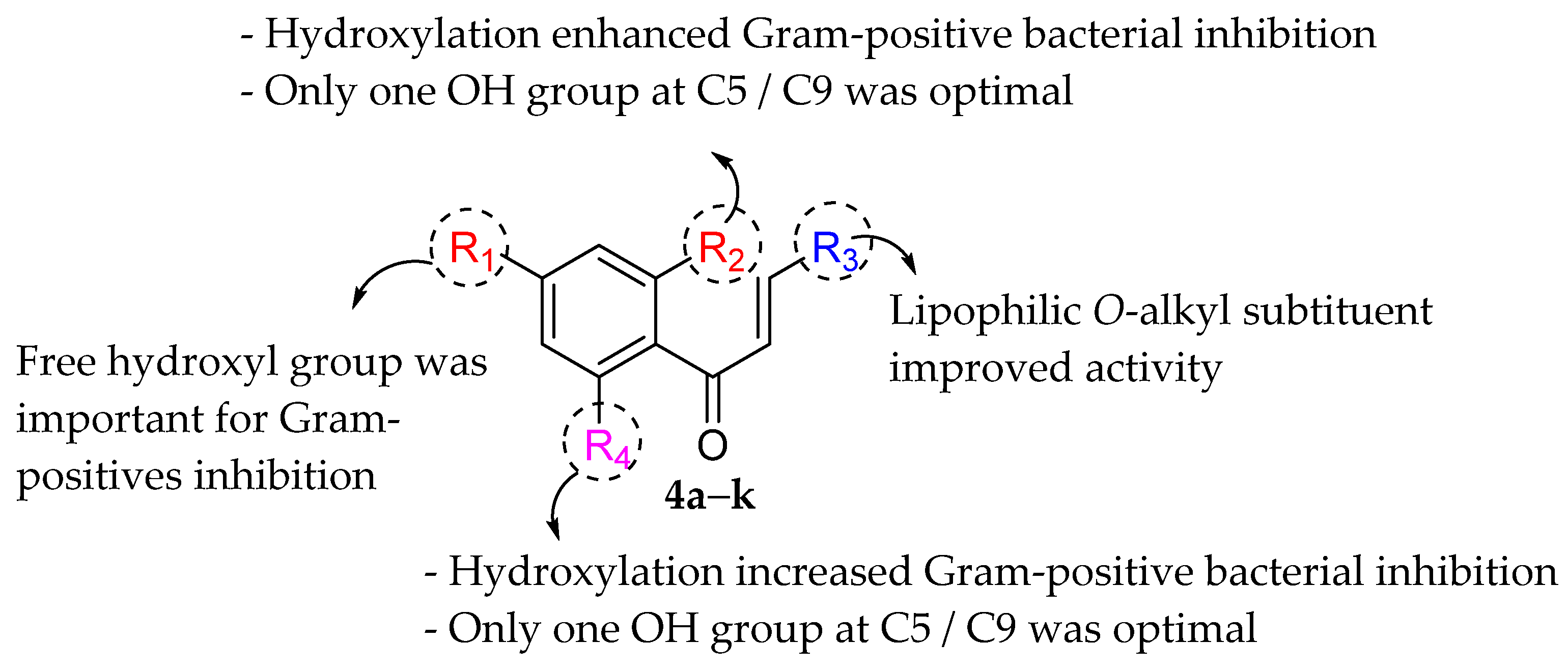
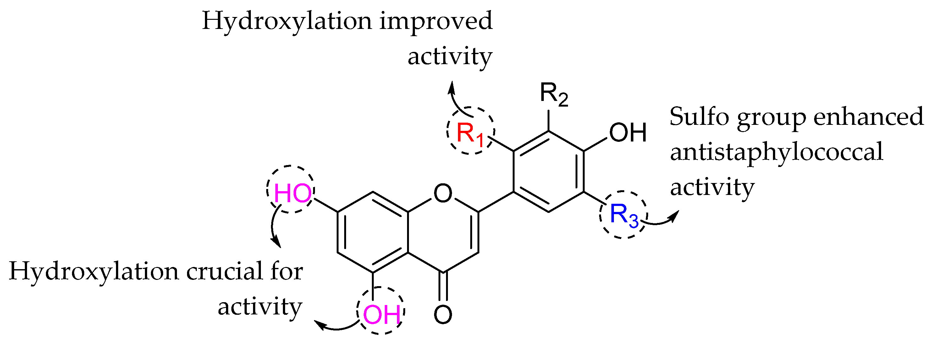
| Com. No. | R1 | R2 | R3 | MIC (μg/mL) | |||||
|---|---|---|---|---|---|---|---|---|---|
| * B.c | * B.s | * E.f | * L.m | * S.a | * S.e | ||||
| * 2a | H | OMe | OH | 4 | 4 | 4 | 4 | 2 | 4 |
| * 2b | H | OMe | OMe | 4 | 4 | 4 | 4 | 4 | 4 |
| * 2c | OH | OMe | H | 2 | 4 | 4 | 2 | 2 | 2 |
| * 2d | H | OMe | H | 4 | 8 | 8 | 4 | 8 | 4 |
| * 2e | H | H | H | 4 | 4 | 4 | 4 | 8 | 4 |
| * 2f | H | OH | H | 4 | 4 | 4 | 4 | 4 | 4 |
| Ciprofloxacin and nystatin | - | - | - | 1 | 2 | 1 | 1 | 0.5 | 1 |
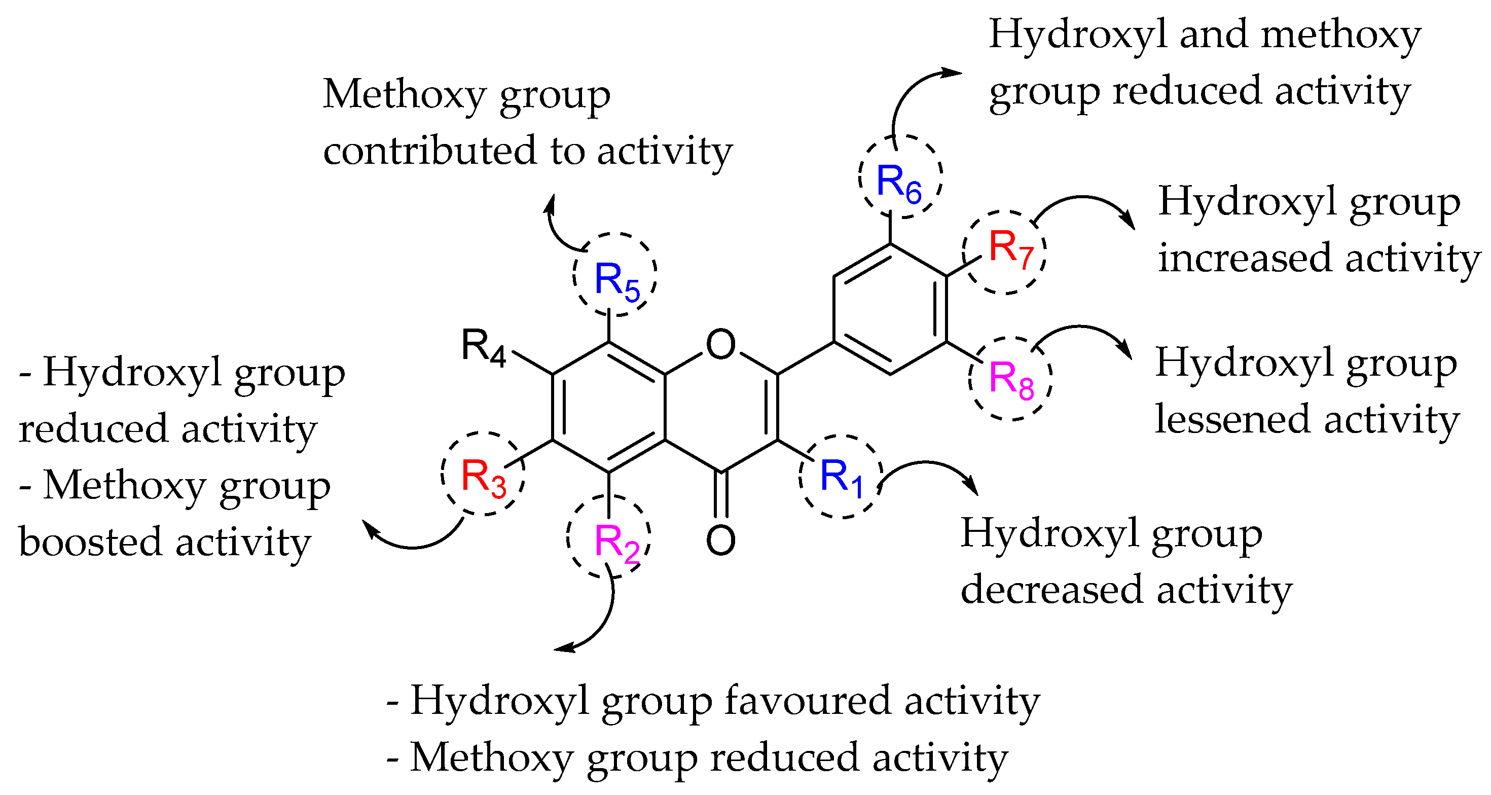
| Flavonoids | R1 | R2 | R3 | R4 | R5 | R6 | R7 | R8 | MIC50 (μg/mL) |
|---|---|---|---|---|---|---|---|---|---|
| Tangeritin | H | OMe | OMe | OMe | OMe | H | OMe | H | 137 |
| 5,6,7,4′-tetramethoxyflavone | H | OMe | OMe | OMe | H | H | OMe | H | 156 |
| Nobiletin | H | OMe | OMe | OMe | OMe | OMe | OMe | H | 177 |
| Chrysin | H | OH | H | OH | H | H | H | H | 37 |
| Galangin | OH | OH | H | OH | H | H | H | H | 53 |
| Quercetin | OH | OH | H | OH | H | OH | OH | H | 36 |
| Baicalein | H | OH | OH | OH | H | H | H | H | 71 |
| Luteolin | H | OH | H | OH | H | OH | OH | H | 67 |
| Kaempferol | OH | OH | H | OH | H | H | OH | H | 25 |
| Myricetin | OH | OH | H | OH | H | OH | OH | OH | 142 |
| Flavones/Flavonols | MIC50 (μg/mL) | Isoflavones | MIC50 (μg/mL) |
|---|---|---|---|
| Chrysin | 36.72 | Daidzein | 120.0 |
| Kaempferol | 25.00 | Puerarin | 1500 |
| 5,6,7,4′-tetramethoxyflavone | 156.3 | Genistin | 238.0 |
| Luteolin | 67.25 | Ononin | 712.5 |
| Baicalein | 70.94 | ||
| Quercetin (Flavonol) | 35.76 | ||
| Tangeritin | 137.1 |
| Flavonoids | MIC (μg/μL) | |||||||
|---|---|---|---|---|---|---|---|---|
| E. cloacae | E. coli | K. pneumoniae | P. mirabilis | B. cereus | B. coagulans | B. subtilis | S. aureus | |
| Naringenin | 2 | 4 | >4 | 2 | 2 | 2 | 2 | >4 |
| Pinocembrin | 1 | 4 | 1 | 4 | 2 | 1 | 1 | >4 |
| 7-O-Methyleriodictyol | 2 | 4 | >4 | 0.5 | 2 | 1 | 2 | 4 |
| Quercetin | >4 | >4 | >4 | 0.5 | 2 | 2 | >4 | 2 |
| Galangin | 1 | 1 | 0.5 | 0.25 | 0.25 | 0.25 | 0.25 | 0.5 |
| 3-O-Methylisorhamnetin | >4 | >4 | >4 | >4 | 2 | 1 | >4 | >4 |
| 3-O-Methylgalangin | 1 | 0.5 | 0.5 | 0.25 | 0.25 | 0.38 | 0.38 | 0.5 |
| 3,7-O-Dimethylgalangin | >4 | >4 | >4 | >4 | >4 | >4 | >4 | >4 |
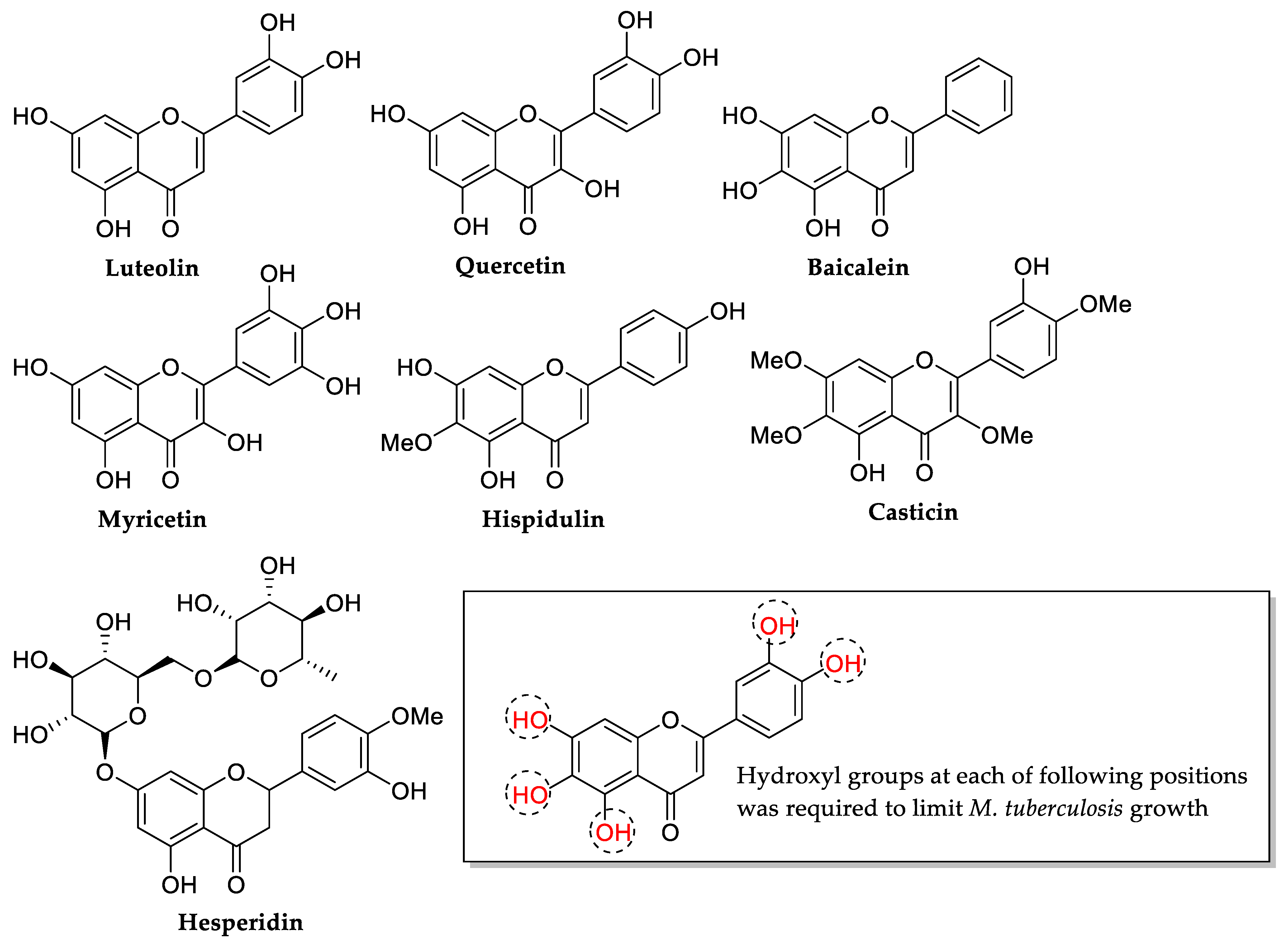
| Flavonoids | MIC (μg/mL) | ||
|---|---|---|---|
| MRSA | VRE | B. cepacia | |
| Luteolin | 512 | >512 | >512 |
| Datiscetin | 512 | >512 | >512 |
| Kaempferol | >512 | >512 | >512 |
| Quercetin | 256 | 512 | >512 |
| Myricetin | 128 | 128 | 32 |
| Flavonoids | MIC (mM) | Flavonoids | MIC (mM) | ||
|---|---|---|---|---|---|
| S. aureus | S. epidermidis | S. aureus | S. epidermidis | ||
| Corylifol A | 0.147 | 0.147 | Bavachin | 0.037 | 0.037 |
| Corylifol B | 0.037 | 0.037 | Bavachinin | 0.018 | 0.018 |
| Corylifol C | >0.147 | >0.147 | Corylin | >0.147 | >0.147 |
| Neobavaisoflavone | 0.037 | 0.037 | 1-[2,4-dihydroxy-3-(2-hydroxy-3-methyl-3-butenyl)phenyl]-3-(4-hydroxyphenyl)-2-propen-1-one | >0.147 | >0.147 |
| Isobavachalcone | 0.018 | 0.009 | 8-prenyldaidzein | >0.147 | >0.147 |
| 7,8-dihydro-8-(4-hydroxyphenyl)-2,2-dimethyl-2H,6H-benzo[1,2-b:5,4-b′]dipyran-6-one | 0.037 | 0.037 | Bakuchalcone | >0.147 | >0.147 |
| Isoneobavaiso-flavone | 0.073 | 0.037 | Brosimacutin G | >0.147 | >0.147 |
| Bavachalcone | 0.037 | 0.018 | Erythrinin A | 0.018 | 0.018 |
| Bakuchiol (Control) | 0.037 | 0.018 | Magnolol (Control) | 0.037 | 0.018 |
| Com. No. | R1 | R2 | R3 | R4 | R5 | R6 | MIC (μg/mL) | |||||
|---|---|---|---|---|---|---|---|---|---|---|---|---|
| * B.e | * E.f | * L.m | * S.a | * S.e | * S.p | |||||||
| * 3a | H | OMe | OH | H | H | OMe | - | - | - | - | - | - |
| * 3b | H | OMe | OMe | H | H | OMe | - | - | - | - | - | - |
| * 3c | H | H | OMe | H | H | OMe | - | - | - | - | - | - |
| * 3d | OMe | H | H | OMe | H | OMe | - | - | - | - | - | - |
| * 3e | H | OMe | OH | H | H | H | - | - | - | - | - | - |
| * 3f | H | OH | OH | H | H | OH | 32 | - | 128 | 16 | 32 | 64 |
| * 3g | H | H | OH | H | OH | OH | - | 128 | 128 | - | - | - |
| * 3h | OH | H | OMe | H | H | OMe | - | - | - | - | - | - |
| * 3i | OH | H | OH | H | H | OMe | 64 | - | 64 | - | - | 32 |
| * 3j | H | H | OH | OH | H | OH | - | - | - | - | 128 | - |
| * 3k | OH | H | OMe | H | H | OH | - | - | - | - | - | - |
| * 3l | H | OMe | OH | H | H | OH | - | - | - | - | - | - |
| * 3m | H | H | OH | H | H | OH | - | - | - | - | - | - |
| * 3n | OH | H | OH | H | H | OH | 128 | - | - | - | - | 64 |
| * 3o | H | H | OH | H | H | OMe | - | - | - | - | - | - |
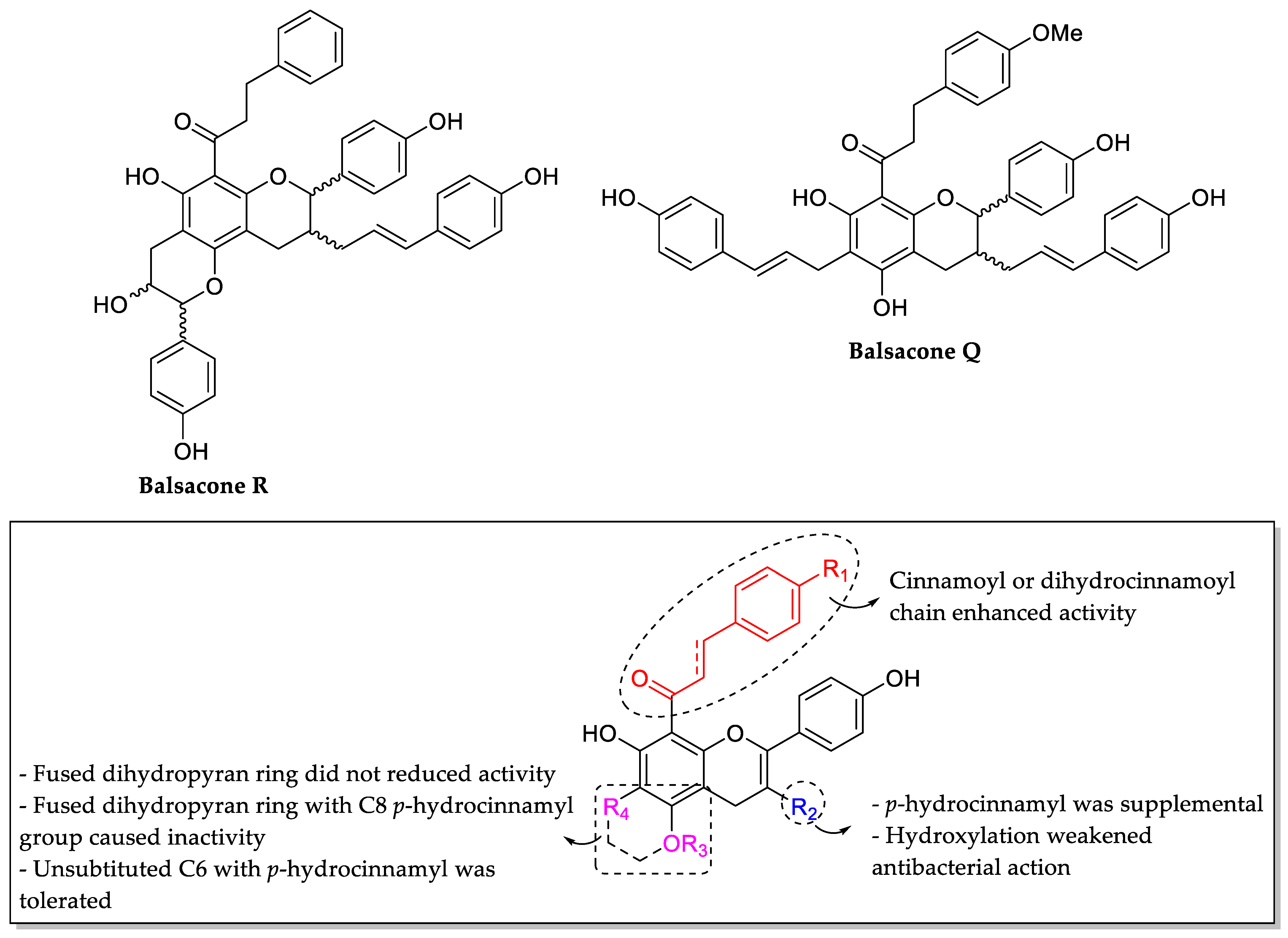
| Com. No. | R1 | R2 | R3 | R4 | MIC (μg/mL) | |||||
|---|---|---|---|---|---|---|---|---|---|---|
| M. tuberculosis | E. faecalis | MSSA | MRSA | E. coli (K12) | E. coli (ΔtolC) | |||||
| * 4a | OH | OH |  | - | 100 | 3.13 | 1.56 | 1.56 | >200 | 1.56 |
| * 4b | MOMO | OH |  | 100 | >200 | >200 | >200 | >200 | 25 | |
| * 4c | OH | OH |  | OH | >200 | 25 | >200 | 200 | >200 | 6.25 |
| * 4d | OH | OH |  | - | 200 | 12.5 | 6.25 | 0.39 | >200 | 3.13 |
| * 4e | OH | OH |  | - | >200 | 1.56 | 3.13 | 0.78 | >200 | >200 |
| * 4f | OH | OH |  | - | >200 | 1.56 | 25 | 6.25 | >200 | >200 |
| * 4g | OH | OH |  | - | 100 | 25 | 3.13 | 3.13 | >200 | 12.5 |
| * 4h | OH | OH |  | OH | >200 | 50 | >200 | 25 | >200 | 12.5 |
| * 4i | OH | - |  | - | 200 | 100 | 12.5 | 6.25 | >200 | 6.25 |
| * 4j | OH | OH |  | - | 50 | >200 | 6.25 | 12.5 | >200 | 3.13 |
| * 4k | OH | OH |  | - | 50 | 100 | 25 | >200 | >200 | >200 |
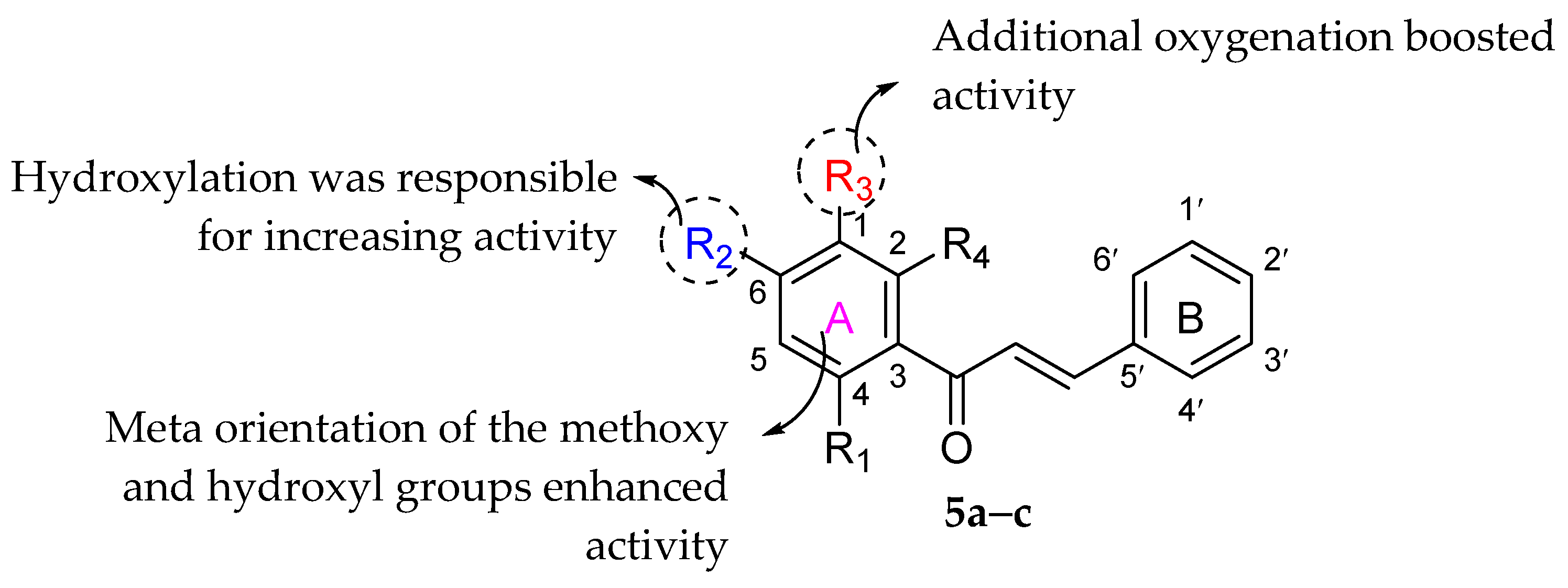
| Com. No. | R1 | R2 | R3 | R4 | MIC (μg/mL) | |||
|---|---|---|---|---|---|---|---|---|
| MRSA3 | MRSA4 | MRSA6 | MRSA8 | |||||
| * 5a | OH | OH | H | OMe | - | 64 | 128 | - |
| * 5b | OMe | OH | OMe | OH | 128 | 16 | 32 | 64 |
| * 5c | OCOMe | OMe | H | OCOMe | - | 128 | - | - |
| Compounds | MIC (μg/mL) | |||||||
|---|---|---|---|---|---|---|---|---|
| S. aureus (ATCC 29213) | MRSA (T144) | MRSA (65322) | E. faecium | VRE (CAU 383) | VRE (CAU 369) | E. coli (ATCC 25922) | E. coli (B2) | |
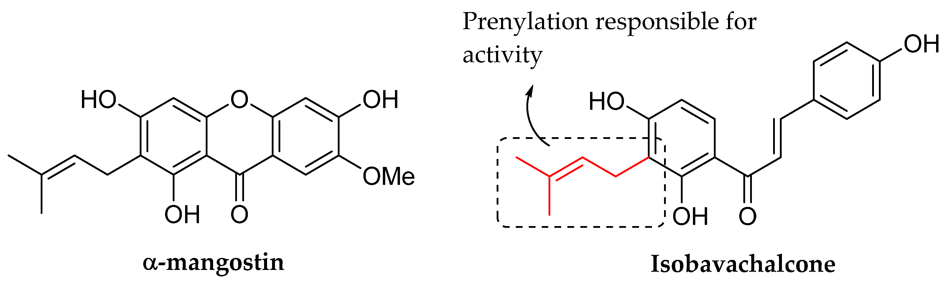
| α-mangostin | 1 | 1 | 0.5 | 0.5 | 0.5 | 2 | >128 | >128 |
| Isobavachalcone | 4 | 4 | 4 | 8 | 4 | 1 | >128 | >128 |
| Vancomycin | 1 | 0.5 | 1 | >128 | >128 | >128 | >128 | >128 |
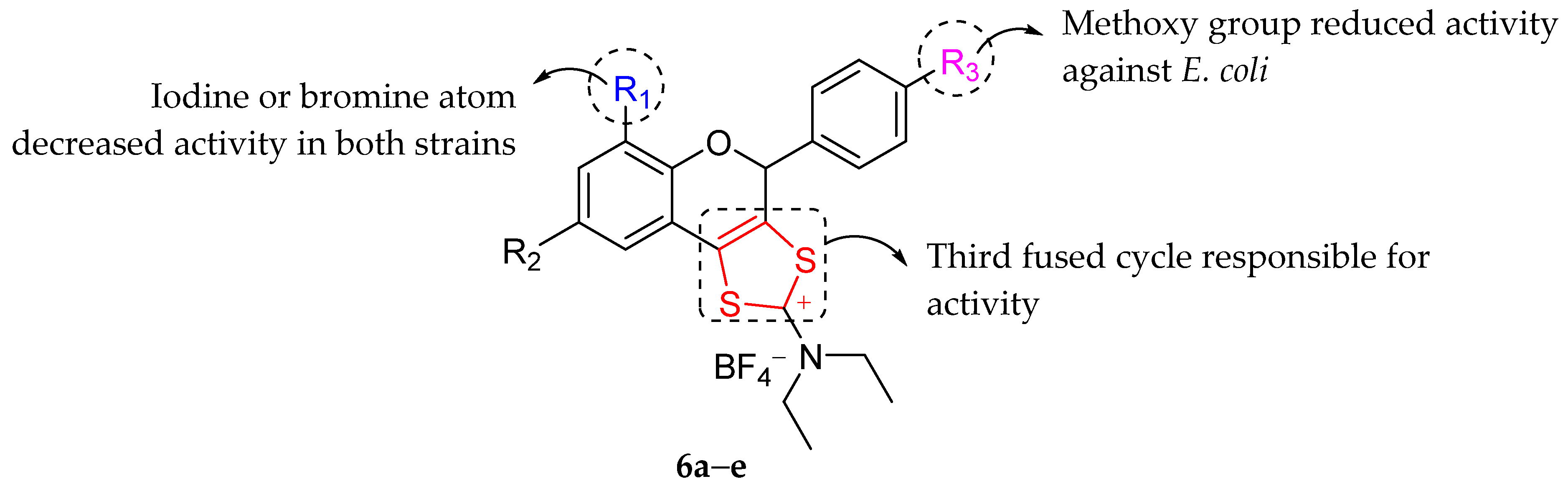
| Com. No. | R1 | R2 | R3 | MIC (μg/mL) | |
|---|---|---|---|---|---|
| S. aureus | E. coli | ||||
| * 6a | H | Br | Cl | 0.48 | 3.9 |
| * 6b | H | Br | OMe | 1.95 | 125 |
| * 6c | I | I | Cl | 0.97 | 15.62 |
| * 6d | Br | Br | Cl | 1.95 | 7.81 |
| * 6e | H | H | Cl | 1.95 | 62.5 |
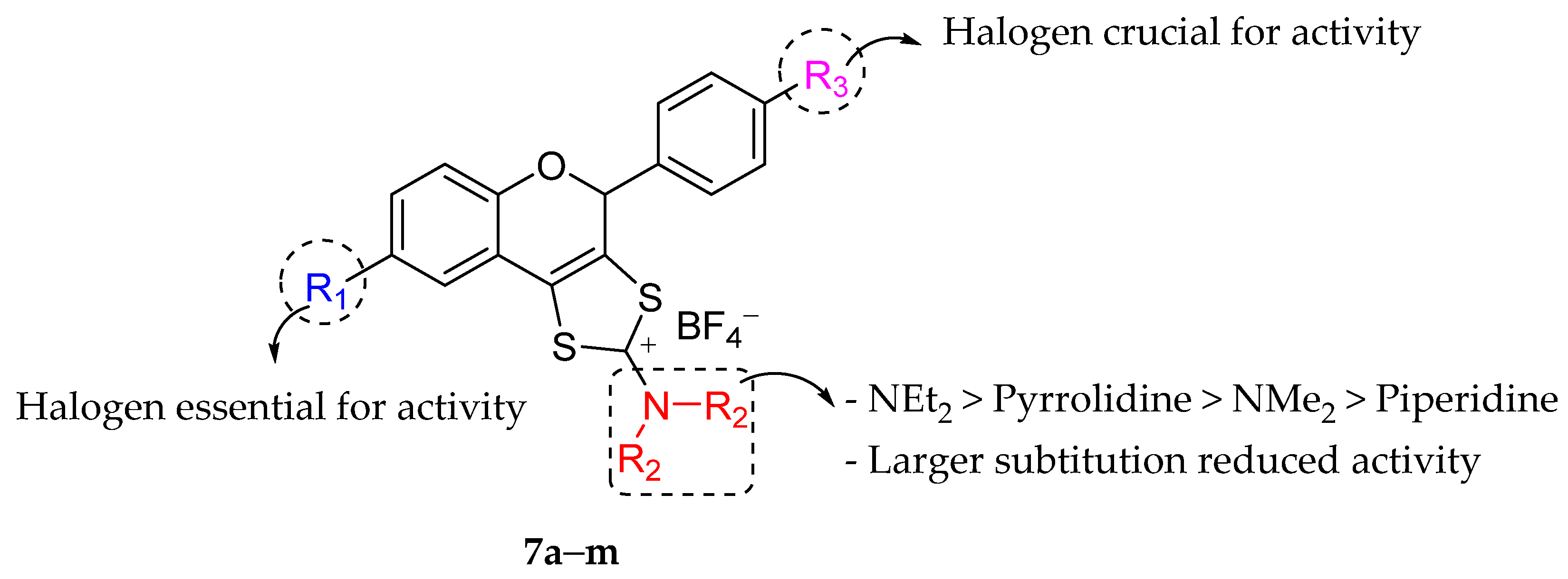
| Com. No. | R1 | R2 | R3 | MIC (μg/mL) | Com. No. | R1 | R2 | R3 | MIC (μg/mL) | ||
|---|---|---|---|---|---|---|---|---|---|---|---|
| S. aureus | E. coli | S. aureus | E. coli | ||||||||
| * 7a | Br | NMe2 | Cl | 7.81 | 15.62 | * 7i | I | NEt2 | Cl | 0.48 | 3.9 |
| * 7b | Br | Pyrrolidine | Cl | 1.95 | 3.90 | * 7j | I | NEt2 | Br | 0.48 | 3.9 |
| * 7c | Br | Piperidine | Cl | 62.5 | 125 | * 7k | I | NEt2 | I | 0.48 | 3.9 |
| * 7d | Br | NEt2 | F | 1.95 | 15.62 | * 7l | I | NEt2 | H | 0.97 | 7.81 |
| * 7e | Br | NEt2 | Br | 0.48 | 3.9 | * 7m | H | NEt2 | H | 62.5 | 62.5 |
| * 7f | Br | NEt2 | I | 0.48 | 3.9 | Kanamycin | - | - | - | 1.95 | 7.81 |
| * 7g | Br | NEt2 | H | 1.95 | 7.81 | Ampicillin | - | - | - | 7.81 | 7.81 |
| * 7h | I | NEt2 | F | 1.95 | 7.81 | ||||||
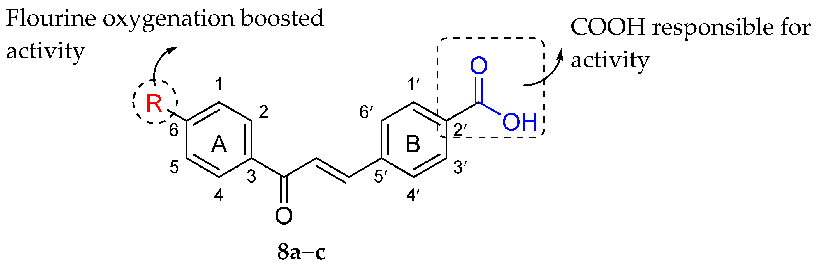
| Com. No. | R | Antibacterial Action against S. aureus | |
|---|---|---|---|
| Inhibition Zone (mm) | MIC (ppm) | ||
| * 8a | H | 11 | 92 |
| * 8b | F | 13 | 88 |
| * 8c | Cl | 9 | 93 |
| Ampicillin | - | 11 | - |
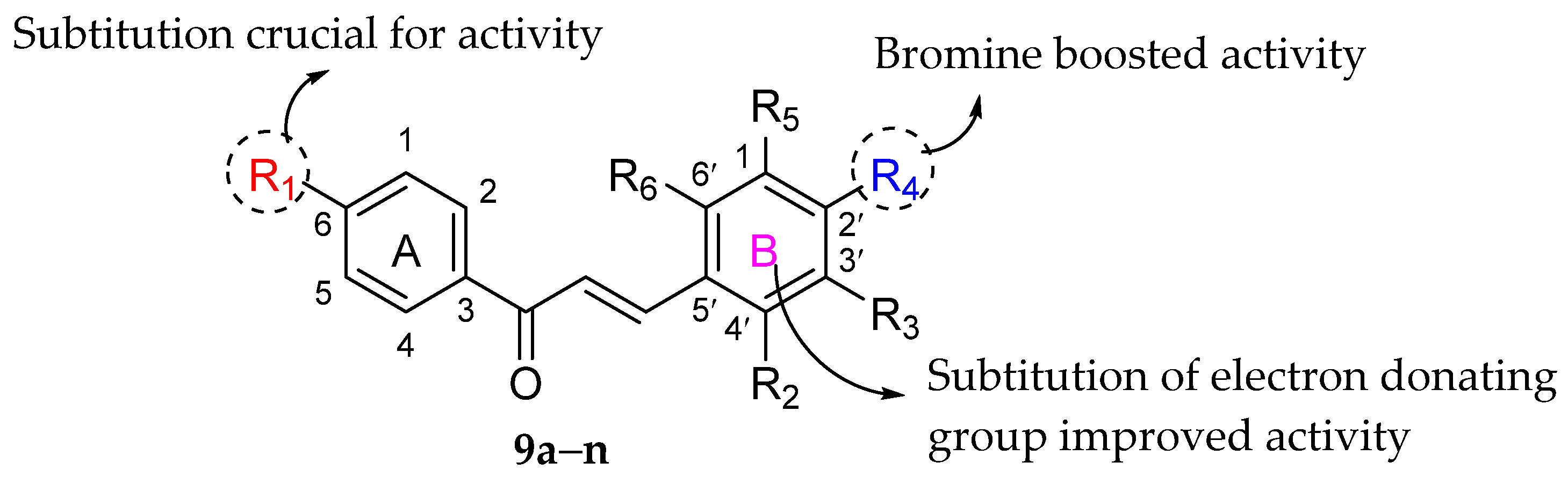
| Com. No. | R1 | R2 | R3 | R4 | R5 | R6 | MIC (μg/mL) | ||||
|---|---|---|---|---|---|---|---|---|---|---|---|
| S. aureus | B. subtilis | E. coli | P. aeruginosa | S. enterica | |||||||
| * 9a | NO2 | H | H | OH | H | H | 4.64 | 4.64 | 2.32 | 1.16 | 2.32 |
| * 9b | NO2 | H | NO2 | H | H | H | 16.76 | 4.19 | 1.05 | 2.10 | 4.19 |
| * 9c | NO2 | H | OMe | OMe | OMe | H | 3.64 | 1.82 | 1.82 | 1.82 | 3.64 |
| * 9d | NO2 | NO2 | H | H | H | H | 2.10 | 4.19 | 2.10 | 2.10 | 4.19 |
| * 9e | NO2 | H | OEt | OH | H | H | 1.00 | 3.99 | 3.99 | 1.99 | 1.99 |
| * 9f | NO2 | H | OMe | OH | H | H | 4.18 | 2.09 | 2.09 | 2.09 | 4.18 |
| * 9g | NO2 | H | H | N(Et)2 | H | H | 3.85 | 3.85 | 1.93 | 1.93 | 1.93 |
| * 9h | NO2 | H | H | Br | H | H | 3.76 | 3.76 | 0.94 | 1.88 | 1.88 |
| * 9i | NO2 | H | H | NO2 | H | H | 4.19 | 4.19 | 2.10 | 2.10 | 4.19 |
| * 9j | NO2 | H | OMe | OMe | H | H | 1.00 | 1.99 | 1.00 | 1.99 | 3.99 |
| * 9k | NO2 | OMe | H | H | H | H | 4.41 | 2.21 | 4.41 | 2.21 | 2.21 |
| * 9l | NO2 | OH | H | H | H | H | 2.32 | 4.64 | 4.64 | 2.32 | 4.64 |
| * 9m | NH2 | H | H | Cl | H | H | 2.43 | 4.85 | 4.85 | 2.43 | 2.43 |
| * 9n | OH | H | OMe | OMe | OMe | H | 1.99 | 3.98 | 3.98 | 1.99 | 3.98 |
| Cefadroxil | - | - | - | - | - | - | 1.72 | 1.72 | 1.72 | 1.72 | 1.72 |
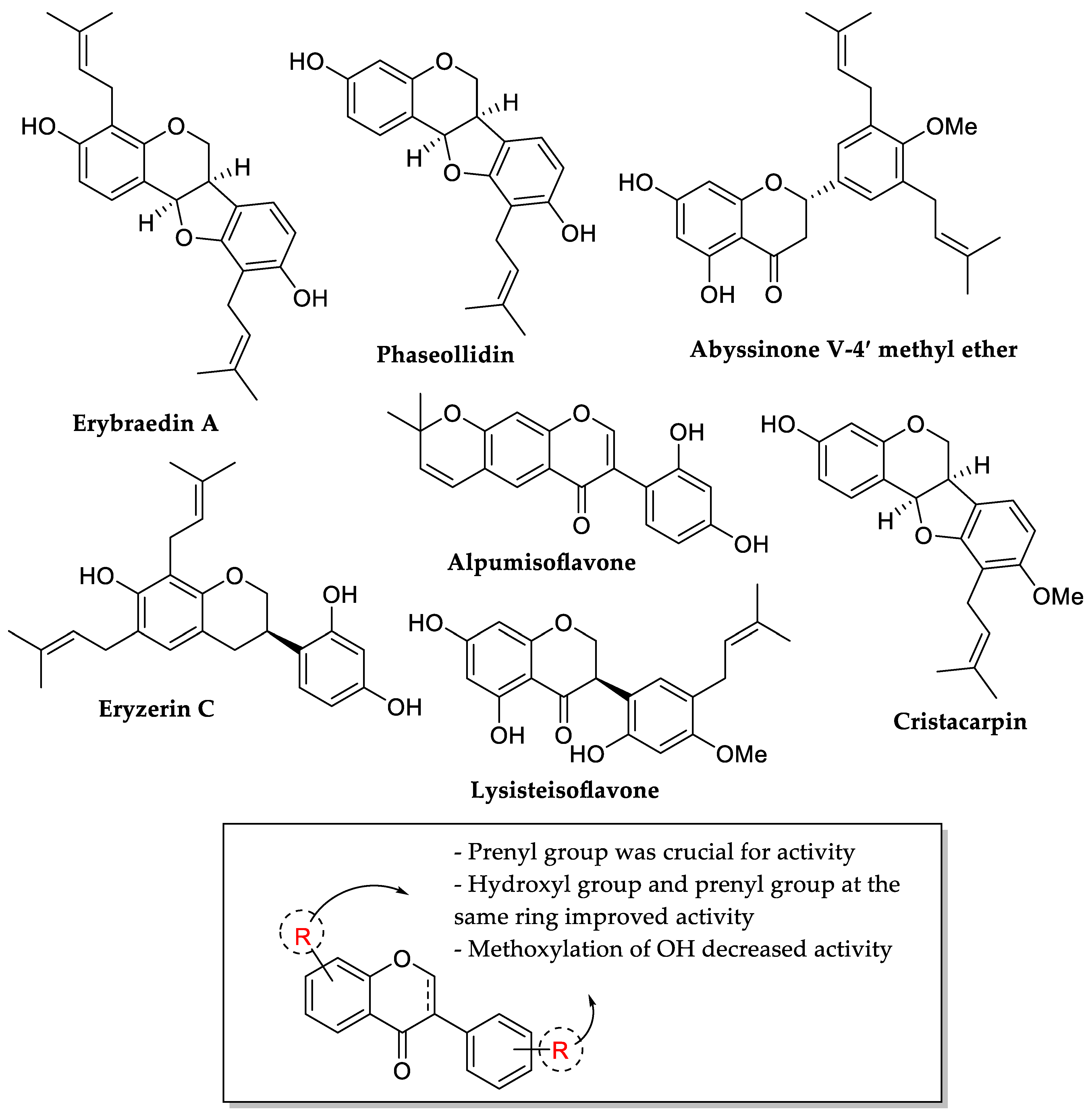
| Compounds | MIC (μg/mL) | ||||
|---|---|---|---|---|---|
| B. cereus | S. aureus | S. epidermidis | E. coli | P. aeruginosa | |
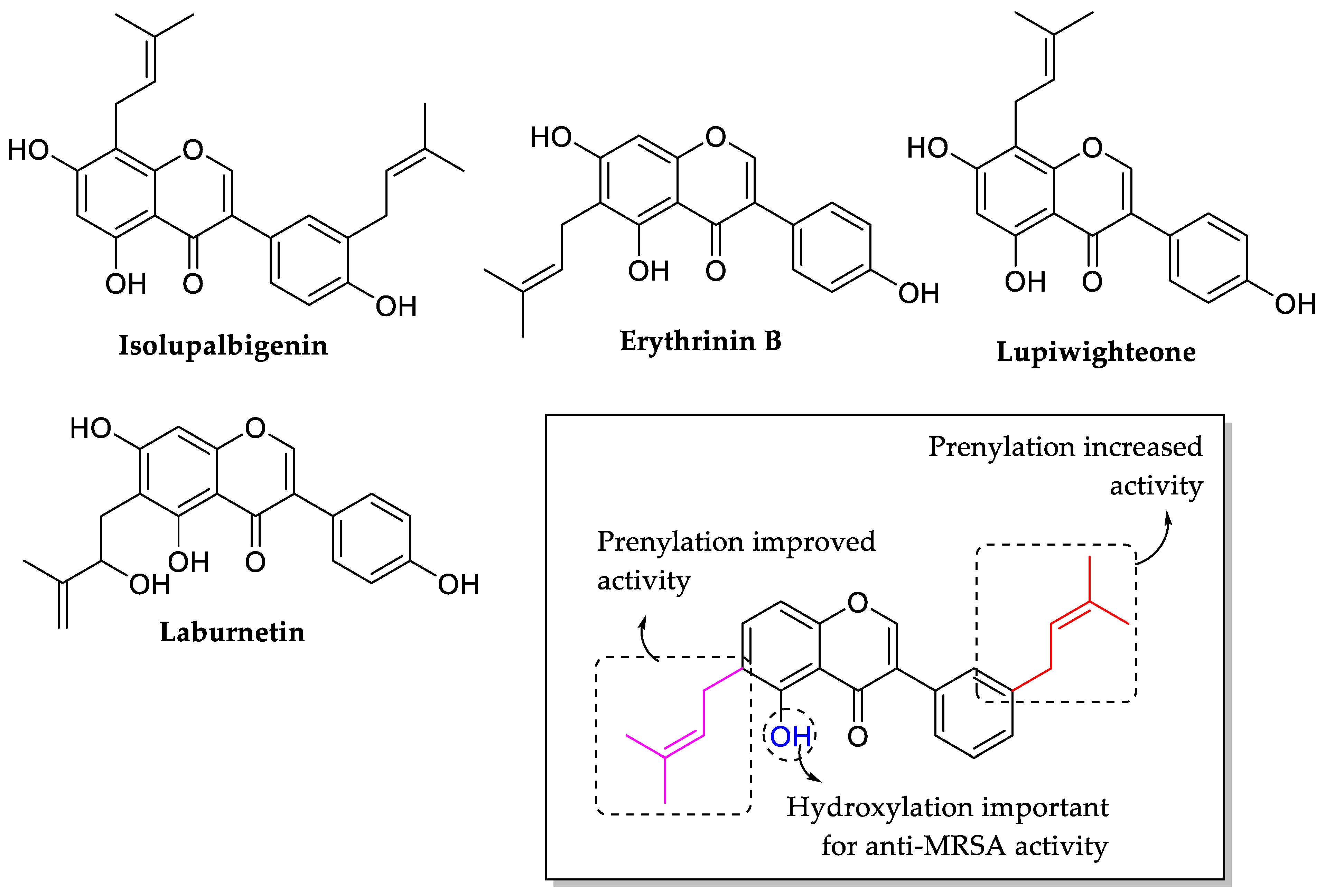
| Erybraedin A | 1 | 2 | 2 | 2 | 20 |
| Phaseollidin | 10 | 10 | 5 | 20 | 20 |
| Abyssinone V-4′ methyl ether | 26 | 59 | 117 | 260 | 260 |
| Eryzerin C | 10 | 5 | 2 | 5 | 5 |
| Alpumisoflavone | 31 | 31 | 125 | 125 | 20 |
| Cristacarpin | 156 | 156 | 412 | 625 | 78 |
| Lysisteisoflavone | 2 | 62 | 26 | 6 | 31 |
| Compounds | MIC (μg/mL) | ||||||
|---|---|---|---|---|---|---|---|
| Streptococcus mutans | S. pyrogenes | B. subtilis | S. aureus | S. epidermidis | P. aeruginosa | E. coli | |
| Dihydromorin | 31.25 | 15.62 | 62.5 | 62.5 | 31.25 | - | - |
| Norartocarpetin | 125.0 | 31.25 | 250.0 | 125.0 | 250.0 | - | - |
| Ampicillin | 0.50 | 0.50 | 0.50 | 0.50 | 0.50 | 31.25 | 0.25 |
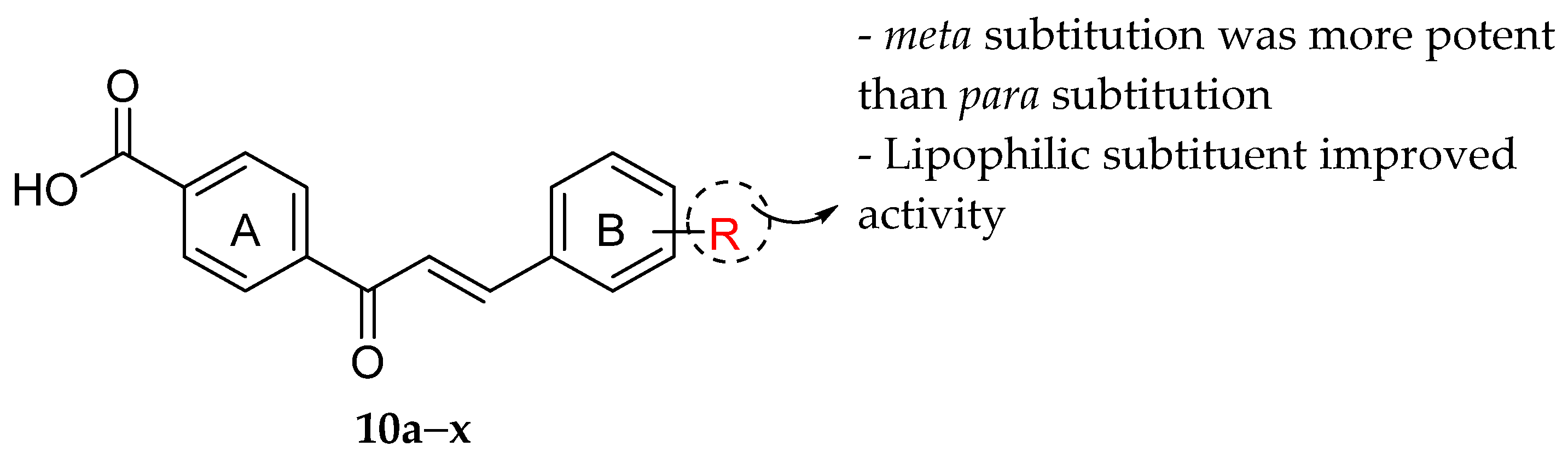
| Com. No. | R | MIC (μM) | Com. No. | R | MIC (μM) | Com. No. | R | MIC (μM) | Com. No. | R | MIC (μM) |
|---|---|---|---|---|---|---|---|---|---|---|---|
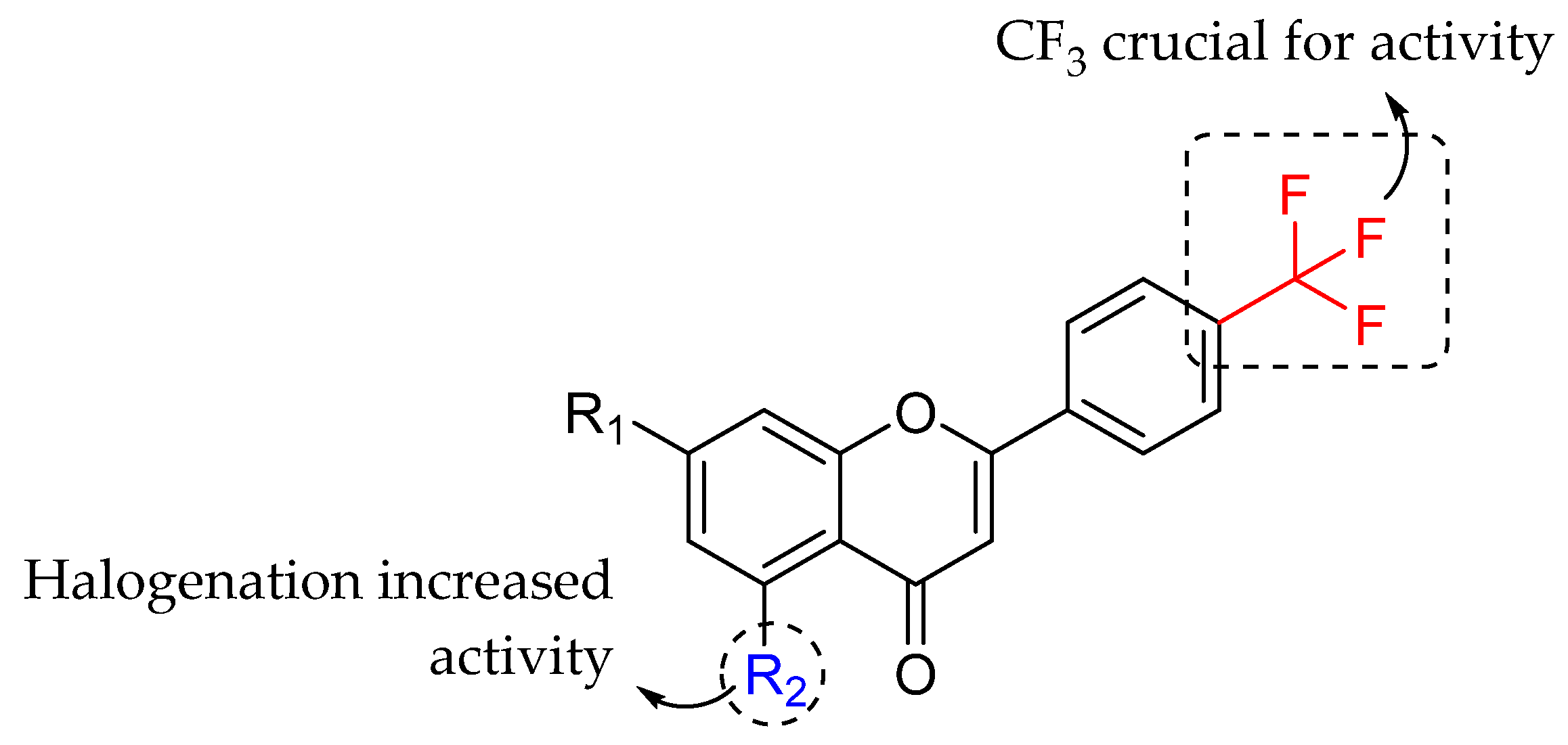
| * 10a | H | >300 | * 10g | 4-OPh | - | * 10m | 3-OPh | - | * 10s | 3,5-Di-CF3 | 2 |
| * 10b | 4-CF3 | 150 | * 10h | 3-CF3 | 40 | * 10n | 2-CF3 | 300 | * 10t | 3,5-Di-Br | 2 |
| * 10c | 4-Cl | 150 | * 10i | 3-Br | 75 | * 10o | 2-Br | 150 | * 10u | 3,5-Di-Cl | 40 |
| * 10d | 4-Me | >300 | * 10j | 3-Cl | 75 | * 10p | 2-Cl | 150 | * 10v | 3,5-Di-Me | 75 |
| * 10e | 4-OMe | >300 | * 10k | 3-NO2 | 300 | * 10q | 2-OH | >300 | * 10w | 3,5-Di-F | 150 |
| * 10f | 4-OH | >300 | * 10l | 3-OH | >300 | * 10r | 2-OBu | - | * 10x | 3,5-Di-OMe | >300 |
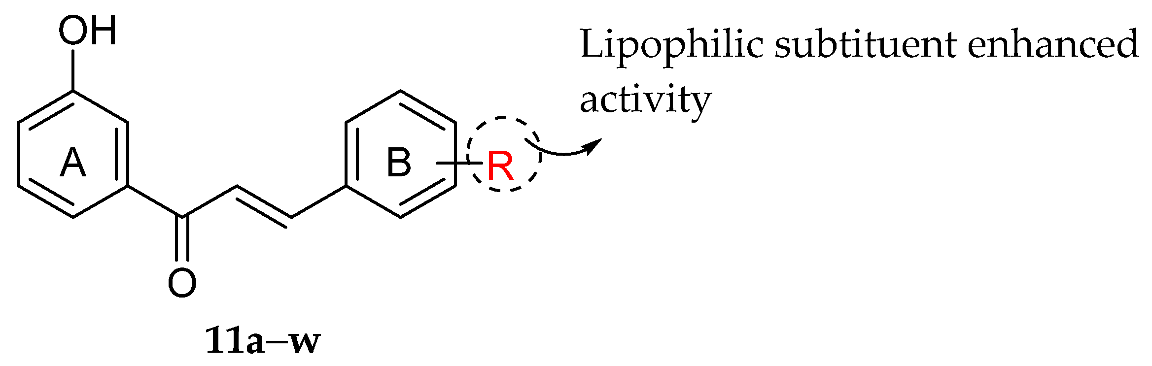
| Com. No. | R | (MIC: mg/mL) | Com. No | R | (MIC: mg/mL) | Com. No | R | (MIC: mg/mL) | Com. No | R | (MIC: mg/mL) |
|---|---|---|---|---|---|---|---|---|---|---|---|
| * 11a | H | 0.5 | * 11g | 4-OMe | 0.6 | * 11m | 2-Cl | 0.5 | * 11s | 3-NO2 | 0.8 |
| * 11b | 2-OH | 0.7 | * 11h | 3,4-OMe | - | * 11n | 3-Cl | 0.3 | * 11t | 4-NO2 | 1.0 |
| * 11c | 3-OH | 0.7 | * 11i | 3-OH, 4-OMe | - | * 11o | 4-Cl | 0.2 | * 11u | 4-NMe2 | - |
| * 11d | 4-OH | 0.8 | * 11j | 2-F | - | * 11p | 3-Br | 0.2 | * 11v | 4-Me | 0.7 |
| * 11e | 2-OMe | 0.5 | * 11k | 3-F | - | * 11q | 4-Br | - | * 11w | 2-Me, 3,4-OMe | - |
| * 11f | 3-OMe | 1.0 | * 11l | 4-F | 0.4 | * 11r | 2-NO2 | 1.0 |
| Compounds | R1 | R2 | R3 | MIC (mg/mL) | |||||
|---|---|---|---|---|---|---|---|---|---|
| E. coli | P. aeruginosa | S. aureus | |||||||
| ATCC 25922 | Clinical Isolates | ATCC 27853 | Clinical Isolates | ATCC 29213 | Clinical Isolates | ||||
| Quercetin | H | OH | H | 62.5 | 62.5 | 62.5 | 62.5 | 62.5 | 62.5 |
| Morin | OH | H | H | 3.9 | 3.9 | 3.9 | 62.5 | 31.2 | 31.2 |
| NaQSA | H | OH | SO3Na | 1000.0 | 62.5 | 31.2 | 1000.0 | 3.9 | 31.2 |
| NaMSA | OH | H | SO3Na | 62.5 | 31.2 | 31.2 | 31.2 | 3.9 | 31.2 |
| Compounds | R | MIC (μg/mL) | |||||||
|---|---|---|---|---|---|---|---|---|---|
| P. aeruginosa | E. coli | S. aureus | E. faecalis | ||||||
| ATCC 27853 | PA004 | ATCC 25922 | PA002 | ATCC 29213 | Sa1 | ATCC 29212 | S007 | ||
| * 12a | OH | >512 | >512 | >512 | >512 | >512 | >512 | >512 | >512 |
| * 12b |  | >512 | >512 | >512 | >512 | >512 | >512 | >512 | >512 |
| * 12c |  | >512 | 128 | 8 | 4 | >512 | >512 | >512 | >512 |
| * 12d |  | 256 | 32 | 2 | 2 | 128 | >512 | 8 | 8 |
| * 12e |  | 128 | 16 | 2 | 1 | 64 | 512 | 8 | 4 |
| * 12f |  | >512 | >512 | >512 | >512 | >512 | >512 | >512 | >512 |
| Com. No. | R1 | R2 | MIC (μg/mL) | ||
|---|---|---|---|---|---|
| B. subtilis | S. aureus | P. aeruginosa | |||
| * 13a | H | H | 12.5 | 25 | 25 |
| * 13b | OMe | H | 50 | 50 | 37.5 |
| * 13c | H | Br | 12.5 | 6.25 | 6.25 |
| Ciprofloxacin | - | - | 6.25 | 6.25 | 6.25 |
| Chalcones | |||||||||||
| References | Ring A | Ring B | Ring C | ||||||||
| C5 | C6 | C7 | C8 | C2′ | C3′ | C4′ | C5′ | C2 | C3 | C4 | |
| [77] | OH ↑ | OH ↑ | C2=C3 ↑ | ||||||||
| [78] | OH ↑ | OH ↑ | |||||||||
| [79] | OH ↑ OMe ↓ | Broken ring C ↑ | C=O ↑ | ||||||||
| [82] | OH ↑ | OH ↑ | O-alkyl chain ↑ | ||||||||
| [84] | OH ↑ | Oxygenation ↑ | |||||||||
| [65] | Prenyl ↑ | ||||||||||
| [92] | Halogen ↑ | COOH ↑ | |||||||||
| [94] | Bromine ↑ | ||||||||||
| [97] | Lipophilic subtituent ↑ | ||||||||||
| [98] | Lipophilic subtituent ↑ | ||||||||||
| Flavanones | |||||||||||
| References | Ring A | Ring B | Ring C | ||||||||
| C5 | C6 | C7 | C8 | C2′ | C3′ | C4′ | C5′ | C2 | C3 | C4 | |
| [71] | OH ↑ | Geranyl ↑ | OMe ↓ | OMe ↓ | OMe ↓ | ||||||
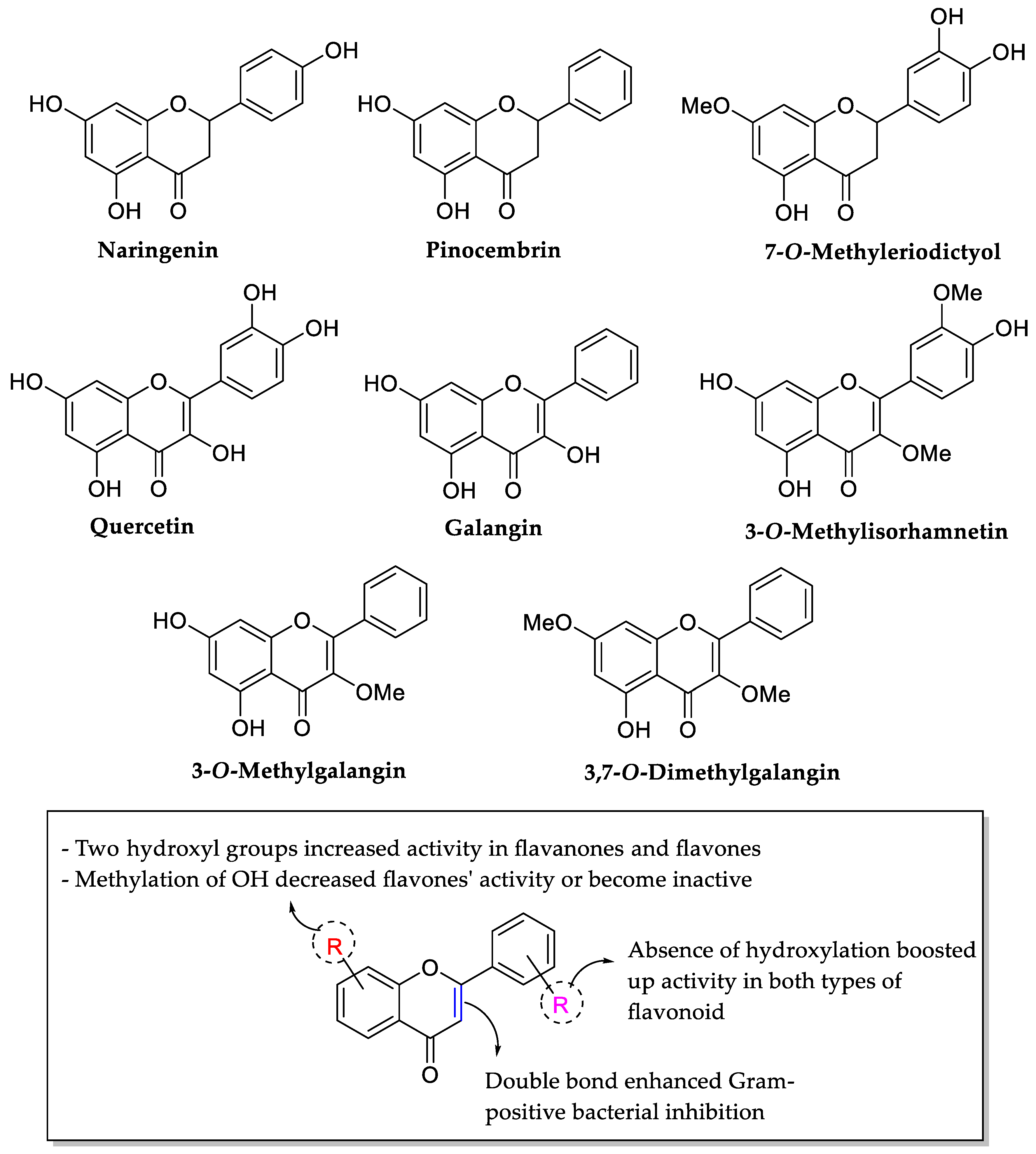
| [76] | Two hydroxyl groups ↑ | Absence of hydroxyl groups ↑ | |||||||||
| [79] | OH ↑ OMe ↓ | C=O ↑ | |||||||||
| [80] | OH ↑ | OH ↑ | Trihydroxylation ↑ Glycosylation of hydroxyl groups ↓ | Saturation of C2=C3 ↑ | |||||||
| [85] | Saturation of C2=C3 ↑ | ||||||||||
| [86] | Glycosyl ↑ | ||||||||||
| [96] | C2=C3 ↓ OH ↑ | ||||||||||
| Flavones | |||||||||||
| References | Ring A | Ring B | Ring C | ||||||||
| C5 | C6 | C7 | C8 | C2′ | C3′ | C4′ | C5′ | C2 | C3 | C4 | |
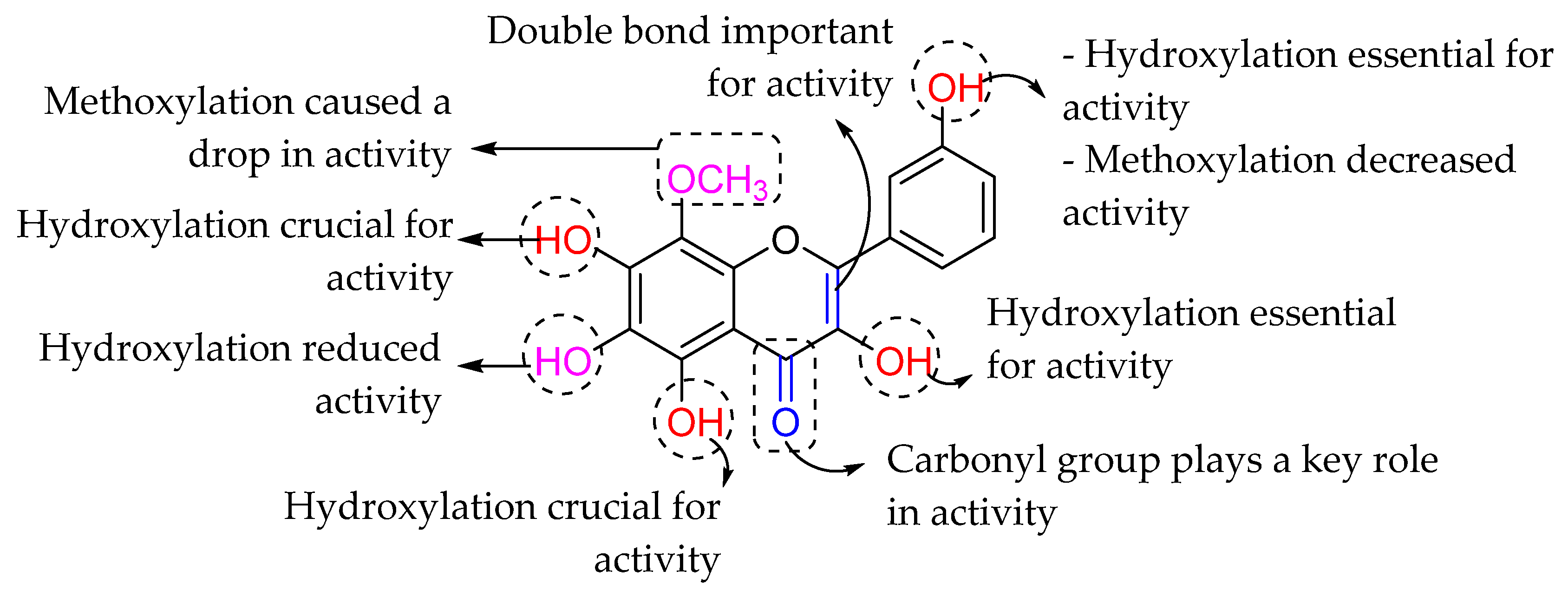
| [62] | OH ↑ OMe ↓ | OH ↓ OMe ↑ | OMe ↑ | OH ↓ OMe ↓ | OH ↑ | OH ↓ | OH ↓ | ||||
| [64] | OH ↑ | OH ↑ | OH ↑ | C2=C3 ↑ | OH ↑ | ||||||
| [73] | OMe ↑ | OH ↑ | |||||||||
| [74] | OH ↑ | OH ↑ | OH ↑ | OH ↑ | OH ↑ | ||||||
| [75] | OMe ↑ | OMe ↑ NO2 ↓ F ↑ | NO2 ↓ | ||||||||
| [76] | Two hydroxyl groups ↑ | Absence of hydroxyl groups ↑ | C2=C3 ↑ | ||||||||
| [78] | Prenyl ↑ | OMe ↑ | |||||||||
| [79] | OH ↑ OMe ↓ | ||||||||||
| [80] | OH ↑ | OH ↑ | Trihydroxylation ↑ Glycosylation of hydroxyl groups ↓ | ||||||||
| [100] | OH ↑ | OH ↑ | OH ↑ | Sulfo ↑ | |||||||
| [102] | Halogen ↑ | CF3 ↑ | |||||||||
| [104] | OH ↑ | OH ↓ | OH ↑ | OMe ↓ | C2=C3 ↑ | OH ↑ | |||||
| Isoflavones | |||||||||||
| References | Ring A | Ring B | Ring C | ||||||||
| C5 | C6 | C7 | C8 | C2′ | C3′ | C4′ | C5′ | C2 | C3 | C4 | |
| [73] | Glycosyl ↓ | Glycosyl ↓ | |||||||||
| [81] | OH ↑ OMe ↓ | OH ↑ OMe ↓ | OH ↑ OMe ↓ | OMe ↓ | OH ↑ OMe ↓ | ||||||
| [95] | Prenyl ↑ OH ↑ OMe ↓ | Prenyl ↑ OH ↑ OMe ↓ | |||||||||
| [103] | OH ↑ | Prenyl ↑ | |||||||||
| Flavan-3-ols | |||||||||||
| References | Ring A | Ring B | Ring C | ||||||||
| C5 | C6 | C7 | C8 | C2′ | C3′ | C4′ | C5′ | C2 | C3 | C4 | |
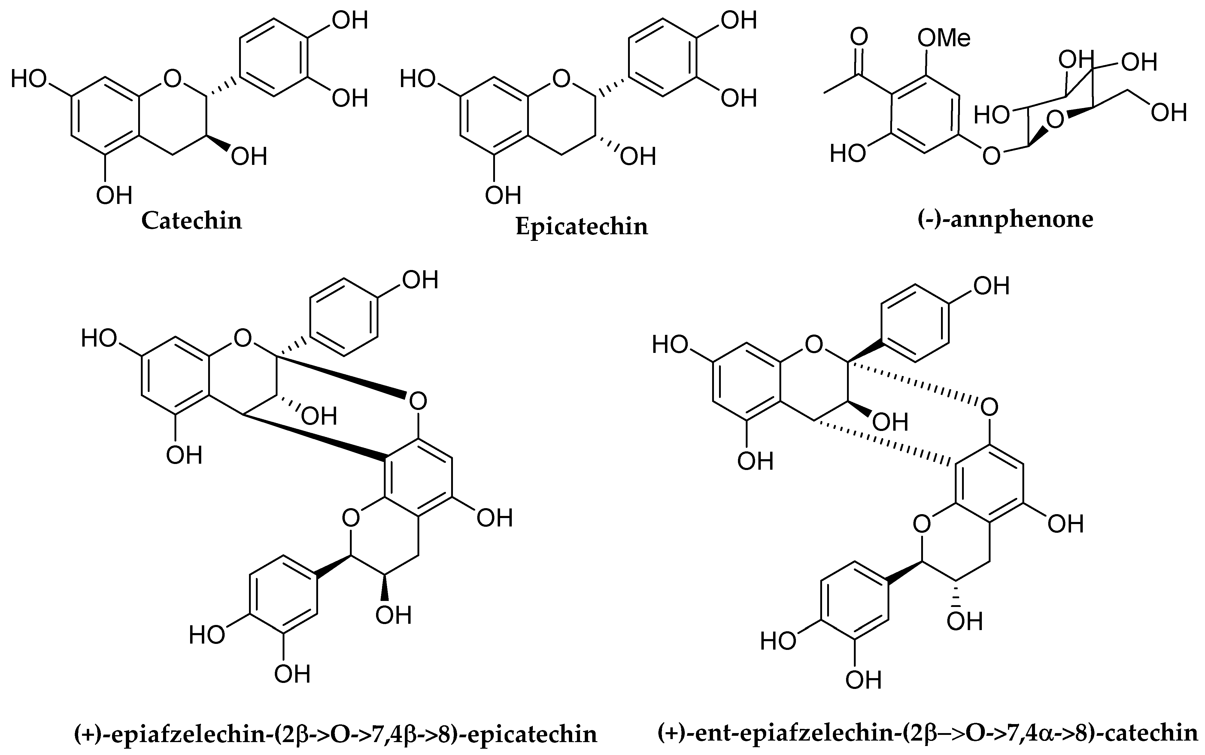
| [70] | OH ↑ | OH ↑ | OH ↑ | Long acyl chain ↑ | |||||||
| [73] | OH ↑ | ||||||||||
| Anthocyanidins | |||||||||||
| References | Ring A | Ring B | Ring C | ||||||||
| C5 | C6 | C7 | C8 | C2′ | C3′ | C4′ | C5′ | C2 | C3 | C4 | |
| [101] | Glycosyl ↑ | ||||||||||
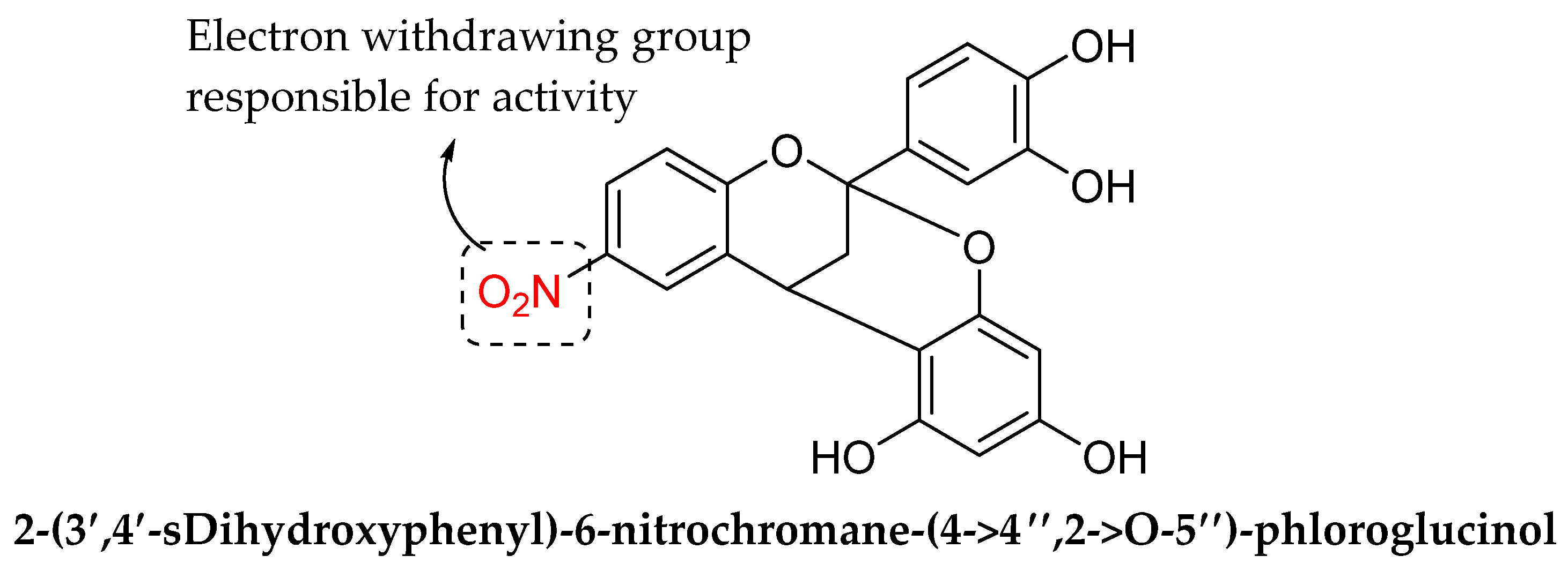
| [105] | Electron withdrawing group ↑ | ||||||||||
Publisher’s Note: MDPI stays neutral with regard to jurisdictional claims in published maps and institutional affiliations. |
© 2022 by the authors. Licensee MDPI, Basel, Switzerland. This article is an open access article distributed under the terms and conditions of the Creative Commons Attribution (CC BY) license (https://creativecommons.org/licenses/by/4.0/).
Share and Cite
Shamsudin, N.F.; Ahmed, Q.U.; Mahmood, S.; Ali Shah, S.A.; Khatib, A.; Mukhtar, S.; Alsharif, M.A.; Parveen, H.; Zakaria, Z.A. Antibacterial Effects of Flavonoids and Their Structure-Activity Relationship Study: A Comparative Interpretation. Molecules 2022, 27, 1149. https://0-doi-org.brum.beds.ac.uk/10.3390/molecules27041149
Shamsudin NF, Ahmed QU, Mahmood S, Ali Shah SA, Khatib A, Mukhtar S, Alsharif MA, Parveen H, Zakaria ZA. Antibacterial Effects of Flavonoids and Their Structure-Activity Relationship Study: A Comparative Interpretation. Molecules. 2022; 27(4):1149. https://0-doi-org.brum.beds.ac.uk/10.3390/molecules27041149
Chicago/Turabian StyleShamsudin, Nur Farisya, Qamar Uddin Ahmed, Syed Mahmood, Syed Adnan Ali Shah, Alfi Khatib, Sayeed Mukhtar, Meshari A. Alsharif, Humaira Parveen, and Zainul Amiruddin Zakaria. 2022. "Antibacterial Effects of Flavonoids and Their Structure-Activity Relationship Study: A Comparative Interpretation" Molecules 27, no. 4: 1149. https://0-doi-org.brum.beds.ac.uk/10.3390/molecules27041149