
Doxorubicin-Conjugated Zinc Oxide Nanoparticles, Biogenically Synthesised Using a Fungus Aspergillus niger, Exhibit High Therapeutic Efficacy against Lung Cancer Cells

 , , ,
, , ,  and
and 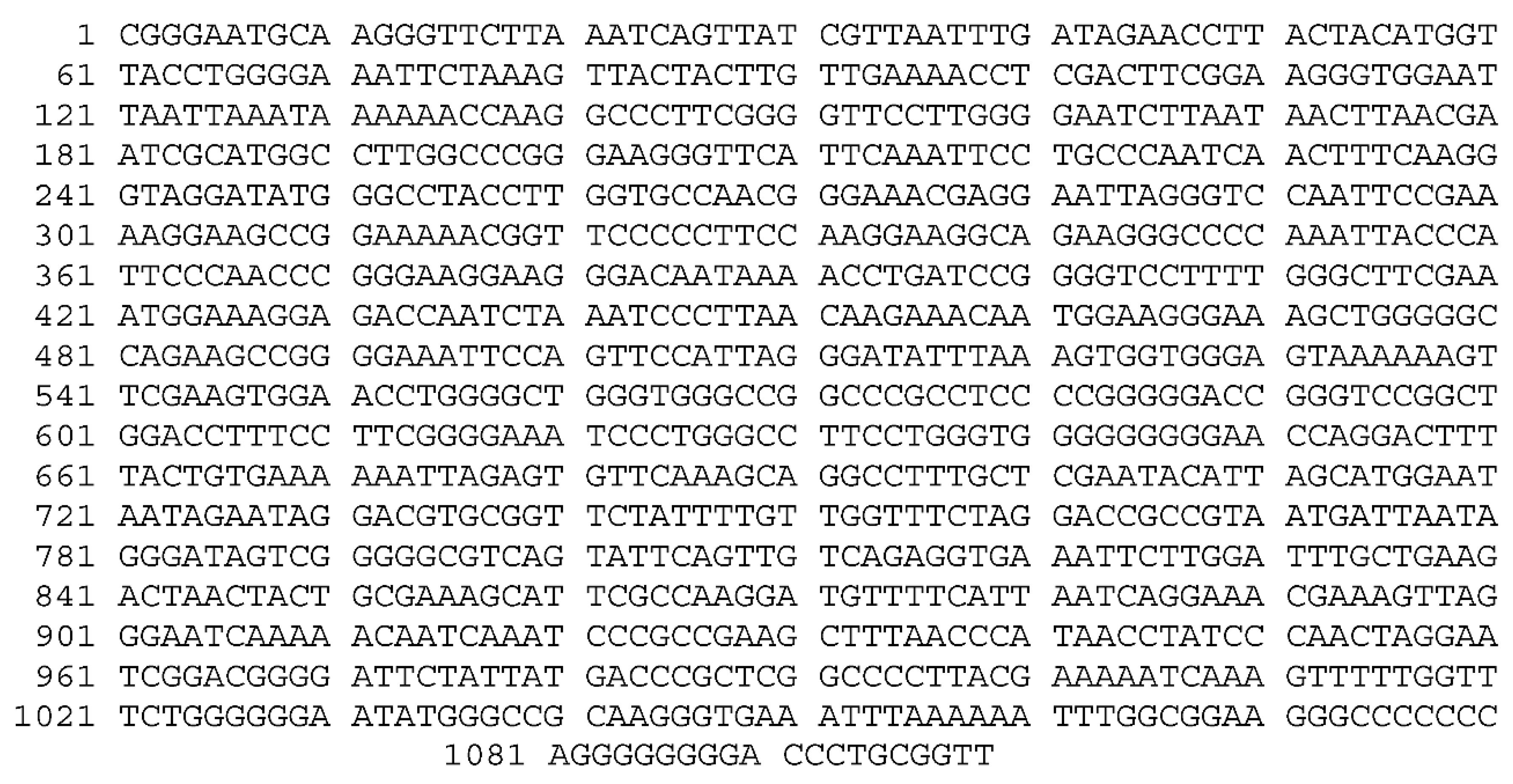
Abstract
:1. Introduction
2. Material and Methods
2.1. Materials
2.2. Methods
2.2.1. Fungus Isolation
2.2.2. Taxonomic Identification
2.2.3. Biosynthesis of ZnONPs
2.2.4. Bioconjugation of ZnONPs with Anti-Cancer Drug DOX
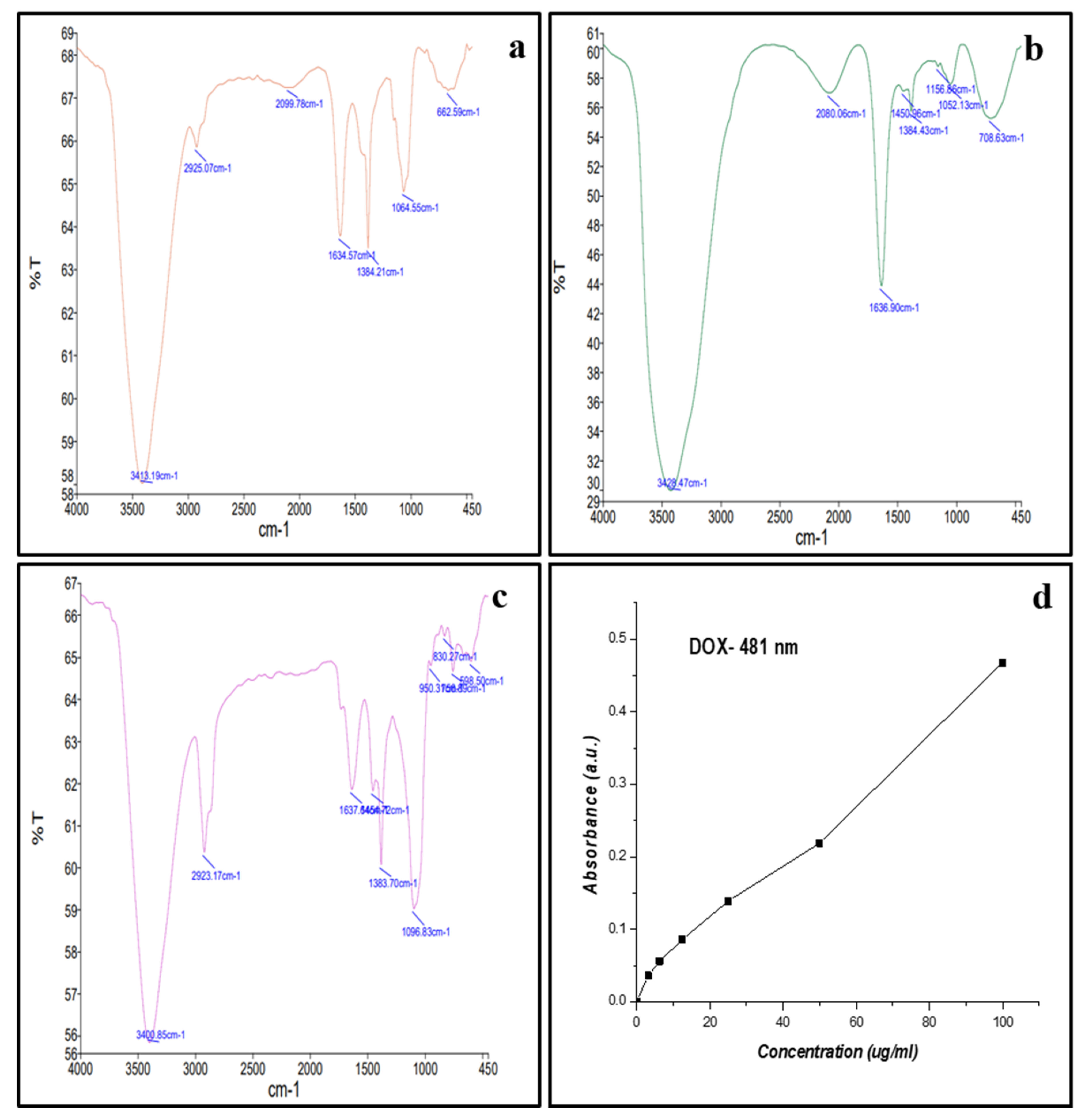
2.2.5. Estimation of Loading Efficiency of DOX on ZnONPs by UV–Visible Spectrophotometer
2.2.6. Characterisation of ZnONPs and DOX ZnONPs
2.2.7. Cell Culture
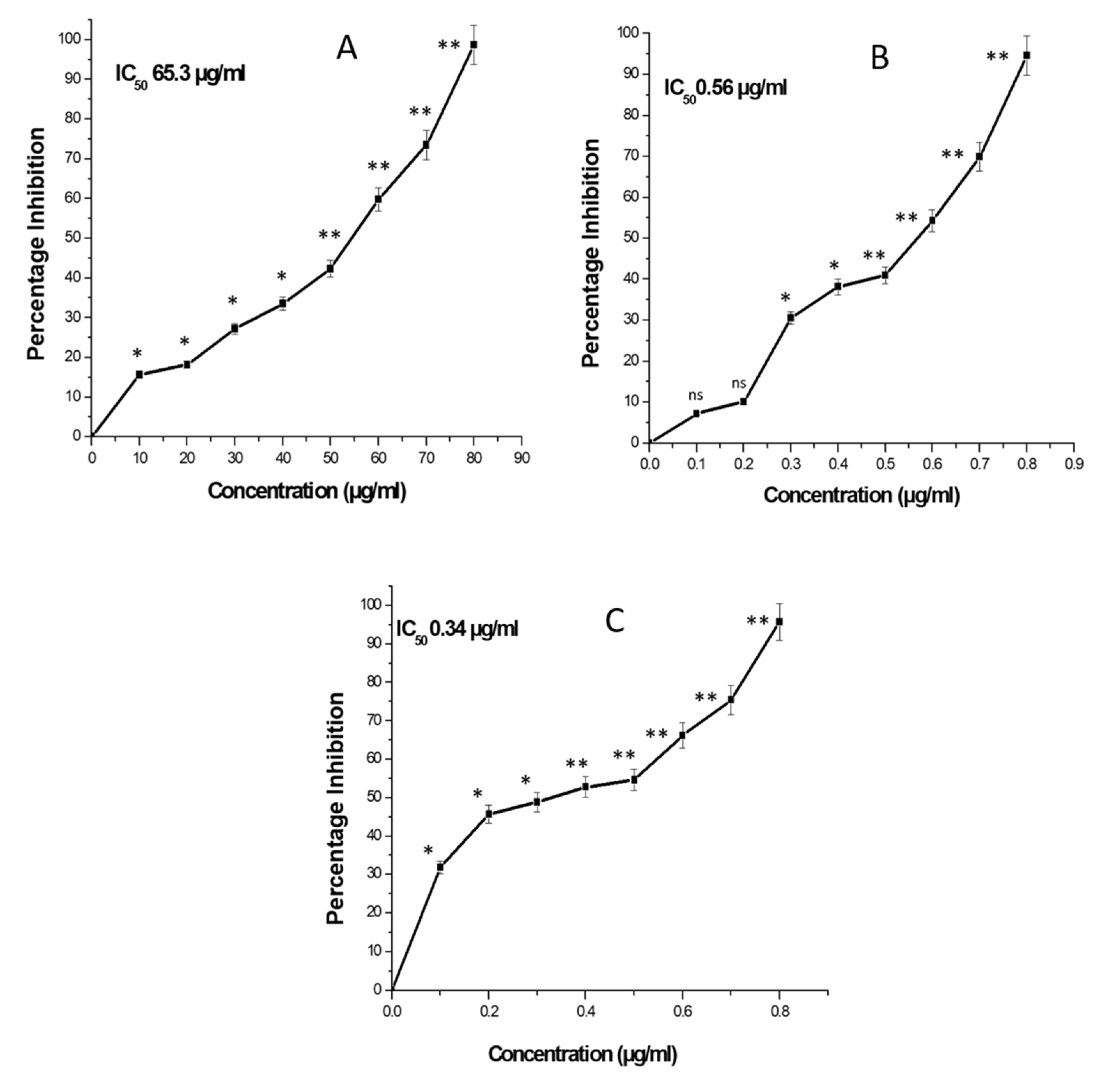
2.2.8. Measurement of Cytotoxicity
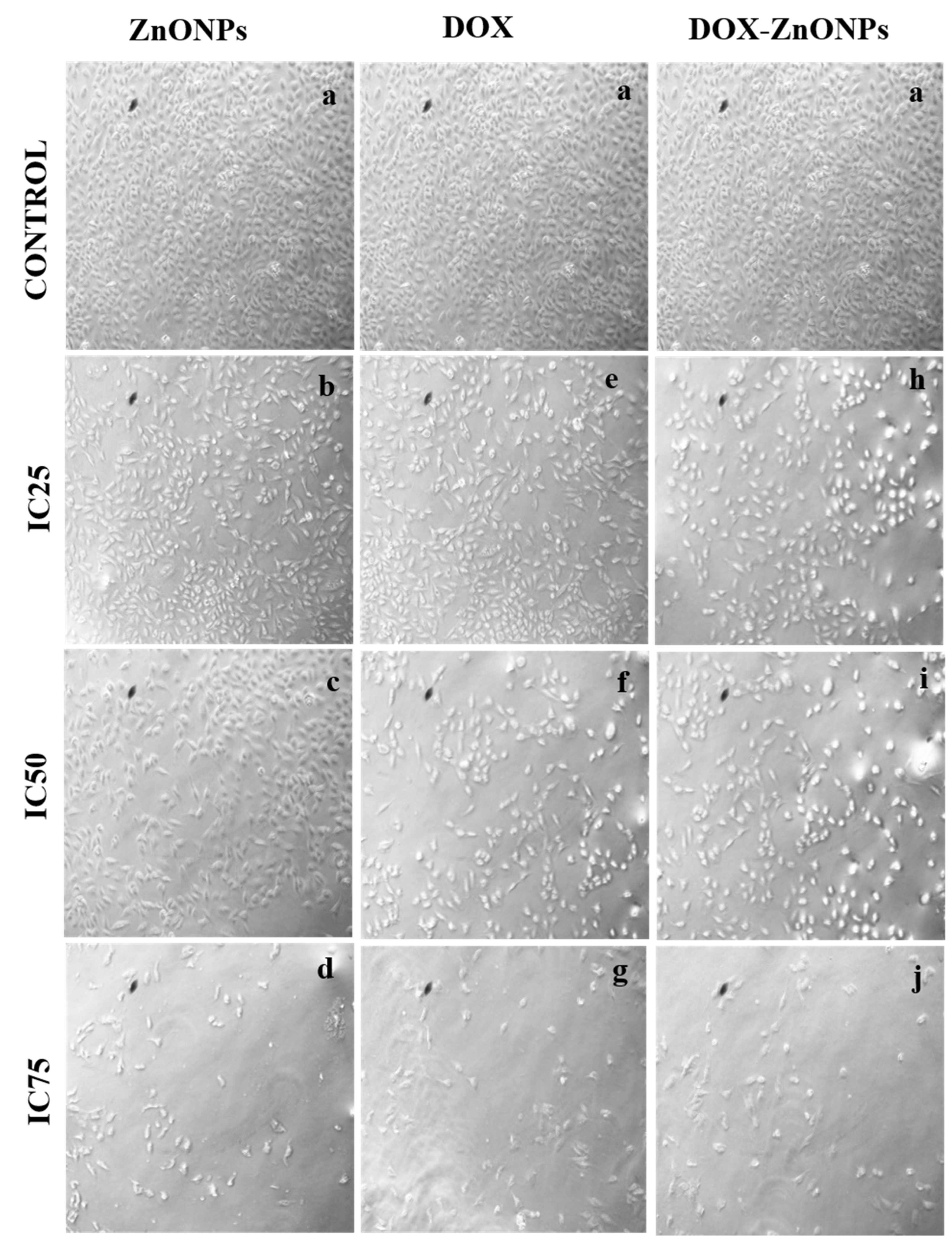
2.2.9. Assessment of Morphological Changes in A549 Cells
2.2.10. Evaluation of Intracellular Reactive Oxygen Species (ROS) Generation
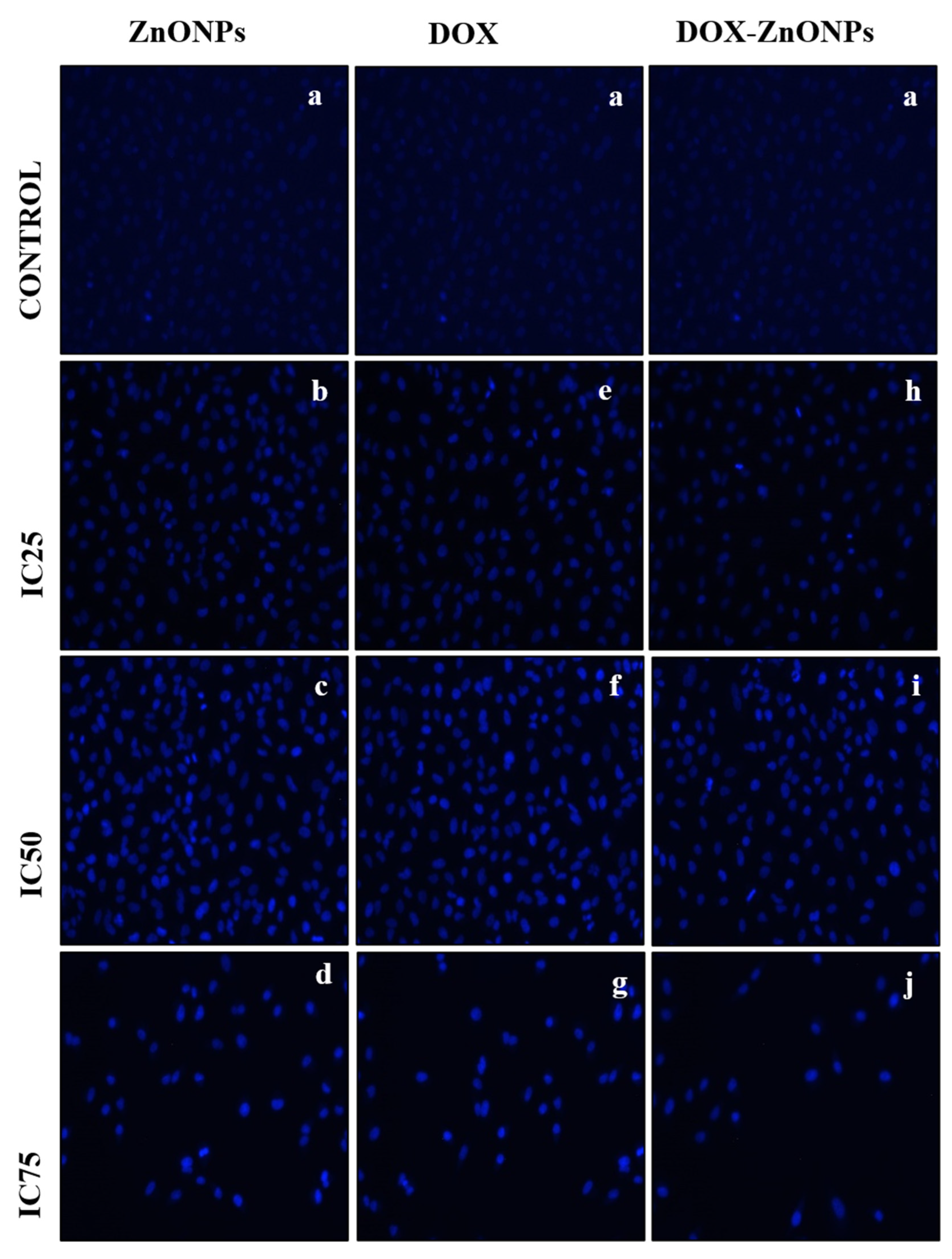
2.2.11. Analysis of Nuclear Morphological Changes
2.2.12. Assessment of Mitochondrial Membrane Potential (ΔΨ m) by Mito Tracker Red
2.2.13. Measurement of Caspase-3 Activity
2.2.14. Statistical Analysis
3. Results
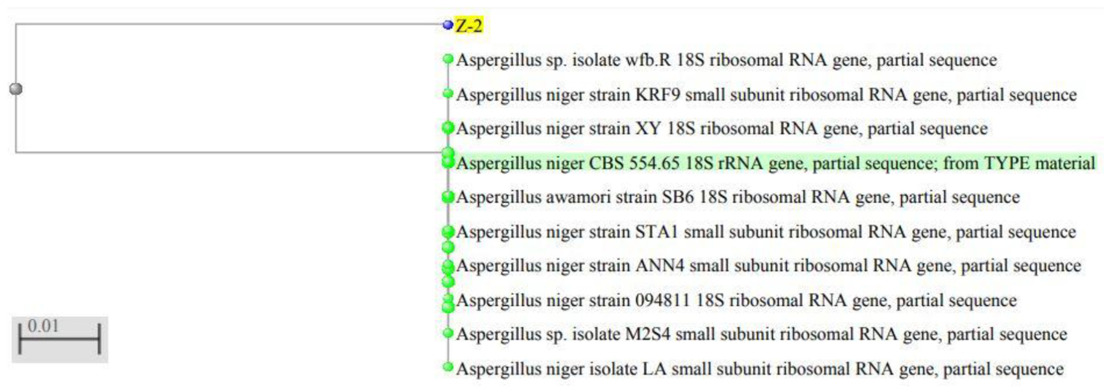
3.1. Isolation, Molecular Characterisation and Phylogenetic Analysis of Fungal Species
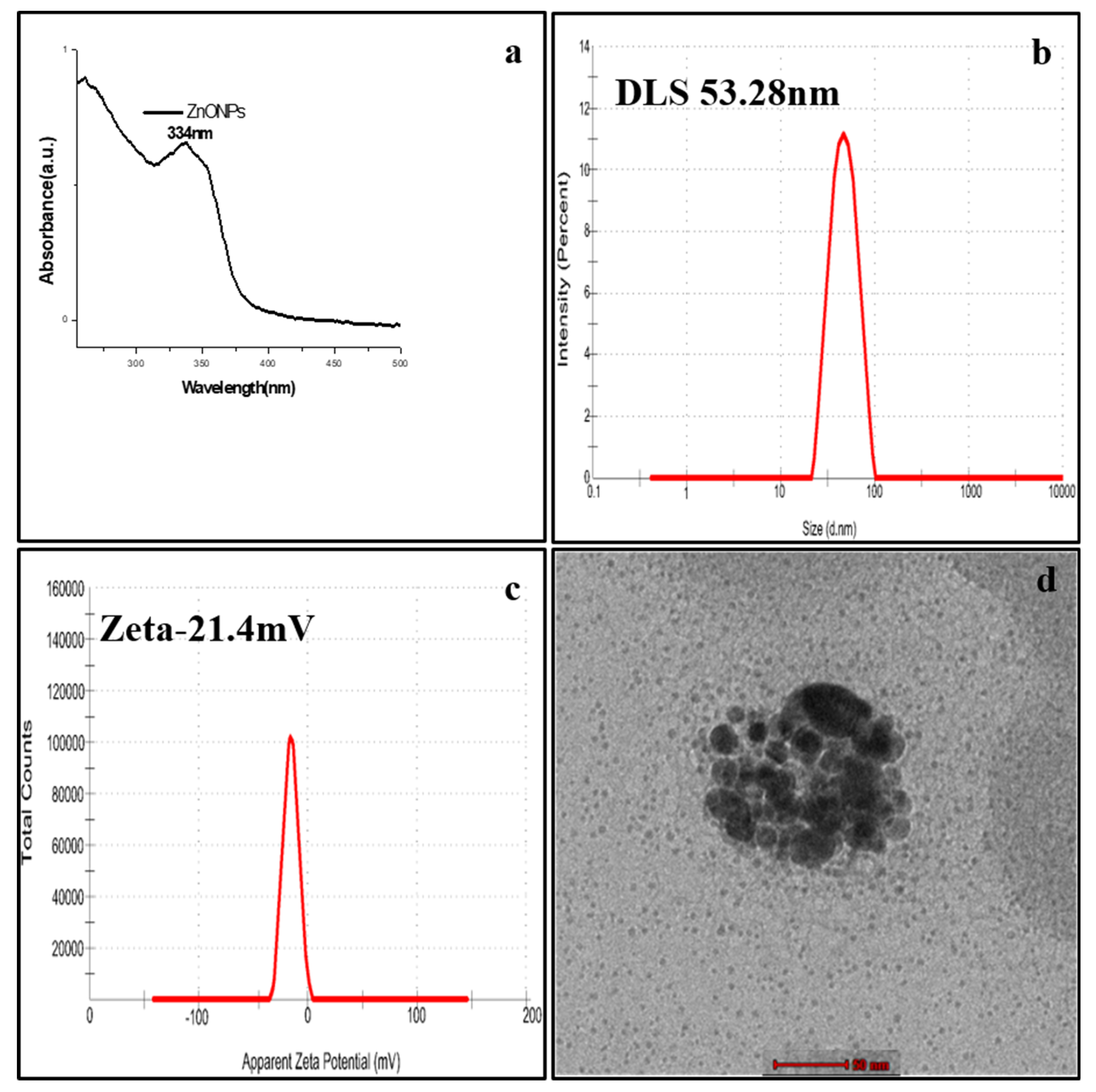
3.2. Biosynthesis of ZnONPs Nanoparticles and Characterisation
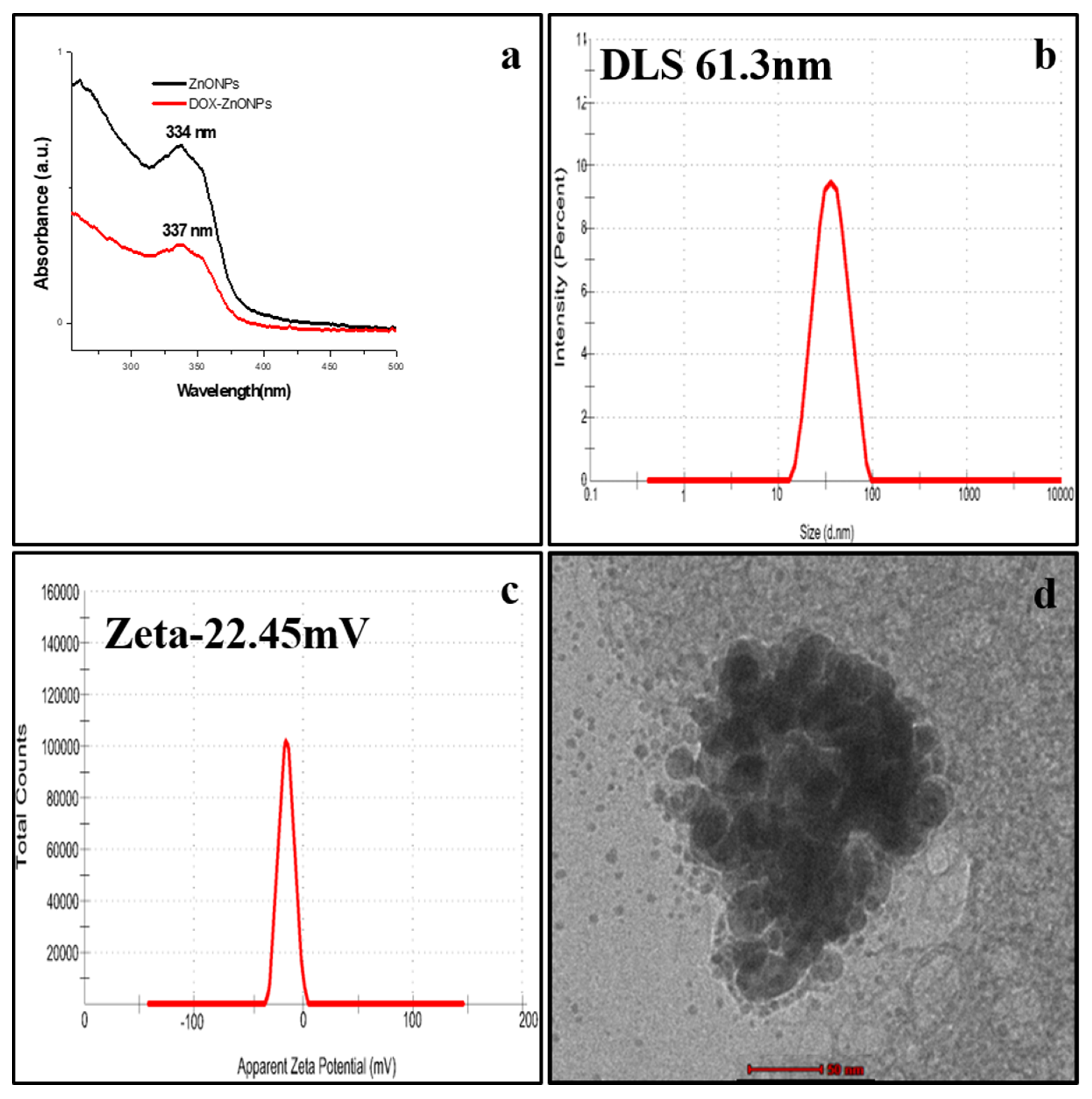
3.3. Bioconjugation of DOX with ZnONPs
3.4. Drug Loading Efficiency
3.5. In Vitro Cytotoxicity of ZnONPs, DOX and DOX-ZnONPs
3.6. Evaluation of Morphological Changes in the A549 Cells
3.7. Analysis of Changes in the Nuclear Morphology
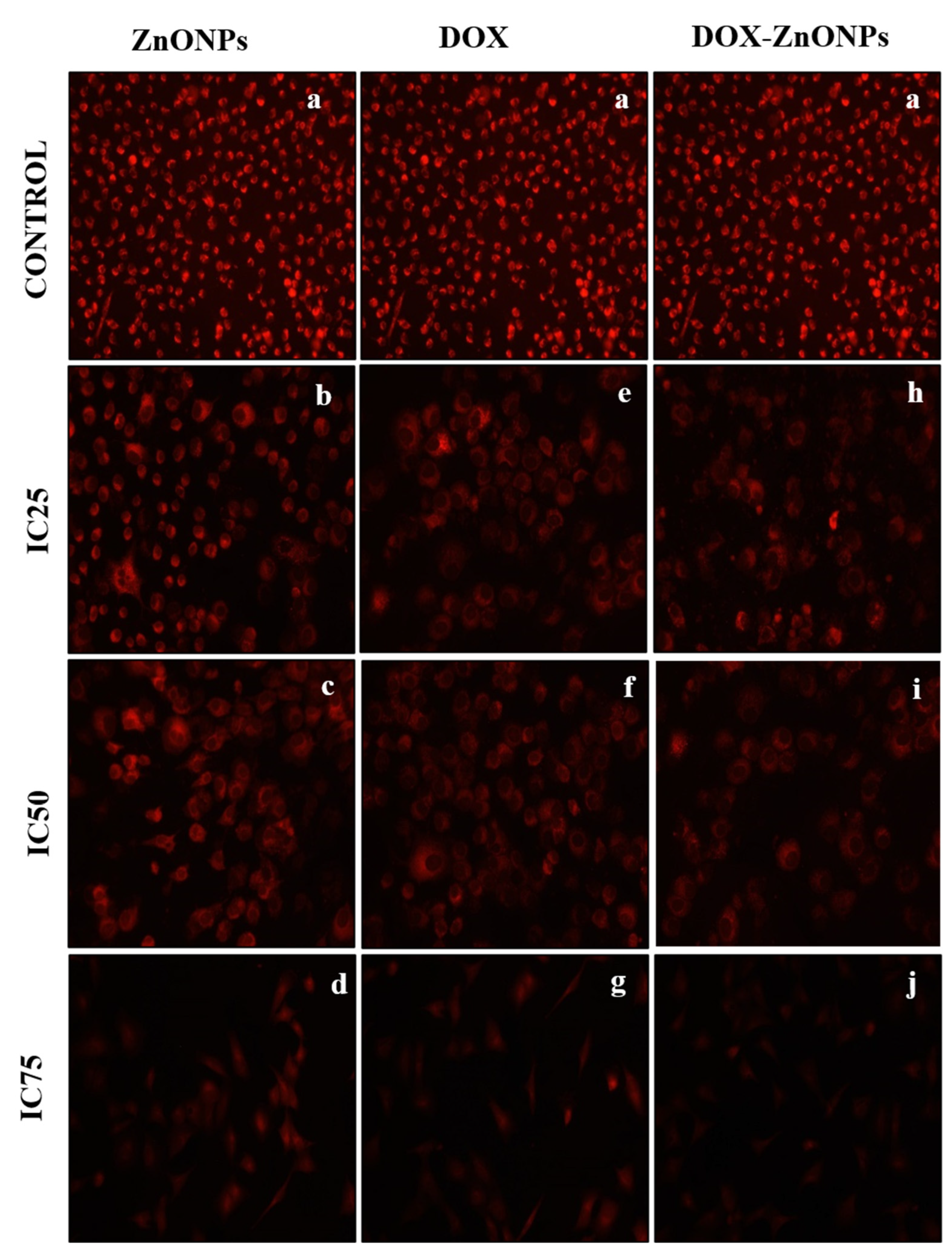
3.8. Analysis of Disruption of the Mitochondrial Membrane Potential (ΔΨm)
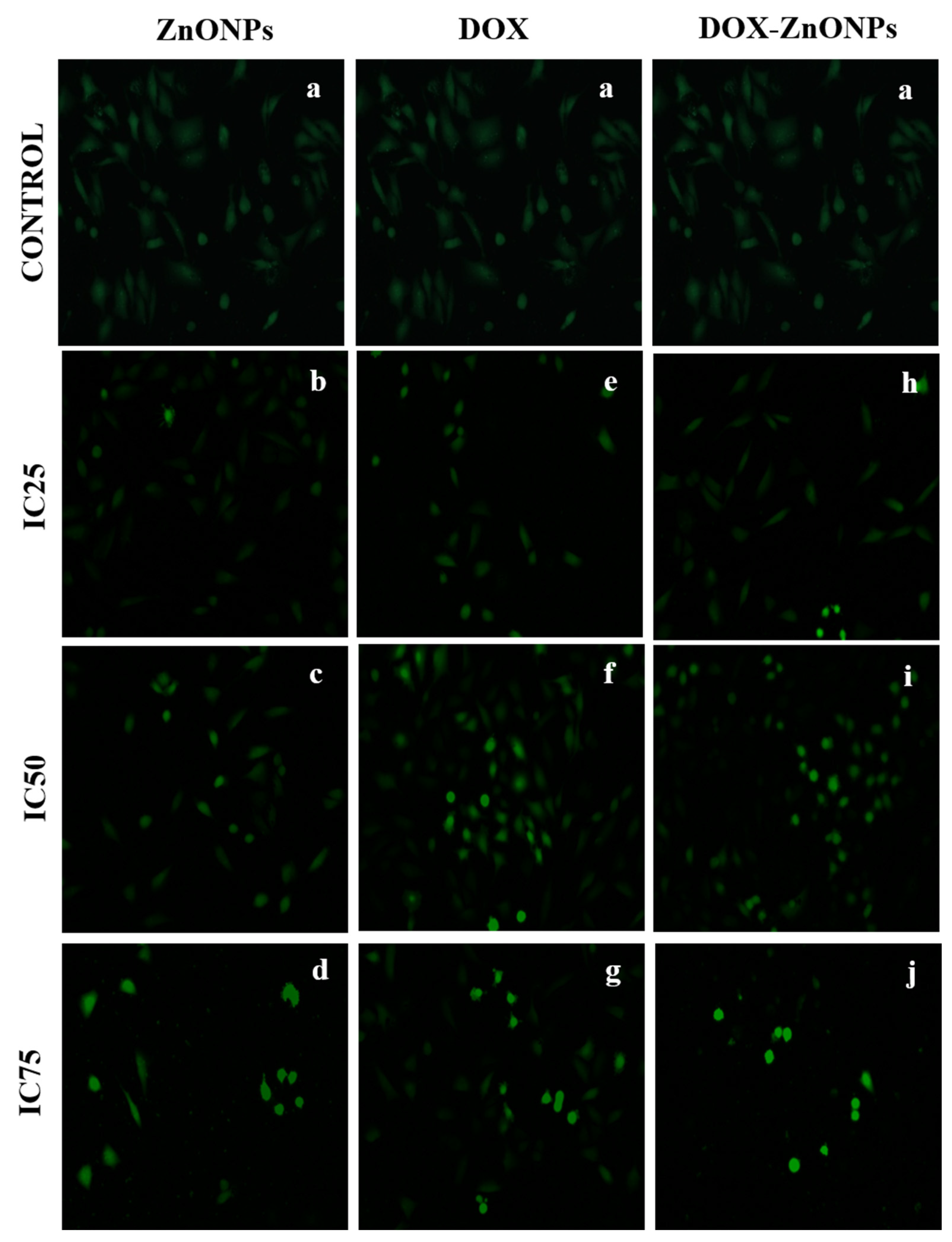
3.9. Assessment of ROS Generation
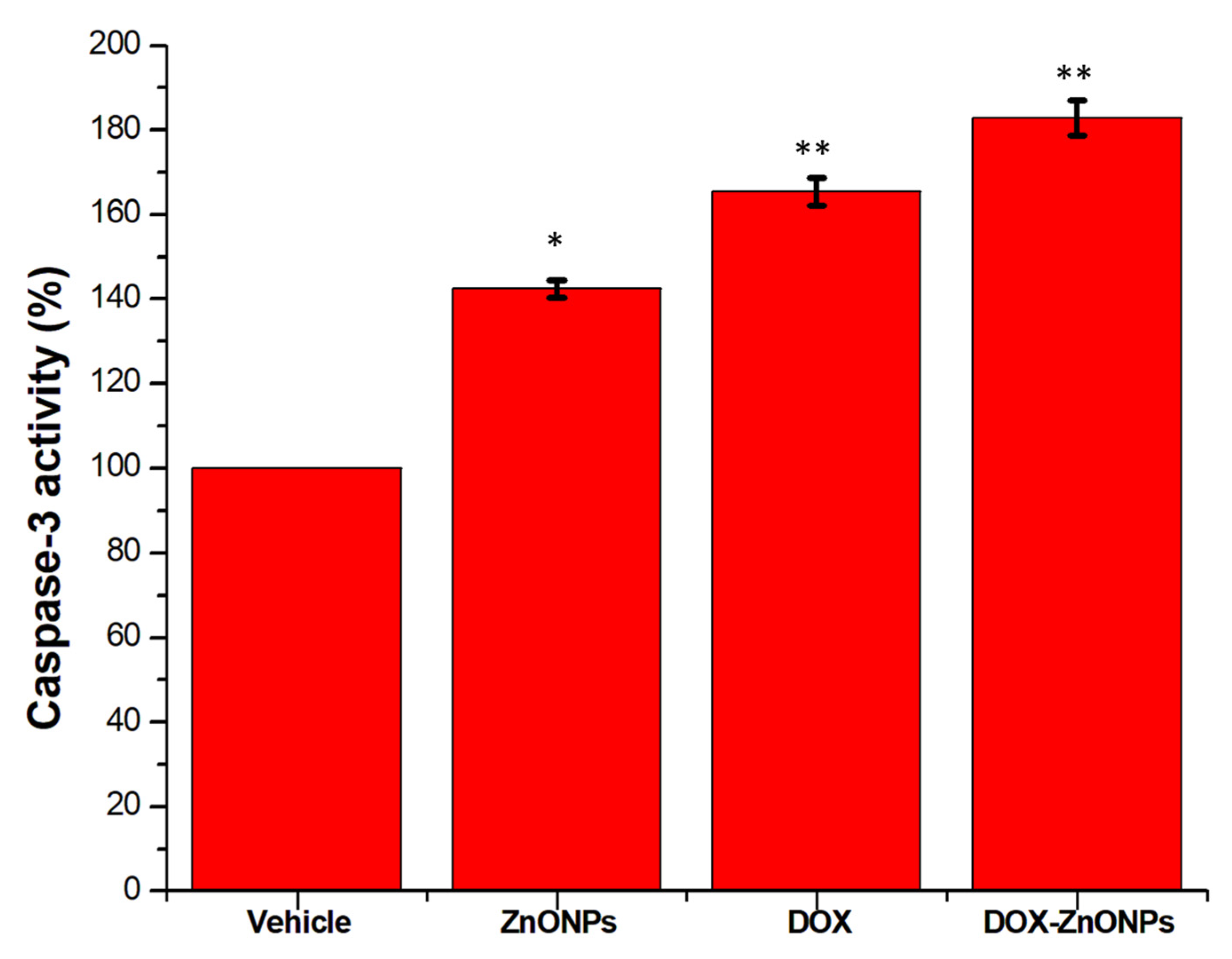
3.10. Activation of Caspase-3
4. Discussion
5. Conclusions
Author Contributions
Funding
Institutional Review Board Statement
Informed Consent Statement
Data Availability Statement
Acknowledgments
Conflicts of Interest
Sample Availability
References
- Steward, B.W.; Kleihues, P. World Cancer Report; IARC Press: Lyon, France, 2003. [Google Scholar]
- Torre, L.A.; Bray, F.; Siegel, R.L.; Ferlay, J.; Lortet-Tieulent, J.; Jemal, A. Global cancer statistics, 2012. CA Cancer J. Clin. 2015, 65, 87–108. [Google Scholar] [CrossRef] [PubMed] [Green Version]
- Punia, R.; Raina, K.; Agarwal, R.; Singh, R.P. Acacetin enhances the therapeutic efficacy of doxorubicin in non-small-cell lung carcinoma cells. PLoS ONE 2017, 12, e0182870. [Google Scholar] [CrossRef] [PubMed]
- Zhou, Z.Y.; Wan, L.L.; Yang, Q.J.; Han, Y.L.; Li, D.; Lu, J.; Guo, C. Nilotinib reverses ABCB1/P-glycoprotein-mediated multidrug resistance but increases cardiotoxicity of doxorubicin in a MDR xenograft model. Toxicol. Lett. 2016, 259, 124–132. [Google Scholar] [CrossRef] [PubMed] [Green Version]
- Henderson, I.C.; Frei, T.E., 3rd. Adriamycin and the heart. N. Engl. J. Med. 1979, 300, 310–312. [Google Scholar]
- Christiansen, S.; Autschbach, R. Doxorubicin in experimental and clinical heart failure. Eur. J. Cardio-Thorac. Surg. 2006, 30, 611–616. [Google Scholar] [CrossRef] [Green Version]
- Binaschi, M.; Capranico, G.; Dal Bo, L.; Zunino, F. Relationship between Lethal Effects and Topoisomerase II-Mediated Double-Stranded DNA Breaks Produced by Anthracyclines with Different Sequence Specificity. Mol. Pharmacol. 1997, 51, 1053–1059. [Google Scholar] [CrossRef]
- Hennig, U.; Rudd, N.; Hoar, D. Kinetochore immunofluorescence in micronuclei: A rapid method for the in situ detection of aneuploidy and chromosome breakage in human fibroblasts. Mutat. Res. Mutagen. Relat. Subj. 1988, 203, 405–414. [Google Scholar] [CrossRef]
- Boucek, R.J.; Dodd, D.A.; Atkinson, J.B.; Oquist, N.; Olson, R.D. Contractile Failure in Chronic Doxorubicin-induced Cardiomyopathy. J. Mol. Cell. Cardiol. 1997, 29, 2631–2640. [Google Scholar] [CrossRef]
- Doroshow, J.H. Effect of anthracycline antibiotics on oxygen radical formation in rat heart. Cancer Res. 1983, 43, 460–472. [Google Scholar]
- Ichihara, S.; Yamada, Y.; Kawai, Y.; Osawa, T.; Furuhashi, K.; Duan, Z.; Ichihara, G. Roles of oxidative stress and Akt signaling in doxorubicin cardiotoxicity. Biochem. Biophys. Res. Commun. 2007, 359, 27–33. [Google Scholar] [CrossRef]
- Temma, K.; Chugun, A.; Akera, T.; Hara, Y.; Sasaki, T.; Kondo, H. Ca2+ overloading causes the negative inotropic effect of doxorubicin in myocytes isolated from guinea-pig hearts. Eur. J. Pharmacol. 1997, 322, 235–242. [Google Scholar] [CrossRef]
- Tong, N.; Zhang, J.; Chen, Y.; Li, Z.; Luo, Y.; Zuo, H.; Zhao, X. Berberine sensitizes mutliple human cancer cells to the anticancer effects of doxorubicin in vitro. Oncol. Lett. 2012, 3, 1263–1267. [Google Scholar] [CrossRef] [PubMed] [Green Version]
- Riehle, K.J.; Dan, Y.Y.; Campbell, J.S.; Fausto, N. New concepts in liver regeneration. J. Gastroenterol. Hepatol. 2011, 26 (Suppl. 1), 203–212. [Google Scholar] [CrossRef] [PubMed] [Green Version]
- Oh, J.-M.; Park, C.-B.; Choy, J.-H. Intracellular Drug Delivery of Layered Double Hydroxide Nanoparticles. J. Nanosci. Nanotechnol. 2011, 11, 1632–1635. [Google Scholar] [CrossRef]
- Zhang, X.-F.; Liu, Z.-G.; Shen, W.; Gurunathan, S. Silver Nanoparticles: Synthesis, Characterisation, Properties, Applications, and Therapeutic Approaches. Int. J. Mol. Sci. 2016, 17, 1534. [Google Scholar] [CrossRef]
- Patra, J.K.; Baek, K.-H. Green Nanobiotechnology: Factors Affecting Synthesis and Characterisation Techniques. J. Nanomater. 2014, 2014, 1–12. [Google Scholar] [CrossRef] [Green Version]
- Ahmad, A.; Senapati, S.; Khan, M.I.; Kumar, R.; Sastry, M. Extracellular Biosynthesis of Monodisperse Gold Nanoparticles by a Novel Extremophilic Actinomycete, Thermomonospora sp. Langmuir 2003, 19, 3550–3553. [Google Scholar] [CrossRef]
- Smijs, T.G.; Pavel, S. Titanium dioxide and zinc oxide nanoparticles in sunscreens: Focus on their safety and effectiveness. Nanotechnol. Sci. Appl. 2011, 4, 95–112. [Google Scholar] [CrossRef] [Green Version]
- Ruszkiewicz, J.A.; Pinkas, A.; Ferrer, B.; Peres, T.V.; Tsatsakis, A.; Aschner, M. Neurotoxic effect of active ingredients in sunscreen products, a contemporary review. Toxicol. Rep. 2017, 4, 245–259. [Google Scholar] [CrossRef]
- Mishra, P.K.; Mishra, H.; Ekielski, A.; Talegaonkar, S.; Vaidya, B. Zinc oxide nanoparticles: A promising nanomaterial for biomedical applications. Drug Discov. Today 2017, 22, 1825–1834. [Google Scholar] [CrossRef]
- Zhang, H.; Ji, Z.; Xia, T.; Meng, H.; Low-Kam, C.; Liu, R.; Pokhrel, S.; Lin, S.; Wang, X.; Liao, Y.-P.; et al. Use of Metal Oxide Nanoparticle Band Gap To Develop a Predictive Paradigm for Oxidative Stress and Acute Pulmonary Inflammation. ACS Nano 2012, 6, 4349–4368. [Google Scholar] [CrossRef] [PubMed]
- Kim, S.; Lee, S.Y.; Cho, H.-J. Doxorubicin-Wrapped Zinc Oxide Nanoclusters for the Therapy of Colorectal Adenocarcinoma. Nanomaterials 2017, 7, 354. [Google Scholar] [CrossRef] [PubMed] [Green Version]
- Xiong, H.-M. ZnO Nanoparticles Applied to Bioimaging and Drug Delivery. Adv. Mater. 2013, 25, 5329–5335. [Google Scholar] [CrossRef] [PubMed]
- Rasmussen, J.W.; Martinez, E.; Louka, P.; Wingett, D.G. Zinc oxide nanoparticles for selective destruction of tumor cells and potential for drug delivery applications. Expert Opin. Drug Deliv. 2010, 7, 1063–1077. [Google Scholar] [CrossRef] [Green Version]
- Khan, S.; Ahmad, K.; Ahmad, A.; Raish, M.; Jan, B.L.; Khan, A.; Khan, M.S. Biogenic pentagonal silver nanoparticles for safer and more effective antibacterial therapeutics. Int. J. Nanomed. 2018, 13, 7789–7799. [Google Scholar] [CrossRef] [Green Version]
- Gomathy, M.; Sabarinathan, K.G. Microbial mechanisms of heavy metal tolerance—A review. Agric. Rev. 2010, 31, 133–138. [Google Scholar]
- Castro-Aceituno, V.; Ahn, S.; Simu, S.Y.; Singh, P.; Mathiyalagan, R.; Lee, H.A.; Yang, D.C. Anticancer activity of silver nanoparticles from Panax ginseng fresh leaves in human cancer cells. Biomed. Pharmacother. 2016, 84, 158–165. [Google Scholar] [CrossRef]
- Mann, S. Biomimetic Materials Chemistry; John Wiley & Sons: New York, NY, USA, 1996; pp. 935–946. [Google Scholar]
- Castro-Longoria, E.; Vilchis-Nestor, A.R.; Avalos-Borja, M. Biosynthesis of silver, gold and bimetallic nanoparticles using the filamentous fungus Neurospora crassa. Colloids Surf. B Biointerfaces 2011, 83, 42–48. [Google Scholar] [CrossRef]
- Zeinab, S.; Mojtaba, S.; Farzad, K. Biological synthesis of gold nanoparticles by fungus Epicoccum nigrum. J. Clus. Sci. 2011, 22, 661–665. [Google Scholar]
- Wang, H.; Wingett, D.; Engelhard, M.H. Fluorescent dye capsulated ZnO particles with cell-specific toxicity for potential use in biomedical applications. J. Mater. Sci. Mater. Med. 2009, 20, 11–22. [Google Scholar] [CrossRef]
- Hanley, C.; Layne, J.; Punnoose, A. Preferential killing of cancer cells and activated human Tcells using zinc oxide nanoparticles. Nanotechnology 2008, 19, 295103. [Google Scholar] [CrossRef] [PubMed] [Green Version]
- Mekky, A.E.; Farrag, A.A.; Hmed, A.A.; Sofy, A.R. Preparation of Zinc Oxide Nanoparticles using Aspergillus niger as Antimicrobial and Anticancer Agents. J. Pure Appl. Microbiol. 2021, 15, 1547–1566. [Google Scholar] [CrossRef]
- Rasha, E.; Alkhulaifi, M.M.; AlOthman, M.; Khalid, I.; Doaa, E.; Alaa, K.; Awad, M.A.; Abdalla, M. Effects of Zinc Oxide Nanoparticles Synthesized Using Aspergillus niger on Carbapenem-Resistant Klebsiella pneumonia In Vitro and In Vivo. Front. Cell. Infect. Microbiol. 2021, 11, 748739. [Google Scholar] [CrossRef] [PubMed]
- Aldalbahi, A.; Alterary, S.; Almoghim, R.A.A.; Awad, M.A.; Aldosari, N.S.; Alghannam, S.F.; Alabdan, A.N.; Alharbi, S.; Alateeq, B.A.M.; Al Mohsen, A.A.; et al. Greener Synthesis of Zinc Oxide Nanoparticles: Characterisation and Multifaceted Applications. Moleculees 2020, 25, 4198. [Google Scholar] [CrossRef]
- Leite, F.L.; Bueno, C.C.; Da Róz, A.L.; Ziemath, E.C.; Oliveira, J.O.N. Theoretical Models for Surface Forces and Adhesion and Their Measurement Using Atomic Force Microscopy. Int. J. Mol. Sci. 2012, 13, 12773–12856. [Google Scholar] [CrossRef]
- Kavitha, S.; Dhamodaran, M.; Prasad, R.; Ganesan, M. Synthesis and characterisation of zinc oxide nanoparticles using terpenoid fractions of Andrographis paniculata leaves. Int. Nano Lett. 2017, 7, 141–147. [Google Scholar] [CrossRef] [Green Version]
- Vimala, K.; Sundarraj, S.; Paulpandi, M.; Vengatesan, S.; Kannan, S. Green synthesized doxorubicin loaded zinc oxide nanoparticles regulates the Bax and Bcl-2 expression in breast and colon carcinoma. Process Biochem. 2014, 49, 160–172. [Google Scholar] [CrossRef]


Publisher’s Note: MDPI stays neutral with regard to jurisdictional claims in published maps and institutional affiliations. |
© 2022 by the authors. Licensee MDPI, Basel, Switzerland. This article is an open access article distributed under the terms and conditions of the Creative Commons Attribution (CC BY) license (https://creativecommons.org/licenses/by/4.0/).
Share and Cite
Mishra, P.; Ahmad, A.; Al-Keridis, L.A.; Alshammari, N.; Alabdallah, N.M.; Muzammil, K.; Saeed, M.; Ansari, I.A. Doxorubicin-Conjugated Zinc Oxide Nanoparticles, Biogenically Synthesised Using a Fungus Aspergillus niger, Exhibit High Therapeutic Efficacy against Lung Cancer Cells. Molecules 2022, 27, 2590. https://0-doi-org.brum.beds.ac.uk/10.3390/molecules27082590
Mishra P, Ahmad A, Al-Keridis LA, Alshammari N, Alabdallah NM, Muzammil K, Saeed M, Ansari IA. Doxorubicin-Conjugated Zinc Oxide Nanoparticles, Biogenically Synthesised Using a Fungus Aspergillus niger, Exhibit High Therapeutic Efficacy against Lung Cancer Cells. Molecules. 2022; 27(8):2590. https://0-doi-org.brum.beds.ac.uk/10.3390/molecules27082590
Chicago/Turabian StyleMishra, Prakriti, Afza Ahmad, Lamya Ahmed Al-Keridis, Nawaf Alshammari, Nadiyah M. Alabdallah, Khursheed Muzammil, Mohd Saeed, and Irfan Ahmad Ansari. 2022. "Doxorubicin-Conjugated Zinc Oxide Nanoparticles, Biogenically Synthesised Using a Fungus Aspergillus niger, Exhibit High Therapeutic Efficacy against Lung Cancer Cells" Molecules 27, no. 8: 2590. https://0-doi-org.brum.beds.ac.uk/10.3390/molecules27082590