
Antitumor Activity and Physicochemical Properties of New Thiosemicarbazide Derivative and Its Co(II), Ni(II), Cu(II), Zn(II) and Cd(II) Complexes

 ,
,  ,
,  , , , , and
Abstract
:1. Introduction
2. Results and Discussion
2.1. Thermogravimetric Analysis (TGA) Analysis
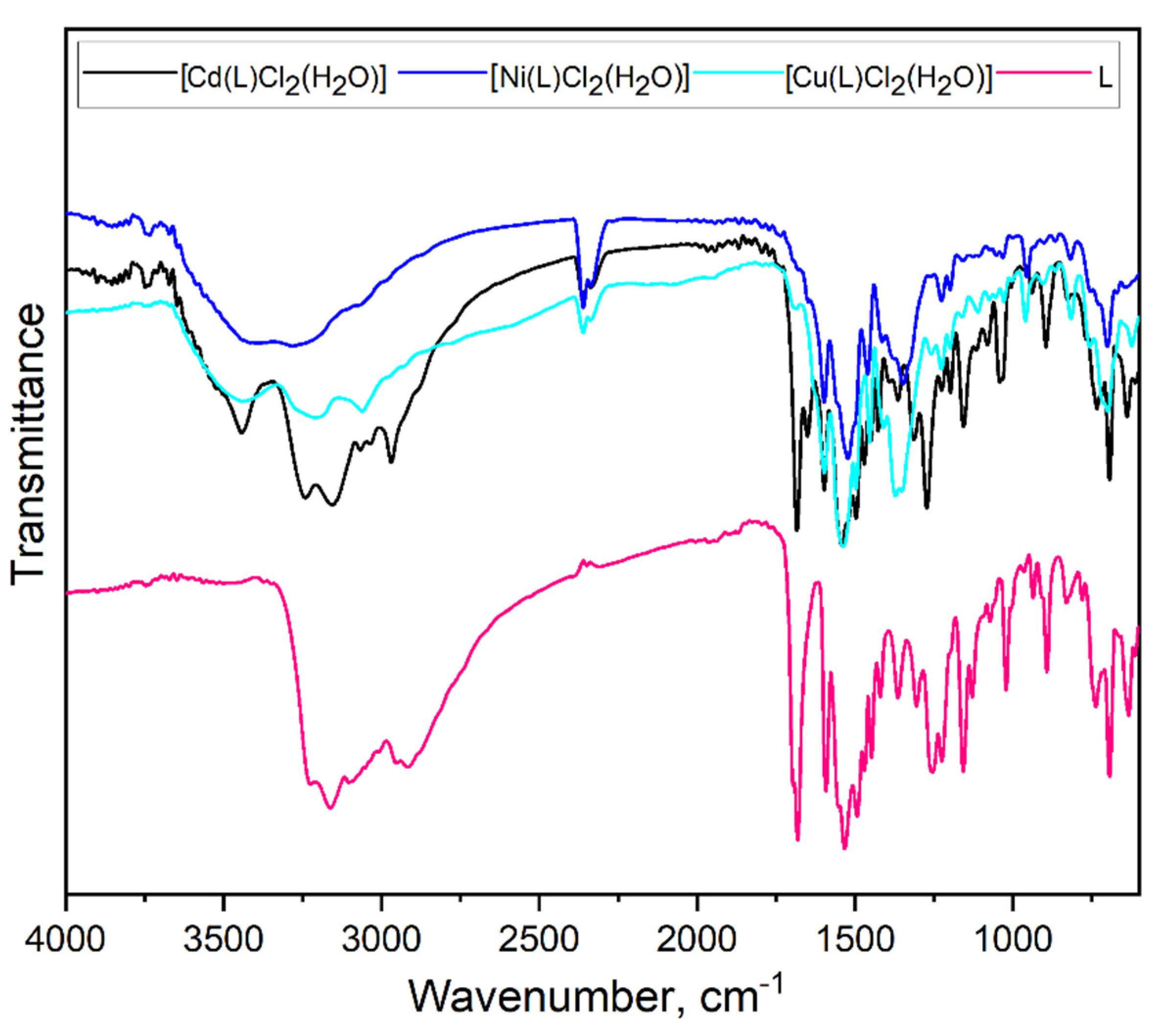
2.2. Fourier-Transform Infrared Spectroscopy (FTIR) Analysis
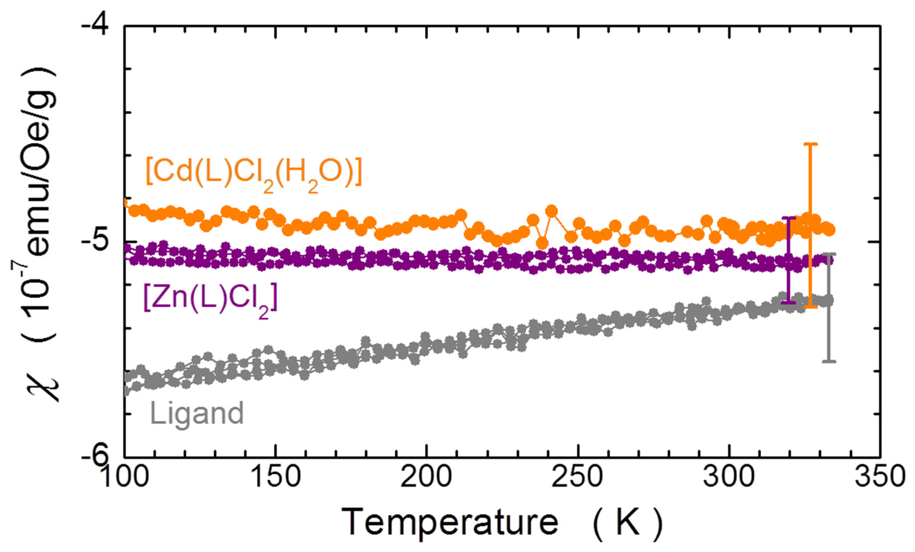
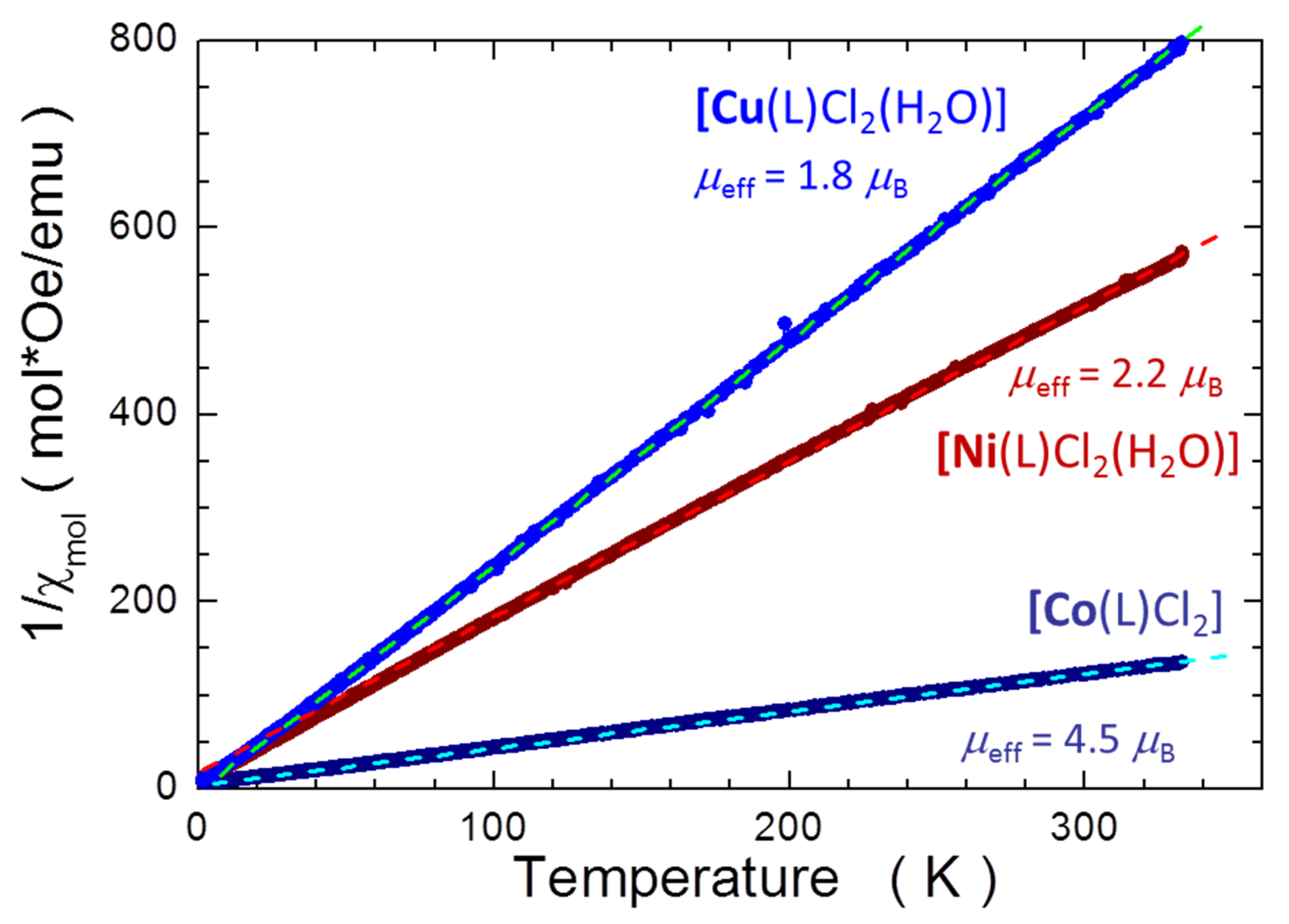
2.3. Magnetic Properties
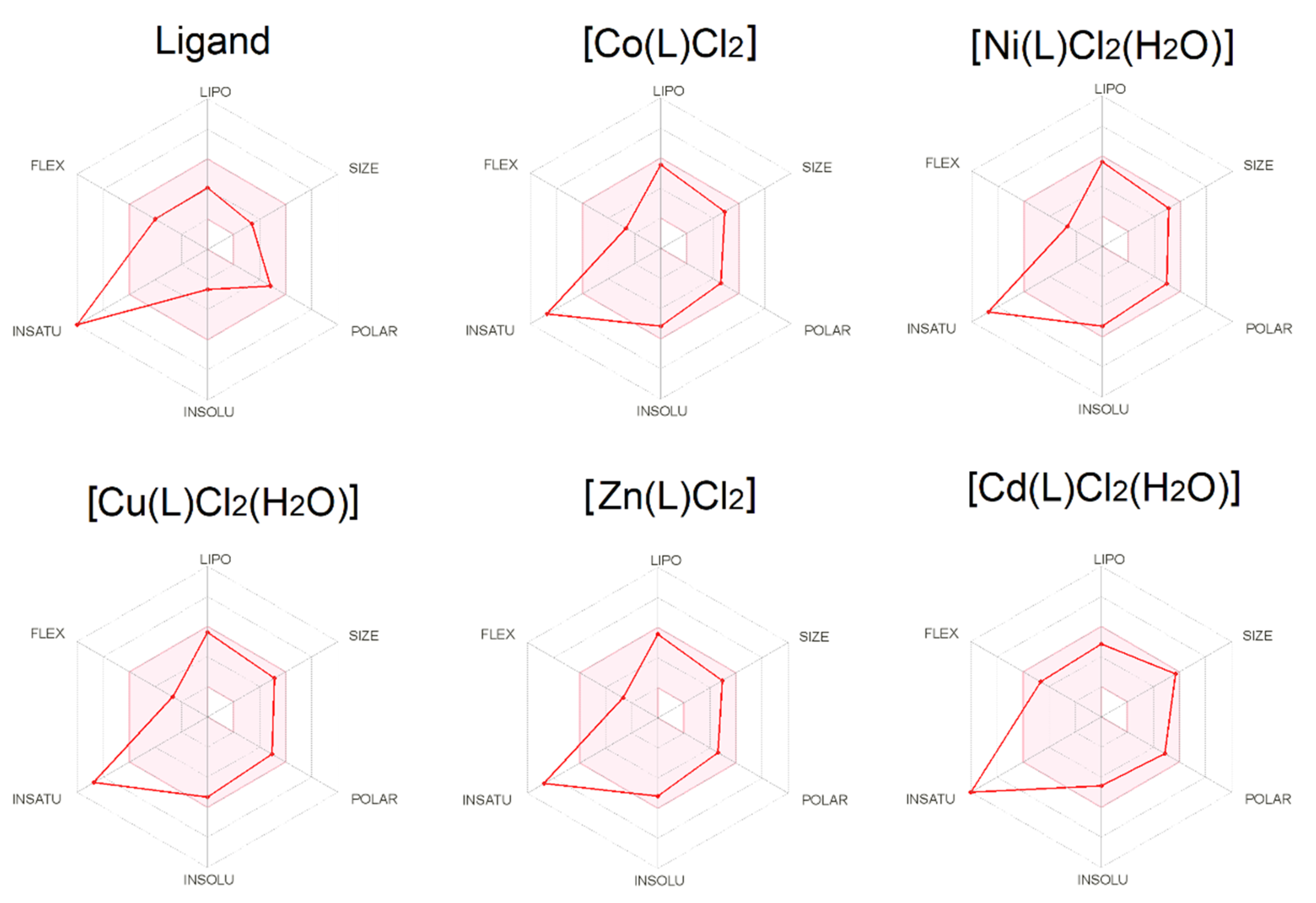
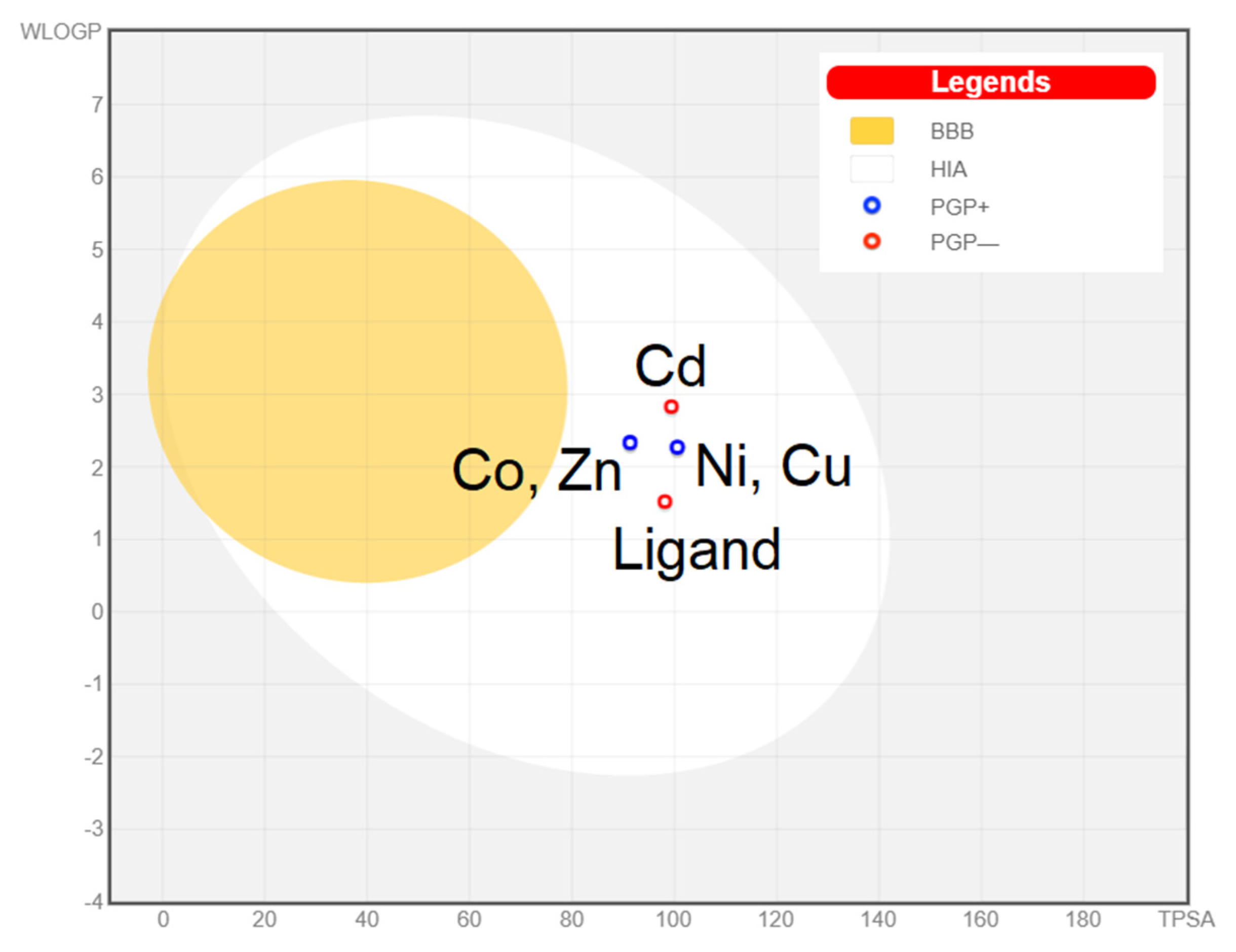
2.4. ADMET
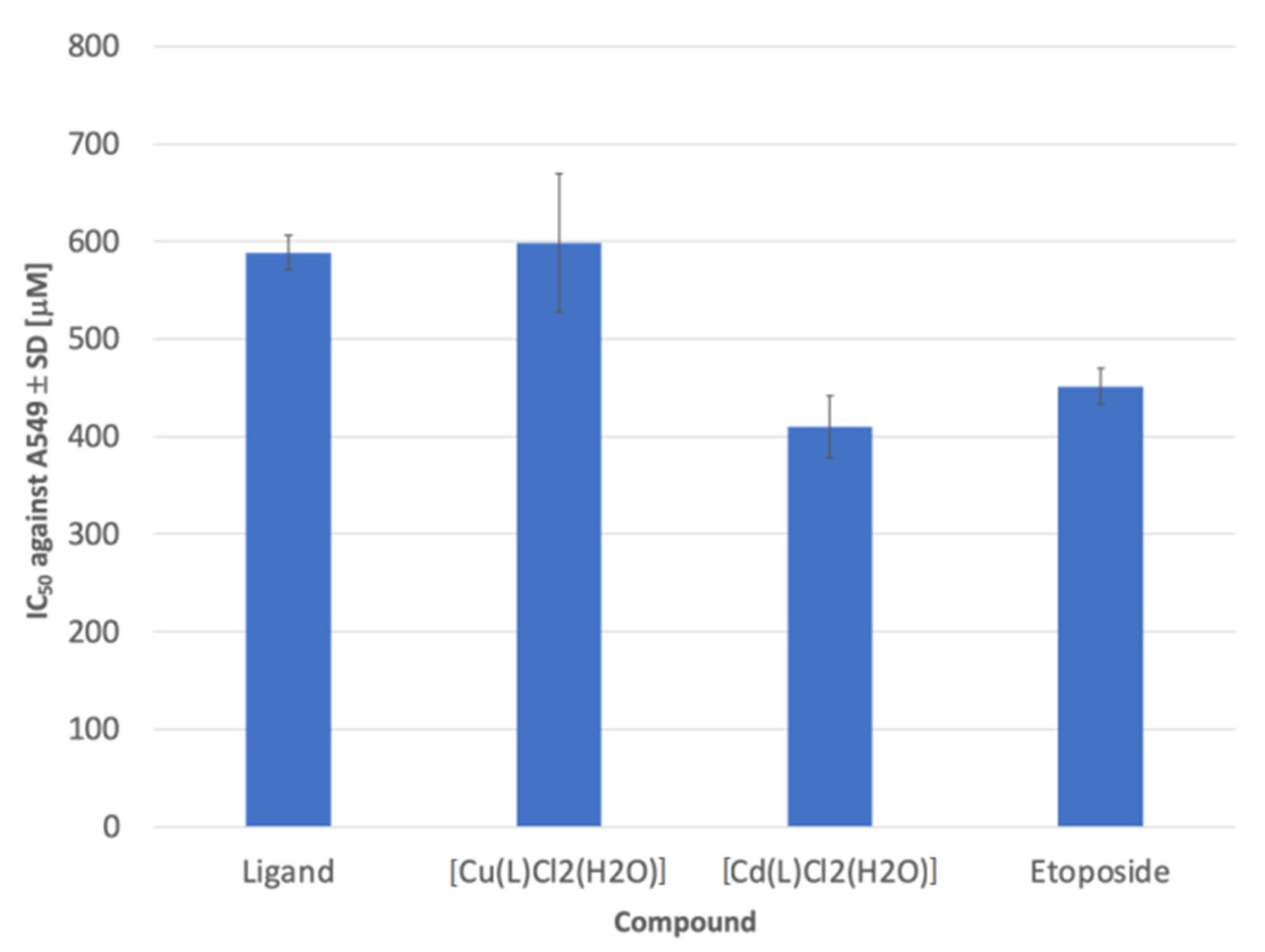
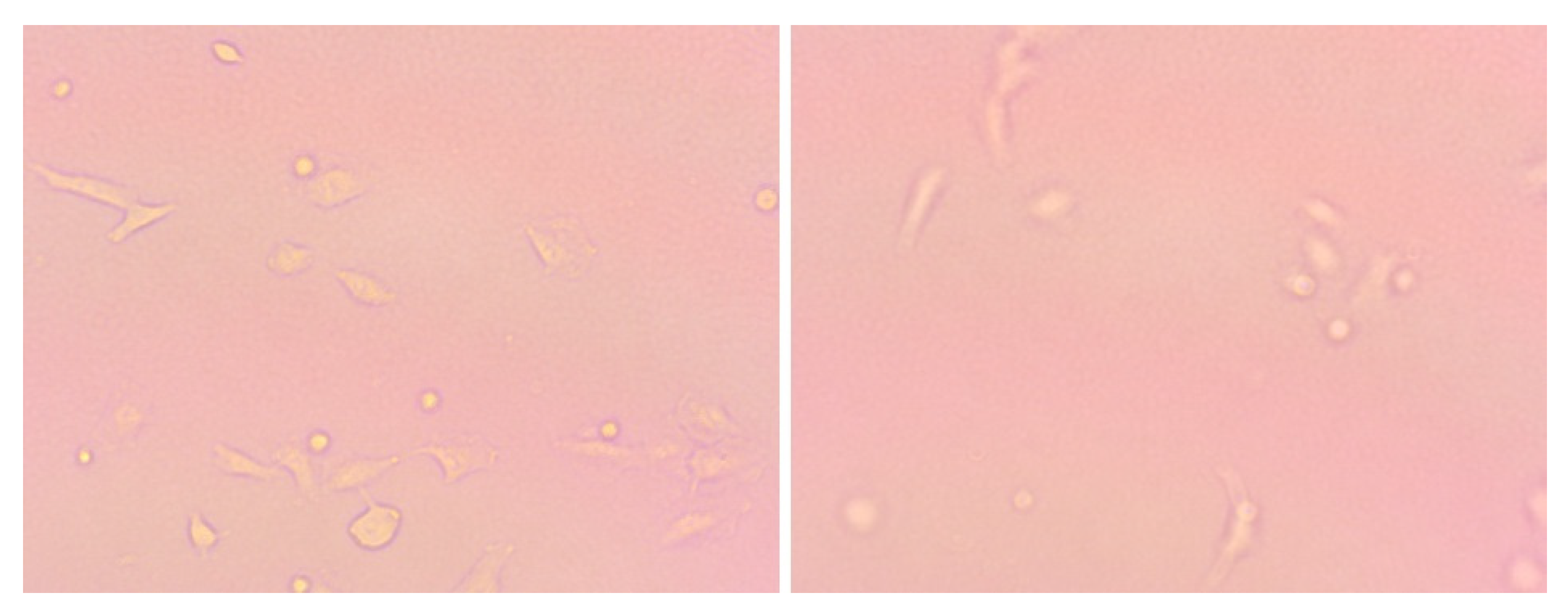
2.5. Cytotoxicity Assay
3. Materials and Methods
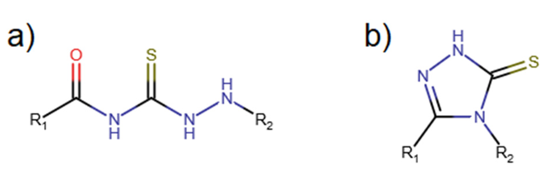
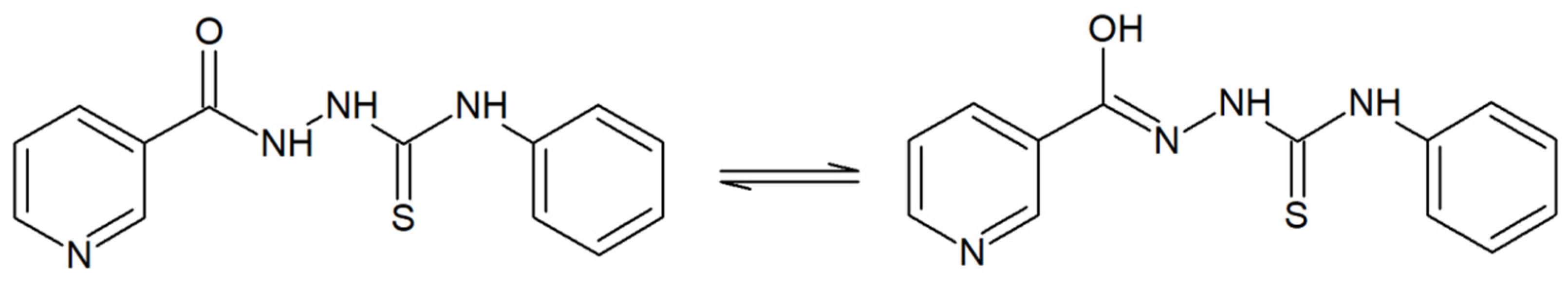
3.1. Synthesis of the L Ligand
3.2. Synthesis of the Coordination Compounds
3.3. Chemistry
3.4. ADMET
3.5. Cytotoxicity Assay
3.6. Magnetic Measurements
4. Conclusions
Author Contributions
Funding
Institutional Review Board Statement
Informed Consent Statement
Data Availability Statement
Acknowledgments
Conflicts of Interest
Sample Availability
References
- Abhale, Y.K.; Shinde, A.; Deshmukh, K.K.; Nawale, L.; Sarkar, D.; Mhaske, P.C. Synthesis, antitubercular and antimicrobial potential of some new thiazole substituted thiosemicarbazide derivatives. Med. Chem. Res. 2017, 26, 25572567. [Google Scholar] [CrossRef]
- Al-Mutabagani, L.A.; Abdelrazek, F.M.; Gomha, S.M.; Hebishy, A.S.; Abdelfattah, M.S.; Hassan, S.M.; Sayed, A.R.; Elaasser, M.M. Synthesis and Biological Evaluation of Thiazolyl-Ethylidene Hydrazino-Thiazole Derivatives: A Novel Heterocyclic System. Appl. Sci. 2021, 11, 8908. [Google Scholar] [CrossRef]
- Plech, T.; Wujec, M.; Siwek, A.; Kosikowska, U.; Malm, A. Synthesis and antimicrobial activity of thiosemicarbazides, s-triazoles and their Mannich bases bearing 3-chlorophenyl moiety. Eur. J. Med. Chem. 2011, 46, 241248. [Google Scholar] [CrossRef] [PubMed]
- Kalhor, M.; Shabani, M.; Nikokar, I.; Reyhaneh Banisaeed, S. Synthesis, Characterization and Antibacterial Activity of some Novel Thiosemicarbazides, 1,2,4-Triazol-3-thiols and their S-substituted Derivatives. Iran. J. Pharm. Res. IJPR 2015, 14, 6775. [Google Scholar]
- Zhao, G.-F.; Zhang, L.; Fu, Y.; Jin, G.-Y. The Synthesis and Bioactivity of New Thiosemicarbazides and Thiadiazole Derivatives Including Substituted Pyrazolyl group. Chem. J. Chin. Univ. 1998, 19, 222. [Google Scholar]
- Popovici, C.; Pavel, C.-M.; Sunel, V.; Cheptea, C.; Dimitriu, D.G.; Dorohoi, D.O.; David, D.; Closca, V.; Popa, M. Optimized Synthesis of New Thiosemicarbazide Derivatives with Tuberculostatic Activity. Int. J. Mol. Sci. 2021, 22, 12139. [Google Scholar] [CrossRef]
- Küçükgüzel, G.; Kocatepe, A.; De Clercq, E.; Sahin, F.; Güllüce, M. Synthesis and biological activity of 4-thiazolidinones, thiosemicarbazides derived from diflunisal hydrazide. Eur. J. Med. Chem. 2006, 41, 353359. [Google Scholar] [CrossRef]
- Cihan-Üstündag, G.; Gürsoy, E.; Naesens, L.; Ulusoy-Güzeldemirci, N.; Çapan, G. Synthesis and antiviral properties of novel indole-based thiosemicarbazides and 4-thiazolidinones. Bioorg. Med. Chem. 2016, 24, 240246. [Google Scholar] [CrossRef]
- Basyouni, W.M.; Abbas, S.Y.; El-Bayouki, K.A.M.; Daawod, R.M.; Elawady, M.K. Synthesis and antiviral evaluation of 5-(arylazo)salicylaldehyde thiosemicarbazone derivatives as potent anti-bovine viral diarrhea virus agents. Synth. Commun. 2021, 51, 2168–2174. [Google Scholar] [CrossRef]
- Kalinowski, D.S.; Quach, P.; Richardson, D.R. Thiosemicarbazones: The new wave in cancer treatment. Future Med. Chem. 2009, 1, 11431151. [Google Scholar] [CrossRef]
- Bahojb Noruzi, E.; Kheirkhahi, M.; Shaabani, B.; Geremia, S.; Hickey, N.; Asaro, F.; Nitti, P.; Kafil, H.S. Design of a Thiosemicarbazide-Functionalized Calix[4]arene Ligand and Related Transition Metal Complexes: Synthesis, Characterization, and Biological Studies. Front. Chem. 2019, 7, 663. [Google Scholar] [CrossRef] [PubMed] [Green Version]
- Aly, A.A.; Hassan, A.A.; Makhlouf, M.M.; Bräse, S. Chemistry and Biological Activities of 1,2,4- Triazolethiones-Antiviral and Anti-Infective Drugs. Molecules 2020, 25, 3036. [Google Scholar] [CrossRef] [PubMed]
- Ahmad, A.; Varshney, H.; Rauf, A.; Sherwani, A.; Owais, M. Synthesis and anticancer activity of long chain substituted 1,3,4-oxadiazol-2-thione, 1,2,4-triazol-3-thione and 1,2,4-triazolo[3,4-b]-1,3,4-thiadiazine derivatives. Arab. J. Chem. 2017, 10, 33473357. [Google Scholar] [CrossRef] [Green Version]
- Kulaba, N.; Tatar, E.; Bingöl Özakpinar, Z.; Özsavci, D.; Pannecouque, C.; De Clercq, E.; Küçükgüzel, I. Synthesis and antiproliferative evaluation of novel 2-(4H-1,2,4-triazole-3-ylthio)acetamide derivatives as inducers of apoptosis in cancer cells. Eur. J. Med. Chem. 2016, 121, 5870. [Google Scholar] [CrossRef] [PubMed]
- Mohamed, G.G.; Omar, M.M.; Ibrahim, A.A. Biological activity studies on metal complexes of novel tridentate Schiff base ligand. Spectroscopic and thermal characterization. Eur. J. Med. Chem. 2009, 44, 48014812. [Google Scholar] [CrossRef] [PubMed]
- Brewer, G.J. The risks of copper toxicity contributing to cognitive decline in the aging population and to Alzheimer’s disease. J. Am. Coll. Nutr. 2009, 28, 238242. [Google Scholar] [CrossRef] [PubMed]
- White, A.R.; Multhaup, G.; Maher, F.; Bellingham, S.; Camakaris, J.; Zheng, H.; Bush, A.I.; Beyreuther, K.; Masters, C.L.; Cappai, R. The Alzheimer’s Disease Amyloid Precursor Protein Modulates Copper-Induced Toxicity and Oxidative Stress in Primary Neuronal Cultures. J. Neurosci. 1999, 19, 91709179. [Google Scholar] [CrossRef] [Green Version]
- Chen, X.; Zhang, X.; Chen, J.; Yang, Q.; Yang, L.; Xu, D.; Zhang, P.; Wang, X.; Liu, J. Hinokitiol copper complex inhibits proteasomal deubiquitination and induces paraptosis-like cell death in human cancer cells. Eur. J. Pharmacol. 2017, 815, 147155. [Google Scholar] [CrossRef]
- Sangeetha, S.; Murali, M. Non-covalent DNA binding, protein interaction, DNA cleavage and cytotoxicity of [Cu(quamol)Cl]·H2O. Int. J. Biol. Macromol. 2018, 107, 25012511. [Google Scholar] [CrossRef]
- Czylkowska, A.; Drozd, M.; Biernasiuk, A.; Rogalewicz, B.; Malm, A.; Pitucha, M. Synthesis, Spectral, Thermal and Biological Studies of 4-Cyclohexyl-3-(4-nitrophenyl)methyl-1,2,4-triazolin-5-thione and Its Copper(II) Coordination Compound, [CuCl2(H2O)2L2]. Materials 2020, 13, 4135. [Google Scholar] [CrossRef]
- Pitucha, M.; Wujec, M.; Dobosz, M. Synthesis of 3-(pyridin-4-ylmethyl)-4-substituted-1,2,4-triazoline-5-thione. J. Chin. Chem. Soc. 2007, 54, 6973. [Google Scholar] [CrossRef]
- Pitucha, M.; Polak, B.; Swatko-Ossor, M.; Popiołek, Ł.; Ginalska, G. Determination of the Lipophilicity of Some New Derivatives of Thiosemicarbazide and 1,2,4-triazoline-5-thione with Potential Antituberculosis Activity. Croat. Chem. Acta 2010, 83, 299. [Google Scholar]
- Pitucha, M.; Wos, M.; Miazga-Karska, M.; Klimek, K.; Miroslaw, B.; Pachuta-Stec, A.; Gadysz, A.; Ginalska, G. Synthesis, antibacterial and antiproliferative potential of some new 1-pyridinecarbonyl-4-substituted thiosemicarbazide derivatives. Med. Chem. Res. 2016, 25, 16661677. [Google Scholar] [CrossRef] [PubMed] [Green Version]
- Wos, M.; Miazga-Karska, M.; Kaczor, A.A.; Klimek, K.; Karczmarzyk, Z.; Kowalczuk, D.; Wysocki, W.; Ginalska, G.; Urbaczyk-Lipkowska, Z.; Morawiak, M.; et al. Novel thiosemicarbazide derivatives with 4-nitrophenyl group as multi-target drugs: Glucosidase inhibitors with antibacterial and antiproliferative activity. Biomed. Pharmacother. 2017, 93, 1269–1276. [Google Scholar] [CrossRef] [PubMed]
- Ali, B.; Mohammed Khan, K.; Arshia, N.; Kanwal, N.; Hussain, S.; Hussain, S.; Ashraf, M.; Riaz, M.; Wadood, A.; Perveen, S. Synthetic nicotinic/isonicotinic thiosemicarbazides: In vitro urease inhibitory activities and molecular docking studies. Bioorg. Chem. 2018, 79, 3445. [Google Scholar] [CrossRef]
- Latheef, L.; Kurup MR, P.; Suresh, E. Synthesis, crystallographic structure and hirshfeld surface analysis of nickel(II) chelate derived from O,N,S-donor tridentate thiosemicarbazone. Chem. Data Collect. 2021, 35, 100758. [Google Scholar] [CrossRef]
- Al-Riyahee, A.A.; Shenta, A.; Saud, K. The Coordination chemistry and cyclic voltammetry exploration of Cu (II), Co (II), Ni (II) and Zn (II) complexes of novel (E)-3, 4-dichloro-N-(2-(1-(pyridin-2-yl) ethylidene) hydrazine-1-carbonothioyl) benzamide ligand. Egypt. J. Chem. 2021, 64, 56. [Google Scholar] [CrossRef]
- Coey, J. Magnetism and Magnetic Materials; Cambridge University Press: Cambridge, UK, 2010; p. 408. [Google Scholar] [CrossRef] [Green Version]
- Majumdar, A.K.; Bhattacharyya, B. Anomalous magnetic behaviour of bis(N-methylsalicylaldimine) nickel(II) complex. J. Inorg. Nucl. Chem. 1965, 27, 143–147. [Google Scholar] [CrossRef]
- Arriortua, M.I.; Cortes, A.R.; Lezam, L.; Rojo, T.; Solans, X.; Font-Bardia, M. Crystal structure and magnetic properties of [Ni(terpy)(N3)2]2·2H2O, a nickel(II) dinuclear complex with ferromagnetic interaction. Inorg. Chim. Acta 1990, 174, 263–269. [Google Scholar] [CrossRef]
- Ballhausen, C.J.; Liehr, A.D. Some Comments on the Anomalous Magnetic Behavior of Certain Ni(II) Complexes. J. Am. Chem. Soc. 1959, 81, 538–542. [Google Scholar] [CrossRef]
- Melson, G.A.; Busch, D.H. The Magnetic Properties of Nickel(II) Complexes Containing a Macrocyclic Ligand and the Existence of a High Spin-Low Spin Equilibrium. J. Am. Chem. Soc. 1964, 86, 4830–4833. [Google Scholar] [CrossRef]
- Basto, M.C.R.; Silva, G.V.A.; Machado, A.A.S.C. Nickel (II) complexes of imidazole-derived ligands with an amide group: Some compounds with complex magnetic behavior. Synth. React. Inorg. Met.-Org. Chem. 2002, 2, 305–327. [Google Scholar] [CrossRef]
- Ranjan, R.; Sinha, N.; Kumar, S.; Chandra, C.M.; Sharma, S. Abnormal Magnetic Moment and Zero Field Splitting of Some Nickel (II) Complexes. Appl. Sci. 2017, 7, 34–41. [Google Scholar] [CrossRef] [Green Version]
- Deneef, S.N.; Mehdi, H.A. Co (II), Cu (II), Ni (II), Fe (II) and Cr (III) Complexes of N, O-Type Schiff base ligand derived from 4, 4,-methylenedianiline Synthesis, Characterization and antibacterial studies. Univ. Thi-Qar J. Sci. 2019, 7, 89–95. [Google Scholar]
- Castillo, O.L.A.; Roman, P.; Lloret, F.; Julve, M. Syntheses, Crystal Structures, and Magnetic Properties of One-Dimensional Oxalato-Bridged Co (II), Ni (II), and Cu (II) Complexes with n-Aminopyridine (n = 2 − 4) as Terminal Ligand. Inorg. Chem. 2001, 22, 5526–5535. [Google Scholar] [CrossRef]
- Lipinski, C.A.; Lombardo, F.; Dominy, B.W.; Feeney, P.J. Experimental and computational approaches to estimate solubility and permeability in drug discovery and development settings. Adv. Drug Deliv. Rev. 2012, 64, 4–17. [Google Scholar] [CrossRef]
- Ghose, A.K.; Viswanadhan, V.N.; Wendoloski, J.J. A knowledge-based approach in designing combinatorial or medicinal chemistry libraries for drug discovery. 1. A qualitative and quantitative characterization of known drug databases. J. Comb. Chem. 1999, 1, 55–68. [Google Scholar] [CrossRef]
- Egan, W.J.; Merz, K.M.; Baldwin, J.J. Prediction of drug absorption using multivariate statistics. J. Med. Chem. 2000, 43, 3867–3877. [Google Scholar] [CrossRef]
- Veber, D.F.; Johnson, S.R.; Cheng, H.Y.; Smith, B.R.; Ward, K.W.; Kopple, K.D. Molecular properties that influence the oral bioavailability of drug candidates. J. Med. Chem. 2002, 45, 2615–2623. [Google Scholar] [CrossRef]
- Muegge, I.; Heald, S.L.; Brittelli, D. Simple selection criteria for drug-like chemical matter. J. Med. Chem. 2001, 44, 1841–1846. [Google Scholar] [CrossRef]
- Dobosz, M.; Pitucha, M.; Wujec, M. The reactions of cyclization of thiosemicarbazide derivatives to 1,2,4-triazole or 1,3,4-thiadiazole system. Acta Pol. Pharm. 1996, 53, 31–38. [Google Scholar]
- Daina, A.; Michielin, O.; Zoete, V. SwissADME: A free web tool to evaluate pharmacokinetics, drug-likeness and medicinal chemistry friendliness of small molecules. Sci. Rep. 2017, 7, 13. [Google Scholar] [CrossRef] [PubMed] [Green Version]
- Daina, A.; Zoete, V. A BOILED-Egg To Predict Gastrointestinal Absorption and Brain Penetration of Small Molecules. ChemMedChem 2016, 11, 1117–1121. [Google Scholar] [CrossRef] [Green Version]
- Banerjee, P.; Eckert, A.O.; Schrey, A.K.; Preissner, R. ProTox-II: A webserver for the prediction of toxicity of chemicals. Nucleic Acids Res. 2018, 46, 257–263. [Google Scholar] [CrossRef] [PubMed] [Green Version]
- Akbarzadeh, A.; Samiei, M.; Joo, S.W.; Anzaby, M.; Hanifehpour, Y.; Nasrabadi, H.T. Synthesis, characterization and in vitro stud- ies of doxorubicin-loaded magnetic nanoparticles grafted to smart copolymers on A549 lung cancer cell line. J. Nanobiotechnol. 2012, 10, 46. [Google Scholar] [CrossRef] [PubMed] [Green Version]
- Girek, M.; Kłosiński, K.; Grobelski, B.; Pizzimenti, S.; Cucci, M.A.; Daga, M.; Barrera, G.; Pasieka, Z.; Czarnecka, K.; Szymański, P. Novel tetrahydroacridine derivatives with iodobenzoic moieties induce G0/G1 cell cycle arrest and apoptosis in A549 non-small lung cancer and HT-29 colorectal cancer cells. Mol. Cell Biochem. 2019, 460, 12350. [Google Scholar] [CrossRef] [PubMed] [Green Version]
- Tun Nur Iskandar, N.A.J.; Guan-Yeow, Y.; Maeta, N.; Ito, M.M.; Nakamura, Y.; Gas, K.; Sawicki, M. Anisotropic and magnetic properties in non-metal and non-radical organic aggregates of tri-substituted phenyl derivatives. N. J. Chem. 2020, 44, 210–217. [Google Scholar] [CrossRef]
- Świątkowski, M.; Lanka, S.; Czylkowska, A.; Gas, K.; Sawicki, M. Structural, Spectroscopic, Thermal, and Magnetic Properties of a New Dinuclear Copper Coordination Compound with Tiglic Acid. Materials 2021, 14, 2148. [Google Scholar] [CrossRef] [PubMed]
- Gas, K.; Sawicki, M. In Situ Compensation Method for Precise Integral SQUID Magnetometry of Miniscule Biological, Chemical, and Powder Specimens Requiring the Use of Capsules. Materials 2022, 15, 495. [Google Scholar] [CrossRef]
- Sawicki, M.; Stefanowicz, W.; Ney, A. Sensitive SQUID magnetometry for studying nanomagnetism. Semicond. Sci. Technol. 2011, 26, 064006. [Google Scholar] [CrossRef] [Green Version]
- Abyar, S.; Khandar, A.A.; Salehi, R.; Abolfazl Hosseini-Yazdi, S.; Alizadeh, E.; Mahkam, M.; Jamalpoor, A.; White, J.M.; Shojaei, M.; Aizpurua-Olaizola, O.; et al. In vitro nephrotoxicity and anticancer potency of newly synthesized cadmium complexes. Sci. Rep. 2019, 9, 14686. [Google Scholar] [CrossRef] [PubMed] [Green Version]
- Rani, A.; Kumar, A.; Lal, A.; Pant, M. Cellular mechanisms of cadmium-induced toxicity: A review. Int. J. Environ. Health Res. 2014, 24, 378–399. [Google Scholar] [CrossRef] [PubMed]
- Messner, B.; Turkcan, A.; Ploner, C.; Laufer, G.; Bernhard, D. Cadmium overkill: Autophagy, apoptosis and necrosis signalling In endothelial cells exposed to cadmium. Cell Mol. Life Sci. 2016, 73, 1699–1713. [Google Scholar] [CrossRef] [PubMed] [Green Version]


| Compound | Stages | Temperature Range | Final Solid Product of the Thermal Decomposition | |||
|---|---|---|---|---|---|---|
| I | II | III | IV | |||
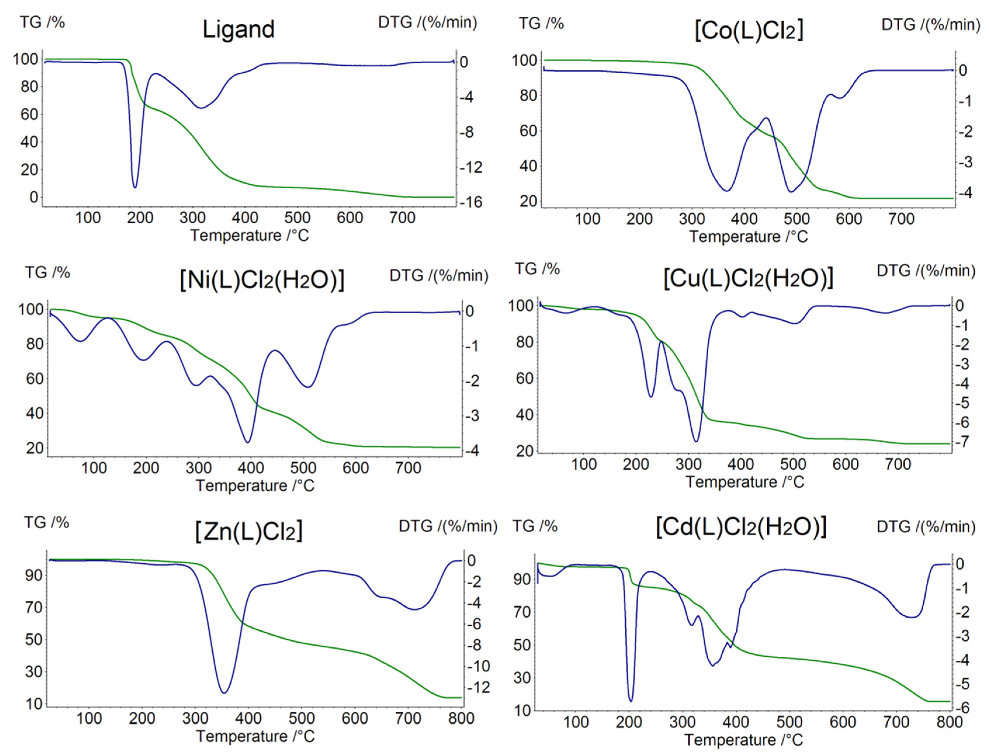
| Ligand | 3 | 180–210 | 210–400 | 550–700 | - | - |
| [Co(L)Cl2] | 3 | 275–460 | 460–550 | 550–640 | - | CoO |
| [Ni(L)Cl2(H2O)] | 4 | 40–120 | 120–250 | 250–420 | 420–600 | NiO |
| [Cu(L)Cl2(H2O)] | 4 | 40–150 | 150–250 | 250–380 | 380–720 | CuO |
| [Zn(L)Cl2] | 2 | 280–500 | 500–800 | - | - | ZnO |
| [Cd(L)Cl2(H2O)] | 3 | 40–200 | 210–450 | 450–760 | - | CdO |
| Compound | IC50 against A549 ± SD (μM) |
|---|---|
| Ligand | 589 ± 18 |
| [Co(L)Cl2] | >600 |
| [Ni(L)Cl2(H2O)] | >600 |
| [Cu(L)Cl2(H2O)] | 599 ± 71 |
| [Zn(L)Cl2] | >600 |
| [Cd(L)Cl2(H2O)] | 410 ± 31 |
| Etoposide | 452 ± 18 |
Publisher’s Note: MDPI stays neutral with regard to jurisdictional claims in published maps and institutional affiliations. |
© 2022 by the authors. Licensee MDPI, Basel, Switzerland. This article is an open access article distributed under the terms and conditions of the Creative Commons Attribution (CC BY) license (https://creativecommons.org/licenses/by/4.0/).
Share and Cite
Rogalewicz, B.; Climova, A.; Pivovarova, E.; Sukiennik, J.; Czarnecka, K.; Szymański, P.; Szczesio, M.; Gas, K.; Sawicki, M.; Pitucha, M.; et al. Antitumor Activity and Physicochemical Properties of New Thiosemicarbazide Derivative and Its Co(II), Ni(II), Cu(II), Zn(II) and Cd(II) Complexes. Molecules 2022, 27, 2703. https://0-doi-org.brum.beds.ac.uk/10.3390/molecules27092703
Rogalewicz B, Climova A, Pivovarova E, Sukiennik J, Czarnecka K, Szymański P, Szczesio M, Gas K, Sawicki M, Pitucha M, et al. Antitumor Activity and Physicochemical Properties of New Thiosemicarbazide Derivative and Its Co(II), Ni(II), Cu(II), Zn(II) and Cd(II) Complexes. Molecules. 2022; 27(9):2703. https://0-doi-org.brum.beds.ac.uk/10.3390/molecules27092703
Chicago/Turabian StyleRogalewicz, Bartłomiej, Alina Climova, Ekaterina Pivovarova, Jarosław Sukiennik, Kamila Czarnecka, Paweł Szymański, Małgorzata Szczesio, Katarzyna Gas, Maciej Sawicki, Monika Pitucha, and et al. 2022. "Antitumor Activity and Physicochemical Properties of New Thiosemicarbazide Derivative and Its Co(II), Ni(II), Cu(II), Zn(II) and Cd(II) Complexes" Molecules 27, no. 9: 2703. https://0-doi-org.brum.beds.ac.uk/10.3390/molecules27092703