Article

A Comparative Evaluation of the Photosensitizing Efficiency of Porphyrins, Chlorins and Isobacteriochlorins toward Melanoma Cancer Cells

by
Kelly A. D. F. Castro
1,2,*,
Nuno M. M. Moura
2,*,
Mário M. Q. Simões
2,*,
Mariana M. Q. Mesquita
2,
Loyanne C. B. Ramos
1,
Juliana C. Biazzotto
1,
José A. S. Cavaleiro
2,
M. Amparo F. Faustino
2,
Maria Graça P. M. S. Neves
2 and
Roberto S. da Silva
1
1
Department of Biomolecular Sciences, Faculty of Pharmaceutical Sciences of Ribeirão Preto, University of São Paulo, São Paulo 05508-220, Brazil
2
LAQV-REQUIMTE, Department of Chemistry, University of Aveiro, 3810-193 Aveiro, Portugal
*
Authors to whom correspondence should be addressed.
Submission received: 11 May 2023 / Revised: 3 June 2023 / Accepted: 8 June 2023 / Published: 12 June 2023
(This article belongs to the Special Issue Nitrogen Ligands)

Abstract

:
Skin cancer is one of the cancers that registers the highest number of new cases annually. Among all forms of skin cancer, melanoma is the most invasive and deadliest. The resistance of this form of cancer to conventional treatments has led to the employment of alternative/complementary therapeutic approaches. Photodynamic therapy (PDT) appears to be a promising alternative to overcome the resistance of melanoma to conventional therapies. PDT is a non-invasive therapeutic procedure in which highly reactive oxygen species (ROS) are generated upon excitation of a photosensitizer (PS) when subjected to visible light of an adequate wavelength, resulting in the death of cancer cells. In this work, inspired by the efficacy of tetrapyrrolic macrocycles to act as PS against tumor cells, we report the photophysical characterization and biological assays of isobacteriochlorins and their corresponding chlorins and porphyrins against melanoma cancer cells through a photodynamic process. The non-tumoral L929 fibroblast murine cell line was used as the control. The results show that the choice of adequate tetrapyrrolic macrocycle-based PS can be modulated to improve the performance of PDT.

Graphical Abstract

1. Introduction

Worldwide, the cancer incidence rate has increased, being one of the main causes of death. Currently, some means of treating cancer include surgery in the case of localized solid tumors, and radiotherapy and chemotherapy (for non-localized tumors) [1]. The development of new therapeutic agents that are selective, efficient, and cause minimal damage to the patient has become one of the central challenges of medicinal chemistry. Melanoma is a type of skin cancer that originates in melanocytes [2], which are found in the basal layer of the epidermis [2,3]. The resistance of this form of cancer to conventional treatments has led to the employment of some alternatives [3].
Photodynamic therapy (PDT) is a therapeutic methodology that requires the combination of a photosensitizer (PS), dioxygen (3O2), and light which, under specific conditions, generate reactive oxygen species (ROS) that are highly cytotoxic, inducing a reduction in the viability of cancer cells [1,4,5,6]. This is a therapeutic approach that has been pointed out by the scientific community as a promising complement or alternative to the conventional anticancer approaches, such as surgery or radiotherapy. PDT is a non-invasive therapy with high selectivity for cancer cells and reduced side effects due to its controllability and high spatiotemporal approach.
Porphyrins and related compounds are known for their excellent performance as photosensitizers (PS) in PDT [7,8,9,10,11,12,13]. Currently, applied PS are often based on porphyrins, chlorins, and isobacteriochlorin-type derivatives due to their optical features, namely, their strong absorption in the “therapeutic window” ranging from 650 to 850 nm, and their ability to produce ROS that can be lethal to the undesired tissue when activated by light [14,15,16,17,18]. Porphyrins and their derivatives that contain fluorine atoms in their structures have been investigated for more than 20 years [19,20,21,22,23,24]. The presence of fluorine atoms aims to modulate the pharmacokinetic and photophysical features, such as photostability, ROS production, and lipophilicity, among others. On the other hand, chlorin-based macrocycles exhibit photophysical properties similar to porphyrins, but with intensified and red-shifted Q bands, making chlorins the most promising candidates for PDT [4,17,25,26]. Like chlorins, isobacteriochlorins are also porphyrin-based reduced forms, but due to the presence of two reduced pyrrolic units in adjacent positions, they are more prone to oxidative processes. Synthetic strategies, such as the presence of bulky substituents next to the reduced pyrrolic-type unit or the modification of the porphyrinic core with exocyclic-fused rings, are usually used to avoid these oxidative processes [27,28].
Natural chlorins such as chlorophylls, which play a vital role in photosynthetic processes, are considered promising PS. However, the use of these naturally reduced porphyrins in PDT requires laborious extraction and modification of natural derivatives or total synthesis approaches, which are both a tremendous disadvantage [29,30]. To overcome this issue, efficient synthetic approaches to prepare chlorin-type macrocycles and other meso-tetraarylporphyrin reduced derivatives (e.g., (iso)bacteriochlorins) were developed, namely, those involving porphyrin precursors in cycloaddition reactions, such as Diels–Alder reactions and 1,3-dipolar cycloadditions [31].
We have recently studied the efficiency of 5,10,15-tris(pentafluorophenyl)-20-(4-pyridyl)porphyrin (Por2) and of the corresponding chlorin (Chl2) and isobacteriochlorin (Iso2) toward the B16F10 melanotic cell line. The Chl2 and Iso2 display a PDT effect and can induce a decrease of up to ca. 90% in the viability of the resistant B16F10 cell line after short irradiation periods with a maximum total light dose of 5.4 J.cm−2. In this study, it was found that the light irradiation on the therapeutic window (PDT effect) was strongly dependent on different factors, namely, the PS structure, 1O2-generation ability, PS cell uptake, and subcellular localization [32]. Additionally, porphyrin Por1 and its reduced derivatives (Chl1 and Iso1) were evaluated as PS toward prostate cancer PC-3 cells after incorporation into polyvinylpyrrolidone (PVP) formulations. The authors used this strategy to prevent aggregation issues and found that PS-PVP formulations bearing the porphyrin-reduced derivatives (Chl1 and Iso1) display apoptosis-mediated PDT activity when irradiated with a red light and a total light dose of 10.6 J.cm−2 [33]. Drain and co-workers prepared a series of thioglycosylated compounds similar to Por1, Chl1, and Iso1 and evaluated them as diagnostic agents. The uptake for such derivatives was evaluated into MDA-MB-231 breast cancer and K:Molv NIH 3T3 mouse fibroblasts, showing that the uptake at a nanomolar range of the thioglycosylated bacteriochlorin derivative makes it appropriate for tagging applications, while the chlorin analog is suitable for both targeting and treating diseased tissues due to its high absorption into the near-infrared region [34]. Later, Samaroo and co-workers [35] evaluated the interaction of those photosensitizers in the presence of plasma proteins, bovine serum albumin (BSA), and human serum albumin (HSA) through spectrophotometric and spectrofluorimetric titrations and theoretical studies. The interaction with the protein’s hemin site by porphyrinic-based PS leads to the formation of stable complexes, showing the potential of those proteins to be used as effective drug-delivery systems for further therapeutic purposes.
In the present work, we decided to evaluate and compare how the efficiency of isobacteriochlorin, chlorin, and porphyrin analogues against melanoma cancer cells would be affected by the type of meso-substituted scaffold selected—A4-type versus A3B-type. For this, Por1, Chl1, and Iso1 were chosen as examples of meso-substituted A4-type macrocycles while for the A3B-type series, Por2, Chl2, and Iso2 were selected. To investigate that influence, the photophysical/photochemical and biological features of both meso-substituted series A4-type and A3B-type derivatives were performed. The carried-out assays allowed us to compare how the photophysical and biological properties of the studied derivatives are modulated by inducing changes in one of the substituents at the meso-position. This comparative study can be a driving force for further studies using both the PS design and PDT assays as targets.

2. Results and Discussion

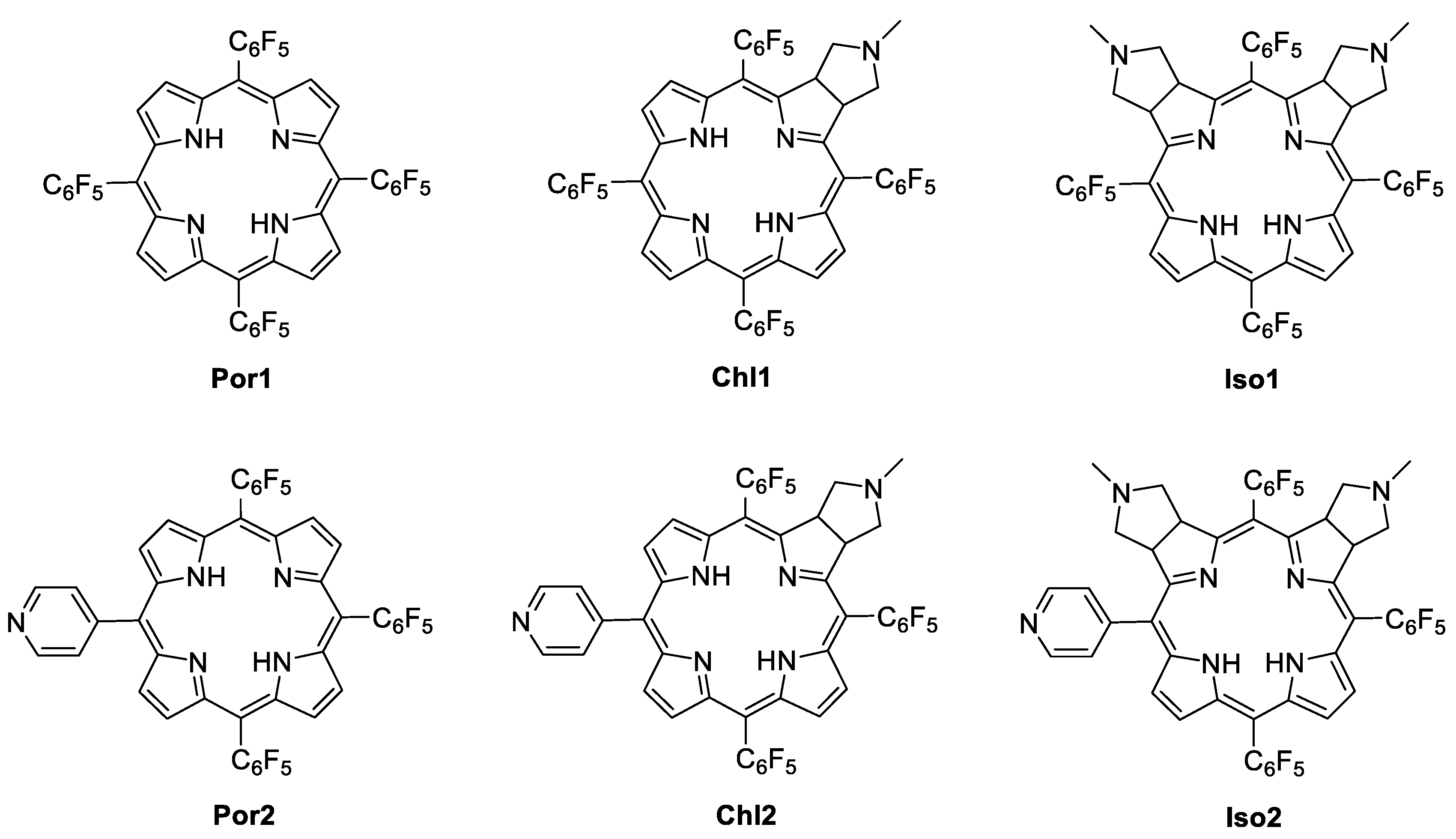
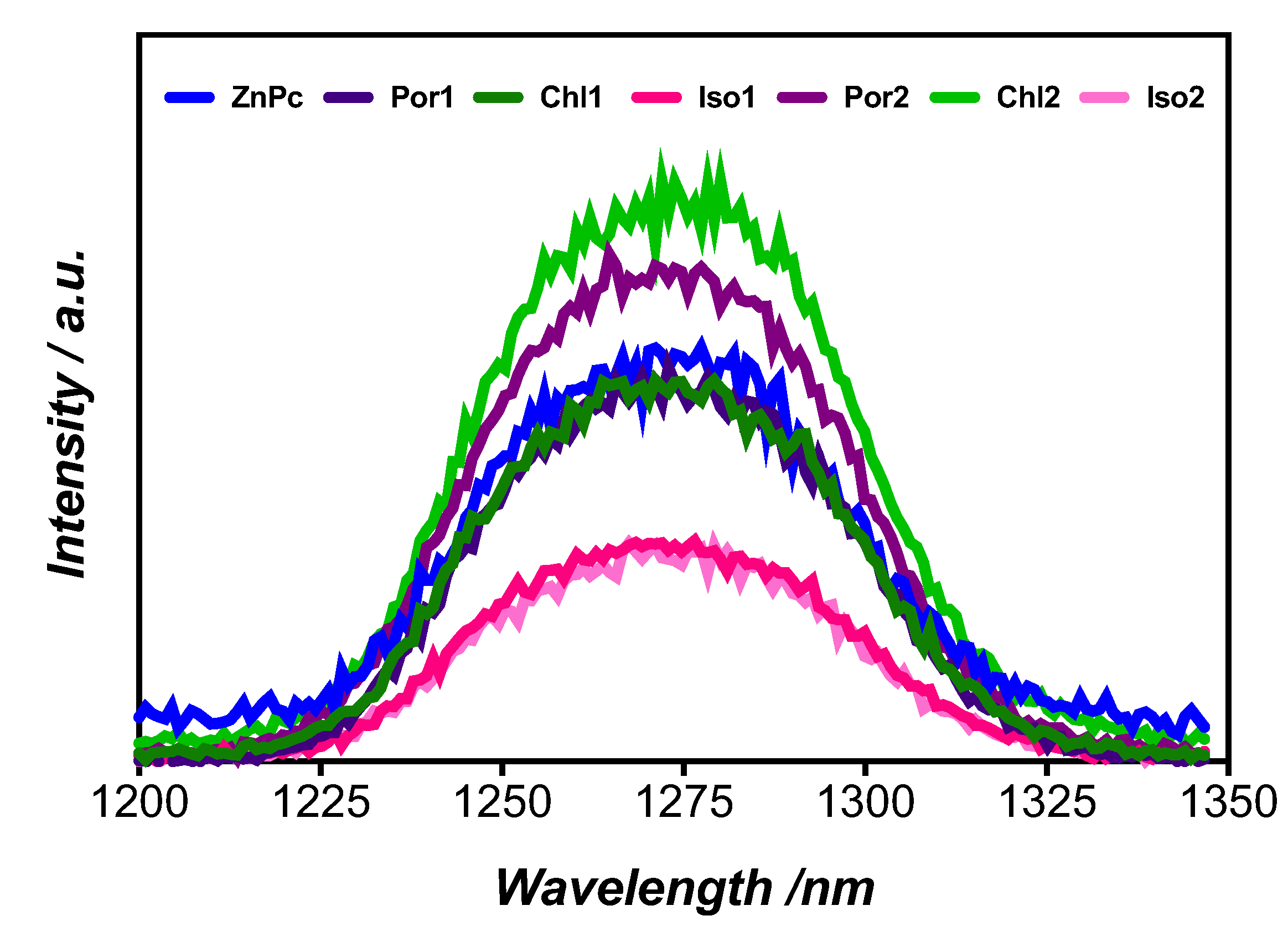
2.1. Photosensitizers: Synthesis and Characterization
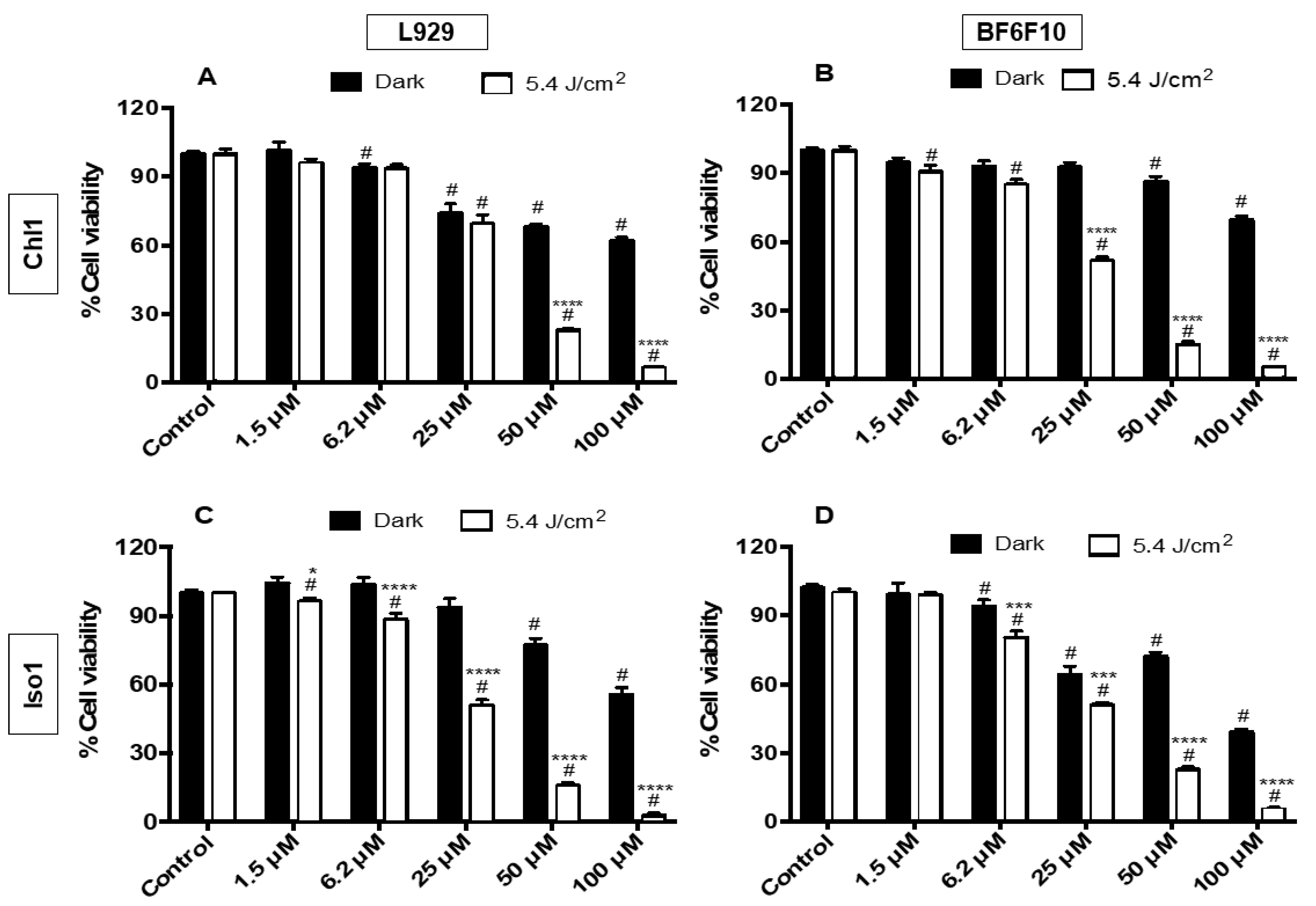
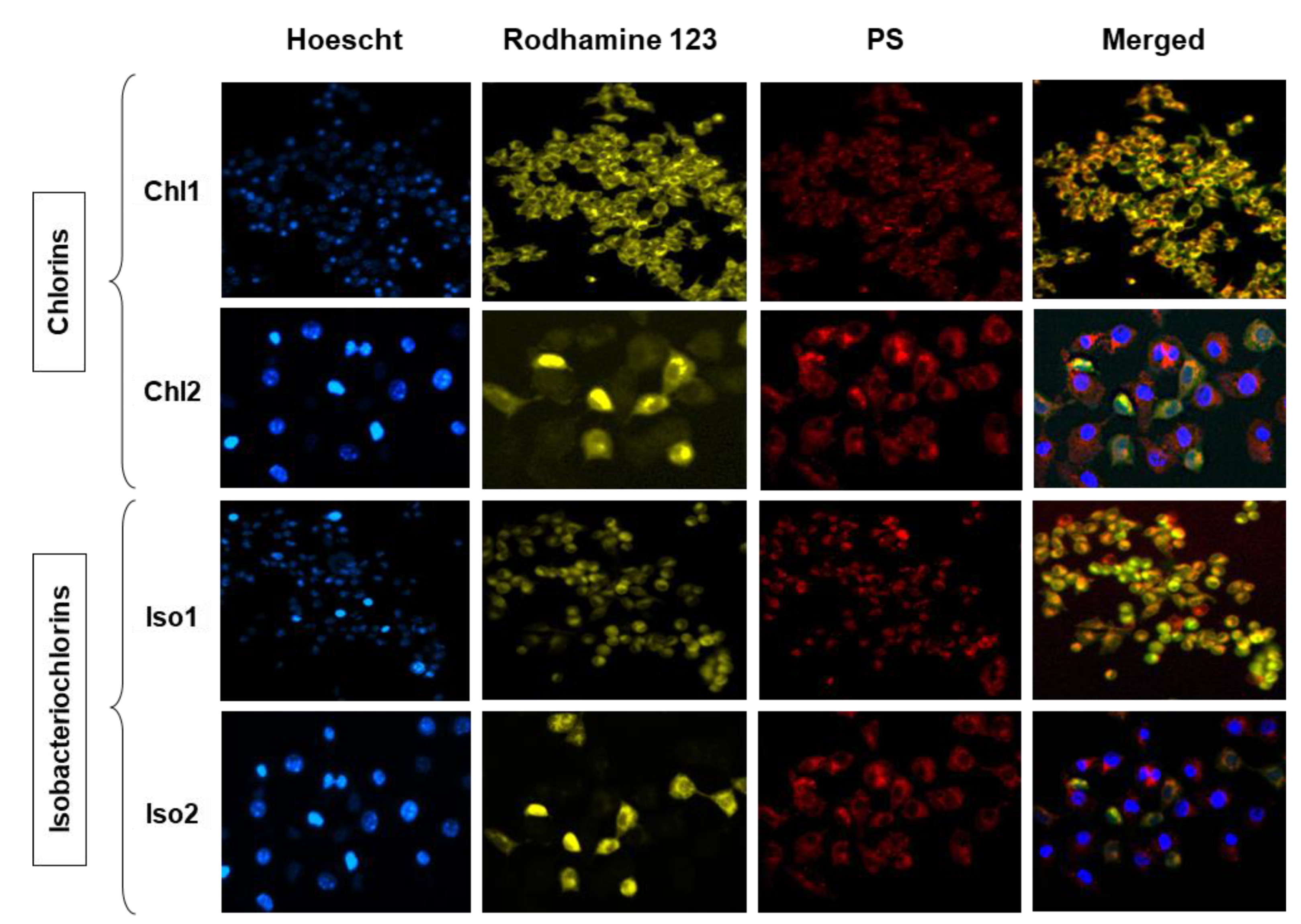
5,10,15,20-Tetrakis(pentafluorophenyl)porphyrin (Por1) and 5,10,15-tris(pentafluorophenyl)-20-(4-pyridyl)porphyrin (Por2) were obtained directly by condensation of pyrrole with the adequate aldehydes in acidic conditions, according to previously described procedures [32,36,37]. The pyrrolidine-fused chlorins (Chl1 and Chl2) and isobacteriochlorins (Iso1 and Iso2) were prepared throughout the 1,3-dipolar cycloaddition of the appropriate porphyrin (Por1 or Por2) and the azomethine ylide generated from N-methylglycine and paraformaldehyde (Figure 1). The reduced derivatives were attained in yields similar to those reported in the literature (70% for Chl1 and 49% for Chl2; 18% for Iso1 and 15% for Iso2). The structures of all the compounds were confirmed by 1H and 19F NMR, UV–Vis spectroscopy, and mass spectrometry and agreed with the data reported in the literature [32,33,37,38,39,40].
The absorption, emission, and excitation spectra of porphyrins (Por1,2) and their reduced analogs, chlorins (Chl1,2), and isobacteriochlorins (Iso1,2), were acquired in N,N-dimethylformamide (DMF) and are summarized in Table 1. The photophysical characterization of Por1 and Por2 was assessed since they are the chemical precursors of the reduced derivatives Chl1, Chl2, Iso1, and Iso2. The absorption spectra of Por1 and Por2 exhibit the typical features of free-base meso-substituted porphyrin derivatives [41], displaying an intense Soret band at 410 and 412 nm, respectively, due to S0→S2 transitions, followed by four weak Q bands in the visible region ranging from 504 to 638 nm, attributed to the S0→S1 transitions. The Soret band on the absorption spectra of chlorins (Chl1,2) was slightly blue-shifted (ca. 5 nm) when compared with the corresponding porphyrin precursor (Figure S1). Concerning the Q bands region, significant red shifts (~14 nm) were observed for the two bands at higher wavelengths.
The UV–Vis spectrum of both isobacteriochlorins Iso1 and Iso2 shows Soret bands even more blue-shifted (20–25 nm) than those of the corresponding chlorin derivatives Chl1 and Chl2; the last Q bands also show significant red shifts (10–15 nm) when compared with porphyrins. This red shift in the last Q band for both reduced series is of high interest for PDT, since this transition absorbs in the so-called therapeutic window (600–800 nm). As expected, Chl1 and Chl2 display the last Q band at ca. 650 nm with a relatively high intensity when compared with the other Q bands. The pyridyl group at the meso-position of the porphyrin macrocycle did not induce noticeable changes in the absorption features of the free-base porphyrin and reduced derivatives.
Concerning the steady-state emission spectra, Por1, Por 2, and Iso2 showed similar emission spectra with two well-defined bands at 638 and ~700 nm. The Iso1 emission spectrum displayed a similar profile; however, the first vibrational mode of the fluorescence was blue-shifted 38 nm to 600 nm. Both chlorin derivatives Chl1 and Chl2 only had one emission band at 654 and 649 nm, respectively (Figure S2). The emission spectra of the studied compounds are typical of free-base meso-tetraarylporphyrins and of their reduced derivatives [42,43,44].
The fluorescence quantum yields (ΦF) (Table 1) were determined in DMF using meso-tetraphenylporphyrin (TPP) as the standard (ΦF = 0.11 in DMF) [45,46]. The fluorescence properties of each compound type appeared quite similar, which is consistent with the similarity of their chemical structure. The higher fluorescence quantum yields observed for Chl1, Chl2, Iso1, and Iso2, if compared to Por1 and Por2, can be ascribed to the differences in their electronic structures and molecular geometries. Chlorins and isobacteriochlorin generally have a more distorted and non-planar structure compared to porphyrins [47]. This distortion can lead to a decrease in the non-radiative relaxation pathways, such as vibrational relaxation and internal conversion, thereby enhancing the fluorescence efficiency. Compound Iso2 shows the highest fluorescence quantum yield (ΦF = 0.21), followed by Chl2F = 0.16), Chl1F = 0.15), and finally, Iso1F = 0.13). In fact, all the isobacteriochlorin and chlorin-type derivatives presented a greater fluorescence quantum yield (ΦF) than TPP (0.11). However, opposite behavior was observed for the starting porphyrins Por1 and Por2, which showed a lower fluorescence quantum yield (ΦF) than TPP (respectively, 0.01 and 0.06 versus 0.11). The fluorescence lifetime of a singlet state (τ) found for porphyrins varied between 10 ns and 11.1 ns. The lifetime reduction observed for Chl1 (6.02 ns) and Chl2 (6.90 ns) when compared to Por1 and Por2 can be explained by the presence of a reduced pyrrole-type ring, leading to a reduction in the π-conjugation effect and improving the macrocycle distortion in the excited state, thus resulting in a decrease in radiative decay rates [48]. The emission decay profile for Iso1 and Iso2 derivatives can be fitted (Figure S3) by two decay components. For Iso1, we have 5.4 ns (~97%) and 1.2 ns (~3%), whereas the short-lived component for Iso2 had a contribution of 77%, and the contribution of the long-lived component was lower when compared to Iso1. The reduction observed for the fluorescence lifetimes of isobacteriochlorins Iso1 and Iso2 can be ascribed to the presence of an additional reduced pyrrolic-type ring at the macrocycle core. The increase in the number of reduced pyrrolic units conducts, in general, to an increase in the conformational flexibility (lower rigidity) of the macrocycle, affecting the electronic structure. Pyrrolidine-type units should decrease the π-conjugation effect, and they may improve the macrocycle distortion in the excited state, contributing to the reduction observed for the fluorescence lifetimes of isobacteriochlorins Iso1 and Iso2 and of chlorins Chl1 and Ch2, leading to a fast internal conversion.
The long-lived triplet state’s (τT) profile decay of the studied compounds range between 0.50 and 0.98 μs. For Por2, bearing a pyridyl group at one of the meso-positions, the value of the triplet lifetime is lower than the one displayed by Por1, with no pyridyl substituent (0.76 versus 0.98 μs). However, for the reduced derivatives, a different behavior is observed. Chl2 (0.74 μs) and Iso2 (0.63 μs) display higher triplet excited state lifetime values when compared with the corresponding analogs Chl1 (0.51 μs) and Iso1 (0.50 μs). The remarkable dependence of the triplet lifetime as a function of the substituent and its position is well established in the literature [44,49,50].
The efficiency of a PS in PDT is also related to its ability to generate reactive oxygen species (ROS), mainly singlet oxygen (1O2). ROS acts as signaling molecules, but they can also promote cellular damage by rapidly oxidizing cellular components. The measurement of the 1O2 quantum yield (ΦΔ) was assessed by the luminescence method, measuring the 1O2 phosphorescence at 1270 nm (Figure 2) and using ZnPc (zinc(II) phthalocyanine) as the reference. It is worth noting that this approach is not dependent on the dye concentration but only on the number of photons absorbed [51]. In general, the ΦΔ increased when the pyridyl group was attached to the macrocycle backbone (except for Iso2) in the following order: Chl2 (0.81) > Por2 (0.65) > Por1 (0.55) > Chl1 (0.42) > Iso2 (0.35) > Iso1 (0.31) (Table 1). Additionally, the triplet lifetime (τT) increased for chlorin-type derivatives as a function of the substituent, which consequently increased the ΦΔ [52]. The presence of halogens influences the ability to generate 1O2 [53], photostability, and photophysical features [16,19]. The differences observed for the ΦΔ are probably related to the macrocycle planar distortion in Por1, Chl1, and Iso1 derivatives. The presence of halogen atoms leads to an improved intersystem crossing from the photosensitizer´s excited singlet and triplet states [53]; however, for the evaluated compounds, the presence of more halogen atoms did not favor 1O2 generation of Por1 and its reduced analogs (Chl1 and Iso1) when compared with those bearing a pyridyl unit (Por2, Chl2, and Iso2). This is undoubtedly related to the lower amount of T1 states quenched by 3O2 due to the occurrence of competing processes, reducing the formation of 1O2. Probably, different restrictions in the aryl ring rotations ascribed to the presence of three C6F5 and one pyridyl unit, higher asymmetry, and different electronic effects contributed to improve the ability of Por2, Chl2, and Iso2 to generate 1O2 when compared with the corresponding Por1 and reduced analogs Chl1 and Iso1 with four C6F5 substituents [54]. Singlet oxygen generation is strongly related to the probability of molecular dioxygen (3O2) colliding with the PS in the triplet state, which can be affected by steric effects, often associated with a decrease in the amount of the excited triplet state. Data in Table 1 agree with this and can be related to the presence of the pyrrolidine-fused rings on both reduced derivatives, resulting in a decrease in collision frequencies between the macrocycles and 3O2. The high ΦΔ of Chl2 seems to indicate higher success in the collisions involving 3O2 and this reduced derivative. The best 1O2 generator was Chl2Δ = 0.81); however, all the compounds studied are able to generate this highly cytotoxic species (ROS), which makes them suitable to be used in PDT against cancer cells.

2.2. PDT Assays

With the exception of Por1, due to its low solubility in the RPMI/DMSO (1%) medium (vide infra), the ability of all the other derivatives Chl1, Iso1, Por2, Chl2, and Iso2 to act as PS in the PDT assays was investigated, considering their efficiency to generate 1O2. The cytotoxicity of the compounds was assessed by an in vitro MTT colorimetric assay [3-(4,5-dimethylthiazol-2-yl)-2,5-diphenyl tetrazolium bromide] [55]. The viability of B16F10 murine melanoma cells was evaluated after treatment with Chl1, Iso1, Por2, Chl2, and Iso2 at different concentrations (1.5, 6.2, 25, 50, and 100 μM), without and under red light irradiation (660 nm; 5.4 J/cm2) (Figure 3 and Figure 4). A non-tumoral fibroblast L929 murine cell line was used as the control.
The results show that in general, the compounds displayed cytotoxicity under non-irradiated conditions (dark) against both melanoma cells and non-tumoral cells at 25 μM or higher concentrations. However, in general, photo-stimulation increased their cytotoxicity. Of all the PS studied, globally speaking, Chl1 was the least cytotoxic (Figure 3A,B) in the dark at 100 μM (38% and 31% cell death for L929 and B16F10, respectively). Both chlorins were effective toward B16F10 at 25 μM under red light irradiation (660 nm) with a reduction in the cell viability of 43% (Chl1) and 42% (Chl2), taking into account their toxicity under dark conditions (Figure 3B and Figure 4D). This phototoxicity improves at higher concentrations, attaining cell deaths of ca 64% at 100 μM, considering also the cytotoxicity under dark conditions. Despite the efficiency of both chlorins, these PS displayed small selectivity for B16F10 cells, causing the death of the non-tumoral cells ranging from 30% to 35% (Figure 3A and Figure 4C).
Although both isobacteriochlorins induced a reduction in tumoral cells, Iso2 was more efficient and selective for B16F10 cells (p < 0.001) (Figure 4E,F) than Iso1 (Figure 3C,D). For instance, Iso1 at 25 μM displayed cytotoxicity under both dark (~36%) and light conditions (~49%) toward B16F10 cells and similar phototoxicity toward the non-tumoral cells (cell death of ~50%). Although there was less cytotoxicity for non-tumoral cell lines, the photocytotoxicity was more relevant than the cytotoxicity at higher concentrations. No improvement in the selectivity toward B16F10 cells was observed for isobacteriochlorin Iso1. Moreover, Iso2 at 25 μM presented lower cytotoxicity in the dark and was able to cause 72% of cell death of the treated melanoma cells, and no significative cell death (20%) was achieved in non-tumoral cell lines under similar conditions.
Therefore, when comparing porphyrin and chlorin-type derivatives using the same concentration, chlorins seem to be more efficient PS after being irradiated with red light (660 nm) due to their strongest absorption in the red region. In general, the more photocytotoxic compounds contain the pyridyl group in their structure (Chl2 and Iso2). In fact, there is a strong relationship between structure and activity. The efficiency of Chl2 can be attributed to its photophysical and photochemical properties, mainly the production of 1O2, but no selectivity for B16F10 cancer cells was observed; this PS was also able to induce a significant reduction in the non-tumoral fibroblast L929 murine cell line. Additionally, its dark activity can be due to the formation of adducts with biological structures that affect the cell survival [56], since the production of ROS is limited. Therefore, cellular uptake and cellular sublocalization should also be considered.
Several factors can influence the PS activity; the formation of aggregates is the feature that contributes the most to the decrease in its efficiency, due to the limitation of ROS production and cellular uptake [57,58]. Aggregation studies were performed in order to understand the potential effect of the porphyrin-type scaffold selected (meso-substituted—A4-type versus A3B type) on the cytotoxicity observed. UV–Vis studies assessing different biological PS concentrations in RPMI medium and DMSO (1%) were performed (see ESI, Figures S4–S8). It was noticed that the PS strictly followed the Beer–Lambert law to established concentrations, thus suggesting no aggregation in RPMI at concentrations below 3 μM for Chl1, 12.5 μM for Iso1, 6.0 μM for Por2, 3.0 μM for Chl2, and 3.0 μM for Iso2. Por1 showed low solubility in biological conditions; so, it was decided not to evaluate this compound in the further PDT assays. The aggregation studies showed that the pyridyl group increased the PS solubility of Por2, but no noticeable changes were observed for Chl2, although leading to a decrease in the solubility of derivative Iso2 when compared with the corresponding A4-type derivative.
As expected, more fluorine atoms increase the lipophilicity of the PS, which is corroborated by cellular uptake studies (Figure S9).
The octanol:water partition coefficients (Log p) were calculated for all the macrocycles using Molinspiration WebME Editor 3.81 [47]. The A4-type macrocycles exhibited Log p values between 9.98 and 9.75, while for the A3B-type macrocycles, the Log p values ranged from 9.69 to 9.39. Considering the possibility that at physiological pH the pyrrolidine units must be almost entirely in the cationic form (pyrrolidine pKa = 11.3), partition coefficients were also calculated for chlorin and isobacteriochlorin derivatives while taking that into account. A slight decrease was observed for Log p values of both series of protonated compounds, being 9.21 (Chl1) and 9.01 (Iso1) for A4-type reduced macrocycles and 8.60 and 7.93 for the corresponding A3B-type macrocycles Chl2 and Iso2, respectively.
The uptake of Chl1 and Iso1 increased slightly as a function of concentration. On the other hand, both Chl2 and Iso2 showed a reduction in the uptake at 25 μM after 4 h of incubation in B16F10 cells evaluated by fluorescence; if compared to the concentrations of 6.1 μM and 12.5 μM, this behavior can be partially justified by the aggregation phenomena, corroborating the main findings in aggregation studies.
When the stability of the PS in RPMI/DMSO (1%) mixture was evaluated in the dark over 24 h (Figure S10), a noticeable decrease (~40%) in the stability was observed for Chl1 and Chl2, and even after this long period in the solution, Iso1 and Por2 showed an acceptable stability, with a decrease of around 20%. Iso2 showed the lowest stability in solution, with a decrease of around 45% in the absorption percentage of this PS after the same period. These data show that, under dark conditions, Iso1 and Por2 were the most stable in the RPMI/DMSO (1%) mixture at 3.1 μM after 24 h. When irradiated for 30 min under the same irradiation conditions of biological assays, the studied compounds showed good stability. Apart from Chl2 (Figure S11D), which displays a noticeable decrease in the absorption intensity at Soret band, the UV–Vis spectra of the other compounds remained almost unchanged (Figure S11).
To demonstrate the intracellular distribution in B16F10 cells, images of fluorescence microscopy were acquired. Red fluorescence was observed in all cases, which led us to suggest that the PS was efficiently internalized (Figure 5). The two panels on the left side displayed the PS internalized in the B16F10 cells, with Hoechst staining the nucleus (blue) and Rhodamine staining the mitochondria (green). Chlorins (Chl1 and Chl2) and isobacteriochlorins (Iso1 and Iso2) showed similar fluorescence patterns with the mitochondria probe. The overlapping fluorescence of Chl2 and Iso2 suggests that the compounds are preferentially localized in regions near mitochondria, while Chl1 and Iso1 are apparently spread throughout the cell, including the nucleus.
Despite the lack of selectivity for cancer cells (B16F10) for most of the tested PS at high concentrations, we propose to employ metronomic PDT to improve the tumor-specific response. Wilson et al. created the term metronomic PDT, which consists of administering both the PS and light during an extended period at very low doses and over many hours in order to increase the selective reduction on the viability of cancer cells by apoptosis. In their study, the authors aimed to compare standard or acute PDT with metronomic PDT. At the end, they found that metronomic PDT enhanced tumor-specific cell death, while decreasing the harm to adjacent normal tissues [59]. Therefore, herein we suggest that metronomic delivery or several PDT treatments are required to increase the selectivity of the PS we have tested, namely, of the chlorin-type derivatives.

3. Materials and Methods

3.1. Generalities

Absorption spectra were obtained on a Shimadzu UV-2501PC spectrophotometer in the 350–800 nm range. The fluorescence spectra were recorded in DMF in 1 cm × 1 cm quartz optical cells under normal atmospheric conditions on a computer-controlled F4500—Hitachi spectrofluorometer. The widths of both excitation and emission slits were set at 3.0 nm. To calculate the fluorescence quantum yield (ΦF), TPP in DMF was used as reference using Equation (1). In this equation, ΦF is the fluorescence quantum yield of the sample; Φst is the fluorescence quantum yield of TPPexc = 420 nm, ΦF = 0.11 in DMF); Ast is the absorbance of TPP and A is the absorbance of the sample at the excitation wavelength; Sst and S represent the integrated emission band of the TPP and sample, respectively.
ΦF = ΦF = Φst × S/Sst × Ast/A
Laser flash photolysis experiments for detection of the triplet state in solution were performed on a system using a Quanta Ray Lab-130 4 Hz Nd:YAG laser at 420 nm from Spectra Physics as the excitation source. The fluorescence lifetime was determined on a Fluorescence Correlation Spectroscopy/Fluorescence Lifetime Imaging coupled to a laser with 420 nm excitation wavelength. Direct measurement of singlet oxygen was performed by luminescence method from Equation (2), as described in the literature [60], using ZnPc as standard (λexc = 660 nm, ΦΔ = 0.56 in DMF) [61,62].
ΦΔ = Φst (Is/Ist)
where ΦΔ is the singlet oxygen quantum yield of the sample; Φst the singlet oxygen quantum yield of ZnPc; Ist is the phosphorescence intensity of 1O2 at 1270 nm for ZnPc; and Is the phosphorescence intensity for the sample.

3.2. Synthesis of the Photosensitizers (PS)

The photosensitizers were synthesized as described in the literature [32,36,37].

3.3. Cell Culture

The B16F10 (melanoma murine) and L929 (fibroblast murine) cells were obtained from the American Type Culture Collection. The cell line was cultured in RPMI medium with 10% supplemental fetal bovine fetal serum, 100 IU mL−1 of penicillin G, 100 mg mL−1 of streptomycin, and 1 µg mL−1 of amphotericin. Cells were seeded until 75–90% confluence in 96-well plates and cultured in a humidified incubator at 37 °C with 5.0% CO2 for 24 h.

3.4. Cell Viability Assay

Evaluation of cell cytotoxicity by porphyrin derivatives was performed against two different cell lines: the murine melanoma, B16F10, and murine fibroblast, L929. To this end, 2 × 10⁴ cells were incubated for 24 h in 96-well cell culture plates. After this period, the treatments with Chl1, Iso1, Por2, Chl2, and Iso2 that were previously dissolved in DMSO (1.5, 6.2, 25, 50, and 100 μM), then dissolved in the culture medium, and serially diluted to the appropriate concentration to give a final DMSO concentration of 1%, were conducted with and without red light irradiation (emitted by an array of 96 light-emitting diodes (LEDs), λ = 660 nm). After the indicated treatment, the cells were incubated at 37 °C for 3 h in a culture medium containing 10 mol L−1 MTT in RPMI without supplemental fetal bovine fetal serum. The blue MTT formazan precipitate was then dissolved in 50 μL of DMSO, and the absorbance was measured at 480 nm with a multi-well plate reader. The cell viability was expressed as the percentage of the absorption values in the treated cells relative to the non-treated (control) cells. The data are presented as an average of three independent experiments with replicates.

3.5. Statistical Analysis

Statistical analysis was performed by two-way ANOVA. Equality of variance was assumed with Bonferroni’s post hoc test for pair-wise comparisons. Results with p < 0.05 were considered statistically significant.

3.6. Fluorescence Studies

Cellular uptake of porphyrin derivatives by B16F10 cells was performed by fluorescence spectroscopy and microscopy, as described in the literature [32]. The fluorescence was measured with a microplate reader (Spectra Max Paradigm) set at λexc = 420 nm, matching the Soret band and corresponding emission wavelength to each PS at the initial time = after 4 h of incubation. For fluorescence microscopy, specific organelles’ staining probes were used. Mitochondria were stained with Rhodamine 123, and Hoechst 33342 was used as the nuclei probe. After washing two times with PBS, the cells were examined by fluorescence microscopy (Nikon Eclipse Ti Microscope model TI-FL) using the following filters: DAPI (λexc at 340/380 nm and λem at 435 to 485 nm), FITC (λexc at 488 nm and λem at 505 to 527 nm), and Cy5 (λexc at 620/660 nm and λem at 662.5/737.5 nm) for porphyrin derivatives detection.

3.7. Stability Studies

The stability of each porphyrin derivative was verified by UV–Vis at regular intervals for up to 24 h under dark conditions. Photostability studies of the porphyrin derivatives were performed by irradiation of a solution of each derivative in RPMI and DMSO (1%) under the same conditions of PDT assays. The solutions were stirred and kept at ambient temperature.

4. Conclusions

In this work, two different series of neutral tetrapyrrolic macrocycle-based PS were successfully prepared and characterized. All the compounds are able to generate ROS, namely 1O2, which shows their potential to be used as PS in PDT against tumor cancer cells. However, due to the high aggregation observed for the A4-type Por1 under the conditions where biological assays were carried out, the PS activity of this compound was not evaluated. So, the PDT efficiency of the A4-type reduced derivatives (Chl1 and Iso1) as well as all the A3B-type porphyrinic PS prepared (Por2, Chl2, and Iso2) was evaluated toward murine melanoma cells (B16F10 cells) and non-tumor fibroblast murine cell lines (L929).
The cytotoxicity of the compounds evaluated is strongly dependent on their structure, level of reduction in the tetrapyrrolic macrocycle, and concentration. Iso2 and Chl2 were the most efficient PS against B16F10 cancer cells; however, Chl2 also displayed higher (photo)cytotoxicity for non-tumor L929 cells.
Although both chlorins can be considered efficient PS against B16F10 cancer cells, they also showed high (photo)cytotoxicity for non-tumoral L929 cells. A different situation was observed with Iso2, whereby at 25 μM, it displayed good selectivity for B16F10 tumoral cells, being able to induce a cell viability reduction of 72% and only 20% of cell death of non-melanoma L929 cells; however, Iso1, with the same degree of reduction and efficiency to generate 1O2, was cytotoxic under both dark and light conditions. These results confirm how a simple manipulation of the porphyrin core can affect the photodynamic action of this type of reduced PS.
In sum, the results showed that the choice of an adequate tetrapyrrolic macrocycle can be modulated to improve the performance of PDT. The presence of a pyridyl unit affords tetrapyrrolic macrocycle-based PS with improved features to be used in PDT. Additionally, the PDT efficiency was highly dependent on the subcellular localization mechanism and cellular uptake.
This study is a pertinent contribution to both tetrapyrrolic macrocycle-based PS development and PDT fields, providing significant data to modulate the tetrapyrrolic macrocycle core and aiming to fine-tune its photophysical/photochemical properties. In the future, it will help the scientific community to design and better prepare PS with improved features and PDT activity to overcome the challenges of effectively reducing the viability of cancer cell lines.

Supplementary Materials

The following supporting information can be downloaded at https://0-www-mdpi-com.brum.beds.ac.uk/article/10.3390/molecules28124716/s1, Figure S1: UV–Vis spectra; Figure S2: Fluorescence spectra; Figure S3: emission decay fittings; Figures S4–S8: Aggregation studies; Figure S9: Cellular uptake; Figure S10: Stability; Figure S11: Photostability studies.

Author Contributions

Conceptualization, K.A.D.F.C. and N.M.M.M.; methodology, K.A.D.F.C. and N.M.M.M.; validation, M.M.Q.S., M.A.F.F., M.G.P.M.S.N. and R.S.d.S.; investigation, K.A.D.F.C., M.M.Q.M., L.C.B.R. and J.C.B.; resources, M.G.P.M.S.N. and R.S.d.S.; writing—original draft preparation, K.A.D.F.C., N.M.M.M. and M.M.Q.S.; writing—review and editing, K.A.D.F.C., N.M.M.M., M.M.Q.S., J.A.S.C., M.A.F.F., M.G.P.M.S.N. and R.S.d.S.; supervision, K.A.D.F.C., N.M.M.M. and M.M.Q.S. All authors have read and agreed to the published version of the manuscript.

Funding

This research work received financial support from PT national funds (FCT/MCTES, Fundação para a Ciência e a Tecnologia and Ministério da Ciência, Tecnologia e Ensino Superior) through the projects UIDB/50006/2020 and UIDP/50006/2020, and the FCT project PORP2PS (EXPL/QUI-QOR/0586/2021). NMM Moura thanks FCT for funding through program DL 57/2016—Norma transitória (CDL-CTTRI-048-88-ARH/2018). The authors are grateful to CNPq, CAPES, FAPESP grant 2019/19448-8; 2022/15336-3, and Universidade de São Paulo for financial support.

Informed Consent Statement

Not applicable.

Data Availability Statement

The data that support the findings of this study are available from the corresponding authors upon reasonable request.

Acknowledgments

This work received support from PT national funds (FCT/MCTES, Fundação para a Ciência e a Tecnologia and Ministério da Ciência, Tecnologia e Ensino Superior) through the projects LAQV-REQUIMTE (UIDB/50006/2020 and UIDP/50006/2020) through national funds, and to the Portuguese NMR Network. NMM Moura thanks FCT for funding through program DL 57/2016—Norma transitória (CDL-CTTRI-048-88-ARH/2018). K.A.D.F. Castro thanks CAPES for the post-doctoral scholarship granted (PNPD/CAPES). Thanks are due to Coordenação de Aperfeiçoamento de Pessoal de Nível Superior (CAPES), Conselho Nacional de Desenvolvimento Científico e Tecnológico (CNPq), Fundação de Amparo à Pesquisa do Estado de São Paulo (FAPESP 2019/19448-8; 2022/15336-3). We would like to thank Maurício da Silva Baptista (Cepid Redoxoma 2013/07937-8) from the Institute of Chemistry, University of São Paulo, for his laboratory and equipment support and Helena Couto Junqueira for technical assistance.

Conflicts of Interest

The authors declare no conflict of interest.

Sample Availability

Samples of the compounds Por1, Por2, Chl1, Chl2, Iso1, and Iso2 are available from the authors.

References

  1. Ethirajan, M.; Chen, Y.; Joshi, P.; Pandey, R.K. The role of porphyrin chemistry in tumor imaging and photodynamic therapy. Chem. Soc. Rev. 2011, 40, 340–362. [Google Scholar] [CrossRef]
  2. Swavey, S.; Tran, M. Porphyrin and Phthalocyanine Photosensitizers as PDT Agents: A New Modality for the Treatment of Melanoma. In Recent Advances in the Biology, Therapy and Management of Melanoma; Davids, L.M., Ed.; InTech: Rijejka, Croatia, 2013. [Google Scholar]
  3. Huang, Y.Y.; Vecchio, D.; Avci, P.; Yin, R.; Garcia-Diaz, M.; Hamblin, M.R. Melanoma resistance to photodynamic therapy: New insights. Biol. Chem. 2013, 394, 239–250. [Google Scholar] [CrossRef] [PubMed]
  4. Gomes, A.T.P.C.; Faustino, M.A.F.; Neves, M.G.P.M.S.; Ferreira, V.F.; Juarranz, A.; Cavaleiro, J.A.S.; Sanz-Rodríguez, F. Photodynamic effect of glycochlorin conjugates in human cancer epithelial cells. RSC Adv. 2015, 5, 33496–33502. [Google Scholar] [CrossRef]
  5. Dougherty, T.J.; Gomer, C.J.; Henderson, B.W.; Jori, G.; Kessel, D.; Korbelik, M.; Moan, J.; Peng, Q. Photodynamic therapy. JNCI-J. Natl. Cancer I. 1998, 90, 889–905. [Google Scholar] [CrossRef] [PubMed] [Green Version]
  6. Yang, B.; Chen, Y.; Shi, J. Reactive Oxygen Species (ROS)-Based Nanomedicine. Chem. Rev. 2019, 119, 4881–4985. [Google Scholar] [CrossRef]
  7. Korolchuk, A.M.; Zolottsev, V.A.; Misharin, A.Y. Conjugates of Tetrapyrrolic Macrocycles as Potential Anticancer Target-Oriented Photosensitizers. Top. Curr. Chem. 2023, 381, 10. [Google Scholar] [CrossRef]
  8. Pan, L.; Ma, Y.; Wu, X.; Cai, H.; Qin, F.; Wu, H.; Li, C.Y.; Jia, Z. A Brief Introduction to Porphyrin Compounds used in Tumor Imaging and Therapies. Mini Rev. Med. Chem. 2021, 21, 1303–1313. [Google Scholar] [CrossRef]
  9. Zheng, B.-D.; Ye, J.; Zhang, X.-Q.; Zhang, N.; Xiao, M.-T. Recent advances in supramolecular activatable phthalocyanine-based photosensitizers for anti-cancer therapy. Coord. Chem. Rev. 2021, 447, 214155. [Google Scholar] [CrossRef]
  10. Kou, J.; Dou, D.; Yang, L. Porphyrin photosensitizers in photodynamic therapy and its applications. Oncotarget 2017, 8, 81591–81603. [Google Scholar] [CrossRef] [Green Version]
  11. Dandash, F.; Leger, D.Y.; Diab-Assaf, M.; Sol, V.; Liagre, B. Porphyrin/Chlorin Derivatives as Promising Molecules for Therapy of Colorectal Cancer. Molecules 2021, 26, 7268. [Google Scholar] [CrossRef]
  12. Pucelik, B.; Sulek, A.; Drozd, A.; Stochel, G.; Pereira, M.M.; Pinto, S.M.A.; Arnaut, L.G.; Dabrowski, J.M. Enhanced Cellular Uptake and Photodynamic Effect with Amphiphilic Fluorinated Porphyrins: The Role of Sulfoester Groups and the Nature of Reactive Oxygen Species. Int. J. Mol. Sci. 2020, 21, 2786. [Google Scholar] [CrossRef] [PubMed] [Green Version]
  13. Tsolekile, N.; Nelana, S.; Oluwafemi, O.S. Porphyrin as Diagnostic and Therapeutic Agent. Molecules 2019, 24, 2669. [Google Scholar] [CrossRef] [PubMed] [Green Version]
  14. Figueira, F.; Pereira, P.M.R.; Silva, S.; Cavaleiro, J.A.S.; Tomé, J.P.C. Porphyrins and Phthalocyanines Decorated with Dendrimers: Synthesis and Biomedical Applications. Curr. Org. Synth. 2014, 11, 110–126. [Google Scholar] [CrossRef]
  15. Simplicio, F.I.; Maionchi, F.n.; Hioka, N. Terapia fotodinâmica: Aspectos farmacológicos, aplicações e avanços recentes no desenvolvimento de medicamentos. Quim. Nova 2002, 25, 801–807. [Google Scholar] [CrossRef]
  16. Dabrowski, J.M.; Arnaut, L.G.; Pereira, M.M.; Monteiro, C.J.; Urbanska, K.; Simoes, S.; Stochel, G. New halogenated water-soluble chlorin and bacteriochlorin as photostable PDT sensitizers: Synthesis, spectroscopy, photophysics, and in vitro photosensitizing efficacy. ChemMedChem 2010, 5, 1770–1780. [Google Scholar] [CrossRef]
  17. Habermeyer, B.; Guilard, R. Some activities of PorphyChem illustrated by the applications of porphyrinoids in PDT, PIT and PDI. Photochem. Photobiol. Sci. 2018, 17, 1675–1690. [Google Scholar] [CrossRef]
  18. Abrahamse, H.; Hamblin, M.R. New photosensitizers for photodynamic therapy. Biochem. J. 2016, 473, 347–364. [Google Scholar] [CrossRef] [Green Version]
  19. Goslinski, T.; Piskorz, J. Fluorinated porphyrinoids and their biomedical applications. J. Photochem. Photobiol. C Photochem. Rev. 2011, 12, 304–321. [Google Scholar] [CrossRef]
  20. Oleinick, N.L.; Morris, R.L.; Belichenko, I. The role of apoptosis in response to photodynamic therapy: What, where, why, and how. Photochem. Photobiol. Sci. 2002, 1, 1–21. [Google Scholar] [CrossRef]
  21. Hao, E.; Friso, E.; Miotto, G.; Jori, G.; Soncin, M.; Fabris, C.; Sibrian-Vazquez, M.; Vicente, M.G. Synthesis and biological investigations of tetrakis(p-carboranylthio-tetrafluorophenyl)chlorin (TPFC). Org. Biomol. Chem. 2008, 6, 3732–3740. [Google Scholar] [CrossRef]
  22. Kralova, J.; Synytsya, A.; Pouckova, P.; Koc, M.; Dvorak, M.; Kral, V. Novel Porphyrin Conjugates with a Potent Photodynamic Antitumor Effect: Differential Efficacy of Mono- and Bis-β-cyclodextrin Derivatives In Vifro and In Vivo. Photochem. Photobiol. 2006, 82, 432–438. [Google Scholar] [CrossRef] [PubMed]
  23. Králová, J.; Bříza, T.; Moserová, I.; Dolenský, B.; Vašek, P.; Poučková, P.; Kejík, Z.; Kaplánek, R.; Martásek, P.; Dvořák, M.; et al. Glycol Porphyrin Derivatives as Potent Photodynamic Inducers of Apoptosis in Tumor Cells. J. Med. Chem. 2008, 51, 5964–5973. [Google Scholar] [CrossRef] [PubMed]
  24. Pereira, P.M.; Silva, S.; Ramalho, J.S.; Gomes, C.M.; Girão, H.; Cavaleiro, J.A.; Ribeiro, C.A.; Tomé, J.P.; Fernandes, R. The role of galectin-1 in in vitro and in vivo photodynamic therapy with a galactodendritic porphyrin. Eur. J. Cancer 2016, 68, 60–69. [Google Scholar] [CrossRef] [PubMed]
  25. Pereira, N.A.M.; Laranjo, M.; Pina, J.; Oliveira, A.S.R.; Ferreira, J.D.; Sanchez-Sanchez, C.; Casalta-Lopes, J.; Goncalves, A.C.; Sarmento-Ribeiro, A.B.; Pineiro, M.; et al. Advances on photodynamic therapy of melanoma through novel ring-fused 5,15-diphenylchlorins. Eur. J. Med. Chem. 2018, 146, 395–408. [Google Scholar] [CrossRef] [PubMed]
  26. Diogo, P.; Mota, M.; Fernandes, C.; Sequeira, D.; Palma, P.; Caramelo, F.; Neves, M.; Faustino, M.A.F.; Goncalves, T.; Santos, J.M. Is the chlorophyll derivative Zn(II)e(6)Me a good photosensitizer to be used in root canal disinfection? Photodiagnosis Photodyn. Ther. 2018, 22, 205–211. [Google Scholar] [CrossRef]
  27. Pandey, R.K.; Zheng, G. Applications: Past, Present and Future. In The Porphyrin Handbook; Kadish, K.M., Smith, K.M., Guilard, R., Eds.; Academic Press: New York, NY, USA, 2000; Volume 6. [Google Scholar]
  28. Stockert, J.C.; Cañete, M.; Juarranz, A.; Villanueva, A.; Horobin, R.W.; Borrell, J.I.; Teixidó, J.; Nonell, S. Porphycenes: Facts and prospects in photodynamic therapy of cancer. Curr. Med. Chem. 2007, 14, 997–1026. [Google Scholar] [CrossRef]
  29. Senge, M.O.; Sergeeva, N.N.; Hale, K.J. Classic highlights in porphyrin and porphyrinoid total synthesis and biosynthesis. Chem. Soc. Rev. 2021, 50, 4730–4789. [Google Scholar] [CrossRef]
  30. Koifman, O.I.; Stuzhin, P.A.; Travkin, V.V.; Pakhomov, G.L. Chlorophylls in thin-film photovoltaic cells, a critical review. RSC Adv. 2021, 11, 15131–15152. [Google Scholar] [CrossRef]
  31. Moura, N.M.M.; Monteiro, C.J.P.; Tomé, A.C.; Neves, M.G.P.M.S.; Cavaleiro, J.A.S. Synthesis of chlorins and bacteriochlorins from cycloaddition reactions with porphyrins. ARKIVOC 2022, 2022, 54–98. [Google Scholar] [CrossRef]
  32. Castro, K.A.D.F.; Ramos, L.; Mesquita, M.; Biazzotto, J.C.; Moura, N.M.M.; Mendes, R.F.; Almeida Paz, F.A.; Tomé, A.C.; Cavaleiro, J.A.S.; Simões, M.M.Q.; et al. Comparison of the Photodynamic Action of Porphyrin, Chlorin, and Isobacteriochlorin Derivatives toward a Melanotic Cell Line. ACS Appl. Bio Mater. 2021, 4, 4925–4935. [Google Scholar] [CrossRef]
  33. Mesquita, M.Q.; Ferreira, A.R.; Neves, M.d.G.P.M.S.; Ribeiro, D.; Fardilha, M.; Faustino, M.A.F. Photodynamic therapy of prostate cancer using porphyrinic formulations. J. Photochem. Photobiol. B 2021, 223, 112301. [Google Scholar] [CrossRef]
  34. Singh, S.; Aggarwal, A.; Thompson, S.; Tomé, J.P.; Zhu, X.; Samaroo, D.; Vinodu, M.; Gao, R.; Drain, C.M. Synthesis and photophysical properties of thioglycosylated chlorins, isobacteriochlorins, and bacteriochlorins for bioimaging and diagnostics. Bioconjug. Chem. 2010, 21, 2136–2146. [Google Scholar] [CrossRef] [Green Version]
  35. Samaroo, D.; Zahran, M.; Wills, A.C.; Guevara, J.; Tatonetti, A. In vitro interaction and computational studies of glycosylated photosensitizers with plasma proteins. J. Porphyrins Phthalocyanines 2019, 23, 437–452. [Google Scholar] [CrossRef]
  36. Castro, K.A.D.F.; Pires, S.M.G.; Ribeiro, M.A.; Simões, M.M.Q.; Neves, M.G.P.M.S.; Schreiner, W.H.; Wypych, F.; Cavaleiro, J.A.S.; Nakagaki, S. Manganese chlorins immobilized on silica as oxidation reaction catalysts. J. Colloid Interface Sci. 2015, 450, 339–352. [Google Scholar] [CrossRef]
  37. Maestrin, A.P.J.; Ribeiro, A.O.; Tedesco, A.C.; Neri, C.R.; Vinhado, F.S.; Serra, O.A.; Martins, P.R.; Iamamoto, Y.; Silva, A.M.G.; Tomé, A.C.; et al. A novel chlorin derivative of meso-tris(pentafluorophenyl)-4-pyridylporphyrin: Synthesis, photophysics and photochemical properties. J. Braz. Chem. Soc. 2004, 15, 923–930. [Google Scholar] [CrossRef] [Green Version]
  38. Mesquita, M.Q.; Menezes, J.C.J.M.D.S.; Neves, M.G.P.M.S.; Tomé, A.C.; Cavaleiro, J.A.S.; Cunha, Â.; Almeida, A.; Hackbarth, S.; Roder, B.; Faustino, M.A.F. Photodynamic inactivation of bioluminescent Escherichia coli by neutral and cationic pyrrolidine-fused chlorins and isobacteriochlorins. Bioorg. Med. Chem. Lett. 2014, 24, 808–812. [Google Scholar] [CrossRef] [PubMed]
  39. Silva, A.M.G.; Tomé, A.C.; Neves, M.G.P.M.S.; Silva, A.M.S.; Cavaleiro, J.A.S. meso-tetraarylporphyrins as dipolarophiles in 1,3-dipolar cycloaddition reactions. Chem. Commun. 1999, 1767–1768. [Google Scholar] [CrossRef]
  40. Silva, A.M.G.; Tomé, A.C.; Neves, M.G.P.M.S.; Silva, A.M.S.; Cavaleiro, J.A.S. 1,3-dipolar cycloaddition reactions of porphyrins with azomethine ylides. J. Org. Chem. 2005, 70, 2306–2314. [Google Scholar] [CrossRef] [PubMed]
  41. Gouterman, M. Spectra of Porphyrins. J. Mol. Spectrosc. 1961, 6, 138–163. [Google Scholar] [CrossRef]
  42. Uttamlal, M.; Sheila Holmes-Smith, A. The excitation wavelength dependent fluorescence of porphyrins. Chem. Phys. Lett. 2008, 454, 223–228. [Google Scholar] [CrossRef]
  43. Hyland, M.A.; Morton, M.D.; Brückner, C. meso-Tetrakis(pentafluorophenyl)porphyrin-Derived Chromene-Annulated Chlorins. J. Org. Chem. 2012, 77, 3038–3048. [Google Scholar] [CrossRef] [PubMed]
  44. Sobotta, L.; Sniechowska, J.; Ziental, D.; Dlugaszewska, J.; Potrzebowski, M.J. Chlorins with (trifluoromethyl)phenyl substituents—Synthesis, lipid formulation and photodynamic activity against bacteria. Dye. Pigm. 2019, 160, 292–300. [Google Scholar] [CrossRef]
  45. Seybold, P.G.; Gouterman, M.; Callis, J. Calorimetric, photometric and lifetime determinations of fluorescence yields of fluorescein dyes. Photochem. Photobiol. 1969, 9, 229–242. [Google Scholar] [CrossRef]
  46. Taniguchi, M.; Lindsey, J.S.; Bocian, D.F.; Holten, D. Comprehensive review of photophysical parameters (ε, Φf, τs) of tetraphenylporphyrin (H2TPP) and zinc tetraphenylporphyrin (ZnTPP)—Critical benchmark molecules in photochemistry and photosynthesis. J. Photochem. Photobiol. C Photochem. Rev. 2021, 46, 100401. [Google Scholar] [CrossRef]
  47. Jiménez-Osés, G.; García, J.I.; Silva, A.M.G.; Santos, A.R.N.; Tomé, A.C.; Neves, M.G.P.M.S.; Cavaleiro, J.A.S. Mechanistic insights on the site selectivity in successive 1,3-dipolar cycloadditions to meso-tetraarylporphyrins. Tetrahedron 2008, 64, 7937–7943. [Google Scholar] [CrossRef]
  48. Gentemann, S.; Medforth, C.J.; Forsyth, T.P.; Nurco, D.J.; Smith, K.M.; Fajer, J.; Holten, D. Photophysical Properties of Conformationally Distorted Metal-Free Porphyrins. Investigation into the Deactivation Mechanisms of the Lowest Excited Singlet State. J. Am. Chem. Soc. 1994, 116, 7363–7368. [Google Scholar] [CrossRef]
  49. Cavaleiro, J.A.S.; Görner, H.; Lacerda, P.S.S.; MacDonald, J.G.; Mark, G.; Neves, M.G.P.M.S.; Nohr, R.S.; Schuchmann, H.-P.; von Sonntag, C.; Tomé, A.C. Singlet oxygen formation and photostability of meso-tetraarylporphyrin derivatives and their copper complexes. J. Photochem. Photobiol. A Chem. 2001, 144, 131–140. [Google Scholar] [CrossRef]
  50. Souza, T.G.B.d.; Vivas, M.G.; Mendonça, C.R.; Plunkett, S.; Filatov, M.A.; Senge, M.O.; Boni, L.D. Studying the intersystem crossing rate and triplet quantum yield of meso-substituted porphyrins by means of pulse train fluorescence technique. J. Porphyrins Phthalocyanines 2016, 20, 282–291. [Google Scholar] [CrossRef] [Green Version]
  51. Spiller, W.; Kliesch, H.; Wöhrle, D.; Hackbarth, S.; Röder, B.; Schnurpfeil, G. Singlet Oxygen Quantum Yields of Different Photosensitizers in Polar Solvents and Micellar Solutions. J. Porphyrins Phthalocyanines 1998, 02, 145–158. [Google Scholar] [CrossRef]
  52. Nifiatis, F.; Athas, J.C.; Gunaratne, K.D.D.; Gurung, Y.; Monette, K.M.; Shivokevich, P.J. Substituent Effects of Porphyrin on Singlet Oxygen Generation Quantum Yields. Open Spectrosc. J. 2011, 5, 1–12. [Google Scholar] [CrossRef]
  53. Topkaya, D.; Arnoux, P.; Dumoulin, F. Modulation of singlet oxygen generation and amphiphilic properties of trihydroxylated monohalogenated porphyrins. J. Porphyrins Phthalocyanines 2015, 19, 1081–1087. [Google Scholar] [CrossRef]
  54. Le Guern, F.; Ouk, T.S.; Yerzhan, I.; Nurlykyz, Y.; Arnoux, P.; Frochot, C.; Leroy-Lhez, S.; Sol, V. Photophysical and Bactericidal Properties of Pyridinium and Imidazolium Porphyrins for Photodynamic Antimicrobial Chemotherapy. Molecules 2021, 26, 1122. [Google Scholar] [CrossRef]
  55. Mosmann, T. Rapid colorimetric assay for cellular growth and survival: Application to proliferation and cytotoxicity assays. J. Immunol. Methods 1983, 65, 55–63. [Google Scholar] [CrossRef]
  56. Stornetta, A.; Zimmermann, M.; Cimino, G.D.; Henderson, P.T.; Sturla, S.J. DNA Adducts from Anticancer Drugs as Candidate Predictive Markers for Precision Medicine. Chem. Res. Toxicol. 2017, 30, 388–409. [Google Scholar] [CrossRef]
  57. Salomao, G.H.A.; Fernandes, A.U.; Baptista, M.S.; Tardivo, J.P.; Gianssante, S.; Veridiano, J.M.; Toledo, O.M.S.; Petri, G.; Christofolini, D.M.; Correa, J.A. A new Chlorin formulation promotes efficient photodynamic action in choriocapillaris of rabbit’s eyes. Bioorg. Med. Chem. Lett. 2018, 28, 1870–1873. [Google Scholar] [CrossRef] [PubMed]
  58. Kramer-Marek, G.; Serpa, C.; Szurko, A.; Widel, M.; Sochanik, A.; Snietura, M.; Kus, P.; Nunes, R.M.; Arnaut, L.G.; Ratuszna, A. Spectroscopic properties and photodynamic effects of new lipophilic porphyrin derivatives: Efficacy, localisation and cell death pathways. J. Photochem. Photobiol. B Biol. 2006, 84, 1–14. [Google Scholar] [CrossRef] [PubMed] [Green Version]
  59. Bisland, S.K.; Lilge, L.; Lin, A.; Rusnov, R.; Wilson, B.C. Metronomic photodynamic therapy as a new paradigm for photodynamic therapy: Rationale and preclinical evaluation of technical feasibility for treating malignant brain tumors. Photochem. Photobiol. 2004, 80, 22–30. [Google Scholar] [CrossRef] [PubMed]
  60. Castro, K.A.D.F.; Prandini, J.A.; Biazzotto, J.C.; Tomé, J.P.C.; da Silva, R.S.; Lourenço, L.M.O. The Surprisingly Positive Effect of Zinc-Phthalocyanines With High Photodynamic Therapy Efficacy of Melanoma Cancer. Front. Chem. 2022, 10, 825716. [Google Scholar] [CrossRef]
  61. Gümrükçü, G.; Karaoğlan, G.K.; Erdoğmuş, A.; Gül, A.; Avcıata, U. Photophysical, Photochemical, and BQ Quenching Properties of Zinc Phthalocyanines with Fused or Interrupted Extended Conjugation. J. Chem. 2014, 2014, 435834. [Google Scholar] [CrossRef] [Green Version]
  62. Mazur, L.M.; Roland, T.; Leroy-Lhez, S.; Sol, V.; Samoc, M.; Samuel, I.D.W.; Matczyszyn, K. Efficient Singlet Oxygen Photogeneration by Zinc Porphyrin Dimers upon One- and Two-Photon Excitation. J. Phys. Chem. B 2019, 123, 4271–4277. [Google Scholar] [CrossRef] [Green Version]
Figure 1. Structures of porphyrins, chlorins, and isobacteriochlorins studied in this work.
Molecules 28 04716 g001
Figure 2. Near IR emission spectra of singlet oxygen produced by ZnPc, Por1, Chl1, Iso1, Por2, Chl2, and Iso2 in DMF (λexc = 660 nm) with an OD = 0.1.
Molecules 28 04716 g002
Figure 3. Cytotoxicity (dark) and photocytotoxicity (5.4 J/cm2) of Chl1 (A,B) and Iso1 (C,D) against L929 (A,C) and B16F10 (B,D) cells after irradiation (λ = 660 nm; light dose of 5.4 J/cm2). The results are presented as mean ± standard deviation. Significant differences relative to control cell cultures are presented with an # and relative to irradiated and non-irradiated by *. Statistical significance: # p < 0.05, * p < 0.05, *** p < 0.001, and **** p < 0.0001.
Molecules 28 04716 g003
Figure 4. Cytotoxicity (dark) and photocytotoxicity (5.4 J/cm2) of Por2 (A,B), Chl2 (C,D), and Iso2 (E,F) against L929 (A,C,E) and B16F10 (B,D,F) cells after red light irradiation (λ = 660 nm; light dose of 5.4 J/cm2). The results are presented as mean ± standard deviation. Significant differences relative to irradiated and non-irradiated by *. Statistical significance: * p < 0.05, ** p < 0.01; *** p < 0.001, and **** p < 0.0001.
Molecules 28 04716 g004
Figure 5. Fluorescence microscopy images of B16F10 cells treated with chlorins (Chl1 and Chl2) and isobacteriochlorins (Iso1 and Iso2). From left to right: blue fluorescence (Hoechst 33342), green fluorescence (Rhodamine 123), red fluorescence (Chl1, Iso1, Chl2, and Iso2) and merged images.
Molecules 28 04716 g005
Table 1. Selected photophysical properties of porphyrins, chlorins, and isobacteriochlorins in DMF.
CompoundSoret Band λmax (nm)Q Bands λmax (nm)a λemission (nm)b ΦFc τ1 (ns)c τ2 (ns)c τT (μs)d ΦΔ
Por1410504539582638638/6980.0110.0
(100)
-0.980.55
Chl14055055405956536540.156.02
(100)
-0.510.42
Iso1380510548586648600/6540.135.40
(97)
1.20
(3.0)
0.500.31
Por2412506538582635638/7000.0611.1
(100)
-0.760.65
Chl24065045325956496490.166.90
(100)
-0.740.81
Iso2386506540580660638/7000.216.39
(23)
1.58
(77)
0.630.35
(a) Optical density of all samples was 0.05 at 420 nm; (b) using TPP as a reference in DMF (ΦF = 0.11); (c) λexc = 420 nm; (d) using ZnPc as a reference in DMF (ΦΔ = 0.56) at 660 nm.
Disclaimer/Publisher’s Note: The statements, opinions and data contained in all publications are solely those of the individual author(s) and contributor(s) and not of MDPI and/or the editor(s). MDPI and/or the editor(s) disclaim responsibility for any injury to people or property resulting from any ideas, methods, instructions or products referred to in the content.

