Review

Targeting Proliferation Signals and the Cell Cycle Machinery in Acute Leukemias: Novel Molecules on the Horizon

by
Andrea Ghelli Luserna di Rorà
1,2,†,
Mouna Jandoubi
1,†,
Giovanni Martinelli
3,* and
Giorgia Simonetti
1
1
Biosciences Laboratory, IRCCS Istituto Romagnolo per lo Studio dei Tumori (IRST) “Dino Amadori”, Via Piero Maroncelli 40, 47014 Meldola, Italy
2
Fondazione Pisana per Scienza ONLUS, 56017 San Giuliano Terme, Italy
3
Scientific Directorate, IRCCS Istituto Romagnolo per lo Studio dei Tumori (IRST) “Dino Amadori”, Via Piero Maroncelli 40, 47014 Meldola, Italy
*
Author to whom correspondence should be addressed.
These authors contributed equal to this work.
Submission received: 4 November 2022 / Revised: 4 January 2023 / Accepted: 24 January 2023 / Published: 26 January 2023
(This article belongs to the Special Issue Design and Synthesis of Organic Molecules as Antineoplastic Agents II)

Abstract

:
Uncontrolled proliferative signals and cell cycle dysregulation due to genomic or functional alterations are important drivers of the expansion of undifferentiated blast cells in acute myeloid leukemia (AML) and acute lymphoblastic leukemia (ALL) cells. Therefore, they are largely studied as potential therapeutic targets in the field. We here present the most recent advancements in the evaluation of novel compounds targeting cell cycle proteins or oncogenic mechanisms, including those showing an antiproliferative effect in acute leukemia, independently of the identification of a specific target. Several new kinase inhibitors have been synthesized that showed effectiveness in a nanomolar to micromolar concentration range as inhibitors of FLT3 and its mutant forms, a highly attractive therapeutic target due to its driver role in a significant fraction of AML cases. Moreover, we introduce novel molecules functioning as microtubule-depolymerizing or P53-restoring agents, G-quadruplex-stabilizing molecules and CDK2, CHK1, PI3Kδ, STAT5, BRD4 and BRPF1 inhibitors. We here discuss their mechanisms of action, including the downstream intracellular changes induced by in vitro treatment, hematopoietic toxicity, in vivo bio-availability and efficacy in murine xenograft models. The promising activity profile demonstrated by some of these candidates deserves further development towards clinical investigation.

1. Introduction

In hematopoietic cells, proliferation is finely tuned by intrinsic and extrinsic factors regulating the physiological generation of new precursors and the substitution of exhausted cells, while avoiding an uncontrolled cellular growth. In acute leukemias, as in many other cancer types, this balance is broken, leading to the accumulation of proliferating malignant cells that are unable to terminally differentiate into the myeloid or lymphoid lineage. These features are among the well-known hallmarks of cancer [1]. Genetic and functional alterations drive the maintenance of the proliferative and undifferentiated status of blast cells. As such, they represent potential therapeutic targets to selectively kill the malignant cells. The synthesis of novel compounds directed against intrinsic proliferative stimuli or components of the cell cycle machinery executing them is a very active research field in acute myeloid leukemia (AML) and acute lymphoblastic leukemia (ALL).
In this review, we describe relevant proliferative signals [driven by FMS-like tyrosine kinase 3 (FLT3)], molecular and cellular players involved in cell cycle progression/regulation and the related alterations characterizing acute leukemias. Moreover, we present and discuss the advancements made in the last five years (from 2017 until September 2022) through the evaluation of newly synthesized molecules targeting these deregulated processes in acute leukemias.

2. Altered Cell Proliferation in Acute Leukemias

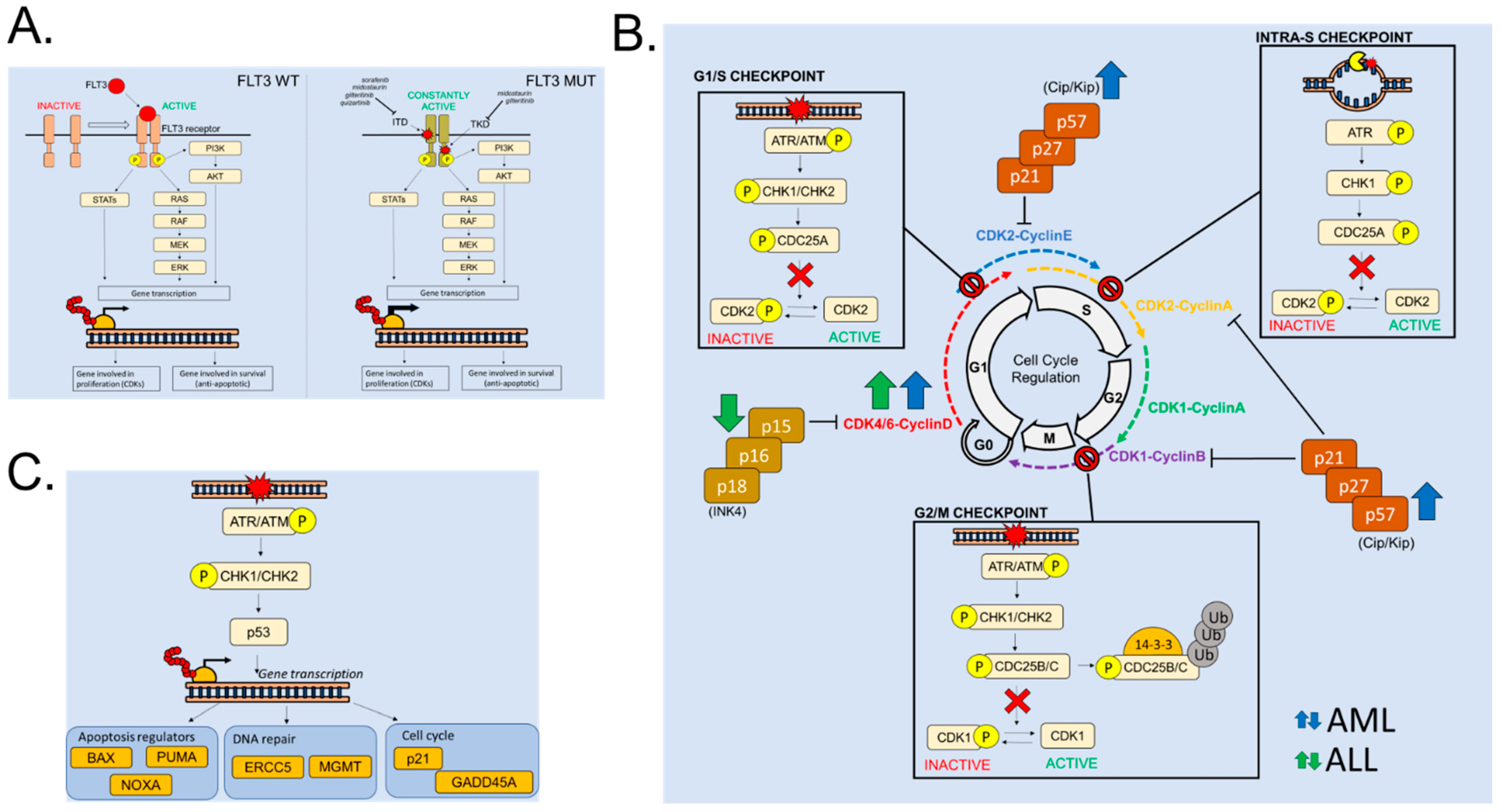
2.1. FLT3 Alterations in Acute Leukemia: A Driver of Cell Proliferation

Extracellular growth factor receptors or tyrosine kinases (TK) associated with extracellular receptors are frequently over-expressed or mutated in acute leukemia cells. A paradigmatic example is FLT3, a receptor tyrosine kinase able to translate external stimuli into pro-proliferative signaling cascades. It consists of five immunoglobulin-like domains in the extracellular region, a juxtamembrane (JM) domain, a TK domain separated by a kinase insert domain and a C-terminal domain in the intracellular region [2]. The binding of the ligand to the FLT3 receptor has an important role in the regulation of cell proliferation, differentiation and survival through various signaling pathways, including RAS/mitogen-activated protein kinases (RAS/MAPK), Janus kinase/signal transducer and activator of transcription 5 (JAK/STAT5) and phosphatidylinositol 3-kinase/protein kinase B (PI3K/AKT) [3] (Figure 1A). In normal conditions, FLT3 is expressed by early hematopoietic stem cells (HSC) [4], multipotent progenitors [5], common lymphoid and myeloid progenitors [6] and mature dendritic cells [7].
FLT3 is altered in about one-third of AML patients [3] and, with lower frequency, in ALL cases [8]. FLT3 represents one of the most frequently mutated genes in AML, with mutations generally belonging to the internal tandem duplication (ITD) or tyrosine kinase domain (TKD) categories. ITDs represent a driver mutation in AML and are located in the JM domain that inhibits receptor auto-activation. FLT3-ITD mutations alter this inhibitory effect, leading to an uncontrolled activation [9,10]. FLT3-ITD occurs in 20.4% of adult AML patients [11], who are classified as intermediate-risk cases in the absence of adverse-risk genetic lesions, according to the European LeukemiaNet 2022 classification [12]. TKD mutations have been detected in 4.8–7.7% of adult cases [11,13]. This type of mutation also induces aberrant activation of FLT3 signaling [14]. The prognostic impact of FLT3-TKD mutations is less clear and these alterations can associate with other mutations and cytogenetic changes [15]. As a consequence of both FLT3-ITD and FLT3-TKD mutations, the intracellular signals mediated by the TK are constitutively translated, thus promoting the cell cycle progression and, in particular, stimulating the transition from G1 to S phase.
The identification of FLT3 mutations is highly relevant since it can drive the selection of personalized therapies for AML patients. FLT3 inhibitors can be classified into two groups [16] (Figure 1A). Type I inhibitors (e.g., midostaurin and gilteritinib) interact with the ATP-binding site in the intracellular active pocket of the enzyme and are active against both FLT3-TKD and FLT3-ITD mutated receptors. Type II inhibitors (e.g., quizartinib and sorafenib) bind to the ATP-binding site and interact with an adjacent hydrophobic pocket that is exposed in the inactive conformation and is made inaccessible by TKD mutations. The current therapy of FLT3-mut AML relies on the inhibition of FLT3 with midostaurin during induction chemotherapy [17] and with single-agent gilteritinib at relapse [18]. However, patients are prone to the acquisition of secondary mutations conferring resistance to these inhibitors, including, but not limited to, on-target FLT3 mutations [19]. Several subclones carrying FLT3-ITDD835V/Y/F or different TKD mutations alone were responsible for resistance to quizartinib [20], while FLT3N676K and FLT3F691L gene mutations were associated with resistance to midostaurin [21] and gilteritinib [22] treatment.

2.2. Cell Cycle Regulation in Hematopoietic Cells and in Acute Leukemias

The cell cycle is the physiological process promoting cell replication, which is fundamental to sustain tissue growth and repair. In the hematopoietic tissue, the rate of cell division becomes more frequent moving from HSC to differentiated and lineage-committed cells [23,24]. It is well established that HSC are kept in a non-proliferative state, called quiescence, and their proliferation is activated only after specific extracellular stimuli. In all hematopoietic cells, the cell cycle is divided into four consecutive phases characterized by specific events ending with the generation of two daughter cells (Figure 1B). Briefly, the first one is a preparatory synthetic phase (G1) that aims to increase cell size in anticipation of DNA replication (S phase) in which the genome is duplicated. Cells then proceed through a second synthetic phase (G2 phase) to prepare the segregation of the duplicated DNA condensed in twin chromosomes and intracellular organelles to daughter cells (M phase) [25]. The transition from one phase to another is regulated by different multiprotein complexes called cell cycle checkpoints, whose function is to control the proper execution of all steps of each specific phase prior to moving the following one. In eukaryotic cells, four consecutive checkpoints control the transitions through the different phases of the cell cycle and, in particular, the following control steps have been characterized: the G1/S, intra-S, G2/M and mitotic (M) checkpoints [26]. These multi-protein complexes regulate the activity and stability of different Cyclin Dependent Kinase (CDK)–Cyclin complexes whose function is critical to move from one cell cycle phase to the next [25]. Indeed, the core molecular machinery controlling cell cycle progression consists of a family of serine/threonine protein kinases called CDKs [27]. These kinases, representing the catalytic subunits, are activated in most cases by association with regulatory proteins called Cyclins. Cyclins were originally named because their concentration varies in a cyclical fashion during the cell cycle. Thus, their level of expression and, consequently, their availability controls the activity of the different CDK–Cyclin complexes (Figure 1B). The transition throughout the G1 phase is regulated by Cyclin D, whose level starts to grow in the early G1 phase. In this transition, Cyclin D complexes with CDK4 or CDK6 kinases [28]. In HSC, CDK4/6–Cyclin D complexes are crucial as they promote the transition from G0 (quiescence) to G1 phase [29]. The transition from G0 to G1 phase is also regulated by the CDK3–Cyclin C complex [30]. Cyclin E accumulates during G1 phase and is essential, together with CDK2, for the transition from G1 to S phase. The level of Cyclin A starts to increase from the late G1 phase and reaches the highest level at the end of the G2 phase. Cyclin A complexes with CDK2 to promote the transition through the S phase. Lastly, Cyclin B regulates the transition from G2 to M phase as its expression starts to grow from the S phase and reaches the highest level during the G2 phase. To promote mitotic entry, Cyclin B complexes with CDK1, thus generating the mitotic promoting factor (MPF) [28].
As mentioned above, the intracellular level of Cyclins controls the activity of CDK–Cyclin complexes. However, additional regulations such as CDK inhibitors (CKIs) and post-transcriptional modifications have been identified. CKIs are families of protein acting, as CDK–Cyclin complexes, in a phase-specific manner and competing with Cyclins for binding with CDKs. Two main groups of CKIs have been identified: the CIP/KIP family and the INK4 family [31,32] (Figure 1B). The first group, composed of p21CIP, p27KIP1 and p57KIP2, acts in multiple phases of the cell cycle by inhibiting: (i) Cyclin D–CDK4/6 activity during the early G1 phase [33]; (ii) Cyclin E–CDK2 during late G1 [32]; (iii) Cyclin A–CDK2 in S phase [33]. Members of the Ink4 family, p16Ink4a, p15Ink4b, p18Ink4c and p19Ink4d, act specifically during the early G1 phase by inhibiting the catalytic activity of CDK4/6–Cyclin D complexes through allosteric competition for binding to Cyclins [32]. As already mentioned, CDK–Cyclin complexes are regulated by post-transcriptional modifications as a consequence of cell cycle checkpoint activation. Indeed, in the event of DNA damage or replicative stress, different intracellular cascades promote the phosphorylation or the dephosphorylation of CDK complexes, thus inducing their activation or inactivation. In the case of DNA damage, different kinases involved in the DNA damage response (DDR) pathway directly or indirectly promote the inhibition of CDK–Cyclin complexes. The function of DDR pathways is to inhibit cell cycle progression until the DNA damage is repaired by specialized pathways. Eukaryotic cells have evolved different DDR pathways to respond to different types of DNA damage and depending on the specific cell cycle phase in which the damage has been detected. During S phase, replicative stress causing stalled replicative forks or single-strand breaks (SSBs) of the DNA activates the ATR-CHK1 pathway. On the other hand, double-strand breaks (DSBs) of the DNA structure activate the ATM-CHK2 pathway that acts on the transition from S, G2 and M phases. Following an intracellular cascade, the ATR/ATM kinases activate their downstream target cell cycle checkpoint kinase 1 (CHK1) and 2 (CHK2), respectively (Figure 1B). After their activation, CHK1 and CHK2 promote the phosphorylation of several target proteins and, in particular, of different phase-specific phosphatases called CDC25A/B/C [34,35]. This final event indirectly regulates the activity of CDKs. Indeed, the three phosphatases remove the inhibitory phosphorylations from CDKs, leaving them available for Cyclin binding. In the transition through S phase, the CDC25A phosphatase removes the inhibitory phosphorylation on CDK2 (Thr14 and Tyr15) promoting the generation of CDK2–Cyclin E/A complexes. In the presence of DNA damage, CHK1 and CHK2 kinases phosphorylate CDC25A (Ser136), thus promoting its ubiquitination and proteasomal degradation. As a consequence, the inhibition of CDC25A causes an S-phase delay. Similarly, CDC25B and CDC25C regulate the transition from G2 to M phase by modulating the activity of CDK1 and, in particular, by removing the inhibitory phosphorylation at two sites (Thr14 and Tyr15). During DDR, CHK1 phosphorylates CDC25B (Ser323) and promotes its binding to 14-3-3, thus blocking its catalytic activity [36]. CDC25C activity is also regulated by phosphorylation (on Ser216) induced by both CHK1/CHK2 kinases. The events that follow CDC25C phosphorylation are similar to those following CDC25A phosphorylation and end with binding to 14-3-3 and its functional inhibition. The degradation of CDC25C impairs CDK1–Cyclin B activation and consequently compromises the transition through the G2/M phase. Lastly, a central role in the regulation of the cell cycle and DNA repair is played by the guardian of the genome, the tumor suppressor p53. Following the identification of DNA damage, p53 is activated by phosphorylation in an ATM/CHK2- or ATR/CHK1-dependent manner. p53 then promotes the transcription of several target proteins, including apoptotic regulators (e.g., PUMA, NOXA and BAX), DNA repair elements (e.g., ERCC5 and MGMT) and cell cycle regulators (e.g., p21Cip1 and Gadd45) (Figure 1C). The p53–p21-dependent response to DNA damage mediates different effects on cell cycle progression: (i) p21Cip1 interferes directly with CDK–Cyclin complexes involved in the transition from the G1 to the S phase; (ii) p53 inhibits the transcription of genes involved in the G2/M checkpoint such as the Cyclin B1 gene (CCNB1). In summary, the activation of the p53–p21 pathway promotes a robust G1 phase arrest [37,38].

2.3. Cell Cycle Alterations in Acute Leukemia

Alterations in the functionality of key factors involved in cell cycle regulation can lead to severe consequences, such as malignant transformation. Dysregulation of CDKs and associated Cyclins is frequently observed in hematological malignancies. CDK6 is predominantly expressed in the hematopoietic compartment [39] and loss of CDK6 results in impaired generation of several blood cell types [40]. As an opposite mechanism, CDK6 hyperactivation promotes cell proliferation in ALL and lymphoma patients [41,42] (Figure 1B). In acute leukemia patients, CDK6 over-expression has been frequently associated with KMT2A translocations, as it has been demonstrated that CDK6 is a direct target of KMT2A fusion proteins [43]. Moreover, CDK6 can be activated by Cyclin D2 and D3 upregulation mediated by FLT3-ITD in AML [44]. In addition to CDK/Cyclin functional dysregulations, cancer cells are frequently characterized by molecular alterations in different CKIs (Figure 1B). Indeed, the CDKN2A/B gene locus, located at chromosome 9p21, is one of the most frequently deleted, mutated and epigenetically silenced sites in cancer cells. In ALL patients both p16INK4a and p15INK4b are frequently deleted [45]. In particular, T-ALL is frequently associated with p16INK4a loss, while p15INK4b deletions are more often observed in pediatric ALL [46]. In mouse models of BCR-ABL1-positive ALL, the deletion of p19ARF initiates a more aggressive disease and confers resistance to the kinase inhibitor imatinib [47]. Regarding the CIP/KIP family, inactivating mutations or deletions of p21Cip1/Waf are very rare in acute leukemias. Conversely, he over-expression of p21Cip1/Waf has been found in AML patients expressing an AML1-ETO fusion transcript [48]. Regarding p27Kip1, its levels of expression correlate with the prognosis of different hematological malignancies. For instance, high p27Kip1 levels are associated with increased disease-free survival in AML [49]. Moreover, Cell division cycle 20 homologue (CDC20), which is involved in mitotic progression, is frequently over-expressed in acute leukemias [50,51], as well as in other blood and solid tumors [52,53].

3. Novel Molecules Targeting Proliferative Mechanisms in Acute Leukemias

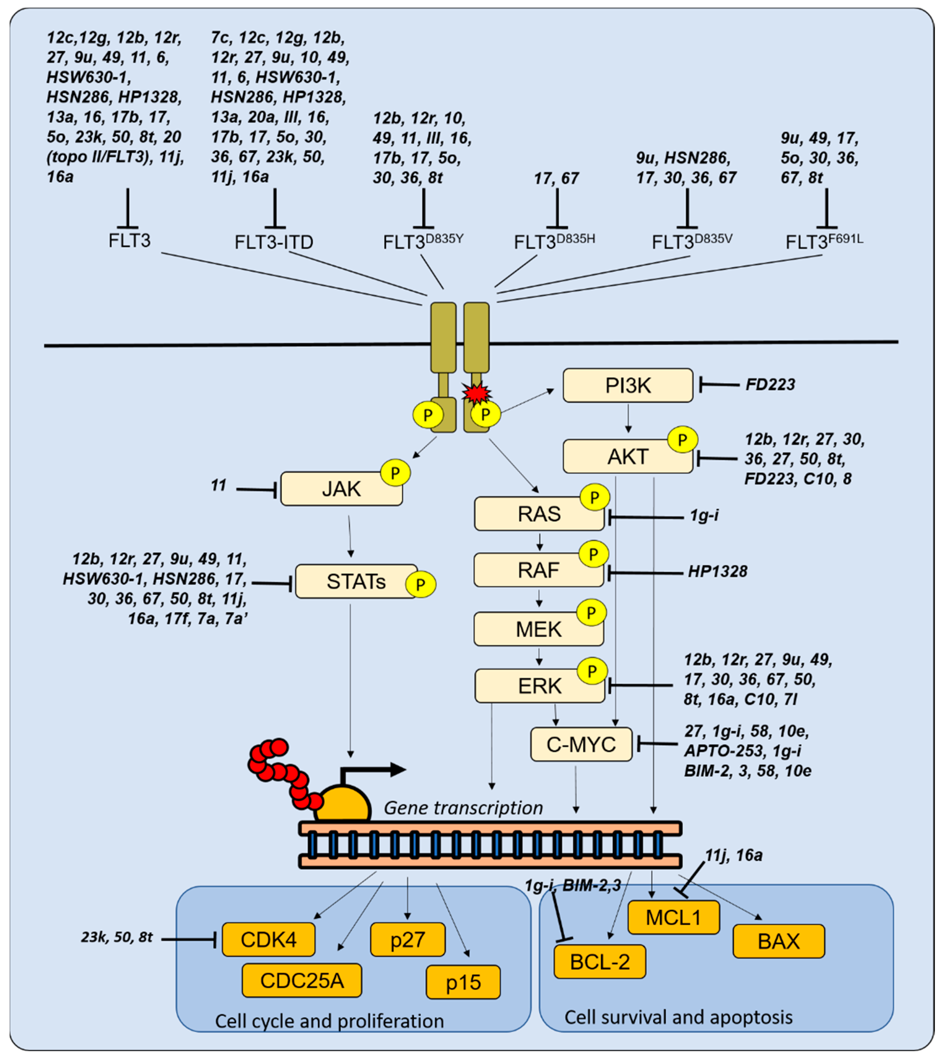
3.1. Novel Molecules Targeting FLT3

Given the high frequency of FLT3 mutations and the development of resistance to drugs targeting them, there is a continuous interest in novel FLT3 inhibitors (Table 1 and Table 2 and Figure 2) to be used in combination therapies (e.g., through multi-targeted approaches), with the aim of eradicating leukemia while decreasing toxicity and enhancing treatment tolerability. Some of them have also been shown to induce cell cycle arrest (Table 2).
A 5-(4-fluorophenyl)-N-phenyloxazol-2-amine (compound 7c) inhibited the proliferation and increased apoptosis of FLT3-ITD cells in vitro and reduced the in vivo growth of tumors obtained by xenotransplantation of the FLT3-ITD MV4-11 AML cell line, without causing obvious toxicities [54]. Similar to other FLT3 inhibitors [80], compound 7c induced downregulation of DDR genes belonging to the homologous recombination (BRCA1, BRCA2, BARD1 and RAD51) and non-homologous end joining (XRCC4, XRCC5 and XRCC6) pathways and accumulation of DNA damage [54]. As a consequence, compound 7c sensitized FLT3-ITD AML cells to treatment with the PARP inhibitor olaparib [54,81].
4-((6,7-dimethoxyquinoline-4-yl)oxy)aniline derivatives possessing the semicarbazide moiety as the linker (in particular compound 12c and 12g) were active as FLT3 inhibitors and reduced the growth of MV4-11 and HL-60 cells [55]. 4-azaaryl-N-phenylpyrimidin-2-amine derivatives (compounds 12b and 12r) inhibited the activity of FLT3 kinase, FLT3-ITD and other mutant forms and of the downstream signaling cascade (STAT5, ERK, AKT, S6RP and eIF4E) and induced apoptosis [56]. They displayed an antiproliferative specificity for FLT3-ITD cells over those expressing the wild-type (wt) kinase or even lacking FLT3 expression.
An important feature in the development of FLT3 inhibitors is kinase selectivity, which is directly related to off-target toxicity. Indeed, several novel compounds showed a specific anti-FLT3 activity, but inhibited a spectrum of additional kinases.
N-(4-(6-Acetamidopyrimidin-4-yloxy)phenyl)-2-(2-(trifluoromethyl)phenyl)acetamide (CHMFL-FLT3-335, compound 27) showed a specific anti-leukemic activity against FLT3-ITD AML models (MOLM-13, MOLM-14, MV4-11 AML cell lines) when compared with other AML subtypes [68]. However, it showed a poor activity against TKD mutations. Compound 27 also inhibited PDGFRβ, HPK1, CSF1R, PDGFRα and c-KIT (though to a lower extent compared with FLT3-ITD). It reduced the phosphorylation levels of the downstream signaling pathway mediators STAT5, AKT and ERK, and also the expression of the c-MYC oncogene. Treated cells were arrested at the G0/G1 phase of the cell cycle and underwent apoptosis. The anti-leukemic effect was confirmed in primary AML cells expressing FLT3-ITD and in xenograft models obtained by MV4-11 cell inoculation, by a reduction in tumor growth. The same in vitro and in vivo effects were also observed by treating AML cell lines with pyrrolo[2,3-d]pyrimidine derivatives, an in particular compound 9u [69]. Notably, this molecule was also active against FLT3-ITDD835V and FLT3-ITDF691L cells. The ability to target FLT3 mutations arising as a mechanism of resistance represents another relevant feature in the development of novel compounds, as it allows us to answer a clinical need.
A 8,9,10,11-tetrahydro-3H-pyrazolo[4,3-a]phenanthridine (HSD1169, compound 10) was active against MV4-11, MOLM-14 cell lines and MOLM-13 harboring the FLT3-ITDD835Y mutation by inducing cell cycle arrest at the G1 phase [70]. The compound also inhibited the activity of LRRK2, MELK, DYRK1B, CLK1, MNK2, TOPK, ROCK2, MSK2, p70S6K, PKG1a and CDK2–Cyclin A1 [71] and decreased the expression of TOPK, a kinase collaborating with FLT3 that is highly expressed in AML [82]. A derivative of compound 10, obtained by substitution of position 1 of the 3H-pyrazolo[4,3-f]quinoline core (compound 49), showed a higher selectivity towards FLT3 proteins [71]. It reduced the phosphorylation level of STAT5 and ERK1/2 signaling proteins, induced cell cycle arrest at the G1 phase, activated the apoptotic cascade and inhibited leukemia growth in a mouse-disseminated AML model.
Among flavones and flavonols, an O-methylated flavonol (compound 11) showed an elevated cytotoxic activity against acute leukemia cells [72]. In particular, it was selectively more active against the AML MOLM-13, MV4-11, HL-60 and T-ALL MOLT-4 models compared with solid cancer cell lines. It favored the accumulation of cells in the G0/G1 phase of the cell cycle and induced apoptosis. The molecule reduced the levels of the phosphorylated forms of FLT3, RSK2, STAT3, eIF4E and p53, and also exhibited inhibitory activity against JAK2, JAK3, MNK2 and NTRK2, as observed by kinase profiling. Moreover, compound 11 inhibited the activity of FLT3-ITD and FLT3D835Y. Additional flavonoids and their derivatives were also shown to target FLT3 (including its mutated forms) [73] or MNKs [83], resulting in cell apoptosis and G0/G1 cell cycle arrest.
Semisynthetic derivatives of fradcarbazole A (and in particular compound 6) also inhibited FLT3 kinase along with c-KIT and the cell cycle protein CDK2, while inducing arrest of the cell cycle at the G0/G1 phase and apoptosis of MV4-11 cells [74]. Notably, these compounds were not active against normal peripheral blood mononuclear cells (PBMCs), with IC50 values greater than 10 μM.
Ma et al. showed that 3-aminoisoquinoline analogs (HSW630-1 and its analogs) are potent FLT3 inhibitors [57]. They reduced the growth of MV4-11 and MOLM-14 AML cells and the phosphorylation of FLT3 and STAT5. Moreover, they also displayed an activity against c-KIT and TRKC enzymes. Starting from these molecules, the group synthesized novel amino isoquinoline, quinoline or quinazoline compounds [58]. In particular, HSN286 and its analogs inhibited both FLT3 and downstream SRC-family kinases, leading to reduced phosphorylation of FLT3, STAT5, STAT3 and p38. This approach could represent a valuable strategy to inhibit cell proliferation [84] even in the event of FLT3 mutations.
A derivative of N-(3,4-dimethoxybenzyl)-1-phenyl-1H-benzimidazol-5-amine (compound HP1328) was designed as an FLT3-ITD inhibitor with good aqueous solubility [59]. The compound induced apoptotic cell death of Ba/F3 expressing FLT3-ITD, MOLM13 and MV4-11 in a dose-dependent manner and of primary FLT3-ITD AML cells. Moreover, it potentiated the cytotoxicity of the anthracycline idarubicin. HP1328 activity has been confirmed in xenograft models, by improvement of the survival of leukemia-bearing mice. The treatment was suitable for oral administration and well tolerated. The kinase profile of inhibitory effects caused by HP1328 revealed a substantial selectivity over potential off-target kinases as BRAF, VEGFR and FGFR. However, the molecule selectively targeted c-KIT, thus limiting its further development due to the in vivo induction of myelosuppression.
Since c-KIT inhibition is largely responsible for myeloid immunosuppression, a number of studies have focused on newly synthesized molecules able to selectively target FLT3 and its mutant forms over c-KIT. Among them, the pyrimidine-4,6-diamine derivative compound 13a inhibited the growth of MOLM-14 and MV4-11 AML cell lines and of Ba/F3 cells expressing FLT3-ITD by accessing the FLT3 allosteric pocket while sparing c-KIT [60]. However, this compound was not effective against the FLT3D835Y mutation.
Novel amino-acid-substituted sunitinib analogues released active compound candidates have been developed in order to improve selectivity towards FLT3 [61]. Among them, the mono-carboxylic acid compound 20a provided the best results in terms of affinity against FLT3-ITD and activity against MV4-11 cells.
Promising results have been achieved with phenylethenylquinazoline derivatives in terms of selectivity [62]. They inhibited the activity of FLT3-ITD, FLT3D835Y and the quizartinib-resistant FLT3-ITDD835Y double-mutant enzyme while exerting almost no effect over c-KIT. Compound III, the most promising derivative, inhibited cell growth while inducing apoptosis in vitro, and effectively reduced the tumor size while being well-tolerated by murine xenograft models.
Derivatives of bis(1H-indol-2-yl)methanones, and in particular compound 16 and its carbamate 17b, selectively inhibited (among 249 kinases) FLT3, RET and ZAK [63], the latter being involved in the TGFβ/JNK pathway [85] and in the regulation of the cell cycle [86]. These compounds were effective against FLT3-ITD and FLT3D835Y mutant proteins; they inhibited the growth and induced apoptosis of MV4-11, MOLM-13 and EOL-1 cells [63], which shared KMT2A alterations (rearrangements or partial tandem duplication) associated with differentiation blockade, and FLT3 aberrations (EOL-1 was characterized by FLT3 overexpression [87] and mutant PDGFRA [88]). in vivo studies revealed a good tolerability of compound 17b [63].
A (Z)-N-(5-((5-Fluoro-2-oxoindolin-3-ylidene)methyl)-4-methyl-1H-pyrrol-3-yl)-3-(pyrrolidin-1-yl)propanamide (compound 17) showed promising in vitro and in vivo properties as an FLT3 inhibitor [64]. The molecule inhibited the proliferation of MV4-11 and MOLM-13 cells and was active against the FLT3D835Y/D835H/D835V and FLT3-ITDD835Y/D835V/F691L mutant forms. It reduced the phosphorylation of the downstream signaling pathway mediators STAT5 and ERK and induced cell cycle arrest in the sub-G1 phase and apoptosis, while displaying a very low activity on FLT3-wt AML cells and other cancer cell lines. Compound 17 had a favorable pharmacokinetic profile with high oral bioavailability and the mechanism of action has been confirmed in murine xenograft models obtained by AML cell line transplantation that also showed low levels of treatment toxicity. Notably, compound 17 was able to reduce the growth of sunitinib- and quizartinib-resistant cells.
Zhang et al. synthesized and tested imidazo[1,2-a]pyridine thiophene compound inhibitors of FLT3 and its mutant forms [65]. Among the tested molecules, compound 5o was effective against MOLM-14 AML cells, even when manipulated to express the mutant FLT3-ITDD835Y or FLT3-ITDF691L forms. Moreover, compound 5o was more potent than the FLT3 inhibitor quizartinib and was not cytotoxic against Ba/F3 normal pro-B cells.
On the same line of reasoning, 2-aminopyrimidine derivatives were synthesized with the aim of selectively inhibiting FLT3-ITD and its mutant forms while sparing the c-KIT kinase, thus reducing the myelosuppressive effect [66]. Compounds 30 and 36 inhibited the growth of MV4-11 cells and of Ba/F3 cells engineered to express FLT3-ITD, FLT3D835V/F, FLT3F691L, FLT3-ITDF691L and FLT3-ITDD835Y. The compound showed good liver microsomal stability, poor antiproliferative effect against FLT3-wt tumor cells, low toxicity to normal cells and weaker inhibitory activity against c-KIT compared with the FLT3 inhibitor gilteritinib in a zebrafish model. The molecules reduced FLT3 phosphorylation, thus affecting the downstream pathway, as revealed by reduced activation of STAT5, AKT and ERK. Moreover, they exerted an inhibitory activity against additional kinases including TRKA and Aurora A. The compound’s activity was confirmed in the murine xenograft model obtained by transplantation of MV4-11 cells by inhibition of the tumor growth rate and in mice inoculated with Ba/F3 expressing FLT3-ITDD835Y by prolongation of animal survival.
Starting from the observation that new compounds with pyrazole amine scaffolds exhibited potent inhibitory activity and selectivity against FLT3-ITD AML cells [89], novel 3-amine-pyrazole-5-benzimidazole compounds were designed and tested in order to overcome secondary resistance mutations [75]. Among them, compound 67 exerted an antiproliferative activity not only against FLT3-ITD cell lines, but also against Ba/F3 FLT3-ITDD835V, Ba/F3 FLT3-ITDF691L and Ba/F3 FLT3-ITDY842H. It effectively induced apoptosis and cell cycle arrest at the G1 phase by blocking the phosphorylation of FLT3, STAT5 and ERK. Compound 67 inhibited tumor growth in xenograft models (by intravenous injection due to poor oral bioavailability), while showing limited hematological and systemic toxicity.
To potentiate the anti-leukemic activity, novel molecules with a dual specificity were also synthetized. Indeed, monotherapies against one single target have shown limited efficacy in acute leukemias, due to the rapid development of escape strategies by malignant cells. For example, activation of RAS/MAPK signaling through functional [90] or genomic alterations [19,22] is a common mechanism of resistance to inhibitors of FLT3 or the antiapoptotic protein BCL-2, which are currently used in the treatment of AML. Therefore, co-targeting of two leukemogenic pathways may provide improved therapeutic benefits while sparing off-target toxicities.
Li et al. presented a series of 6-(pyrimidin-4-yl)- 1H-pyrazolo[4,3-b]pyridine derivatives that inhibited FLT3 and CDK4 kinases [76]. Some of them were more effective than the selective CDK4/6 inhibitor Abemaciclib and compound 23k showed the highest potency. Molecular docking studies showed that 23k could form a relatively stable protein–small-molecule complex by binding to both CDK4 and FLT3. The molecule induced cell cycle arrest in the G0/G1 phase and apoptosis in a concentration-dependent manner and inhibited phosphorylation of the downstream CDK2/4 factor RB and of FLT3 in the MV4-11 AML cell line. Compound 23k was also tested in vivo in the MV4-11 xenograft tumor model and reached a 67% tumor growth inhibition rate at a high dose, suggesting poor oral bio-availability.
Similar results were obtained by testing a series of 1-H-pyrazole-3-carboxamide derivatives, acting as FLT3 and CDK inhibitors [77]. Among them, compound 50 potently inhibited FLT3, CDK2, CDK4 and CDK6, resulting in the suppression of phosphorylation of RB, FLT3, ERK, AKT and STAT5, cell cycle arrest at the G0/G1 phase, induction of apoptosis and in vivo activity in AML. Aiming to improve the FLT3/CDK inhibitory activity, novel 1H-pyrazole-3-carboxamide derivatives were also developed from compound 50 [67]. Compound 8t had a multi-kinase inhibitory capacity, by targeting KDR/VEGFR2, ERK7, FLT1, FLT4 and GSK3 in addition to FLT3 and CDKs, that was responsible for its broad activity against acute leukemia but also solid tumor models. Moreover, it also exerted an inhibitory activity against a variety of FLT3 mutants. At the intracellular level, compound 8t blocked the phosphorylation of STAT5/AKT/ERK and of RB.
Novel pyrido-dipyrimidines showed activity towards topoisomerase II and/or FLT3, which was confirmed by docking studies [78] and successfully combined in the dual activity of compound 20. Biologically, this molecule induced upregulation of p53 and TNFα, accumulation of cells in the G2/M phase of the cell cycle and apoptosis in the HL-60 cell line.
A series of N-phenyl-4-(thiazol-5-yl)pyrimidin-2-amines and 4-(indol-3-yl)-N-phenylpyrimidin-2-amines was investigated with the aim of achieving dual inhibition of FLT3 and MNK2 as a potential strategy to avoid resistance mediated by the activation of parallel signaling pathways [79]. Indeed, MNK kinases act as downstream effectors of MAPK pathways mediating the phosphorylation of eIF4E, a protein that plays a key role in the regulation of mRNA translation, and counteract chemotherapy-induced antileukemic responses [91]. Three molecules were selected for their promising activity as FLT3 (11j), dual (16a) or MNK2 (16h) inhibitors [79]. They all showed a selective activity against their target(s) over CDK2, B-RAF MAPKs, p38a, PI3K, AKT and mTOR. In the MV4-11 AML model, 11j decreased STAT5 phosphorylation, 16h inhibited eIF4E phosphorylation and showed off-targets effects over STAT5, while 16a reduced phosphorylated STAT5, ERK, p38 and eIF4E. Both 11j and 16a compounds arrested the cells in the G1 phase of the cell cycle in a dose-dependent manner and induced apoptosis along with downregulation of the pro-survival protein MCL-1.

3.2. Targeting Other Proliferative Signals in AML

In addition to FLT3 inhibition, targeting of other proliferative signals, including kinases (e.g., PI3Kδ and STAT5) and oncogenic mechanisms (e.g., c-MYC), has raised interest in recent years (Table 3 and Figure 2).
A PI3Kδ inhibitor (FD223, compound 13) was obtained by bioisosteric replacements based on an indole scaffold [92]. The compound inhibited the proliferation of several AML cell lines by suppressing AKT phosphorylation, thus arresting the cell cycle at the G1 phase and inducing apoptosis. Pharmacokinetic studies revealed an acceptable oral bioavailability and in vivo experiments showed a dose-dependent tumor growth inhibition, with induction of tumor necrosis, decreased expression of Ki67 and no signs of acute toxicity.
Among the mediators of oncogenic signals in AML, STAT5 plays an important role in multiple disease subtypes [101,102]. PPARα/γ ligand derivatives, and in particular compound 17f, which is characterized by a free nitrogen on the indole ring along with a C-6 tetrahydroquinoline 3-pyridinyl substitution, selectively inhibited STAT5 activity, resulting in an antiproliferative effect against AML cellular models [93]. The compound also restored sensitivity to cytarabine in resistant AML cells through the inhibition of STAT5B but not STAT5A protein expression [103]. The 7a and 7a’ analogs of 17f showed a higher potency as inhibitors of STAT5 activity and expression, resulting in the downregulation of its targets PIM1 and CISH [94]. Both positions of the pyridinyl moiety on the tetrahydroquinoline aromatic part and its nitrogen position were essential in the antileukemic properties of 7a and 7a’.
Interest has been raised in recent years in the development of molecules able to bind G-quadruplexes, noncanonical nucleic acid structures found in specific guanine-rich DNA or RNA sequences, including the promoter regions of the c-MYC, K-RAS and c-KIT oncogenes and of the anti-apoptotic gene BCL-2. The stabilization of G-quadruplex structures can be therapeutically exploited to attenuate the transcription of pro-tumorigenic genes, leading to an antiproliferative effect [104]. Treatment of AML cells with the small-molecule 2-indolylimidazole[4,5-d]phenanthroline derivative APTO-253 [105] induced CDKN1A expression, G0/G1 cell cycle arrest, and apoptosis through stabilization of G-quadruplex DNA, inhibition of MYC expression and induction of DNA damage [106]. APTO-253 also entered clinical investigation for patients with relapsed/refractory AML or myelodysplastic syndrome (#NCT02267863). However, its clinical development has been stopped by the company. In the meantime, the development of novel heterocycles binding to DNA G-quadruplexes has led to the synthesis of a series of new substituted 2,9-bis [(substituted-aminomethyl)phenyl]-1,10-phenanthroline derivatives, showing a selective activity against human leukemia cells over normal PBMCs [107]. This was the starting point for the synthesis of a new series of 2,9-bis[(substituted-aminomethyl)phenyl]-1,10-phenanthroline derivatives [95]. Among them, compounds 1gi provided the most promising results in terms of activity against AML cell lines, stabilization of c-MYC, BCL-2 and K-RAS promoter G-quadruplexes and inhibition of telomerase activity.
The biimidazole derivative BIM-2 also showed a promising anti-leukemia activity as a dual c-MYC/BCL-2–quadruplex binder [96]. It reduced the expression of both c-MYC and BCL-2 and triggered cell cycle arrest at the G0/G1 phase and apoptosis in AML cells, thus showing a promising example of dual targeting, with activity against signals supporting both proliferation and survival of leukemic cells.
c-MYC downregulation was also a consequence of quinizarin derivative treatment in T-ALL cells [97]. Quinizarin quaternary ammonium salt 3 inhibited the growth of AML and T-ALL cell lines. However, it displayed a similar toxicity against normal human embryonic kidney HEK-293 cells. The compound led to G0/G1 phase arrest with a decreased S phase cell population, induction of apoptosis and accumulation of intracellular reactive oxygen species. Although the direct targets of compound 3 were not investigated, the treatment also triggered the downregulation of BCL-2 and NOTCH1.
Aiming at c-MYC inhibition, Feng et al. synthesized a series of 3,5-dimethylisoxazole derivatives targeting the epigenetic reader bromodomain-containing protein 4 (BRD4). In particular, compound 58 effectively inhibited the proliferation of AML cell lines by causing a reduction in c-MYC protein levels, cell cycle arrest in G1 and apoptosis [98]. Structure–activity relationship (SAR) and docking studies revealed that the 3-hydroxyisoindolin-1-one chemical scaffold could also act as BRD4 inhibitor [99]. Among the synthesized 3-Hydroxyisoindolin-1-one derivates, compound 10e displayed antiproliferative properties against AML cell lines through c-MYC downregulation and activation of the intrinsic apoptotic pathway. In addition to BRD4, the bromodomain and PHD finger-containing (BRPF) family has been studied as a potential therapeutic target in AML. Indeed, BRPF1 is highly expressed in acute leukemia cell lines [108] and sustains the expression of HOX genes [109], which play a pathogenic role in diverse AML subtypes [110,111,112,113]. A series of Quinolin-2-one Hits has been synthesized and tested in order to achieve selective inhibition of BRPF proteins. Among them, 13-d was experimentally confirmed as a potent inhibitor of BRPF1 [100]. The compound exerted an antiproliferative activity on AML cell lines and displayed an acceptable oral bioavailability according to pharmacokinetic studies conducted in mouse models. Recently, aiming to achieve a potent anti-leukemic activity, novel dual HDAC8/BRPF1 (sulfonamide derivatives 23a-b of an HDAC8 inhibitor) and HDAC6/BRPF1 (compound 37) inhibitors were synthesized [114] starting from benzhydroxamic acids, and they proved to be potent and selective HDAC8 inhibitors [115,116]. However, they demonstrated a poor cellular activity [114].

3.3. Novel Compounds with Unknown Targets That Inhibit Acute Leukemia Cell Proliferation

A group of novel compounds showing an antiproliferative activity against acute leukemia cells remains to be characterized in terms of target specificity (Table 4).
A 3′,5′-diprenylated chalcone belonging to the flavone family, (E)-1-(2-hydroxy-4-methoxy-3,5-diprenyl)phenyl-3-(3-pyridinyl)-propene-1-one (C10), inhibited the proliferation and induced apoptosis of the human erythroleukemia cell line HEL in a time- and dose-dependent manner [117]. The molecule caused a downregulation of the transcription factor Friend Leukemia Integration 1 (FLI-1) in HEL cells and a reduction of the phosphorylation levels of p38/MAPK and ERK1/2, suggesting an activity on the MAPK pathway, supporting cell proliferation (Figure 2). A potential effect on cell autophagy has been also hypothesized, based on the inhibitory effect exerted on the AKT/mTOR pathway (Figure 2).
The MAPK pathway has been also inhibited by a dithiocarbamate ester of parthenolide (compound 7l) in AML [118]. The molecule induced apoptosis and suppressed the clonogenic capacity of primary leukemia stem cells while sparing normal cells from healthy donors. Moreover, it prolonged the survival of AML-patient-derived xenograft mice.
In addition to dithiocarbamate esters of parthenolide, neglectin derivatives were active against AML models [119]. Compound 8, carrying an N,N-dimethylamino ethoxyl moiety at the C-6 position, induced cell cycle arrest at the G0/G1 phase in HL-60 cells by suppressing the activation of AKT (Figure 2) and S6K1, a serine/threonine kinase belonging to the mTORC1 pathway. A new 2,4-dinitrobenzenesulfonamide derivative (S1) triggered G0/G1 blockade, intrinsic apoptosis and reduction of Survivin expression in the Jurkat T-ALL model, while showing no cytotoxicity towards normal erythrocytes and mononuclear cells [120]. Analogs based on the fungi-derived polyenylpyrrole products rumbrin and auxarconjugatin-B (and in particular the octatetraenylpyrrole analog 3s) were also able to suppress the proliferation of T-ALL cells [121].

3.4. Novel Molecules Targeting the Cell Cycle Machinery

Starting from the observation that functional alterations of the cell cycle machinery occur in acute leukemias and the expression of some of its components is deregulated, the cell cycle and DDR pathway have become appealing targets for the development of anti-leukemic therapies (Table 5).
Thirty 2-carbomethoxy-3-substituted indoles have been synthetized and tested against acute leukemia cells [122]. Among them, compound 20 showed a selective anti-leukemic activity while sparing healthy T lymphocytes up to a dose of 100 μM. The molecule induced cell cycle arrest at G2/M and apoptosis in HL-60 cells, CCRF-CEM and RS4;11 T-ALL and B-ALL models, respectively. Mechanistically, its activity has been linked to microtubule depolymerization, as supported by the observation of reduced tubulin expression, abnormal chromosomal segregation and fragmented centrosomal material. Accordingly, gene expression profiling highlighted enrichment of gene sets involved in the G2/M checkpoint, the mitotic spindle and NOTCH signaling and a decrease in the cholesterol homeostasis in treated HL-60 cells. Moreover, compound 20 induced differentiation of HL-60 cells, as revealed by upregulation of the CD11b and CD14 myeloid markers. Of note, the treatment improved the survival of mice engrafted with RS4;11 leukemic cells.
Induction of microtubule depolymerization was also the mechanism of action of (Z)-2-((2-((1-ethyl-5-methoxy-1H-indol-3-yl)methylene)-3-oxo-2,3-dihydrobenzofuran-6-yl)oxy)acetonitrile (5a) and (Z)-6-((2,6-dichlorobenzyl)oxy)-2-(pyridin-4-ylmethylene)benzofuran-3(2H)-one (5b), which showed a selectivity for the colchicine-binding site on tubulin [123]. The molecules inhibited the growth of T- and B-ALL cell lines while being less effective against normal B-lymphoblast cells and blocked disease progression in a myc-induced T-ALL zebrafish model.
With the aim of targeting the mitotic checkpoint, we have designed a series of tryptamine derivatives [127]. Compound 9, which is characterized by 2-aminopyrimidyl- and trichloroethyl- moieties, similarly to those found in Apcin [128], a small molecule that blocks the interaction between the anaphase-promoting complex/cyclosome (APC/C) and CDC20 [53], showed a selective activity against acute leukemia models over solid tumors. Indeed, it inhibited the growth of MV4-11 AML cells, the REH B-ALL model and especially the T-ALL cell line Jurkat-6. Notably, the pyrimidine ring and aminal nitrogens, together with the hydrophobic trichloromethyl group, were identified as important structural motifs for Apcin activity SAR experiments. Future studies are needed to define the intracellular activity of compound 9.
Induction of blast cell differentiation is emerging as a promising strategy, not only against acute promyelocytic leukemia, but also in the AML field [129]. A CDK2-targeted proteolysis-targeting chimera (PROTAC) has been developed to selectively degrade CDK2 while sparing the other CDKs [124]. The molecule (CPS2) exerted an antiproliferative effect (also favored by the downregulation of Aurora Kinase A), induced remarkable differentiation of AML cell lines and primary cells and arrested leukemic cell growth in vivo. Moreover, CPS2 sensitized the cells to CDK4/6 or PI3K inhibition and enforced the induction of differentiation by all-trans retinoic acid. The compound altered the expression of genes involved in myeloid leukocyte differentiation and activation, and cytokine-mediated signaling pathways and some of them were not altered by inhibition of CDK2 enzymatic activity, thus confirming the importance of degrading the molecule in order to achieve a stronger effect on cell differentiation.
As highlighted in the previous paragraphs, one key step in the cell cycle is the maintenance of DNA integrity. CHK1 is a central component in the DDR. Diaminopyrimidine derivatives (and in particular compound 13) were able to inhibit its activity, as demonstrated by inhibition of its autophosphorylation [125]. Compound 13 suppressed the proliferation of MV4-11 AML cells, while exhibiting low inhibitory potency against normal PBMCs. Moreover, the molecule also displayed a moderate oral bioavailability in vivo, suggesting its suitability for further studies on murine models.
Novel 1-substituted pyrido[4,3-b]carbazole derivatives of olivacine [130] also exerted a role in the maintenance of DNA integrity in leukemic cells [126]. Indeed, they forced the expression of p53 and p21Cip1 proteins in the TP53-mutant CCRF-CEM T-ALL model, thus causing a decrease in the number of cells in the S phase of the cell cycle and the induction of cell death via apoptosis. Notably, several attempts have been made in recent years in the development of novel molecules and strategies to restore the functionality of p53 in cancers [131], which may in the future answer an unmet clinical need in acute leukemias.

4. Conclusions

We have here provided a comprehensive review of recently synthesized compounds targeting the cell cycle or cell proliferation in AML and ALL. They include microtubule-depolymerizing or p53-restoring agents, G-quadruplex-stabilizing molecules and CDK2, CHK1, STAT5, PI3K-δ, BRD4, BRPF1 and FLT3 inhibitors. Different strategies led to the development or discovery of these compounds: some of them were synthesized with the aim of targeting specific molecules, as for many FLT3 inhibitors, and their structure and moieties have been progressively replaced in order to improve their efficacy and selectivity. Conversely, other agents are products of synthesis showing an antitumor activity in general and/or in leukemia in particular, that have been tested against potential targets as a second step. For a minority of them, SAR and/or docking studies have not been conducted, and the cellular target still needs to be identified. This step is crucial for the definition of the molecule’s specificity and selectivity so that it can be adjusted through the synthesis of derivatives and novel or improved therapeutic combinations against acute leukemia cells can be suggested. The mechanisms of action, including the downstream intracellular changes induced by in vitro treatment, have been investigated for a number of molecules, though with different depth levels (from selective cell phenotype analysis to signaling activation or transcriptome analysis, allowing a broader view of their effects). Some of them showed promising results, with active concentrations in the nanomolar range, while others were effective at micromolar concentrations. Few compounds have also been tested on primary leukemia samples. The compounds showing a preclinical activity at lower doses, along with elevated bio-availability and lower toxicity in vivo, represent good candidates for future clinical investigation. Moreover, a deeper investigation of the transcriptional changes induced by the newly synthesized molecules is recommended, since it can uncover the spectrum of biological effects induced at the intracellular level. This knowledge is crucial to design novel effective therapeutic strategies, aiming to target multiple oncogenic mechanisms while reducing the drug doses [132] through synergic combinations.

Author Contributions

Conceptualization, A.G.L.d.R., G.M. and G.S.; writing—original draft preparation, A.G.L.d.R., M.J. and G.S.; writing—review and editing, G.M.; funding acquisition, G.M. All authors have read and agreed to the published version of the manuscript.

Funding

This work was supported by Italian Association for Cancer Research IG 2019 within the Project Code: 23810 (PI Martinelli G.).

Institutional Review Board Statement

Not applicable.

Data Availability Statement

Not applicable.

Conflicts of Interest

G.M. has competing interests with Novartis, BMS, Roche, Pfizer, ARIAD and MSD. None of these agencies have had a role in the preparation of this manuscript. All other authors have no conflict of interest.

References

  1. Hanahan, D. Hallmarks of cancer: New dimensions. Cancer Discov. 2022, 12, 31–46. [Google Scholar] [CrossRef] [PubMed]
  2. Knight, T.E.; Edwards, H.; Meshinchi, S.; Taub, J.W.; Ge, Y. “FLipping” the story: FLT3-mutated acute myeloid leukemia and the evolving role of FLT3 inhibitors. Cancers 2022, 14, 3398. [Google Scholar] [CrossRef] [PubMed]
  3. Kazi, J.U.; Rönnstrand, L. FMS-like tyrosine kinase 3/FLT3: From basic science to clinical implications. Physiol. Rev. 2019, 99, 1433–1466. [Google Scholar] [CrossRef] [PubMed]
  4. Adolfsson, J.; Borge, O.J.; Bryder, D.; Theilgaard-Mönch, K.; Åstrand-Grundström, I.; Sitnicka, E.; Sasaki, Y.; Jacobsen, S.E.W. Upregulation of Flt3 expression within the bone marrow Lin-Sca1+c-Kit+ stem cell compartment is accompanied by loss of self-renewal capacity. Immunity 2001, 15, 659–669. [Google Scholar] [CrossRef] [PubMed] [Green Version]
  5. Buza-Vidas, N.; Woll, P.; Hultquist, A.; Duarte, S.; Lutteropp, M.; Bouriez-Jones, T.; Ferry, H.; Luc, S.; Jacobsen, S.E.W. FLT3 expression initiates in fully multipotent mouse hematopoietic progenitor cells. Blood 2011, 118, 1544–1548. [Google Scholar] [CrossRef] [PubMed]
  6. Sitnicka, E.; Bryder, D.; Theilgaard-Mönch, K.; Buza-Vidas, N.; Adolfsson, J.; Jacobsen, S.E.W. Key role of Flt3 ligand in regulation of the common lymphoid progenitor but not in maintenance of the hematopoietic stem cell pool. Immunity 2002, 17, 463–472. [Google Scholar] [CrossRef] [Green Version]
  7. Karsunky, H.; Merad, M.; Cozzio, A.; Weissman, I.L.; Manz, M.G. Flt3 ligand regulates dendritic cell development from Flt3+ lymphoid and myeloid-committed progenitors to Flt3+ dendritic cells in vivo. J. Exp. Med. 2003, 198, 305–313. [Google Scholar] [CrossRef] [Green Version]
  8. Zhang, Y.; Zhang, Y.; Wang, F.; Wang, M.; Liu, H.; Chen, X.; Cao, P.; Ma, X.; Teng, W.; Zhang, X.; et al. The mutational spectrum of FLT3 gene in acute lymphoblastic leukemia is different from acute myeloid leukemia. Cancer Gene Ther. 2020, 27, 81–88. [Google Scholar] [CrossRef]
  9. Kiyoi, H.; Ohno, R.; Ueda, R.; Saito, H.; Naoe, T. Mechanism of constitutive activation of FLT3 with internal tandem duplication in the juxtamembrane domain. Oncogene 2002, 21, 2555–2563. [Google Scholar] [CrossRef] [Green Version]
  10. Hayakawa, F.; Towatari, M.; Kiyoi, H.; Tanimoto, M.; Kitamura, T.; Saito, H.; Naoe, T. Tandem-duplicated Flt3 constitutively activates STAT5 and MAP kinase and introduces autonomous cell growth in IL-3-dependent cell lines. Oncogene 2000, 19, 624–631. [Google Scholar] [CrossRef] [Green Version]
  11. Thiede, C.; Steudel, C.; Mohr, B.; Schaich, M.; Schäkel, U.; Platzbecker, U.; Wermke, M.; Bornhäuser, M.; Ritter, M.; Neubauer, A.; et al. Analysis of FLT3-activating mutations in 979 patients with acute myelogenous leukemia: Association with FAB subtypes and identification of subgroups with poor prognosis. Blood 2002, 99, 4326–4335. [Google Scholar] [CrossRef] [PubMed] [Green Version]
  12. Dohner, H.; Wei, A.H.; Appelbaum, F.R.; Craddock, C.; DiNardo, C.D.; Dombret, H.; Ebert, B.L.; Fenaux, P.; Godley, L.A.; Hasserjian, R.P.; et al. Diagnosis and management of AML in adults: 2022 Recommendations from an international expert panel on behalf of the ELN. Blood 2022, 140, 1345–1377. [Google Scholar] [CrossRef] [PubMed]
  13. Bacher, U.; Haferlach, C.; Kern, W.; Haferlach, T.; Schnittger, S. Prognostic relevance of FLT3-TKD mutations in AML: The combination matters an analysis of 3082 patients. Blood 2008, 111, 2527–2537. [Google Scholar] [CrossRef] [Green Version]
  14. Kiyoi, H.; Kawashima, N.; Ishikawa, Y. FLT3 mutations in acute myeloid leukemia: Therapeutic paradigm beyond inhibitor development. Cancer Sci. 2020, 111, 312–322. [Google Scholar] [CrossRef] [PubMed] [Green Version]
  15. Guan, W.; Zhou, L.; Li, Y.; Yang, E.; Liu, Y.; Lv, N.; Fu, L.; Ding, Y.; Wang, N.; Fang, N.; et al. Profiling of somatic mutations and fusion genes in acute myeloid leukemia patients with FLT3-ITD or FLT3-TKD mutation at diagnosis reveals distinct evolutionary patterns. Exp. Hematol. Oncol. 2021, 10, 27–41. [Google Scholar] [CrossRef]
  16. Weisberg, E.; Roesel, J.; Furet, P.; Bold, G.; Imbach, P.; Flörsheimer, A.; Caravatti, G.; Jiang, J.; Manley, P.; Ray, A.; et al. Antileukemic effects of novel first- and second-generation FLT3 inhibitors: Structure-affinity comparison. Genes Cancer 2010, 1, 1021–1032. [Google Scholar] [CrossRef] [Green Version]
  17. Stone, R.M.; Mandrekar, S.J.; Sanford, B.L.; Laumann, K.; Geyer, S.; Bloomfield, C.D.; Thiede, C.; Prior, T.W.; Döhner, K.; Marcucci, G.; et al. Midostaurin plus chemotherapy for acute myeloid leukemia with a FLT3 mutation. N. Engl. J. Med. 2017, 377, 454–464. [Google Scholar] [CrossRef]
  18. Perl, A.E.; Martinelli, G.; Cortes, J.E.; Neubauer, A.; Berman, E.; Paolini, S.; Montesinos, P.; Baer, M.R.; Larson, R.A.; Ustun, C.; et al. Gilteritinib or chemotherapy for relapsed or refractory FLT3 -mutated AML. N. Engl. J. Med. 2019, 381, 1728–1740. [Google Scholar] [CrossRef]
  19. Alotaibi, A.S.; Yilmaz, M.; Kanagal-Shamanna, R.; Loghavi, S.; Kadia, T.M.; DiNardo, C.D.; Borthakur, G.; Konopleva, M.; Pierce, S.A.; Wang, S.A.; et al. Patterns of resistance differ in patients with acute myeloid leukemia treated with type I versus type II FLT3 inhibitors. Cancer Discov. 2021, 2, 125–134. [Google Scholar] [CrossRef]
  20. Smith, C.C.; Paguirigan, A.; Jeschke, G.R.; Lin, K.C.; Massi, E.; Tarver, T.; Chin, C.S.; Asthana, S.; Olshen, A.; Travers, K.J.; et al. Heterogeneous resistance to quizartinib in acute myeloid leukemia revealed by single-cell analysis. Blood 2017, 130, 48–58. [Google Scholar] [CrossRef]
  21. Schmalbrock, L.K.; Dolnik, A.; Cocciardi, S.; Sträng, E.; Theis, F.; Jahn, N.; Panina, E.; Blätte, T.J.; Herzig, J.; Skambraks, S.; et al. Clonal evolution of acute myeloid leukemia with FLT3-ITD mutation under treatment with midostaurin. Blood 2021, 137, 3093–3104. [Google Scholar] [CrossRef] [PubMed]
  22. Smith, C.C.; Levis, M.J.; Perl, A.E.; Hill, J.E.; Rosales, M.; Bahceci, E. Molecular profile of FLT3-mutated Relapsed/refractory patients with AML in the phase 3 ADMIRAL study of Gilteritinib. Blood Adv. 2022, 6, 2144–2155. [Google Scholar] [CrossRef] [PubMed]
  23. Laurenti, E.; Frelin, C.; Xie, S.; Ferrari, R.; Dunant, C.F.; Zandi, S.; Neumann, A.; Plumb, I.; Doulatov, S.; Chen, J.; et al. CDK6 levels regulate quiescence exit in human hematopoietic stem cells. Cell Stem Cell 2015, 16, 302–313. [Google Scholar] [CrossRef] [PubMed] [Green Version]
  24. Hao, S.; Chen, C.; Cheng, T. Cell cycle regulation of hematopoietic stem or progenitor cells. Int. J. Hematol. 2016, 103, 487–497. [Google Scholar] [CrossRef] [Green Version]
  25. Satyanarayana, A.; Kaldis, P. Mammalian cell-cycle regulation: Several Cdks, numerous cyclins and diverse compensatory mechanisms. Oncogene 2009, 28, 2925–2939. [Google Scholar] [CrossRef] [Green Version]
  26. Lukas, J.; Lukas, C.; Bartek, J. Mammalian cell cycle checkpoints: Signalling pathways and their organization in space and time. DNA Repair 2004, 3, 997–1007. [Google Scholar] [CrossRef]
  27. Morgan, D.O. Cyclin-dependent kinases: Engines, clocks, and microprocessors. Annu. Rev. Cell Dev. Biol. 2003, 13, 261–291. [Google Scholar] [CrossRef]
  28. Hochegger, H.; Takeda, S.; Hunt, T. Cyclin-dependent kinases and cell-cycle transitions: Does one fit all? Nat. Rev. Mol. Cell Biol. 2008, 9, 910–916. [Google Scholar] [CrossRef]
  29. Gudmundsson, K.O.; Du, Y. Quiescence regulation by normal haematopoietic stem cells and leukaemia stem cells. FEBS J. 2022; ahead of print. [Google Scholar] [CrossRef]
  30. Ren, S.; Rollins, B.J. Cyclin C/Cdk3 promotes Rb-dependent G0 exit. Cell 2004, 117, 239–251. [Google Scholar] [CrossRef] [Green Version]
  31. Quereda, V.; Porlan, E.; Canãmero, M.; Dubus, P.; Malumbres, M. An essential role for Ink4 and Cip/Kip cell-cycle inhibitors in preventing replicative stress. Cell Death Differ. 2015, 23, 430–441. [Google Scholar] [CrossRef] [PubMed] [Green Version]
  32. Sherr, C.J.; Roberts, J.M. Inhibitors of mammalian G1 cyclin-dependent kinases. Genes Dev. 1995, 9, 1149–1163. [Google Scholar] [CrossRef] [PubMed] [Green Version]
  33. Blain, S.W.; Montalvo, E.; Massagué, J. Differential interaction of the cyclin-dependent kinase (Cdk) inhibitor P27Kip1 with cyclin A-Cdk2 and cyclin D2-Cdk4. J. Biol. Chem. 1997, 272, 25863–25872. [Google Scholar] [CrossRef] [PubMed] [Green Version]
  34. Perry, J.A.; Kornbluth, S. Cdc25 and Wee1: Analogous opposites? Cell Div. 2007, 2, 12–23. [Google Scholar] [CrossRef] [PubMed] [Green Version]
  35. Aressy, B.; Ducommun, B. Cell cycle control by the CDC25 phosphatases. Anticancer Agents Med. Chem. 2012, 8, 818–824. [Google Scholar] [CrossRef] [PubMed]
  36. Schmitt, E.; Boutros, R.; Froment, C.; Monsarrat, B.; Ducommun, B.; Dozier, C. CHK1 phosphorylates CDC25B during the cell cycle in the absence of DNA damage. J. Cell Sci. 2006, 119, 4269–4275. [Google Scholar] [CrossRef] [Green Version]
  37. Donehower, L.A. Phosphatases reverse P53-mediated cell cycle checkpoints. Proc. Natl. Acad. Sci. USA 2014, 111, 7172–7173. [Google Scholar] [CrossRef] [Green Version]
  38. Engeland, K. Cell cycle regulation: P53-P21-RB signaling. Cell Death Differ. 2022, 29, 946–960. [Google Scholar] [CrossRef]
  39. Della Ragione, F.; Borriello, A.; Mastropietro, S.; Della Pietra, V.; Monno, F.; Gabutti, V.; Locatelli, F.; Bonsi, L.; Bagnara, G.P.; Iolascon, A. Expression of G1-phase cell cycle genes during hematopoietic lineage. Biochem. Biophys. Res. Commun. 1997, 231, 73–76. [Google Scholar] [CrossRef]
  40. Malumbres, M.; Sotillo, R.; Santamaría, D.; Galán, J.; Cerezo, A.; Ortega, S.; Dubus, P.; Barbacid, M. Mammalian cells cycle without the D-type cyclin-dependent kinases Cdk4 and Cdk6. Cell 2004, 118, 493–504. [Google Scholar] [CrossRef] [Green Version]
  41. Jena, N.; Sheng, J.; Hu, J.K.; Li, W.; Zhou, W.; Lee, G.; Tsichlis, N.; Pathak, A.; Brown, N.; Deshpande, A.; et al. CDK6-mediated repression of CD25 is required for induction and maintenance of Notch1-induced T-Cell acute lymphoblastic leukemia. Leukemia 2016, 30, 1033–1043. [Google Scholar] [CrossRef] [PubMed] [Green Version]
  42. Kollmann, K.; Sexl, V. CDK6 and P16INK4A in lymphoid malignancies. Oncotarget 2013, 4, 1858–1859. [Google Scholar] [CrossRef]
  43. Placke, T.; Faber, K.; Nonami, A.; Putwain, S.L.; Salih, H.R.; Heidel, F.H.; Krämer, A.; Root, D.E.; Barbie, D.A.; Krivtsov, A.V.; et al. Requirement for CDK6 in MLL-rearranged acute myeloid leukemia. Blood 2014, 124, 13–23. [Google Scholar] [CrossRef] [PubMed] [Green Version]
  44. Wang, L.; Wang, J.; Blaser, B.W.; Duchemin, A.-M.; Kusewitt, D.F.; Liu, T.; Caligiuri, M.A.; Briesewitz, R. Pharmacologic inhibition of CDK4/6: Mechanistic evidence for selective activity or acquired resistance in acute myeloid leukemia. Blood 2007, 110, 2075–2083. [Google Scholar] [CrossRef] [PubMed]
  45. Takeuchi, S.; Bartram, C.R.; Seriu, T.; Miller, C.W.; Tobler, A.; Janssen, J.W.G.; Reiter, A.; Ludwig, W.D.; Zimmermann, M.; Schwaller, J.; et al. Analysis of a family of cyclin-dependent kinase inhibitors: P15/MTS2/INK4B, P16/MTS1/INK4A, and P18 genes in acute lymphoblastic leukemia of childhood. Blood 1995, 86, 755–760. [Google Scholar] [CrossRef] [PubMed] [Green Version]
  46. Schirripa, A.; Sexl, V.; Kollmann, K. Cyclin-dependent kinase inhibitors in malignant hematopoiesis. Front. Oncol. 2022, 12, 4100. [Google Scholar] [CrossRef]
  47. Williams, R.T.; Roussel, M.F.; Sherr, C.J. Arf gene loss enhances oncogenicity and limits imatinib response in mouse models of Bcr-Abl-induced acute lymphoblastic leukemia. Proc. Natl. Acad. Sci. USA 2006, 103, 6688–6693. [Google Scholar] [CrossRef] [Green Version]
  48. Berg, T.; Fliegauf, M.; Burger, J.; Staege, M.S.; Liu, S.; Martinez, N.; Heidenreich, O.; Burdach, S.; Haferlach, T.; Werner, M.H.; et al. Transcriptional upregulation of P21/WAF/Cip1 in myeloid leukemic blasts expressing AML1-ETO. Haematologica 2008, 93, 1728–1733. [Google Scholar] [CrossRef]
  49. Yokozawa, T.; Towatari, M.; Iida, H.; Takeyama, K.; Tanimoto, M.; Kiyoi, H.; Motoji, T.; Asou, N.; Saito, K.; Takeuchi, M.; et al. Prognostic significance of the cell cycle inhibitor P27Kip1 in acute myeloid leukemia. Leukemia 2000, 14, 28–33. [Google Scholar] [CrossRef] [Green Version]
  50. Simonetti, G.; Padella, A.; do Valle, I.F.; Fontana, M.C.; Fonzi, E.; Bruno, S.; Baldazzi, C.; Guadagnuolo, V.; Manfrini, M.; Ferrari, A.; et al. Aneuploid acute myeloid leukemia exhibits a signature of genomic alterations in the cell cycle and protein degradation machinery. Cancer 2019, 125, 712–725. [Google Scholar] [CrossRef] [Green Version]
  51. Moison, C.; Lavallée, V.P.; Thiollier, C.; Lehnertz, B.; Boivin, I.; Mayotte, N.; Gareau, Y.; Fréchette, M.; Blouin-Chagnon, V.; Corneau, S.; et al. Complex karyotype AML displays G2/M signature and hypersensitivity to PLK1 inhibition. Blood Adv. 2019, 3, 552–563. [Google Scholar] [CrossRef] [PubMed]
  52. Simonetti, G.; Bruno, S.; Padella, A.; Tenti, E.; Martinelli, G. Aneuploidy: Cancer strength or vulnerability? Int. J. Cancer 2019, 144, 8–25. [Google Scholar] [CrossRef] [Green Version]
  53. Bruno, S.; Ghelli Luserna di Rorà, A.; Napolitano, R.; Soverini, S.; Martinelli, G.; Simonetti, G. CDC20 in and out of mitosis: A prognostic factor and therapeutic target in hematological malignancies. J. Exp. Clin. Cancer Res. 2022, 41, 159–174. [Google Scholar] [CrossRef] [PubMed]
  54. Kim, H.J.; Ryu, H.; Song, J.Y.; Hwang, S.G.; Jalde, S.S.; Choi, H.K.; Ahn, J. Discovery of oxazol-2-amine derivatives as potent novel FLT3 inhibitors. Molecules 2020, 25, 5154. [Google Scholar] [CrossRef]
  55. Xu, Q.; Dai, B.; Li, Z.; Xu, L.; Yang, D.; Gong, P.; Hou, Y.; Liu, Y. Design, synthesis, and biological evaluation of 4-((6,7-dimethoxyquinoline-4-Yl)oxy)aniline derivatives as FLT3 inhibitors for the treatment of acute myeloid leukemia. Bioorg. Med. Chem. Lett. 2019, 29, 126630–126636. [Google Scholar] [CrossRef] [PubMed]
  56. Long, Y.; Yu, M.; Ochnik, A.M.; Karanjia, J.D.; Basnet, S.K.; Kebede, A.A.; Kou, L.; Wang, S. Discovery of novel 4-azaaryl-N-phenylpyrimidin-2-amine derivatives as potent and selective FLT3 inhibitors for acute myeloid leukaemia with FLT3 mutations. Eur. J. Med. Chem. 2021, 213, 113215–113232. [Google Scholar] [CrossRef]
  57. Ma, X.; Zhou, J.; Wang, C.; Carter-Cooper, B.; Yang, F.; Larocque, E.; Fine, J.; Tsuji, G.; Chopra, G.; Lapidus, R.G.; et al. Identification of new FLT3 inhibitors that potently inhibit AML cell lines via an azo click-it/staple-it approach. ACS Med. Chem. Lett. 2017, 8, 492–497. [Google Scholar] [CrossRef] [Green Version]
  58. Larocque, E.; Naganna, N.; Ma, X.; Opoku-Temeng, C.; Carter-Cooper, B.; Chopra, G.; Lapidus, R.G.; Sintim, H.O. Aminoisoquinoline benzamides, FLT3 and Src-family kinase inhibitors, potently inhibit proliferation of acute myeloid leukemia cell lines. Future Med. Chem. 2017, 9, 1213–1225. [Google Scholar] [CrossRef]
  59. Tian, T.; Zhang, S.; Luo, B.; Yin, F.; Lu, W.; Li, Y.; Huang, K.; Liu, Q.; Huang, P.; Garcia-Manero, G.; et al. Identification of the benzoimidazole compound as a selective FLT3 inhibitor by cell-based high-throughput screening of a diversity library. J. Med. Chem. 2022, 65, 3597–3605. [Google Scholar] [CrossRef]
  60. Bharate, J.B.; McConnell, N.; Naresh, G.; Zhang, L.; Lakkaniga, N.R.; Ding, L.; Shah, N.P.; Frett, B.; Li, H.Y. Rational design, synthesis and biological evaluation of pyrimidine-4,6-diamine derivatives as type-II inhibitors of FLT3 selective against c-KIT. Sci. Rep. 2018, 8, 3722–3739. [Google Scholar] [CrossRef] [Green Version]
  61. Nemes, Z.; Takács-Novák, K.; Völgyi, G.; Valko, K.; Béni, S.; Horváth, Z.; Szokol, B.; Breza, N.; Dobos, J.; Szántai-Kis, C.; et al. Synthesis and characterization of amino acid substituted sunitinib analogues for the treatment of AML. Bioorg. Med. Chem. Lett. 2018, 28, 2391–2398. [Google Scholar] [CrossRef]
  62. Baska, F.; Sipos, A.; Őrfi, Z.; Nemes, Z.; Dobos, J.; Szántai-Kis, C.; Szabó, E.; Szénási, G.; Dézsi, L.; Hamar, P.; et al. Discovery and development of extreme selective inhibitors of the ITD and D835Y mutant FLT3 kinases. Eur. J. Med. Chem. 2019, 184, 111710–111722. [Google Scholar] [CrossRef] [PubMed]
  63. Sellmer, A.; Pilsl, B.; Beyer, M.; Pongratz, H.; Wirth, L.; Elz, S.; Dove, S.; Henninger, S.J.; Spiekermann, K.; Polzer, H.; et al. A series of novel aryl-methanone derivatives as inhibitors of FMS-like tyrosine kinase 3 (FLT3) in FLT3-ITD-positive acute myeloid leukemia. Eur. J. Med. Chem. 2020, 193, 112232–112251. [Google Scholar] [CrossRef] [PubMed]
  64. Wang, J.; Pan, X.; Song, Y.; Liu, J.; Ma, F.; Wang, P.; Liu, Y.; Zhao, L.; Kang, D.; Hu, L. Discovery of a potent and selective FLT3 inhibitor (Z)- N-(5-((5-fluoro-2-oxoindolin-3-ylidene)methyl)-4-methyl-1 H-pyrrol-3-Yl)-3-(pyrrolidin-1-Yl)propanamide with improved drug-like properties and superior efficacy in FLT3-ITD-positive acute myeloid leu. J. Med. Chem. 2021, 64, 4870–4890. [Google Scholar] [CrossRef]
  65. Zhang, L.; Lakkaniga, N.R.; Bharate, J.B.; Mcconnell, N.; Wang, X.; Kharbanda, A.; Leung, Y.K.; Frett, B.; Shah, N.P.; Li, H. Discovery of imidazo[1,2-a]pyridine-thiophene derivatives as FLT3 and FLT3 mutants inhibitors for acute myeloid leukemia through structure-based optimization of an NEK2 inhibitor. Eur. J. Med. Chem. 2021, 225, 113776–113787. [Google Scholar] [CrossRef]
  66. Tong, L.; Wang, P.; Li, X.; Dong, X.; Hu, X.; Wang, C.; Liu, T.; Li, J.; Zhou, Y. Identification of 2-aminopyrimidine derivatives as FLT3 kinase inhibitors with high selectivity over c-KIT. J. Med. Chem. 2022, 65, 3229–3248. [Google Scholar] [CrossRef]
  67. Zhi, Y.; Wang, Z.; Yao, C.; Li, B.; Heng, H.; Cai, J.; Xiang, L.; Wang, Y.; Lu, T.; Lu, S. Design and synthesis of 4-(heterocyclic substituted amino)-1h-pyrazole-3-carboxamide derivatives and their potent activity against acute myeloid leukemia (AML). Int. J. Mol. Sci. 2019, 20, 5739. [Google Scholar] [CrossRef] [Green Version]
  68. Liang, X.; Wang, B.; Chen, C.; Wang, A.; Hu, C.; Zou, F.; Yu, K.; Liu, Q.; Li, F.; Hu, Z.; et al. Discovery of N-(4-(6-acetamidopyrimidin-4-yloxy)phenyl)-2-(2-(trifluoromethyl)phenyl)acetamide (CHMFL-FLT3-335) as a potent FMS-like tyrosine kinase 3 internal tandem duplication (FLT3-ITD) mutant selective inhibitor for acute myeloid leukemia. J. Med. Chem. 2019, 62, 875–892. [Google Scholar] [CrossRef]
  69. Yuan, X.; Chen, Y.; Zhang, W.; He, J.; Lei, L.; Tang, M.; Liu, J.; Li, M.; Dou, C.; Yang, T.; et al. Identification of pyrrolo[2,3-d]pyrimidine-based derivatives as potent and orally effective Fms-like tyrosine receptor kinase 3 (FLT3) inhibitors for treating acute myelogenous leukemia. J. Med. Chem. 2019, 62, 4158–4173. [Google Scholar] [CrossRef]
  70. Dayal, N.; Opoku-Temeng, C.; Hernandez, D.E.; Sooreshjani, M.A.; Carter-Cooper, B.A.; Lapidus, R.G.; Sintim, H.O. Dual FLT3/TOPK inhibitor with activity against FLT3-ITD secondary mutations potently inhibits acute myeloid leukemia cell lines. Future Med. Chem. 2018, 10, 823–835. [Google Scholar] [CrossRef] [PubMed] [Green Version]
  71. Dayal, N.; Řezníčková, E.; Hernandez, D.E.; Peřina, M.; Torregrosa-Allen, S.; Elzey, B.D.; Škerlová, J.; Ajani, H.; Djukic, S.; Vojáčková, V.; et al. 3 H-pyrazolo[4,3- f]quinoline-based kinase inhibitors inhibit the proliferation of acute myeloid leukemia cells in vivo. J. Med. Chem. 2021, 64, 10981–10996. [Google Scholar] [CrossRef]
  72. Yen, S.-C.; Wu, I.-W.; Huang, C.-C.; Chao, M.-W.; Tu, H.-J.; Chen, L.-C.; Lin, T.E.; Sung, T.-Y.; Tseng, H.-J.; Chu, J.-C.; et al. O-methylated flavonol as a multi-kinase inhibitor of leukemogenic kinases exhibits a potential treatment for acute myeloid leukemia. Phytomedicine 2022, 100, 154061–154072. [Google Scholar] [CrossRef] [PubMed]
  73. Yen, S.C.; Chen, L.C.; Huang, H.L.; Ngo, S.T.; Wu, Y.W.; Lin, T.E.; Sung, T.Y.; Lien, S.T.; Tseng, H.J.; Pan, S.L.; et al. Investigation of selected flavonoid derivatives as potent FLT3 inhibitors for the potential treatment of acute myeloid leukemia. J. Nat. Prod. 2021, 84, 1–10. [Google Scholar] [CrossRef] [PubMed]
  74. Li, M.; Xu, Y.; Zuo, M.; Liu, W.; Wang, L.; Zhu, W. Semisynthetic derivatives of fradcarbazole A and their cytotoxicity against acute myeloid leukemia cell lines. J. Nat. Prod. 2019, 82, 2279–2290. [Google Scholar] [CrossRef]
  75. Wang, Z.; Cai, J.; Ren, J.; Chen, Y.; Wu, Y.; Cheng, J.; Jia, K.; Huang, F.; Cheng, Z.; Sheng, T.; et al. Discovery of a potent FLT3 inhibitor (LT-850-166) with the capacity of overcoming a variety of FLT3 mutations. J. Med. Chem. 2021, 64, 14664–14701. [Google Scholar] [CrossRef]
  76. Li, X.; Yang, T.; Hu, M.; Yang, Y.; Tang, M.; Deng, D.; Liu, K.; Fu, S.; Tan, Y.; Wang, H.; et al. Synthesis and biological evaluation of 6-(pyrimidin-4-Yl)-1H-pyrazolo[4,3-b]pyridine derivatives as novel dual FLT3/CDK4 inhibitors. Bioorg. Chem. 2022, 121, 105669–105689. [Google Scholar] [CrossRef]
  77. Wang, Y.; Zhi, Y.; Jin, Q.; Lu, S.; Lin, G.; Yuan, H.; Yang, T.; Wang, Z.; Yao, C.; Ling, J.; et al. Discovery of 4-((7H-pyrrolo[2,3-d]pyrimidin-4-Yl)amino)-N-(4-((4-methylpiperazin-1-Yl)methyl)phenyl)-1H-pyrazole-3-carboxamide (FN-1501), an FLT3- and CDK-kinase inhibitor with potentially high efficiency against acute myelocytic leukemia. J. Med. Chem. 2018, 61, 1499–1518. [Google Scholar] [CrossRef]
  78. Abdelgawad, M.A.; Mohamed, F.E.A.; Lamie, P.F.; Bukhari, S.N.A.; Al-Sanea, M.M.; Musa, A.; Elmowafy, M.; Nayl, A.A.; Karam Farag, A.; Ali, S.M.; et al. Design, synthesis, and biological evaluation of novel pyrido-dipyrimidines as dual topoisomerase II/FLT3 inhibitors in leukemia cells. Bioorg. Chem. 2022, 122, 105752–105768. [Google Scholar] [CrossRef] [PubMed]
  79. Diab, S.; Abdelaziz, A.M.; Li, P.; Teo, T.; Basnet, S.K.C.; Noll, B.; Rahaman, M.H.; Lu, J.; Hou, J.; Yu, M.; et al. Dual inhibition of Mnk2 and FLT3 for potential treatment of acute myeloid leukaemia. Eur. J. Med. Chem. 2017, 139, 762–772. [Google Scholar] [CrossRef]
  80. Maifrede, S.; Nieborowska-Skorska, M.; Sullivan-Reed, K.; Dasgupta, Y.; Podszywalow-Bartnicka, P.; Le, B.V.; Solecka, M.; Lian, Z.; Belyaeva, E.A.; Nersesyan, A.; et al. Tyrosine kinase inhibitor—Induced defects in DNA repair sensitize FLT3(ITD)-positive leukemia cells to PARP1 inhibitors. Blood 2018, 132, 67–77. [Google Scholar] [CrossRef]
  81. Padella, A.; Ghelli Luserna Di Rorà, A.; Marconi, G.; Ghetti, M.; Martinelli, G.; Simonetti, G. Targeting PARP proteins in acute leukemia: DNA damage response inhibition and therapeutic strategies. J. Hematol. Oncol. 2022, 15, 10–30. [Google Scholar] [CrossRef]
  82. Alachkar, H.; Mutonga, M.; Malnassy, G.; Park, J.H.; Fulton, N.; Woods, A.; Meng, L.; Kline, J.; Raca, G.; Odenike, O.; et al. T-LAK cell-originated protein kinase presents a novel therapeutic target in FLT3-ITD mutated acute myeloid leukemia. Oncotarget 2015, 6, 33410–33425. [Google Scholar] [CrossRef] [PubMed] [Green Version]
  83. Chen, L.C.; Huang, H.L.; Huangfu, W.C.; Yen, S.C.; Ngo, S.T.; Wu, Y.W.; Lin, T.E.; Sung, T.Y.; Lien, S.T.; Tseng, H.J.; et al. Biological evaluation of selected flavonoids as inhibitors of MNKs targeting acute myeloid leukemia. J. Nat. Prod. 2020, 83, 2967–2975. [Google Scholar] [CrossRef]
  84. Ozawa, Y.; Williams, A.H.; Estes, M.L.; Matsushita, N.; Boschelli, F.; Jove, R.; List, A.F. Src family kinases promote AML cell survival through activation of signal transducers and activators of transcription (STAT). Leuk. Res. 2008, 32, 893–903. [Google Scholar] [CrossRef]
  85. Vin, H.; Ching, G.; Ojeda, S.S.; Adelmann, C.H.; Chitsazzadeh, V.; Dwyer, D.W.; Ma, H.; Ehrenreiter, K.; Baccarini, M.; Ruggieri, R.; et al. Sorafenib suppresses JNK-dependent apoptosis through inhibition of ZAK. Mol. Cancer Ther. 2013, 13, 221–229. [Google Scholar] [CrossRef] [PubMed] [Green Version]
  86. Yang, J.J. A novel zinc finger protein, ZZaPK, interacts with ZAK and stimulates the ZAK-expressing cells re-entering the cell cycle. Biochem. Biophys. Res. Commun. 2003, 301, 71–77. [Google Scholar] [CrossRef]
  87. Reiter, K.; Polzer, H.; Krupka, C.; Maiser, A.; Vick, B.; Rothenberg-Thurley, M.; Metzeler, K.H.; Dörfel, D.; Salih, H.R.; Jung, G.; et al. Tyrosine kinase inhibition increases the cell surface localization of FLT3-ITD and enhances FLT3-directed immunotherapy of acute myeloid leukemia. Leukemia 2018, 32, 313–322. [Google Scholar] [CrossRef] [PubMed] [Green Version]
  88. Kampa-Schittenhelm, K.M.; Heinrich, M.C.; Akmut, F.; Döhner, H.; Döhner, K.; Schittenhelm, M.M. Quizartinib (AC220) is a potent second generation class III tyrosine kinase inhibitor that displays a distinct inhibition profile against mutant-FLT3, -PDGFRA and -KIT isoforms. Mol. Cancer 2013, 12, 19–33. [Google Scholar] [CrossRef]
  89. Heng, H.; Wang, Z.; Li, H.; Huang, Y.; Lan, Q.; Guo, X.; Zhang, L.; Zhi, Y.; Cai, J.; Qin, T.; et al. Combining structure- and property-based optimization to identify selective FLT3-ITD inhibitors with good antitumor efficacy in AML cell inoculated mouse xenograft model. Eur. J. Med. Chem. 2019, 176, 248–267. [Google Scholar] [CrossRef] [PubMed]
  90. Zhang, Q.; Riley-Gillis, B.; Han, L.; Jia, Y.; Lodi, A.; Zhang, H.; Ganesan, S.; Pan, R.; Konoplev, S.N.; Sweeney, S.R.; et al. Activation of RAS/MAPK pathway confers MCL-1 mediated acquired resistance to BCL-2 inhibitor venetoclax in acute myeloid leukemia. Signal Transduct. Target. Ther. 2022, 7, 51–63. [Google Scholar] [CrossRef] [PubMed]
  91. Altman, J.K.; Glaser, H.; Sassano, A.; Joshi, S.; Ueda, T.; Watanabe-Fukunaga, R.; Fukunaga, R.; Tallman, M.S.; Platanias, L.C. Negative regulatory effects of Mnk kinases in the generation of chemotherapy-induced antileukemic responses. Mol. Pharmacol. 2010, 78, 778–784. [Google Scholar] [CrossRef] [PubMed] [Green Version]
  92. Yang, C.; Xu, C.; Li, Z.; Chen, Y.; Wu, T.; Hong, H.; Lu, M.; Jia, Y.; Yang, Y.; Liu, X.; et al. Bioisosteric replacements of the indole moiety for the development of a potent and selective PI3Kδ inhibitor: Design, synthesis and biological evaluation. Eur. J. Med. Chem. 2021, 223, 113661–113671. [Google Scholar] [CrossRef] [PubMed]
  93. Juen, L.; Brachet-Botineau, M.; Parmenon, C.; Bourgeais, J.; Hérault, O.; Gouilleux, F.; Viaud-Massuard, M.C.; Prié, G. New inhibitor targeting signal transducer and activator of transcription 5 (STAT5) signaling in myeloid leukemias. J. Med. Chem. 2017, 60, 6119–6136. [Google Scholar] [CrossRef] [PubMed]
  94. Polomski, M.; Brachet-Botineau, M.; Juen, L.; Viaud-Massuard, M.C.; Gouilleux, F.; Prié, G. Inhibitors targeting STAT5 signaling in myeloid leukemias: New tetrahydroquinoline derivatives with improved antileukemic potential. ChemMedChem 2021, 16, 1034–1046. [Google Scholar] [CrossRef]
  95. Guillon, J.; Denevault-Sabourin, C.; Chevret, E.; Brachet-Botineau, M.; Milano, V.; Guédin-Beaurepaire, A.; Moreau, S.; Ronga, L.; Savrimoutou, S.; Rubio, S.; et al. Design, synthesis, and antiproliferative effect of 2,9-bis[4-(pyridinylalkylaminomethyl)phenyl]-1,10-phenanthroline derivatives on human leukemic cells by targeting G-quadruplex. Arch. Pharm. 2021, 354, 2000450–2000463. [Google Scholar] [CrossRef]
  96. Hu, M.H.; Yu, B.Y.; Wang, X.; Jin, G. Drug-like Biimidazole Derivatives Dually Target c-MYC/BCL-2 G-Quadruplexes and Inhibit Acute Myeloid Leukemia. Bioorg. Chem. 2020, 104, 104264–104271. [Google Scholar] [CrossRef]
  97. Hu, X.; Cao, Y.; Yin, X.; Zhu, L.; Chen, Y.; Wang, W.; Hu, J. Design and synthesis of various quinizarin derivatives as potential anticancer agents in acute T lymphoblastic leukemia. Bioorg. Med. Chem. 2019, 27, 1362–1369. [Google Scholar] [CrossRef]
  98. Feng, Z.; Chen, A.; Shi, J.; Zhou, D.; Shi, W.; Qiu, Q.; Liu, X.; Huang, W.; Li, J.; Qian, H.; et al. Design, synthesis, and biological activity evaluation of a series of novel sulfonamide derivatives as BRD4 inhibitors against acute myeloid leukemia. Bioorg. Chem. 2021, 111, 104849–104857. [Google Scholar] [CrossRef]
  99. Chen, P.; Yang, Y.; Yang, L.; Tian, J.; Zhang, F.; Zhou, J.; Zhang, H. 3-hydroxyisoindolin-1-one derivates: Synthesis by palladium-catalyzed C–H activation as BRD4 inhibitors against human acute myeloid leukemia (AML) cells. Bioorg. Chem. 2019, 86, 119–125. [Google Scholar] [CrossRef]
  100. Igoe, N.; Bayle, E.D.; Fedorov, O.; Tallant, C.; Savitsky, P.; Rogers, C.; Owen, D.R.; Deb, G.; Somervaille, T.C.P.; Andrews, D.M.; et al. Design of a biased potent small molecule inhibitor of the bromodomain and PHD finger-containing (Brpf) proteins suitable for cellular and in vivo studies. J. Med. Chem. 2017, 60, 668–680. [Google Scholar] [CrossRef]
  101. Choudhary, C.; Brandts, C.; Schwable, J.; Tickenbrock, L.; Sargin, B.; Ueker, A.; Böhmer, F.D.; Berdel, W.E.; Müller-Tidow, C.; Serve, H. Activation mechanisms of STAT5 by oncogenic Flt3-ITD. Blood 2007, 110, 370–374. [Google Scholar] [CrossRef] [PubMed] [Green Version]
  102. Heuser, M.; Sly, L.M.; Argiropoulos, B.; Kuchenbauer, F.; Lai, C.; Weng, A.; Leung, M.; Lin, G.; Brookes, C.; Fung, S.; et al. Modeling the functional heterogeneity of leukemia stem cells: Role of STAT5 in leukemia stem cell self-renewal. Blood 2009, 114, 3983–3993. [Google Scholar] [CrossRef] [PubMed]
  103. Brachet-Botineau, M.; Deynoux, M.; Vallet, N.; Polomski, M.; Juen, L.; Hérault, O.; Mazurier, F.; Viaud-Massuard, M.C.; Prié, G.; Gouilleux, F. A novel inhibitor of Stat5 signaling overcomes chemotherapy resistance in myeloid leukemia cells. Cancers 2019, 11, 2043. [Google Scholar] [CrossRef] [PubMed] [Green Version]
  104. Awadasseid, A.; Ma, X.; Wu, Y.; Zhang, W. G-quadruplex stabilization via small-molecules as a potential anti-cancer strategy. Biomed. Pharmacother. 2021, 139, 111550–111558. [Google Scholar] [CrossRef]
  105. Tyner, J.W.; Tognon, C.E.; Bottomly, D.; Wilmot, B.; Kurtz, S.E.; Savage, S.L.; Long, N.; Schultz, A.R.; Traer, E.; Abel, M.; et al. Functional genomic landscape of acute myeloid leukaemia. Nature 2018, 562, 526–531. [Google Scholar] [CrossRef]
  106. Local, A.; Zhang, H.; Benbatoul, K.D.; Folger, P.; Sheng, X.; Tsai, C.Y.; Howell, S.B.; Rice, W.G. APTO-253 stabilizes G-quadruplex DNA, inhibits MYC expression, and induces DNA damage in acute myeloid leukemia cells. Mol. Cancer Ther. 2018, 17, 1177–1186. [Google Scholar] [CrossRef] [Green Version]
  107. Gueddouda, N.M.; Hurtado, M.R.; Moreau, S.; Ronga, L.; Das, R.N.; Savrimoutou, S.; Rubio, S.; Marchand, A.; Mendoza, O.; Marchivie, M.; et al. Design, synthesis, and evaluation of 2,9-bis[(substituted-aminomethyl)phenyl]-1,10-phenanthroline derivatives as G-quadruplex ligands. ChemMedChem 2017, 12, 146–160. [Google Scholar] [CrossRef] [Green Version]
  108. Barretina, J.; Caponigro, G.; Stransky, N.; Venkatesan, K.; Margolin, A.A.; Kim, S.; Wilson, C.J.; Lehár, J.; Kryukov, G.V.; Sonkin, D.; et al. The cancer cell line encyclopedia enables predictive modelling of anticancer drug sensitivity. Nature 2012, 483, 603–607. [Google Scholar] [CrossRef] [Green Version]
  109. Shima, H.; Yamagata, K.; Aikawa, Y.; Shino, M.; Koseki, H.; Shimada, H.; Kitabayashi, I. Bromodomain-PHD finger protein 1 is critical for leukemogenesis associated with MOZ-TIF2 fusion. Int. J. Hematol. 2014, 99, 21–31. [Google Scholar] [CrossRef]
  110. Brunetti, L.; Gundry, M.C.; Sorcini, D.; Guzman, A.G.; Huang, Y.H.; Ramabadran, R.; Gionfriddo, I.; Mezzasoma, F.; Milano, F.; Nabet, B.; et al. Mutant NPM1 maintains the leukemic state through HOX expression. Cancer Cell 2018, 34, 499–512.e9. [Google Scholar] [CrossRef] [Green Version]
  111. Dorrance, A.M.; Liu, S.; Yuan, W.; Becknell, B.; Arnoczky, K.J.; Guimond, M.; Strout, M.P.; Feng, L.; Nakamura, T.; Yu, L.; et al. Mll partial tandem duplication induces aberrant Hox expression in vivo via specific epigenetic alterations. J. Clin. Investig. 2006, 116, 2707–2716. [Google Scholar] [CrossRef] [PubMed] [Green Version]
  112. Camós, M.; Esteve, J.; Jares, P.; Colomer, D.; Rozman, M.; Villamor, N.; Costa, D.; Carrió, A.; Nomdedéu, J.; Montserrat, E.; et al. Gene expression profiling of acute myeloid leukemia with translocation t(8;16)(P11;P13) and MYST3-CREBBP rearrangement reveals a distinctive signature with a specific pattern of Hox gene expression. Cancer Res. 2006, 66, 6947–6954. [Google Scholar] [CrossRef] [PubMed] [Green Version]
  113. Klein, B.J.; Lalonde, M.E.; Côté, J.; Yang, X.J.; Kutateladze, T.G. Crosstalk between epigenetic readers regulates the MOZ/MORF HAT complexes. Epigenetics 2014, 9, 186–193. [Google Scholar] [CrossRef] [Green Version]
  114. Ghazy, E.; Zeyen, P.; Herp, D.; Hügle, M.; Schmidtkunz, K.; Erdmann, F.; Robaa, D.; Schmidt, M.; Morales, E.R.; Romier, C.; et al. Design, synthesis, and biological evaluation of dual targeting inhibitors of histone deacetylase 6/8 and bromodomain BRPF1. Eur. J. Med. Chem. 2020, 200, 112338–112357. [Google Scholar] [CrossRef] [PubMed]
  115. Heimburg, T.; Kolbinger, F.R.; Zeyen, P.; Ghazy, E.; Herp, D.; Schmidtkunz, K.; Melesina, J.; Shaik, T.B.; Erdmann, F.; Schmidt, M.; et al. Structure-based design and biological characterization of selective histone deacetylase 8 (HDAC8) inhibitors with anti-neuroblastoma activity. J. Med. Chem. 2017, 60, 10188–10204. [Google Scholar] [CrossRef]
  116. Heimburg, T.; Chakrabarti, A.; Lancelot, J.; Marek, M.; Melesina, J.; Hauser, A.T.; Shaik, T.B.; Duclaud, S.; Robaa, D.; Erdmann, F.; et al. Structure-based design and synthesis of novel inhibitors targeting HDAC8 from schistosoma mansoni for the treatment of schistosomiasis. J. Med. Chem. 2016, 59, 2423–2435. [Google Scholar] [CrossRef] [Green Version]
  117. Zhang, Y.Q.; Wen, Z.H.; Wan, K.; Yuan, D.; Zeng, X.; Liang, G.; Zhu, J.; Xu, B.; Luo, H. A novel synthesized 3′ 5′-diprenylated chalcone mediates the proliferation of human leukemia cells by regulating apoptosis and autophagy pathways. Biomed. Pharmacother. 2018, 106, 794–804. [Google Scholar] [CrossRef]
  118. Ding, Y.; Yang, Z.; Ge, W.; Kuang, B.; Xu, J.; Yang, J.; Chen, Y.; Zhang, Q. Synthesis and biological evaluation of dithiocarbamate esters of parthenolide as potential anti-acute myelogenous leukaemia agents. J. Enzyme Inhib. Med. Chem. 2018, 33, 1376–1391. [Google Scholar] [CrossRef] [Green Version]
  119. Wu, J.; Chen, Y.; Liu, X.; Gao, Y.; Hu, J.; Chen, H. Discovery of novel negletein derivatives as potent anticancer agents for acute myeloid leukemia. Chem. Biol. Drug Des. 2018, 91, 924–932. [Google Scholar] [CrossRef]
  120. de Almeida, P.A.; de Souza, L.F.S.; Maioral, M.F.; Duarte, B.F.; Walter, L.O.; Pirath, Í.M.S.; Speer, D.B.; Sens, L.; Tizziani, T.; de Oliveira, A.S.; et al. Cell cycle arrest and apoptosis induction by a new 2,4- dinitrobenzenesulfonamide derivative in acute leukemia cells. J. Pharm. Pharm. Sci. 2021, 24, 23–36. [Google Scholar] [CrossRef]
  121. Yoshida, C.; Higashi, T.; Hachiro, Y.; Fujita, Y.; Yagi, T.; Takechi, A.; Nakata, C.; Miyashita, K.; Kitada, N.; Saito, R.; et al. Synthesis of polyenylpyrrole derivatives with selective growth inhibitory activity against T-Cell acute lymphoblastic leukemia cells. Bioorg. Med. Chem. Lett. 2021, 37, 127837–127840. [Google Scholar] [CrossRef] [PubMed]
  122. Cury, N.M.; Capitão, R.M.; do Canto Borges de Almeida, R.; Artico, L.L.; Ronchi Corrêa, J.; Dos Santos, E.F.S.; Yunes, J.A.; Correia, C.R.D. Synthesis and evaluation of 2-carboxy indole derivatives as potent and selective antileukemic agents. Eur. J. Med. Chem. 2019, 181, 111570–111587. [Google Scholar] [CrossRef] [PubMed]
  123. Xie, Y.; Kril, L.M.; Yu, T.; Zhang, W.; Frasinyuk, M.S.; Bondarenko, S.P.; Kondratyuk, K.M.; Hausman, E.; Martin, Z.M.; Wyrebek, P.P.; et al. Semisynthetic aurones inhibit tubulin polymerization at the colchicine-binding site and repress PC-3 tumor xenografts in nude Mice and Myc-induced T-ALL in zebrafish. Sci. Rep. 2019, 9, 6439–6453. [Google Scholar] [CrossRef] [PubMed] [Green Version]
  124. Wang, L.; Shao, X.; Zhong, T.; Wu, Y.; Xu, A.; Sun, X.; Gao, H.; Liu, Y.; Lan, T.; Tong, Y.; et al. Discovery of a first-in-class CDK2 selective degrader for AML differentiation therapy. Nat. Chem. Biol. 2021, 17, 567–575. [Google Scholar] [CrossRef]
  125. Jin, T.; Wang, P.; Long, X.; Jiang, K.; Song, P.; Wu, W.; Xu, G.; Zhou, Y.; Li, J.; Liu, T. Design, synthesis, and biological evaluation of orally bioavailable CHK1 inhibitors active against acute myeloid leukemia. ChemMedChem 2021, 16, 1477–1487. [Google Scholar] [CrossRef]
  126. Gębarowski, T.; Wiatrak, B.; Gębczak, K.; Tylińska, B.; Gąsiorowski, K. Effect of new olivacine derivatives on P53 protein level. Pharmacol. Rep. 2020, 72, 214–224. [Google Scholar] [CrossRef] [Green Version]
  127. Simonetti, G.; Boga, C.; Durante, J.; Micheletti, G.; Telese, D.; Caruana, P.; Di Rorà, A.G.L.; Mantellini, F.; Bruno, S.; Martinelli, G.; et al. Synthesis of novel tryptamine derivatives and their biological activity as antitumor agents. Molecules 2021, 26, 683. [Google Scholar] [CrossRef]
  128. Sackton, K.L.; Dimova, N.; Zeng, X.; Tian, W.; Zhang, M.; Sackton, T.B.; Meaders, J.; Pfaff, K.L.; Sigoillot, F.; Yu, H.; et al. Synergistic blockade of mitotic exit by two chemical inhibitors of the APC/C. Nature 2014, 514, 646–649. [Google Scholar] [CrossRef] [Green Version]
  129. Takahashi, S. Kinase inhibitors and interferons as other myeloid differentiation inducers in leukemia therapy. Acta Haematol. 2022, 145, 113–121. [Google Scholar] [CrossRef]
  130. Jasztold-Howorko, R.; Tylińska, B.; Bbiaduń, O.; Barowski, T.G.; Gasiorowski, K. New pyridocarbazole derivatives. synthesis and their in vitro anticancer activity. Acta Pol. Pharm.-Drug Res. 2013, 70, 823–832. [Google Scholar]
  131. Hassin, O.; Oren, M. Drugging P53 in cancer: One protein, many targets. Nat. Rev. Drug Discov. 2022; ahead of print. [Google Scholar] [CrossRef] [PubMed]
  132. Yang, C.; Boyson, C.A.; Di Liberto, M.; Huang, X.; Hannah, J.; Dorn, D.C.; Moore, M.A.S.; Chen-Kiang, S.; Zhou, P. CDK4/6 inhibitor PD 0332991 sensitizes acute myeloid leukemia to cytarabine-mediated cytotoxicity. Cancer Res. 2015, 75, 1838–1845. [Google Scholar] [CrossRef] [PubMed] [Green Version]
Figure 1. Altered cell proliferation in acute leukemias. (A) Schematic representation of the downstream signaling of wild-type (wt, left) and mutated (mut, right) FLT3 receptors. FLT3 inhibitors targeting FLT3-ITD and FLT3-TKD cells are indicated in the figure. (B) Cell cycle regulation in eukaryotic cells. The boxes represent the mechanism of action of the different cell cycle checkpoints. The arrows refer to altered activity (due to functional or genomic aberrations) of different proteins involved in cell cycle regulation found in AML (blue) and ALL (green) patients. (C) Role of p53 in the regulation of cell cycle and DNA repair.
Molecules 28 01224 g001
Figure 2. Novel molecules targeting proliferative mechanisms in acute leukemias. In the picture the different molecules targeting proliferative mechanisms presented in the manuscript are reported in italics.
Molecules 28 01224 g002
Table 1. New molecules targeting FLT3 with no available evidence on cell cycle effects.
Molecule.Target(s)Disease(s)IC50 /GI50ReferenceSAR/Docking Studies
5-(4-fluorophenyl)-N-phenyloxazol-2-amine (compound 7c)FLT3, FLT3-ITDAMLMV4-11: 95.51 ± 1.16 nM
MOLM-13: 61.9 ± 2.45 nM
[54]Yes
4-((6,7-dimethoxyquinoline-
4-yl)oxy)aniline derivatives (compound 12c)
FLT3AMLMV4-11: 8.29 ± 0.24 µM
HL-60: 14.16 ± 0.22 µM
[55]Yes
4-((6,7-dimethoxyquinoline-
4-yl)oxy)aniline derivatives (compound 12g)
FLT3AMLMV4-11: 5.80 ± 0.42 µM
HL-60: 8.91 ± 0.66 µM
[55]Yes
4-azaaryl-N-phenylpyrimidin-2-amine derivative (compound 12b)FLT3, FLT3-ITD and their mutantsAMLMV4-11: 0.074 ± 0.010 µM
MOLM-13 0.023 ± 0.001 µM
[56]Yes
4-azaaryl-N-phenylpyrimidin-2-amine derivative (compound 12r)FLT3, FLT3-ITD and their mutantsAMLMV4-11: 0.017 ± 0.012 µM
MOLM-13: 0.0004 ± 0.0002 µM
[56]Yes
3-aminoisoquinoline analogs (HSW630-1)FLT3AMLMV4-11: 0.15 µM
MOLM-14: 0.15 µM
[57]Yes
3-amino and 1-aminoisoquinoline benzamides (HSN286)FLT3, SRC kinasesAMLMV4-11: 0.492 nM
MOLM-14: 0.721 nM
[58]Yes
N-(3,4-dimethoxybenzyl)-1-phenyl-1H-benzimidazol-5-amine derivative (compound HP1328) FLT3, FLT3-ITD, c-KITAMLBa/F3 FLT3-ITD: 75.4 ± 3.2 nM
MV4-11: 165.0 ± 17.5 nM
MOLM-13: 66.8 ± 4.8 nM
[59]Yes
pyrimidine-4,6-diamine derivative (compound 13a)FLT3AMLBa/F3 FLT3-ITD: 131.2 nM
MV4-11: 9.9 nM
MOLM-14: 24.4 nM
MOLM-14D835Y:1842 nM
MOLM-14F691L: 1345 nM
[60]Yes
amino acid-substituted sunitinib analogue released active compound candidates (20a) FLT3-ITD AMLMV4-11: 2.2 ± 0.3 µM [61]Yes
phenylethenylquinazoline derivatives (compound III)
FLT3-ITD, FLT3D835Y, FLT3-ITDD835YAMLMV4-11: 0.03 ± 0.00 µM [62]Yes
bis(1H-indol-2-yl)methanones, detivatives (compound 16)
FLT3, FLT3-ITD, FLT3D835Y, RET, ZAKAMLMV4-11: <0.001 µM
MOLM-13: <0.001 µM
EOL-1: <0.001 µM
[63]Yes
(Z)-N-(5-((5-Fluoro-2-oxoindolin-3-ylidene)methyl)-4-methyl-1H-pyrrol-3-yl)-3-(pyrrolidin-1-yl)propanamide (compound 17)FLT3-ITD and its mutantsAMLMV4-11: 23.5 ± 1.2 nM
MOLM-13: 35.5 ± 2.1 nM
Ba/F3 FLT3-ITD: 12.7 ± 0.1 nM
Ba/F3 FLT3-ITDD835Y: 36.7 ± 0.7 nM
Ba/F3 FLT3-ITDD835V: 26.8 ± 1.5 nM
Ba/F3 FLT3-ITDF691L: 43.6 ± 3.1 nM
[64]Yes
imidazo[1,2-a]pyridine-thiophene derivative (compound 5o)FLT3 and its mutants AMLMOLM-14: 0.52 ± 0.062 µM
MOLM-14 FLT3-ITDD835Y: 0.53 ± 0.022 µM
MOLM-14 FLT3-ITDF691L: 0.57 ± 0.058 µM
[65]Yes
2-Aminopyrimidine derivative (compound 30)FLT3, TRKA, Aurora AAMLMV4-11: 3.20 ± 0.77 nM
Ba/F3 FLT3-ITD: 23.32 ± 8.27 nM
Ba/F3 FLT3D835V: 1.41 ± 0.11 nM
Ba/F3 FLT3D835F: 5.02 ± 0.87 nM
Ba/F3 FLT3F691L: 28.84 ± 5.01 nM
Ba/F3 FLT3-ITDD835Y: 19.23 ± 10.46 nM
Ba/F3 FLT3-ITDF691L: 99.62 ± 3.22 nM
[66]Yes
2-Aminopyrimidine derivative (compound 36)FLT3, TRKA, Aurora AAMLMV4-11: 0.75 ± 0.11 nM
Ba/F3 FLT3-ITD: 0.84 ± 0.33 nM
Ba/F3 FLT3D835V: 1.29 ± 0.10 nM
Ba/F3 FLT3D835F: 0.16 ± 0.03 nM
Ba/F3 FLT3F691L: 3.56 ± 0.48 nM
Ba/F3 FLT3-ITDD835Y: 1.71 ± 0.54 nM
Ba/F3 FLT3-ITDF691L: 14.50 ± 1.02 nM
[66]Yes
1H-pyrazole-3-carboxamide derivatives (compound 8t) FLT3, CDKs, KDR/VEGFR2, ERK7, FLT1, FLT4, GSK3AML, T-ALLMV4-11: 1.22 nM
HL-60 1.15 µM
CCRF-CEM: 0.22 µM
MOLT-4 0.08 µM
[67]Yes
Table 2. New molecules targeting FLT3 that alter cell cycle progression.
MoleculeTarget(s)Disease(s)IC50 /GI50Cell Cycle Arrest (Phase, Model, Dose, Time Point)Reference(s)SAR/Docking Studies
N-(4-(6-Acetamidopyrimidin-4-yloxy)phenyl)-2-(2-(trifluoromethyl)phenyl)acetamide (CHMFL-FLT3-335, compound 27) FLT3-ITD, PDGFRβ, HPK1, CSF1R, PDGFRα, c-KITAMLMV4-11: 0.284 ± 0.018 µM
MOLM-13: 0.466 ± 0.026 µM
MOLM-14: 0.343 ± 0.025 µM
G0/G1,
MV4-11: 0.1–3 µM, 24 h MOLM-13: 0.1–3 µM, 12 h
MOLM-14: 0.3–3 µM, 24 h
[68]Yes
pyrrolo[2,3-d]pyrimidine derivatives (compound 9u)FLT3-ITD, PDGFRα, ABL1 and their mutants, HCK, LCK, RET, LYN, MET, MER, TIE2AMLMV4-11: 0.089 ± 0.001 nM
MOLM-13: 0.022 ± 0.003 nM
Ba/F3 FLT3-ITD: 0.92 ± 0.01 nM
Ba/F3 FLT3-ITDD835Y: 20.71 ± 2.32 nM
Ba/F3 FLT3-ITDF691L: 12.99 ± 0.87 nM
G0/G1
MV4-11: 1–30 nM, 24 h
MOLM-13: 3–30 nM, 24 h
[69]Yes
8,9,10,11-tetrahydro-3H-pyrazolo[4,3-a]phenanthridine (HSD1169, compound 10)FLT3, FLT3-ITD and its mutants, LRRK2, MELK, DYRK1B, CLK1, TRKC, MNK2, TOPK, ROCK2, MSK2, p70S6K, PKG1a, CDK2/Cyclin A1AMLMV4-11: 5.4 nM
MOLM-14: 4.9 nM
MOLM-13 FLT3-ITDD835Y: 5.1 nM
G1,
MV4-11: 62.5 nM, 48–72 h
[70,71]Yes
8,9,10,11-tetrahydro-3H-pyrazolo[4,3-a]phenanthridine derivative (compound 49)FLT3, FLT3-ITD and its mutants, LRRK2, MELK, DYRK1B, CLK1, TRKCAMLMV4-11: 0.070 µM
MOLM-13: 0.068 µM
MOLM-14: 0.026 µM
MOLM14 FLT3D835Y: 0.036 µM
MOLM14 FLT3F691L: 0.053 µM
Ba/F3 FLT3-ITD: 0.062 µM
G1,
MV4-11: 20–500 nM, 24 h
[71]Yes
O-methylated flavonol (compound 11)FLT3, other kinasesAML, ALLMOLM-13: 2.65 ± 0.28 μM
MV4-11: 1.99 ± 0.25 μM
HL-60: 12 ± 4.39 µM
MOLT-4: 7.95 ± 1.9 µM
G0/G1,
MOLM-13: 3 μM, 16–72 h
MV4-11: 3 μM, 16–72 h
[72]Yes
Flavonoid derivatives (compound 31)FLT3, FLT3D835Y, FLT3-ITDAMLMOLM-13: 2.6 ± 0.20 μM
MV4-11: 2.6 ± 0.46 μM
G0/G1
MOLM-13: 3 μM, 72 h
MV4-11: 3–30 μM, 72 h
[73]Yes
Flavonoid derivatives (compound 32)FLT3, FLT3D835Y, FLT3-ITDAMLMOLM-13: 32 5.9 ± 0.58 μM
MV4-11: 7.9 ± 0.20 μM
G0/G1
MV4-11: 3–10 μM, 72 h
[73]Yes
5,7,4′-trihydroxy-6-methoxyflavone (compound 40)FLT3, FLT3D835Y, FLT3-ITDAMLMOLM-13: 40 7.0 ± 0.71 μM
MV4-11: 6.8 ± 0.66 μM
G0/G1
MOLM-13: 3 μM, 72 h
MV4-11: 3–10 μM, 72 h
[73]Yes
Fradcarbazole A derivative (compound 6)FLT3, c-KIT, CDK2AML
MV4-11: 0.32 ± 0.03 µMG0/G1,
MV4-11: 0.15–0.6 μM, 24 h
[74]No
3-amine-pyrazole-5-benzimidazole compounds (67)FLT3-ITD and its mutantsAMLMOLM-13: 9.85 ± 1.03 nM
MV4-11: 2.93 ± 0.31 nM
Ba/F3 FLT3-ITD: 7.60 ± 0.58 nM
Ba/F3 FLT3-ITDF691L: 8.30 ± 0.51 nM
Ba/F3 TEL-FLT3D835V, Ba/F3 FLT3-ITDY842H, Ba/F3 FLT3-ITDD835V: <1.50 nM
G1,
MV4-11: 10–100 nM, 24 h
[75]Yes
6-(pyrimidin-4-yl)- 1H-pyrazolo[4,3-b]pyridine derivative (compound 23k)FLT3, CDK4AMLMV4-11: 70 ± 8 nM G0/G1,
MV4-11: 200 nM, 24 h
[76]Yes
1-H-pyrazole-3-carboxamide derivatives (compound 50)FLT3, CDK2,4,6AMLMV4-11: 0.008 ± 0.001 µM
G0/G1,
MV4-11: 0.02–0.2 μM, 24 h
[77]Yes
pyrido-dipyrimidines (compound 20)topoisomerase II, FLT3AMLHL-60: 0.48 ± 0.08 µM G2/M,
HL-60: 2.26 μM, 24 h
[78]Yes
N-phenyl-4-(thiazol-5-yl)pyrimidin-2-amines and 4-(indol-3-yl)-N-phenylpyrimidin-2-amines (16a)FLT3, MNK2AMLMV4-11: 0.60 ± 0.10 µMG1,
MV4-11: 4.8 μM, 48 h
[79]Yes
Table 3. New molecules targeting other proliferative signals.
MoleculeTarget(s)Disease(s)IC50 /GI50ReferenceSAR/Docking Studies
Indole scaffold derivative (FD223, compound 13)PI3KδAMLHL-60: 2.25 μM,
MOLM-16: 0.87 μM
EOL-1: 2.82 μM
KG-1: 5.82 μM
[92]Yes
PPARα/γ ligand derivative (compound 17f)STAT5AMLKG1a: 2.638 ± 0.51 μM
MV4-11: 3.549 ± 0.47 μM
[93]Yes
17f analogs (compounds 7a and 7a’)STAT5AMLKG-1a: 7a, 7.8±0.9 μM
7a’, 6.9±0.8 μM
MOLM-13: 7a, 5.4±0.5 μM
7a’, 4.7±1.0 μM
[94]No
2,9-bis[4-(pyridinylalkylaminomethyl)phenyl]-1,10-phenanthroline derivatives (compound 1gi)G- quadruplexesAMLMV4-11: 1g, 2.1 ± 0.5 µM
1h, 1.3 ± 0.3 µM
1i, 1.6 ± 0.4 µM
U937: 1 g, 2.0 ± 0.8 µM
1h, 2.0 ± 0.7 µM
1i, 3.0 ± 0.9 µM
HL-60: 1 g, 3.0 ± 1.0 µM
1h, 8.0 ± 0.9 µM
1i, 3.0 ± 0.8 µM
[95]FRET melting experiments and native electrospray mass spectrometry
biimidazole derivative (BIM-2)G- quadruplexesAMLU937: 9.2 μM[96]Yes
Quinizarin quaternary ammonium salt 3unknownAML, T-ALLHL-60: 1.40 ± 0.81 μM
MOLT-4: 2.61 ± 0.15 μM
Jurkat: 2.80 ± 0.22 μM
[97]Yes
3,5-dimethylisoxazole derivatives (compound 58)BRD4AMLMV4-11: 0.15 ± 0.02 µM
HL-60: 1.21 ± 0.02 µM
[98]Yes
3-Hydroxyisoindolin-1-one derivate (compound 10e)BRD4AMLMV4-11: 0.420 ± 0.011 µM
HL-60: 0.365 ± 0.018 µM
[99]Yes
Quinolin-2-one Hit (compound 13-d)BRPF1AMLOCI-AML2: 1.3 µM
NOMO-1: 4.6 µM
THP-1: 5.7 µM
KG-1: 7.0 µM
MV4-11: 9.9 µM
[100]Yes
Table 4. New molecules targeting proliferation with unknown targets.
MoleculeTarget(s)DiseaseIC50 /GI50ReferenceSAR/Docking Studies
3′, 5′-diprenylated chalconeunknownErythroleukemia HEL: 2.027 ± 0.523 µmol/L [117]No
dithiocarbamate esters of parthenolide (compound 7l)unknownAMLKG-1a: 0.7 ± 0.2 μM.
HL-60: 1.7 ± 0.5 μM.
[118]No
Neglectin derivative (compound 8)unknownAMLHL-60: 7.24 ± 0.15 µM[119]No
2,4-dinitrobenzenesulfonamide derivative (S1)unknownT-ALLJurkat: 6.0 ± 0.4 μM[120]No
Octatetraenylpyrrole (compound 3s)unknownT-ALLCCRF-CEM: 0.27 μM.[121]No
Table 5. New molecules targeting the cell cycle machinery.
MoleculeTarget(s)Disease(s)IC50 /GI50ReferenceSAR/Docking Studies
2-carbomethoxy-3-substituted indoles (compound 20)Tubulin (suggested by cell phenotype)ALL, AMLCEM: 0.22 µM
RS4;11: 0.30 µM
[122]No
Z)-2-((2-((1-ethyl-5-methoxy-1H-indol-3-yl)methylene)-3-oxo-2,3-dihydrobenzofuran-6-yl)oxy)acetonitrile (5a)TubulinT-ALL, B-ALLCRF-CEM: 244 nM
DND41: 210 nM
Jurkat: 273 nM
HBP-ALL: 94 nM
Loucy: 334 nM
MOLT-4: 241 nM
MOLT-16: 234 nM
RPMI8402: 301 nM
NALM-16: 272 nM
REH: 287 nM
[123]Yes
CDK2-PROTACCDK2AMLMV4-11: 100 nM < IC50 < 1 µM[124]Yes
Diaminopyrimidine derivatives (compound 13)CHK1AMLMV4-11: 0.035 ± 0.007 μM[125]Yes
1-substituted pyrido[4,3-b]carbazole derivatives of olivacineTP53 restorationT-ALLCCFR/CEM:
Compound 1: 0.442 ± 0.062 µM
Compound 2: 0.520 ± 0.185 µM
Compound 3: 0.359 ± 0.109 µM
[126]No
Disclaimer/Publisher’s Note: The statements, opinions and data contained in all publications are solely those of the individual author(s) and contributor(s) and not of MDPI and/or the editor(s). MDPI and/or the editor(s) disclaim responsibility for any injury to people or property resulting from any ideas, methods, instructions or products referred to in the content.

