Communication

Bioactive Antidiabetic Flavonoids from the Stem Bark of Cordia dichotoma Forst.: Identification, Docking and ADMET Studies

by
Nazim Hussain
1,
Bibhuti Bhushan Kakoti
2,
Mithun Rudrapal
3,*,
Khomendra Kumar Sarwa
4,
Ismail Celik
5,
Emmanuel Ifeanyi Attah
6,
Shubham Jagadish Khairnar
7,
Soumya Bhattacharya
8,
Ranjan Kumar Sahoo
9 and
Sanjay G. Walode
3
1
Kingston Imperial Institute of Technology and Science, Dunga, Dehradun 248007, India
2
Department of Pharmaceutical Sciences, Dibrugarh University, Dibrugarh 786004, India
3
Rasiklal M. Dhariwal Institute of Pharmaceutical Education & Research, Chinchwad, Pune 411019, India
4
Department of Pharmacy, Government Girls Polytechnic, Raipur 492001, India
5
Department of Pharmaceutical Chemistry, Faculty of Pharmacy, Erciyes University, Talas, Kayseri 38280, Turkey
6
Department of Pharmaceutical and Medicinal Chemistry, University of Nigeria, Nsukka 410001, Nigeria
7
MET Institute of Pharmacy, Bhujbal Knowledge City, Nashik 422003, India
8
Guru Nanak Institute of Pharmaceutical Science and Technology, 157/F, Nilgunj Road, Panihati, Kolkata 700114, India
9
School of Pharmacy and Life Sciences, Centurion University of Technology and Management, Bhubaneswar 752050, India
*
Author to whom correspondence should be addressed.
Molbank 2021, 2021(2), M1234; https://0-doi-org.brum.beds.ac.uk/10.3390/M1234
Submission received: 7 May 2021 / Revised: 8 June 2021 / Accepted: 9 June 2021 / Published: 11 June 2021
(This article belongs to the Special Issue Synthesis of Flavonoids or Other Nature-Inspired Small Molecules)

Abstract

:
Cordia dichotoma Forst. (F. Boraginaceae) has been traditionally used for the management of a variety of human ailments. In our earlier work, the antidiabetic activity of methanolic bark extract of C. dichotoma (MECD) has been reported. In this paper, two flavonoid molecules were isolated (by column chromatography) and identified (by IR, NMR and mass spectroscopy/spectrometry) from the MECD with an aim to investigate their antidiabetic effectiveness. Molecular docking and ADMET studies were carried out using AutoDock Vina software and Swiss ADME online tool, respectively. The isolated flavonoids were identified as 3,5,7,3′,4′-tetrahydroxy-4-methoxyflavone-3-O-l-rhamnopyranoside and 5,7,3′-trihydroxy-4-methoxyflavone-7-O-l-rhamnopyranoside (quercitrin). Docking and ADMET studies revealed the promising binding affinity of flavonoid molecules for human lysosomal α-glucosidase and human pancreatic α-amylase with acceptable ADMET properties. Based on computational studies, our study reports the antidiabetic potential of the isolated flavonoids with predictive pharmacokinetics profile.

1. Introduction

Cordia dichotoma Forst. (also known as Indian cherry, F. Boraginaceae) is a traditionally important deciduous medicinal plant widely grown in India, Sri Lanka and other tropical countries of the world [1]. This plant has been traditionally (Ayurveda, Unani and Siddha medicines) used for the management of a variety of human ailments/disorders [1]. Leaves and stem bark have been used traditionally in the treatment of fever, dyspepsia, diarrhea, leprosy, gonorrhea and wounds [2]. Leaves, seeds, bark and fruits have been reported to exhibit anti-inflammatory, anthelmintic, antibacterial, antileprotic, antiviral, diuretic, astringent, demulcent, laxative/purgative, expectorant/antitussive, tonic, immunomodulatory, hepatoprotective and gastroprotective/antiulcer activities [3,4].
Recent literature have also reported the anti-inflammatory [3], antidiabetic [4], anticancer [2] and antioxidant [3] activities for the bark extract of C. dichotoma. Phytoconstituents like3′,5-dihydroxy-4′-methoxyflavanone-7-O-α-l-rhamnopyranoside, β-sitosteryl-3β-glucopyranoside-6′-O-palmitate, quercitrin and β-sitosterol have already been isolated from the leaves of this plant [5]. Betulin, lupeol-3-rhamnoside, β-sitosterol, taxifolin-3-5-dirhamnoside, hesperitin-7-rhamnoside, rutin, chlorogenic acid and caffeic acid have been reported from seeds of C. dichotoma [2,3,4,5]. Flavonoids, the most important class of plant polyphenolics possessing diverse range of biological/pharmacological potential [6,7,8], are attributed to be the most dominant phytochemical components in various plant parts of C. dichotoma. In this work, the isolation and identification of bioactive flavonoids from the methanolic bark extract of C. dichotoma Forst was carried out. The isolated flavonoid molecules were further investigated for their antidiabetic potential and pharmacokinetic properties by molecular docking and ADMET studies.

2. Results and Discussion

The phytochemical analysis revealed the presence of flavonoids, alkaloids, glycosides, saponins, steroids, carbohydrates and proteins in the methanolic bark extract of C. dichotoma Forst (MECD) [2,3,4].

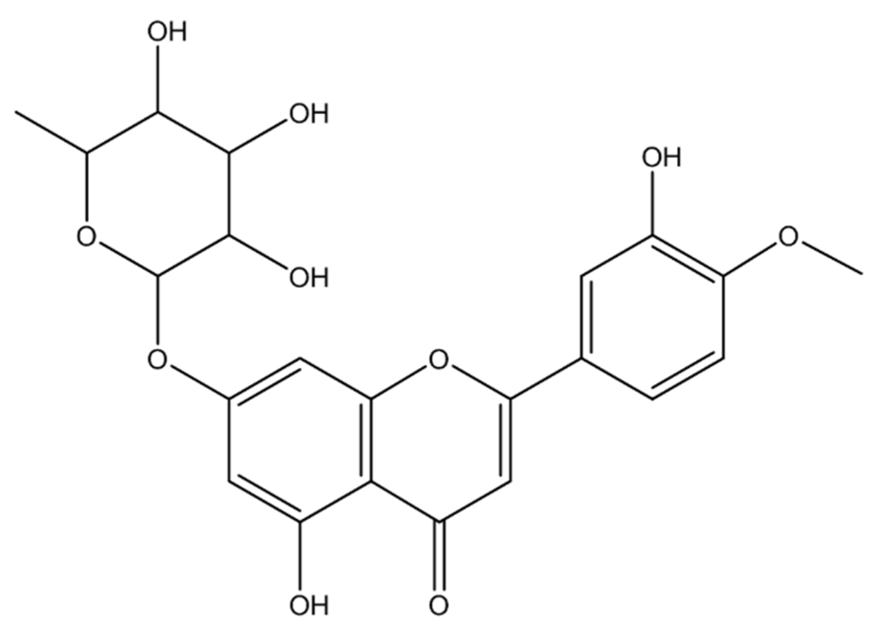
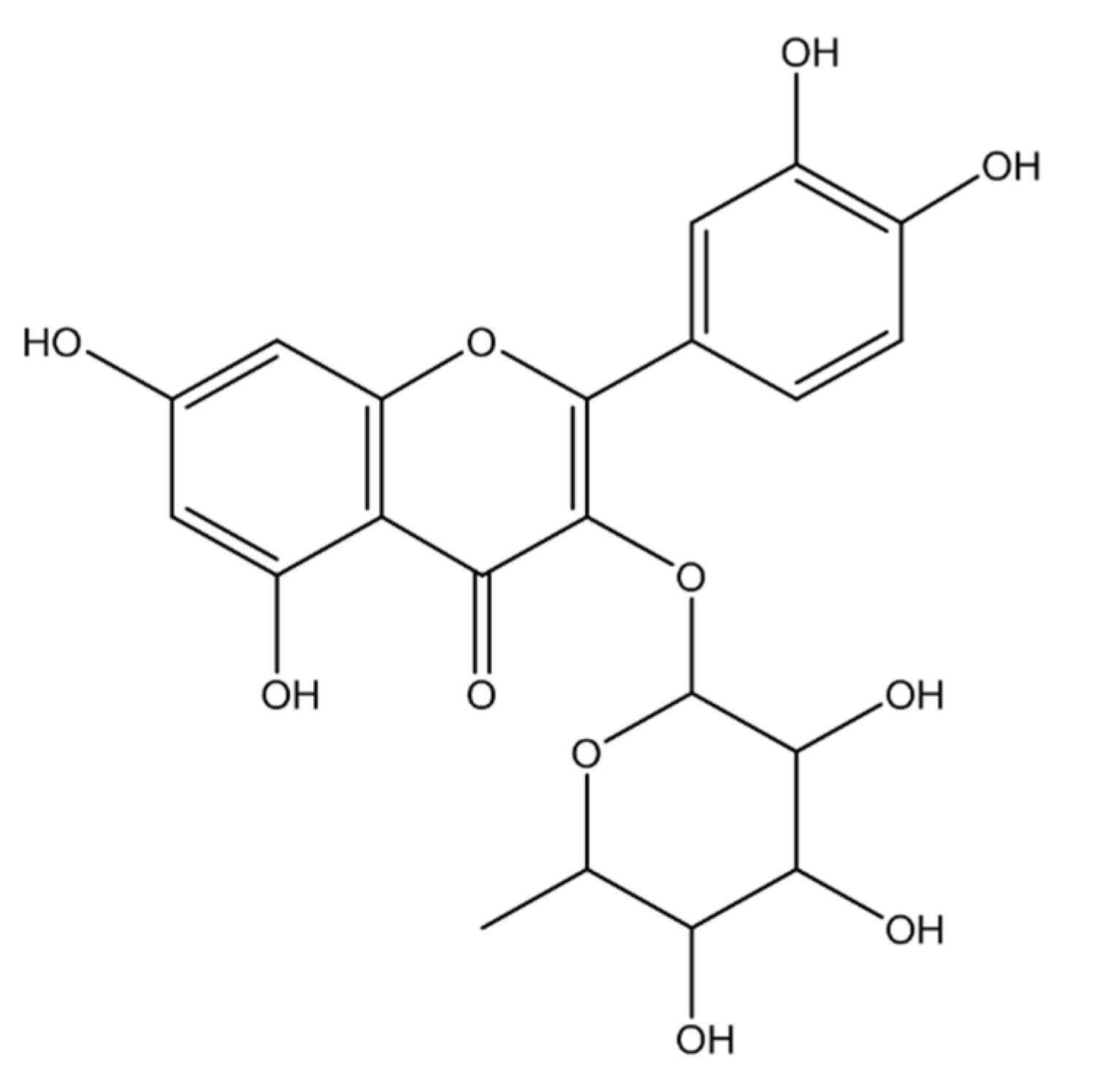
2.1. Identification of Isolated Phytocompounds

2.1.1. Compound 1 (MECD-1)

Subfraction 20–78 was purified by column chromatography on silica using methanol:ethyl acetoacetate to yield the pure compound 1 (120 mg). The isolated compound 1 was obtained as pale yellow amorphous powder with a melting range of 163–165 °C and a [α]25D value of +0.34 (conc. 0.25 mg/mL, MeOH). The structure of the isolated compound 1, represented in Figure 1, was elucidated by UV, IR, 1H-NMR, 13CNMR and mass spectroscopic/spectrometric analyses (Figure S1A–D).
UV (λmax, nm, MeOH): 307, 334; FT-IR (υmax, cm−1, KBR): 3265, 2960, 2895, 1668, 1616, 1429, 1367; 1H-NMR (δ, 400 MHz, DMSO-d6): 0.79 (J = 6.0 Hz), 3.98 (J = 15.0, 3.5 Hz), 2.96 (J = 15.0, 11.0 Hz), 3.02 (J = 11.0, 3.5 Hz), 4.41, 4.43, 5.25, 6.21, 6.42 (J = 2.0 Hz), 7.26 (J = 6.5, 2.0 Hz); 13CNMR (δ, 100 MHz, DMSO-d6): 193.56, 102.25, 55.78, 52.55.
The IR bands at 3265 cm−1 (O-H stretching) revealed the presence of the hydroxyl group in the structure of compound 1. Other prominent absorption bands at 2960 and 2895 cm−1 (aliphatic C-H stretching), 1668 cm−1 (C=O stretching) and 1616 cm−1 (aromatic C=H stretching) indicated the presence of methyl group (CH3), α,β unsaturated carbonyl group and aromatic rings. In 1H-NMR spectrum, three one-proton double doublets at δ 3.98 (J = 15.0, 3.5 Hz), 3.02 (J = 11.0, 3.5 Hz) and 2.96 (J = 15.0, 11.0 Hz) were ascribed to H-2, H-3α and H-3β protons, respectively of ring C of a flavone moiety. Two one-proton doublets at δ 6.21 and 6.42 (J = 2.0 Hz, each) were assigned to H-6 and H-8 aromatic protons. Two doublets at δ 7.26 (J = 6.5, 2.0 Hz), each integrating for one proton, were ascribed correspondingly to H-2, H-5 and H-6 of aromatic protons. A three-proton singlet at δ 3.61 was attributed to methoxy (OCH3) protons. A broad singlet at δ 5.25 was accounted to H-1″ anomeric proton, while a three proton doublet at δ 0.79 (J = 6.0 Hz) was appeared due to secondary methyl proton H-6″ of rhamnose unit. The remaining protons of sugar unit appeared between δ 4.43 and 4.41. The 13CNMR spectra showed twenty two distinct signals suggesting that the compound contains twenty two carbon atoms. The important signals appeared at δ 193.56 (C-4, carbonyl carbon), 102.25 (C-1 anomeric carbon), 55.78 (methoxy carbon OCH3) and 17.95 (C-6″ methyl carbon). The presence of an aromatic methoxy group was confirmed by position of the methyl signal at δ 52.55. The molecular ion [M]+ peak was obtained at m/z 446.0, which concord the molecular formula of the compound 1 as C22H22O11. The NMR spectral data also supported the structure of the compound. A thorough spectral interpretation suggests that the compound 1 (MECD-1) is 5,7,3,-trihydroxy-4-methoxyflavone-7-O-l-rhamnopyranoside.

2.1.2. Compound 2 (MECD-2)

Subfraction 120–184 was purified by column chromatography on silica gel using methanol:ethyl acetoacetate to obtain the pure compound 1 (160 mg). The isolated compound 1 was obtained as pale yellow crystalline powder with a melting range of 176–178 °C and a [α]25D value of +0.36 (conc. 0.25 mg/mL, MeOH). The structure of the isolated compound 1, represented in Figure 2 was elucidated by UV, IR, 1H-NMR, 13CNMR and mass spectroscopic/spectrometric analyses (Figure S2A–D).
UV (λmax, nm, MeOH): 312, 346; FT-IR (υmax, cm−1, KBR): 3265, 2950, 2880, 1654, 1502, 1454, 1354, 810. 1H-NMR (δ, 400 MHz, DMSO-d6): 0.73 (J = 7.1 Hz), 3.96–4.81, 6.11, 6.29 (J = 2.1 Hz), 6.78 (J = 9.6 Hz), 7.14 (J = 9.6, 2.2 Hz), 7.20 (J = 2.2 Hz), 12.55; 13CNMR (δ, 100 MHz, DMSO-d6): 156.90 (C-2), 134.69 (C-3), 178.20 (C-4), 104.56 (C-4), 161.75 (C-5), 99.15(C-6), 164.63 (C-7), 64.09 (C-8), 157.76(C-8), 121.22 (C-1′), 116.13 (C-2′),145.65 (C-3′), 148.88 (C-4′), 2115.93 (C-5′), 121.58 (C-6′), 102.29 (C-1″), 71.03 (C-2″), 70.83(C-3″), 71.66 (C-4″), 70.51 (C-5″), 17.95 (C-6″).
The IR bands at 3265 cm−1 (O-H stretching) revealed the presence of hydroxyl group in the structure of compound 2. Other prominent absorption bands at 2950 and 2880 cm−1 (aliphatic C-H stretching), 1654 cm−1 (C=O stretching) and 1502 cm−1 (aromatic C=H stretching) indicated the presence of methyl group (CH3), α,β unsaturated carbonyl group and aromatic rings. The 1H-NMR spectrum exhibited a set of two coupled doublets at δ 6.11 and 6.29 (J = 2.1 Hz, each), which was ascribed to H-6 and H-8 aromatic protons. Another set of coupled signals consisting of two doublets at δ 7.20 (J = 2.2 Hz), 6.78 (J = 9.6 Hz) and a double-doublet at δ 7.14 (J = 9.6, 2.2 Hz) were ascribed to H-2′, H-5′ and H-6′ aromatic protons of ring B. A doublet at δ 5.15 (J = 8.1 Hz) was assigned to H-1″ anomeric proton, while as another doublet at δ 0.73 (J = 7.1 Hz) was attributed to methyl protons (H-6″) of rhamnose unit. The remaining protons of rhamnose resonated from δ 4.81 to 3.96. A single proton singlet at δ 12.55 was attributed to hydroxyl proton. The 13CNMR spectrum displayed signals for twenty-one carbons. Important signals appeared for carbonyl carbon (δ 178.20, C-4), anomeric carbon (δ 102.29, C-1″) and methyl carbon (δ 17.94, C-6″). The molecular ion [M]+ peak was obtained at m/z 448.0, which concord the molecular formula of the compound as C21H20O11. The 1H and 13C NMR data was compared with other reported flavonoids and was found to be 3,5,7,3,4,-tetrahydroxy-4-methoxyflavone-3-O-l-rhamnopyranoside (Quercitrin (Quercetin-3-O-l-rhamnoside).

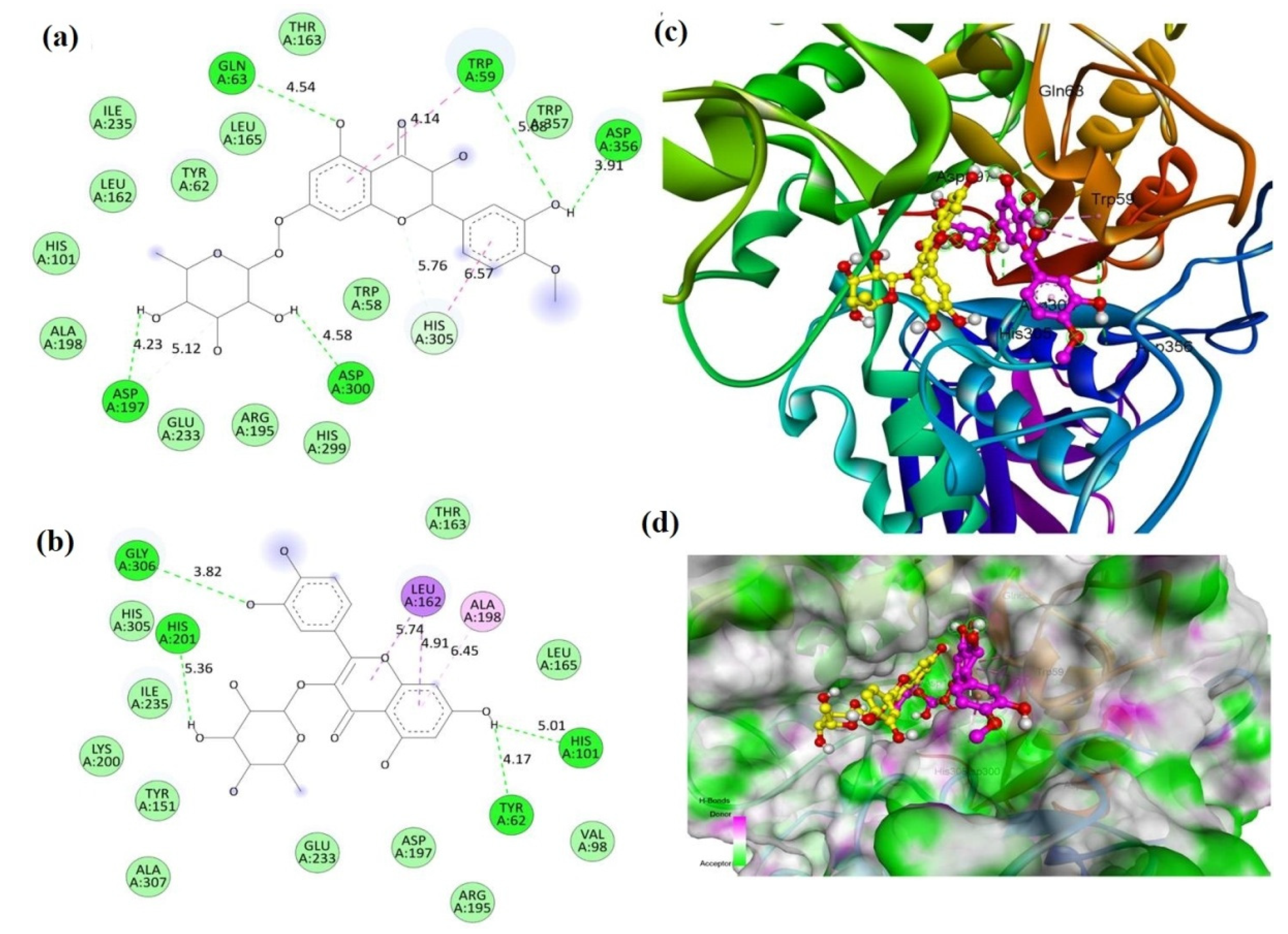
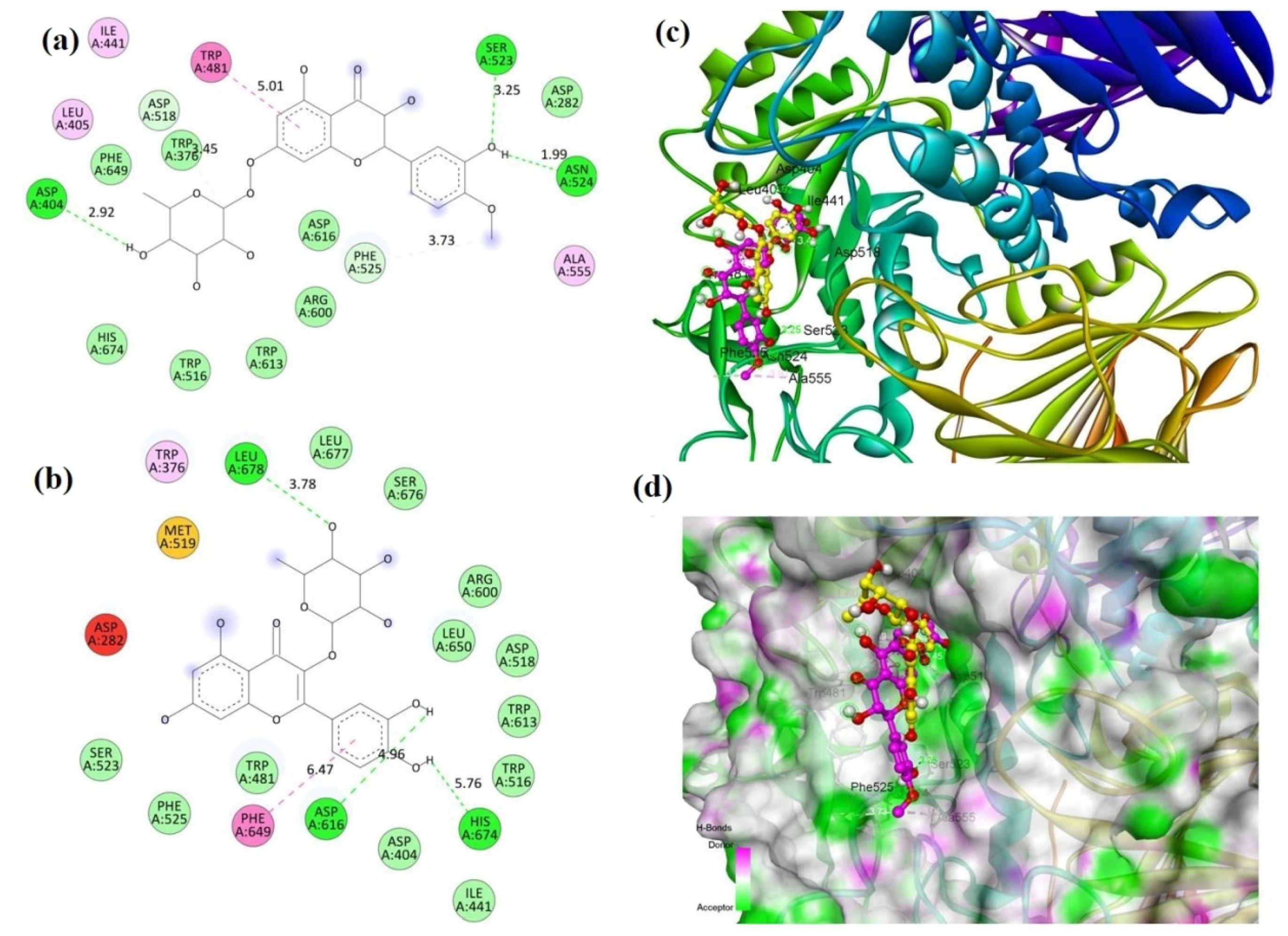
2.2. Molecular Docking

Molecular docking is used to understand the drug–receptor interaction, binding affinity and binding orientation of bioactive molecules into the target protein molecule. The objective behind docking study is to predict a particular biological activity based on the binding orientation/affinity of small molecule ligands to the appropriate target active site [9]. In the docking study, the binding affinity was predicted in terms of the interaction energy (kcal/mole). Results of docking (binding) energies are given in Table 1. Both the compounds exhibited very good binding affinity against both α-glucosidase and α-amylase enzyme. Compound 1 (MECD-1) exhibited more binding affinity against alpha-amylase compared to the alpha-glucosidase. On the hand, the compound 2 (MECD-2) showed better affinity against alpha-glucosidase than alpha-amylase. Not much variation in binding energies between these two enzymes were observed. Docking scores of isolated compounds were compared with that of the standard drug, acarbose. Against α-glucosidase, the binding affinity of compounds 1 and 2 was comparatively more affinity than the standard drug. On the other hand, the binding affinity of compounds 1 and 2 were less to some extent than that the standard drug against α-amylase. No significant difference in activities between isolated test compounds and the standard compound was observed. The test compounds were found to have α-glucosidase and α-amylase inhibitory potential to a similar degree as that of the standard drug, acarbose. Overall, both the isolated flavonoids exhibited significant inhibitory potential of human glucosidase and amylase enzymes.
Post-docking visualization of protein–ligand complexes revealed that the compounds interacted with active site residues of the protein molecules through the formation of predominantly hydrogen bonding interactions (Figure 3 and Figure 4). From the observation of 2D interaction diagrams of compound 1-α-glucosidase complexes, it is clear that compound 1 formed H-bonds with Trp59, Gln63, Asp197, Asp300 and Asp356 residues, whereas the compound 2 interacted with Tyr62, His101, His201 and Gly306 residues through H-bonds (Figure 3a,b). The 3D diagrams revealed the binding conformation and binding poses of the compounds at the catalytic site of α-glucosidase (Figure 3c,d) were observed.
From the observation of 2D interaction diagrams of compound 2-α-amylase complexes, it is clear that compound 1 formed prominent H-bonds with Asp404, Ser523 and Ser524 residues, whereas the compound 2 interacted with Asp616, His674 and Leu678 residues through H-bonds (Figure 3b and Figure 4a). The 3D diagrams revealed the binding conformation and binding poses of the compounds at the catalytic site of alpha-amylase (Figure 3d and Figure 4c) were observed.
Upon critical analysis of protein–ligand interactions, favorable binding orientations and/or binding modes of flavonoid molecules for both the α-glucosidase and α-amylase enzyme were evident. Both the compounds structurally represent glycosides of flavones, which are abundantly found in plant kingdom. The glycone part (sugar), i.e., the rhamnose moiety is similar in both the compounds, while the aglycone part (non-sugar bioactive principle, flavanone moiety) is dissimilar. In compound 1, it is 5,7,3′-trihydroxy-4-methoxyflavone, whereas, it is 3,5,7,3′,4′-tetrahydroxy-4-methoxyflavone, i.e., quercetin in compound 2. The aglycone, i.e., the flavonoid moiety is a polyhydroxylated C6-C3-C6 tricyclic heteroaromatic system (phenylchromone) [10,11,12] with distinct structural features, particularly in terms of nature and pattern of ring substitutions. There is a close structural resemblance between these two isolated flavonoid glycosides. The basic flavone skeleton along with hydroxylated/methoxylated aromatic ring interacted predominantly with the active site residues of target protein molecules. Polar groups such as phenolic hydroxy groups and carbonyl moiety contributed significantly in protein-ligand interactions with the formation of hydrogen bonds. Apart from hydrogen bonding, other non-bonding interactions such as hydrophobic interactions also exist, but to a lesser extent. Aromatic bulky moieties chromone system and phenyl ring were mainly involved in non-polar hydrophobic interactions.
The human lysosomal α-glucosidase and pancreatic α-amylase enzymes play an important role in the digestion of dietary long-chain complex carbohydrates (breakdown of starch and disaccharides to glucose) and hence, their inhibition is believed to facilitate the reduction of post-prandial (post meal) blood glucose level in type 2 diabetes [13]. The traditional usefulness about the antidiabetic potential of C. dichotoma is mentioned in literature [2,3,4]. In our earlier studies, the antidiabetic activity of the methanolic bark extract of C. dichotoma has already been reported [4]. Moreover, the literature suggest that the flavonoids content demonstrates antidiabetic efficacy of many plants [14,15,16,17,18]. Our docking study validates the antidiabetic claim about C. dichotoma reported in traditional medicines and in recent literature. Although the isolated phytocompounds are already established bioactive flavonoids with many scientific reports from past literature, their antidiabetic potential determined by in silico (molecular docking) methods with α-glucosidase and α-amylase inhibitory activities has been reported for the first time. Our study may thus provide an avenue for further investigation with these bioflavonoids for their development as potent antidiabetic molecules with alpha-glucosidase and alpha-amylase inhibitory agents for the treatment of type 2 diabetes mellitus.

2.3. ADMET

Results of predicted ADMET (absorption, distribution, metabolism, excretion and toxicity) data showed that both the isolated compounds possess good solubility profile, which is in favor of their oral bioavailability. There is a prediction of poor intestinal absorption, while the compounds were predicted to be non-inhibitors of the cytochromes (CYP450) [19]. Poor intestinal absorption might be due to their limited oil/water partition coefficient (logP) values (−1.64 and −1.84). CYP450 enzymes are largely involved in drug metabolism. Non-inhibition of CYP450 enzymes suggests that compounds do not suppress the metabolic function of the enzymes. Inhibition can lead to increased bioavailability of compounds that normally undergo extensive first-pass elimination or to decreased elimination of compounds dependent on metabolism for systemic clearance. Compounds did not exhibit the property of blood brain barrier (BBB) penetration. It substantiates that the compounds are devoid of producing CNS toxicities. Furthermore, quercitrin (compound 2) was predicted to be a substrate to permeability of glycoprotein (p-gp), whereas the other flavonoid molecule (compound 1) did not show such property. Glycoprotein is responsible for the efflux of drug molecules out of the target cells [20]. A good drug candidate should not only have sufficient efficacy against the therapeutic target, but also show appropriate ADMET properties at a therapeutic dose. It is therefore inevitable to evaluate the ADMET profile of drug-like molecules to avoid the failure of candidate drugs at the clinical stage of drug development [21].

3. Materials and Methods

3.1. Collection of Plant

The barks of Cordia dichotoma Forst. were collected from the Duhai forest of Ghaziabad, Uttar Pradesh, India. The plant species was identified from National Institute of Science Communication and Information Resources, New Delhi, India. The voucher specimen (NISCAIR/RHMD/Consult/2012-13/2025/33) of the bark of Cordia dichotoma Forst. was submitted at the herbarium of the department for future reference.

3.2. Preparation of Methanolic Bark Extract

The shade-dried barks of C. dichotoma were pulverized to a coarse powder and defatted using petroleum ether by the cold maceration method [2] to remove fat, latex and non-polar compounds of high molecular weights. The defatted plant residues were then macerated successively with methanol to obtain the desired extract [3,4]. The collected extract was filtered through Whatman No. 1 filter paper. Rotary evaporator was used to concentrate the filtrate. The concentrated extract was preserved in refrigerator at 4 °C for further use. The percentage yield of the methanolic bark extract of C. dichotoma (MECD) was 7.11%.

3.3. Phytochemical Analysis

Chemical tests for the screening and detection of phytochemical constituents of the MECD were carried out using the standard procedures [22,23].

3.4. Isolation of Phytocompounds

The MECD was subjected to column chromatographic separation using silica gel (packed column, 100–200 mesh) and a glass column (6.0 × 3 inch dimension) [6] for the isolation of bioactive phytoconstituents in pure form. The elution was carried out by gradient separation technique using the solvent system of n-hexane/ethyl acetate. The column was eluted successively with n-hexane:ethyl acetate in increasing order of polarity (98:2, 95:5, 90:10, 80:20, 60:40, 50:50, 35:65, 30:70, 25:75, 20:80 and 100%). The fractions collected were subjected to thin-layer chromatography (TLC) to check their homogeneity. Chromatographically identical fractions (having the same Rf values) were combined together and concentrated. The concentrated fractions were purified by crystallization with methanol/benzene and confirmed by their sharp melting points.

3.5. Identification of Isolated Compounds

Ultraviolet (UV)–visible spectra were recorded on Shimadzu UV-1700 UV–visible spectrophotometer (Shimadzu, Kyoto, Japan) and the wave lengths of maximum absorption (λmax, nm) were reported. Infrared (IR) spectra were obtained on a Bruker alpha Fourier transform (FT-IR) spectrometer (Bruker, MA, USA) using the KBR disc and reported in terms of frequency of absorption (υmax, cm−1). 1H and 13C nuclear magnetic resonance (NMR) spectra were recorded on Bruker Avance II 400 FT-NMR spectrometer (Bruker, MA, USA) at 400 and 100 MHz, respectively using tetramethylsilane (TMS) as an internal standard (δ 0.00 ppm) and CDCl3 as a solvent. Mass spectra were obtained on a LC–MS Water 4000 ZQ instrument (Waters, Massachusetts, USA) using electrospray ionization (ES+). The m/z values were recorded in the range of m/z between 100 and 500 and the m/z values of the most intense molecular ion [M]+ peak were considered. Melting points were determined on an electric melting point apparatus (JSGW, Model 3046). (Jain Scientific Glass Works, Ambala, India)

3.6. Molecular Docking

The X-ray crystal structure of proteins, viz., human lysosomal acid α-glucosidase (PDB ID: 5NN8) and human pancreatic α-amylase (PDB ID: 1B2Y) were reposited by Roig-Zamboni et al. [24] and Nahoun et al. [25] having resolution of 2.45 Å and 3.20 Å, respectively were retrieved from the RCSB protein data bank (http://www.rcsb.org/ (accessed on 13 March 2021)).
Prior to docking, The docking was performed in the AutoDock Vina software(The Scripps Research Institute, La Jolla, CA, USA) [26] in accordance with the standard procedure. The protein crystal structure was prepared prior to the docking process. Hydrogen atoms were added to the protein structure, and all ionizable residues were set at their default protonation at pH 7.4. The active site coordinates were determined with the dimensions of x = −15.941, y = −37.643, z = 92.912 for 5NN8 and x = 22.116, y = 4.749, z = 45.878 for 1B2Y, and, a grid box with radius of 25 × 25 × 25 A3 was generated for both the proteins. Similarly, the ligands were prepared and energy minimized using Chem3D 17.0 software. During the docking process, the receptor was rigidly held, while the ligands were allowed to flex during the refinement. Binding energies of docking were recorded and analyzed. The best docked poses and binding modes of protein–ligand interactions were obtained using the Discovery Studio visualizer.

3.7. ADMET Prediction

Predictive ADMET (pharmacokinetics) parameters were studied using web-based Swiss ADME tool developed by Daina et al. [27]. Solubility, intestinal absorption, oil/water partition coefficient (logP), CYP450 inhibition, blood brain barrier penetration and p-gp substrate were predicted [28].

4. Conclusions

This study reports two bioactive flavonoids, viz., 3,5,7,3′,4′-tetrahydroxy-4-methoxyflavone-3-O-l-rhamnopyranoside and 5,7,3′-trihydroxy-4-methoxyflavone-7-O-l-rhamnopyranoside (Quercitrin) isolated and identified from the methanolic bark extract Cordia dichotoma Forst. The molecular docking study investigated the antidiabetic potential of the isolated flavonoids against human lysosomal acid α-glucosidase and human pancreatic α-amylase enzymes. The predictive ADMET study demonstrated acceptable pharmacokinetics of the isolated compounds. The in silico study needs to be further validated by in vitro and in vivo experimental assays in order to confirm the antidiabetic effectiveness for the flavonoids reported herein. Our study may thus provide an avenue for further investigation with these bioflavonoids for their development as potent antidiabetic molecules with α-glucosidase and α-amylase inhibitory agents for the treatment of type 2 diabetes mellitus.

Supplementary Materials

The following are available online, Figure S1A: FT-IR spectrum of compound 1, Figure S1B: 1H-NMR spectrum of compound 1, Figure S1C: 13C-NMR spectrum of compound 1, Figure S1D: Mass spectrum of compound 1, Figure S2A: FT-IR spectrum of compound 2, Figure S2B: 1H-NMR spectrum of compound 2, Figure S2C(1) and Figure S2C(2): 13H-NMR spectrum of compound 2, Figure S2D: Mass spectrum of compound 2.

Author Contributions

Conceptualization, N.H. and B.B.K.; methodology, N.H.; software, I.C.; validation, I.C. and M.R.; formal analysis, M.R. and E.I.A.; investigation, N.H. and I.C.; resources, B.B.K. and I.C.; data curation, M.R., I.C. and S.J.K.; writing—original draft preparation, E.I.A. and S.J.K.; writing—review and editing, M.R. and E.I.A.; visualization, E.I.A.; supervision, B.B.K.; project administration, B.B.K.; funding acquisition, S.J.K., K.K.S., S.B., R.K.S. and S.G.W. All authors have read and agreed to the published version of the manuscript.

Funding

This research received no external funding.

Institutional Review Board Statement

Not applicable.

Informed Consent Statement

Not applicable.

Data Availability Statement

Additional data will be made available on request.

Acknowledgments

Authors gratefully acknowledge the TÜBİTAK (The Scientific and Technological Research Council of Turkey), ULAKBİM (Turkish Academic Network and Information Centre), High Performance and Grid, Computing Centre (TRUBA resources), Turkey for providing necessary facilities for performing the computational studies. Authors extend their sincere thanks to Sagarika Chandra for her needful help towards editing the images incorporated in the manuscript.

Conflicts of Interest

The authors declare no conflict of interest.

References

  1. Jamkhande, P.G.; Barde, S.R.; Patwekar, S.L.; Tidke, P.S. Plant profile, phytochemistry and pharmacology of Cordia dichotoma (Indian cherry): A review. Asian Pac. J. Trop. Biomed. 2013, 3, 1009–1012. [Google Scholar] [CrossRef] [Green Version]
  2. Hussain, N.; Kakoti, B.B.; Rudrapal, M.; Junejo, J.A.; Laskar, M.A.; Lal, M.; Sarwa, K.K. Anticancer and Antioxidant Activities of Cordia dichotoma Forst. Int. J. Green. Pharm. 2020, 14, 265–273. [Google Scholar]
  3. Hussain, N.; Kakoti, B.B.; Rudrapal, M.; Sarwa, K.K. Anti-inflammatory and Antioxidant Activities of Cordia dichotoma Forst. Biomed. Pharmacol. J. 2020, 13, 2093–2099. [Google Scholar] [CrossRef]
  4. Hussain, N.; Kakoti, B.B.; Rudrapal, M.; Rahman, Z.; Rahman, M.; Chutia, D.; Sarwa, K.K. Antidiabetic Activity of the bark of Indian Cherry, Cordia dichotoma. Biosci. Biotech. Res. Comm. 2020, 13, 2211–2216. [Google Scholar] [CrossRef]
  5. Ragasa, C.Y.; EbajoJr, V.D.; Mariquit, M.; Mandia, E.H.; Tan, M.C.S.; Brkljača, R.; Urban, S. Chemical constituents of Cordia dichotoma G. Forst. J. Appl. Pharm. Sci. 2015, 5, 16–21. [Google Scholar]
  6. Junejo, J.A.; Rudrapal, M.; Mohammed, A.; Zaman, K. New flavonoid with antidiabetic potential from Tetrastigma angustifolia (Roxb.) Deb leaves. Braz. J. Pharm. Sci. 2020, 56, e18806. [Google Scholar] [CrossRef]
  7. Junejo, J.A.; Mondal, P.; Rudrapal, M.; Zaman, K. Antidiabetic assessment of the hydro-alcoholicleaf extracts of the plant Tetrastigma angustifolia (Roxb.), a traditionally used North-Eastern Indian vegetable. Biomed. Pharmacol. J. 2014, 7, 635–644. [Google Scholar] [CrossRef]
  8. Junejo, J.A.; Gogoi, G.; Islam, J.; Rudrapal, M.; Mondal, P.; Hazarika, H.; Zaman, K. Exploration ofantioxidant, antidiabetic and hepatoprotective activity of Diplazium esculentum, a wild edible plant from North Eastern region of India. Future J. Pharm. Sci. 2018, 4, 93–101. [Google Scholar] [CrossRef]
  9. Kumar, S.; Kaushik, A.; Narasimhan, B.; Shah, S.A.; Lim, S.M.; Ramasamy, K.; Mani, V. Molecular docking, synthesis and biological significance of pyrimidine analogues as prospective antimicrobial and antiproliferative agents. BMC Chem. 2019, 13, 85. [Google Scholar] [CrossRef] [PubMed] [Green Version]
  10. Wen, W.; Alseekh, S.; Fernie, A.R. Conservation and diversification of flavonoid metabolism in the plant kingdom. Curr. Opin. Plant. Biol. 2020, 55, 100–108. [Google Scholar] [CrossRef]
  11. Arora, S.; Itankar, P. Extraction, isolation and identification of flavonoid from Chenopodium album aerial parts. J. Trad. Comp. Med. 2018, 8, 476–482. [Google Scholar] [CrossRef] [PubMed]
  12. Kishore, N.; Twilley, D.; Blom van Staden, A.; Verma, P.; Singh, B.; Cardinali, G.; Lall, N. Isolation of flavonoids and flavonoid glycosides from Myrsine africana and their inhibitory activities against mushroom tyrosinase. J. Nat. Prod. 2018, 81, 49–56. [Google Scholar] [CrossRef] [PubMed]
  13. Tundis, R.; Loizzo, M.R.; Menichini, F. Natural products as α-amylase and α-glucosidase inhibitors and their hypoglycaemic potential in the treatment of diabetes: An update. Mini Rev. Med. Chem. 2010, 10, 315–331. [Google Scholar] [CrossRef]
  14. Junejo, J.A.; Zaman, K.; Rudrapal, M.; Hussain, N. Antidiabetic and Antioxidant Activity of Hydro-alcoholic Extract of Oxalis debilis Kunth. Leaves in Experimental Rats. Biosci. Biotech. Res. Comm. 2020, 13, 860–867. [Google Scholar] [CrossRef]
  15. Junejo, J.A.; Rudrapal, M.; Zaman, K. Antidiabetic activity of Carallia brachiata Lour. Leaves hydro-alcoholic extract (HAE) with antioxidant potential in diabetic rats. Indian J. Nat. Prod. Resour. 2020, 11, 18–29. [Google Scholar]
  16. Junejo, J.A.; Rudrapal, M.; Nainwal, L.M.; Zaman, K. Antidiabetic activity of hydro-alcoholic stem bark extract of Callicarpa arborea Roxb. with antioxidant potential in diabetic rats. Biomed. Pharmacother. 2017, 95, 84–94. [Google Scholar] [CrossRef]
  17. Rashed, K.N.; Butnariu, M. Isolation and antimicrobial and antioxidant evaluation of bioactive compounds from Eriobotrya japonica stem. Adv. Pharm. Bull. 2014, 4, 75–81. [Google Scholar]
  18. Conforti, F.; Statti, G.A.; Tundis, R.; Menichini, F.; Houghton, P. Antioxidant activity of methanolic extract of Hypericum triquetrifolium Turra aerial part. Fitoterapia 2002, 73, 479–483. [Google Scholar] [CrossRef]
  19. Lynch, T.; Price, A.L. The effect of cytochrome P450 metabolism on drug response, interactions, and adverse effects. Am. Fam. Physician. 2007, 76, 391–396. [Google Scholar]
  20. Feng, B.; Mills, J.B.; Davidson, R.E.; Mireles, R.J.; Janiszewski, J.S.; Troutman, M.D.; de Morais, S.M. In vitro P-glycoprotein assays to predict the in vivo interactions of P-glycoprotein with drugs in the central nervous system. Drug Metab. Dispos. 2008, 36, 268–275. [Google Scholar] [CrossRef]
  21. Isyaku, Y.; Uzairu, A.; Uba, S. Computational studies of a series of 2-substituted phenyl-2-oxo-,2-hydroxyl-and 2-acylloxyethylsulfonamides as potent anti-fungal agents. Heliyon 2020, 6, e03724. [Google Scholar] [CrossRef]
  22. Onah, O.E.; Babangida, K.J. Phytochemical Investigation and Antimicrobial Activity of Hexane, Ethyl Acetate and Methanol Fractions from Stem Bark of Icacina trichantha Oliv (Icacinaceae). J. Chem. Environ. Sci. Appl. 2020, 7, 7–12. [Google Scholar] [CrossRef]
  23. Jigna, P.; Sumitra, C. Phytochemical screening of some plants from western region of India. Plan. Arch. 2008, 8, 657–662. [Google Scholar]
  24. Roig-Zamboni, V.; Cobucci-Ponzano, B.; Iacono, R.; Ferrara, M.C.; Germany, S.; Bourne, Y.; Sulzenbacher, G. Structure of human lysosomal acid α-glucosidase–a guide for the treatment of Pompe disease. Nat. Commun. 2017, 8, 1–10. [Google Scholar] [CrossRef] [PubMed] [Green Version]
  25. Nahoum, V.; Roux, G.; Anton, V.; Rougé, P.; Puigserver, A.; Bischoff, H.; Payan, F. Crystal structures of human pancreatic α-amylase in complex with carbohydrate and proteinaceous inhibitors. Biochem. J. 2000, 346, 201–208. [Google Scholar] [CrossRef] [PubMed]
  26. Trott, O.; Olson, A.J. AutoDockVina: Improving the speed and accuracy of docking with a new scoring function, efficient optimization, and multi threading. J. Comput. Chem. 2010, 31, 455–461. [Google Scholar]
  27. Daina, A.; Michielin, O.; Zoete, V. Swiss ADME: A free web tool to evaluate pharmacokinetics, drug-likeness and medicinal chemistry friendliness of small molecules. Sci. Rep. 2017, 7, 1–13. [Google Scholar] [CrossRef] [Green Version]
  28. Kato-Schwartz, C.G.; deSá-Nakanishi, A.B.; Guidi, A.C.; Gonçalves, D.A.; Bueno, F.G.; Zani, B.P.M.; Peralta, R.M. Carbohydrate digestive enzymes are inhibited by Poincianellapluviosa stem bark extract: Relevance on type 2 diabetes treatment. Clin. Phytosci. 2020, 6, 1–11. [Google Scholar] [CrossRef]
Figure 1. Structure of compound 1 (MECD-1).
Molbank 2021 m1234 g001
Figure 2. Structure of compound 2.
Molbank 2021 m1234 g002
Figure 3. (a) Two-dimensional interactions between compound 1 and α-glucosidase, (b) 2D interactions between compound 2 and α-glucosidase showing hydrogen bonding and other non-covalent interactions with amino acid residues at the active site, (c) 3D representation of protein-ligand interactions showing binding conformation and (d) binding poses/binding modes of both the compounds at the catalytic site of α-glucosidase.
Molbank 2021 m1234 g003
Figure 4. (a) Two-dimensional interactions between compound 1 and α-amylase, (b) 2D interactions between compound 2 and α-glucosidase showing hydrogen bonding and other non-covalent interactions with amino acid residues at the active site, (c) 3D representation of protein–ligand interactions showing binding conformation and (d) binding poses/binding modes of both the compounds at the catalytic site of α-amylase.
Molbank 2021 m1234 g004
Table 1. Docking data of compounds.
CompoundBinding Energy (kcal/mole)
5NN81B2Y
Compound 1 (MECD-1)−7.8−8.6
Compound 2 (MECD-2)−8.0−7.8
Acarbose (Standard drug)−7.6−9.4
5NN8: Human lysosomal acid α-glucosidase; 1B2Y: Human pancreatic α-amylase; MECD: Metanolic bark extract of C. dichotoma.
Publisher’s Note: MDPI stays neutral with regard to jurisdictional claims in published maps and institutional affiliations.

