
Preparation, Characterization, Swelling Potential, and In-Vitro Evaluation of Sodium Poly(Styrene Sulfonate)-Based Hydrogels for Controlled Delivery of Ketorolac Tromethamine


Abstract
:1. Introduction
2. Results and Discussion
2.1. Sol-Gel Analysis
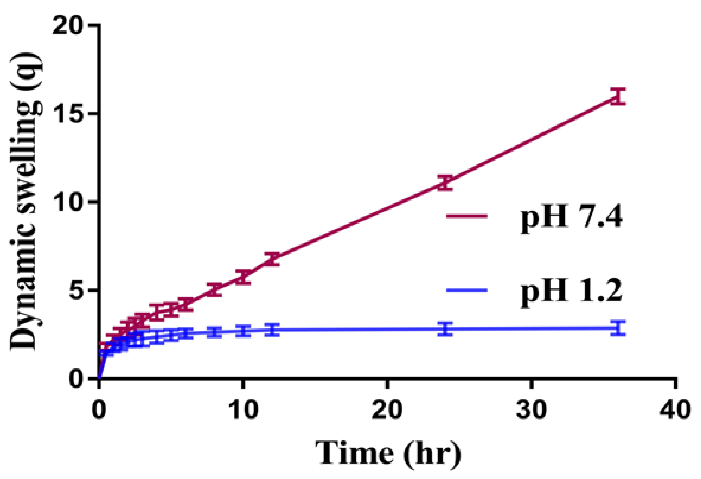
2.2. Dynamic Swelling Studies
2.3. Drug Loading
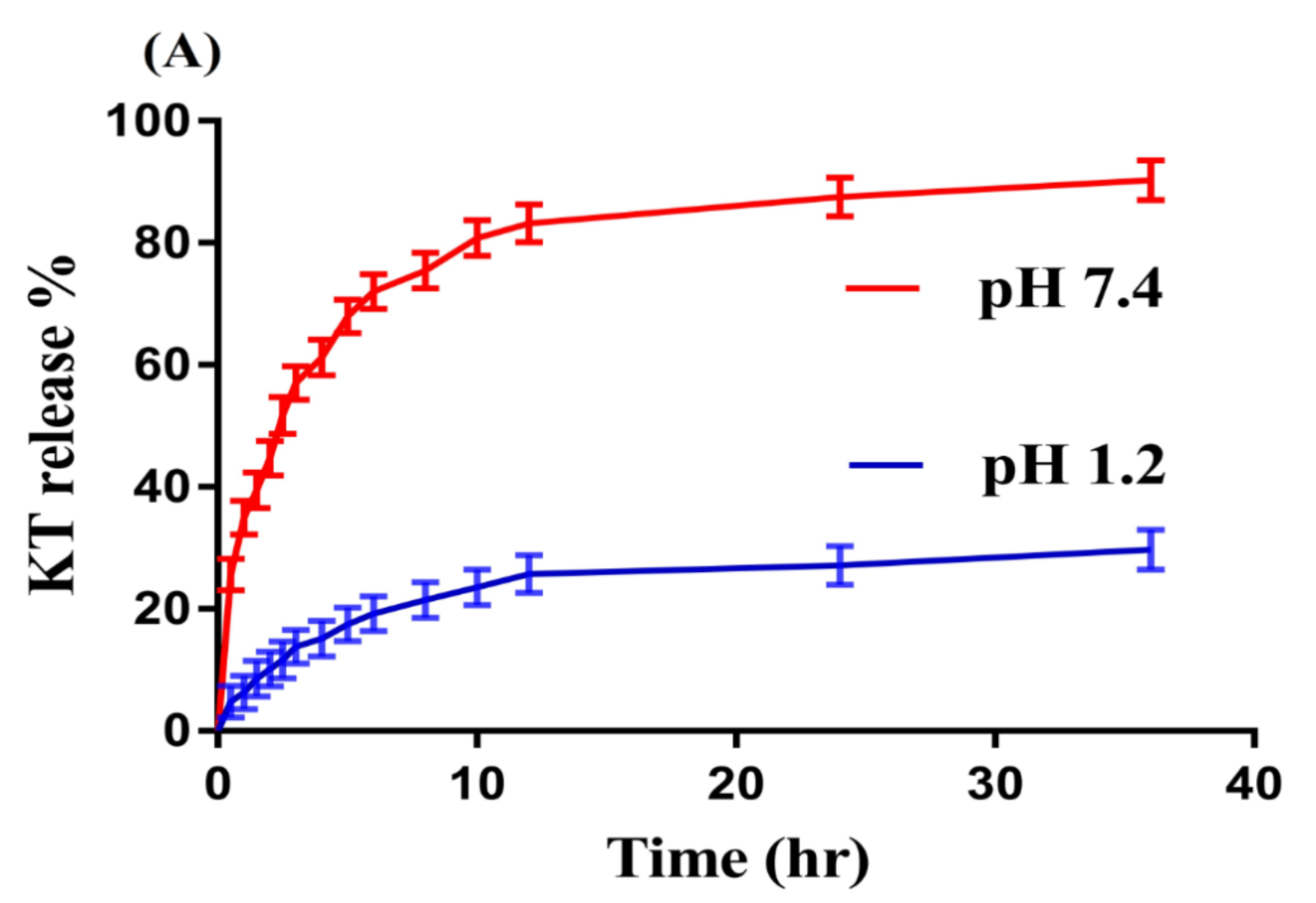
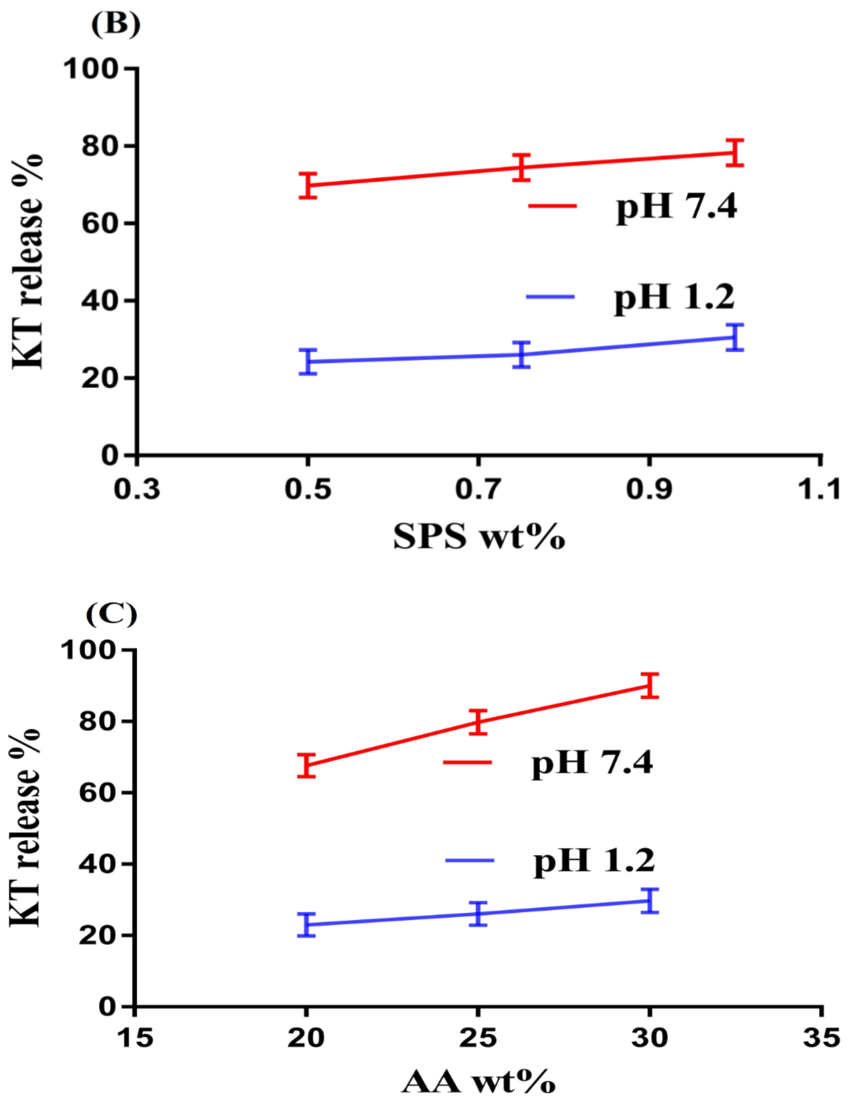
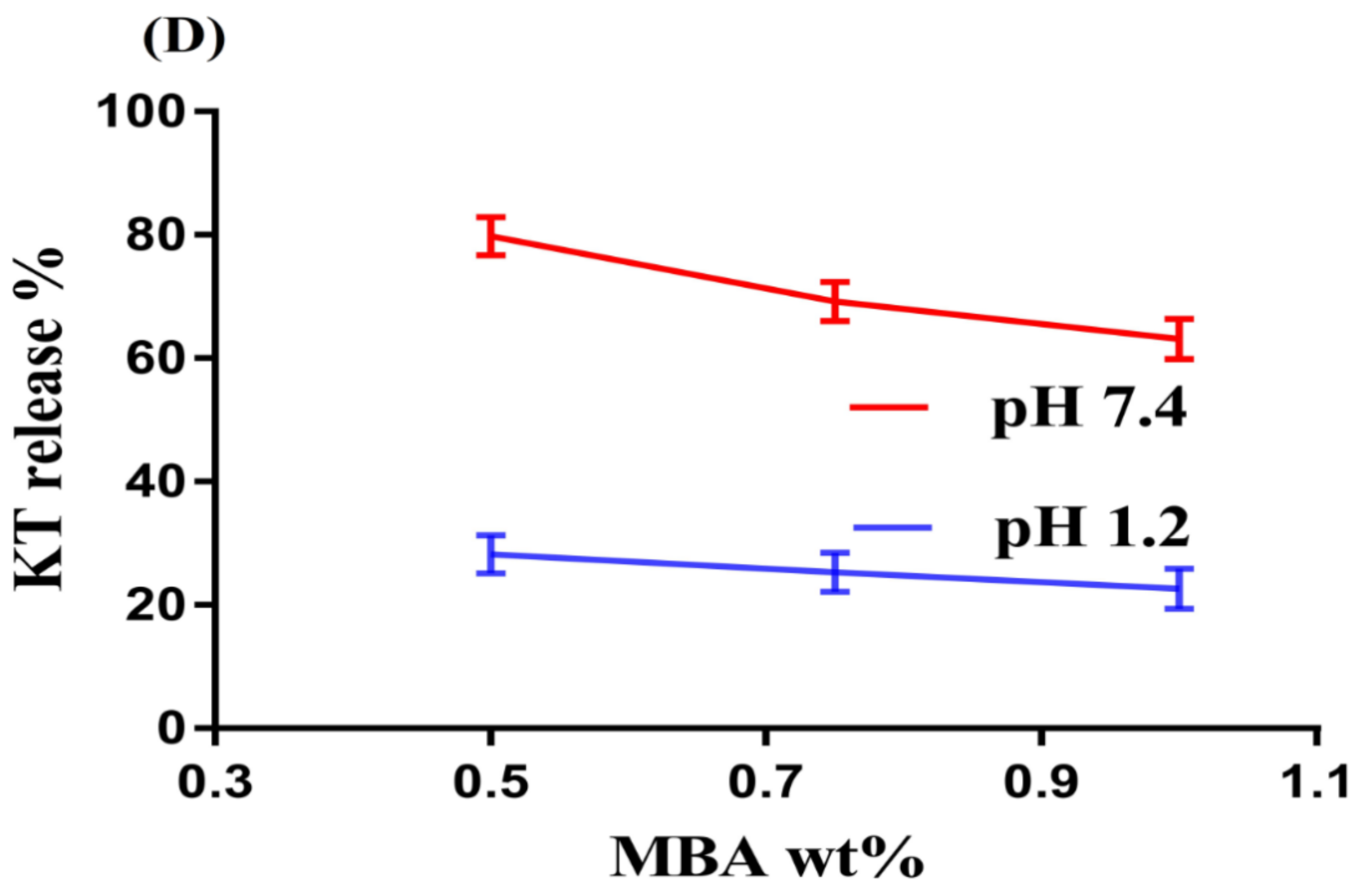
2.4. In-Vitro Drug Release and Kinetic Modeling
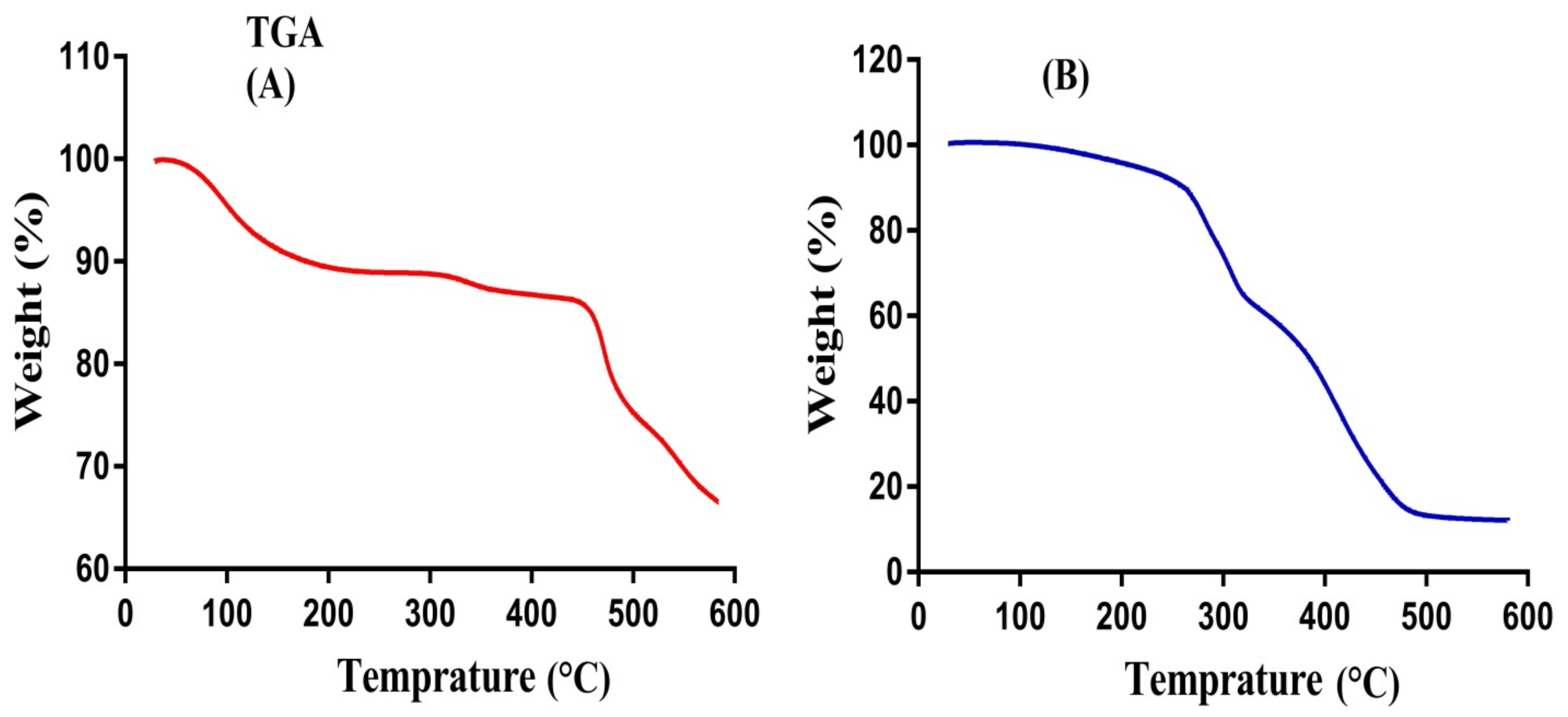
2.5. TGA Analysis
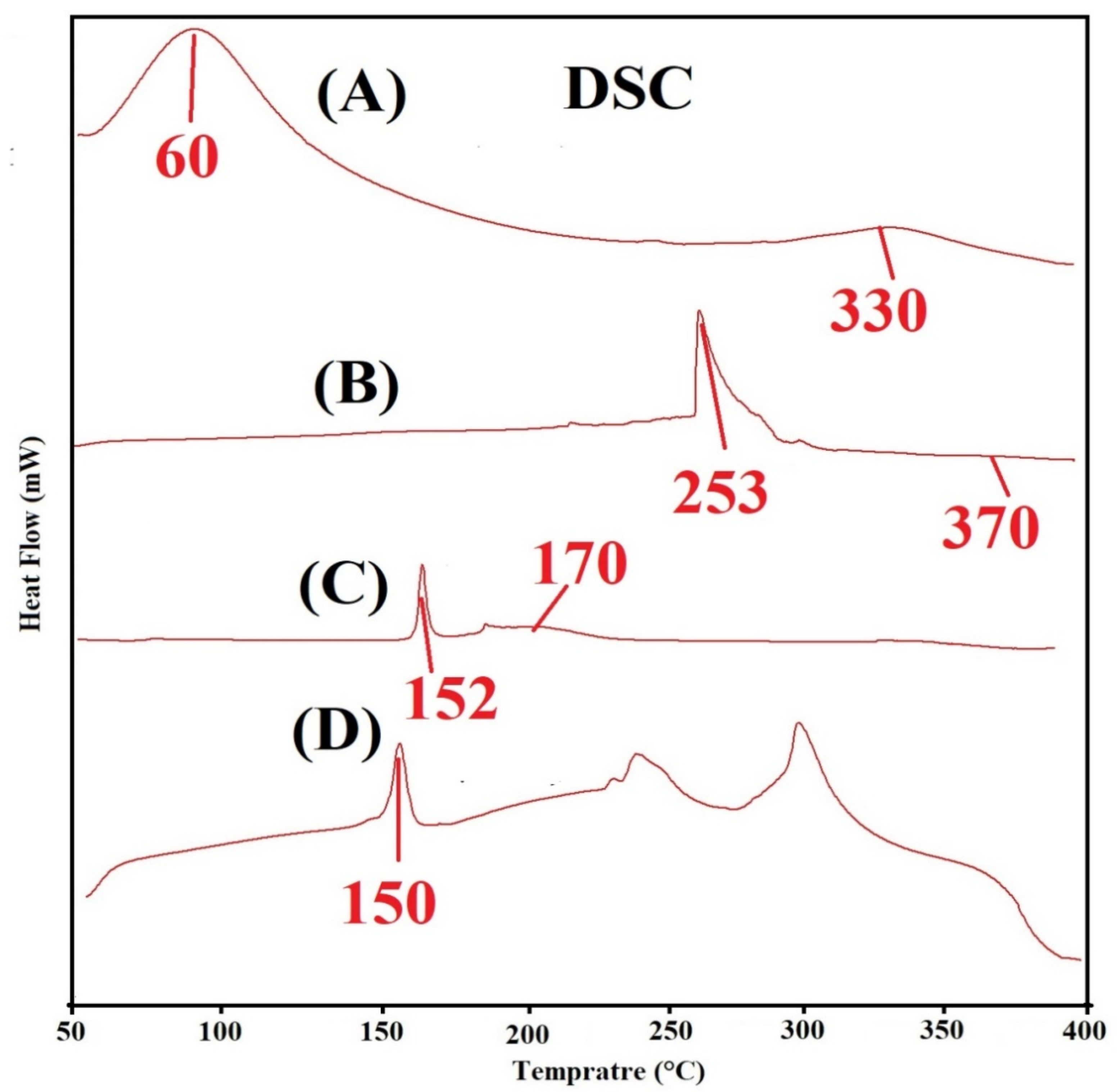
2.6. DSC Analysis
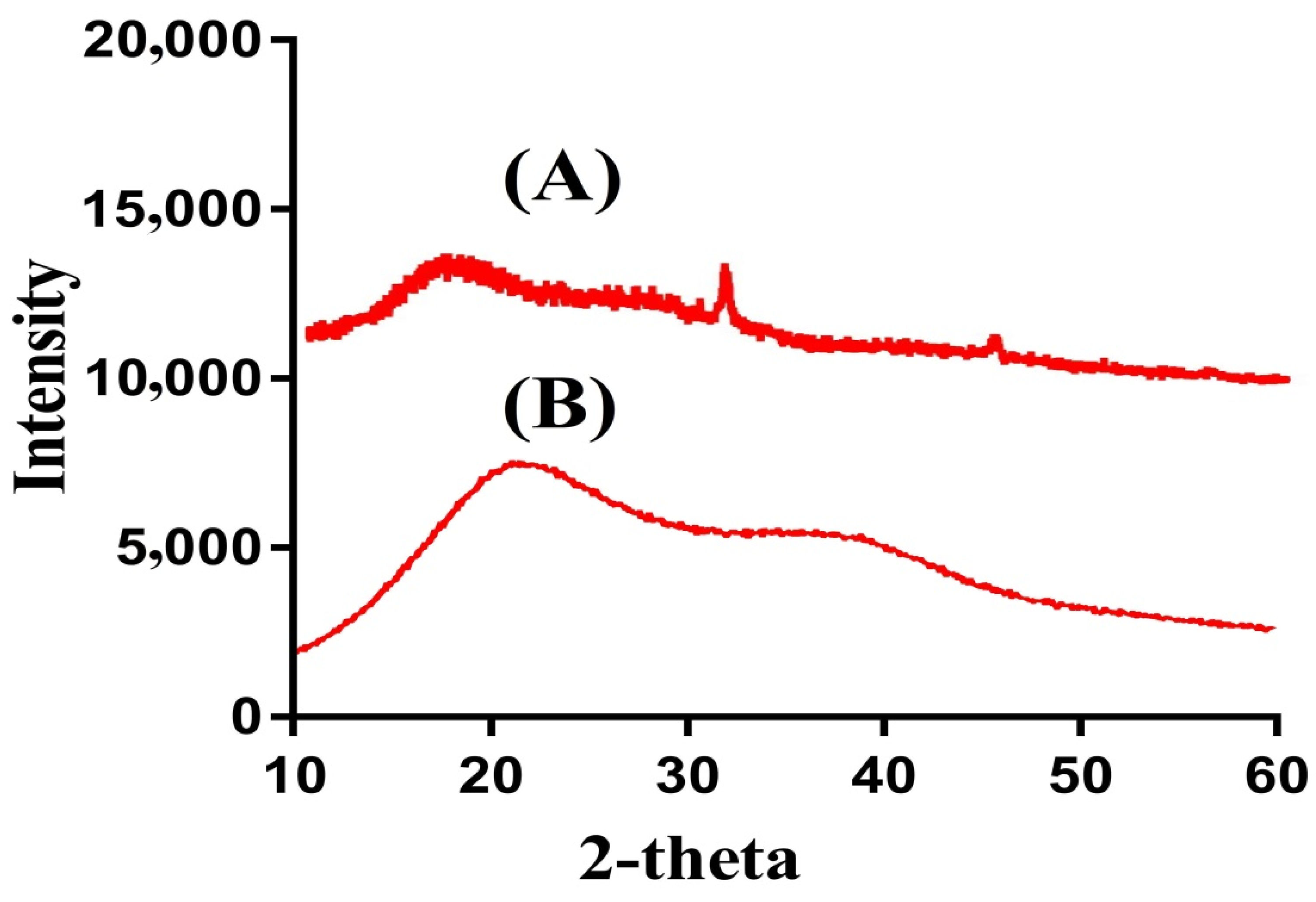
2.7. Powder X-ray Diffraction (PXRD) Analysis
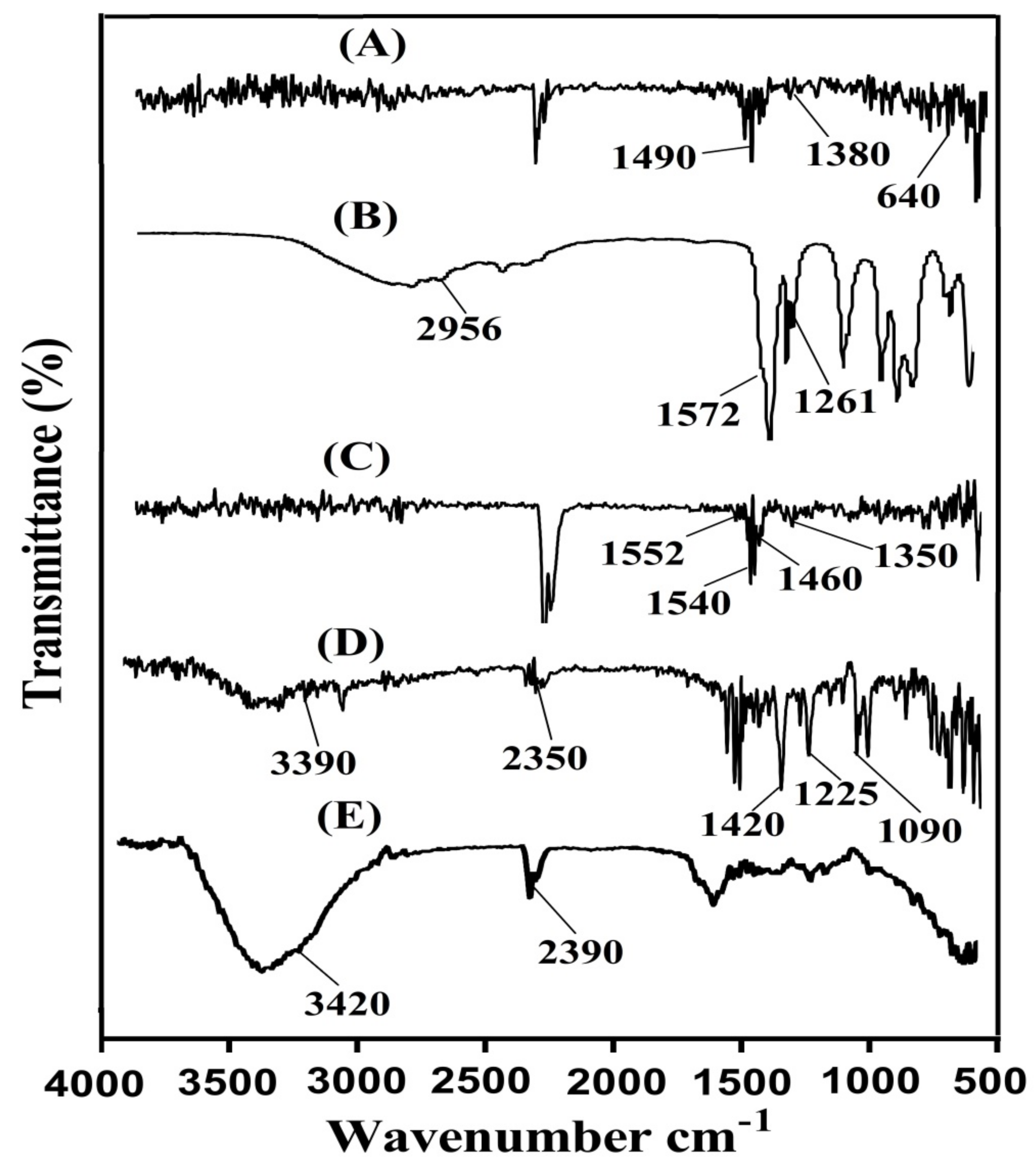
2.8. Fourier Transform Infrared Spectroscopy (FTIR)
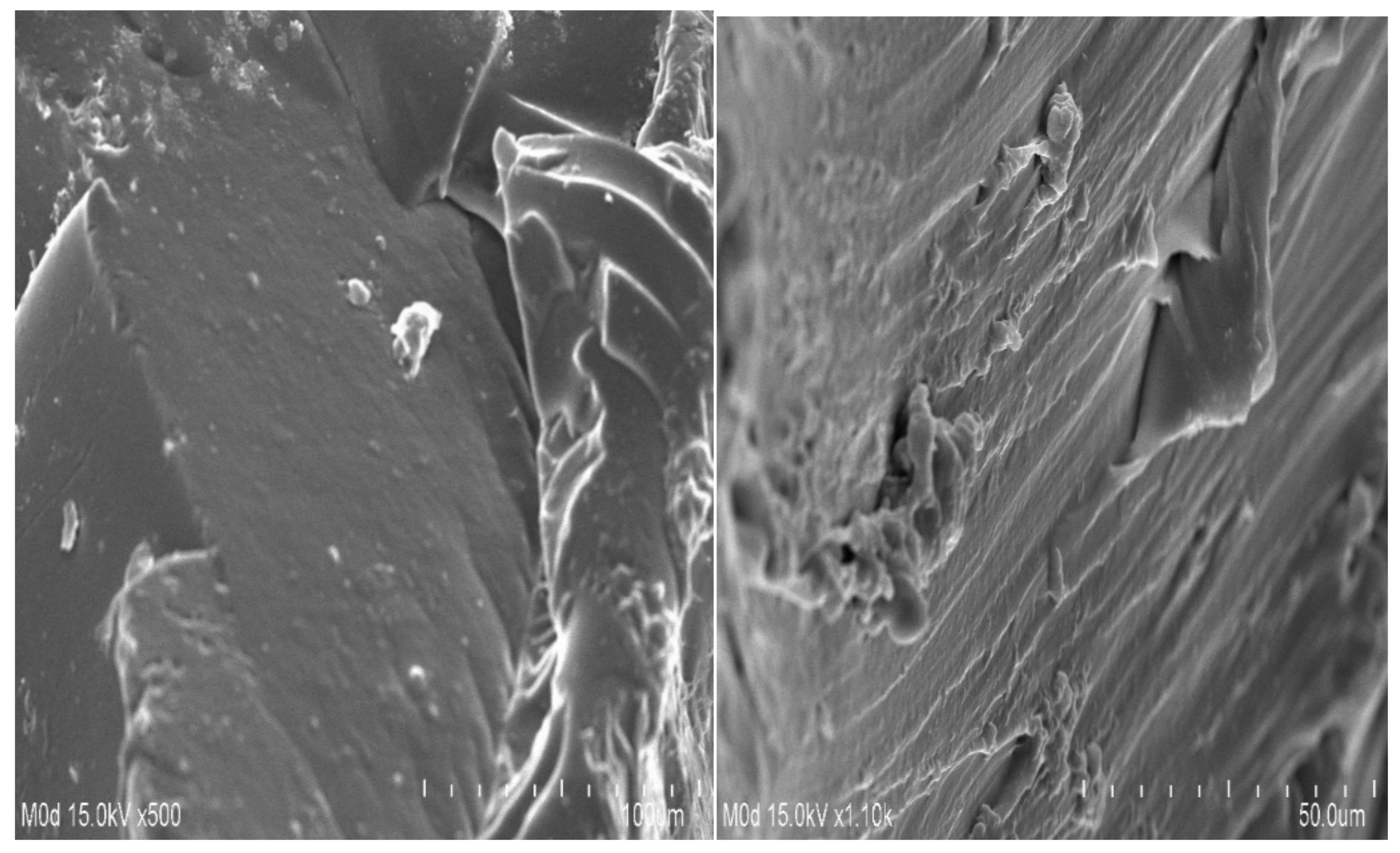
2.9. Scanning Electron Microscopy (SEM)
3. Materials and Methods
3.1. Materials
3.2. Synthesis of SPSPAA Hydrogels
3.3. Sol-Gel Analysis
3.4. Swelling Studies
3.5. Drug Loading
3.6. In-Vitro Drug Release and Kinetics Modeling
3.7. Thermal Analysis
3.8. Powder X-ray Diffraction (PXRD) Analysis
3.9. Fourier Transform Infrared Spectroscopy (FTIR)
3.10. Scanning Electron Microscopy (SEM)
4. Conclusions
Author Contributions
Funding
Institutional Review Board Statement
Informed Consent Statement
Data Availability Statement
Conflicts of Interest
References
- Goerne, T.M.L.; Garcia, M.G.L.; Grada, G.R.; Perez, I.O.; Lopez, E.G.; Lemus, M.A.A. Obtaining of sol-gel ketorolac-silica nanoparticles: Characterization and drug release kinetics. J. Nanomater. 2013, 2013. [Google Scholar] [CrossRef] [Green Version]
- Puglia, C.; Filosa, R.; Peduto, A.; de Caprariis, P.; Rizza, L.; Bonina, F.; Blasi, P. Evaluation of alternative strategies to optimize ketorolac transdermal delivery. AAPS PharmSciTech 2006, 7, E61–E69. [Google Scholar] [CrossRef] [PubMed] [Green Version]
- Wagh, P.; Mujumdar, A.; Naik, J.B. Preparation and characterization of ketorolac tromethamine-loaded ethyl cellulose micro-/nanospheres using different techniques. Part. Sci. Technol. 2019, 37, 347–357. [Google Scholar] [CrossRef]
- Alsarra, I.A.; Bosela, A.A.; Ahmed, S.M.; Mahrous, G.M. Proniosomes as a drug carrier for transdermal delivery of ketorolac. Eur. J. Pharm. Biopharm. 2005, 59, 485–490. [Google Scholar] [CrossRef]
- Mathew, S.T.; Devi, S.G.; Sandhya, K.V. Formulation and evaluation of ketorolac tromethamine-loaded albumin microspheres for potential intramuscular administration. AAPS PharmSciTech 2007, 8, E100–E108. [Google Scholar] [CrossRef] [Green Version]
- Yar, M.; Shahzad, S.; Siddiqi, S.A.; Mahmood, N.; Rauf, A.; Anwar, M.S.; Chaudhry, A.A.; Rehman, I.U. Triethyl orthoformate mediated a novel crosslinking method for the preparation of hydrogels for tissue engineering applications: Characterization and in vitro cytocompatibility analysis. Mat. Sci. Eng. C Mater. 2015, 56, 154–164. [Google Scholar] [CrossRef]
- Suhail, M.; Rosenholm, J.M.; Minhas, M.U.; Badshah, S.F.; Naeem, A.; Khan, K.U.; Fahad, M. Nanogels as drug-delivery systems: A comprehensive overview. Ther. Deliv. 2019, 10, 697–717. [Google Scholar] [CrossRef]
- Samanta, H.S.; Ray, S.K. Controlled release of tinidazole and theophylline from chitosan based composite hydrogels. Carbohydr. Polym. 2014, 106, 109–120. [Google Scholar] [CrossRef]
- Ullah, F.; Othman, M.B.H.; Javed, F.; Ahmad, Z.; Akil, H.M. Classification, processing and application of hydrogels: A review. Mat. Sci. Eng. C Mater. 2015, 57, 414–433. [Google Scholar] [CrossRef]
- Miyata, T.; Uragami, T.; Nakamae, K. Biomolecule-sensitive hydrogels. Adv. Drug Deliv. Rev. 2002, 54, 79–98. [Google Scholar] [CrossRef]
- Atta, S.; Khaliq, S.; Islam, A.; Javeria, I.; Jamil, T.; Athar, M.M.; Shafiq, M.I.; Ghaffar, A. Injectable biopolymer based hydrogels for drug delivery applications. Int. J. Biol. Macromol. 2015, 80, 240–245. [Google Scholar] [CrossRef]
- Buenger, D.; Topuz, F.; Groll, J. Hydrogels in sensing applications. Prog. Polym. Sci. 2012, 37, 1678–1719. [Google Scholar] [CrossRef]
- Khalid, I.; Ahmad, M.; Minhas, M.U.; Barkat, K. Synthesis and evaluation of chondroitin sulfate based hydrogels of loxoprofen with adjustable properties as controlled release carriers. Carbohydr. Polym. 2018, 181, 1169–1179. [Google Scholar] [CrossRef]
- Ali, L.; Ahmad, M.; Usman, M. Evaluation of cross-linked hydroxypropyl methylcellulose graft-methacrylic acid copolymer as extended release oral drug carrier. Cellul. Chem. Technol. 2015, 49, 143–151. [Google Scholar]
- Dergunov, S.A.; Nam, I.K.; Mun, G.A.; Nurkeeva, Z.S.; Shaikhutdinov, E.M. Radiation synthesis and characterization of stimuli-sensitive chitosan-polyvinyl pyrrolidone hydrogels. Radiat. Phys. Chem. 2005, 72, 619–623. [Google Scholar] [CrossRef]
- Atta, A.M. Swelling behaviors of polyelectrolyte hydrogels containing sulfonate groups. Polym. Adv. Technol. 2002, 13, 567–576. [Google Scholar] [CrossRef]
- Ali, A.E.H. Removal of heavy metals from model wastewater by using carboxymehyl cellulose/2-acrylamido-2-methyl propane sulfonic acid hydrogels. J. Appl. Polym. Sci. 2012, 123, 763–769. [Google Scholar]
- Ju, Y.H.; Huynh, L.H.; Kasim, N.S.; Guo, T.J.; Wang, J.H.; Fazary, A.E. Analysis of soluble and insoluble fractions of alkali and subcritical water treated sugarcane bagasse. Carbohydr. Polym. 2011, 83, 591–599. [Google Scholar] [CrossRef]
- Sohail, M.; Ahmad, M.; Minhas, M.U.; Ali, L.; Munir, A.; Khalid, I. Synthesis and characterization of graft pva composites for controlled delivery of valsartan. Lat. Am. J. Pharm. 2014, 33, 1237–1244. [Google Scholar]
- Qudah, Y.H.; Raafat, A.I.; Ali, A. Removal of some heavy metals from their aqueous solutions using 2-acrylamido-2-methyl-1-propane sulfonic acid/polyvinyl alcohol copolymer hydrogels prepared by gamma irradiation. Arab. J. Nucl. Sci. Appl. 2013, 46, 80–91. [Google Scholar]
- Peng, G.; Xu, S.M.; Peng, Y.; Wang, J.; Zheng, L.C. A new amphoteric superabsorbent hydrogel based on sodium starch sulfate. Biores. Technol. 2008, 99, 444–447. [Google Scholar] [CrossRef] [PubMed]
- Wu, W.; Wang, D.S. A fast pH-responsive IPN hydrogel: Synthesis and controlled drug delivery. React. Funct. Polym. 2010, 70, 684–691. [Google Scholar] [CrossRef]
- Murthy, P.S.K.; Mohan, Y.M.; Sreeramulu, J.; Raju, K.M. Semi-IPNs of starch and poly(acrylamide-co-sodium methacrylate): Preparation, swelling and diffusion characteristics evaluation. React. Funct. Polym. 2006, 66, 1482–1493. [Google Scholar] [CrossRef]
- Sullad, A.G.; Manjeshwar, L.S.; Aminabhavi, T.M. Novel pH-sensitive hydrogels prepared from the blends of poly(vinyl alcohol) with acrylic acid-graft-guar gum matrixes for isoniazid delivery. Ind. Eng. Chem. Res. 2010, 49, 7323–7329. [Google Scholar] [CrossRef]
- Rashid, H.; Ahmad, M.; Minhas, M.U.; Sohail, M.; Aamir, M.F. Synthesis and characterization of poly(hydroxyethyl methacrylate-co-methacrylic acid) cross linked polymeric network for the delivery of analgesic agent. J. Chem. Soc. Pak. 2015, 37, 999–1007. [Google Scholar]
- Sanli, O.; Ay, N.; Isiklan, N. Release characteristics of diclofenac sodium from poly(vinyl alcohol)/sodium alginate and poly(vinyl alcohol)-grafted-poly(acrylamide)/sodium alginate blend beads. Eur. J. Pharm. Biopharm. 2007, 65, 204–214. [Google Scholar] [CrossRef] [PubMed]
- Ravindra, S.; Mohan, Y.M.; Varaprasad, K.; Reddy, N.N.; Vimala, K.; Raju, K.M. Surfactant-modified poly(acrylamide-co-acrylamido propane sulphonic acid) hydrogels . Int. J. Polym. Mater. 2009, 58, 278–296. [Google Scholar] [CrossRef]
- Mutar, M.A.; Radia, N.D. Controlled release from crosslinked polyacrylic acid as drug delivery theophylline (NJC). Iraqi Nat. J. Chem. 2012, 45, 67–85. [Google Scholar]
- Korsmeyer, R.W.; Gurny, R.; Doelker, E.; Buri, P.; Peppas, N.A. Mechanisms of potassium chloride release from compressed, hydrophilic, polymeric matrices: Effect of entrapped air. J. Pharm. Sci. 1983, 72, 1189–1191. [Google Scholar] [CrossRef]
- Liu, H.; Gong, B.; Zhou, Y.; Sun, Z.; Wang, X.; Zhao, S. Preparation of high-capacity magnetic polystyrene sulfonate sodium material based on SI-ATRP method and its adsorption property research for sulfonamide antibiotics. BMC Chem. 2020, 14, 3. [Google Scholar] [CrossRef] [Green Version]
- Barkat, K.; Ahmad, M.; Minhas, M.U.; Khalid, I.; Nasir, B. Development and characterization of pH-responsive polyethylene glycol-co-poly(methacrylic acid) polymeric network system for colon target delivery of oxaliplatin: Its acute oral toxicity study. Adv. Polym. Technol. 2018, 37, 1806–1822. [Google Scholar] [CrossRef]
- Barkat, K.; Ahmad, M.; Minhas, M.U.; Khalid, I. Oxaliplatin-loaded crosslinked polymeric network of chondroitin sulfate-co-poly(methacrylic acid) for colorectal cancer: Its toxicological evaluation. J. Appl. Polym. Sci. 2017, 134, 45312. [Google Scholar] [CrossRef]
- Lee, C.T.; Huang, C.P.; Lee, Y.D. Synthesis and characterizations of amphiphilic poly(L-lactide)-grafted chondroitin sulfate copolymer and its application as drug carrier. Biomol. Eng. 2007, 24, 131–139. [Google Scholar] [CrossRef]
- De, R.; Lee, H.; Das, B. Exploring the interactions in binary mixtures of polyelectrolytes: Influence of mixture composition, concentration, and temperature on counterion condensation. J. Mol. Liq. 2018, 251, 94–99. [Google Scholar] [CrossRef]
- Moharram, M.A.; Khafagi, M.G. Application of FTIR spectroscopy for structural characterization of ternary poly(acrylic acid)-metal-poly(vinyl pyrrolidone) complexes. J. Appl. Polym. Sci. 2007, 105, 1888–1893. [Google Scholar] [CrossRef]
- Begum, M.Y.; Shaik, M.R.; Abbulu, K.; Sudhakar, M. Ketorolac tromethamine loaded liposomes of long alkyl chain lipids: Development, characterization and in vitro performance. Int. J. PharmTech Res. 2012, 4, 218–225. [Google Scholar]
- Aşık, M.D.; Uğurlu, N.; Yülek, F.; Tuncer, S.; Türk, M.; Denkbaş, E.B. Ketorolac tromethamine loaded chitosan nanoparticles as a nanotherapeutic system for ocular diseases. Hacet. J. Biol. Chem. 2013, 41, 81–86. [Google Scholar]
- Waghulde, M.; Mujumdar, A.; Naik, J. Preparation and characterization of miglitol-loaded Poly (d, l-lactide-co-glycolide) microparticles using high pressure homogenization-solvent evaporation method. Int. J. Polym. Mater. Polym. Biomater. 2019, 68, 198–207. [Google Scholar] [CrossRef]
- Khalid, I.; Ahmad, M.; Minhas, M.; Barkat, K.; Sohail, M. Cross-linked sodium alginate-g-poly(acrylic acid) structure: A potential hydrogel network for controlled delivery of loxoprofen sodium. Adv. Polym. Technol. 2018, 37, 985–995. [Google Scholar] [CrossRef]
- Suhail, M.; Wu, P.C.; Minhas, M.U. Using carbomer-based hydrogels for control the release rate of diclofenac sodium: Preparation and in vitro evaluation. Pharm. Base 2020, 13, 399. [Google Scholar] [CrossRef]
- Suhail, M.; Khan, A.; Rosenholm, J.M.; Minhas, M.U.; Wu, P.C. Fabrication and Characterization of Diclofenac Sodium Loaded Hydrogels of Sodium Alginate as Sustained Release Carrier. Gels 2021, 7, 10. [Google Scholar] [CrossRef] [PubMed]
- Ranjha, N.M.; Mudassir, J. Swelling and aspirin release study: Cross-linked pH-sensitive vinyl acetate-co-acrylic acid (VAC-co-AA) hydrogels. Drug Dev. Ind. Pharm. 2008, 34, 512–521. [Google Scholar] [CrossRef] [PubMed]
- Khan, S.; Ranjha, N.M. Effect of degree of cross-linking on swelling and on drug release of low viscous chitosan/poly(vinyl alcohol) hydrogels. Polym. Bull. 2014, 71, 2133–2158. [Google Scholar] [CrossRef]
- Peppas, N.A.; Sahlin, J.J. A simple equation for the description of solute release: III. Coupling of diffusion and relaxation. Int. J. Pharm. 1989, 57, 169–172. [Google Scholar] [CrossRef]
- Mahmood, A.; Ahmad, M.; Sarfraz, R.M.; Minhas, M.U.; Yaqoob, A. Formulation and in Vitro Evaluation of Acyclovir Loaded Polymeric Microparticles: A Solubility Enhancement Study. Acta Pol. Pharm. 2016, 73, 1311–1324. [Google Scholar]
- Khalid, I.; Ahmad, M.; Minhas, M.U.; Barkat, K. Preparation and characterization of alginate-PVA-based semi-IPN: Controlled release pH-responsive composites. Polym. Bull. 2018, 75, 1075–1099. [Google Scholar] [CrossRef]
- Sarfraz, R.M.; Khan, H.U.; Mahmood, A.; Ahmad, M.; Maheen, S.; Sher, M. Formulation and evaluation of mouth disintegrating tablets of atenolol and atorvastatin. Ind. J. Pharm. Sci. 2015, 77, 83–90. [Google Scholar] [CrossRef] [PubMed] [Green Version]


| Formulation | Sol Fraction | Gel Fraction | Dynamic Swelling Up to 36 h | Drug Loaded | |
|---|---|---|---|---|---|
| Code | (%) | (%) | pH 1.2 | pH 7.4 | (mg)/400 mg of Dry Gels |
| PSAF-1 | 14.88 | 85.12 | 3.16 ± 0.12 | 14.39 ± 0.24 | 170 ± 1.12 |
| PSAF-2 | 11.10 | 88.90 | 3.18 ± 0.14 | 16.33 ± 0.38 | 186 ± 0.92 |
| PSAF-3 | 9.24 | 90.76 | 3.24 ± 0.23 | 17.17 ± 0.21 | 198 ± 0.98 |
| PSAF-4 | 16.53 | 83.47 | 3.08 ± 0.08 | 9.35 ± 0.33 | 155 ± 1.06 |
| PSAF-5 | 14.88 | 85.12 | 3.16 ± 0.12 | 14.39 ± 0.24 | 170 ± 1.12 |
| PSAF-6 | 10.72 | 89.28 | 3.28 ± 0.38 | 15.98 ± 0.43 | 220 ± 1.22 |
| PSAF-7 | 14.88 | 85.12 | 3.16 ± 0.12 | 14.39 ± 0.24 | 170 ± 1.12 |
| PSAF-8 | 13.12 | 86.88 | 3.04 ± 0.27 | 11.16 ± 0.37 | 110 ± 1.18 |
| PSAF-9 | 10.59 | 89.41 | 2.80 ± 0.24 | 10.32 ± 0.32 | 94 ± 1.14 |
| Formulation | Korsmeyer‒Peppas Model | |
|---|---|---|
| Code | r2 | N |
| PSAF-1 | 0.8949 | 0.3396 |
| PSAF-2 | 0.9219 | 0.3684 |
| PSAF-3 | 0.9724 | 0.3927 |
| PSAF-4 | 0.9743 | 0.5299 |
| PSAF-5 | 0.8949 | 0.3396 |
| PSAF-6 | 0.8832 | 0.3465 |
| PSAF-7 | 0.8949 | 0.3396 |
| PSAF-8 | 0.9370 | 0.3818 |
| PSAF-9 | 0.9829 | 0.4420 |
| Formulation | Polymer | Monomer | Initiator | Cross-Linker |
|---|---|---|---|---|
| (SPS) | (AA) | (APS) | (MBA) | |
| Code | g/100 g | g/100 g | g/100 g | g/100 g |
| PSAF-1 | 0.50 | 25 | 0.5 | 0.50 |
| PSAF-2 | 0.75 | 25 | 0.5 | 0.50 |
| PSAF-3 | 1.00 | 25 | 0.5 | 0.50 |
| PSAF-4 | 0.50 | 20 | 0.5 | 0.50 |
| PSAF-5 | 0.50 | 25 | 0.5 | 0.50 |
| PSAF-6 | 0.50 | 30 | 0.5 | 0.50 |
| PSAF-7 | 0.50 | 25 | 0.5 | 0.50 |
| PSAF-8 | 0.50 | 25 | 0.5 | 0.75 |
| PSAF-9 | 0.50 | 25 | 0.5 | 1.00 |
Publisher’s Note: MDPI stays neutral with regard to jurisdictional claims in published maps and institutional affiliations. |
© 2021 by the authors. Licensee MDPI, Basel, Switzerland. This article is an open access article distributed under the terms and conditions of the Creative Commons Attribution (CC BY) license (https://creativecommons.org/licenses/by/4.0/).
Share and Cite
Suhail, M.; Fang, C.-W.; Minhas, M.U.; Wu, P.-C. Preparation, Characterization, Swelling Potential, and In-Vitro Evaluation of Sodium Poly(Styrene Sulfonate)-Based Hydrogels for Controlled Delivery of Ketorolac Tromethamine. Pharmaceuticals 2021, 14, 350. https://0-doi-org.brum.beds.ac.uk/10.3390/ph14040350
Suhail M, Fang C-W, Minhas MU, Wu P-C. Preparation, Characterization, Swelling Potential, and In-Vitro Evaluation of Sodium Poly(Styrene Sulfonate)-Based Hydrogels for Controlled Delivery of Ketorolac Tromethamine. Pharmaceuticals. 2021; 14(4):350. https://0-doi-org.brum.beds.ac.uk/10.3390/ph14040350
Chicago/Turabian StyleSuhail, Muhammad, Chih-Wun Fang, Muhammad Usman Minhas, and Pao-Chu Wu. 2021. "Preparation, Characterization, Swelling Potential, and In-Vitro Evaluation of Sodium Poly(Styrene Sulfonate)-Based Hydrogels for Controlled Delivery of Ketorolac Tromethamine" Pharmaceuticals 14, no. 4: 350. https://0-doi-org.brum.beds.ac.uk/10.3390/ph14040350