Article

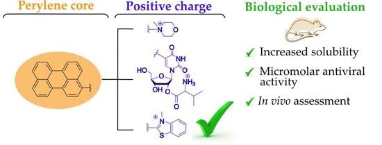
Cationic Perylene Antivirals with Aqueous Solubility for Studies In Vivo

by
Anna A. Shtro
1,*,†,
Anzhelika V. Garshinina
1,†,
Vera A. Alferova
2,3,†,
Polina N. Kamzeeva
2,
Viktor P. Volok
2,4,
Ekaterina S. Kolpakova
4,
Timofei D. Nikitin
2,
Alexey A. Chistov
2,
Evgeny S. Belyaev
5,
Vladimir A. Korshun
2,
Liubov I. Kozlovskaya
4,6,* and
Andrey V. Aralov
2,*
1
Smorodintsev Research Institute of Influenza, 197376 Saint Petersburg, Russia
2
Shemyakin-Ovchinnikov Institute of Bioorganic Chemistry, Russian Academy of Sciences, 117997 Moscow, Russia
3
Gause Institute of New Antibiotics, 119021 Moscow, Russia
4
Chumakov Scientific Center for Research and Development of Immune-and-Biological Products, Russian Academy of Sciences (Institute of Poliomyelitis), 108819 Moscow, Russia
5
Frumkin Institute of Physical Chemistry and Electrochemistry, Russian Academy of Science, 119071 Moscow, Russia
6
Institute of Translational Medicine and Biotechnology, Sechenov Moscow State Medical University, 119991 Moscow, Russia
*
Authors to whom correspondence should be addressed.
These authors contributed equally to this work.
Pharmaceuticals 2022, 15(10), 1178; https://0-doi-org.brum.beds.ac.uk/10.3390/ph15101178
Submission received: 30 August 2022 / Revised: 17 September 2022 / Accepted: 19 September 2022 / Published: 22 September 2022
(This article belongs to the Special Issue Antiviral Drugs 2021)

Abstract

:
Perylene-based compounds are attracting significant attention due to their high broad-spectrum antiviral activity against enveloped viruses. Despite unambiguous results of in vitro studies and high selectivity index, the poor water solubility of these compounds prevented in vivo evaluation of their antiviral properties. In this work, we synthesized a series of compounds with a perylene pharmacophore bearing positively charged substituents to improve the aqueous solubility of this unique type of antivirals. Three types of charged groups were introduced: (1) quaternary morpholinium salts (3a–b); (2) a 2′-O-l-valinyl-uridine hydrochloride residue (8), and (3) a 3-methylbenzothiazolium cation (10). The synthesized compounds were evaluated based both on antiviral properties in vitro (CHIKV, SARS-CoV-2, and IAV) and on solubility in aqueous media. Compound 10 has the greatest aqueous solubility, making it preferable for pre-evaluation by intragastrical administration in a mouse model of lethal influenza pneumonia. The results indicate that the introduction of a positively charged group is a viable strategy for the design of drug candidates with a perylene scaffold for in vivo studies.

Graphical Abstract

1. Introduction

Epidemics of diseases associated with various viruses, such as Influenza A (H1N1) and Ebola viruses, as well as middle east respiratory syndrome coronavirus (MERS-CoV) and severe acute respiratory syndrome coronaviruses 1 and 2 (SARS-CoV-1 and 2) occurred in 2009, 2014, 2012, 2003 and 2019, respectively. Re-emerging EBOVs and new H1N1, MERS, and SARS viruses pose a great threat to humanity and easily cross country borders. The traditional “one-bug – one drug” approach is insufficient for addressing the issue of re-emerging and new viral pathogens, and few drugs are currently available to control epidemic viral diseases [1,2,3,4,5]. Thus, it is critical to develop a class of broad-spectrum antiviral agents.
Antivirals with broad-spectrum activity can be divided into two categories: direct-acting drugs and host-targeting antivirals that engage host-cell machinery important for maintaining various stages of the viral life cycle. Representatives of the former class usually demonstrate an option for repurposing, high selectivity, and low cytotoxicity. Direct-acting antivirals include inhibitors of virus attachment (broadly neutralizing antibodies (bNAbs) [6,7] and sialidase fusion protein DAS181 [8]), virus entry (small molecules 5705213/7402683 [9] and the peptide enfuvirtide [10]), replication (galidesivir [11], favipiravir [12] ribavirin [13], and remdesivir [14,15]), viral assembly and budding (verdinexor [16] and FGI-104 [17]). Among them, replication inhibitors, the most promising candidates for the development of optimal broad-spectrum agents, still exhibit a number of disadvantages such as low plasma concentrations resulting from rapid renal elimination for favipiravir [18,19,20], poor selectivity and toxicity leading to undesirable side effects for ribavirin [21], and poor oral bioavailability and short half-life for remdesivir [22]. As a separate prospective subclass, yet affecting only enveloped viruses, agents targeting the viral envelope (LJ001 [23] and JL118/JL122 [24], derivatives of aromatic methyldiene rhodanine and oxazolidine-2,4-dithione, respectively) can be distinguished.
Enveloped viruses are a large class of pathogens responsible for multiple serious diseases. The membrane of the viral particle and its rearrangements during the replicative cycle appear to be a promising target for the development of broad-spectrum antiviral agents [25]. Moreover, the viral envelope is significantly more vulnerable than the mammalian cellular membrane because the virus possesses no membrane repair systems. Additionally, targeting the viral envelope is attractive due to the anticipated rate of resistance development being negligible. The viral lipid envelope originates from the host cell’s membrane and is thus not affected by natural viral diversity and variability [26].
One of the most potent classes of viral fusion inhibitors with a broad spectrum of activity against enveloped viruses are so-called rigid amphipathic fusion inhibitors (RAFIs) [27]. These compounds contain a perylene moiety and were initially discovered as nucleoside analogs. However, further studies revealed that their structures could be significantly simplified, and neither the nucleobase nor the carbohydrate moiety were vital for their antiviral action [28]. Thorough structural studies led to the development of perylene-based compounds with remarkable subnanomolar in vitro activity against a wide range of enveloped viruses [29]. The mechanism of viral fusion inhibition by perylene derivatives is not yet completely understood. There are two hypotheses, with consensus on the target – the outer lipid membrane of the virion. The first hypothesis implies intercalation of the drug into the virion lipid membrane, leading to mechanical disturbance of its rheological properties [8]. Alternatively, drug-mediated photoinduced generation of singlet oxygen, causing oxidation of unsaturated lipids in the virion membrane, was proposed [30]. Taking into account the rather controversial results of mechanistic studies, perylene derivatives presumably inhibit viral fusion in a structure-dependent combination of these two modes of action.
Despite outstanding in vitro activities, RAFIs have not been tested in vivo. The most challenging problem is the low aqueous solubility of perylene-based compounds. In this work, we aimed to improve the solubility of RAFI by introducing various positively charged moieties. We synthesized a concise series of novel perylene derivatives and assessed their antiviral properties, cytotoxicity, and solubility in aqueous media. Dramatically improved water solubility allowed studying the protective properties of the most promising compound against the influenza virus in mice.

2. Results and Discussion
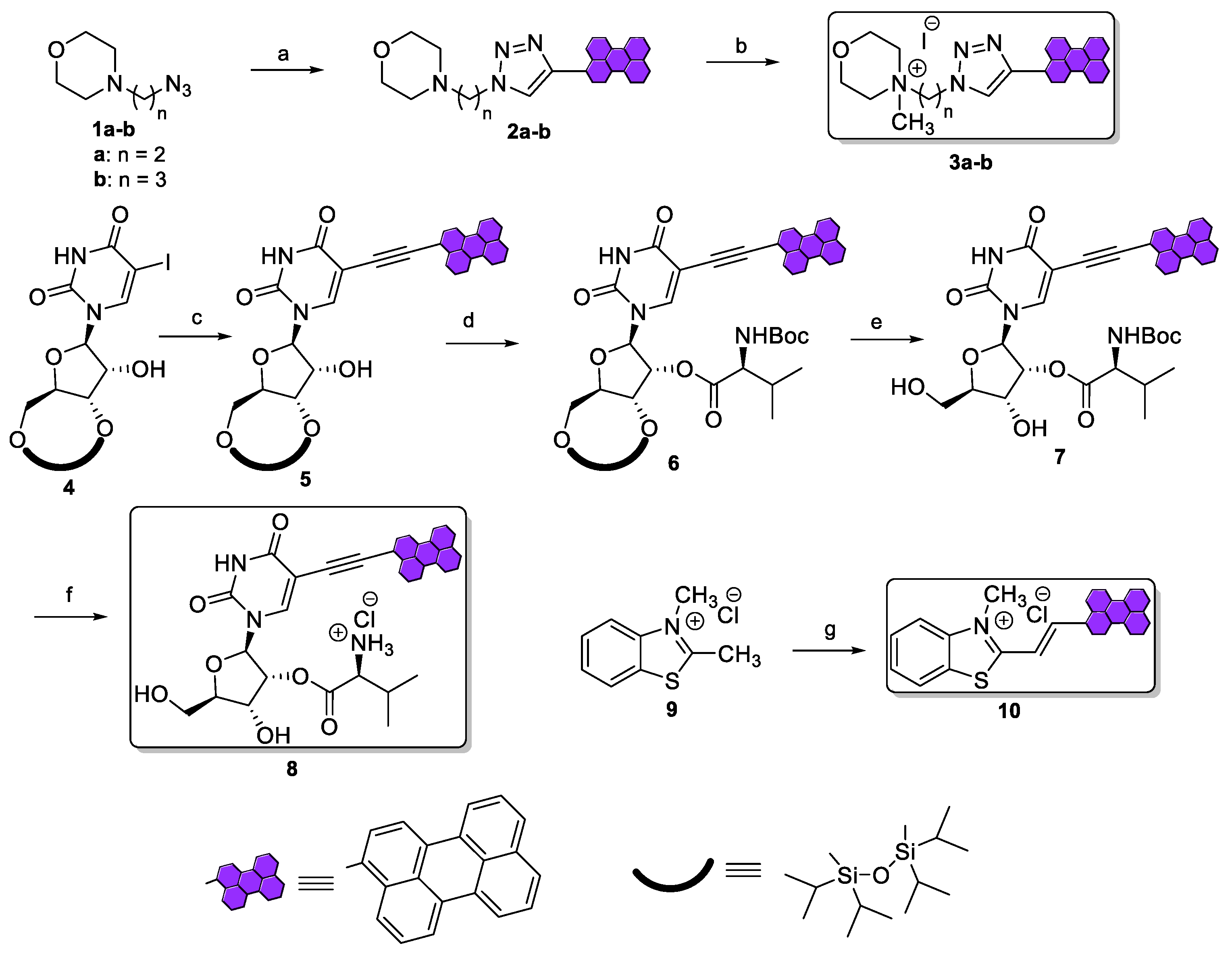
2.1. Chemistry

The synthesis of soluble positively charged perylene-based analogs was carried out using a number of biocompatible moieties bearing a positive charge, such as N-methylmorpholinium [31], O-l-valinyl ester hydrochloride [32], and benzothiazolium [33] residues, and introduced them via well-established synthetic procedures (Scheme 1).
First, N-(ω-azidoalkyl)morpholine 1 was condensed with 3-ethynylperylene under Cu(I)-catalyzed azide-alkyne cycloaddition (CuAAC) conditions [28]. Then, the N atom of the morpholine ring was alkylated with methyl iodide, affording target positively charged derivatives 3a and 3b, with an ethyl (n = 2) or propyl (n = 3) bridge, respectively. Nucleoside derivative 8 was prepared in four steps. 3′,5′-O-silyl protected 5-iodouridine 4 [34] was subjected to Sonogashira coupling with 3-ethynylperylene, yielding 5. Condensation of 5 with N-Boc-l-valine using DCC as the condensing agent afforded 6, which was then subjected to deblocking of 3′- and 5′-hydroxyls groups by treatment with TBAF·3H2O. The Boc protecting group from 7 was removed by hydrolysis in acidic conditions, yielding positively charged nucleoside derivative 8. Finally, 2,3-dimethylbenzo[d]thiazol-3-ium chloride 9 and perylene-3-carbaldehyde, as the carbanion-forming and the carbonyl-containing electrophilic components, respectively, were subjected to a Knoevenagel condensation reaction [35] by refluxing in acetic anhydride to give final compound 10.
NMR spectroscopy and HRMS spectrometry were used to confirm the structure and elemental composition of intermediates and target compounds (see Experimental and Figures S1–S9). The purity of target positively charged derivatives 3a–b, 8, and 10 was >90%, as determined by HPLC (Figures S11–S14).

2.2. In Vitro Activity, Aqueous Solubility, and Serum Stability Assessment

Since determining the scope of applicability of perylene-based compounds as antivirals was the main purpose of this study, we first evaluated in vitro antiviral activity (Table 1) of target compounds 3a–b, 8, 10 and synthetic intermediates 2a–b, and 5–7 against a number of enveloped RNA viruses pathogenic for humans: Chikungunya virus (CHIKV), severe acute respiratory syndrome-related coronavirus 2 (SARS-CoV-2), and influenza A virus (IAV).
Charged compounds retained antiviral activity in the low micromolar-submicromolar concentration range against CHIKV and SARS-CoV-2 with almost no evidence of cytotoxicity. At the same time, some uncharged intermediates did not demonstrate any activity against CHIKV (2b, 6) or SARS-CoV-2 (2b, 5, 6) in concentrations studied. In particular, replacement of the ethyl linker between perylenyltriazolyl and morpholine residues with a propyl one led to complete loss of activity against CHIKV and SARS-CoV-2 in the morpholine series, but the corresponding N-methylated derivative 3b had activity against these two viruses. Surprisingly, 5′,3′-O-protected 5-(perylen-3-ylethynyl)uridine derivative 5 did not show any signs of efficacy against SARS-CoV-2, and the introduction of an additional lipophilic substituent at the 2′-OH function of the ribose residue also caused a complete loss of activity of 6 against CHIKV. Meanwhile, removal of the siloxane 5′,3′-O-protective group restored the activity of 7, suggesting the importance of maintaining the lipophilic perylene residue and polar, in our case hydroxy, groups, for maximum activity. Activity against IAV was substantially lower compared to CHIKV and SARS-CoV-2, surprisingly decreasing with the introduction of the positive charge in the morpholine series (3a–b vs. 2a–b). In contrast, positively charged nucleoside derivative 8 has slightly better activity compared to its non-charged intermediates 6 and 7. Finally, all charged derivatives showed comparable-to-higher activity against CHIKV and SARS-CoV-2 vs. positive control N-hydroxycytidine (NHC) [36]. All compounds had lower activity against IAV vs. Tamiflu as the control antiviral.
Then, an assessment of aqueous solubility of the positively charged compounds was performed to select a candidate for studies in a model of lethal influenza pneumonia in mice (Table 2).
Positively charged derivatives 3a–b and 10 were found to have substantial solubility in water, whereas positively charged and uncharged nucleoside analogs 8 and aUY11 had poor and no solubility, respectively. Among the candidates synthesized, 8 had the highest antiviral activity against influenza A virus; however, its low aqueous solubility limits evaluation in vivo. Since, among the remaining three derivatives, compound 10 had the highest potency and solubility, it was chosen for subsequent evaluation in a mouse model of influenza A virus infection.
Next, human intestinal absorption (cHIA) [38] and blood–brain barrier permeability (clogBB) [39] were estimated using predictive QSAR models [40]. Most of the compounds were able to penetrate the blood–brain barrier effectively, and derivative 10 demonstrated the highest level of intestinal absorption, and even distribution between the brain and blood, which may be useful in the treatment of various brain lesion complications associated with influenza (Table S1) [41,42].
Since in vivo evaluation of the leader compound in mouse model involves intragastric administration, stability in stomach acid-mimicking 0.16 M aqueous HCl was assessed. The compound was stable, with slight signs of decomposition as determined by thin-layer chromatography (TLC) for at least 4 h, which exceeds the average residence time of drugs in the stomach [43]. Then, stability in fetal bovine serum (FBS) was evaluated. In vitro FBS half-life (t1/2) was found to be 167 min (Figure S10), which is comparable to approved drugs and other drug candidates [44,45,46].

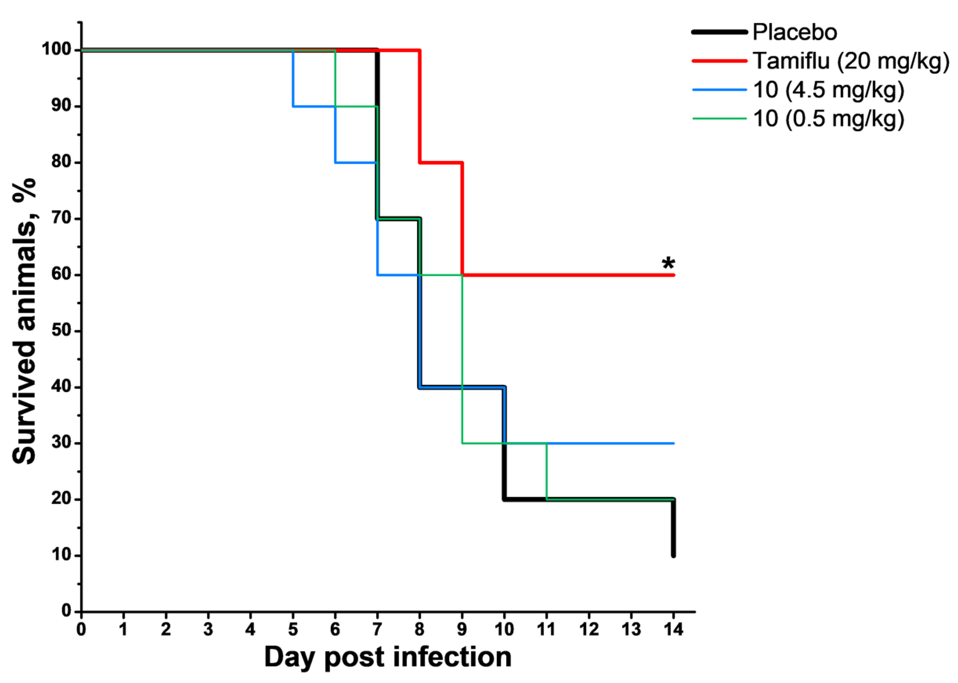
2.3. In Vivo Studies

Protective efficacy for compound 10 was assessed by the ability to prevent the development of lethal influenza pneumonia in mice. BALB/c mice were treated intragastrically with the compound in two different dosing regimens, placebo and comparative drug Tamiflu [47,48] for the period between one day prior and three days after intranasal infection with IAV. Clinical symptom development and body weight were observed daily. Survival rates and curves are presented in Table 3 and Figure 1, respectively, and body weight changes in Figure 2.
Infection with the IAV strain A/California/7/09/MA caused mouse mortality starting from day 5 post infection (p.i.). By the end of the experiment, mortality was 90% in the Placebo group and 40% (p = 0.015) in the comparison group (Tamiflu), which corresponded to a protection index (PI) of 56% and was consistent with available information for this strain [49]. The use of compound 10 in a dose of 0.5 or 4.5 mg/kg did not cause a significant reduction in the mortality rate compared to the Placebo group (p > 0.05).
Influenza virus infection induced a reduction in body weight of all animals in all groups, reaching a maximum of 26.2% on day 9 p.i. and returning to the initial values on day 14 p.i. in the Placebo group. In the comparison group (Tamiflu), the reduction was less pronounced and reached 24.0% on day 10 p.i. The experimental group treated with 4.5 mg/kg of 10 showed less weight loss (up to 21.3% on day 9 p.i.) and faster weight recovery compared to the Placebo group.
To conclude, mice treated with 10 (4.5 mg/kg) showed a lower mortality rate and faster weight recovery, although the differences were not significant.

3. Materials and Methods

3.1. General Information

All solvents and reagents were commercially available and used without purification. Spots on silica gel-precoated TLC plates (60 μm, F254, Merck, Germany) were visualized using UV light (254 and 365 nm), and column chromatography was performed on silica gel (0.040−0.063 mm, Merck, Germany). 1H and 13C NMR spectra were recorded on a Bruker Avance III 600 spectrometer (Bruker BioSpin, Germany) at 600 and 150 MHz, respectively. DMSO-d6 (2.50/39.52 ppm for 1H and 13C spectra, respectively) was used as the internal standard. Signals were described as follows: chemical shift (multiplicity (s = singlet, d = doublet, t = triplet, m = multiplet), coupling constant(s) (Hz), number of protons) for 1H spectra, and chemical shift (number of carbons) for 13C spectra. HRMS spectra were registered in the positive ion mode on a Thermo Scientific LTQ Orbitrap hybrid instrument (Thermo Electron Corp., Bremen, Germany) with direct sample infusion. HPLC and LCMS analysis was performed using an Agilent 1260 Infinity II instrument (with a single quadrupole 6125B mass detector), equipped with a ZORBAX RRHT SB-C18, 2.1 × 50 mm, 1.8 μm column. The column was eluted with a linear gradient (5–95% in 10 min) of LCMS-grade acetonitrile (with 0.1 vol. % formic acid) in LCMS-grade water (with 0.1 vol. % formic acid) at a 0.4 mL/min flow rate. 4-(2-Azidoethyl)morpholine 1a, 4-(3-azidopropyl)morpholine 1b, 1-[3′,5′-O-(tetraisopropyldisiloxan-1,3-diyl)-β-d-ribofuranosyl]-5-iodouracil 4 and 3-ethynylperylene were synthesized according to literature [28,34,50]. An aqueous solution of 2,3-dimethylbenzo[d]thiazol-3-ium iodide [51] was eluted through a column filled with Bio-Rad AG® MP-1M Anion Exchange Resin (chloride form, 10 eq.) to afford 2,3-dimethylbenzo[d]thiazol-3-ium chloride.

3.2. Synthesis

3.2.1. General Procedure for CuAAC Reaction

The corresponding azido-containing morpholine derivative 1 (0.5 mmol), 3-ethynylperylene (138 mg, 0.5 mmol), TBTA (14 mg, 0.025 mmol), CuI (5 mg, 0.025 mmol), and DMSO (5 mL) were mixed, degassed, and then stirred for 16 h at room temperature under argon atmosphere. To the reaction solution, water (15 mL) was added, and the precipitate formed was filtered. The filter cake was dissolved in ethyl acetate (30 mL) and sequentially washed with 0.1 M aqueous EDTA solution (10 mL) and brine (30 mL). The organic layer was dried over Na2SO4 and concentrated in vacuo. The crude product was purified by column chromatography on silica gel (0–2% MeOH in CH2Cl2), yielding the corresponding product.

4-(2-(4-(Perylen-3-yl)-1H-1,2,3-triazol-1-yl)ethyl)morpholine 2a

Yield 190 mg (88%); amorphous brown solid. 1H NMR (600 MHz, DMSO-d6): δ 8.62 (s, 1H), 8.46–8.37 (m, 5H), 7.84–7.77 (m, 3H), 7.63–7.53 (m, 3H), 4.62 (t, J = 6.2 Hz, 2H), 3.60–3.55 (m, 4H), 2.87 (t, J = 6.2 Hz, 2H), 2.50–2.46 (m, 4H, overlapping with DMSO). 13C NMR (150 MHz, DMSO-d6): δ 145.0, 134.0, 131.4, 130.6, 130.3 (2C), 130.0, 128.2, 127.9, 127.8, 127.7, 127.5, 127.4, 127.1, 126.8, 126.7, 125.3, 124.5, 120.8, 120.8, 120.7, 120.3, 66.0 (2C), 57.1, 52.8 (2C), 46.5. HRMS (ESI) m/z: calcd for C28H25N4O+ [M + H]+: 433.2023; found 433.2020.

4-(3-(4-(Perylen-3-yl)-1H-1,2,3-triazol-1-yl)propyl)morpholine 2b

Yield 192 mg (86%); amorphous brown solid. 1H NMR (600 MHz, DMSO-d6): δ 8.65 (s, 1H), 8.49–8.34 (m, 5H), 7.86–7.75 (m, 3H), 7.66–7.51 (m, 3H), 4.58–4.46 (m, 2H), 3.65–3.52 (m, 4H), 2.43–2.30 (m, 6H), 2.17–2.06 (m, 2H). 13C NMR (150 MHz, DMSO-d6): δ 145.3, 134.1, 131.4, 130.6, 130.4, 130.4, 130.1, 128.3, 128.0, 127.9, 127.7, 127.6, 127.5, 127.2, 126.9, 126.9, 125.5, 124.2, 120.9, 120.9, 120.8, 120.4, 66.0 (2C), 54.8, 53.1 (2C), 47.8, 26.5. HRMS (ESI) m/z: calcd for C29H27N4O+ [M + H]+: 447.2179; found 447.2173.

3.2.2. General Procedure for Methylation with Methyl Iodide

To a solution of 2 (0.3 mmol) in CH2Cl2 (30 mL), methyl iodide (0.62 mL, 3 mmol) was added in one portion, and the reaction mixture was stirred at 35 °C overnight. The precipitate that formed was filtered, sequentially washed with Et2O (3 × 10 mL), CH2Cl2 (3 × 10 mL), and 10% MeOH in CH2Cl2 (10 mL, v/v), dried over Na2SO4, and concentrated in vacuo, yielding the corresponding methylated product.

4-Methyl-4-(2-(4-(perylen-3-yl)-1H-1,2,3-triazol-1-yl)ethyl)morpholin-4-ium iodide 3a

Yield 122 mg (71%); amorphous brown solid. HPLC rt 7.5 min (Figure S11). 1H NMR (600 MHz, DMSO-d6): δ 9.24 (s, 1H), 8.60 (d, J = 7.9 Hz, 1H), 8.60 (d, J = 7.9 Hz, 1H), 8.55 (t, J = 7.8 Hz, 1H), 8.49 (d, J = 7.4 Hz, 1H), 7.94 (d, J = 8.1 Hz, 1H), 7.91 (d, J = 8.1 Hz, 1H), 7.87 (d, J = 7.8 Hz, 1H), 7.69–7.61 (m, 3H), 7.57 (d, J = 8.2 Hz, 1H), 4.91–4.86 (m, 2H), 4.17 (s, 3H), 3.63–3.58 (m, 4H), 2.99–2.93 (m, 2H), 2.56–2.52 (m, 4H). 13C NMR (150 MHz, DMSO-d6): δ 139.7, 134.2, 134.0, 132.5, 131.3, 131.1, 130.7, 129.6, 129.4, 129.2, 128.7, 128.4, 128.0, 127.5, 127.1, 127.0, 123.9, 122.3, 121.8, 121.5, 119.9, 118.6, 66.1 (2C), 55.9, 52.6 (2C), 50.3, 38.1. HRMS (ESI) m/z: calcd for C29H27N4O+ [M-I]+: 447.2179; found 447.2182.

4-Methyl-4-(3-(4-(Perylen-3-yl)-1H-1,2,3-triazol-1-yl)propyl)morpholin-4-ium iodide 3b

Yield 115 mg (65%); amorphous brown solid. HPLC rt 7.5 min (Figure S12). 1H NMR (600 MHz, DMSO-d6): δ 8.73 (s, 1H), 8.49–8.45 (m, 3H), 8.44–8.40 (m, 2H), 7.86–7.81 (m, 3H), 7.64 (t, J = 8 Hz, 1H), 7.60–7.56 (m, 2H), 4.62 (t, J = 6.8 Hz, 2H), 3.98–3.91 (m, 4H), 3.64–3.58 (m, 2H), 3.52–3.46 (m, 4H), 3.18 (s, 3H), 2.50–2.44 (m, 2H). 13C NMR (150 MHz, DMSO-d6): δ 145.5, 134.1, 131.4, 130.7, 130.6, 130.4, 130.1, 128.3, 128.1, 128.0, 127.6 (2C), 127.5, 127.2, 126.9, 126.9, 125.5, 124.3, 121.0 (2C), 120.9, 120.4, 59.7 (2C), 59.0 (2C), 48.5, 46.6, 39.4, 22.0. HRMS (ESI) m/z: calcd for C30H29N4O+ [M-I]+: 461.2336; found 461.2345.

3.2.3. 3′,5′-O-(Tetraisopropyldisiloxan-1,3-diyl)-5-(perylen-3-ylethynyl)uridine 5

A mixture of 3′,5′-O-(tetraisopropyldisiloxane-1,3-diyl)-5-iodouridine 4 (0.74 g, 1.2 mmol), 3-ethynylperylene (0.42 g, 1.5 mmol), Pd(PPh3)4 (0.14 g, 0.12 mmol) and CuI (0.05 g, 0.24 mmol) in DMF (20 mL) was evacuated, and then Et3N (0.34 mL, 2.4 mmol) was added under argon atmosphere. The reaction mixture was stirred at room temperature in the dark overnight and then partitioned between EtOAc (200 mL) and water (100 mL). The organic layer was sequentially washed with a 5% aqueous solution of citric acid (20 mL), a 1% EDTA solution (20 mL) and brine (100 mL) and evaporated in vacuo. The resulting brown oil was purified by column chromatography on silica gel (0→50% EtOAc in CH2Cl2), yielding 5 (0.76 g, 83%) as an orange foam. 1H NMR (600 MHz, DMSO-d6): δ 11.85 (br s, 1H), 8.45 (d, J = 7.6 Hz, 1H), 8.42–8.38 (m, 2H), 8.37 (d, J = 8.0 Hz, 1H), 8.26 (d, J = 8.2 Hz, 1H), 7.85–7.80 (m, 3H), 7.68–7.64 (m, 2H), 7.57 (t, J = 7.8 Hz, 2H), 6.11 (d, J = 7.8 Hz, 1H), 5.94 (d, J = 6.0 Hz, 1H), 4.41–4.36 (m, 1H), 4.17–4.12 (m, 1H), 4.07 (dd, J = 13.1 Hz, J = 2.0 Hz, 1H), 3.94 (dd, J = 13.1 Hz, J = 2.1 Hz, 1H), 3.77–3.73 (m, 1H), 1.08–0.89 (m, 28H). 13C NMR (150 MHz, DMSO-d6): δ 161.3, 149.3, 143.5, 134.1, 133.8, 131.2, 130.9, 130.3, 130.0, 129.7, 128.6, 128.2, 127.7 (2C), 127.5, 126.9, 126.8, 125.6, 121.6, 121.3, 121.2, 120.2, 119.2, 97.7, 90.7, 88.0, 82.9, 79.1, 74.3, 74.2, 59.9, 17.1 (2C), 17.1, 17.0, 16.8, 16.7 (2C), 16.6, 12.8, 12.3, 12.0, 11.8. HRMS (ESI) m/z: calcd for C43H49N2O7Si2+ [M + H]+: 761.3073; found 761.3036.

3.2.4. 2′-O-(N-Boc-l-valinyl)-3′,5′-O-(tetraisopropyldisiloxane-1,3-diyl)-5-(perylen-3-ylethynyl)uridine 6

To a solution of 5 (0.71 g, 0.93 mmol) and Boc-l-valine (0.61 g, 2.79 mmol) in dry CH2Cl2 (50 mL) 4-dimethylaminopyridine (DMAP) (0.01 g, 0.08 mmol) was added followed by N,N’-dicyclohexylcarbodiimide (DCC) (0.77 g, 3.72 mmol). The reaction mixture was stirred at room temperature for 4 h, washed with 5% solution of citric acid (20 mL), and concentrated in vacuo. The residue was purified by column chromatography on silica gel (50→100% CH2Cl2 in hexane), yielding 6 (0.77 g, 87%) as an orange foam. 1H NMR (600 MHz, DMSO-d6): δ 8.46 (br s, 1H), 8.36 (d, J = 8.2Hz, 1H), 8.24 (d, J = 7.4Hz, 1H), 8.21 (d, J = 7.6Hz, 1H), 8.19 (d, J = 7.6Hz, 1H), 8.13 (d, J = 7.9Hz, 1H), 7.87 (s, 1H), 7.72–7.67 (m, 3H), 7.61 (dd, J = 7.4 Hz, J = 8.2Hz, 1H), 7.51–7.47 (m, 2H), 6.28 (d, J = 4.7 Hz, 1H), 5.65–5.59 (m, 1H), 4.90 (d, J = 9.3 Hz, 1H), 4.60–4.51 (m, 1H), 4.30 (dd, J = 9.3 Hz, J = 3.8 Hz, 1H), 4.15 (dd, J = 3.7 Hz, J = 12.8 Hz, 1H), 4.10 (dd, J = 3.0 Hz, J = 12.8 Hz, 1H), 3.90–3.86 (m, 1H), 2.13–2.04 (m, 1H), 1.43 (s, 9H), 1.13–1.01 (m, 28H), 0.96 (d, J = 6.8 Hz, 3H), 0.81 (d, J = 6.8 Hz, 3H). 13C NMR (150 MHz, DMSO-d6): δ 171.2, 160.6, 155.4, 148.6, 141.8, 134.6, 134.6, 132.2, 131.3, 131.0, 130.8, 130.7, 128.4 (2C), 128.0, 127.5 (2C), 126.6, 126.5, 126.4, 120.9, 120.8, 120.6, 119.6, 119.4, 100.7, 93.2, 85.7, 81.4, 79.9, 77.0 (1C overlaps with CDCl3), 73.0 (2C), 60.7, 58.2, 30.7, 28.2 (3C), 19.3, 17.4 (3C), 17.2, 16.9, 16.8 (3C), 16.6, 13.5, 13.0, 12.7, 12.3. HRMS (ESI) m/z: calcd for C53H66N3O10Si2+ [M + H]+: 960.4281; found 960.4279.

3.2.5. 2′-O-(N-Boc-l-valinyl)-5-(perylen-3-ylethynyl)uridine 7

To a solution of 6 (0.72 g, 0.75 mmol) in THF (20 mL), tetra-n-butylammonium fluoride trihydrate (TBAF·3H2O) (0.52 g, 1.65 mmol) was added, and the resulting solution was allowed to stand for 1 h at room temperature. After concentration in vacuo the residue was purified by column chromatography on silica gel (0→2% MeOH in CH2Cl2), yielding 7 (0.48 g, 90%) as a brown foam. 1H NMR (600 MHz, DMSO-d6): δ 11.90 (br s, 1H), 8.47 (d, J = 7.3 Hz, 1H), 8.44–8.37 (m, 2H), 8.38 (d, J = 8.0 Hz, 1H), 8.31–8.28 (m, 2H), 7.87–7.83 (m, 2H), 7.74 (d, J = 7.8 Hz, 1H), 7.69 (t, J = 7.9 Hz, 1H), 7.60–7.56 (m, 2H), 7.14 (d, J = 8.5 Hz, 1H), 6.20 (d, J = 4.8 Hz, 1H), 5.89 (d, J = 4.7 Hz, 1H), 5.29–5.25 (m, 1H), 5.21–5.16 (m, 1H), 4.19–4.14 (m, 1H), 4.01–3.96 (m, 1H), 3.92–3.88 (m, 1H), 3.73–3.66 (m, 2H), 2.02–1.95 (m, 1H), 1.38 (s, 9H), 0.81 (d, J = 6.8 Hz, 3H), 0.74 (d, J = 6.8 Hz, 3H). 13C NMR (150 MHz, DMSO-d6): δ 170.6, 161.2, 155.7, 148.9, 143.7, 134.1, 133.7, 131.1, 130.9, 130.5, 130.0, 129.7, 128.6, 128.3, 127.8, 127.7, 127.5, 126.9, 126.9, 125.7, 121.6, 121.3, 121.2, 120.2, 119.3, 98.1, 90.6, 88.2, 84.7, 83.4, 78.3, 77.0, 72.9, 60.1, 58.7, 29.2 (3C), 28.0, 19.0, 17.1. HRMS (ESI) m/z: calcd for C41H40N3O9+ [M + H]+: 718.2759; found 718.2763.

3.2.6. 2′-O-l-Valinyl-5-(perylen-3-ylethynyl)uridine 8

To a solution of 7 (0.43 g, 0.60 mmol) in a mixture of MeOH:CH2Cl2 (30 mL, 1:100, v/v), 6.6 M HCl in 1,4-dioxane (1 mL) were added, and the resulting brown solution was stirred for 12 h at room temperature. The precipitate that formed was filtered, washed with CH2Cl2 (2×10 mL), and dried in vacuo, affording 8 (0.29 g, 73%) as a brown amorphous solid. HPLC rt 7.6 min (Figure S13). 1H NMR (600 MHz, DMSO-d6): δ 8.89 (s, 1H), 8.53–8.47 (m, 7H), 8.47–8.44 (m, 2H), 8.20 (d, J = 8.5 Hz, 1H), 7.91 (d, J = 7.9 Hz, 1H), 7.90–7.86 (m, 2H), 7.71 (t, J = 8.0 Hz, 1H), 7.63–7.59 (m, 2H), 7.31 (s, 1H), 6.34 (d, J = 4.2 Hz, 1H), 5.52–5.49 (m, 1H), 4.31–4.28 (m, 1H), 4.10–4.07 (m, 1H), 3.82–3.78 (m, 1H), 3.75–3.69 (m, 2H), 1.91–1.84 (m, 1H), 0.74 (d, J = 7.0 Hz, 3H), 0.62 (d, J = 7.0 Hz, 3H). 13C NMR (150 MHz, DMSO-d6): δ 171.2, 167.6, 153.4, 153.2, 139.4, 134.0, 132.3, 131.1, 130.9, 130.1, 129.6, 128.8, 128.3, 128.3, 128.0, 127.9, 127.5, 127.0, 127.0, 124.9, 124.4, 121.8, 121.5, 121.3, 120.3, 107.1, 104.0, 86.3, 86.0, 77.6, 73.6, 60.4, 57.3, 28.7, 17.5, 17.3. HRMS (ESI) m/z: calcd for C36H32N3O7+ [M-Cl-]+: 618.2235; found 618.2225.

3.2.7. 3-Methyl-2-(2-(perylen-3-yl)vinyl)benzo[d]thiazol-3-ium chloride 10

A solution of perylene-3-carbaldehyde (0.28 g, 1.0 mmol) and 2,3-dimethylbenzo[d]thiazol-3-ium chloride 9 (0.24 g, 1.2 mmol) in Ac2O (5 mL) was heated at 150 °C for 2 h. Then, water (2 mL) was added, and the resulting dark purple solution was heated at 50 °C for 30 min. After concentration in vacuo, the residue was purified by column chromatography on silica gel (0→12% MeOH in CH2Cl2), yielding 10 (0.27 g, 54%) as a dark purple amorphous solid. HPLC rt 9 min (Figure S14). 1H NMR (600 MHz, DMSO-d6): δ 8.75 (d, J = 15.5 Hz, 1H), 8.56 (d, J = 7.4 Hz, 1H), 8.52 (d, J = 7.9 Hz, 2H), 8.49–8.42 (m, 3H), 8.36 (d, J = 8.4 Hz, 1H), 8.26 (d, J = 8.4 Hz, 1H), 8.16 (d, J = 15.5 Hz, 1H), 7.92–7.83 (m, 3H), 7.81–7.73 (m, 2H), 7.62 (t, J = 7.8 Hz, 1H), 7.58 (t, J = 7.7 Hz, 1H), 4.40 (s, 3H). 13C NMR (150 MHz, DMSO-d6): δ 171.2, 143.5, 141.9, 134.8, 133.9, 132.5, 131.0, 129.9, 129.8, 129.5, 129.5, 129.3, 128.5, 128.3 (2C), 128.2, 128.1, 127.9, 127.3, 127.1, 127.0, 124.0, 123.1, 122.7, 121.8, 121.4, 120.5, 116.7, 115.1, 36.3. HRMS (ESI) m/z: calcd for C30H20NS+ [M-Cl-]+: 426.1311; found 426.1299.

3.3. Solubility

The samples were dissolved in DMSO, diluted in series, and photometrically analyzed. The average molar absorption coefficient was calculated from the obtained optical densities of calibration solutions at the absorption maxima. Solubility values were determined as previously described [52] by adding weighed amounts of compounds to distilled water followed by sonication (30 min), centrifugation (12,000 rpm, 10 min), and photometry.

3.4. In Vitro Biological Studies

3.4.1. Cells and Viruses

The green monkey kidney (Vero) cell line originated from WHO Biologicals, Switzerland (10–87), and the Madin-Darby canine kidney (MDCK) cell line was obtained from the American Type Culture Collection (ATCC CCL-34).
Viruses and strains used in the present work were severe acute respiratory syndrome-related coronavirus 2 (SARS-CoV-2, strain PIK35, GISAID EPI_ISL_428852), Chikungunya virus (CHIKV, strain Nic, GenBank IDs MN271691-2) and influenza A virus (H1N1, strain A/PR/8/34, ATCC VR-95).

3.4.2. Methods

Cell viability Assay in Vero Cells

Two-fold dilutions of the compounds (starting from 100 μM) were prepared in DMEM (Chumakov FSC R&D IBP RAS, Russia) and added to confluent cell monolayers. After 5 days of incubation (37 °C, 5% CO2), the cultural supernatant was substituted with a resazurin solution (25 μg/mL), followed by 4 h incubation (37 °C, 5% CO2). Then, 20 µL of 10% SDS were added to stop the reaction. Fluorescence intensity was measured with Promega GloMax-Multi Detection System (Ex525/Em580–640). Cells treated with dilutions of the compounds and DMSO, but not resazurin, were used to subtract the background fluorescence. All experimental procedures were performed in two replicates. Fluorescence curves were analyzed with Microsoft Excel 2013. 50% Cytotoxic concentrations (CC50) were calculated.

Cell Viability Assay in MDCK Cells

Compound solutions were prepared in DMSO (20 mg/mL), and then 2-fold dilutions in cultural medium were prepared starting from 1000 μg/mL. One-day MDCK cell monolayers (over 95% confluency) in 96-well plates (6×10⁵ cells/well) were washed twice with serum-free alpha-MEM (Thermo-Fisher Scientific, USA) and treated with 100 μL of compound dilutions in 2 replicates. The plates were then incubated for 24 h at 37 °C in a CO2 incubator. Cell viability was assessed using the tetrazolium test (MTT, ApplyChem) [53]. Optical density was measured on a Wallac 1420 Victor2 microplate reader (Perkin Elmer, Ramsey, MN, USA) at 535 nm, then CC50 values were calculated.

IAV Virus Yield Reduction Assay

Compound solutions were prepared in DMSO (20 mg/mL). The compounds were weighed in 2 mg amounts and dissolved in 100 μL of DMSO. Then, a series of 3-fold dilutions was prepared, starting from 300 µg/mL, in a cultural medium. The MDCK cell monolayer was treated with compound dilutions and incubated for 1 h in a CO2 incubator at 37 °C. Untreated cells were used as negative control. Afterward, the virus was added (MOI 1), and cells were incubated for 24 h at 37 °C in a CO2 incubator. After incubation, the culture medium was collected, and viral yields were determined by titration. In brief, a series of 10-fold dilutions (10−7–10−1) of viral supernatants were prepared in a cultural medium and applied to daily MDCK cell monolayers. Infected cells were incubated for 72 h at 37 °C in a CO2 incubator. The presence of the virus in the culture supernatants was determined using the hemagglutination assay with a 1% suspension of chicken erythrocytes [54]. Viral titer was calculated according to the Reed-and-Muench method and was expressed as 50% tissue infectious doses (TID50) per 100 μL of volume. Inhibition (%) was calculated by comparison with non-treated cells, and EC50 was calculated from inhibitory curves.

SARS-CoV-2 and CHIKV Cytopathic Effect (CPE) Inhibition Test

The procedure was described previously [55]. In brief, two-fold dilutions of the compounds were prepared in Dulbecco MEM (Chumakov FSC R&D IBP RAS, Russia) and mixed 1:1 with a virus suspension containing 50–200 TCID50 and incubated for 1 h at 37 °C in a CO2 incubator. The final concentration series started from 100 μM. Then, compound-virus mixtures were added to confluent Vero cell monolayers in 2 replicates. After incubation at 37 °C in a CO2-incubator for 5 days, CPE signs (cell death) were assessed via microscope, and 50% effective concentration (EC50) (compound concentration required to decrease CPE by 50%) was calculated using the Karber method [56].

Plasma Stability Test

In vitro stability of 10 was studied in Gibco FBS (Thermo-Fisher Scientific, USA) by following the reported procedure with minor modifications [57]. A 10 mM solution of the compound in DMSO (20 μL) was added to 80% FBS in 0.05 M PBS (pH 7.4) at 37 °C to yield the final concentration of 200 μM. The assays were carried out in triplicate in a water bath at 37 °C. Samples (100 μL) were taken at 0, 15, 30, 45, 60, 90, 120, 180, 240, and 300 min and added to 400 μL acetonitrile in order to deproteinize the plasma. The samples were then shaken for 1 min and centrifuged at 37 °C at 15,000 rpm for 5 min. The supernatants were analyzed by HPLC (detection at 530 nm). AUC values were determined and averaged from three independent experiments. In vitro serum half-life (t1/2) was calculated using the equation t1/2 = 0.693/b, where b is the slope found by linear fitting of the negative natural logarithm of the fraction remaining (AUC0-300/AUC0) vs. incubation time.

3.5. In Vivo Biological Studies

3.5.1. Animals

Female BALB/c mice (5–7 weeks old) were obtained from the “Stolbovaya” branch of the Scientific center for biomedical technology of the Federal medical-biological agency (Moscow region, Russia). All animal procedures were performed in accordance with the Helsinki declaration and Russian animal protection regulations.

3.5.2. Virus

Influenza A virus strain A/California/7/09/MA adapted to mice by 8 consecutive passages and accumulated on chicken embryos was used.

3.5.3. Design of the Experiment to Determine the Antiviral Activity of Compounds

The test compound (0.5 and 4.5 mg/kg), placebo (PBS as a negative control), and reference drug Tamiflu (20 mg/kg, oseltamivir phosphate as a positive control) were administered once per day to the animals using a gastric tube in a 0.2 mL volume according to the therapeutic and prophylactic scheme (24 and 1 h before infection and 1, 2, and 3 days post infection). The mice were intranasally infected with 5 MLD50 of virus in a 50 μL volume. The animals were observed and weighed daily for 14 days post infection. Kaplan–Meier survival curves were built with GraphPad Prism 6.0. The Mantel–Cox test was used for posterior pairwise comparisons with the Placebo group. The protection index was calculated as a ratio (Mc − Me)/Mc × 100%, where Mc and Me are mortality rates in the placebo and experimental groups, respectively, at the end of the experiment (14 days post infection). Differences in weight between the groups were established with one-way ANOVA with Dunnett’s test for pairwise comparisons.

4. Conclusions

While previously reported data on perylene RAFIs have demonstrated remarkable subnanomolar antiviral activity in vitro, low water solubility and a rather controversial mode of action have discouraged in vivo studies. The introduction of positively charged moieties led to a dramatic improvement in the solubility of these compounds without a significant reduction in efficacy in vitro. We demonstrated 22% protective activity and accelerated weight recovery upon intragastrical administration of the most promising compound 10 in a mouse model of lethal influenza pneumonia, but the differences were not significant. This is the first example of in vivo efficacy assessment of RAFIs, paving the way for the development of water-soluble perylene-based antiviral agents and their preclinical studies.

Supplementary Materials

The following supporting information can be downloaded at: https://0-www-mdpi-com.brum.beds.ac.uk/article/10.3390/ph15101178/s1, Figures S1–S14: 1H and 13C NMR spectra of the compounds, Plasma stability evaluation, HPLC trace for compounds; Table S1: Predicted ADMET profiles of the synthesized compounds.

Author Contributions

Conceptualization, A.A.S., L.I.K. and A.V.A.; Funding acquisition, A.V.A.; Investigation, A.V.G., P.N.K., V.P.V., E.S.K., T.D.N., A.A.C. and E.S.B.; Methodology, A.A.S., V.A.K., L.I.K. and A.V.A.; Project administration, A.V.A.; Visualization, V.A.A.; Writing—original draft, A.A.S., V.A.A., V.A.K., L.I.K. and A.V.A. All authors have read and agreed to the published version of the manuscript.

Funding

This work was supported by the Russian Science Foundation (grant 22-25-00161).

Institutional Review Board Statement

All animal procedures were performed in accordance with the Helsinki declaration and Russian animal protection regulations (protocol N 37/2, 14 April 2021).

Informed Consent Statement

Not applicable.

Data Availability Statement

Not applicable.

Conflicts of Interest

The authors declare no conflict of interest. The funders had no role in the design of the study; in the collection, analyses, or interpretation of data; in the writing of the manuscript; or in the decision to publish the results.

References

  1. Chan, J.F.W.; Chan, K.-H.; Kao, R.Y.T.; To, K.K.W.; Zheng, B.-J.; Li, C.P.Y.; Li, P.T.W.; Dai, J.; Mok, F.K.Y.; Chen, H.; et al. Broad-Spectrum Antivirals for the Emerging Middle East Respiratory Syndrome Coronavirus. J. Infect. 2013, 67, 606–616. [Google Scholar] [CrossRef]
  2. Raveh, A.; Delekta, P.C.; Dobry, C.J.; Peng, W.; Schultz, P.J.; Blakely, P.K.; Tai, A.W.; Matainaho, T.; Irani, D.N.; Sherman, D.H.; et al. Discovery of Potent Broad Spectrum Antivirals Derived from Marine Actinobacteria. PLoS ONE 2013, 8, e82318. [Google Scholar] [CrossRef] [PubMed]
  3. Carossino, M.; Thiry, E.; de la Grandière, A.; Barrandeguy, M.E. Novel Vaccination Approaches against Equine Alphavirus Encephalitides. Vaccine 2014, 32, 311–319. [Google Scholar] [CrossRef] [PubMed]
  4. Zhu, J.-D.; Meng, W.; Wang, X.-J.; Wang, H.-C.R. Broad-Spectrum Antiviral Agents. Front. Microbiol. 2015, 6, 517. [Google Scholar] [CrossRef] [PubMed]
  5. Geraghty, R.; Aliota, M.; Bonnac, L. Broad-Spectrum Antiviral Strategies and Nucleoside Analogues. Viruses 2021, 13, 667. [Google Scholar] [CrossRef]
  6. Balazs, A.B.; Chen, J.; Hong, C.M.; Rao, D.S.; Yang, L.; Baltimore, D. Antibody-Based Protection against HIV Infection by Vectored Immunoprophylaxis. Nature 2012, 481, 81–84. [Google Scholar] [CrossRef]
  7. Johnson, P.R.; Schnepp, B.C.; Zhang, J.; Connell, M.J.; Greene, S.M.; Yuste, E.; Desrosiers, R.C.; Reed Clark, K. Vector-Mediated Gene Transfer Engenders Long-Lived Neutralizing Activity and Protection against SIV Infection in Monkeys. Nat. Med. 2009, 15, 901–906. [Google Scholar] [CrossRef] [PubMed]
  8. Hedlund, M.; Aschenbrenner, L.M.; Jensen, K.; Larson, J.L.; Fang, F. Sialidase-based Anti–Influenza Virus Therapy Protects against Secondary Pneumococcal Infection. J. Infect. Dis. 2010, 201, 1007–1015. [Google Scholar] [CrossRef] [PubMed]
  9. Elshabrawy, H.A.; Fan, J.; Haddad, C.S.; Ratia, K.; Broder, C.C.; Caffrey, M.; Prabhakar, B.S. Identification of a Broad-Spectrum Antiviral Small Molecule against Severe Acute Respiratory Syndrome Coronavirus and Ebola, Hendra, and Nipah Viruses by Using a Novel High-Throughput Screening Assay. J. Virol. 2014, 88, 4353–4365. [Google Scholar] [CrossRef] [PubMed]
  10. Eckert, D.M.; Kim, P.S. Mechanisms of Viral Membrane Fusion and Its Inhibition. Annu. Rev. Biochem. 2001, 70, 777–810. [Google Scholar] [CrossRef] [Green Version]
  11. Warren, T.K.; Wells, J.; Panchal, R.G.; Stuthman, K.S.; Garza, N.L.; Van Tongeren, S.A.; Dong, L.; Retterer, C.J.; Eaton, B.P.; Pegoraro, G.; et al. Protection against Filovirus Diseases by a Novel Broad-Spectrum Nucleoside Analogue BCX4430. Nature 2014, 508, 402–405. [Google Scholar] [CrossRef]
  12. Oestereich, L.; Lüdtke, A.; Wurr, S.; Rieger, T.; Muñoz-Fontela, C.; Günther, S. Successful Treatment of Advanced Ebola Virus Infection with T-705 (Favipiravir) in a Small Animal Model. Antivir. Res. 2014, 105, 17–21. [Google Scholar] [CrossRef]
  13. Parker, W.B. Metabolism and Antiviral Activity of Ribavirin. Virus Res. 2005, 107, 165–171. [Google Scholar] [CrossRef] [PubMed]
  14. Warren, T.K.; Jordan, R.; Lo, M.K.; Ray, A.S.; Mackman, R.L.; Soloveva, V.; Siegel, D.; Perron, M.; Bannister, R.; Hui, H.C.; et al. Therapeutic Efficacy of the Small Molecule GS-5734 against Ebola Virus in Rhesus Monkeys. Nature 2016, 531, 381–385. [Google Scholar] [CrossRef] [PubMed]
  15. Grein, J.; Ohmagari, N.; Shin, D.; Diaz, G.; Asperges, E.; Castagna, A.; Feldt, T.; Green, G.; Green, M.L.; Lescure, F.-X.; et al. Compassionate Use of Remdesivir for Patients with Severe COVID-19. N. Engl. J. Med. 2020, 382, 2327–2336. [Google Scholar] [CrossRef] [PubMed]
  16. Perwitasari, O.; Johnson, S.; Yan, X.; Howerth, E.; Shacham, S.; Landesman, Y.; Baloglu, E.; McCauley, D.; Tamir, S.; Tompkins, S.M.; et al. Verdinexor, a Novel Selective Inhibitor of Nuclear Export, Reduces Influenza a Virus Replication in Vitro and in Vivo. J. Virol. 2014, 88, 10228–10243. [Google Scholar] [CrossRef]
  17. Kinch, M.S.; Yunus, A.S.; Lear, C.; Mao, H.; Chen, H.; Fesseha, Z.; Luo, G.; Nelson, E.A.; Li, L.; Huang, Z.; et al. FGI-104: A Broad-Spectrum Small Molecule Inhibitor of Viral Infection. Am. J. Transl. Res. 2009, 1, 87–98. [Google Scholar] [CrossRef]
  18. Mentré, F.; Taburet, A.-M.; Guedj, J.; Anglaret, X.; Keïta, S.; de Lamballerie, X.; Malvy, D. Dose Regimen of Favipiravir for Ebola Virus Disease. Lancet Infect. Dis. 2015, 15, 150–151. [Google Scholar] [CrossRef]
  19. Nguyen, T.H.T.; Guedj, J.; Anglaret, X.; Laouénan, C.; Madelain, V.; Taburet, A.-M.; Baize, S.; Sissoko, D.; Pastorino, B.; Rodallec, A.; et al. Favipiravir Pharmacokinetics in Ebola-Infected Patients of the JIKI Trial Reveals Concentrations Lower than Targeted. PLoS Negl. Trop. Dis. 2017, 11, e0005389. [Google Scholar] [CrossRef]
  20. Agrawal, U.; Raju, R.; Udwadia, Z.F. Favipiravir: A New and Emerging Antiviral Option in COVID-19. Med. J. Armed Forces India 2020, 76, 370–376. [Google Scholar] [CrossRef]
  21. Müller, W.E.G.; Maidhof, A.; Taschner, H.; Zahn, R.K. Virazole (1-β-d-Ribofuranosyl-1,2,4-triazole-3-carboxamide; A Cytostatic Agent. Biochem. Pharmacol. 1977, 26, 1071–1075. [Google Scholar] [CrossRef]
  22. Jorgensen, S.C.J.; Kebriaei, R.; Dresser, L.D. Remdesivir: Review of Pharmacology, Pre-clinical Data, and Emerging Clinical Experience for COVID-19. Pharmacotherapy 2020, 40, 659–671. [Google Scholar] [CrossRef] [PubMed]
  23. Wolf, M.C.; Freiberg, A.N.; Zhang, T.; Akyol-Ataman, Z.; Grock, A.; Hong, P.W.; Li, J.; Watson, N.F.; Fang, A.Q.; Aguilar, H.C.; et al. A Broad-Spectrum Antiviral Targeting Entry of Enveloped Viruses. Proc. Natl. Acad. Sci. USA 2010, 107, 3157–3162. [Google Scholar] [CrossRef] [PubMed]
  24. Vigant, F.; Lee, J.; Hollmann, A.; Tanner, L.B.; Akyol Ataman, Z.; Yun, T.; Shui, G.; Aguilar, H.C.; Zhang, D.; Meriwether, D.; et al. A Mechanistic Paradigm for Broad-Spectrum Antivirals That Target Virus-Cell Fusion. PLoS Pathog. 2013, 9, e1003297. [Google Scholar] [CrossRef] [PubMed]
  25. Vigant, F.; Santos, N.C.; Lee, B. Broad-Spectrum Antivirals against Viral Fusion. Nat. Rev. Microbiol. 2015, 13, 426–437. [Google Scholar] [CrossRef]
  26. Palombi, N.; Brai, A.; Gerace, M.; Di Maria, S.; Orofino, F.; Corelli, F. Viral Envelope Membrane: A Special Entry Pathway and a Promising Drug Target. Curr. Med. Chem. 2021, 28, 6957–6976. [Google Scholar] [CrossRef] [PubMed]
  27. St. Vincent, M.R.; Colpitts, C.C.; Ustinov, A.V.; Muqadas, M.; Joyce, M.A.; Barsby, N.L.; Epand, R.F.; Epand, R.M.; Khramyshev, S.A.; Valueva, O.A.; et al. Rigid Amphipathic Fusion Inhibitors, Small Molecule Antiviral Compounds against Enveloped Viruses. Proc. Natl. Acad. Sci. USA 2010, 107, 17339–17344. [Google Scholar] [CrossRef] [PubMed]
  28. Aralov, A.V.; Proskurin, G.V.; Orlov, A.A.; Kozlovskaya, L.I.; Chistov, A.A.; Kutyakov, S.V.; Karganova, G.G.; Palyulin, V.A.; Osolodkin, D.I.; Korshun, V.A. Perylenyltriazoles Inhibit Reproduction of Enveloped Viruses. Eur. J. Med. Chem. 2017, 138, 293–299. [Google Scholar] [CrossRef]
  29. Slesarchuk, N.A.; Khvatov, E.V.; Chistov, A.A.; Proskurin, G.V.; Nikitin, T.D.; Lazarevich, A.I.; Ulanovskaya, A.A.; Ulashchik, E.A.; Orlov, A.A.; Jegorov, A.V.; et al. Simplistic Perylene-Related Compounds as Inhibitors of Tick-Borne Encephalitis Virus Reproduction. Bioorg. Med. Chem. Lett. 2020, 30, 127100. [Google Scholar] [CrossRef]
  30. Vigant, F.; Hollmann, A.; Lee, J.; Santos, N.C.; Jung, M.E.; Lee, B. The Rigid Amphipathic Fusion Inhibitor DUY11 Acts through Photosensitization of Viruses. J. Virol. 2014, 88, 1849–1853. [Google Scholar] [CrossRef] [Green Version]
  31. Kumari, M.; Singh, U.K.; Singh, P.; Patel, R. Effect of N-Butyl-N-methyl-morpholinium Bromide Ionic Liquid on the Conformation Stability of Human Serum Albumin. ChemistrySelect 2017, 2, 1241–1249. [Google Scholar] [CrossRef]
  32. Pierra, C.; Amador, A.; Benzaria, S.; Cretton-Scott, E.; D’Amours, M.; Mao, J.; Mathieu, S.; Moussa, A.; Bridges, E.G.; Standring, D.N.; et al. Synthesis and Pharmacokinetics of Valopicitabine (Nm283), an Efficient Prodrug of the Potent Anti-Hcv Agent 2‘-C-Methylcytidine. J. Med. Chem. 2006, 49, 6614–6620. [Google Scholar] [CrossRef]
  33. Sigmundová, I.; Zahradník, P.; Magdolen, P.; Bujdáková, H. Synthesis and Study of New Antimicrobial Benzothiazoles Substituted on Heterocyclic Ring. Arkivoc 2008, 2008, 183–192. [Google Scholar] [CrossRef]
  34. Matsuda, A.; Takenuki, K.; Tanaka, M.; Sasaki, T.; Ueda, T. Nucleosides and Nucleotides. 97. Synthesis of New Broad Spectrum Antineoplastic Nucleosides, 2′-Deoxy-2′-methylidenecytidine (DMDC) and Its Derivatives. J. Med. Chem. 1991, 34, 812–819. [Google Scholar] [CrossRef] [PubMed]
  35. Klochko, O.P.; Fedyunyayeva, I.A.; Khabuseva, S.U.; Semenova, O.M.; Terpetschnig, E.A.; Patsenker, L.D. Benzodipyrrolenine-Based Biscyanine Dyes: Synthesis, Molecular Structure and Spectroscopic Characterization. Dyes Pigm. 2010, 85, 7–15. [Google Scholar] [CrossRef]
  36. Sheahan, T.P.; Sims, A.C.; Zhou, S.; Graham, R.L.; Pruijssers, A.J.; Agostini, M.L.; Leist, S.R.; Schäfer, A.; Dinnon, K.H.; Stevens, L.J.; et al. An Orally Bioavailable Broad-Spectrum Antiviral Inhibits SARS-CoV-2 in Human Airway Epithelial Cell Cultures and Multiple Coronaviruses in Mice. Sci. Transl. Med. 2020, 12, eabb5883. [Google Scholar] [CrossRef]
  37. Colpitts, C.C.; Ustinov, A.V.; Epand, R.F.; Epand, R.M.; Korshun, V.A.; Schang, L.M. 5-(Perylen-3-yl)ethynyl-arabino-uridine (AUY11), an Arabino-Based Rigid Amphipathic Fusion Inhibitor, Targets Virion Envelope Lipids to Inhibit Fusion of Influenza Virus, Hepatitis C Virus, and Other Enveloped Viruses. J. Virol. 2013, 87, 3640–3654. [Google Scholar] [CrossRef]
  38. Radchenko, E.V.; Dyabina, A.S.; Palyulin, V.A.; Zefirov, N.S. Prediction of Human Intestinal Absorption of Drug Compounds. Russ. Chem. Bull. 2016, 65, 576–580. [Google Scholar] [CrossRef]
  39. Dyabina, A.S.; Radchenko, E.V.; Palyulin, V.A.; Zefirov, N.S. Prediction of Blood-Brain Barrier Permeability of Organic Compounds. Dokl. Biochem. Biophys. 2016, 470, 371–374. [Google Scholar] [CrossRef]
  40. ADMET Prediction Service. Available online: http://Qsar.Chem.Msu.Ru/Admet/ (accessed on 26 August 2022).
  41. Chen, Q.; Li, P.; Li, S.; Xiao, W.; Yang, S.; Lu, H. Brain Complications with Influenza Infection in Children. Behav. Brain Sci. 2020, 10, 129–152. [Google Scholar] [CrossRef] [Green Version]
  42. Hosseini, S.; Wilk, E.; Michaelsen-Preusse, K.; Gerhauser, I.; Baumgärtner, W.; Geffers, R.; Schughart, K.; Korte, M. Long-Term Neuroinflammation Induced by Influenza A Virus Infection and the Impact on Hippocampal Neuron Morphology and Function. J. Neurosci. 2018, 38, 3060–3080. [Google Scholar] [CrossRef] [PubMed]
  43. Alqahtani, M.S.; Kazi, M.; Alsenaidy, M.A.; Ahmad, M.Z. Advances in Oral Drug Delivery. Front. Pharmacol. 2021, 12, 618411. [Google Scholar] [CrossRef] [PubMed]
  44. Smith, D.A.; Beaumont, K.; Maurer, T.S.; Di, L. Relevance of Half-Life in Drug Design: Miniperspective. J. Med. Chem. 2018, 61, 4273–4282. [Google Scholar] [CrossRef] [PubMed]
  45. Lima, L.M.; Zapata-Sudo, G.; da Costa Nunes, I.K.; Segundo Chaves de Araujo, J.; da Silva, J.; Manhães Trachez, M.; Fernandes da Silva, T.; da Costa, F.P.; Sudo, R.; Barreiro, E. Synthesis, Solubility, Plasma Stability, and Pharmacological Evaluation of Novel Sulfonylhydrazones Designed as Anti-Diabetic Agents. Drug Des. Devel. Ther. 2016, 10, 2869–2879. [Google Scholar] [CrossRef] [PubMed]
  46. Qu, H.; Hu, X.; Shi, X.; Wang, C.; Wang, L.; Wang, G. Pharmacokinetic, Metabolic Stability, Plasma Protein Binding and CYP450s Inhibition/Induction Assessment Studies of N-(2-Pyridylmethyl)-2-hydroxiymethyl-1-pyrrolidinyl-4-(3-chloro-4-methoxy-benzylamino)-5-pyrimidine-carboxamide as Potential Type 5 Phosphodiesterase Inhibitors. Anim. Cells Syst. 2019, 23, 155–163. [Google Scholar] [CrossRef]
  47. Sidwell, R.W.; Bailey, K.W.; Bemis, P.A.; Wong, M.-H.; Eisenberg, E.J.; Huffman, J.H. Influence of Treatment Schedule and Viral Challenge Dose on the in Vivo Influenza Virus-Inhibitory Effects of the Orally Administered Neuraminidase Inhibitor GS 4104. Antivir. Chem. Chemother. 1999, 10, 187–193. [Google Scholar] [CrossRef]
  48. Govorkova, E.A.; Ilyushina, N.A.; McClaren, J.L.; Naipospos, T.S.P.; Douangngeun, B.; Webster, R.G. Susceptibility of Highly Pathogenic H5N1 Influenza Viruses to the Neuraminidase Inhibitor Oseltamivir Differs in Vitro and in a Mouse Model. Antimicrob. Agents Chemother. 2009, 53, 3088–3096. [Google Scholar] [CrossRef]
  49. Rakers, C.; Schwerdtfeger, S.-M.; Mortier, J.; Duwe, S.; Wolff, T.; Wolber, G.; Melzig, M.F. Inhibitory Potency of Flavonoid Derivatives on Influenza Virus Neuraminidase. Bioorg. Med. Chem. Lett. 2014, 24, 4312–4317. [Google Scholar] [CrossRef]
  50. Taleli, L.; de Kock, C.; Smith, P.J.; Pelly, S.C.; Blackie, M.A.L.; van Otterlo, W.A.L. In Vitro Antiplasmodial Activity of Triazole-Linked Chloroquinoline Derivatives Synthesized from 7-Chloro-N-(prop-2-yn-1-yl)quinolin-4-amine. Bioorg. Med. Chem. 2015, 23, 4163–4171. [Google Scholar] [CrossRef]
  51. Chen, X.; Peng, W.; Huang, S.; Yang, C.; Hu, M.; Yang, S.; Yang, S.; Xie, Y.; Chen, H.; Lei, N.; et al. Novel Mitochondria-Targeted and Fluorescent DNA Alkylation Agents with Highly Selective Activity against Cancer Cells. Dyes Pigm. 2019, 170, 107610. [Google Scholar] [CrossRef]
  52. Proskurin, G.V.; Orlov, A.A.; Brylev, V.A.; Kozlovskaya, L.I.; Chistov, A.A.; Karganova, G.G.; Palyulin, V.A.; Osolodkin, D.I.; Korshun, V.A.; Aralov, A.V. 3′-O-Substituted 5-(Perylen-3-ylethynyl)-2′-deoxyuridines as Tick-Borne Encephalitis Virus Reproduction Inhibitors. Eur. J. Med. Chem. 2018, 155, 77–83. [Google Scholar] [CrossRef]
  53. Mosmann, T. Rapid Colorimetric Assay for Cellular Growth and Survival: Application to Proliferation and Cytotoxicity Assays. J. Immunol. Methods 1983, 65, 55–63. [Google Scholar] [CrossRef]
  54. Reed, L.J.; Muench, H. A Simple Method of Estimating Fifty per Cent Endpoints. Am. J. Epidemiol. 1938, 27, 493–497. [Google Scholar] [CrossRef]
  55. Kozlovskaya, L.I.; Volok, V.P.; Shtro, A.A.; Nikolaeva, Y.V.; Chistov, A.A.; Matyugina, E.S.; Belyaev, E.S.; Jegorov, A.V.; Snoeck, R.; Korshun, V.A.; et al. Phenoxazine Nucleoside Derivatives with a Multiple Activity against RNA and DNA Viruses. Eur. J. Med. Chem. 2021, 220, 113467. [Google Scholar] [CrossRef] [PubMed]
  56. Kärber, G. Beitrag zur kollektiven Behandlung pharmakologischer Reihenversuche. Arch. Exp. Pathol. Pharmakol. 1931, 162, 480–483. [Google Scholar] [CrossRef]
  57. Konsoula, R.; Jung, M. In Vitro Plasma Stability, Permeability and Solubility of Mercaptoacetamide Histone Deacetylase Inhibitors. Int. J. Pharm. 2008, 361, 19–25. [Google Scholar] [CrossRef] [Green Version]
Scheme 1. Synthetic route to positively charged perylene-based derivatives. Reagents and conditions: (a) 3-ethynylperylene, TBTA, CuI, DMSO, rt; (b) methyl iodide, CH2Cl2, 35 °C; (c) 3-ethynylperylene, CuI, Pd(PPh3)4, TEA, DMF, rt; (d) N-Boc-l-valine, DCC, DMAP, CH2Cl2, rt; (e) TBAF·3H2O, THF, rt; (f) HCl, 1,4-dioxane-CH2Cl2, rt; (g) perylene-3-carbaldehyde, Ac2O, reflux.
Pharmaceuticals 15 01178 sch001
Figure 1. Kaplan–Meier survival curves during experimental influenza pneumonia in BALB/c mice upon infection with influenza A/California/7/09/MA virus (N = 10); *—difference between Placebo and Tamiflu groups is statistically significant (p = 0.02084).
Pharmaceuticals 15 01178 g001
Figure 2. Dynamics of changes in body weight of BALB/c mice with experimental influenza pneumonia upon infection with influenza A/California/7/09/MA virus (N = 10).
Pharmaceuticals 15 01178 g002
Table 1. In vitro antiviral activity and cytotoxicity of perylene-based compounds against CHIKV, SARS-CoV-2, and IAV (M ± SD).
CompoundAntiviral Activity, EC50 ± SD, µMCytotoxicity, CC50 ± SD, µM
CHIKVSARS-CoV-2IAVVeroMDCK
5 d24 h
2a0.47 ± 0.1611.3 ± 1.820.6 ± 8 >100>100
2b>100>10062 ± 22>100>100
3a11.3 ± 9.01.88 ± 0.8849 ± 19>100>100
3b10.7 ± 2.69.2 ± 4.3>100>100>100
51.17 ± 0.55>100ND>100ND
6>100>10031 ± 10>100>100
7<0.0160.075 ± 0.0184.6 ± 1.098 ± 1762 ± 24
80.55 ± 0.331.28 ± 0.403.8 ± 1.3>100>100
100.99 ± 0.151.45 ± 0.9522.7 ± 10.0>10069 ± 27
Positive controlNHC a
9.7 ± 2.8
NHC a
7.4 ± 3.6
Tamiflu b
0.02 ± 0.01
NHC a
>100
Tamiflu b
>100
a N4-hydroxycytidine; b Oseltamivir phosphate.
Table 2. Solubility of perylene-based compounds in water.
Compoundε, M−1·cm−1Solubility in Water, mM
3a20,0003.4
3b20,0005.0
834,0000.1
1060,00011.0
aUY11 [37] 41,000insoluble
Table 3. Infection characteristics of BALB/c mice with experimental influenza pneumonia upon infection with influenza A/California/7/09/MA virus.
CmpdDose,
mg/kg
Mortality Rate,
%
p (Mortality Rates Comparison)Average Life Expectancy
(M ± SEM), days
p (Average Life Expectancy Comparison)Protective
Index (PI)
Placebo908.40 ± 0.90
Tamiflu20400.02084 *11.4 ± 1.10.0134 *56
104.5700.72958.6 ± 1.20.448322
100.5800.73628.6 ± 1.00.452411
* Difference between Placebo and Tamiflu groups is statistically significant (p values < 0.05 are shown in bold).
Publisher’s Note: MDPI stays neutral with regard to jurisdictional claims in published maps and institutional affiliations.

