Case Report of Misleading Features of a Rare Sertoli Cell Testicular Tumor

 ,
,
Abstract
:1. Introduction
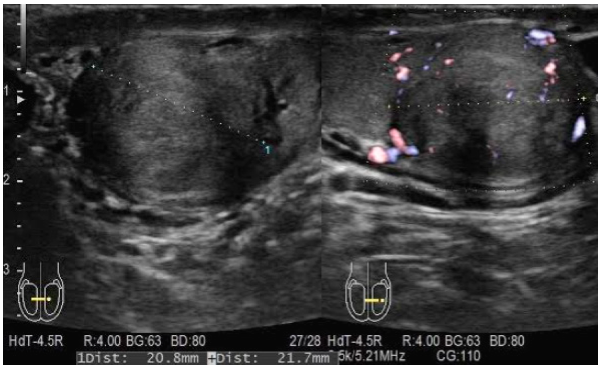
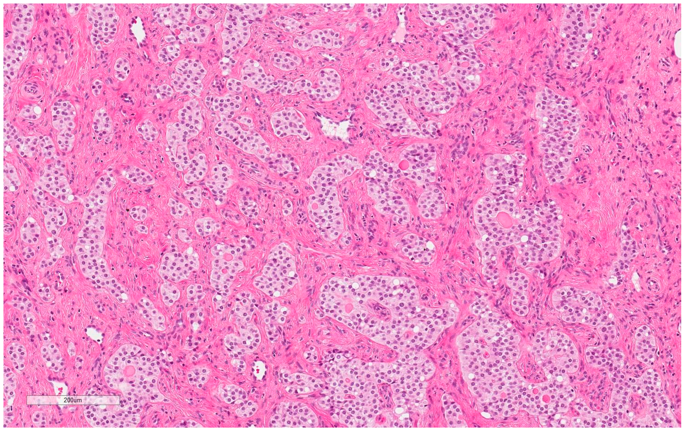
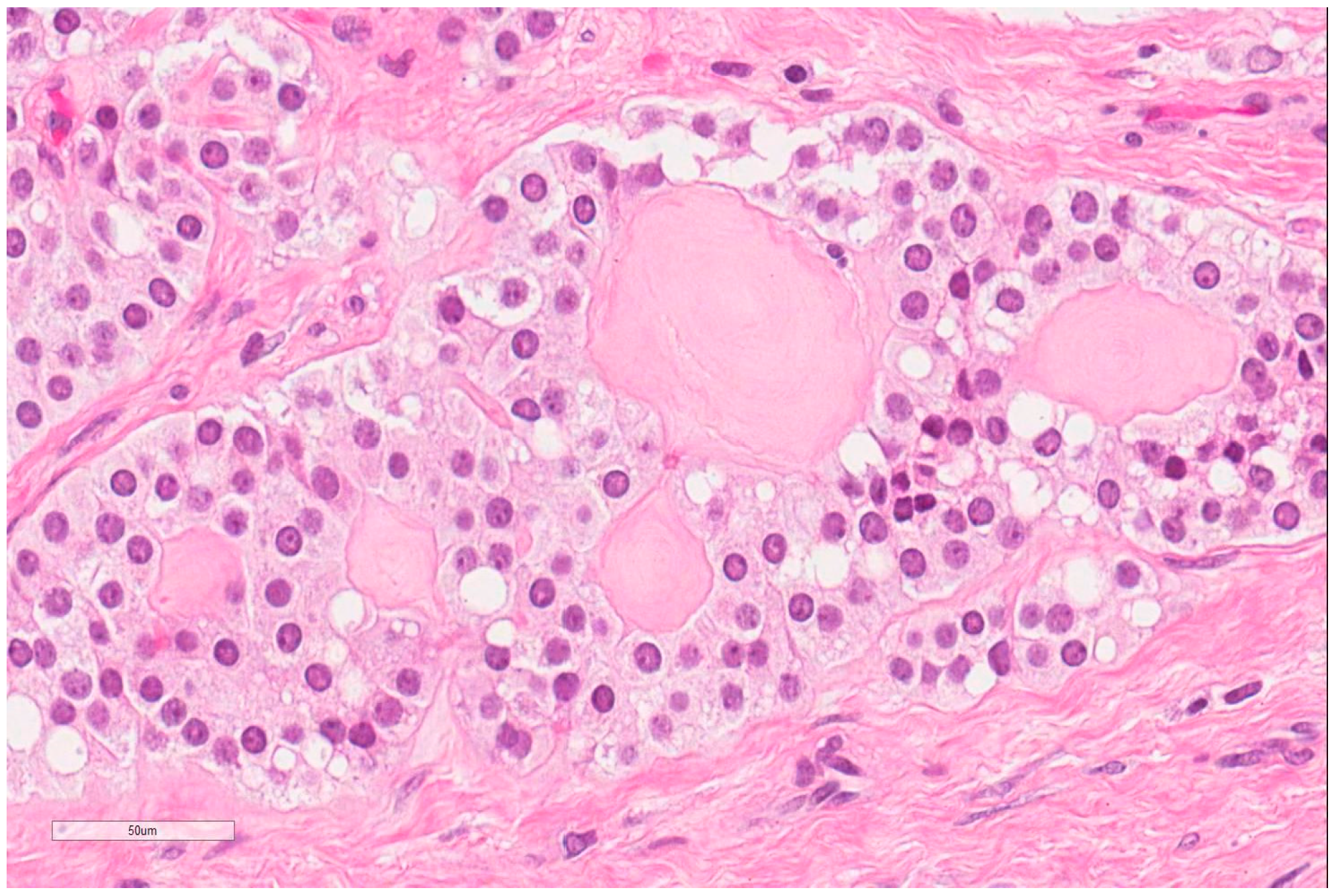
2. Case Report
3. Discussion
4. Conclusions
Author Contributions
Funding
Conflicts of Interest
References
- Banerji, J.S.; Odem-Davis, K.; Wolff, E.M.; Nichols, C.R.; Porter, C.R. Patterns of care and survival outcomes for malignant sex cord stromal testicular cancer: Results from the National Cancer Data Base. J. Urol. 2016, 196, 1117–1122. [Google Scholar] [CrossRef] [PubMed]
- Morgan, M.A. Sertoli Cell Tumour of the Testis, 2016. Radiopaedia. Available online: https://radiopaedia.org/articles/sertoli-cell-tumour-of-the-testis (accessed on 12 June 2016).
- Eble, J.N.; Sauter, G.; Epstein, J.I.; Sesterhenn, I.A. Pathology and Genetics of Tumours of the Urinary System and Male Genital Organs. World Health Organization Classification of Tumours; IARC: Lyon, France, 2004; pp. 250–255. [Google Scholar]
- Kratzer, S.S.; Ulbright, T.M.; Talerman, A.; Srigley, J.R.; Roth, L.M.; Wahle, G.R.; Moussa, M.; Stephens, J.K.; Millos, A.; Young, R.H.; et al. Large cell calcifying Sertoli cell tumor of the testis: Contrasting features of six malignant and six benign tumors and a review of the literature. Am. J. Surg. Pathol. 1997, 21, 1271–1280. [Google Scholar] [CrossRef] [PubMed]
- Henley, J.D.; Young, R.H.; Ulbright, T.M. Malignant Sertoli cell tumors of the testis: A study of 13 examples of a neoplasm frequently misinterpreted as seminoma. Am. J. Surg. Pathol. 2002, 26, 541–550. [Google Scholar] [CrossRef] [PubMed]
- Huddart, R.; Hazell, S.; Horwich, A. Rare tumors of the testis and paratesticular tissues. In Textbook of Uncommon Cancer, 4th ed.; Raghavan, D., Blanke, D.C., Johnson, H.D., Moots, L.P., Reaman, H.G., Rose, G.P., Sekeres, A.M., Eds.; Wiley: Hoboken, NJ, USA, 2012; pp. 77–96. [Google Scholar]
- Perrone, F.; Bertolotti, A.; Montemurro, G.; Paolini, B.; Pierotti, M.A.; Colecchia, M. Frequent mutation and nuclear localization of β-catenin in sertoli cell tumors of the testis. Am. J. Surg. Pathol. 2014, 38, 66–71. [Google Scholar] [CrossRef] [PubMed]
- Moch, H.; Cubilla, A.L.; Humphrey, P.A.; Reuter, V.E.; Ulbright, T.M. The 2016 WHO Classification of Tumours of the Urinary System and Male Genital Organs–Part A: Renal, Penile, and Testicular Tumours, 4th ed.; International Agency for Research on Cancer: Lyon, France, 2016; pp. 93–105. [Google Scholar]
- Albers, P.; Albrecht, W.; Algaba, F.; Bokemeyer, C.; Cohn-Cedermark, G.; Fizazi, K.; Horwich, A.; Laguna, M.P. Guidelines on Testicular Cancer European Association of Urology Guidelines; Elsevier: Amsterdam, The Netherlands, 2010; pp. 32–33. [Google Scholar]


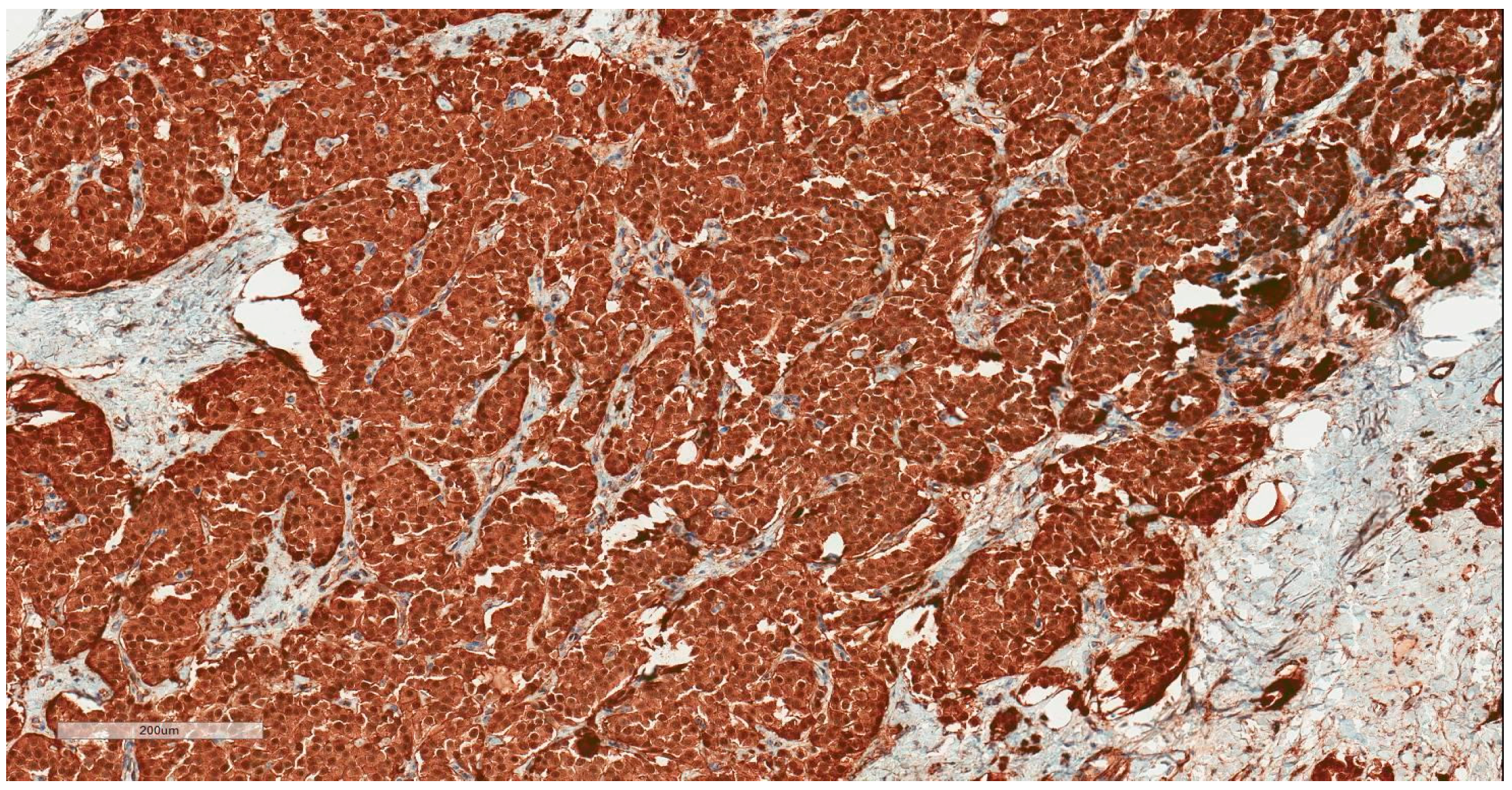
| Immunohistochemical Marker | Our Case | Typical Sertoli Cell Tumor [3] | Seminoma [3] |
|---|---|---|---|
| Vimentin | +++ | positive | |
| Beta-Catenin | +++ | positive (60–70% tumors) | |
| SF1 | ++ | positive (“typically”) | |
| CD99 | ++ | positive (“typically”) | |
| CD56 | + | ||
| S100 | + | positive | |
| EMA | +/- | variable | negative |
| Synaptophysin | +/- | often positive | |
| Chromogranin A | - | often positive | |
| PanCytokeratin | - | often positive | variable (20–36% tumors) |
| Inhibin | - | positive (50% tumors) | |
| Calretinin | - | positive (“typically”) | |
| MelanA (A103) | - | positive (“typically”) | |
| PLAP | - | positive (90–100% tumors) | |
| SALL4 | - | positive (100% tumors) | |
| OCT4 | - | positive (100% tumors) |
© 2019 by the authors. Licensee MDPI, Basel, Switzerland. This article is an open access article distributed under the terms and conditions of the Creative Commons Attribution (CC BY) license (http://creativecommons.org/licenses/by/4.0/).
Share and Cite
Anglickis, M.; Stulpinas, R.; Anglickienė, G.; Gabrilevičius, J.; Jaškevičius, A. Case Report of Misleading Features of a Rare Sertoli Cell Testicular Tumor. Medicina 2019, 55, 170. https://0-doi-org.brum.beds.ac.uk/10.3390/medicina55050170
Anglickis M, Stulpinas R, Anglickienė G, Gabrilevičius J, Jaškevičius A. Case Report of Misleading Features of a Rare Sertoli Cell Testicular Tumor. Medicina. 2019; 55(5):170. https://0-doi-org.brum.beds.ac.uk/10.3390/medicina55050170
Chicago/Turabian StyleAnglickis, Marius, Rokas Stulpinas, Giedrė Anglickienė, Justinas Gabrilevičius, and Arūnas Jaškevičius. 2019. "Case Report of Misleading Features of a Rare Sertoli Cell Testicular Tumor" Medicina 55, no. 5: 170. https://0-doi-org.brum.beds.ac.uk/10.3390/medicina55050170