Article

Carbohydrate Antigen 50: Values for Diagnosis and Prognostic Prediction of Intrahepatic Cholangiocarcinoma

by
Sukanya Luang
1,2,
Karuntarat Teeravirote
1,2,
Waraporn Saentaweesuk
3,
Prasertsri Ma-In
1 and
Atit Silsirivanit
1,2,*
1
Department of Biochemistry, Faculty of Medicine, Khon Kaen University, Khon Kaen 40002, Thailand
2
Cholangiocarcinoma Research Institute, Khon Kaen University, Khon Kaen 40002, Thailand
3
Faculty of Pharmacy, Mahasarakham University, Maha Sarakham 44150, Thailand
*
Author to whom correspondence should be addressed.
Submission received: 16 October 2020 / Revised: 6 November 2020 / Accepted: 10 November 2020 / Published: 16 November 2020

Abstract

:
Background and objectives: Cancer-associated carbohydrate antigen 50 (CA50) is a marker for detection of gastrointestinal cancers, especially of pancreatic and colon cancer. In this study, the power of CA50 as a diagnostic and prognostic marker was evaluated in intrahepatic cholangiocarcinoma (iCCA). Materials and Methods: Serum CA50 levels of iCCA patients and non-cholangiocarcinoma controls (non-CCA, including healthy persons and patients with benign biliary diseases and other gastrointestinal cancers) were measured using MAGLUMI®800 CLIA analyzer. Diagnostic and prognostic values of serum CA50 levels were evaluated. Results: CA50 levels in the sera of iCCA patients were significantly higher than those of non-CCA controls (p < 0.001, Mann–Whitney U test). Using cut-off value of 25 U/mL, CA50 provided 65.9% sensitivity, 87.3% specificity, and 80.1% accuracy for diagnosis of iCCA. Serum CA50 levels were increased and associated with the severity of bile duct pathology. In addition, a higher level of CA50 was associated with poor clinical outcome and shorter survival in iCCA patients. Multivariate survival analysis by Cox regression model revealed the potential of CA50 as an independent poor prognostic indicator for iCCA, regardless of the age, sex, histological types, or tumor stages. Conclusions: CA50 can be a diagnostic and poor prognostic marker candidate for iCCA.

Graphical Abstract

1. Introduction

Cholangiocarcinoma (CCA) is one of the most common primary liver malignancies. It is classified into three types, intrahepatic, perihilar, and distal CCA, based on their anatomic location to help the therapeutic treatment plan [1]. CCA incidence and mortality is increasing worldwide due to delayed diagnosis and treatment because the disease is asymptomatic at the early stage. Therefore, several CCA markers, such as carbohydrate antigens, have been applied for diagnosis, prognostic stratification, and treatment prediction. As the clinically nonspecific marker, carbohydrate antigen 19-9 (CA19-9) and carcinoembryonic antigen (CEA) are either individually or combinedly used for CCA diagnosis and prognosis with variable sensitivity and specificity [2,3,4,5]. However, a continual discovery of potential tumor markers for CCA is still crucial for these purposes.
Cancer-associated carbohydrate antigen 50 (CA50) is a ganglioside glycoprotein and is a biomarker for the diagnosis and prognosis of gastrointestinal malignancies, especially of pancreatic and colorectal cancers [6,7]. The expression level of CA50 has been measured for pancreatic and colorectal cancers and also for other cancers such as breast, lung, renal, prostatic, and ovarian cancers [6]. In addition, it can distinguish the patients of benign disease of atrophic gastritis, pancreatitis, ulcerative colitis, pneumonia, cirrhosis, and autoimmune diseases from healthy persons [8]. The monoclonal antibody that defines CA50 reacts with both the afucosyl form of sialylated Lewis A carbohydrate moiety and sialylated Lewis A moiety on the cancer cell surface [9]. CA50 epitope is assumed to be similar to CA19-9 epitope (sialyl-Lewis A). In fact, both CA19-9 and CA50 are valuable markers for the differentiation of pancreatic cancer from benign diseases of the pancreatobiliary tract [10,11]. However, in contrast to the limited expression of CA 19-9 in gastrointestinal malignancy, high CA50 levels can also be seen in malignant tumors outside the digestive tract. In spite of extensive study on the diagnostic value of CA50 for various malignancies, its expression level in the sera of CCA patients has not much been explored. The previous studies by Haglund et al. and Watanabe et al. demonstrated that CA50 was elevated in CCA tissues [12,13]. In our study, we have investigated the potential of CA50 to be a marker for diagnosis of CCA by measuring the CA50 level in serum from intrahepatic cholangiocarcinoma (iCCA) patients compared with non-CCA controls. In addition, the power of CA50 in prognosis of CCA was also demonstrated.

2. Materials and Methods

2.1. Patients and Samples

Serum samples from healthy individuals (HE) were obtained from asymptomatic persons with a normal level of fasting blood sugar (<110 mg/dL), aspartate aminotransferase (AST, ≤35 U/mL), alanine aminotransferase (ALT, ≤37 U/mL), and alkaline phosphatase (ALP, < 121 U/mL) who visited Srinagarind Hospital, Khon Kaen University, Khon Kaen, Thailand, for their annual health check-up. The serum samples from benign biliary diseases (BBD, n = 23), other gastro-intestinal cancers (OCA, n = 33), and iCCA (n = 85) were obtained from the Specimen Bank of the Cholangiocarcinoma Research Institute, Khon Kaen University. The patients of BBD, OCA, and iCCA were definitely diagnosed by histopathological study of resected tumor tissues at the Department of Pathology, Srinagarind Hospital. The OCA group consisted of hepatoma and carcinoma of ampulla of Vater (n = 6), colon and colorectal cancer (n = 10), pancreatic cancer (n = 7), and gastric cancer (n = 10). The informed consents were obtained from each subject. The study protocol was approved by the Khon Kaen University Ethics Committee for Human Research based on the Declaration of Helsinki and the ICH Good Clinical Practice Guidelines (HE621176).

2.2. CA50 and CA19-9 Analysis

The serum CA50 and CA19-9 levels were measured using an automatic chemiluminescence immunoassay (CLIA) on the MAGLUMI®800 CLIA analyzer from SNIBE Co., Ltd. (Shenzhen New Industries Biomedical Engineering Co., Ltd.), Shenzhen, China.

2.3. Statistical Analysis

The statistical analyses were performed using IBM-SPSS statistics version 26 (IBM Corp., Armonk, NY, USA) and GraphPad-Prism version 8.4.2 (GraphPad Software, San Diego, CA, USA). The difference of CA50 among the groups of subjects was analyzed by Mann–Whitney U test. Univariate and multivariate Cox regression analyses were used to identify the factors that influence the survival of CCA patients. Kaplan–Meier plot and log-rank test were used to analyze the association of CA50 and median survival time of the patients.

3. Results

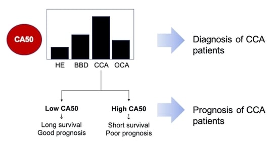
3.1. CA50 Was Elevated in the Sera of CCA Patients and Has a Diagnostic Value for iCCA

To investigate the potential of serum CA50 level for CCA diagnosis, we measured the expression level of CA50 in preoperative serum samples of iCCA patients (n = 85) and compared with those of 166 non-CCA controls, including 110 HE, 23 BBD, and 33 OCA (Table 1). Serum CA50 levels of iCCA patients varied, ranging from 0.5 to 125,000 U/mL with the median value of 197.3 U/mL, which is significantly higher than that of non-CCA controls (median = 4.6 U/mL) (Figure 1A, p < 0.001). Receiver operating characteristic (ROC) curve analysis revealed that CA50 could differentiate iCCA patients from non-CCA controls with the area under curve (AUC) of 0.806 (Figure 1B, p < 0.001). Using the cut-off value of 25 U/mL, CA50 was found to provide 65.9% sensitivity, 87.3% specificity, and 80.1% accuracy to discriminate iCCA patients from non-CCA controls (Table 2). The diagnostic values of CA50 were comparable with those of CA19-9, a standard marker for diagnosis of CCA.

3.2. The Increase of Serum CA50 Was Associated with the Severity of Bile Duct Pathology

Comparing between iCCA and BBD, the level of CA50 in iCCA was significantly higher than that in BBD (Figure 1C, p < 0.05). The median values of serum CA50 level of both BBD and iCCA groups were significantly higher than that of OCA group (4.1 U/mL) (Figure 1C, p < 0.0001). Using the cut-off value of 25.0 U/mL, 47.8% (11/23) of BBD and 27.3% (9/33) of OCA cases were found to be positive for CA50 (Table 1). As shown in Figure 1D, ROC analysis revealed that CA50 can differentiate iCCA from BBD with an AUC of 0.641 (p < 0.05). Using Youden’s index calculated from ROC analysis [14], a new cut-off value of CA50 was determined to be 85.3 U/mL, which provided the specificity to exclude 82% (19/23) of BBD, 84% (28/33) of OCA, and 100% (110/110) of HE cases, although the sensitivity to detect iCCA was decreased to 55.3% (47/85), as shown in Table 2.

3.3. High Level of CA50 of iCCA Patients Indicates Poor Prognosis and Shorter Survival

To investigate the potential of using CA50 as a prognostic marker for iCCA, the correlation between patient clinical outcomes and serum CA50 level was analyzed. Differences of CA50 level between iCCA subgroups were determined using Mann–Whitney U test (Figure 2). Serum CA50 level of the iCCA subgroup with tumor stage IVB and survival ≤1 year was significantly higher than that of the subgroup with stage I–III/IVA and survival >1 year (p < 0.05). However, serum CA50 level was not associated with age, sex, histopathological types, tumor size, and total bilirubin level of the patients. Then, iCCA patients were subcategorized according to the median (197.3 U/mL) and mean (8348.0 U/mL) values of serum CA50 levels into 1) Low (≤197.3 U/mL, n = 43), 2) Medium (197.4–8348.0 U/mL, n = 24), and 3) High (>8348.0 U/mL, n = 18) groups. Univariate Cox proportional hazard analysis revealed that the iCCA patients with high CA50 have 3.629 times higher hazard ratio compared with those with low CA50 (Table 3, p < 0.05). Multivariate survival analysis suggested the potential of using CA50 as an independent prognostic indicator for iCCA regardless of age, sex, histological type, and tumor stage of the patients (Table 3, p < 0.05). As shown in Figure 2H, Kaplan–Meier plot and log-rank analysis showed that the patients with a high level of CA50 had the shortest survival (127.0 days, 95% CI of 35.5–128.5 days), compared with those with medium CA50 (172.0 days, 95% CI of 91.6–252.4 days) and low CA50 (227.0 days, 95% CI of 161.5–292.5 days) (p < 0.001). The overall median survival of iCCA patients and 95% CI were 189.0 and 152.9–225.1 days, respectively.

4. Discussion

Altered glycosylation is seen in CCA, and aberrant expression of glycans plays significant roles in CCA progression, leading to poor survival of patients. Beside involving in tumor progression [15,16,17,18,19], the CCA-associated glycans were found to be the potential biomarkers for diagnosis and prognostic prediction of the disease [18,20,21].
In this study, we have analyzed the serum level of sialyl-associated glycan antigen CA50 in iCCA, BBD, and healthy control groups. Our data showed that CA50 level was associated with the severity of bile duct pathology, as its level gradually increased in BBD and iCCA, suggesting the potential use of CA50 as the indicator of bile ducts pathology. The previous study by Haglund et al. and Watanabe et al. demonstrated the increasing of CA50 level in CCA tissues and showed it could be secreted from CCA cells, suggesting that CA50 is a tumor origin tumor marker [12,13]. Our present study was performed in a larger cohort of CCA (n = 85) and various non-CCA cases (n = 166). When we set the cut-off value at 25 U/mL, CA50 was able to differentiate iCCA patients from non-CCA control with 65.9% sensitivity, 87.3% specificity, and 80.1% accuracy. The diagnostic values provided by CA50 were slightly higher than that of a general serum marker for diagnosis of CCA, CA19-9 (65.9% sensitivity, 86.1% specificity, and 79.3% accuracy). Combined analysis of these markers may improve their power in diagnosis of CCA.
When the cut-off value was set at 85.3 U/mL, the CA50 could exclude 82% (19/23) of BBD, 84% (28/33) of OCA, and 100% (110/110) of HE cases, suggesting that the patients with high CA50 have high possibility of having iCCA. Although CA50 was previously reported to increase in patients with gastrointestinal cancers, for example, gastric cancer and pancreatic cancer [8], our results demonstrated the degree of increase of CA50 in iCCA patients was significantly higher than that of OCA and other control groups. Related to this, Tao et al. previously demonstrated that the serum CA50 levels in CCA and hepatoma patients were comparable and statistically not different from each other [22]. This difference might be due to the different etiology of CCA in Thailand and other countries. The CCA in Thailand is almost exclusively associated with liver fluke, Opisthorchis viverrini infection, while the CCA in other countries is associated with various factors, for example, primary sclerosing cholangitis and primary biliary cirrhosis. Previous studies comparing the epigenetics and gene expression profile revealed that CCA with different etiology exhibits different gene expression patterns [23,24,25], therefore the increase of a particular molecule may not be observed and applicable for the diagnosis of all types of CCA. To improve the detection of CCA, combined analysis of multiple markers may possibly provide the better answer.
In this study, serum CA50 level was correlated with the clinical outcomes of CCA patients. High level of CA50 in the serum was associated with the advanced tumor stage and shorter survival of iCCA patients. Previous studies on CA50 expression in gastric and pancreatic cancer patients showed that the high level of CA50 is significantly associated with shorter survival of the patients [26,27,28]. Previous studies by Juntavee et al. (2005) and Wattanavises et al. (2019) demonstrated the increase of sialylation, as represented by sialyl Lewis A antigen and MAL-II binding glycan, in CCA, compared with normal bile ducts and parenchymal cells, in association with poor prognosis and short survival of CCA patients [16,19]. These glycans are involved in metastasis and chemoresistance of CCA cells [16,19]. Thus, CA50, another sialylated glycan, may also play roles in CCA metastasis and chemoresistance, resulting in poor prognosis.

5. Conclusions

CA50 could be a candidate marker for CCA diagnosis and prognostic prediction, as the higher level of CA50 was associated with advanced bile duct pathology and shorter survival of iCCA patients. Further analysis using a combination of CA50 and other CCA markers may enhance the sensitivity and specificity in diagnosis of CCA, resulting in better outcomes of CCA treatments.

Author Contributions

Conceptualization, S.L. and A.S.; methodology, S.L. and W.S.; software, A.S.; validation, A.S.; formal analysis, S.L, P.M.-I., K.T. and W.S.; investigation, K.T., A.S.; resources, A.S.; data curation, A.S.; writing—original draft preparation, S.L, W.S..; writing—review and editing, A.S.; project administration, P.M.-I.; All authors have read and agreed to the published version of the manuscript.

Funding

This research received no external funding.

Acknowledgments

We would like to thank Yukifumi Nawa for English editing via KKU Publication Clinic.

Conflicts of Interest

A.S. was supported by Snibe as a consultant.

References

  1. Razumilava, N.; Gores, G.J. Classification, diagnosis, and management of cholangiocarcinoma. Clin. Gastroenterol. Hepatol. 2013, 11, 13Ce4. [Google Scholar] [CrossRef] [PubMed] [Green Version]
  2. Levy, C.; Lymp, J.; Angulo, P.; Gores, G.J.; Larusso, N.; Lindor, K.D. The value of serum CA 19-9 in predicting cholangiocarcinomas in patients with primary sclerosing cholangitis. Dig. Dis. Sci. 2005, 50, 1734–1740. [Google Scholar] [CrossRef] [PubMed]
  3. Charatcharoenwitthaya, P.; Enders, F.B.; Halling, K.C.; Lindor, K.D. Utility of serum tumor markers, imaging, and biliary cytology for detecting cholangiocarcinoma in primary sclerosing cholangitis. Hepatology 2008, 48, 1106–1117. [Google Scholar] [CrossRef] [PubMed]
  4. Li, Y.; Li, D.J.; Chen, J.; Liu, W.; Li, J.W.; Jiang, P.; Zhao, X.; Guo, F.; Li, X.W.; Wang, S.G. Application of joint detection of AFP, CA19-9, CA125 and CEA in identification and diagnosis of cholangiocarcinoma. Asian Pac. J. Cancer Prev. 2015, 16, 3451–3455. [Google Scholar] [CrossRef] [Green Version]
  5. Loosen, S.H.; Roderburg, C.; Kauertz, K.L.; Koch, A.; Vucur, M.; Schneider, A.T.; Binnebösel, M.; Ulmer, T.F.; Lurje, G.; Schoening, W. CEA but not CA19-9 is an independent prognostic factor in patients undergoing resection of cholangiocarcinoma. Sci. Rep. 2017, 7, 16975. [Google Scholar] [CrossRef] [Green Version]
  6. Holmgren, J.; Lindholm, L.; Persson, B.; Lagergård, T.; Nilsson, O.; Svennerholm, L.; Rudenstam, C.M.; Unsgaard, B.; Yngvason, F.; Pettersson, S. Detection by monoclonal antibody of carbohydrate antigen CA 50 in serum of patients with carcinoma. Br. Med. J. 1984, 288, 1479–1482. [Google Scholar] [CrossRef] [Green Version]
  7. Månsson, J.E.; Fredman, P.; Nilsson, O.; Lindholm, L.; Holmgren, J.; Svennerholm, L. Chemical structure of carcinoma ganglioside antigens defined by monoclonal antibody C-50 and some allied gangliosides of human pancreatic adenocarcinoma. Biochim. Biophys. Acta 1985, 834, 110–117. [Google Scholar] [CrossRef]
  8. Shan, M.; Tian, Q.; Zhang, L. Serum CA50 levels in patients with cancers and other diseases. Prog. Mol. Biol. Transl. Sci. 2019, 162, 187–198. [Google Scholar]
  9. Bunworasate, U.; Voravud, N. CA 50: A tumor marker for gastrointestinal malignancies. J. Med. Assoc. Thail. 1995, 78, 255–270. [Google Scholar]
  10. Haglund, C.; Kuusela, P.; Jalanko, H.; Roberts, P.J. Serum CA 50 as a tumor marker in pancreatic cancer: A comparison with CA 19-9. Int. J. Cancer 1987, 39, 477–481. [Google Scholar] [CrossRef]
  11. Harmenberg, U.; Wahren, B.; Wiechel, K.L. Tumor markers carbohydrate antigens CA 19-9 and CA-50 and carcinoembryonic antigen in pancreatic cancer and benign diseases of the pancreatobiliary tract. Cancer Res. 1988, 48, 1985–1988. [Google Scholar] [PubMed]
  12. Haglund, C.; Lindgren, J.; Roberts, P.J.; Nordling, S. Difference in tissue expression of tumour markers CA 19-9 and CA 50 in hepatocellular carcinoma and cholangiocarcinoma. Br. J. Cancer 1991, 63, 386–389. [Google Scholar] [CrossRef] [PubMed] [Green Version]
  13. Watanabe, M.; Chigusa, M.; Takahashi, H.; Nakamura, J.; Tanaka, H.; Ohno, T. High level of CA19-9, CA50, and CEA-producible human cholangiocarcinoma cell line changes in the secretion ratios in vitro or in vivo. In Vitro Cell. Dev. Biol. Anim. 2000, 36, 104–109. [Google Scholar] [CrossRef]
  14. Youden, W.J. Index for rating diagnostic tests. Cancer 1950, 3, 32–35. [Google Scholar] [CrossRef]
  15. Detarya, M.; Sawanyawisuth, K.; Aphivatanasiri, C.; Chuangchaiya, S.; Saranaruk, P.; Sukprasert, L.; Silsirivanit, A.; Araki, N.; Wongkham, S.; Wongkham, C. The O-GalNAcylating enzyme GALNT5 mediates carcinogenesis and progression of cholangiocarcinoma via activation of AKT/ERK signaling. Glycobiology 2020, 30, 312–324. [Google Scholar] [CrossRef] [PubMed]
  16. Juntavee, A.; Sripa, B.; Pugkhem, A.; Khuntikeo, N.; Wongkham, S. Expression of sialyl Lewis(a) relates to poor prognosis in cholangiocarcinoma. World J. Gastroenterol. 2005, 11, 249–254. [Google Scholar] [CrossRef] [PubMed]
  17. Phoomak, C.; Park, D.; Silsirivanit, A.; Sawanyawisuth, K.; Vaeteewoottacharn, K.; Detarya, M.; Wongkham, C.; Lebrilla, C.B.; Wongkham, S. O-GlcNAc-induced nuclear translocation of hnRNP-K is associated with progression and metastasis of cholangiocarcinoma. Mol. Oncol. 2019, 13, 338–357. [Google Scholar] [CrossRef]
  18. Silsirivanit, A.; Araki, N.; Wongkham, C.; Vaeteewoottacharn, K.; Pairojkul, C.; Kuwahara, K.; Narimatsu, Y.; Sawaki, H.; Narimatsu, H.; Okada, S. CA-S27: A novel Lewis A associated carbohydrate epitope is diagnostic and prognostic for cholangiocarcinoma. Cancer Sci. 2013, 104, 1278–1284. [Google Scholar] [CrossRef]
  19. Wattanavises, S.; Silsirivanit, A.; Sawanyawisuth, K.; Cha’on, U.; Waraasawapati, S.; Saentaweesuk, W.; Luang, S.; Chalermwat, C.; Wongkham, C.; Wongkham, S. Increase of MAL-II binding alpha2,3-sialylated glycan is associated with 5-FU resistance and short survival of cholangiocarcinoma patients. Medicina 2019, 55, 761. [Google Scholar] [CrossRef] [Green Version]
  20. Saentaweesuk, W.; Silsirivanit, A.; Vaeteewoottacharn, K.; Sawanyawisuth, K.; Pairojkul, C.; Cha’on, U.; Indramanee, S.; Pinlaor, S.; Boonmars, T.; Araki, N. Clinical significance of GalNAcylated glycans in cholangiocarcinoma: Values for diagnosis and prognosis. Clin. Chim. Acta 2018, 477, 66–71. [Google Scholar] [CrossRef]
  21. Silsirivanit, A.; Araki, N.; Wongkham, C.; Pairojkul, C.; Narimatsu, Y.; Kuwahara, K.; Narimatsu, H.; Wongkham, S.; Sakaguchi, N. A novel serum carbohydrate marker on mucin 5AC: Values for diagnostic and prognostic indicators for cholangiocarcinoma. Cancer 2011, 117, 3393–3403. [Google Scholar] [CrossRef] [PubMed]
  22. Tao, L.Y.; Cai, L.; He, X.D.; Liu, W.; Qu, Q. Comparison of serum tumor markers for intrahepatic cholangiocarcinoma and hepatocellular carcinoma. Am. Surg. 2010, 76, 1210–1213. [Google Scholar] [CrossRef] [PubMed]
  23. Chan-On, W.; Nairismagi, M.L.; Ong, C.K.; Lim, W.K.; Dima, S.; Pairojkul, C.; Lim, K.H.; McPherson, J.R.; Cutcutache, I.; Heng, H.L. Exome sequencing identifies distinct mutational patterns in liver fluke-related and non-infection-related bile duct cancers. Nat. Genet. 2013, 45, 1474–1478. [Google Scholar] [CrossRef] [PubMed]
  24. Jinawath, N.; Chamgramol, Y.; Furukawa, Y.; Obama, K.; Tsunoda, T.; Sripa, B.; Pairojkul, C.; Nakamura, Y. Comparison of gene expression profiles between Opisthorchis viverrini and non-Opisthorchis viverrini associated human intrahepatic cholangiocarcinoma. Hepatology 2006, 44, 1025–1038. [Google Scholar] [CrossRef] [PubMed]
  25. Jusakul, A.; Cutcutache, I.; Yong, C.H.; Lim, J.Q.; Huang, M.N.; Padmanabhan, N.; Nellore, V.; Kongpetch, S.; Ng, A.W.T.; Ng, L.M. Whole-genome and epigenomic landscapes of etiologically distinct subtypes of cholangiocarcinoma. Cancer Discov. 2017, 7, 1116–1135. [Google Scholar] [CrossRef] [PubMed] [Green Version]
  26. Feng, L.; Gu, S.; Wang, P.; Chen, H.; Chen, Z.; Meng, Z.; Liu, L. Pretreatment values of bilirubin and albumin are not prognostic predictors in patients with advanced pancreatic cancer. Cancer Med. 2018, 7, 5943–5951. [Google Scholar] [CrossRef]
  27. Liu, X.; Cai, H.; Wang, Y. Prognostic significance of tumor markers in T4a gastric cancer. World J. Surg. Oncol. 2012, 10, 68. [Google Scholar] [CrossRef] [Green Version]
  28. Mittal, A.; Gupta, S.P.; Jha, D.K.; Sathian, B.; Poudel, B. Impact of various tumor markers in prognosis of gastric cancer. A Hospital Based Study from Tertiary Care Hospital of Kathmandu Valley. Asian Pac. J. Cancer Prev. 2013, 14, 1965–1967. [Google Scholar] [CrossRef] [Green Version]
Figure 1. Serum CA50 level of iCCA patients and non-CCA control. (A) Scatter plot showed the level of CA50 in iCCA patients, compared with non-CCA control. (B) ROC analysis to analyze the power of CA50 in discriminating iCCA from non-CCA control. (C) Scatter plot showed the level of CA50 in healthy persons (HE), patients of benign biliary diseases (BBD), other gastrointestinal cancers (OCA) and iCCA. (D) Receiver operating characteristic (ROC) analysis to analyze the power of CA50 in discriminating iCCA from BBD cases. The significant difference of CA50 level among the cases is indicated by stars (**) for p < 0.001, while the small letter indicates p < 0.05 (a vs. HE, b vs. BBD, and c vs. OCA).
Medicina 56 00616 g001
Figure 2. Correlation between CA50 level and clinicopathological data of iCCA patients. The level of CA50 was compared among the patients with different (A) age, (B) sex, (C) histological types, (D) tumor size, (E) total bilirubin, (F) tumor stages, and (G) 1 year survival; using Mann–Whitney U test (* p < 0.05). (H) Kaplan–Meier plot and log-rank test for survival analysis of CA50.
Medicina 56 00616 g002
Table 1. Serum level of carbohydrate antigen CA50 in non-cholangiocarcinoma (non-CCA) and intrahepatic cholangiocarcinoma (iCCA) patients.
IndexGroup of Subjects
Healthy (HE)Benign (BBD)Other Cancers (OCA)iCCA
n110233385
CA50 Level (U/mL)
Mean5.11195.0156.98348.0
Median4.015.54.1197.3
Min0.50.50.50.5
Max25.612,460.02664.0125,000.0
Number of Cases at Cut-Off CA50 = 25.0 U/mL
CA50 ≤ 25.0 U/ml109122429
CA50 > 25.0 U/ml111956
Number of Cases at Cut-Off CA50 = 85.3 U/mL
CA50 ≤ 85.3 U/ml110192838
CA50 > 85.3 U/ml04547
Table 2. Diagnostic values of CA50 and carbohydrate antigen 19-9 (CA19-9).
AnalysesNon-CCA vs. iCCABBD vs. iCCA
CA19-9CA50CA50
Mann–Whitney U test
p<0.001<0.0010.039
ROC Analysis
AUC0.7970.8060.641
p<0.001<0.0010.039
Cut-off value37 U/mL25 U/mL85.3 U/mL
Sensitivity (%)65.965.955.3
Specificity (%)86.187.382.6
Positive predictive value (%)70.972.792.2
Negative predictive value (%)83.183.333.3
False positive (%)13.912.717.4
False negative (%)34.134.144.7
Accuracy (%)79.380.161.1
Table 3. Univariate and multivariate Cox regression analyses.
ParametersnUnivariate AnalysisMultivariate Analysis
HR95% CIpHR95% CIp
Age (years)
≤56491- 1-
>56361.2360.777–1.9650.3701.5060.908–2.5000.113
Sex
Male 601- 1-
Female250.7290.444–1.1990.2130.7250.425–1.2350.236
Histological types
Papillary201 1-
Non-papillary651.9031.060–3.4180.0311.9631.036–3.7200.039
Tumor stage
I–III71- -
IVA251.9540.783–4.8810.1511.8010.691–4.6960.229
IVB522.7251.129–6.5770.0261.7440.676–4.4980.250
CA50 (U/mL)
Low431- -
Medium241.6590.964–2.8540.0681.4470.689–3.0400.329
High463.6291.961–6.714<0.0012.9881.269–7.0320.012
Publisher’s Note: MDPI stays neutral with regard to jurisdictional claims in published maps and institutional affiliations.

