
Subcutaneous and Intraosseous Fat Necrosis Associated with Chronic Pancreatitis

 , ,
, , 
Abstract
:1. Introduction
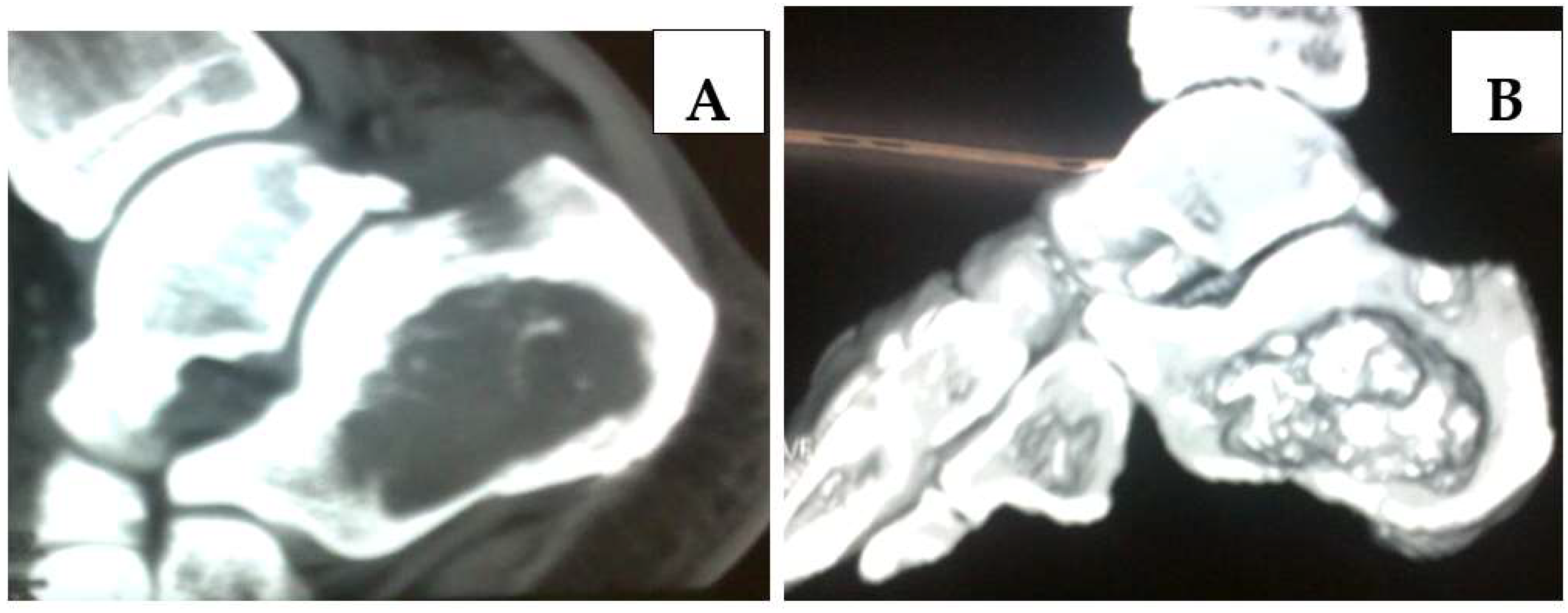
2. Case Report
3. Discussion
4. Conclusions
Author Contributions
Funding
Institutional Review Board Statement
Informed Consent Statement
Data Availability Statement
Conflicts of Interest
References
- Čolović, R.; Grubor, N.; Radak, V.; Čolović, N.; Ranković, V.; Latinčić, S.; Matić, S. Disseminated subcutaneous fat necrosis and elbow joint arthritis as a complication of pancreatitis. VSP 2008, 65, 703. [Google Scholar] [CrossRef] [PubMed]
- Haller, J.; Greenway, G.; Resnick, D.; Kindynis, P.; Kang, H.S. Intraosseous fat necrosis associated with acute pancreatitis: MR imaging. Radiology 1989, 173, 193–195. [Google Scholar] [CrossRef]
- Karasick, D.; Schweitzer, M.E. Case 4: Intraosseous, fat necrosis associated with pancreatitis. Radiology 1998, 209, 521–524. [Google Scholar] [CrossRef] [PubMed]
- Kotilainen, P.; Saario, R.; Mattila, K.; Nylamo, E.; Aho, H. Intraosseous fat necrosis simulating septic arthritis and osteomyelitis in a patient with chronic pancreatitis. Arch. Orthop. Trauma Surg. 1998, 118, 174–175. [Google Scholar] [CrossRef] [PubMed]
- Morita, O.; Ogose, A.; Hotta, T.; Kawashima, H.; Higuchi, T.; Suzuki, K.; Endo, N. Pathological fractures due to intraosseous fat necrosis associated with pancretitis. Reumatology 2003, 42, 394–395. [Google Scholar] [CrossRef] [Green Version]
- Baba, T.; Shitoto, K.; Yoshioka, C.; Kaneko, H. Pathological fracture due to vertebral osteonecrosis associated with pancreatitis. Arch. Orthop. Trauma Surg. 2011, 131, 11–14. [Google Scholar] [CrossRef]
- Norimura, D.; Mizuta, Y.; Ohba, K.; Oh, J.; Oohara, H.; Nakahara, N.; Yamaguchi, N.; Ohnita, K.; Isomoto, H.; Shikuwa, S.; et al. Intraosseous fat necrosis associated with alcoholic pancreatitis. Clin. J. Gastroenterol. 2009, 2, 425–430. [Google Scholar] [CrossRef]
- Tupalli, A.; Angamuthu, M.; Prashanth, A.; Singh, R.; Kumar, R. Intraosseous Medullary Fat Necrosis on 99mTc-MDP Bone Scan of Patient With Acute Pancreatitis. Clin. Nucl. Med. 2020, 45, e294–e295. [Google Scholar] [CrossRef]
- Tannenbaum, H.; Anderson, L.G.; Schur, P.H. Association of polyarthritis, subcutaneous nodules, and pancreatic disease. J. Rheumatol. 1975, 2, 15–20. [Google Scholar]
- Kushner, D.S.; Szanto, P.B. Fulminant polyarthritis, fever, and cutaneous nodules in an alcoholic patient. J. Am. Med. Assoc. 1958, 167, 1625–1632. [Google Scholar]
- Kim, E.J.; Chu, M.S.; Sohn, K.C.; Cho, D.H.; Na, G.H.; Kim, H.C.; Cho, E.Y. Pancreatic Panniculitis in Patients with Chronic Pancreatitis: Case Report and Review of Literature. Korean J. Gastroenterol. 2017, 69, 83–86. [Google Scholar] [CrossRef] [PubMed] [Green Version]
- Simkin, P.A.; Brunzell, J.D.; Wisner, D.; Fiechtner, J.J.; Carlin, J.S.; Willkens, R.F. Free fatty acids in the pancreatitic arthritis syndrome. Arthritis Rheum. 1983, 26, 127–132. [Google Scholar] [CrossRef] [PubMed]
- Schutte, H.E.; Wackwitz, J.D. Case report 171: Metastatic fat necrosis involving the tubular bones of the hands (and probably the feet) secondary to traumatic pancreatitis. Skelet. Radiol. 1981, 7, 147–149. [Google Scholar] [CrossRef] [PubMed]
- Smukler, N.M.; Schumacher, H.R.; Pascual, E.; Brown, S.; Ryan, W.E.; Sadeghian, M.R. Synovial fat necrosis associated with ischemic pancreatic disease. Arthritis Rheum. 1979, 22, 547–553. [Google Scholar] [CrossRef] [PubMed]
- Haber, R.M.; Assaad, D.M. Panniculitis associated with a pancreas divisum. J. Am. Acad. Dermatol. 1986, 14, 331–334. [Google Scholar] [CrossRef]
- Virshup, A.M.; Sliwinski, A.J. Polyarthritis and subcutaneous nodules associated with carcinoma of the pancreas. Arthritis Rheum. 1973, 16, 388–392. [Google Scholar] [CrossRef]
- Radin, D.R.; Colletti, P.M.; Forrester, D.M.; Tang, W.W. Pancreatic acinar cell carcinoma with subcutaneous and intraosseous fat necrosis. Radiology 1986, 158, 67–68. [Google Scholar] [CrossRef] [Green Version]
- Takeuchi, Y. Pancreatitis, panniculitis, and polyarthritis syndrome complicated with terminal pancreatic adenocarcinoma managed with intra-articular knee aspiration, intra-articular lidocaine and corticosteroid injection, and decompression of panniculitis: A case report. J. Gen. Fam. Med. 2020, 22, 87–89. [Google Scholar]
- Zhang, G.; Cao, Z.; Yang, G.; Wu, W.; Zhang, T.; Zhao, Y. Pancreatic panniculitis associated with pancreatic carcinoma: A case report. Medicine 2016, 95, e4374. [Google Scholar] [CrossRef]
- Narváez, J.; Bianchi, M.M.; Santo, P.; de la Fuente, D.; Ríos-Rodriguez, V.; Bolao, F.; Narváez, J.A.; Nolla, J.M. Pancreatitis, panniculitis, and polyarthritis. Semin. Arthritis Rheum. 2010, 39, 417–423. [Google Scholar] [CrossRef]
- Watts, R.A.; Kelly, S.; Hacking, J.C.; Lomas, D.; Hazleman, B.L. Fat necrosis. An unusual cause of polyarthritis. J. Rheumatol. 1993, 20, 1432–1435. [Google Scholar] [PubMed]
- Ferrari, R.; Wendelboe, M.; Ford, P.M.; Corbett, W.E.; Anastassiades, T.P. Pancreatitis arthritis with periarticular fat necrosis. J. Rheumatol. 1993, 20, 1436–1437. [Google Scholar] [PubMed]
- Immelman, E.; Bank, S.; Krige, H. Roentgenologic and clinical features of intramedullary fat necrosis in bones in acute and chronic pancreatitis. Am. J. Med. 1964, 36, 96–105. [Google Scholar] [CrossRef]
- Phillips, R.M., Jr.; Sulser, R.E.; Songcharoen, S. Inflammatory arthritis and subcutaneous fat necrosis associated with acute and chronic pancreatitis. Arthritis Rheum. 1980, 23, 355–360. [Google Scholar] [PubMed]
- Dieker, W.; Derer, J.; Henzler, T.; Schneider, A.; Rückert, F.; Wilhelm, T.J.; Krüger, B. Pancreatitis, panniculitis and polyarthritis (PPP-) syndrome caused by post-pancreatitis pseudocyst with mesenteric fistula. Diagnosis and successful surgical treatment. Case report and review of literature. Int. J. Surg. Case Rep. 2017, 31, 170–175. [Google Scholar] [CrossRef] [PubMed]
- Castro, J.P.; Atanásio, G.; Canelas, M.A.; Ferreira, A.; Barbosa, A.R.; Barbedo, M.; Abreu, R. A case report of pancreatitis-panniculitis-polyarthritis syndrome-An unusual but serious presentation of pancreatic disease. Scott. Med. J. 2020, 65, 19–23. [Google Scholar] [CrossRef]
- Rodriguez, M.; Lopez, G.L.; Prieto, P.; Fernandez, L.; Willisch, A.; Arce, M. Massive Subcutaneous and Intraosseous Fat Necrosis Associated with Pancreatitis. Natural Evolution of the Radiographic Picture. Clin. Rheumatol. 1997, 16, 199–203. [Google Scholar] [CrossRef]
- Obatake, M.; Yamane, Y.; Tokunaga, T.; Taura, Y.; Inamura, Y.; Nagayasu, T. Arthralgia and Osteolytic Lesions Associated with Traumatic Pancreatitis in a 10-Year-Old Girl. Int. J. Pediatrics 2009, 2009, 950687. [Google Scholar] [CrossRef]
- Arbeláez-Cortés, A.; Vanegas-García, A.L.; Restrepo-Escobar, M.; Correa-Londoño, L.A.; González-Naranjo, L.A. Polyarthritis and pancreatic panniculitis associated with pancreatic carcinoma: Review of the literature. J. Clin. Rheumatol. 2014, 20, 433–436. [Google Scholar] [CrossRef]
- Langenhan, R.; Reimers, N.; Probst, A. Osteomyelitis: A rare complication of pancreatitis and PPP-syndrome. Jt. Bone Spine 2016, 83, 221–224. [Google Scholar] [CrossRef]
- Keating, J.P.; Shackelford, G.D.; Shackelford, P.G.; Ternberg, J.L. Pancreatitis and osteolytic lesions. J. Pediatrics 1972, 81, 350–353. [Google Scholar] [CrossRef]
- Boswell, S.H.; Baylin, G.J. Metastatic fat necrosis and lytic lesions in a patient with painless pancreatitis. Radiology 1973, 106, 85–86. [Google Scholar] [CrossRef] [PubMed]
- Callachand, F.; Milligan, D.; Wilson, A. Atraumatic Pantalar Avascular Necrosis in a Patient with Alcohol Dependence. J. Foot Ankle Surg. 2016, 55, 207–208. [Google Scholar] [CrossRef]
- Loverdos, I.; Swan, M.C.; Shekherdimian, S.; Al-Rasheed, A.A.; Schneider, R.; Fish, J.S.; Ngan, B.Y.; Adeli, K.; Lowe, M.E.; Singh, V.P.; et al. A case of pancreatitis, panniculitis and polyarthritis syndrome: Elucidating the pathophysiologic mechanisms of a rare condition. J. Pediatrics Surg. Case Rep. 2015, 3, 223–226. [Google Scholar] [CrossRef] [PubMed] [Green Version]


Publisher’s Note: MDPI stays neutral with regard to jurisdictional claims in published maps and institutional affiliations. |
© 2022 by the authors. Licensee MDPI, Basel, Switzerland. This article is an open access article distributed under the terms and conditions of the Creative Commons Attribution (CC BY) license (https://creativecommons.org/licenses/by/4.0/).
Share and Cite
Zivadinovic, J.D.; Stojanovic, M.M.; Stosic, M.D.; Zivadinovic, A.R.; Jankovic, R.; Gmijovic, M.D.; Golubovic, I.; Stosic, B.; Ignjatovic, N.S.; Stojanovic, M.P. Subcutaneous and Intraosseous Fat Necrosis Associated with Chronic Pancreatitis. Medicina 2022, 58, 802. https://0-doi-org.brum.beds.ac.uk/10.3390/medicina58060802
Zivadinovic JD, Stojanovic MM, Stosic MD, Zivadinovic AR, Jankovic R, Gmijovic MD, Golubovic I, Stosic B, Ignjatovic NS, Stojanovic MP. Subcutaneous and Intraosseous Fat Necrosis Associated with Chronic Pancreatitis. Medicina. 2022; 58(6):802. https://0-doi-org.brum.beds.ac.uk/10.3390/medicina58060802
Chicago/Turabian StyleZivadinovic, Jelena D., Marko M. Stojanovic, Marija D. Stosic, Aleksandar R. Zivadinovic, Radmilo Jankovic, Marko D. Gmijovic, Ilija Golubovic, Biljana Stosic, Nebojsa S. Ignjatovic, and Miroslav P. Stojanovic. 2022. "Subcutaneous and Intraosseous Fat Necrosis Associated with Chronic Pancreatitis" Medicina 58, no. 6: 802. https://0-doi-org.brum.beds.ac.uk/10.3390/medicina58060802