
Different Causes of Functional Tricuspid Valve Regurgitation Are Linked to Differences in Tricuspid Valve and Right-Sided Heart Geometry and Function: 3D Echocardiography Study
 , ,
, ,
Abstract
:1. Introduction
2. Materials and Methods
2.1. Study Population
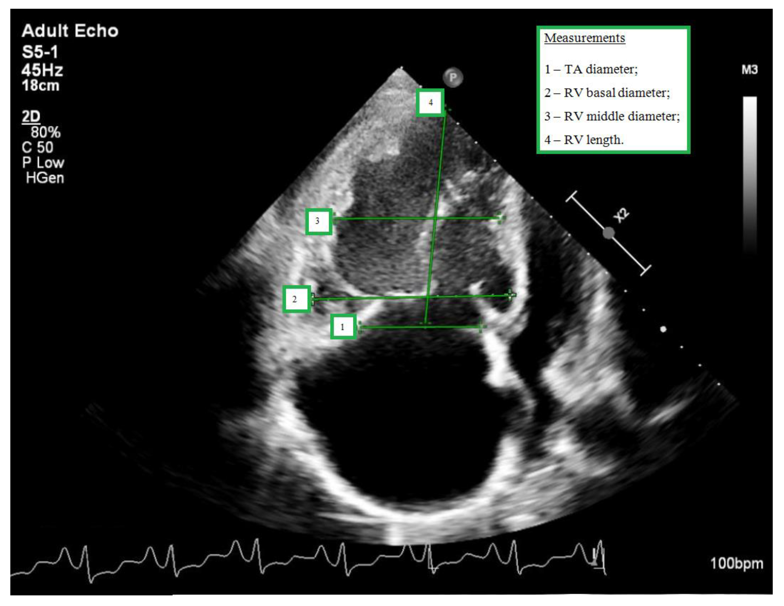
2.2. Transthoracic Echocardiography
2.3. Statistical Analysis
3. Results
3.1. Study Population
3.2. Comparison and Difference in 3D TV Geometry and Right Heart Remodelling between Controls and fTR
3.3. Comparison of Left and Right Heart Remodelling and 3D TV Geometry between Different Etiology fTR Groups
3.4. Comparison of RV Geometry and Function between Different Etiology fTR Groups
3.5. Comparison of TV Geometry between Different Etiology fTR Groups
3.6. Relation of 3D TV and RV Geometry and Severity of fTR
3.7. Prediction of Severe fTR in Different fTR Etiologies
4. Discussion
5. Study Limitations
6. Conclusions
Supplementary Materials
Author Contributions
Funding
Institutional Review Board Statement
Informed Consent Statement
Data Availability Statement
Acknowledgments
Conflicts of Interest
References
- Muraru, D.; Guta, A.-C.; Ochoa-Jimenez, R.C.; Bartos, D.; Aruta, P.; Mihaila, S.; Popescu, B.A.; Iliceto, S.; Basso, C.; Badano, L.P. Functional Regurgitation of Atrioventricular Valves and Atrial Fibrillation: An Elusive Pathophysiological Link Deserving Further Attention. J. Am. Soc. Echocardiogr. 2020, 33, 42–53. [Google Scholar] [CrossRef] [PubMed]
- Lancellotti, P.; Moura, L.; Pierard, L.A.; Agricola, E.; Popescu, B.A.; Tribouilloy, C.; Hagendorff, A.; Monin, J.-L.; Badano, L.; Zamorano, J.L.; et al. European Association of Echocardiography recommendations for the assessment of valvular regurgitation. Part 2: Mitral and tricuspid regurgitation (native valve disease). Eur. J. Echocardiogr. 2010, 11, 307–332. [Google Scholar] [CrossRef] [PubMed] [Green Version]
- Galiè, N.; Humbert, M.; Vachiery, J.; Gibbs, S.; Lang, I.; Torbicki, A.; Simmonneau, G.; Peacock, A.; Vonk Noordegraaf, A.; Beghetti, M.; et al. 2015 ESC/ERS Guidelines for the diagnosis and treatment of pulmonary hypertension: The Joint Task Force for the Diagnosis and Treatment of Pulmonary Hypertension of the European Society of Cardiology (ESC) and the European Respiratory Society (ERS): Endorsed by: Association for European Paediatric and Congenital Cardiology (AEPC), International Society for Heart and Lung Transplantation (ISHLT). Eur. Heart J. 2016, 37, 67–119. [Google Scholar] [CrossRef] [PubMed]
- Vahanian, A.; Beyersdorf, F.; Praz, F.; Milojevic, M.; Baldus, S.; Bauersachs, J.; Capodanno, D.; Conradi, L.; De Bonis, M.; De Paulis, R.; et al. 2021 ESC/EACTS Guidelines for the management of valvular heart disease. Eur. Heart J. 2022, 43, 561–632, Erratum in Eur. Heart J. 2022, 43, 2022. [Google Scholar] [CrossRef] [PubMed]
- Nath, J.; Foster, E.; Heidenreich, P.A. Impact of tricuspid regurgitation on long-term survival. J. Am. Coll. Cardiol. 2004, 43, 405–409. [Google Scholar] [CrossRef] [PubMed] [Green Version]
- Topilsky, Y.; Khanna, A.; Le Tourneau, T.; Park, S.; Michelena, H.; Suri, R.; Mahoney, D.W.; Enriquez-Sarano, M. Clinical context, and mechanism of functional tricuspid regurgitation in patients with and without pulmonary hypertension. Circ. Cardiovasc. Imaging 2012, 5, 314–323. [Google Scholar] [CrossRef] [PubMed]
- Shiran, A.; Sagie, A. Tricuspid regurgitation in mitral valve disease incidence, prognostic implications, mechanism, and management. J. Am. Coll. Cardiol. 2009, 53, 401–408. [Google Scholar] [CrossRef] [Green Version]
- Tornos Mas, P.T.; Rodríguez-Palomares, J.F.; Antunes, M.J. Secondary tricuspid valve regurgitation: A forgotten entity. Heart 2015, 101, 1840–1848. [Google Scholar] [CrossRef] [Green Version]
- Neuhold, S.; Huelsmann, M.; Pernicka, E.; Graf, A.; Bonderman, D.; Adlbrecht, C.; Binder, T.; Maurer, G.; Pacher, R.; Mascherbauer, J. Impact of tricuspid regurgitation on survival in patients with chronic heart failure: Unexpected findings of a long-term observational study. Eur. Heart J. 2013, 34, 844–852. [Google Scholar] [CrossRef] [Green Version]
- Badano, L.P.; Agricola, E.; De Isla, L.P.; Gianfagna, P.; Zamorano, J.L. Evaluation of the tricuspid valve morphology and function by transthoracic real-time three-dimensional echocardiography. Eur. J. Echocardiogr. 2009, 10, 477–484. [Google Scholar] [CrossRef]
- Addetia, K.; Yamat, M.; Mediratta, A.; Medvedofsky, D.; Patel, M.; Ferrara, P.; Mor-Avi, V.; Lang, R.M. Comprehensive Two-Dimensional Interrogation of the Tricuspid Valve Using Knowledge Derived from Three-Dimensional Echocardiography. J. Am. Soc. Echocardiogr. 2016, 29, 74–82. [Google Scholar] [CrossRef] [PubMed] [Green Version]
- Muraru, D.; Badano, L.P.; Sarais, C.; Soldà, E.; Iliceto, S. Evaluation of tricuspid valve morphology and function by transthoracic three-dimensional echocardiography. Curr. Cardiol. Rep. 2011, 13, 242–249. [Google Scholar] [CrossRef] [PubMed]
- Muraru, D.; Hahn, R.T.; Soliman, O.I.; Faletra, F.F.; Basso, C.; Badano, L.P. 3-Dimensional Echocardiography in Imaging the Tricuspid Valve. JACC Cardiovasc. Imaging 2019, 12, 500–515. [Google Scholar] [CrossRef] [PubMed]
- Lang, R.M.; Badano, L.P.; Mor-Avi, V.; Afilalo, J.; Armstrong, A.; Ernande, L.; Flachskampf, F.A.; Foster, E.; Goldstein, S.A.; Kuznetsova, T.; et al. Recommendations for cardiac chamber quantification by echocardiography in adults: An update from the American Society of Echocardiography and the European Association of Cardiovascular Imaging. Eur. Heart J. Cardiovasc. Imaging 2015, 16, 233–271, Erratum in Eur. Heart J. Cardiovasc. Imaging 2016, 17, 412; Erratum in Eur. Heart J. Cardiovasc. Imaging 2016, 17, 969. [Google Scholar] [CrossRef] [PubMed]
- Maffessanti, F.; Muraru, D.; Esposito, R.; Gripari, P.; Ermacora, D.; Santoro, C.; Tamborini, G.; Galderisi, M.; Pepi, M.; Badano, L.P. Age-, body size-, and sex-specific reference values for right ventricular volumes and ejection fraction by three-dimensional echocardiography: A multicenter echocardiographic study in 507 healthy volunteers. Circ. Cardiovasc. Imaging 2013, 6, 700–710. [Google Scholar] [CrossRef] [Green Version]
- Hahn, R.T.; Zamorano, J.L. The need for a new tricuspid regurgitation grading scheme. Eur. Heart J. Cardiovasc. Imaging 2017, 18, 1342–1343. [Google Scholar] [CrossRef] [Green Version]
- Hahn, R.T.; Thomas, J.D.; Khalique, O.K.; Cavalcante, J.L.; Praz, F.; Zoghbi, W.A. Imaging Assessment of Tricuspid Regurgitation Severity. JACC Cardiovasc. Imaging 2019, 12, 469–490. [Google Scholar] [CrossRef]
- Song, J.-M.; Jang, M.-K.; Kim, Y.-J.; Kim, D.-H.; Kang, D.-H.; Song, J.-K. Right ventricular remodeling determines tricuspid valve geometry and the severity of functional tricuspid regurgitation: A real-time 3-dimensional echocardiography study. Korean Circ. J. 2010, 40, 448–453. [Google Scholar] [CrossRef] [Green Version]
- Baumgartner, H.; Falk, V.; Bax, J.J.; De Bonis, M.; Hamm, C.; Holm, P.J.; Iung, B.; Lancellotti, P.; Lansac, E.; Rodriguez Muñoz, D.; et al. 2017 ESC/EACTS Guidelines for the management of valvular heart disease. Eur. Heart J. 2017, 38, 2739–2791. [Google Scholar] [CrossRef]
- Badano, L.P.; Muraru, D.; Enriquez-Sarano, M. Assessment of functional tricuspid regurgitation. Eur. Heart J. 2013, 34, 1875–1885. [Google Scholar] [CrossRef]
- Spinner, E.M.; Lerakis, S.; Higginson, J.; Pernetz, M.; Howell, S.; Veledar, E.; Yoganathan, A.P. Correlates of tricuspid regurgitation as determined by 3D echocardiography: Pulmonary arterial pressure, ventricle geometry, annular dilatation, and papillary muscle displacement. Circ. Cardiovasc. Imaging 2012, 5, 43–50, Erratum in Circ. Cardiovasc. Imaging 2012, 5, e54. [Google Scholar] [CrossRef] [PubMed] [Green Version]
- Utsunomiya, H.; Itabashi, Y.; Mihara, H.; Berdejo, J.; Kobayashi, S.; Siegel, R.J.; Shiota, T. Functional Tricuspid Regurgitation Caused by Chronic Atrial Fibrillation: A Real-Time 3-Dimensional Transesophageal Echocardiography Study. Circ. Cardiovasc. Imaging 2017, 10, e004897. [Google Scholar] [CrossRef] [PubMed] [Green Version]
- Utsunomiya, H.; Harada, Y.; Susawa, H.; Ueda, Y.; Izumi, K.; Itakura, K.; Hidaka, T.; Shiota, T.; Nakano, Y.; Kihara, Y. Tricuspid valve geometry and right heart remodelling: Insights into the mechanism of atrial functional tricuspid regurgitation. Eur. Heart J. Cardiovasc. Imaging 2020, 21, 1068–1078. [Google Scholar] [CrossRef] [PubMed]
- Florescu, D.-R.; Figliozzi, S.; Guta, A.; Vicini, S.; Tomaselli, M.; Târtea, G.C.; Istrătoaie, O.; Parati, G.; Badano, L.; Muraru, D. Atrial functional tricuspid regurgitation: A novel and underappreciated clinical entity. Rom. J. Cardiol. 2021, 31, 27–35. [Google Scholar] [CrossRef]
- Muraru, D.; Addetia, K.; Guta, A.C.; Ochoa-Jimenez, R.C.; Genovese, D.; Veronesi, F.; Basso, C.; Iliceto, S.; Badano, L.P.; Lang, R.M. Right atrial volume is a major determinant of tricuspid annulus area in functional tricuspid regurgitation: A three-dimensional echocardiographic study. Eur. Heart J. Cardiovasc. Imaging 2021, 22, 660–669, Erratum in Eur. Heart J. Cardiovasc. Imaging 2021, 22, 669. [Google Scholar] [CrossRef]
- Hai, T.; Amador, Y.; Mahmood, F.; Jeganathan, J.; Khamooshian, A.; Knio, Z.O.; Matyal, R.; Nicoara, A.; Liu, D.C.; Senthilnathan, V.; et al. Changes in Tricuspid Annular Geometry in Patients with Functional Tricuspid Regurgitation. J. Cardiothorac. Vasc. Anesthesia 2017, 31, 2106–2114. [Google Scholar] [CrossRef]
- Caravita, S.; Figliozzi, S.; Florescu, D.-R.; Volpato, V.; Oliverio, G.; Tomaselli, M.; Torlasco, C.; Muscogiuri, G.; Cernigliaro, F.; Parati, G.; et al. Recent advances in multimodality imaging of the tricuspid valve. Expert Rev. Med. Devices 2021, 18, 1069–1081. [Google Scholar] [CrossRef]


| LVSP | PH | Controls | p-Value for LSVP vs. PH | p-Value for LSVP vs. Controls | p-Value for PH vs. Controls | |
|---|---|---|---|---|---|---|
| Clinical characteristics | ||||||
| Age, years | 68 (8.55) | 58 (14.86) | 62 (12.05) | 0.001 | 0.029 | 0.629 |
| Sex, females % | 46.4 | 72.5 | 52.6 | 0.026 | 0.165 | 0.029 |
| Body mass index, kg/m2 | 27.9 (5.06) | 26.9 (6.14) | 27.4 (4.51) | 0.374 | 0.675 | 0.782 |
| PA pressures | ||||||
| Max. systolic PAP, mmHg | 53.44 (23.25) | 73.16 (38.48) | 24.16 (5.4) | <0.001 | <0.001 | <0.001 |
| Mean PAP, mmHg | 38 (11.75) | 42 (7.5) | 14 (9) | 0.001 | <0.001 | <0.001 |
| RA parameters | ||||||
| RA diameter, mm | 50 (9) | 56.5 (15.75) | 38 (5) | 0.004 | <0.001 | <0.001 |
| RA area, cm2 | 27.8 (10.18) | 30.89 (13.17) | 17.3 (5.4) | 0.484 | <0.001 | <0.001 |
| RA volume, mL | 97.7 (59.26) | 130 (70.75) | 47.77 (21) | 0.286 | <0.001 | <0.001 |
| RV parameters | ||||||
| RV parasternal diastolic diameter, mm | 36 (7.5) | 40 (8.5) | 31 (6.5) | 0.019 | 0.003 | <0.001 |
| RV basal diameter, mm | 45 (7) | 52 (11.75) | 34 (5) | 0.001 | <0.001 | <0.001 |
| RV middle diameter, mm | 36 (9.25) | 47 (12.5) | 28.5 (3.25) | <0.001 | 0.001 | <0.001 |
| RV length, mm | 63 (17.5) | 71 (8.72) | 66 (11.5) | <0.001 | 0.504 | 0.043 |
| RV sphericity index, % | 0.57 (0.17) | 0.66 (0.19) | 0.44 (0.06) | <0.001 | <0.001 | <0.001 |
| RV end-diastolic area, cm2 | 22 (10.44) | 31.14 (15.57) | 17.6 (4.98) | <0.001 | 0.028 | <0.001 |
| RV end-systolic area, cm2 | 14 (7.12) | 23.1 (14.1) | 10 (3.4) | <0.001 | 0.002 | <0.001 |
| FAC, % | 0.32 (0.1) | 0.25 (0.1) | 0.43 (0.08) | <0.001 | <0.001 | <0.001 |
| TAPSE, mm | 17.5 (5.5) | 16 (5.25) | 24 (7) | 0.054 | <0.001 | <0.001 |
| RV S’, cm/s | 10 (5) | 9 (2.75) | 12 (4.5) | 0.039 | 0.007 | <0.001 |
| RV septal wall strain, % | −10.7 (4.75) | −11 (3.9) | −21.1 (3.9) | 0.909 | <0.001 | <0.001 |
| RV lateral wall strain, % | −20.15 (6.3) | −16.6 (6.3) | −25.8 (3.2) | 0.028 | <0.001 | <0.001 |
| TV parameters | ||||||
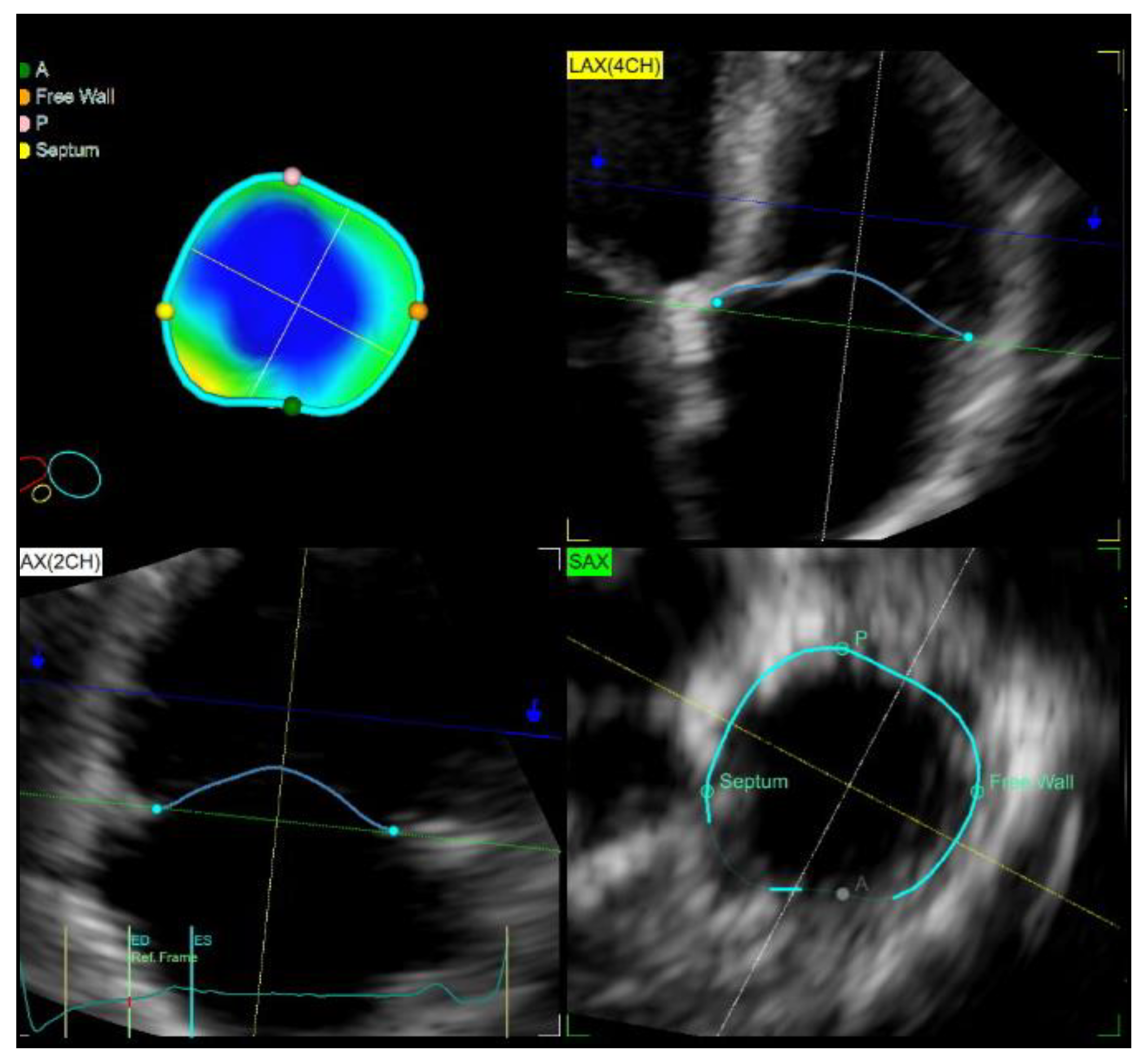
| TA diastolic diameter, mm | 43 (5) | 43 (5.5) | 32 (6.25) | 0.604 | <0.001 | <0.001 |
| TA diastolic diameter index, mm/m2 | 21.86 (3.4) | 23.22 (5.53) | 17.09 (2.95) | 0.061 | <0.001 | <0.001 |
| TA systolic diameter, mm | 39 (5.5) | 40 (4.75) | 29 (6.13) | 0.73 | <0.001 | <0.001 |
| TA systolic diameter index, mm/m2 | 20 (2.54) | 21.25 (5.27) | 15.62 (2.42) | 0.102 | <0.001 | <0.001 |
| TV leaflet tethering height, mm | 5.5 (2.3) | 7.8 (3.7) | 3.3 (2) | <0.001 | <0.001 | <0.001 |
| TV tenting area (cm2) | 1.25 (0.7) | 1.79 (1.04) | 0.68 (0.32) | <0.001 | <0.001 | <0.001 |
| TV EROA, mm2 | 28.94 (20.97) | 29.96 (23.76) | 0.464 | |||
| LVSP | PH | Controls | p-Value for LSVP vs. PH | p-Value for LSVP vs. Controls | p-Value for PH vs. Controls | |
|---|---|---|---|---|---|---|
| RV parameters | ||||||
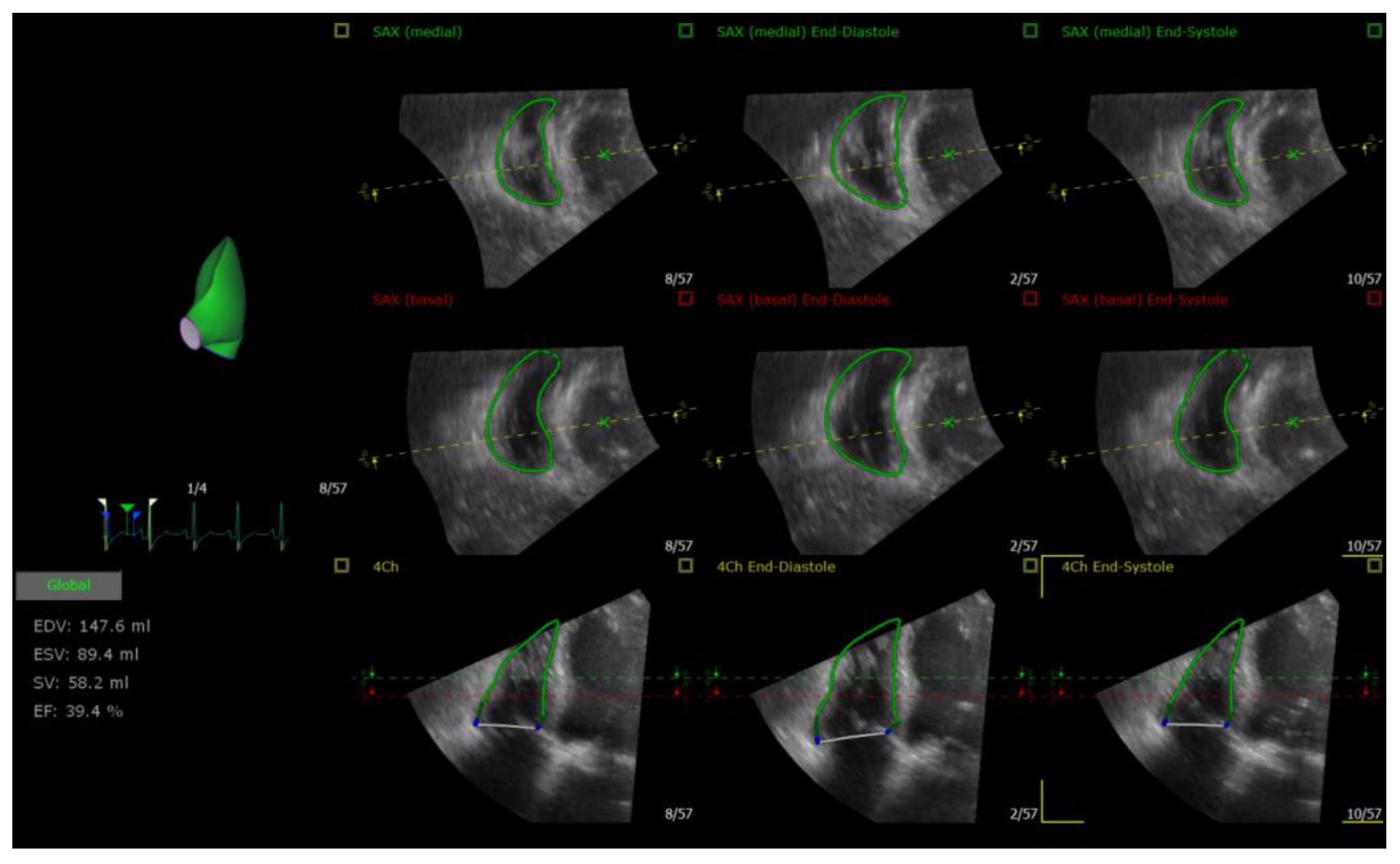
| RV end-diastolic volume, mL | 147.05 (103.08) | 182.75 (105.03) | 85.5 (86.75) | 0.007 | 0.003 | <0.001 |
| RV end-systolic volume, mL | 86.65 (65.23) | 129.5 (76.68) | 44.9 (46.9) | 0.003 | <0.001 | <0.001 |
| RV EF, % | 40.5 (6.73) | 30.85 (10.48) | 49.8 (5.85) | 0.001 | <0.001 | <0.001 |
| TV parameters | ||||||
| TA area, cm2 | 14.3 (4.4) | 13.2 (3.4) | 8.1 (0.93) | 0.649 | <0.001 | <0.001 |
| TA area index (cm2/m2) | 7.13 (2.25) | 6.88 (2.17) | 4.45 (0.97) | 0.713 | <0.001 | <0.001 |
| TA perimeter, mm | 130 (23) | 133 (17) | 102 (7.5) | 0.979 | <0.001 | <0.001 |
| TA perimeter index (cm/m2) | 66.4 (15.8) | 71.1 (19.3) | 55 (9.8) | 0.065 | 0.004 | <0.001 |
| Septal-Lateral Systolic TA Diameter, mm | 42 (5) | 44 (7) | 33.5 (4.5) | 0.37 | <0.001 | <0.001 |
| Septal-Lateral Systolic TA Diameter Index, cm/m2 | 21.5 (3.1) | 23.6 (7.1) | 18.1 (2.7) | 0.021 | 0.003 | <0.001 |
| Septal-Lateral Diastolic TA Diameter, mm | 45 (5) | 46 (7) | 36.5 (5.8) | 0.329 | <0.001 | <0.001 |
| Septal-Lateral Diastolic TA Diameter Index, mm/m2 | 22.9 (4) | 25.1 (4.9) | 19.1 (1.8) | 0.023 | 0.006 | <0.001 |
| Anterior–Posterior TA Diameter, mm | 40 (8) | 35 (7) | 28 (4.5) | 0.037 | <0.001 | <0.001 |
| Anterior–Posterior TA Diameter Index, mm/m2 | 20.3 (4.7) | 18.8 (5.5) | 15.6 (4.5) | 0.411 | <0.001 | 0.003 |
| Major Axis Systolic TA Diameter, mm | 46 (5) | 46 (6) | 36 (2.8) | 0.791 | <0.001 | <0.001 |
| Major Axis Systolic TA Diameter Index, mm/m2 | 23.2 (4.7) | 25 (5.2) | 20.1 (1.9) | 0.074 | 0.003 | <0.001 |
| Major Axis Diastolic TA Diameter, mm | 48 (6.5) | 48 (8) | 39.5 (5) | 0.777 | <0.001 | <0.001 |
| Major Axis Diastolic TA Diameter Index, mm/m2 | 23.9 (4.4) | 26.2 (5.2) | 20.3 (2.8) | 0.044 | 0.006 | <0.001 |
| Minor Axis Diastolic TA Diameter, mm | 39 (8) | 36 (6) | 27 (3.5) | 0.222 | <0.001 | <0.001 |
| Minor Axis Diastolic TA Diameter Index, mm/m2 | 19.8 (4.4) | 19.7 (5.9) | 15.2 (3.7) | 0.908 | <0.001 | <0.001 |
| TV Leaflet Coaptation point Height, mm | 9 (5.5) | 13 (3) | 6.5 (2) | <0.001 | 0.012 | <0.001 |
| TV Leaflet Tenting Volume, mL | 3.9 (2) | 5 (2.9) | 1.55 (0.25) | 0.025 | <0.001 | <0.001 |
| TV Sphericity Index, % | 83.67 (11.33) | 80 (14.04) | 73.61 (12.4) | 0.04 | 0.002 | 0.13 |
| Univariate | Multivariate | |||||
|---|---|---|---|---|---|---|
| OR | 95% CI | p-Value | OR | 95% CI | p-Value | |
| 3D echo-derived TV parameters | ||||||
| TA area, cm2 | 1.434 | 1.163–1.769 | <0.001 | |||
| TA perimeter, mm | 1.058 | 1.021–1.096 | 0.002 | 0.421 | 0.221–0.975 | 0.042 |
| Septal-Lateral Systolic TA Diameter, mm | 1.507 | 1.233–1.842 | <0.001 | 1.651 | 1.042–2.385 | 0.028 |
| Septal-Lateral Diastolic TA Diameter, mm | 1.431 | 1.193–1.718 | <0.001 | |||
| Major Axis Systolic TA Diameter, mm | 1.386 | 1.176–1.632 | <0.001 | |||
| Major Axis Diastolic TA Diameter, mm | 1.363 | 1.165–1.594 | <0.001 | |||
| TV Leaflet Tenting Volume, mL | 1.580 | 1.177–2.120 | 0.002 | |||
| 2D echo-derived TV parameter | ||||||
| 4-Chambers Systolic Diameter, mm | 1.255 | 1.106–1.424 | <0.001 | |||
| RV parameters | ||||||
| 2D RV basal diameter, mm | 1.149 | 1.073–1.230 | <0.001 | 1.198 | 1.021–1.409 | 0.029 |
| 2D RV middle diameter, mm | 1.083 | 1.036–1.133 | <0.001 | |||
| RV end-diastolic area, cm2 | 1.096 | 1.042–1.152 | <0.001 | |||
| RV end-systolic area, cm2 | 1.112 | 1.050–1.178 | <0.001 | |||
| RV EF, % | 0.911 | 0.855-.0971 | 0.004 | |||
| LSVP | PH | |||||
|---|---|---|---|---|---|---|
| OR | 95% CI | p-Value | OR | 95% CI | p-Value | |
| TA area, cm2 | 1.282 | 1.001–1.641 | 0.05 | 2.198 | 1.138–4.244 | 0.02 |
| TA perimeter, mm | 1.032 | 0.995–1.070 | 0.09 | 1.242 | 1.033–1.495 | 0.02 |
| Septal-Lateral Systolic TA Diameter, mm | 1.430 | 1.123–1.820 | <0.001 | 1.717 | 1.149–2.566 | 0.01 |
| Septal-Lateral Diastolic TA Diameter, mm | 1.394 | 1.108–1.754 | <0.001 | 1.498 | 1.089–2.062 | 0.01 |
| Anterior-Posterior TA Diameter, mm | 1.147 | 0.999–1.316 | 0.051 | 1.246 | 1.022–1.518 | 0.03 |
| Major Axis Systolic TA Diameter, mm | 1.268 | 1.066–1.508 | 0.01 | 1.934 | 1.149–3.256 | 0.01 |
| Major Axis Diastolic TA Diameter, mm | 1.253 | 1.059–1.484 | 0.01 | 1.716 | 1.154–2.550 | 0.01 |
| Minor Axis Diastolic TA Diameter, mm | 1.118 | 0.976–1.281 | 0.11 | 1.228 | 1.009–1.493 | 0.04 |
| TV Leaflet Tenting Volume, mL | 1.398 | 0.995–1.965 | 0.53 | 2.149 | 1.143–4.040 | 0.02 |
Disclaimer/Publisher’s Note: The statements, opinions and data contained in all publications are solely those of the individual author(s) and contributor(s) and not of MDPI and/or the editor(s). MDPI and/or the editor(s) disclaim responsibility for any injury to people or property resulting from any ideas, methods, instructions or products referred to in the content. |
© 2022 by the authors. Licensee MDPI, Basel, Switzerland. This article is an open access article distributed under the terms and conditions of the Creative Commons Attribution (CC BY) license (https://creativecommons.org/licenses/by/4.0/).
Share and Cite
Krivickienė, A.; Verikas, D.; Krečkauskienė, R.; Padervinskienė, L.; Hoppenot, D.; Miliauskas, S.; Vaškelytė, J.J.; Ereminienė, E. Different Causes of Functional Tricuspid Valve Regurgitation Are Linked to Differences in Tricuspid Valve and Right-Sided Heart Geometry and Function: 3D Echocardiography Study. Medicina 2023, 59, 57. https://0-doi-org.brum.beds.ac.uk/10.3390/medicina59010057
Krivickienė A, Verikas D, Krečkauskienė R, Padervinskienė L, Hoppenot D, Miliauskas S, Vaškelytė JJ, Ereminienė E. Different Causes of Functional Tricuspid Valve Regurgitation Are Linked to Differences in Tricuspid Valve and Right-Sided Heart Geometry and Function: 3D Echocardiography Study. Medicina. 2023; 59(1):57. https://0-doi-org.brum.beds.ac.uk/10.3390/medicina59010057
Chicago/Turabian StyleKrivickienė, Aušra, Dovydas Verikas, Rita Krečkauskienė, Lina Padervinskienė, Deimantė Hoppenot, Skaidrius Miliauskas, Justina Jolanta Vaškelytė, and Eglė Ereminienė. 2023. "Different Causes of Functional Tricuspid Valve Regurgitation Are Linked to Differences in Tricuspid Valve and Right-Sided Heart Geometry and Function: 3D Echocardiography Study" Medicina 59, no. 1: 57. https://0-doi-org.brum.beds.ac.uk/10.3390/medicina59010057