1. Introduction
Therapeutic protein has attracted extensive attention in recent years due to its tremendous therapeutic benefits in treating widespreading and severe health problems such as cancer, infectious diseases, autoimmune diseases, AIDS/HIV, and related conditions [
1,
2]. Unfortunately, most proteins are highly vulnerable, as their activity can be strongly influenced in vitro by many factors such as pH, ionic strength, temperature, high pressure, non-aqueous solvents, etc. Additionally, proteins also can be easily degraded by enzymes and proteases in vivo [
3], not only at the administration site but also en route to the site of pharmacological action, resulting in a reduced bioavailability.
For these reasons, competent drug delivery systems, typically nanoparticles (NPs), are commonly utilized to load these proteins as cargos, potentially producing essential benefits, such as enhancing solubility and thermodynamic stability of proteins, controlling the release of protein molecules, improving biodistribution and prolonged half-lives of proteins, as well as targeting the diseased tissue in vivo [
4,
5]. In fact, different drug-loaded NPs have their unique advantages. For example, anticancer protein-loaded NPs are able to penetrate tumors due to their small particle size, and the leaky nature of tumor microvasculature [
6,
7]. For infectious diseases, NPs are often used as vehicles for co-delivery of antigen and immunostimulatory molecules to the same APC, which can promote cross-presentation and help elicit a broader cellular response [
8]. In order to reduce the cytotoxicity of inorganic nanoparticles during drug administration and to expand the potential applications of nanomedicine technology, plenty of researches have been extensively invested into. More recently, various natural biomaterials, especially polysaccharides, have been greatly introduced and developed for protein-loaded NPs, because of their outstanding physical and biological properties. Chitosan (CS), the only natural cationic biopolymer, is one of the most up-and-coming biopolymers among all the natural polymers and [
9] is widely applied because of remarkable properties such as biocompatibility, non-toxicity, biodegradability, and low immunogenicity. Most CS are present in the cuticles of arthropods, especially in the shells of marine animals such as crabs and shrimps [
10]. The free amino groups of CS can spontaneously form NPs with anionic molecules, and this ionic gelation process is relatively mild, avoiding the use of harsh organic chemicals and high temperatures, so that proteins can be successfully encapsulated. It has been reported that Chitosan nanoparticles (CS-NPs) loading proteins (e.g., bovine serum albumin (BSA), ovalbumin (OVA), insulin, antigens, lysozyme) [
9] were prepared via the ionic gelation process to obtain high encapsulation efficiency (EE), with no change in the biological activity of proteins, which can be a very promising candidate for drug delivery applications [
11].
A large number of studies have been taken on NPs characteristics (e.g., particle size distribution, surface charge) of different therapeutic proteins; very few of these studies have focused on the microstructure of protein-loaded NPs, which may closely relate to the in vivo pharmacokinetics/pharmacodynamics processes [
12,
13]. For example, high molecular weight chitosan (HMWC) is generally associated with higher EE, because high molecular weight polymer chains facilitate the immobilization of proteins within the chitosan matrix [
14,
15], accompanied by more complex NP microstructures. Meanwhile, the release was much faster when the loading capacity (LC) was higher, due to the release of proteins mainly located on the NP surface [
16]. It is generally recognized that the release mechanisms of proteins loaded in chitosan NP are desorption, dissociation [
17], diffusion, and erosion [
18,
19]. In addition, the mechanism of protein release is greatly affected by the interaction forces and microstructure in NPs, which ultimately affect the pharmacokinetic process in vivo. The conventional methods of nano preparations have numerous disadvantages, for instance, longtime consumption, low yield, low repeatability, and easy contamination. For this reason, nanoparticles have been studied in the laboratory thoroughly but rarely applied to clinical treatment.
In this study, the dynamic high pressure microfluidization (DHPM) was used to address most of the ongoingcritical challenges in amplification reactions during the manufacturing processes of nanoparticle. As shown in
Figure 1, the microjet is a fluid witchthrough a valve with a very small aperture under the ultra-high pressure (310 MPa) to achieve dispersion, homogenization, emulsification etc. It is able to expand the fluid handling from merely micro scale down to a more precise nanoscale [
20], which is a typical technical equipment developed based on the micro-jet technology to achieve the specific purpose of material emulsification and homogenization. The implementation of nanoparticle production through DHPM was reproducible and highly controllable. In addition, DHPM requires less media volumes, shorter time, and is more efficient compared with traditional amplification reactions [
21].
The present study aims to design and develop novel therapeutic protein-loaded NPs with CS, a typical natural biomaterial, to improve the therapeutic effect and bioavailability of such protein. Freeze-drying studies are performed to produce re-dispersible dry powders to improve the stability of BSA-loaded NPs and using the variety of spectroscopy techniques to figure out the interaction forces and microstructure in CS-NPs.
2. Results and Discussion
2.1. NPs Preparation and Characterization
In our preliminary experiment, low molecular weight chitosan (LMWC), medium molecular weight chitosan (MMWC), and high molecular weight chitosan (HMWC) were chosen to prepare NPs. However, as the molecular weight increased, the viscosity of CS also become higher, leading to some problems in the preparation process. For example, CS cannot pass through the filter membrane to remove the impurities, so we chose LMWC as the material for the preparation of NPs.
LMWC is a natural polymer having a number-average molecular weight (MW) of 166,000 (ranging from 104,000 to 225,000). In previous studies, BSA was encapsulated into NPs through a facile self-assembly approach in which electrostatic interaction of positively charged CS with oppositely charged Tripolyphosphate pentasod (TPP) is the primary driving force for nanostructure formation by a self-assembly process. Preliminary experiments were addressed to discovery the main influencing factor in NP formation. The particle size and
Zeta potential of the NPs were determined at 25 °C by the dynamic light scattering (DLS) (Zetasizer Nano ZS90, Malvern, UK). Particle size values were reported as the mean hydrodynamic diameter (MHD), standard deviation (SD), and polydispersity index (PDI). Each batch was analyzed in triplicate. During this process, the opalescence was used as an indicator, which was also confirmed by means of dynamic light scattering [
22]. As shown in
Figure 2A, BSA-loaded NPs suspension was transparent with weak opalescence, indicating the small particle sizes.
Compared with blank NPs (CS: TPP = 3:1), a significant decrease in particle size and PDI were observed in BSA-loaded NPs, showing that BSA was favorable for the formation and stability of NPs, which was consistent with the fluorescence quenching measurement. The effect of CS: TPP weight ratio on particle characteristics is also reported in
Table 1. The obtained results illustrated that the increase in weight ratio was accompanied by an increase in particle size and
Zeta potential. Similar observations have also previously been reported [
23]. All NPs showed positive
Zeta potentials due to protonated amine groups of CS that were responsible for such a positive surface charge. The PDI values of all samples were smaller than 0.3, which suggested the narrow distribution of particle size. EE and loading capacity of “pharmaceutical agents” in NPs are important factors to be considered to evaluate the therapeutic efficacy of nanocarriers by in vitro and in vivo models [
24]. As shown in
Table 1, the highest EE (52.7%) and LC (66.9%) of BSA were observed in the NPs which CS: TPP (
w/w) was 3:1. The reduced usage of TPP would lead to a retarded interaction between the BSA and TPP and a reduced encapsulation of BSA into nanoparticles due to the inadequate crosslinker (TPP), consequently, the weight of the formed NPs was too low (lower than 2 mg) to be weighted. Meanwhile, the increase in BSA EE was also dependent on the concentration of the CS in the NPs (
Table S1), suggesting a high affinity of the positively charged CS binding to BSA.
The situation in which the drug LC is greater than the encapsulation rate probably was caused by the calculation of drug LC, which is greatly related to the weight of nanoparticles, yet the value of the electronic balance was inaccurate when the weight of the analyte was less than 2.0 mg. That is why we use microfluidics for amplification experiments to further make the nanoparticles more uniform. The DHPM can massively produce nanoparticles with a PDI values less than 0.2 (20 times of traditional preparation method), and the calculated drug LC through DHPM is more reliable. This is capable to provide theoretical and practical basis for the industrialization and mass production of nanoparticles.
2.2. Preparation of the NPs through DHPM
To improve the drug LC with an acceptable uniformity of nanoparticles as well as satisfactory repeatability of experimental results, DHPM was selected for post-treating of NPs. After determining the optimal CS: TPP ratio (w/w), according to this ratio, BSA solution (1 mg/mL) in deionized water were added directly to the CS solution, then placed in a high pressure homogenizer for 5 min and TPP solution (0.7 mg/mL) was added to the CS solution dropwise while homogenizing. The nanoparticle crude solution was then placed into the DHPM and circulated under different pressures and different cycle times.
In our study, LMWC (1 mg/mL) was mixed with BSA (1 mg/mL) and dissolved in Tris-HCl buffer (pH 5.5), the CS: TPP (
w/w) = 3:1 was chosen as the optimal ratio for the preparation of NPs. According to this ratio, BSA solution (1 mg/mL) in deionized water were added directly to the CS solution, placed in a high pressure homogenizer for 5 min and TPP solution (0.7 mg/mL) was added to the CS solution dropwise while homogenizing. The effect of pressures and cycle times on particle characteristics is also reported in
Table 2 and
Table 3.
As shown in
Table 2, compared to the coarse NPs, the EE of those post-treated NPs with DHPM has increased significantly, probably because in the high-pressure environment, the free drug on the surface of the nanoparticle is further adsorbed into the nanoparticles. The drug loading the results of the
Table 2 indicated that different pressures had no effect on the particle size,
Zeta potential and EE of the nanoparticles. As the pressure increased, the drug LC of nanoparticles had a significant increase. Because of the limitations of the instrument, we did not choose a higher pressure. The instrument would be rapidly heated to 80 °C in a short time if the pressure continued to increase, leading to an inactivation of the encapsulated protein drug.
As shown in
Table 3, the cycle time had no influence on the particle size,
Zeta potential, morphology, EE, and drug LC. On the contrary, when increasing the cycle time from 0–8 min, the drug LC firstly increased from 33.4% to 39.8%, indicating that the more cycles, the more drug LC of the nanoparticles. However, further increasing the cycle time to 10 min led to partial desorption of the nanoparticles, which will reduce the drug LC.
The morphology of freshly prepared NPs was observed under transmission electron microscope (TEM), as shown in
Figure 2B, which exhibited spherical shape and uniform-size distribution consistent with the DLS measurement (
Figure 2C). DLS showed that the particle size of NPs is 137 nm while TEM indicated about 130 nm. This is probably because DLS reflects the hydrodynamic diameter of NPs swelling in aqueous solution, whereas TEM reflects the diameter of dried NPs. Therefore, the diameter determined by TEM was smaller than that measured by DLS [
25].
2.3. NPs Lyophilization
NPs are often produced in an aqueous suspension form. However, NP suspension is unstable. For example, particle aggregation and fusion, polymers degradation and drug leakage, and growth of microorganisms [
26,
27] often occur during long periods of storage. Freeze-drying of aqueous suspensions into solid powders has been used to improve the long-term stability of NPs [
28]. However, this dehydration process may generate freezing and desiccation stresses on the NPs, accompanied by irreversible aggregation of the NPs. These stresses can be minimized by using cryoprotectants and lyoprotectants, possibly due to the formation of amorphous cryoconcentrated suspension or ice during the freezing or drying steps. The cryoconcentrated phase is constituted by nanoparticles, surfactants, unloaded drugs, etc. A high concentration of nanoparticles may cause aggregation or fusion in the process of cryoconcentrating, and the crystallization of ice may cause mechanical stress to nanoparticles. The excipients were added to protect nanoparticles from freezing stresses [
29]. In our study, it was found that most of the NPs without any cryo- or lyoprotectants were aggregated. Therefore, cryo- and lyoprotectants were necessary for the lyophilization process. On the premise of adding cryo- and lyoprotectants, the DLS was used to characterize the nanoparticles before and after lyophilization by the method described in 2.1. The increase of particle size of re-dissolved BSA-loaded NPs after lyophilizing was calculated to evaluate the protective ability of cryo- and lyoprotectants during the lyophilization process of nanoparticles. With the purpose of cryoprotection and cryopreservation for the lyophilized samples, different sugars such as trehalose, glucose, sucrose, and mannitol were used [
30]. NPs lyophilized with mannitol could not completely re-dissolve in water [
12]. Re-dispersibility is defined as the ability of dried NPs to recover to their original state in an aqueous system, evaluated in terms of particle size, PDI, and morphology. After lyophilization, NPs with sucrose, glucose or trehalose as cryoprotectant exhibited almost common behavior when the concentration was 10%, and the particle size of those were all smaller than 200 nm (
Figure 3A). With the increase of mass ratio of cryoprotectant to NPs (5:1 to 10:1), the particle size of lyophilized samples was smaller, which was similar to newly synthesized NPs. Re-dissolved BSA-loaded NPs were transparent in their appearance with more obvious opalescence owing to the NPs enrichment (
Figure 3B).
Trehalose could stabilize biological macromlecules (e.g., proteins) in the folded state under conditions that would normally promote their denaturation [
31]. Fonte et al. [
32] demonstrated that trehalose showed superior insulin stability in insulin-loaded PLGA NPs. Trehalose was selected as a cryoprotectant with albumin-loaded NPs, and the main mechanism was explored regarding cryoprotection [
33]. As shown in
Figure 3C, comparing the different concentrations of trehalose (
w/v), it emerged that at a concentration of 5, lyophilized NPs appeared with a greater increase in particle size than at 10 and 15. In contrast, the increase in particle size was less than 50 nm and the PDI was less than 0.25 when the concentration of trehalose was 10% and 15%, suggesting that a complete re-dispersion was obtained. Furthermore, the effect of trehalose on the formulation at a concentration of 15% was studied by analyzing the TEM images (
Figure 3D), indicating that the particle size of BSA-loaded NPs with trehalose was similar to the NPs before lyophilization (
Figure 2B). Meanwhile, NP aggregation resulted in higher particle size and PDI in DLS.
Therefore, it can be concluded that for BSA-loaded NPs, the use of trehalose with the appropriate concentration as lyoprotectant is very effective in controlling the particle size of NPs without affecting the physicochemical characteristics after re-dispersion of the freeze-dried samples.
2.4. Microstructure of NPs
2.4.1. Fluorescence Quenching of BSA
Tryptophan (Trp) fluorescence quenching assay is considered to be a useful method to provide information on the binding properties of drugs to proteins, such as the quenching mechanisms, binding mechanisms, binding constants, binding patterns, and binding sites [
34]. As shown in
Figure 4A, the fluorescence quenching spectra produced maximum emission at 338 nm, indicating that the binding sites were close to Trp residues. Meanwhile, with the addition of CS, a noticeable decrease occurred in the fluorescence intensity of BSA, suggesting a direct interaction between CS and BSA. Similar fluorescence quenching was reported upon the interaction of various drugs with BSA [
35,
36]. The binding kinetics between CS and BSA were further investigated using the Stern–Volmer equation (Equation (1)):
where
F0 and
F are the fluorescence intensities of BSA aqueous solution in the absence and presence of the quencher, respectively.
Kq is quenching rate constant.
τ0 is the lifetime of the fluorophore in the absence of quencher and its value is 5.9 ns for BSA aqueous solution [
35]. [Q] is the quencher concentration and
Ksv is the Stern–Volmer quenching constant.
The values of
Kq and
Ksv obtained from the slope of Stern–Volmer plots (
Figure 4B) are reported in
Table 4.
In general, the fluorescence quenching mechanisms are classified as dynamic quenching (collision encounter), static quenching (non-fluorescent complex formation), or combination mechanism [
37]. Furthermore, dynamic and static quenching can be distinguished by their different factors in terms of temperature, viscosity, and excited-state lifetime. For dynamic quenching, higher temperature results in faster diffusion collisions and larger quenching constant [
38]. In this study, the
Ksv value was positively correlated with the rising temperature, which may indicate that dynamic quenching is involved in the quenching process. Besides, the
Kq value was greater than the maximum diffusion collision quenching rate constant (2 × 10
10 M
−1 s
−1), indicating the presence of static quenching during the binding process. Therefore, we can perorate that the fluorescence quenching mechanism of BSA with CS is triggered by a combination mechanism.
2.4.2. Interaction Parameter and the Combination Types
The non-covalent interactions between polysaccharides and proteins in aqueous solutions can be driven by hydrogen bonding, hydrophobic and electrostatic interactions [
39]. In order to elucidate the nature of the binding force between CS and BSA, the fluorescence data was also used to determine the thermodynamic parameters of the complex by the following equations (Equation (2)):
where
Kb is the binding constant, and
n is the number of binding sites.
The value of Δ
H0 can be regarded as a constant when there is no obvious change in temperature. Then the values of Δ
H, Δ
S, and Δ
G can be evaluated by the van’t Hoff and thermodynamic equations (Equations (3) and (4)):
where
R is the gas constant. The experimental temperatures used were 298, 303 and 308 K.
The results are tabulated in
Table 5. The sign of ∆
G determines the feasibility of the binding reaction between polymer and protein. If Δ
H > 0 and Δ
S > 0, the main force is a hydrophobic force. If Δ
H < 0 and Δ
S > 0, the electrostatic force dominates the interaction. Finally, if Δ
H < 0 and Δ
S < 0, both van der Waals forces and hydrogen bonds are established [
40]. The negative sign of
∆G indicates the spontaneous binding of CS to BSA.
Table 5 shows ∆
H > 0 (407.40 kJ mol
−1) and ∆
S > 0 (1.43 kJ mol
−1 K
−1), implying that hydrophobic force is the main force between CS and BSA. Furthermore, it is generally agreed that if
Kb for the interaction of BSA with various substances is in the range of 115 × 10
4 M
−1, the binding affinity is moderate [
41]. Interestingly, at 308K, the binding constant of CS and BSA was 5.988 × 10
5 M
−1, and
Kb increased with higher temperature, suggesting that the binding affinity of CS and BSA was high under the preparation conditions.
2.4.3. Fourier Transform Infrared Spectroscopy (FT-IR)
FT-IR was used to determine variations in chemical functional groups in the samples (
Figure 4A). Generally, according to the previously published literature [
42,
43,
44], CS (
Figure 4C(a)) showed specific peaks at 3354.9 (–OH and –NH
2 stretching), 2873.2 (–CH stretching), 1645.0 (amide I, C O stretching), 1588.4 (amide II, N–H bending), 1419.8 (–CH
2 bending), 1375.3 (–CH
3 symmetric deformation), 1026.1 (C–O–C stretching), and 895.1 cm
−1 (glucose ring) [
45]. Compared with the spectrum of CS, the peak of blank NPs (
Figure 4C(b)) shifted from 3354.9 to 3249.2 cm
−1 and became wider and flatter, indicating that hydrogen bonding was enhanced. The peaks of amide I and amide II in blank NPs shifted to 1633.3 and 1535.8 cm
−1, respectively, due to the electrostatic interactions between phosphoric groups of TPP and amino groups of CS in NPs. These observations were consistent with the results reported previously [
46,
47]. BSA (
Figure 4C(c)) showed specific peaks at 3283.9 (–NH
2 stretching), 2930.1 (–CH stretching), 1638.6 (amide I), 1532.6 (amide II), and 1391.5 cm
−1 (amide III, C–N stretching). In comparison with blank NPs, the BSA-loaded NPs (
Figure 4C(d)) showed a significant increase in the intensity of the amino acid characteristic peak at 1650–1500 cm
−1, and the peak shape was similar to BSA, reflecting the successful encapsulation of BSA in NPs.
2.4.4. CD Spectra Studies
CD spectroscopy has been widely used to determine the secondary and tertiary structure of proteins in solution, which is sufficiently precise for biopharmaceutical protein characteristics [
48]. Therefore, CD analysis is sensitive enough to detect changes in protein content [
49]. The experimental CD spectra of the BSA-loaded NPs suspension and supernatant are presented in
Figure 4D. BSA is predominantly α-helical, as expected, and the CD spectrum exhibits a strong negative band (ellipticity) at 240–200 nm [
50]. Previous studies [
51,
52] have reported that the manufacturing process of NPs could potentially induce an alteration of the molecular conformation of proteins in such NPs. However, in this study, the obviously raised ellipticity of BSA-loaded NPs suspension, which is an indicator of α- helical structure of BSA, demonstrated that this ionic gelation process is adequately friendly to the BSA and did not change its molecular structure. In this study, the increase of the alpha-helix of BSA in CS NPs may come from the strong hydrophobic force between CS and BSA [
53,
54,
55]. However, more thorough research is necessary to further explain this interesting fact directly and scientifically.
2.5. In Vitro Release of NPs
In order to investigate the feasibility of CS-NPs as drug carriers for therapeutic proteins, an in vitro release study of BSA-loaded NPs was performed. Previous studies have reported that drug molecule diffusion plays a predominant role in release profile when the particle size of an encapsulated drug molecule is much smaller than the formed NPs [
46]. Proteins are macromolecules, some of which react with CS [
56,
57]; therefore, the release process is particularly complex.
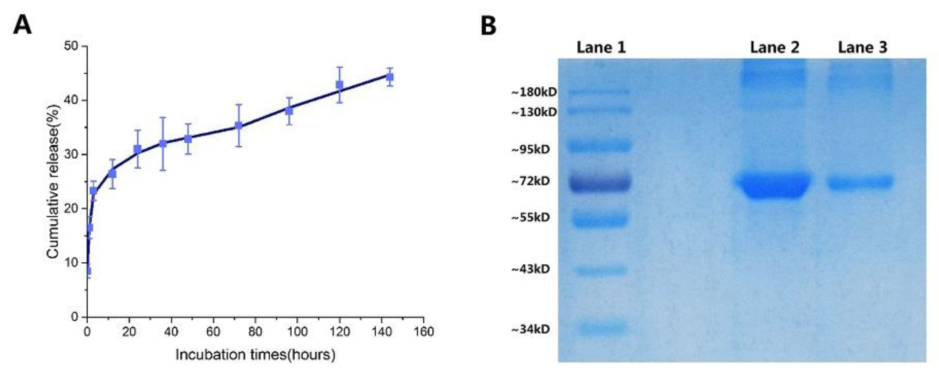
Analysis of BSA release showed a very rapid initial burst (0–12 h), followed by a slow release in the samples (
Figure 5A). Gan and Wang’s research shows that after the burst release of protein, considerable particles swelled with loss of density, increase in particle size, and loss of physical integrity. So Gan and Wang concluded that the burst release of BSA is more likely a consequence of rapid surface desorption of large amounts of protein molecules from a huge specific surface area provided by large numbers of NPs [
19]. Consistent with FT-IR spectra that the BSA-loaded NPs decreased intensity in the main characteristic bands of carbonyl (C=O–NHR) and amine groups (–NH
2) at 1636 cm
−1 and 1540 cm
−1 before and after release at the first 12 h [
44], the degree of deacetylation of chitosan used in this study is high (75–85%), leading to a denser formed nanostructure due to the more available cations and anions. It was then shown that BSA is slowly released after 12 h because protein macromolecules are difficult to release from the pores of the NPs produced by polymer erosion. The high binding affinity of CS to BSA (in
Section 2.4.2) is also a factor responsible for the slow release of BSA. This resulted in the small accumulative release of BSA (44.3% at 144 h) as well.
The integrity of the encapsulated proteins was evaluated by sodium dodecyl sulfate-polyacrylamide gel electrophoresis (SDS-PAGE) analysis. SDS-PAGE analysis followed by Coomassie blue staining revealed identical bands for the native and entrapped proteins (
Figure 5B). This indicates that the preparation and freeze-drying conditions did not cause any irreversible aggregation or cleavage of the proteins [
58].
2.6. Cytotoxicity Analysis
To determine if the lyophilized BSA-loaded NPs were innocuous, the cell viability of L929 cells was measured using the MTT assay. First, L929 cells were added into the 96-well plate at a certain density (dulbecco’s modified eagle’s medium + 10% (
v/v) fetal bovine serum), and then the lyophilized BSA-loaded NPs solution was added to the 96-well plate at different concentrations. After 4 days of cultivation in the CO
2 incubator, the cell survival rate was calculated through the absorbance in the microplate reader. The results are presented in
Table 6, good cell viability (>80%) was observed for L929 cells containing re-dissolved BSA-loaded NPs with varying concentrations from 10 to 100 μg/mL. These results suggest that CS-NPs with trehalose have excellent biocompatibility and these proved the CS-NPs to be a safe vehicle for the delivery of proteins.

 ,
, 


