Article

The Common Sunstar Crossaster papposus—A Neurotoxic Starfish

1
Centre for Environment Fisheries and Aquaculture Science (CEFAS), Barrack Road, Weymouth DT4 8UB, UK
2
Department of Chemistry, University of Surrey, Guildford GU2 7XH, UK
3
Biotoxin Metrology, National Research Council Canada, Halifax, NS B3Z 3H1, Canada
4
Centre for Environment Fisheries and Aquaculture Science (CEFAS), Pakefield Road, Lowestoft NR33 0HT, UK
*
Author to whom correspondence should be addressed.
Submission received: 17 November 2021 / Revised: 1 December 2021 / Accepted: 1 December 2021 / Published: 7 December 2021
(This article belongs to the Special Issue Marine Toxins in Non-traditional Vectors)

Abstract

:
Saxitoxins (STXs) are a family of potent neurotoxins produced naturally by certain species of phytoplankton and cyanobacteria which are extremely toxic to mammalian nervous systems. The accumulation of STXs in bivalve molluscs can significantly impact animal and human health. Recent work conducted in the North Sea highlighted the widespread presence of various saxitoxins in a range of benthic organisms, with the common sunstar (Crossaster papposus) demonstrating high concentrations of saxitoxins. In this study, an extensive sampling program was undertaken across multiple seas surrounding the UK, with 146 starfish and 5 brittlestars of multiple species analysed for STXs. All the common sunstars analysed (n > 70) contained quantifiable levels of STXs, with the total concentrations ranging from 99 to 11,245 µg STX eq/kg. The common sunstars were statistically different in terms of toxin loading to all the other starfish species tested. Two distinct toxic profiles were observed in sunstars, a decarbomylsaxitoxin (dcSTX)-dominant profile which encompassed samples from most of the UK coast and an STX and gonyautoxin2 (GTX2) profile from the North Yorkshire coast of England. Compartmentalisation studies demonstrated that the female gonads exhibited the highest toxin concentrations of all the individual organs tested, with concentrations >40,000 µg STX eq/kg in one sample. All the sunstars, male or female, exhibited the presence of STXs in the skin, digestive glands and gonads. This study highlights that the common sunstar ubiquitously contains STXs, independent of the geographical location around the UK and often at concentrations many times higher than the current regulatory limits for STXs in molluscs; therefore, the common sunstar should be considered toxic hereafter.

1. Introduction

The saxitoxins (STXs) are a group of structurally related neurotoxic alkaloids responsible for the human health syndrome paralytic shellfish poisoning (PSP) [1]. The parent compound saxitoxin (STX), as well as over 50 known analogues, have been described (the common STXs are detailed in Figure 1), all with varying toxicities [2,3]. STXs bind to site one of the voltage-gated Na+ channel, thus stemming the flow of sodium ions into excitable cells. Symptoms include tingling in the extremities, numbness of the lips, vomiting, headaches, ataxia, paralysis and, in severe intoxications, death via respiratory arrest [4]. The toxins are commonly associated with harmful algal blooms of the genera Alexandrium, Gymondinium and Pyrodinium [5,6], as well as some freshwater cyanobacteria [7]. Due to the filter-feeding capacity of bivalve molluscs, the bioaccumulation of STXs into these foodstuffs is a vector of intoxication to humans and animals. To manage the risk to shellfish consumers from PSP, the regulatory testing of bivalve molluscs is a near global requirement, with a maximum permitted level (MPL) of 800 µg STX eq/kg stipulated in EU law [8,9,10].
In the UK, the most common known producers of STXs are the marine dinoflagellate species Alexandrium catenella [11,12] (reported as Alexandrium tamerense group 1) and A. minutum [13]. The common toxin profiles of each Alexandrium species are well described in UK shellfish [14], with A. catenella from Scotland producing a mixed profile containing gonyautoxins1-4 (GTXs), neosaxitoxin (NEO) and saxitoxin (STX) and A. minutum profiles from England and Wales dominated by GTX2&3 and STX. Multiple species within the genus Alexandrium are capable of producing a resilient ‘rest’ phase in their life cycle [15], transitioning from the water column into the sediment. These cysts are capable of containing high concentrations of STXs, and consumption of these cysts has been implicated in the accumulation of STXs in shellfish [16,17]. Benthic grazers, such as those that feed on echinoderms, have been previously noted to consume algal cysts, and it is therefore possible that exposure to toxic algal cysts through their natural feeding patterns can lead to the accumulation of STXs [18]. Cyanobacteria also have benthic variants capable of producing STXs [19]; however, these are limited to freshwater or marginal environments, and to the authors’ knowledge, STX-producing cyanobacteria have not been detected in UK water bodies to date. The production of STXs by marine bacteria is still questionable, with many suspected STX producers isolated from the known STX-producing dinoflagellates [20,21] and results generated using non-specific detection methods [22,23]. It has been proposed that marine bacteria are involved in the production of the neurotoxin tetrodotoxin (TTX) in marine organisms, including pufferfish [24] and the starfish Astropecten polyacanthus [25]. The biosynthesis of STXs has been mapped in cyanobacteria and dinoflagellates, and the proposed genes attributed to its production have been elucidated [7,26,27,28,29]. The production pathway for these toxins is a multistage synthesis requiring a series of core, regulator, tailoring and transporter genes. The process starts with the sxtA4 gene cluster facilitating the production of a 4-amino-3-oxo-guanidinoheptane intermediate from arginine and ends after a cascade of sxt-gene-controlled reactions with the production of decarbamoylsaxitoxin (dcSTX), which is subsequently transformed into the parent STX after the addition of a carbamoyl group at C-13 (R4 in Figure 1) [26,27].
Traditionally, the detection, accumulation and depuration of STXs has been focused on bivalve molluscs [4,6,14,30,31,32,33,34,35,36,37,38,39], due to their ability to bioaccumulate toxins and their role as an important seafood product. Steadily, research into non-bivalve occurrences has increased understanding, with STXs discovered in many other taxonomic groups, including fish [30,40,41,42,43] and marine mammals [44,45,46,47]. Additionally, multiple investigations into invertebrates have highlighted that STXs are more common in these vectors than previously thought [41,48,49,50,51]. Published literature now provides evidence that STXs are present at multiple trophic levels across wide taxonomic groups. In the winter of 2018, a large winter storm stranded multiple marine organisms along the coast of eastern England. Ingestion of these washed-up organisms resulted in multiple canines falling ill and two fatalities [52]. Subsequent investigation concluded that STXs were the probable cause of death. Surprisingly, the common sunstar (Crossaster papposus) exhibited extreme toxicities, which exceeded 14,000 µg STX eq/kg [52], with crustacean and fish samples also accumulating toxins. The presence of STXs in this location at this time of year was unexpected, given that there had been no historical outbreaks in the area [14], no presence of typical algal producers at the time [53] and no accumulation of STXs in the onshore shellfish beds. The toxin profile discovered was also unexpected, with a high percentage of the toxic burden attributed to dcSTX, and STX and gonyautoxin5 (GTX5) also present in lower concentrations. This toxin profile was unlike that of any known algal producer, both domestically and globally. The high proportion of dcSTX suggested a possible enzymatic change, due to the presence of carbomylase and sulfocarbomoylase [29,54,55] or the presence of STX-transforming bacteria [21]. Recent studies have described the accumulation of STXs in a wide range of taxonomic groups across broad areas of the North Sea. In addition, high toxin concentrations exceeding 1000 µg STX eq/kg were quantified in common sunstars [56] sampled from multiple locations. Although sunstars appeared consistently toxic, the toxin profiles were driven by location, with a high dcSTX profile determined in most North Sea locations and an STX and GTX 2 dominated profile discovered off the North Yorkshire coast (North East England). Sunstar toxicity had never been described before these two studies, and STXs have rarely been reported in other starfish species [41,57,58,59,60] (in these cases, starfish toxicity was linked to a causative algal bloom and subsequent predation of intoxicated molluscs, which differs to the previous two studies described here). The source of STXs in sunstars and the wider benthos has yet to be elucidated.
The common sunstar is present around most of the British Isles [61] and has a broader circumboreal distribution [62]. The common sunstar is primarily predatory, feeding on most appropriately sized invertebrates that are available, but it also displays scavenging and cannibalistic feeding behaviours [18,63,64,65,66,67]. Mature sunstars are rarely predated on by other animals, with larger sunstars (e.g., Solaster spp.), being its most noted predator [63,66]. Sunstars are known to illicit a strong avoidance response in many organisms [66,68,69], including other starfish species, via physical interaction and distance chemoreception. Sunstars often do not exert enough (or any) force on bivalve molluscs; however, they are still successful mollusc predators [68]. This could imply the possible presence of a toxic aid in their predation mechanics. Although the presence of toxic compounds in starfish has been accepted, its use as a tool for predation has been questioned [70]. The anecdotal death of cats fed with sunstars [65,68] has been previously noted, and a series of biologically active saponins have been derived from sunstars [71].
In two previous studies, all examples of C. papposus analysed contained STXs ([52], n = 2, and [56], n = 7), regardless of the location or the time of the year; therefore, there is a potential risk to seafood consumers, as the trophic transfer of STXs through the food chain is common [60,72,73,74]. STXs also have a wide effect on marine organisms [75], such as starfish [60], fish [40,42,76], bivalve molluscs [77,78], gastropods [73] and sea urchins [79]. Exposure to STXs can also affect marine mammals such as whales, seals [44,45] and otters [46], as well as sea birds [80,81,82,83]. This study sought to extensively map out the level of sunstar toxicity around the UK coast and, ultimately, to better understand any geographical or physiological drivers of toxin presence.

2. Results

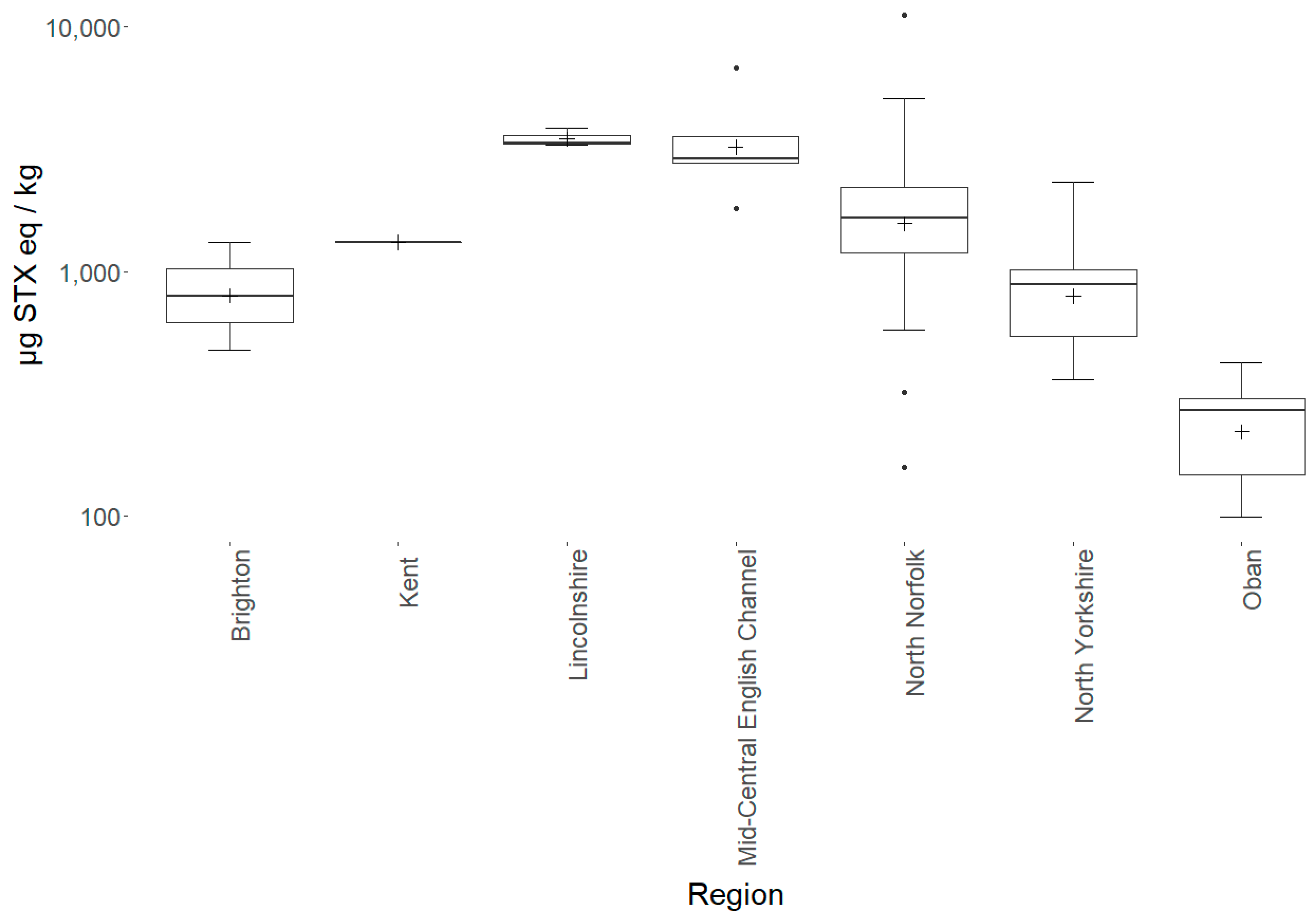
2.1. Starfish Toxicity
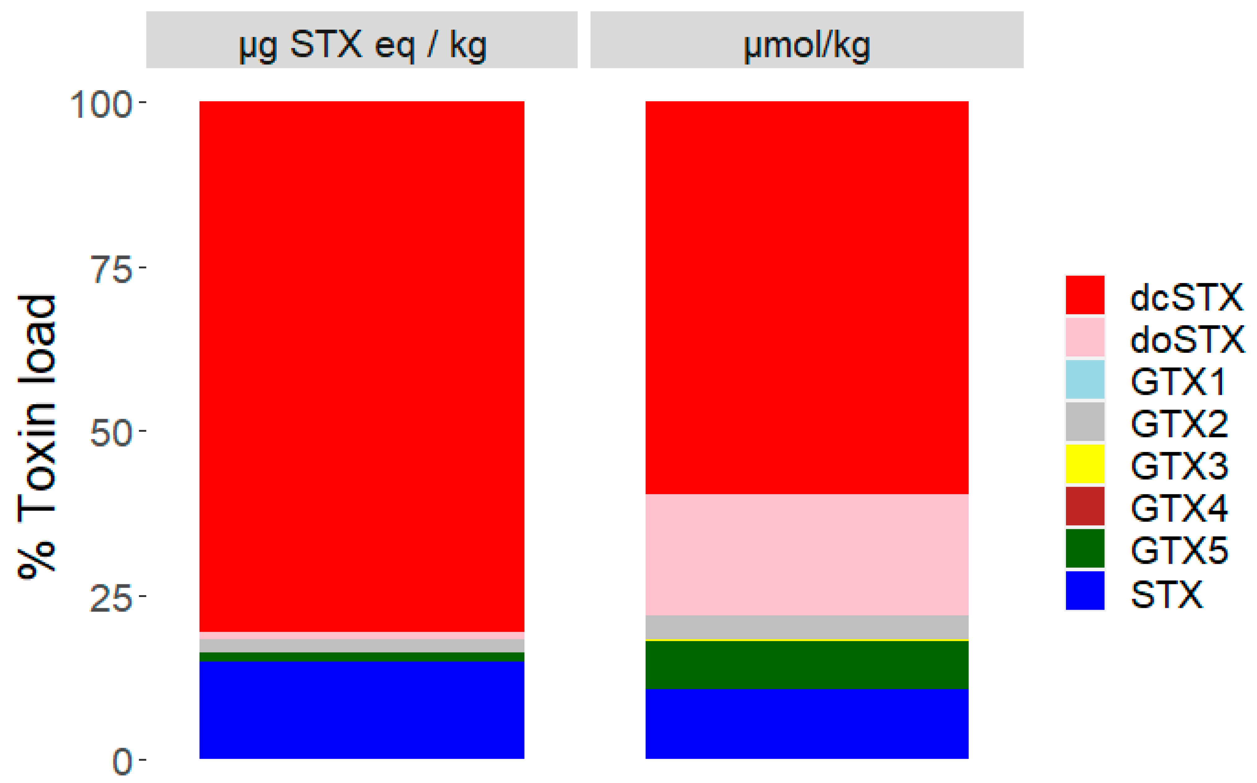
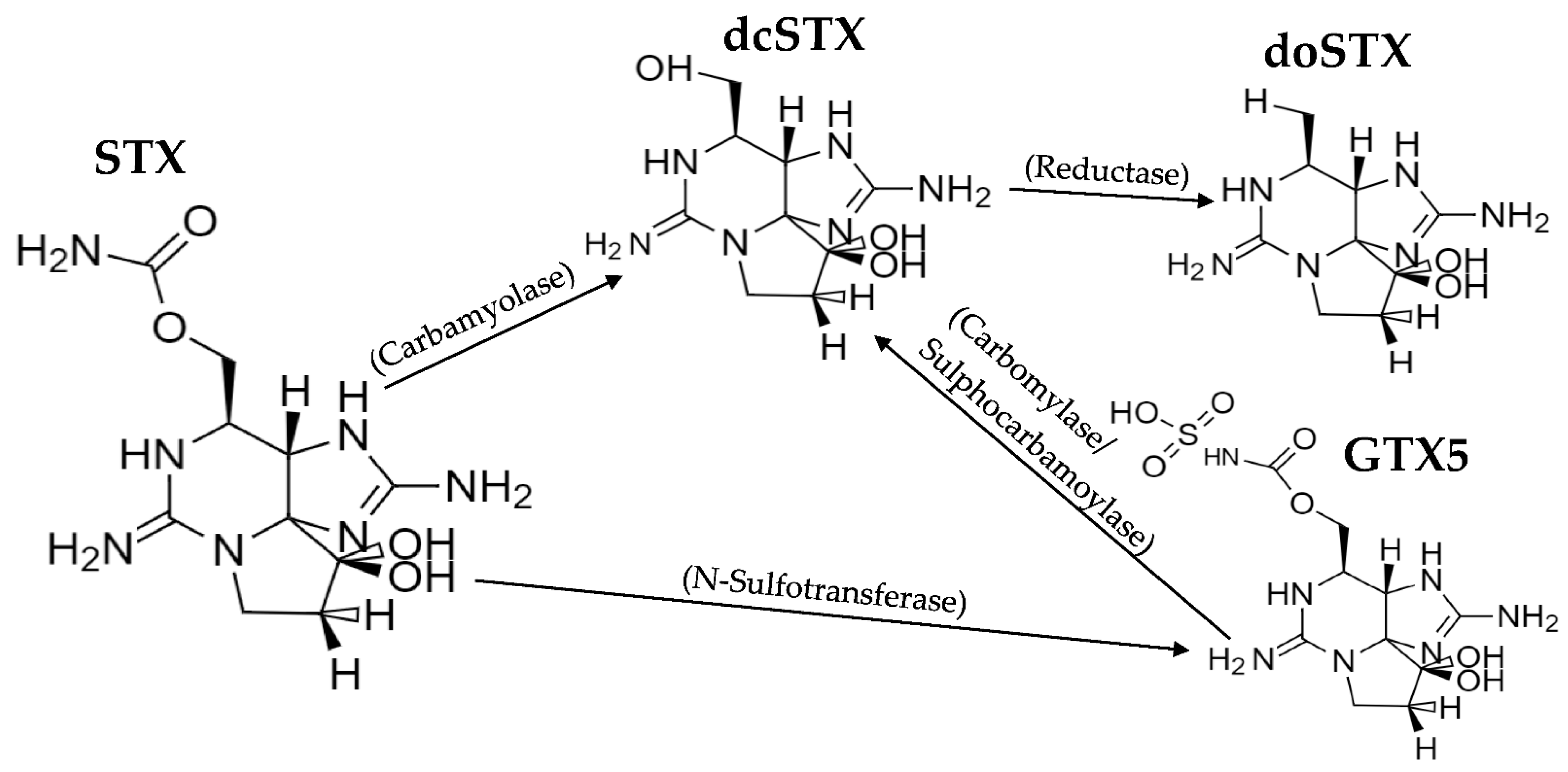
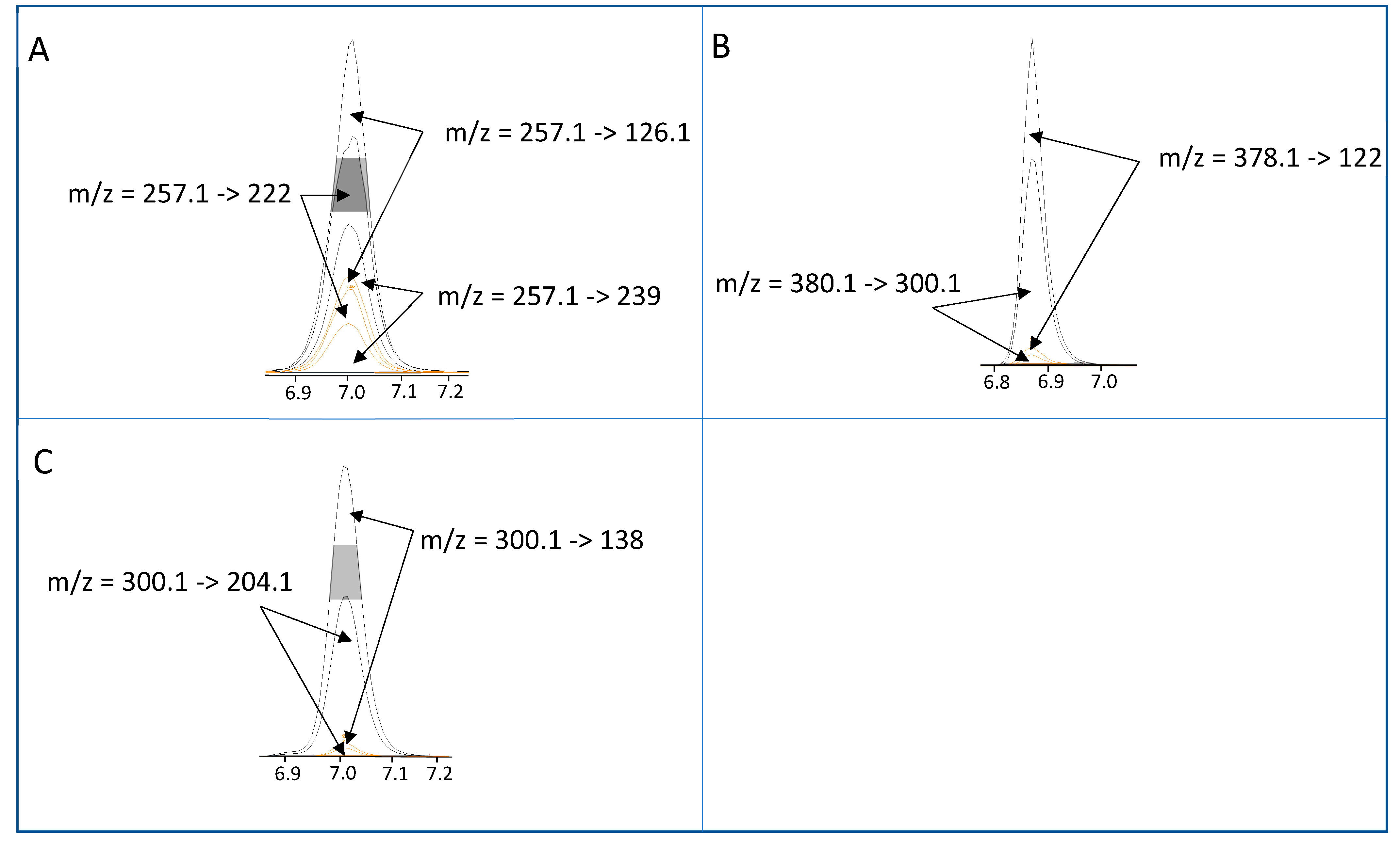
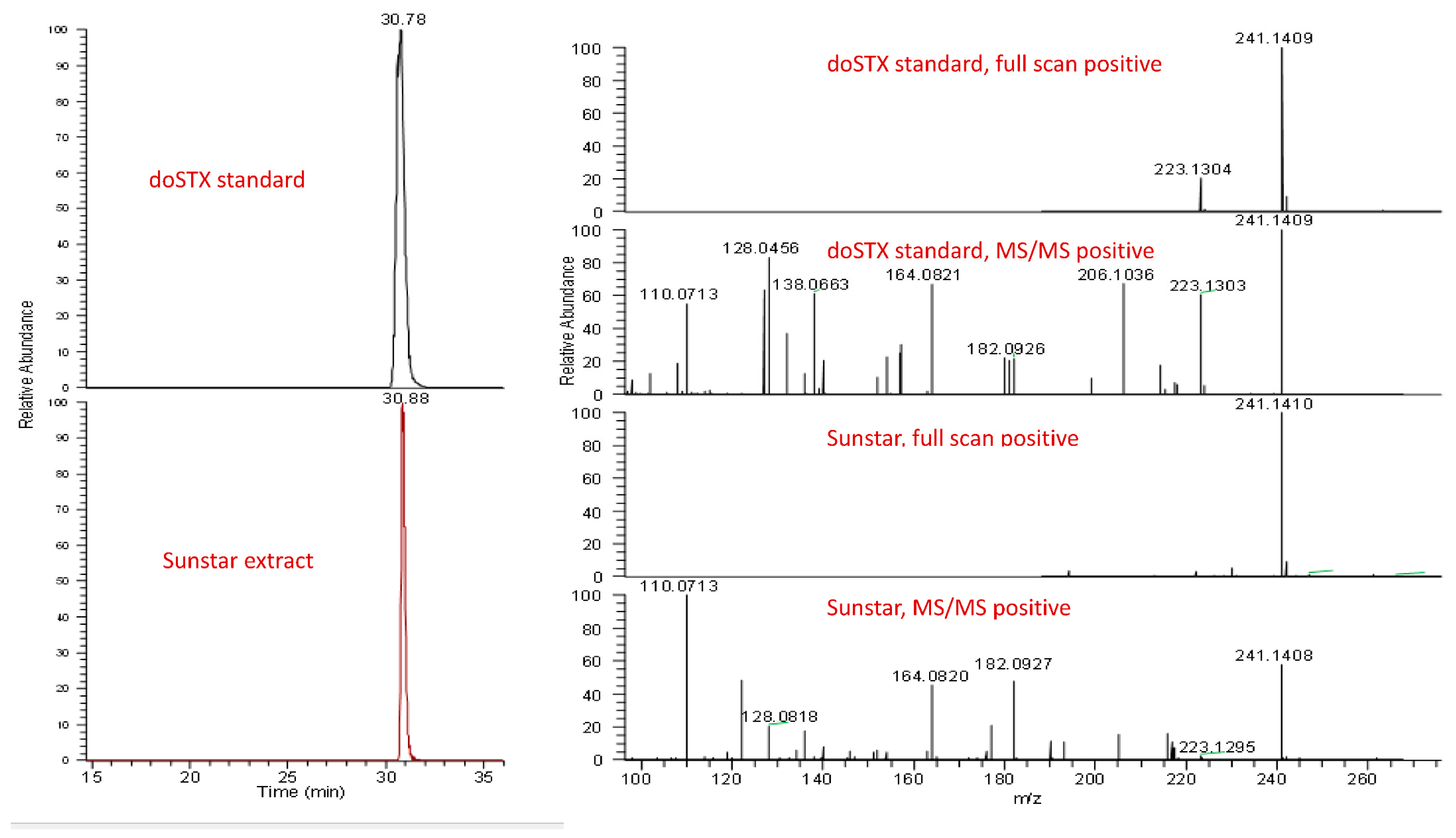
As the common sunstar C. papposus (referred to hereafter as sunstar) was suspected of displaying a consistent presence of STXs, a range of other starfish species were analysed, to act as a control group. Figure 2 summarises the sampling locations of all the starfishes sampled. In total, 151 starfishes were analysed between 2018 and 2021 (Table A1), comprising 73 specimens of sunstars and six other starfish species (n = 73), with one brittlestar species also analysed (n = 5). In sunstars, the presence of STXs was ubiquitous, with a mean total toxin concentration of 1739 µg STX eq/kg (Figure 3 and Table A2). In some cases, extreme toxicity in sunstars was quantified, with a maximum level of 11,245 µg STX eq/kg recorded in one sunstar from north Norfolk.
Conversely, none of the other starfish species showed any consistent toxicity, with the highest non-sunstar toxin concentration quantified in the brittlestar Ophiura ophiura (164 µg STX eq/kg). As some species were under-represented (Table A2), the species were grouped into sunstars and non-sunstars for statistical analysis. The sunstars exhibited higher mean toxicities in comparison with the other starfish species. This was statistically assessed with an ANOVA, which highlighted the species as the most statistically significant factor affecting the toxicity at the 95% significance level (p = <2 × 10⁻¹⁷). Additionally, the geographic region also had a statistically significant effect on the toxicity (p = 3.8 × 10⁻⁸); however, no statistical effect of starfish diameter or temporal variability was found. A linear mixed-effects model was fitted with the sampling region as a fixed variable and the species as a random variable, which highlighted the mid-central English Channel region as being statistically different to other regions (p = 0.0257). A Tukey’s multiple comparison of means test confirmed a significant difference in the toxin levels quantified in samples from the mid-central English Channel compared with those from most other sampling locations. Figure 4 highlights the geographical differences in toxicity across the UK. It should be noted that the sample numbers were low for many regions and most of the sunstars tested were from North Norfolk (n = 40) (see Table A3 for an overview of each region), where the intoxication event in canines occurred, which may have biased the geographical differences and requires further exploration. The interanimal variance in the total toxin concentrations was calculated using 22 sunstars sampled from the Wash in north Norfolk on the same day. The sunstars had a mean of 1558 µg STX eq/kg, a standard deviation of 570 µg STX eq/kg and an RSD of 37%, suggesting moderate inter-animal variability. The RSD of all the sunstars was 89%, showing large variability across the entire population. Therefore, it appeared that factors other than the geographical location may have affected the toxicity, as the sunstars from north Norfolk ranged from 157 to 11,245 µg STX eq/ kg (the low result from Holkham Beach in February 2018 and the high result from the Wash in January 2020). Overall, these data highlight the ubiquitous presence of STXs in sunstars and provide strong evidence that they exhibit STX presence differently to other starfish species. As the LC–MS/MS method utilised for the quantitation of the STXs could quantify the neurotoxin tetrodotoxin (TTX), all the samples were also analysed for the presence of this toxin; however, TTX was not detected in any sample.

2.2. Toxin Profiles

With the non-sunstar starfish (and brittlestars) containing low or nondetectable levels of STXs, toxic profiles were not generated for them. Analyses of the mean profiles were undertaken for sunstars in terms of both micrograms of STX equivalents per kilogram and micromoles per kilogram (Figure 5). The mean sunstar profile was dominated by dcSTX, with smaller relative contributions from deoxydecarbomyl-STX (doSTX), STX, GTX5 and GTX1–4. Large differences in the toxin proportions of doSTX and GTX5 between the micrograms of STX equivalents per kilogram and micromoles per kilogram profiles were noted as a consequence of the low relative TEF of these toxins. The mean profile from each geographic region (Figure 2) highlighted the differing toxic profiles based on the geographic location. There were two discernible profiles described, one dominated by STX and GTX2 from a relatively small sampling area, specifically, off the North Yorkshire coast, and all other regions exhibiting a dcSTX and STX profile. This did not appear to be a latitudinal-driven phenomenon, as susntars from Oban (NW Scotland) also had a dcSTX-dominated profile. This was confirmed by an ANOVA that demonstrated that the dcSTX load was statistically driven by the sampling region (p = <6.9 × 10⁻¹⁴), and a Tukey’s multiple comparison of means test corroborated the ANOVA results by highlighting the sunstars from North Yorkshire as having statistically different dcSTX loads to those of most other sampling regions. The dcSTX-dominated profiles varied slightly based on the location. Specifically, the Lincolnshire, north Norfolk and Kent sunstars showed concentrations of doSTX, which was not present in the South Coast sunstars (Brighton and the English Channel); instead, the doSTX portion was replaced with a GTX5 component.

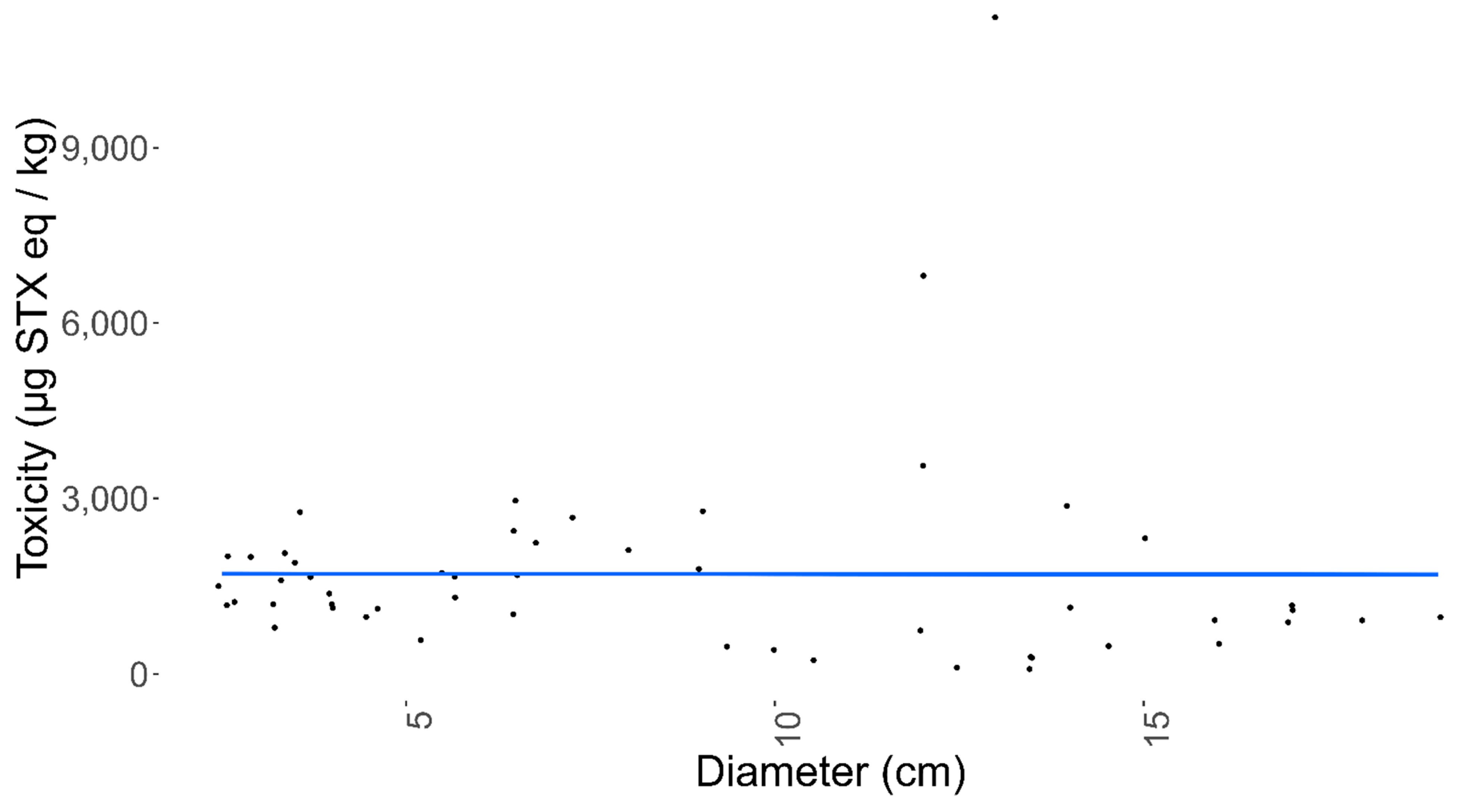
2.3. Sunstar Physiological Analysis

To determine any variability in the STX concentration between the sunstar organs, 13 sunstars from three different batches, each obtained from different sampling locations on different dates, were dissected, and the digestive glands, skin and gonads were removed and tested separately. At this point, the sunstars were sexed visually and, where required, this was confirmed via a histological examination through light microscopy of the gonads. The sunstars that were unable to be sexed or were sexually immature were removed from the analysis. The first batch of sunstars were from North Yorkshire and consisted of three males and two females, and they exhibited the STX- and GTX2-heavy toxin profile. The second batch originated from the Devon coast (Southwest England) and consisted of three males and two females, and the third batch from north Norfolk, which consisted of three females. The sunstars from batches two and three exhibited the dcSTX and STX profile. The highest toxin content (45,766 µg STX eq/kg) was quantified in a female gonad sample, with the female gonads also exhibiting the highest mean toxicity (14,234 µg STX eq/kg) of all the organs analysed (Figure 6, Table A4). In the females, the interorgan variability in toxicity was large, whilst in the males, all the organs appeared to show similar toxicities. An ANOVA assessing the total toxin concentrations in relation to the organisms’ sex, batch and organ type showed that the batch had a statistically significant effect on the toxicity (p = <3.53 × 10⁻¹¹). The mean toxicities for all the organs combined for each batch were 1176, 4056 and 12,589 µg STX eq/kg for batches one to three, respectively. Neither the sex nor the organ type showed a significant effect on the toxicity. To remove the effect of interbatch toxicity on identifying whether sex has a statistically significant effect on toxicity in sunstars (batch three heavily skewed the results towards females), a linear mixed-effects model was fitted with the sex as a random variable and the batch as a fixed variable. This highlighted that there was no statistical difference between male and female toxicity (p = 0.12). As each sex was not fully represented in all the batches and the batch had a statistically significant effect on the toxicity, the analysis of the organs’ toxicity was carried out separately on each sex. When a linear mixed-effects model was fitted for the female sunstars, using the organ type as a random variable and the batch as a fixed variable, the female gonads showed a weak but statistically significant influence on the toxicity (p = 0.02) and a Tukey’s analysis of multiple means highlighted statistically significant differences between the gonads and the skin, and the gonads and the digestive glands (p = 0.001 and p = 0.033, respectively). The same model was fitted for the male sunstars. This highlighted the gonads as being statistically significantly different to the other organs (p = 0.009), with the gonad–digestive gland interaction showing a statistically significant difference (p = 0.007), but the gonad–skin interaction demonstrating no statistical difference. In conclusion, sex had no statistically significant effect on the overall toxicity. However, for each sex, the gonads appeared to show different toxicity levels compared to the other organs: in the females this manifested in a higher toxicity, and in the males in a lower toxicity. Although these data offer some weight to the notion that different organs display different toxicities, the sample sizes were small and, as such, drawing definitive conclusions from them is questionable. In order to assess any potential relationship between the organism size and the total toxin levels, the quantified toxicities were compared between the measured sunstar diameters. Figure 7 illustrates the lack of any apparent correlation between the sunstar size and the toxin levels. This was confirmed by the ANOVA described in 2.1, which highlighted no statistical effect between the diameter of a sunstar and the total toxin concentration.

2.4. Comparison of Detection Techniques

As there are no formally validated methods for the determination of STXs in starfish tissues, quantitation was performed using two independent methods, the precolumn oxidation and liquid chromatography with fluorescence detection (LC–FLD) method and the tandem mass spectrometry utilising HILIC (HILIC–MS/MS) method (See Figure A1 for a comparison between the methods). There was a good agreement between the two quantitative methods, as the correlation coefficient of the total toxicity was 0.87, with dcSTX, STX and GTX5 having the coefficients of 0.87, 0.91 and 0.55, respectively. A paired Student’s t-test confirmed a statistical difference between the two methods as a consequence of the consistent slight overestimation of the LC–FLD method vs. the HILIC–MS/MS method. This was likely due to matrix-related interference during ionisation, as seen in other species [84]. As neither method has been fully validated, it is currently not possible to elucidate which is quantitatively more accurate; as such, the HILIC–MS/MS method was used for the bulk of the analysis to take a conservative approach, and due to its ability to quantify all the toxic epimers individually [10]. Qualitatively, the methods agreed well, with dcSTX, STX and GTX5 always detected in positive samples which exhibited the dcSTX profile. However, doSTX was not detected using the LC–FLD method, although it was detected by HILIC–MS/MS. The preCOX LC–FLD method can detect doSTX [85]; however, due to the rapid chromatographic nature of the LC–FLD method used, it is possible that the doSTX coeluted with the STX, making confirmation by LC–FLD impossible without changing the chromatographic methods. The confirmation of doSTX presence was, however, conducted via an LC–HRMS method. An accurate mass-to-charge-ratio measurement of 241.1413 (∆ = 2 ppm for C9H17N6O2+) was obtained for the [M + H]+ ion of doSTX (SI Ax) (Figure A2). Overall, the methods agreed well enough for good confidence in the HILIC–MS/MS data used for the qualitative and quantitative analysis.

3. Discussion

3.1. Starfish Toxicity

An extensive sampling and toxicity screening program was conducted in waters around the UK coast to assess the prevalence of STXs in starfish. The data obtained provide strong evidence to support the preliminary hypothesis [52,56] that sunstars ubiquitously contain STXs (n = 71). All the sunstars sampled contained quantifiable concentrations of toxins in all the sampling locations across all the sampling dates. The toxicity in the mid-central English Channel (Figure 2 and Figure 4) was statistically different to those in the other sampling regions; however, due to the random nature of the sampling and the fact that only a small number of sunstars were available from regions other than north Norfolk, drawing conclusions on the geographic variability is difficult. The variability over the entire sunstar dataset was large (RSD 89%); however, the interanimal variability of a subset sampled on the same day from the same location showed an RSD of 37%. This represented a relatively low interanimal variability and was lower than is commonly seen in shellfish [31,86,87,88,89]. Published records in peer-reviewed literature for the accumulation of STXs in starfish are rare [41,57,58,59,60]. In these manuscripts, the accumulation of STXs in starfish was linked to their predation on contaminated bivalves following algal blooms. An analysis of annual phytoplankton results [54,90] showed no correlation between toxic sunstars and the presence of vegetative Alexandrium cells in the water columns or contaminated bivalves from routine monitoring points. The only notable occurrence was related to samples collected in Oban in March 2021, when low Alexandrium cell counts were detected in the surrounding area. It should be noted that these routine monitoring points do not specifically relate to the sampling points of sunstars in this study, but they could be used as a general indication of the algal presence in the surrounding area. Furthermore, there were many offshore collections of starfish that had no inshore monitoring point in close proximity; therefore, elucidating the presence of causative phytoplankton species at these sampling points was not possible.
Consistent sunstar toxicity is hard to explain, especially compared to other starfish species occupying the same geographical and ecological niches. The vast geographic range of sunstar toxicity also makes the accumulation via an algal cell/cyst route questionable. The accumulation of STXs via trophic transfer by ingesting intoxicated food sources is also unlikely, as sunstars are both scavenger and predatory by nature [65,69]; therefore, their food sources would be expected to be different depending on what is readily available in each location. Conversely, the presence of STXs in sunstars could result from a dietary source, as, although they occupy similar ecological roles to other starfish species, their feeding habits have been shown to be different [66,67,69,91]. However, C. papposus often consumes the common starfish Asterias rubens as a preference over molluscs or gastropods [18,65,91], and, as the common starfish displayed far fewer STXs (in comparison to the sunstars), sunstars are unlikely to be accumulating STXs via this route. A trophic transfer route of STXs could be explained if sunstars possessed an active storage mechanism for STXs similar to that of other molluscs [36,92,93,94] but that other starfish species do not exhibit. Currently, however, the depuration and uptake kinetics of STXs in sunstars are unknown, and so would be a potential future study of interest. Previously [56], two sources of sunstar toxicity were hypothesised, either that their grazing on algal cysts produced the STXs [15] or that the presence of a symbiosis with bacteria produced the STXs [21]. As sunstars and algal cysts both occupy the benthos, the accumulation of STXs via this route is feasible. Algal cysts beds can be geographically extensive [95,96], cysts can be more toxic than vegetative cells [16] and the ingestion of algal cysts has been previously implicated in shellfish toxicity [17]. For cyst ingestion to be the principal cause of the toxin concentrations observed in C. papposus, the causative cyst bed/s would have to stretch around the entire UK coast and into the English Channel and be capable of producing two different toxin profiles, one of which is completely different to the profiles produced by the known UK vegetative Alexandrium blooms. Additionally, cyst toxin profiles have been shown to be similar to their vegetative counterparts [16]; subsequently, the resulting toxin profiles, if sunstars did ingest Alexandrium cysts, would likely be similar to those described in [14] and would pose the question whether the dcSTX profile came from a source other than Alexandrium cysts. It is also unclear why sunstars would be more susceptible to toxin accumulation via this route compared to other benthic organisms [52,56]. The cyanobacterial production of STXs is possible [7]. Although cyanobacteria are mostly associated with freshwater, there have been reports of both benthic and saltwater colonies producing STXs [19,97,98], which could explain the toxicity in a saline benthic environment; however, the same arguments for algal cysts not being the source of STXs apply to cyanobacteria, in that any cyanobacteria presence would have to be geographically extensive and it would be unclear why the uptake of STXs in sunstars is far more pronounced than in other starfish species. Due to the deeper offshore environments that sunstars often inhabit, the presence of toxins during all months of the year, the unique toxic profile and the statistical differences in the presence of STXs in sunstars, a nontraditional source should not be ruled out. The information discussed above suggests that it is feasible that sunstars accumulate STXs from somewhere other than their diet or the environment, and thus the notion that sunstars synthesise STXs internally, via a microbial symbiosis or other means, can be hypothesised. The production of the similar neurotoxin TTX in pufferfish and starfish has previously been linked to symbiotic vibrio species [24,25], so it is possible that sunstars accumulate STXs in a similar manner, which would explain the geographically widespread yet consistent toxin concentrations that were found. In conjunction, the core genes responsible for the synthesis of STXs already exist in multiple animal kingdoms [28], making the possibility of an unknown novel producer feasible. Elucidating the source could involve a multistep approach encompassing: laboratory studies to experimentally determine the accumulation and depuration kinetics of STXs in sunstars, the toxin testing of bacteria isolated and cultured from sunstars and the genetic analysis of both the microbiome and the gene clusters associated with STX production. Sediment found in sunstar environments could also be analysed; even if traditional Alexandrium cysts are unlikely to be the causative agent, the presence of an unknown benthic species could also be investigated.

3.2. Toxin Profiles

The two dominant toxic profiles are similar to those discovered previously [52,56], specifically, a high dcSTX profile from most regions and an STX- and GTX2-dominated profile from the North Yorkshire coast (Figure 2). These two distinct toxic profiles may imply two different sources of STXs, noting that whilst the high dcSTX profile is unlike any known bivalve toxin profile reported globally, the STX and GTX2 profile is similar to the profile reported from UK shellfish associated with toxin uptake from A. minutum [14], usually detected in the South West of the UK. The Alexandrium cyst beds present off the North East coast of England [99,100,101] are also of note; however, A. catenella (formally A. tamerense) was the species implicated in this region, which in the UK produces a more complex toxin profile, typically containing GTX1–5, C1/2, NEO and STX [14]. Therefore, the presence of A. catenella cysts does not fully explain the toxin profile prevalent off the North Yorkshire coast. The mean toxin profile for all the sunstars was heavily dominated by dcSTX in terms of the micrograms of STX equivalents per kilogram; however, in terms of the micromoles per kilogram, the relative concentrations of the lower TEF compounds doSTX and GTX5 were notably higher. Specifically, doSTX was responsible for nearly 25% of the mean molar toxin suite present, which could imply that ~25% of the dcSTX was converted, via the reduction of the hydroxyl group at R4, to the less toxic doSTX. It is currently unclear whether there was a transformation or whether the doSTX was expressed as part of the naturally produced toxic profile. In the known biosynthesis of STX, dcSTX is the last intermediate before the creation of STX, which requires the sxtL gene to facilitate the addition of the carbomyl group at R4 (Figure 1) [26,27]. The dcSTX-dominant profile could therefore be created by a source that does not possess the sxtL gene that codes for the addition of the carbomyl group at C-13 [26], or perhaps sxtL is less readily transcribed, and therefore only a small portion of the dcSTX is continued along the synthesis chain to form STX. The formation of GTX5 from dcSTX would be unusual, as GTX5 production would require the addition of a carbamoyl group at R4 and the subsequent sulfation of that carbamoyl group. It is also possible that the dcSTX profile could be the product of a series of gene- and enzyme-controlled reactions on the STX itself (Figure 8). The presence of carbomylase, for example, would convert the STX into a dcSTX, and the dcSTX could then be transformed into a doSTX via reduction at R4 (it is noted that this is not a common transformation kinetic of STXs [29]). The GTX5 present could then be a result of the sulphation of the STX at R4. Either proposed synthesis pathway would therefore require a specific set of enzymes and/or genes to be present. In order to determine the production pathway taken, molecular tools must be implemented to help discover the genes present. This will help elucidate the more likely synthesis route. For example, the presence of two distinct, geographically driven profiles infers the existence of two different genetic/enzymatic populations. In the dcSTX-dominated profile, the sulphation of STX to GTX5 could be controlled by the sxtN gene, whereas in the North Yorkshire profile, the sulphation of STX into GTX2 could be mediated by the sxtSUL gene [29]. Conversely to these hypothesis, which are not currently supported by genetic or enzymatic testing, the evidence suggests the presence of both of these profiles across a wide taxonomic range [56], which would make it unlikely that these transformations were happening in all organisms and more likely that they were representative of the toxic source itself. Therefore, if C. papposus can be considered as the hypothetical source of STXs in the benthos [56], then the lower concentrations exhibited by the other organisms [56] and starfish in this study could be attributed to predation on and/or trophic transfer from sunstars. Overall, however, there is currently limited evidence to determine the true nature of the toxin source that has been described in sunstars. Future work should therefore focus on the molecular analysis of the known STX-producing gene clusters at both geographic locations to isolate any obvious genetic differences in the populations, which could help elucidate the biosynthesis pathway.

3.3. Sunstar Physiological Analysis

Both the male and the female sunstars showed a ubiquitous toxin presence, and all the organs tested contained quantifiable concentrations of toxins. The female gonads in particular contained high concentrations of STXs, possibly highlighting their role in reproductive or larval protection. Sunstars reproduce by external fertilisation via spawning (usually in March–May in the Northern Hemisphere), in which males and females eject sperm and eggs into the water column [102]. The presence of a potent mammalian neurotoxin within the eggs and sperm in the water column has a potential ecological advantage for larval survival. There are a range of previous studies analysing the effect of STXs on different marine organisms, and the toxic effect of STXs is not limited to mammalian nervous structures. By far the most extensively researched is the effect that STXs have on molluscs, with the major examples being: reduced feeding, reduced clearance rates, reduced larval survival, reduced heart rate and shell valve closures (reviewed in [75]). In the starfish Pisaster ochraceus, STXs inhibited fertilisation and decreased the ability of the starfish to attach to a substrate [60], with STXs also causing mysid mortality and larval abnormalities in sea urchins [79]. STXs have also elicited negative responses in marine fish, causing neurological symptoms and mortality in multiple species [40,42] and a range of effects on rainbow trout intestinal cells [103]. STXs have also caused altered grazing strategies and reduced reproducibility in some copepods [104,105]. Previously, STXs have been proposed as a pheromone in Alexandrium [28] that aids in reproductive success. If STXs are used as a pheromone by sunstars, they could act as a chemical cue to initiate spawning, thus increasing the likelihood of the successful fertilisation of eggs. Conversely, previous experiments on the ‘keystone’ starfish P. ochraceus noted a decrease in fertilisation with an increase in the STX concentration, showing that it suppressed reproduction [60]. It should be noted that both pheromonal and larval protection could also be provided or could work in conjunction with the variety of saponins known to be produced by starfish [71,106]. In pufferfish, high concentrations of TTX were shown in the ovaries, and the inherited TTX presence in larval pufferfish acted as a deterrent for predation in juvenile pufferfish, even at low concentrations [107,108]. TTX has also been discovered in high concentrations in the eggs of the sea slug Pleurobranchaea maculata [109,110]. It is similarly possible that sunstars could be utilising STXs as a feeding deterrent for juvenile/larval sunstars. The presence of STXs in the digestive glands could imply that they are a potential feeding aid, with sunstars previously reported to have opened molluscs without applying much physical force [68,111] and the use of a toxic compound in predation having been proposed before [68]. STXs can illicit neurotoxicity, oxidative stress and lower metabolic capacity in bivalves [112] and could therefore potentially increase sunstar predatory success by making their prey more susceptible to their enveloping stomach. Furthermore, the presence of STXs in the skin of sunstars highlights the potential use of STXs as a chemical defense, which could work in a similar fashion to the targeted retention of STXs that has been proposed as a defense mechanism protecting some clams from predation by sea otters and siphon-nipping fish [46,113,114]. The anecdotal neurotoxicity to cats described in [68] was almost certainly caused by inherent STXs present in sunstars. Overall, there are multiple known and unknown toxicological effects of STX that act on a wide range of marine organisms; therefore, the presence of a potent neurotoxin has multiple potential ecological benefits. However, the exact role STX plays is unknown, as is whether the accumulation of STXs is passive or targeted or whether STXs are produced by sunstars themselves.

4. Materials and Methods

4.1. Sample Collection

Samples were collected from a range of locations around the coasts of England, from both inshore and offshore areas as bycatch from fishermen or washed up onshore between 16 February 2018 and 11 March 2021. There was a total of 26 individual sampling locations, separated into 12 regions, which provided a spread of data along the UK coast (Figure 2). Once collected, the samples were transported to the Weymouth laboratory where they were stored at −20 °C until required for analysis. As well as common sunstar (Crossaster papposus), six other species of starfish were analysed as a control group (Figure 3, Table A1), namely, sandstar (Astropecten irregularis), seven-armed starfish (Luidia cilaris), common starfish (Asterias rubens), spiny starfish (Marthasterias glacialis), goosefoot starfish (Anserpoda placenta), bloody Henry starfish (Henrica sp.) and brittlestar (Ophiura ophiura).

4.2. Reagents and Chemicals

All solvents, reagents and chemicals were of LC–MS or HPLC grade, depending on the system-specific requirements. LC–MS grade water was produced by a MilliQ water purification system (Merck, Darmstadt, Germany). Certified reference toxins were obtained from Biotoxin Metrology, National Research Council Canada (NRCC, Halifax, NS, Canada). Toxins incorporated included GTX1–6, dcGTX2&3, dcSTX, dcNEO, NEO, STX and C1&2. Non-certified toxin standards were also received from Cawthron Natural Compounds (CNC, Nelson, New Zealand) for C3&4, dcGTX1&4 and doSTX.

4.3. Sample Preparation and Extraction for Toxin Analysis

Individuals of the same species from the same sampling locations were (excluding C. papposus) pooled together to create a representative sample. C. papposus samples were analysed individually to ascertain interanimal variability and whether toxicity was correlated with diameter or any physiological traits. Sunstars subjected to organ analysis were dissected, and samples of the digestive system, gonads and skin were taken. All samples were homogenised using Waring industrial blenders (Stamford, CT, USA) and IKA Ultra-Turrax homogenisers (Oxford, Oxfordshire, UK). Samples unable to be blended into a smooth paste with blenders were instead homogenised with an extraction solvent (1% acetic acid) present. Tissues were extracted using a refined method recently validated for analysis of STXs in crab, whelk and shrimp [84], specifically, a single dispersive method utilising a 1:9 sample–solvent ratio. Samples of 2.0 ± 0.1 g of homogenised tissue were extracted. Where available tissues were <2.0 ± 0.1 g, a scaled-down extraction was performed, with volumes used dependent on the volume of homogenised tissue available. Three different analytical methods were used to detect STXs in starfish samples. These were a precolumn oxidation liquid chromatography with fluorescence detection (LC–FLD) [115] method, a liquid chromatography with hydrophilic interaction chromatography coupled with tandem mass spectrometry method (HILIC–MS/MS) [116] and a LC–HRMS method (qualitative only). Where possible, samples were quantified using both the LC–FLD and HILIC–MS/MS methods; however, analysis by the HILIC–MS/MS method was prioritized due to its higher sensitivity and better analogue specificity. Therefore, all data shown in the manuscript were generated by the HILIC–MS/MS method, unless stated otherwise.

4.4. Sample Analysis

4.4.1. Analysis of STXs by LC–FLD

Once extracted, supernatants from the centrifuged crude acetic acid extracts were subjected to a C18 solid-phase extraction step (SPE) using an automated Gilson (Dunstable, Bedfordshire, UK) ASPEC 271 system. Extracts that had been cleaned up by SPE were subsequently pH-adjusted to 6.5 ± 0.5 using 1 M NaOH and 0.1 M acetic acid and diluted to 4 mL. Analysis of samples was performed in two steps. Firstly, a semiquantitative screen (similar to that validated in [117]) was carried out to identify samples that contained any N-hydroxylated compounds, which if present would be forwarded to an ion-exchange SPE that isolated the individual fractions ready for further analysis—throughout the entire study no samples were forwarded to the ion-exchange SPE. Secondly, full quantitation of samples was achieved by the peroxide oxidation (Figure 9) of C18 SPE eluents. Analysis of unoxidised C18 SPE eluents was conducted to identify any naturally fluorescent coextractives that could interfere with chromatographic toxin peaks. LC–FLD analysis was performed on an Agilent 1200 LC system consisting of a quaternary pump, FLD, vacuum degasser, autosampler and thermostatically controlled column oven. Chromatographic separation was achieved using a Phenomenex Kinetex C18 (150 mm × 4.6 mm × 5 µm) (Torrance, CA, USA) column with a solvent gradient as per [118]. Quantitation of oxidised STXs was achieved using a six-point calibration curve prepared using certified calibrants diluted in 0.01 M acetic acid. In samples where chromatographically unresolvable epimeric pairs were present (for example, GTX2&3), the TEF of the most toxic epimer was applied (Figure 1). The LC–FLD method can quantify the epimeric pairs GTX1&4, GTX2&3, C1&2, C3&4 and dcGTX2&3, as well as the analogues GTX5, GTX6, NEO, dcNEO, dcSTX and STX.

4.4.2. Analysis of STXs by HILIC–MS/MS

Crude acetic acid extracts were subjected to graphite SPE clean-up, using an automated Gilson ASPEC271 system as described in [116,119]. One hundred microlitres of SPE eluents were subsequently diluted with 300 µL of LC-MS/MS-grade acetonitrile, prior to analysis. LC-MS/MS analysis was performed using an Agilent (Manchester, UK) 6495B triple quadrupole tandem mass spectrometer, with chromatography conducted using an Agilent 1290 Infinity II UHPLC system. Chromatographic separation was achieved using either an Agilent Poroshell 120 HILICZ (150 mm × 2.1 mm × 2.7 µm) or a Waters Acquity BEH Amide (150 mm × 2.1 mm × 1.7 µm) (Elstree, Herefordshire, UK) column utilising the gradient solvent delivery method reported in [10]. Analysis of each toxin analogue was achieved using two multiple reaction monitoring (MRM) transitions [10] (Figure 10), with quantitation performed using a six-point calibration curve for each primary transition prepared using certified calibrants diluted in solvent or STX-negative, graphite SPE-cleaned and diluted mussel extract. TEFs applied were those stated in Figure 1. The HILIC–MS/MS method quantified GTX1–6, dcGTX1–4, C1-4, doSTX, dcSTX, dcNEO, NEO, and STX as well as the neurotoxin tetrodotoxin (TTX).

4.5. LC–HRMS Qualitative Analysis of doSTX

Qualitative assessment of doSTX presence was performed using an Agilent 1200 in tandem with an QExactive HF Orbitrap mass spectrometer. HPLC parameters were the same as those detailed in [120], specifically, analysis on a TosoHaas Amide column (250 mm × 2.1 mm × 5 µm), with separation of STXs obtained using mobile phase A (deionised water with 50 mM formic acid and 2 mM ammonium formate) and mobile phase B (100% MeCN). Analytical runs were performed at 200 µL/min using the following gradient: 90%B > 55%B over 25 min, decrease in B to 30% at 27 min and then hold at 30%B until finishing at 36 min. MS analyses were performed using a heated electrospray ionisation probe with 2500 V spray voltage and 275 °C capillary temperature. Full scan data were acquired with a resolution setting 120,000. MS/MS data were acquired using data-dependent acquisition with a resolution setting of 30,000, using stepped normalised collision energies (30, 60 and 80 eV).

4.6. Histological Processing and Analysis of Sunstars

Gonadal tissue from sunstars which could not be sexed through gross visual examination were dissected and fixed in Davidson’s fixative for at least 24 h. Following fixation, samples were processed in a Thermo Scientific Excelsior AS tissue processor following standard overnight routine processing schedule, where tissues were dehydrated through ethanol series, placed in xylene substitute, and then embedded in paraffin wax. Sections 3 µm thick were taken using a Leica HistoCore Multicut semiautomated rotary microtome, stained with hematoxylin and eosin, mounted and coverslipped. Sunstar gonads were sexed via light microscopy using a Nikon Eclipse E800 microscope and compared to images from [121]. Figure 11 illustrates male and female sunstar gonads.

4.7. Data Analysis

Data were analysed using R with ANOVAs, linear mixed-effects models and Tukey’s post hoc tests performed using the packages described in [122,123,124]. All ANOVAs, linear mixed-effects models and Tukey’s analyses were performed using log-transformed data. To make geographically driven profiles easier to analyse, sampling locations were merged into ‘regions’ which encompassed all sites from nearby locations. Analysis of toxin profiles removed all STXs < 10 µg STX eq/kg. Sunstars which were used for organ analysis were removed from the main data set and analysed separately.

5. Conclusions

Based on the analysis of 71 whole and 13 dissected sunstars from multiple locations along the UK coast across multiple years, this manuscript describes strong evidence for the ubiquitous presence of STXs in this species. As such, it should be considered ‘toxic’ hereafter. The sunstars sampled in this study exhibited extreme toxicities (>10,000 µg STX eq/kg) and contained statistically higher concentrations of STXs than all other starfish species tested. All the samples of sunstar skin, digestive glands and gonads (male and female) were found to contain quantifiable concentrations of STXs, with the female gonads displaying the highest total toxin concentrations (>40,000 µg STX eq/kg). The ecological role of these toxins in sunstars has yet to be elucidated; however, there are multiple proposed advantages for producing/accumulating STXs, including larval and adult defense, increasing reproductive success and use as a predation aid. The source remains unknown; however, the evidence described here hints that a traditionally described algal source may not be involved, given the ubiquitous toxin presence across a wide spatial and temporal range. Two distinct toxin profiles were confirmed, a dcSTX profile (from most UK sampling locations) and an STX and GTX2-dominated profile (from North Yorkshire). The total toxin concentrations were also shown to vary largely between the locations. Any investigation to elucidate the source should involve a multipronged strategy encompassing the following: laboratory-tank-based studies to determine the accumulation and depuration kinetics of STX in sunstars, toxin analysis of bacteria cultured from sunstars to describe any potential bacterial symbiosis and molecular analysis of both the microbiome and the gene cluster associated with the synthesis of STXs. This study and several recent manuscripts have highlighted that STXs are far more widespread than traditionally thought and that STXs possibly perform multiple unknown ecological roles in benthic marine environments around the UK coast.

Author Contributions

Conceptualisation, A.D.T. and K.J.D.; methodology, R.P.A., K.J.D., K.M.T., T.C. and R.G.H.; formal analysis, K.J.D., C.D., R.P.A., T.C., V.L., L.N.C., R.H., P.W. and R.G.H.; investigation, K.J.D. and A.D.T.; data curation, K.J.D., M.T.A.; writing—original draft preparation, K.J.D.; writing—review and editing, R.G.H., A.M.L., J.R.E., M.T.A., C.D., A.D.T., K.M.T. and R.P.A.; supervision, A.D.T.; funding acquisition, A.D.T. All authors have read and agreed to the published version of the manuscript.

Funding

This research was funded primarily by the EMFF (European Maritime and Fisheries Fund) Funding Project ENG3313, Cefas contract code C7847, with additional support from Interreg Alertox-Net EAPA-317-2016 (Atlantic Area Program).

Data Availability Statement

Data is contained within the article and Appendix A.

Acknowledgments

The authors would like to thank our colleagues at Eastern IFCA for their excellent support throughout the project.

Conflicts of Interest

The authors declare no conflict of interest.

Appendix A

Table A1. Full summary of all starfishes and brittlestars analysed. nt = not tested, nd = not detected (<1 µg STX eq/kg).
Common NameScientific NameDate SampledLocationRegionDiameter (cm)HILIC–MS/MS Total (µg STX eq/kg)LC–FLD Total (µg STX eq/kg)
Common starfishAsterias rubens6 February 2018FelixstoweSuffolkntndnt
Common starfishAsterias rubens6 February 2018FelixstoweSuffolkntnd1
Common starfishAsterias rubens6 February 2018FelixstoweSuffolkntnd4
Common starfishAsterias rubens6 February 2018FelixstoweSuffolkntndnt
Common starfishAsterias rubens8 February 2018LowestoftEast Suffolkntnd4
Common starfishAsterias rubens8 February 2018LowestoftEast Suffolkntndnd
Common starfishAsterias rubens8 February 2018LowestoftEast Suffolkntnd1
Common starfishAsterias rubens8 February 2018LowestoftEast Suffolkntndnd
SunstarCrossaster papposus16 February 2018Holkham BeachNorth Norfolknt157388
Common starfishAsterias rubens19 February 2018Lulworth BanksDorsetntndnd
Common starfishAsterias rubens20 February 2018Aldeburgh BeachEast Suffolkntnd2
Common starfishAsterias rubens20 February 2018Aldeburgh BeachEast Suffolkntndnd
Common starfishAsterias rubens20 February 2018Aldeburgh BeachEast Suffolkntnd167
Common starfishAsterias rubens3 March 2018Hunstanton BeachNorth Norfolkntnd10
Common starfishAsterias rubens3 March 2018Hunstanton BeachNorth Norfolkntnd5
Common starfishAsterias rubens3 March 2018Hunstanton BeachNorth Norfolkntnd10
Common starfishAsterias rubens5 March 2018Aldeburgh BeachEast Suffolkntnd5
Common starfishAsterias rubens5 March 2018Aldeburgh BeachEast Suffolkntnd3
Common starfishAsterias rubens5 March 2018Aldeburgh BeachEast Suffolkntnd7
SunstarCrossaster papposus5 March 2018BrancasterNorth Norfolknt321781
SunstarCrossaster papposus5 March 2018BrancasterNorth Norfolknt5791357
SunstarCrossaster papposus5 March 2018WellsNorth Norfolknt16404980
SunstarCrossaster papposus5 March 2018Hunstanton BeachNorth Norfolknt27276679
SunstarCrossaster papposus5 March 2018Hunstanton BeachNorth Norfolknt29914778
SunstarCrossaster papposus5 March 2018WellsNorth Norfolknt35438489
SunstarCrossaster papposus5 March 2018WellsNorth Norfolknt510816,513
SunstarCrossaster papposus5 March 2018BrancasterNorth Norfolkntnd13,237
Common starfishAsterias rubens6 March 2018Lincolnshire coastLincolnshirentnd4
Common starfishAsterias rubens6 March 2018Lincolnshire coastLincolnshirentnd1
SunstarCrossaster papposus6 March 2018Lincolnshire coastLincolnshirent38479058
SunstarCrossaster papposus6 March 2018Lincolnshire coastLincolnshirent329210,993
SunstarCrossaster papposus6 March 2018Lincolnshire coastLincolnshirent33435746
Common starfishAsterias rubens6 March 2018Ramsgate beachKentntndnt
Common starfishAsterias rubens6 March 2018Ramsgate beachKentnt2nt
Common starfishAsterias rubens6 March 2018Ramsgate beachKentntndnt
Common starfishAsterias rubens6 March 2018Ramsgate beachKentnt3nt
Common starfishAsterias rubens6 March 2018Ramsgate beachKentnt4nt
SunstarCrossaster papposus6 March 2018Ramsgate beachKentnt13177061
Common starfishAsterias rubens6 March 2018Ramsgate beachKentnt164
Common starfishAsterias rubens12 March 2018FelixstoweSuffolkntndnd
Common starfishAsterias rubens12 March 2018FelixstoweSuffolkntnd7
Common starfishAsterias rubens12 March 2018FelixstoweSuffolkntnd5
Spiny starfishMarthasterias glacialis19 September 2019Cornwall—south of St.AustellSouth Cornwallntndnd
Common starfishAsterias rubens14 October 2019BrixhamSouth Devonntndnd
Common starfishAsterias rubens24 October 2019South of Lyme RegisDorsetntndnd
BrittlestarOphiura ophiura25 October 2019South of Lyme RegisDorsetntndnd
Common starfishAsterias rubens14 November 2019BrightonBrightonntndnd
Common starfishAsterias rubens6 December 2019PlymouthSouth Devonntndnd
Common starfishAsterias rubens17 December 2019PlymouthSouth Devonntndnd
Spiny starfishMarthasterias glacialis17 December 2019PlymouthSouth Devonnt422
Common starfishAsterias rubens17 December 2019East of WhitbyNorth Yorkshirent8948
Common starfishAsterias rubens17 December 2019East of WhitbyNorth Yorkshirent814
Spiny starfishMarthasterias glacialis17 December 2019East of WhitbyNorth Yorkshirent48
SunstarCrossaster papposus17 December 2019East of WhitbyNorth Yorkshirent360686
SunstarCrossaster papposus17 December 2019East of WhitbyNorth Yorkshirent564909
SunstarCrossaster papposus17 December 2019East of WhitbyNorth Yorkshirent8801447
SunstarCrossaster papposus17 December 2019East of WhitbyNorth Yorkshirent548779
SunstarCrossaster papposus17 December 2019East of WhitbyNorth Yorkshirent472625
SunstarCrossaster papposus23 January 2020South of BrightonBrighton9.4479795
SunstarCrossaster papposus23 January 2020South of BrightonBrighton5.713191929
SunstarCrossaster papposus27 January 2020North Norfolk, near the WashNorth Norfolk3.5nt3109
SunstarCrossaster papposus27 January 2020North Norfolk, near the WashNorth Norfolk5.2590837
SunstarCrossaster papposus27 January 2020North Norfolk, near the WashNorth Norfolk3.28051429
SunstarCrossaster papposus27 January 2020North Norfolk, near the WashNorth Norfolk4.59861420
SunstarCrossaster papposus27 January 2020North Norfolk, near the WashNorth Norfolk6.510341667
SunstarCrossaster papposus27 January 2020North Norfolk, near the WashNorth Norfolk4.611301609
SunstarCrossaster papposus27 January 2020North Norfolk, near the WashNorth Norfolk411422115
SunstarCrossaster papposus27 January 2020North Norfolk, near the WashNorth Norfolk2.611881803
SunstarCrossaster papposus27 January 2020North Norfolk, near the WashNorth Norfolk3.212011610
SunstarCrossaster papposus27 January 2020North Norfolk, near the WashNorth Norfolk412031548
SunstarCrossaster papposus27 January 2020North Norfolk, near the WashNorth Norfolk2.712422146
SunstarCrossaster papposus27 January 2020North Norfolk, near the WashNorth Norfolk413861726
SunstarCrossaster papposus27 January 2020North Norfolk, near the WashNorth Norfolk2.515132632
SunstarCrossaster papposus27 January 2020North Norfolk, near the WashNorth Norfolk3.316122864
SunstarCrossaster papposus27 January 2020North Norfolk, near the WashNorth Norfolk3.716692460
SunstarCrossaster papposus27 January 2020North Norfolk, near the WashNorth Norfolk5.716722651
SunstarCrossaster papposus27 January 2020North Norfolk, near the WashNorth Norfolk6.517012545
SunstarCrossaster papposus27 January 2020North Norfolk, near the WashNorth Norfolk5.517382633
SunstarCrossaster papposus27 January 2020North Norfolk, near the WashNorth Norfolk3.519112682
SunstarCrossaster papposus27 January 2020North Norfolk, near the WashNorth Norfolk2.920113500
SunstarCrossaster papposus27 January 2020North Norfolk, near the WashNorth Norfolk2.620223131
SunstarCrossaster papposus27 January 2020North Norfolk, near the WashNorth Norfolk3.420754104
SunstarCrossaster papposus27 January 2020North Norfolk, near the WashNorth Norfolk821283982
SunstarCrossaster papposus27 January 2020North Norfolk, near the WashNorth Norfolk6.822513825
SunstarCrossaster papposus27 January 2020North Norfolk, near the WashNorth Norfolk6.524553910
SunstarCrossaster papposus27 January 2020North Norfolk, near the WashNorth Norfolk7.326823648
SunstarCrossaster papposus27 January 2020North Norfolk, near the WashNorth Norfolk3.627764131
SunstarCrossaster papposus27 January 2020North Norfolk, near the WashNorth Norfolk6.529734745
SunstarCrossaster papposus27 January 2020North Norfolk, near the WashNorth Norfolk1311,24516,244
Common starfishAsterias rubens29 January 2020Kings LynnNorth Norfolkntnd7
Goosefoot starfishAnseropoda placenta6 February 2020Mid-central English ChannelMid-central English Channelntnd23
Common starfishAsterias rubens6 February 2020Mid-central English ChannelMid-central English Channelntnd8
BrittlestarOphiura ophiura6 February 2020Mid-central English ChannelMid-central English Channelnt121112
SunstarCrossaster papposus6 February 2020Mid-central English ChannelMid-central English Channel927904230
SunstarCrossaster papposus6 February 2020Mid-central English ChannelMid-central English Channel1268219510
SunstarCrossaster papposus6 February 2020Mid-central English ChannelMid-central English Channel918043272
SunstarCrossaster papposus6 February 2020Mid-central English ChannelMid-central English Channel1235705500
SunstarCrossaster papposus6 February 2020Mid-central English ChannelMid-central English Channel1428818480
Sand starAstropecten irregularis6 February 2020Mid-central English ChannelMid-central English Channelntnd36
Seven-armed starfishLuidia ciliaris6 February 2020Mid-central English ChannelMid-central English Channelntndnd
Common starfishAsterias rubens3 March 2020South of BridportDorsetntndnd
SunstarCrossaster papposus20 March 2020East of WhitbyNorth Yorkshire16526nt
SunstarCrossaster papposus20 March 2020East of WhitbyNorth Yorkshire141148nt
SunstarCrossaster papposus20 March 2020East of WhitbyNorth Yorkshire19984nt
SunstarCrossaster papposus20 March 2020East of WhitbyNorth Yorkshire17896nt
SunstarCrossaster papposus20 March 2020East of WhitbyNorth Yorkshire16933nt
SunstarCrossaster papposus20 March 2020East of WhitbyNorth Yorkshire14.5492nt
SunstarCrossaster papposus20 March 2020East of WhitbyNorth Yorkshire171184nt
SunstarCrossaster papposus20 March 2020East of WhitbyNorth Yorkshire171104nt
SunstarCrossaster papposus20 March 2020East of WhitbyNorth Yorkshire18930nt
SunstarCrossaster papposus20 March 2020East of WhitbyNorth Yorkshire152327nt
SunstarCrossaster papposus20 March 2020East of WhitbyNorth Yorkshire12756nt
SunstarCrossaster papposus3 May 2020Hunstanton BeachNorth Norfolknt1380940
SunstarCrossaster papposus3 May 2020Hunstanton BeachNorth Norfolknt16071231
Common starfishAsterias rubens3 May 2020Cley shingle beach (from shoreline)North Norfolkntndnd
Goosefoot starfishAnseropoda placenta21 July 2020WeymouthDorsetnt30nd
Sand starAstropecten irregularis28 August 2020South of DartmouthSouth Devonnt3nt
Sand starAstropecten irregularis28 August 2020South of DartmouthSouth Devonnt13nt
Sand starAstropecten irregularis28 August 2020South of DartmouthSouth Devonnt2nt
Common starfishAsterias rubens28 August 2020South of DartmouthSouth Devonntndnt
Goosefoot starfishAnseropoda placenta28 August 2020South of DartmouthSouth Devonnt9nt
Bloody HenryHenrica oculata28 August 2020South of DartmouthSouth Devonnt6nt
BrittlestarOphiura ophiura28 August 2020South of DartmouthSouth Devonnt2nt
BrittlestarOphiura ophiura28 August 2020South of DartmouthSouth Devonnt164nt
BrittlestarOphiura ophiura28 August 2020South of DartmouthSouth Devonntndnt
Seven-armed starfishLuidia ciliaris28 August 2020South of DartmouthSouth Devonnt12nt
Goosefoot starfishAnseropoda placenta28 August 2020South of DartmouthSouth Devonnt4nt
Common starfishAsterias rubens28 August 2020South of DartmouthSouth Devonnt63nt
SunstarCrossaster papposus11 March 2021ObanOban10.5250270
SunstarCrossaster papposus11 March 2021ObanOban13.599125
SunstarCrossaster papposus11 March 2021ObanOban12.5124134
SunstarCrossaster papposus11 March 2021ObanOban10425452
SunstarCrossaster papposus11 March 2021ObanOban13.5291419
SunstarCrossaster papposus11 March 2021ObanOban13.5307320
Table A2. Overview of all starfish and brittlestars analysed. Mean toxicity, standard deviation (sd) and range are displayed in µg STX eq/kg. nd = not detected (<1 µg STX eq/kg), NA = not applicable, % > LOD = number of samples that had detectable levels of STXs above the LC–MS/MS limit of detection.
Common NameScientific NameMean ToxicitysdRangen% > LOD
Bloody HenryHenricia spp.6NANA1100%
BrittlestarOphiura ophiura9684nd–164560%
Common starfishAsterias rubens2635nd–895912%
Goosefoot starfishAnserpoda placenta1414nd–30475%
Sand starAstropecten irregularis66nd–13475%
Seven-armed starfishLuidia ciliaris12NAnd–12250%
Spiny starfishMarthasterias glacialis4NAnd–4333%
SunstarCrossaster papposus1739166999–11,24571100%
Table A3. Summary of each starfish species collected in each region. Mean toxicity, standard deviation (sd) and range are displayed in µg STX eq/kg. nd = not detected (<1 µg STX eq/kg), N/A = not applicable.
RegionSpeciesMean ToxicitysdRangen
BrightonCommon starfishndN/AN/A1
BrightonSunstar899594479-13192
DorsetBrittlestarndN/AN/A1
DorsetCommon starfishndN/AN/A3
DorsetGoosefoot starfish30N/AN/A1
East SuffolkCommon starfishndN/AN/A16
Mid-central English ChannelSeven-armed starfishndN/AN/A1
Mid-central English ChannelBrittlestar121N/AN/A1
Mid-central English ChannelCommon starfishndN/AN/A1
Mid-central English ChannelGoosefoot starfishndN/AN/A1
Mid-central English ChannelSand starndN/AN/A1
Mid-central English ChannelSunstar357319221804–68215
KentCommon starfish24nd-1613
KentSunstar1317N/AN/A1
LincolnshireCommon starfishndN/AN/A3
LincolnshireSunstar34943073292–38473
North NorfolkCommon starfishndN/AN/A5
North NorfolkSunstar19101815157–11,24540
North YorkshireCommon starfish48578–892
North YorkshireSpiny starfish4N/AN/A1
North YorkshireSunstar882466360–232616
ObanSunstar24912299–4256
South CornwallSpiny starfishndN/AN/A1
South DevonSeven-armed starfish12N/AN/A1
South DevonBloody Henry6N/AN/A1
South DevonBrittlestar5594nd–1643
South DevonCommon starfish1328nd–635
South DevonGoosefoot starfish644–92
South DevonSand star662–123
South DevonSpiny starfish4N/AN/A1
SuffolkCommon starfishndN/AN/A9
Table A4. Summary of individual organ analysis of sunstars. Mean toxicity, sd and range are displayed in µg STX eq/kg.
OrganSexMean ToxicitysdRangen
Digestive glandFemale28712115842–56667
Digestive glandMale46724232700–10,3396
GonadFemale14,23416,952669–45,7667
GonadMale23832167234–56346
SkinFemale15741035128–30457
SkinMale31172685766–72496
Figure A1. Comparison of LC–FLD and HILIC–MS/MS methods. Correlation coefficient (top right), scatter plot (bottom left) and distributions (HILIC–MS/MS (top left) and LC–FLD (bottom right)). The x axis is HILIC–MS/MS in µg STX eq/kg and y axis is LC–FLD in µg STX eq/kg.
Marinedrugs 19 00695 g0a1
Figure A2. LC–HRMS chromatogram showing accurate mass and fragmentation pattern of doSTX in a standard (not certified) and a sunstar sample.
Marinedrugs 19 00695 g0a2

References

  1. Botana, L.M. Seafood and Freshwater Toxins: Pharmacology, Physiology and Detection, Chapter 2: Diversity of Marine and FreshwaterAlgal Toxins. In Seafood and Freshwater Toxins: Pharmacology, Physiology and Detection; Marcel Dekker Inc.: New York, NY, USA, 2000; pp. 20–24. ISBN 9780824789565. [Google Scholar]
  2. Wiese, M.; D’Agostino, P.M.; Mihali, T.K.; Moffitt, M.C.; Neilan, B.A. Neurotoxic alkaloids: Saxitoxin and its analogs. Mar. Drugs 2010, 8, 2185–2211. [Google Scholar] [CrossRef] [PubMed] [Green Version]
  3. EFSA Marine biotoxins in shellfish—Saxitoxin group. EFSA J. 2009, 7, 1019.
  4. Etheridge, S.M. Paralytic shellfish poisoning: Seafood safety and human health perspectives. Toxicon 2010, 56, 108–122. [Google Scholar] [CrossRef] [Green Version]
  5. Hallegraeff, G.M. Harmful Algal Blooms: A Global Overview; UNESCO: Paris, France, 2004; ISBN 9231039482. [Google Scholar]
  6. Llewellyn, L.; Negri, A.; Robertson, A. Paralytic shellfish toxins in tropical oceans. Toxin Rev. 2006, 25, 159–196. [Google Scholar] [CrossRef]
  7. Pearson, L.; Mihali, T.; Moffitt, M.; Kellmann, R.; Neilan, B. On the chemistry, toxicology and genetics of the cyanobacterial toxins, microcystin, nodularin, saxitoxin and cylindrospermopsin. Mar. Drugs 2010, 8, 1650–1680. [Google Scholar] [CrossRef] [Green Version]
  8. Anon Regulation (EC) No 854/2004 of the European Parliament and of the Council of 29 April 2004 laying down specific rules for the organisation of official controls on products of animal origin intended for human consumption. Off. J. Eur. Union 2004, L 139, 206–320.
  9. Anon Regulation (EC) No 853/2004 of the European Parliament and of the Council of 29 April 2004 laying down specific hygiene rules for food of animal origin. Off. J. Eur. Union 2004, L 139, 55–205.
  10. Turner, A.D.; Rapkova, M.D.; Fong, S.Y.; Hungerford, J.; McNabb, P.S.; Boundy, M.J.; Harwood, D.T. Ultrahigh-Performance Hydrophilic Interaction Liquid Chromatography with Tandem Mass Spectrometry Method for the Determination of Paralytic Shellfish Toxins and Tetrodotoxin in Mussels, Oysters, Clams, Cockles, and Scallops: Collaborative Study. J. AOAC Int. 2020, 103, 1–30. [Google Scholar] [CrossRef] [PubMed]
  11. Brown, L.; Bresnan, E.; Graham, J.; Lacaze, J.; Turrell, E.; Collins, C.; Brown, L.; Bresnan, E.; Graham, J.; Lacaze, J.; et al. Distribution, diversity and toxin composition of the genus Alexandrium (Dinophyceae) in Scottish waters. Eur. J. Phycol. 2010, 45, 375–393. [Google Scholar] [CrossRef] [Green Version]
  12. Nascimento, S.M.; Purdie, D.A.; Lilly, E.L.; Larsen, J.; Morris, S. Toxin profile, pigment composition, and large subunit rDNA phylogenetic analysis of an Alexandrium minutum (Dinophyceae) strain isolated from the fleet lagoon, United Kingdom. J. Phycol. 2005, 41, 343–353. [Google Scholar] [CrossRef]
  13. Lewis, A.M.; Coates, L.N.; Turner, A.D.; Percy, L.; Lewis, J. A review of the global distribution of Alexandrium minutum (Dinophyceae) and comments on ecology and associated paralytic shellfish toxin profiles, with a focus on northern Europe. J. Phycol. 2018, 54, 581–598. [Google Scholar] [CrossRef]
  14. Turner, A.D.; Stubbs, B.; Coates, L.; Dhanji-Rapkova, M.; Hatfield, R.G.; Lewis, A.M.; Rowland-Pilgrim, S.; O’Neil, A.; Stubbs, P.; Ross, S.; et al. Variability of paralytic shellfish toxin occurrence and profiles in bivalve molluscs from Great Britain from official control monitoring as determined by pre-column oxidation liquid chromatography and implications for applying immunochemical tests. Harmful Algae 2014, 31, 87–99. [Google Scholar] [CrossRef]
  15. Anderson, D.M.; Alpermann, T.J.; Cembella, A.D.; Collos, Y.; Masseret, E.; Montresor, M. The globally distributed genus Alexandrium: Multifaceted roles in marine ecosystems and impacts on human health. Harmful Algae 2012, 14, 10–35. [Google Scholar] [CrossRef] [PubMed] [Green Version]
  16. Oshima, Y.; Bolch, C.J.; Hallegraeff, G.M. Toxin composition of resting cysts of Alexandrium tamerense (Dinophycae). Toxicon 1992, 30, 1539–1544. [Google Scholar] [CrossRef]
  17. Persson, A.; Smith, B.C.; Wikfors, G.H.; Quilliam, M. Grazing on toxic Alexandrium fundyense resting cysts and vegetative cells by the eastern oyster (Crassostrea virginica). Harmful Algae 2006, 5, 678–684. [Google Scholar] [CrossRef]
  18. Hunt, O.D. The Food of the Bottom Fauna of the Plymouth Fishing Grounds. J. Mar. Biol. Assoc. UK 1925, 13, 560–599. [Google Scholar] [CrossRef] [Green Version]
  19. Quiblier, C.; Wood, S.; Echenique Subiabre, I.; Heath, M.; Villeneuve, A.; Humbert, J.-F. A review of current knowledge on toxic benthic freshwater cyanobacteria—Ecology, toxin production and risk management. Water Res. 2013, 47, 5464–5479. [Google Scholar]
  20. Baker, T.R.; Doucette, G.J.; Powell, C.L.; Boyer, G.L.; Plumley, F.G. GTX4imposters: Characterization of fluorescent compounds synthesized by Pseudomonas stutzeri SF/PS and Pseudomonas/Alteromonas PTB-1, symbionts of saxitoxin-producing Alexandrium spp. Toxicon 2003, 41, 339–347. [Google Scholar] [CrossRef]
  21. Gallacher, S.; Smith, E.A. Bacteria and paralytic shellfish toxins. Protist 1999, 150, 245–255. [Google Scholar] [CrossRef]
  22. Martins, C.A.; Alvito, P.; Tavares, M.J.; Pereira, P.; Doucette, G.; Franca, S. Reevaluation of production of paralytic shellfish toxin by bacteria associated with dinoflagellates of the Portuguese Coast. Appl. Environ. Microbiol. 2003, 69, 5693–5698. [Google Scholar] [CrossRef] [Green Version]
  23. Lu, Y.; Chai, T.; Hwang, D. Isolation of bacteria from toxic dinoflagellate Alexandrium minutum and their effects on algae toxicity. J. Nat. Toxins 2000, 9, 409–417. [Google Scholar]
  24. Bane, V.; Lehane, M.; Dikshit, M.; Riordan, A.O.; Furey, A. Tetrodotoxin: Chemistry, Toxicity, Source, Distribution and Detection. Toxins 2014, 6, 693–755. [Google Scholar] [CrossRef] [PubMed] [Green Version]
  25. Narita, H.; Matsubara, S.; Miwa, N.; Akahane, S.; Murakami, M.; Goto, T.; Nara, M.; Noguchi, T.; Saito, T.; Shida, Y.; et al. Vibrio alginolyticus, a TTX-producing Bacterium Isolated from the Starfish Astropecten polyacanthus. Nippon Suisan Gakkaishi 1987, 53, 617–621. [Google Scholar] [CrossRef]
  26. Akbar, M.A.; Yusof, N.Y.M.; Tahir, N.I.; Ahmad, A.; Usup, G.; Sahrani, F.K.; Bunawan, H. Biosynthesis of saxitoxin in marine dinoflagellates: An omics perspective. Mar. Drugs 2020, 18, 103. [Google Scholar] [CrossRef] [PubMed] [Green Version]
  27. Verma, A.; Barua, A.; Ruvindy, R.; Savela, H.; Ajani, P.A.; Murray, S.A. The genetic basis of toxin biosynthesis in dinoflagellates. Microorganisms 2019, 7, 222. [Google Scholar] [CrossRef] [PubMed] [Green Version]
  28. Cusick, K.D.; Sayler, G.S. An overview on the marine neurotoxin, saxitoxin: Genetics, moleculartargets, methods of detection and ecological functions. Mar. Drugs 2013, 11, 991–1018. [Google Scholar] [CrossRef] [Green Version]
  29. Raposo, M.I.C.; Gomes, M.T.S.R.; Jo, M.; Rudnitskaya, A. Paralytic Shellfish Toxins (PST)-Transforming Enzymes: A Review. Toxins 2020, 12, 344. [Google Scholar] [CrossRef] [PubMed]
  30. Kwong, R.W.M.; Wang, W.X.; Lam, P.K.S.; Yu, P.K.N. The uptake, distribution and elimination of paralytic shellfish toxins in mussels and fish exposed to toxic dinoflagellates. Aquat. Toxicol. 2006, 80, 82–91. [Google Scholar] [CrossRef]
  31. Bricelj, V.M.; Shumway, S.E. Paralytic shellfish toxins in bivalve molluscs: Occurrence, transfer kinetics, and biotransformation. Rev. Fish. Sci. 1998, 6, 315–383. [Google Scholar] [CrossRef]
  32. Taleb, H.; Vale, P.; Jaime, E.; Blaghen, M. Study of paralytic shellfish poisoning toxin profile in shellfish from the Mediterranean shore of Morocco. Toxicon 2001, 39, 1855–1861. [Google Scholar] [CrossRef]
  33. Bazzoni, A.M.; Mudadu, A.G.; Lorenzoni, G.; Arras, I.; Lugliè, A.; Vivaldi, B.; Cicotelli, V.; Sanna, G.; Tedde, G.; Ledda, S.; et al. Occurrence of harmful algal species and shellfish toxicity in Sardinia (Italy). Ital. J. Food Saf. 2016, 5. [Google Scholar] [CrossRef] [PubMed] [Green Version]
  34. Garcia, C.; Mardones, P.; Sfeir, A.; Lagos, N. Simultaneous presence of Paralytic and Diarrheic Shellfish Poisoning toxins in Mytilus chilensis samples collected in the Chiloe Island, Austral Chilean fjords. Biol. Res. 2004, 37, 721–731. [Google Scholar] [CrossRef] [PubMed]
  35. Murray, S.A.; O’Connor, W.A.; Alvin, A.; Mihali, T.K.; Kalaitzis, J.; Neilan, B.A. Differential accumulation of paralytic shellfish toxins from Alexandrium minutum in the pearl oyster, Pinctada imbricata. Toxicon 2009, 54, 217–223. [Google Scholar] [CrossRef]
  36. Shumway, S.E. A Review of the Effects of Algal Blooms on Shellfish and Aquaculture. J. World Aquac. Soc. 1990, 21, 65–104. [Google Scholar] [CrossRef]
  37. Wong, C.K.; Hung, P.; Ng, H.C.C.; Lee, S.Y.; Kam, K.M. Cluster analysis of toxins profile pattern as a tool for tracing shellfish contaminated with PSP-toxins. Environ. Res. 2011, 111, 1083–1090. [Google Scholar] [CrossRef] [PubMed]
  38. Blanco, J.; Reyero, M.I.; Franco, J. Kinetics of accumulation and transformation of paralytic shellfish toxins in the blue mussel Mytilus galloprovincialis. Toxicon 2003, 42, 777–784. [Google Scholar] [CrossRef]
  39. DeGrasse, S.; Vanegas, C.; Conrad, S. Paralytic shellfish toxins in the sea scallop Placopecten magellanicus on Georges Bank: Implications for an offshore roe-on and whole scallop fishery. Deep. Sea Res. Part II Top. Stud. Oceanogr. 2014, 103, 301–307. [Google Scholar] [CrossRef]
  40. Montoya, N.; Akselman, R.; Franco, J.; Carreto, J.I. Paralytic shellfish toxins and mackerel (Scomber japonicus) mortality in the Argentine Sea. Harmful Toxic Algal Bloom. 1996, 417–420. [Google Scholar]
  41. Ben-Gigirey, B.; Rossignoli, A.E.; Riobo, P.; Rodriguez, F. First Report of Paralytic Shellfish Toxins in Marine Invertebrates and Fish in Spain. Toxins 2020, 12, 723. [Google Scholar] [CrossRef]
  42. Reis Costa, P. Impact and effects of paralytic shellfish poisoning toxins derived from harmful algal blooms to marine fish. Fish Fish. 2016, 17, 226–248. [Google Scholar] [CrossRef]
  43. Abbott, J.P.; Flewelling, L.J.; Landsberg, J.H. Saxitoxin monitoring in three species of Florida puffer fish. Harmful Algae 2009, 8, 343–348. [Google Scholar] [CrossRef]
  44. Hernandez, M.; Robinson, I.; Aguilar, A.; Gonzalez, L.M.; Lopez-Jurado, L.F.; Reyero, M.; Cacho, E. Did algal toxins cause monk seal mortality? Nature 1998, 393, 28–29. [Google Scholar] [CrossRef] [PubMed] [Green Version]
  45. Jensen, S.; Lacaze, J.; Hermann, G.; Kershaw, J.; Brownlow, A.; Turner, A.; Hall, A. Toxicon Detection and effects of harmful algal toxins in Scottish harbour seals and potential links to population decline. Toxicon 2015, 97, 1–14. [Google Scholar] [CrossRef] [PubMed]
  46. Kvitek, R.G.; DeGange, A.R.; Beitler, M.K. Paralytic shellfish poisoning toxins mediate feeding behavior of sea otters. Limnol. Ocean. 1991, 36, 393–404. [Google Scholar] [CrossRef]
  47. Durbin, E.; Teegarden, G.; Campbell, R.; Cembella, A.; Baumgartner, M.F.; Mate, B.R. North Atlantic right whales, Eubalaena glacialis, exposed to paralytic shellfish poisoning (PSP) toxins via a zooplankton vector, Calanus finmarchicus. Harmful Algae 2002, 1, 243–251. [Google Scholar] [CrossRef]
  48. Deeds, J.R.; Landsberg, J.H.; Etheridge, S.M.; Pitcher, G.C.; Longan, S.W. Non-traditional vectors for paralytic shellfish poisoning. Mar. Drugs 2008, 6, 308–348. [Google Scholar] [CrossRef] [PubMed]
  49. Silva, M.; Barreiro, A.; Kaufmann, M.; Neto, A.I.; Hassouani, M.; Sabour, B.; Botana, A.; Botana, L.M.; Vasconcelos, V. Paralytic Shellfish Toxins Occurrence in Non-Traditional Invertebrate Vectors from North AtlanticWaters (Azores, Madeira, and Morocco). Toxins 2018, 10, 362. [Google Scholar] [CrossRef] [Green Version]
  50. García, C.; Pérez, F.; Contreras, C.; Figueroa, D.; Barriga, A.; López-Rivera, A.; Araneda, O.F.; Contreras, H.R. Saxitoxins and okadaic acid group: Accumulation and distribution in invertebrate marine vectors from Southern Chile. Food Addit. Contam.—Part A Chem. Anal. Control Expo. Risk Assess. 2015, 32, 984–1002. [Google Scholar] [CrossRef]
  51. Silva, M.; Barreiro, A.; Rodriguez, P.; Otero, P.; Azevedo, J.; Alfonso, A.; Botana, L.M.; Vasconcelos, V. New invertebrate vectors for PST, spirolides and okadaic acid in the North Atlantic. Mar. Drugs 2013, 11, 1936–1960. [Google Scholar] [CrossRef] [Green Version]
  52. Turner, A.D.; Dhanji-Rapkova, M.; Dean, K.; Milligan, S.; Hamilton, M.; Thomas, J.; Poole, C.; Haycock, J.; Spelman-Marriott, J.; Watson, A.; et al. Fatal canine intoxications linked to the presence of saxitoxins in stranded marine organisms following winter storm activity. Toxins 2018, 10, 94. [Google Scholar] [CrossRef] [Green Version]
  53. Cefas Harmful Algal Blooms (HABS) Surveillance Programmes and Monitoring. Available online: https://www.cefas.co.uk/data-and-publications/habs/ (accessed on 1 June 2021).
  54. Lin, H.; Cho, Y.; Yashiro, H.; Yamada, T.; Oshima, Y. Purification and characterization of paralytic shellfish toxin transforming enzyme from Mactra chinensis. Toxicon 2004, 44, 657–668. [Google Scholar] [CrossRef]
  55. Cho, Y.; Ogawa, N.; Takahashi, M.; Lin, H.; Oshima, Y. Purification and characterization of paralytic shellfish toxin-transforming enzyme, sulfocarbamoylase I, from the Japanese bivalve Peronidia venulosa. Biochim. Biophys. Acta 2008, 1784, 1277–1285. [Google Scholar] [CrossRef]
  56. Dean, K.J.; Hatfield, R.G.; Lee, V.; Alexander, R.P.; Lewis, A.M.; Maskrey, B.H.; Alves, M.T.; Hatton, B.; Coates, L.N.; Capuzzo, E.; et al. Multiple New Paralytic Shellfish Toxin Vectors in Off shore North Sea Benthos, a Deep Secret Exposed. Mar. Drugs 2020, 18, 400. [Google Scholar] [CrossRef] [PubMed]
  57. Asakawa, M.; Nishimura, F.; Miyazawa, K.; Noguchi, T. Occurance of paralytic shellfish poison in the starfish Asteria amurensis in Kure Bay, Hiroshima prefecture, Japan. Toxicon 1997, 37, 1081–1087. [Google Scholar] [CrossRef]
  58. Ito, K.; Asakawa, M.; Sida, Y.; Miyazawa, K. Occurrence of paralytic shellfish poison (PSP) in the starfish Asterina pectinifera collected from the Kure Bay, Hiroshima Prefecture, Japan. Toxicon 2003, 41, 291–295. [Google Scholar] [CrossRef]
  59. Lin, S.-J.; Tsai, Y.-H.; Lin, H.; Hwang, D. Paralytic toxins in Taiwanese starfish Astrpecten scoparius. Toxicon 1998, 36, 799–803. [Google Scholar] [CrossRef]
  60. Ferrer, R.P.; Lunsford, E.T.; Candido, C.M.; Strawn, M.L.; Pierce, K.M. Saxitoxin and the ochre sea star: Molecule of keystone significance and a classic keystone species. Integr. Comp. Biol. 2015, 55, 533–542. [Google Scholar] [CrossRef] [PubMed] [Green Version]
  61. Ellis, J.R.; Rogers, S.I. The distribution, relative abundance and diversity of echinoderms in the eastern English Channel, Bristol Channel, and Irish Sea. J. Mar. Biol. Assoc. UK 2000, 80, 127–138. [Google Scholar] [CrossRef]
  62. Ringvold, H.; Moum, T. On the genus Crossaster (Echinodermata: Asteroidea) and its distribution. PLoS ONE 2020, 15, e0227223. [Google Scholar]
  63. Birkeland, C. Interactions between a Sea Pen and Seven of Its Predators. Ecol. Monogr. 1974, 44, 211–232. [Google Scholar] [CrossRef]
  64. Carlson, H.R.; Pfister, C.A. A seventeen-year study of the rose star Crossaster papposus population in a coastal bay in southeast Alaska. Mar. Biol. 1999, 133, 223–230. [Google Scholar] [CrossRef]
  65. Hancock, D.A. The feeding behaviour of starfish on Essex oyster beds. J. Mar. Biol. Assoc. UK 1955, 34, 313–331. [Google Scholar] [CrossRef] [Green Version]
  66. Sloan, N.A. Starfish encounters: An experimental study of its advantages. Experientia 1979, 35, 1314–1315. [Google Scholar] [CrossRef]
  67. Himmelman, J.H.; Dutil, C. Distribution, population structure and feeding of subtidal seastars in the northern Gulf of St Lawrence. Mar. Ecol. Prog. Ser. 1991, 76, 61–72. [Google Scholar] [CrossRef]
  68. Hancock, D.A. Some aspects of the biology of the sunstar Crossaster papposus (L.). Ophelia 1974, 13, 1–30. [Google Scholar] [CrossRef]
  69. Sloan, N.A.; Northway, S.M. Chemoreception by the asteroid Crossaster papposus (L.). J. Exp. Mar. Bio. Ecol. 1982, 61, 85–98. [Google Scholar] [CrossRef]
  70. Feder, M.; Christianson, A. Aspects of Asteroid biology. In Physiology of Echinodermata; Inderscience Publishers: Hoboken, NJ, USA, 1966; pp. 88–127. [Google Scholar]
  71. Andersson, L.; Bohlin, L.; Iorizzi, M.; Riccio, R.; Minale, L.; Moreno-Lopez, W. Biological Activity of Saponins and Saponin-like Compounds from Starfish and Brittle-stars. Toxicon 1989, 27, 179–188. [Google Scholar] [CrossRef]
  72. Choi, M.C.; Yu, P.K.N.; Hsieh, D.P.H.; Lam, P.K.S. Trophic transfer of paralytic shellfish toxins from clams (Ruditapes philippinarum) to gastropods (Nassarius festivus). Chemosphere 2006, 64, 1642–1649. [Google Scholar] [CrossRef] [PubMed]
  73. Andrade-Villagrán, P.V.; Navarro, J.M.; Aliste, S.; Chaparro, O.R.; Ortíz, A. Trophic transfer of paralytic shellfish toxin (PST): Physiological and reproductive effects in the carnivorous gastropod Acanthina monodon (Pallas, 1774). Aquat. Toxicol. 2019, 212, 37–46. [Google Scholar] [CrossRef] [PubMed]
  74. Jiang, T.J.; Wang, D.Z.; Niu, T.; Xu, Y.X. Trophic transfer of paralytic shellfish toxins from the cladoceran (Moina mongolica) to larvae of the fish (Sciaenops ocellatus). Toxicon 2007, 50, 639–645. [Google Scholar] [CrossRef]
  75. Landsberg, J.H. The Effects of Harmful Algal Blooms on Aquatic Organisms. Rev. Fish. Sci. 2010, 10, 113–390. [Google Scholar] [CrossRef]
  76. White, A.W. Sensitivity of marine fishes to toxins from the red-tide dinoflagellate Gonyaulax excavata and implications for fish kills. Mar. Biol. 1981, 65, 255–260. [Google Scholar] [CrossRef]
  77. Li, S.; Wang, W.; Hsieh, D.P.H. Effects of toxic dinoflagellate Alexandrium tamarense on the energy budgets and growth of two marine bivalves. Mar. Environ. Res. 2002, 53, 145–160. [Google Scholar] [CrossRef]
  78. Cisternas, B.; Lo, J.A.; Navarro, J.M.; Gonza, K.; Segura, C.J.; Co, M. Contrasting Physiological Responses of Two Populations of the Razor Clam Tagelus dombeii with Different Histories of Exposure to Paralytic Shellfish Poisoning. PLoS ONE 2014, 9, 1–13. [Google Scholar]
  79. Bernardi Bif, M.; Yunes, J.S.; Resgalla, C. Evaluation of mysids and sea urchins exposed to saxitoxins. Environ. Toxicol. Pharmacol. 2013, 36, 819–825. [Google Scholar] [CrossRef] [PubMed]
  80. Fraser, S.M. Seabirds Caused By Paralytic Shellfish Poison. Br. Birds 1968, 61, 1–404. [Google Scholar]
  81. Shumway, S.E.; Allen, S.M.; Boersma, P.D. Marine birds and harmful algal blooms: Sporadic victims or under-reported events? Harmful Algae 2003, 2, 1–17. [Google Scholar] [CrossRef]
  82. Van Hemert, C.; Schoen, S.K.; Litaker, R.W.; Smith, M.M.; Arimitsu, M.L.; Piatt, J.F.; Holland, W.C.; Ransom Hardison, D.; Pearce, J.M. Algal toxins in Alaskan seabirds: Evaluating the role of saxitoxin and domoic acid in a large-scale die-off of Common Murres. Harmful Algae 2020, 92, 101730. [Google Scholar] [CrossRef] [PubMed]
  83. Ben-Gigirey, B.; Soliño, L.; Bravo, I.; Rodríguez, F.; Casero, M.V.M. Paralytic and amnesic shellfish toxins impacts on seabirds, analyses and management. Toxins 2021, 13, 454. [Google Scholar] [CrossRef] [PubMed]
  84. Dean, K.J.; Hatfield, R.G.; Turner, A.D. Performance Characteristics of refined LC-FLD and HILIC-MS/MS methods for the Determination of Paralytic Shellfish Toxins in Shrimp, Whelk and Crab. J. AOAC Int. 2021, 104, 1022–1035. [Google Scholar] [CrossRef] [PubMed]
  85. Harwood, D.T.; Selwood, A.I.; Van Ginkel, R.V.; Waugh, C.; McNabb, P.S.; Munday, R.; Hay, B.; Thomas, K.; Quilliam, M.A.; Malhi, N.; et al. Paralytic shellfish toxins, including deoxydecarbamoyl-STX, in wild-caught tasmanian abalone (Haliotis rubra). Toxicon 2014, 90, 213–225. [Google Scholar] [CrossRef] [PubMed]
  86. Quayle, D. Paralytic shellfish poisoning in British Columbia. Fish. Res. Board Can. 1969, 168, 1–68. [Google Scholar]
  87. Sekiguchi, K.; Sato, S.; Ogata, T.; Kaga, S.; Kodama, M. Accumulation and depuration kinetics of paralytic shellfish toxins in the scallop Patinopecten yessoensis fed Alexandrium tamarense. Mar. Ecol. Prog. Ser. 2001, 220, 213–218. [Google Scholar] [CrossRef] [Green Version]
  88. Baron, R.; Couedel, M.; Joret, C.; Garen, P.; Truquet, P.; Masselin, P.; Bardouil, M.; Lassus, P. Continuous fluorescence recording as a way to improve Pacific oyster (Crassostrea gigas) models of paralytic shellfish toxin accumulation. Aquat. Living Resour. 2006, 19, 77–84. [Google Scholar] [CrossRef] [Green Version]
  89. White, A.W.; Shumway, S.E.; Nassif, J.; Whittaker, D. Variation in levels of paralytic shellfish toxins among individual shellfish. Toxic Phytoplankt. Bloom. Sea 1993, 11, 209. [Google Scholar]
  90. Natural Scotland Scotland’s Aquaculture. Available online: http://aquaculture.scotland.gov.uk/data/data.aspx (accessed on 1 June 2021).
  91. Hancock, D.A. Notes on starfish on an essex oyster bed. J. Mar. Biol. Assoc. UK 1958, 37, 565–589. [Google Scholar] [CrossRef] [Green Version]
  92. Etheridge, S.M.; Pitcher, G.C.; Roesler, C.S. Depuration and transformation of PSP toxins in the South African abalone Haliotis midae. Harmful Algae 2002, 98–101. [Google Scholar]
  93. Medina-Elizalde, J.; García-Mendoza, E.; Turner, A.D.; Sánchez-Bravo, Y.A.; Murillo-Martínez, R. Transformation and depuration of paralytic shellfish toxins in the geoduck clam Panopea globosa from the Northern Gulf of California. Front. Mar. Sci. 2018, 5, 1–13. [Google Scholar] [CrossRef]
  94. Graneli, E.; Sundstrom, B.; Edler, L.; Anderson, D.M. Uptake and distribution of PSP toxins in butter clams. In Toxic Marine Phytoplankton; Elsevier: New York, NY, USA, 1990; pp. 257–262. [Google Scholar]
  95. Martin, J.L.; LeGresley, M.M.; Hanke, A.R. Thirty years—Alexandrium fundyense cyst, bloom dynamics and shellfish toxicity in the Bay of Fundy, eastern Canada. Deep. Sea Res. Part II Top. Stud. Oceanogr. 2014, 103, 27–39. [Google Scholar] [CrossRef]
  96. Anderson, D.M.; Stock, C.A.; Keafer, B.A.; Bronzino, A.; Thompson, B.; Mcgillicuddy, D.J.; Keller, M.; Matrai, P.A.; Martin, J. Alexandrium fundyense cyst dynamics in the Gulf of Maine. Deep. Sea Res. Part II Top. Stud. Oceanogr. 2005, 52, 2522–2542. [Google Scholar] [CrossRef]
  97. Sacilotto Detoni, A.M.; Fonseca Costa, L.D.; Pacheco, L.A.; Yunes, J.S. Toxic Trichodesmium bloom occurrence in the southwestern South Atlantic Ocean. Toxicon 2016, 110, 51–55. [Google Scholar] [CrossRef] [PubMed]
  98. Shunmugam, S.; Gayathri, M.; Prasannabalaji, N.; Thajuddin, N.; Muralitharan, G. Unraveling the presence of multi-class toxins from Trichodesmium bloom in the Gulf of Mannar region of the Bay of Bengal. Toxicon 2017, 135, 43–50. [Google Scholar] [CrossRef]
  99. Lewis, J.; Higman, W.; Kuenstner, S. Occurrence of Alexandrium sp. Cysts in Sediments from the North East Coast of Britain. In Harmful Marine Algal Blooms; Lassus, P., Arzul, G., Erard-Le Denn, E., Gentien, P., Macrcaillou-Le Baut, C., Eds.; Lavoisier: Paris, France, 1995; pp. 175–180. [Google Scholar]
  100. Brown, J.; Fernand, L.; Horsburgh, K.J.; Hill, A.E.; Read, J.W. Paralytic shellfish poisoning on the east coast of the UK in relation to seasonal density-driven circulation. J. Plankton Res. 2001, 23, 105–116. [Google Scholar] [CrossRef] [Green Version]
  101. Joint, I.; Lewis, J.; Aiken, J.; Proctor, R.; Moore, G.; Higman, W.; Donald, M. Interannual variability of PSP outbreaks on the north east UK coast. J. Plankton Res. 1997, 19, 937–956. [Google Scholar] [CrossRef] [Green Version]
  102. Lambert, P. Sea Stars of British Columbia, Southeast Alaska and Puget Sound; UBC Press: Vancouver, BC, Canada, 2000. [Google Scholar]
  103. Painefilú, J.C.; Bianchi, V.A.; Krock, B.; De Anna, J.S.; Kristoff, G.; Luquet, C.M. Ecotoxicology and Environmental Safety Effects of paralytic shellfish toxins on the middle intestine of Oncorhynchus mykiss: Glutathione metabolism, oxidative status, lysosomal function and ATP-binding cassette class C (ABCC) proteins activity. Ecotoxicol. Environ. Saf. 2020, 204, 1–10. [Google Scholar] [CrossRef] [PubMed]
  104. Rangel, L.M.; Silva, L.H.S.; Faassen, E.J.; Lürling, M.; Ger, K.A. Copepod prey selection and grazing efficiency mediated by chemical and morphological defensive traits of cyanobacteria. Toxins 2020, 12, 465. [Google Scholar] [CrossRef]
  105. Abdulhussain, A.H.; Cook, K.B.; Turner, A.D.; Lewis, A.M.; Bibby, T.S.; Mayor, D.J. The Influence of the Toxin-Producing Dinoflagellate, Alexandrium catenella (1119/27), on the Survival and Reproduction of the Marine Copepod, Acartia tonsa, During Prolonged Exposure. Front. Mar. Sci. 2021, 8, 1–10. [Google Scholar] [CrossRef]
  106. Friess, S.L. Mode of Action of Marine Saponins on Neuromuscluar Tissues. Fed. Proc. 1972, 31, 1146–1149. [Google Scholar]
  107. Itoi, S.; Yoshikawa, S.; Tatsuno, R.; Suzuki, M.; Asahina, K.; Yamamoto, S.; Takanashi, S.; Takatani, T.; Arakawa, O.; Sakakura, Y.; et al. Difference in the localization of tetrodotoxin between the female and male pufferfish Takifugu niphobles, during spawning. Toxicon 2012, 60, 1000–1004. [Google Scholar] [CrossRef]
  108. Itoi, S.; Yoshikawa, S.; Asahina, K.; Suzuki, M.; Ishizuka, K.; Takimoto, N.; Mitsuoka, R.; Yokoyama, N.; Detake, A.; Takayanagi, C.; et al. Larval pufferfish protected by maternal tetrodotoxin. Toxicon 2014, 78, 35–40. [Google Scholar] [CrossRef] [PubMed] [Green Version]
  109. Wood, S.A.; Taylor, D.I.; McNabb, P.; Walker, J.; Adamson, J.; Cary, S.C. Tetrodotoxin concentrations in Pleurobranchaea maculata: Temporal, spatial and individual variability from New Zealand Populations. Mar. Drugs 2012, 10, 163–176. [Google Scholar] [CrossRef]
  110. McNabb, P.; Selwood, A.I.; Munday, R.; Wood, S.A.; Taylor, D.I.; MacKenzie, L.A.; van Ginkel, R.; Rhodes, L.L.; Cornelisen, C.; Heasman, K.; et al. Detection of tetrodotoxin from the grey side-gilled sea slug—Pleurobranchaea maculata, and associated dog neurotoxicosis on beaches adjacent to the Hauraki Gulf, Auckland, New Zealand. Toxicon 2010, 56, 466–473. [Google Scholar] [CrossRef] [PubMed]
  111. Mayo, P. Ecological, Behavioural and Biochemical Studies of Aviodance Responses in Sea-Stars; University of Aberdeen: Aberdeen, UK, 1975. [Google Scholar]
  112. Freitas, R.; Marques, F.; De Marchi, L.; Vale, C.; Botelho, M.J. Biochemical performance of mussels, cockles and razor shells contaminated by paralytic shellfish toxins. Environ. Res. 2020, 188, 109846. [Google Scholar] [CrossRef] [PubMed]
  113. Kvitek, R.; Bretz, C. Harmful algal bloom toxins protect bivalve populations from sea otter predation. Mar. Ecol. Prog. Ser. 2004, 271, 233–243. [Google Scholar] [CrossRef]
  114. Kvitek, R.G. Paralytic shellfish toxins sequestered by bivalves as a defense against siphon-nipping fish. Mar. Biol. 1991, 111, 369–374. [Google Scholar] [CrossRef]
  115. AOAC. AOAC Official Method 2005.06 Paralytic Shellfish Poisoning Toxins in Shellfish Prechromatographic Oxidation and Liquid Chromatography with Fluorescence Detection. J. AOAC Int. 2005, 88, 1714–1732. [Google Scholar]
  116. Turner, A.D.; McNabb, P.S.; Harwood, D.T.; Selwood, A.I.; Boundy, M.J. Single-laboratory validation of a multitoxin ultra- performance LC-hydrophilic interaction LC-MS/MS method for quantitation of paralytic shellfish toxins in bivalve shellfish. J. AOAC Int. 2015, 98, 609–621. [Google Scholar] [CrossRef] [PubMed]
  117. Turner, A.D.; Dhanji-Rapkova, M.; Baker, C.; Algoet, M. Assessment of a Semiquantitative Liquid Chromatography- Fluorescence Detection Method for the Determination of Paralytic Shellfish Poisoning Toxin Levels in Bivalve Molluscs from Great Britain. J. AOAC Int. 2014, 97, 492–497. [Google Scholar] [CrossRef]
  118. Hatfield, R.G.; Punn, R.; Algoet, M.; Turner, A.D. A rapid method for the analysis of paralytic shellfish toxins utilizing standard pressure HPLC: Refinement of AOAC 2005.06. J. AOAC Int. 2016, 99, 475–480. [Google Scholar] [CrossRef]
  119. Boundy, M.J.; Selwood, A.I.; Harwood, D.T.; Mcnabb, P.S.; Turner, A.D. Development of a sensitive and selective liquid chromatography—Mass spectrometry method for high throughput analysis of paralytic shellfish toxins using graphitised carbon solid phase extraction. J. Chromatogr. A 2015, 1387, 1–12. [Google Scholar] [CrossRef]
  120. Thomas, K.M.; Beach, D.G.; Reeves, K.L.; Gibbs, R.S.; Kerrin, E.S.; McCarron, P.; Quilliam, M.A. Hydrophilic interaction liquid chromatography-tandem mass spectrometry for quantitation of paralytic shellfish toxins: Validation and application to reference materials. Anal. Bioanal. Chem. 2017, 409, 5675–7687. [Google Scholar] [CrossRef] [PubMed]
  121. Yasuda, N. Distribution Expansion and Historical Population Outbreak Patterns of Crown-of-Thorns Starfish, Acanthaster planci sensu lato, in Japan from 1912 to 2015. In Coral Reef Studies of Japan 2018; Springer: Singapore, 2018; pp. 125–148. [Google Scholar]
  122. Pinheiro, J.; Bates, D.; DebRoy, S.; Sarkar, D. R Core Team nlme: Linear and Nonlinear Mixed Effects Models; R Foundation: Vienna, Austria, 2018. [Google Scholar]
  123. Hothorn, T.; Bretz, F.; Westfall, P. Simultaneous Inference in General Parametric Models. Biom. J. 2008, 346–363. [Google Scholar] [CrossRef] [PubMed] [Green Version]
  124. Chavent, M.; Kuentz-Simonet, V.; Labenne, A.; Saracco, J. Multivariate Analysis of Mixed Data: The R Package PCAmixdata. arXiv 2014, arXiv:1411.4911. [Google Scholar]
Figure 1. Chemical structures and TEFs (toxin equivalence factors) of the common STXs. TEFs primarily based on EFSA recommendations.
Marinedrugs 19 00695 g001
Figure 2. Sampling locations of all starfishes collected, with mean toxic profiles of sunstars at each sampling region expressed as toxin load % in µg STX eq/kg.
Marinedrugs 19 00695 g002
Figure 3. Box-and-whisker plot highlighting species means (cross), 1st and 3rd quartiles, outliers (dots) and interquartile ranges for the starfish and brittlestar species analysed (sample numbers (n) can be found in Table A2).
Marinedrugs 19 00695 g003
Figure 4. Box-and-whisker plot highlighting sampling region means (cross), 1st and 3rd quartiles, outliers (dots) and interquartile ranges for all sunstars analysed.
Marinedrugs 19 00695 g004
Figure 5. Mean toxic profiles of all sunstars expressed as toxin load % in µg STX eq/kg (left) and µmol/kg (right).
Marinedrugs 19 00695 g005
Figure 6. Box-and-whisker plot highlighting individual sunstar organ means for each sex (cross), 1st and 3rd quartiles, outliers (dots) and interquartile ranges.
Marinedrugs 19 00695 g006
Figure 7. The relationship between the diameter of sunstars and toxicity.
Marinedrugs 19 00695 g007
Figure 8. Hypothetical production route for the dcSTX profile. Enzymatic transformations from [29].
Marinedrugs 19 00695 g008
Figure 9. Chromatogram of LC–FLD analysis for a sunstar (top) and for certified standards (bottom). A—dcSTX (quantitative peak), B—dcSTX (qualitative peak), C—GTX2&3, D—GTX5, E—STX, F—Matrix.
Marinedrugs 19 00695 g009
Figure 10. Chromatograms showing dMRMs and associated m/z transitions for (A) dcSTX, (B) GTX5 and (C) STX using the HILIC–MS/MS method. Grey peaks represent analytical standards and orange peaks represent sunstar sample SBS 50. X-axis = time in mins.
Marinedrugs 19 00695 g010
Figure 11. Histological examination of sunstar gonads and determination of male and female animals: (a) male gonads partially spawned, (b) higher magnification of area highlighted by box in (a), (c,d) female gonads spawned and spent. sg, spermatogenic layer; S, spermatozoa; RO residual oocytes.
Marinedrugs 19 00695 g011
Publisher’s Note: MDPI stays neutral with regard to jurisdictional claims in published maps and institutional affiliations.

