Advances in Breast Cancer Management and Extracellular Vesicle Research, a Bibliometric Analysis

 , , , , , , and
Abstract
:1. Introduction
2. Materials and Methods
2.1. Literature Search
2.2. Statistical Analysis
3. Results
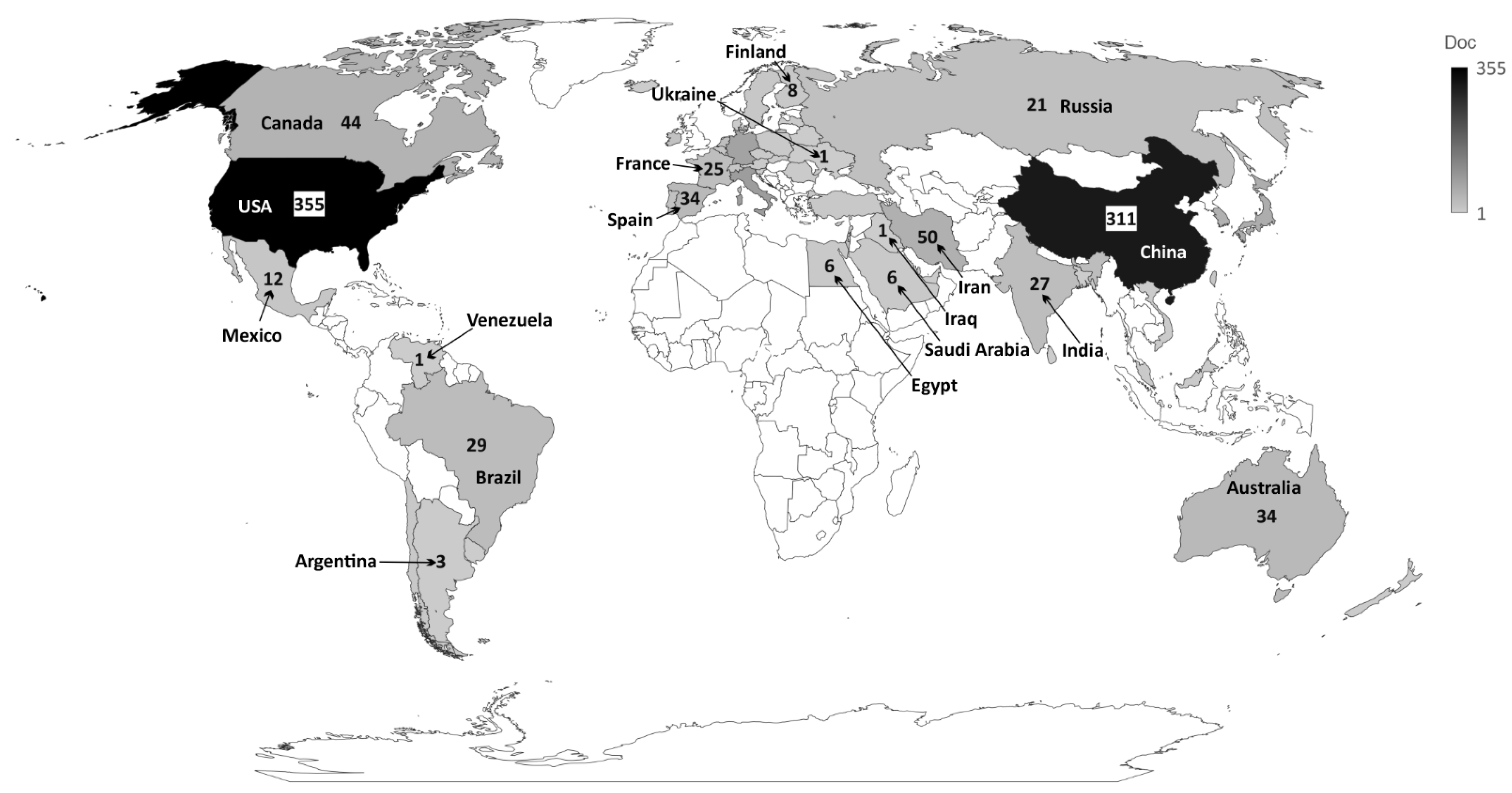
Bibliometric Findings
4. Strengths and Limitations
5. Conclusions
Supplementary Materials
Author Contributions
Funding
Acknowledgments
Conflicts of Interest
References
- Hanahan, D.; Weinberg, R.A. Hallmarks of cancer: The next generation. Cell 2011, 144, 646–674. [Google Scholar] [CrossRef] [PubMed] [Green Version]
- Forouzanfar, M.H.; Afshin, A.; Alexander, L.T.; Biryukov, S.; Brauer, M.; Cercy, K.; Charlson, F.J.; Cohen, A.J.; Dandona, L.; Estep, K.; et al. Global, regional, and national comparative risk assessment of 79 behavioural, environmental and occupational, and metabolic risks or clusters of risks, 1990–2015: A systematic analysis for the Global Burden of Disease Study 2015. Lancet 2016, 388, 1659–1724. [Google Scholar] [CrossRef] [Green Version]
- Harbeck, N.; Gnant, M. Breast cancer. Lancet 2017, 389, 1134–1150. [Google Scholar] [CrossRef]
- Sung, H.; Ferlay, J.; Siegel, R.L.; Laversanne, M.; Soerjomataram, I.; Jemal, A.; Bray, F. Global cancer statistics 2020: GLOBOCAN estimates of incidence and mortality worldwide for 36 cancers in 185 countries. CA Cancer J Clin. 2021, 71, 209–249. [Google Scholar] [CrossRef]
- Guan, X. Cancer metastases: Challenges and opportunities. Acta Pharm. Sin. B 2015, 5, 402–418. [Google Scholar] [CrossRef] [Green Version]
- Van Zijl, F.; Krupitza, G.; Mikulits, W. Initial steps of metastasis: Cell invasion and endothelial transmigration. Mutat. Res./Rev. Mutat. Res. 2011, 728, 23–34. [Google Scholar] [CrossRef]
- VanderVorst, K.; Dreyer, C.A.; Konopelski, S.E.; Lee, H.; Ho, H.Y.H.; Carraway, K.L. Wnt/PCP signaling contribution to carcinoma collective cell migration and metastasis. Cancer Res. 2019, 79, 1719–1729. [Google Scholar] [CrossRef] [Green Version]
- Demicheli, R.; Desmedt, C.; Piccart, M.; Biganzoli, E. Tumor dormancy at bedside: A late awakening. Breast 2019, 45, 61–63. [Google Scholar] [CrossRef]
- Ursini-Siegel, J.; Siegel, P.M. The influence of the pre-metastatic niche on breast cancer metastasis. Cancer Lett. 2016, 380, 281–288. [Google Scholar] [CrossRef]
- Friberg, S.; Nystrom, A. Cancer Metastases: Early Dissemination and Late Recurrences. Cancer Growth Metastasis 2015, 8, CGM.S31244. [Google Scholar] [CrossRef] [Green Version]
- Fares, J.; Kanojia, D.; Rashidi, A.; Ulasov, I.; Lesniak, M.S. Genes that Mediate Metastasis across the Blood–Brain Barrier. Trends Cancer 2020, 6, 660–676. [Google Scholar] [CrossRef]
- Follain, G.; Osmani, N.; Azevedo, A.S.; Allio, G.; Mercier, L.; Karreman, M.A.; Solecki, G.; Garcia Leòn, M.J.; Lefebvre, O.; Fekonja, N.; et al. Hemodynamic Forces Tune the Arrest, Adhesion, and Extravasation of Circulating Tumor Cells. Dev. Cell 2018, 45, 33–52.e12. [Google Scholar] [CrossRef] [Green Version]
- Peinado, H.; Zhang, H.; Matei, I.R.; Costa-silva, B.; Hoshino, A.; Rodrigues, G.; Psaila, B.; Kaplan, R.N.; Bromberg, J.F.; Kang, Y.; et al. Pre-metastatic niches: Organ-specific homes for metastases. Nat. Publ. Gr. 2017, 17, 302–317. [Google Scholar] [CrossRef]
- Qian, C.N.; Mei, Y.; Zhang, J. Cancer metastasis: Issues and challenges. Chin. J. Cancer 2017, 36, 36–39. [Google Scholar] [CrossRef] [PubMed]
- Colombo, M.; Raposo, G.; Théry, C. Biogenesis, secretion, and intercellular interactions of exosomes and other extracellular vesicles. Annu. Rev. Cell Dev. Biol. 2014, 30, 255–289. [Google Scholar] [CrossRef]
- Hoshino, A.; Kim, H.S.; Bojmar, L.; Gyan, K.E.; Cioffi, M.; Hernandez, J.; Zambirinis, C.P.; Rodrigues, G.; Molina, H.; Heissel, S.; et al. Extracellular Vesicle and Particle Biomarkers Define Multiple Human Cancers. Cell 2020, 182, 1044–1061.e18. [Google Scholar] [CrossRef] [PubMed]
- Théry, C.; Witwer, K.W.; Aikawa, E.; Alcaraz, M.J.; Anderson, J.D.; Andriantsitohaina, R.; Antoniou, A.; Arab, T.; Archer, F.; Atkin-Smith, G.K.; et al. Minimal information for studies of extracellular vesicles 2018 (MISEV2018): A position statement of the International Society for Extracellular Vesicles and update of the MISEV2014 guidelines. J. Extracell. Vesicles 2018, 7, 1535750. [Google Scholar] [CrossRef] [Green Version]
- Medina, C.B.; Mehrotra, P.; Arandjelovic, S.; Perry, J.S.A.; Guo, Y.; Morioka, S.; Barron, B.; Walk, S.F.; Ghesquière, B.; Krupnick, A.S.; et al. Metabolites released from apoptotic cells act as tissue messengers. Nature 2020, 580, 130–135. [Google Scholar] [CrossRef] [PubMed]
- Keklikoglou, I.; Cianciaruso, C.; Güç, E.; Squadrito, M.L.; Spring, L.M.; Tazzyman, S.; Lambein, L.; Poissonnier, A.; Ferraro, G.B.; Baer, C.; et al. Chemotherapy elicits pro-metastatic extracellular vesicles in breast cancer models. Nat. Cell Biol. 2019, 21, 190–202. [Google Scholar] [CrossRef] [Green Version]
- Kosaka, N.; Iguchi, H.; Hagiwara, K.; Yoshioka, Y.; Takeshita, F.; Ochiya, T. Neutral Sphingomyelinase 2 (nSMase2)-dependent Exosomal Transfer of Angiogenic MicroRNAs Regulate Cancer Cell Metastasis. J. Biol. Chem. 2013, 288, 10849–10859. [Google Scholar] [CrossRef] [Green Version]
- Zhou, W.; Fong, M.Y.; Min, Y.; Somlo, G.; Liu, L.; Palomares, M.R.; Yu, Y.; Chow, A.; O’Connor, S.T.F.; Chin, A.R.; et al. Cancer-Secreted miR-105 destroys vascular endothelial barriers to promote metastasis. Cancer Cell 2014, 25, 501–515. [Google Scholar] [CrossRef] [PubMed] [Green Version]
- Tominaga, N.; Kosaka, N.; Ono, M.; Katsuda, T.; Yoshioka, Y.; Tamura, K.; Lötvall, J.; Nakagama, H.; Ochiya, T. Brain metastatic cancer cells release microRNA-181c-containing extracellular vesicles capable of destructing blood-brain barrier. Nat. Commun. 2015, 6, 6716. [Google Scholar] [CrossRef] [Green Version]
- Shi, S.; Gao, Y.; Liu, M.; Bu, Y.; Wu, J.; Tian, J.; Zhang, J. Top 100 most-cited articles on exosomes in the field of cancer: A bibliometric analysis and evidence mapping. Clin. Exp. Med. 2020, 21, 181–194. [Google Scholar] [CrossRef] [PubMed]
- Zyoud, S.H.; Al-Jabi, S.W. Mapping the situation of research on coronavirus disease-19 (COVID-19): A preliminary bibliometric analysis during the early stage of the outbreak. BMC Infect. Dis. 2020, 20, 1–8. [Google Scholar] [CrossRef] [PubMed]
- Manoel Alves, J.; Handerson Gomes Teles, R.; do Valle Gomes Gatto, C.; Muñoz, V.R.; Regina Cominetti, M.; Garcia de Oliveira Duarte, A.C. Mapping Research in the Obesity, Adipose Tissue, and MicroRNA Field: A Bibliometric Analysis. Cells 2019, 8, 1581. [Google Scholar] [CrossRef] [Green Version]
- Broadus, R.N. Toward a definition of “bibliometrics”. Scientometrics 1987, 12, 373–379. [Google Scholar] [CrossRef]
- Ellegaard, O.; Wallin, J.A. The bibliometric analysis of scholarly production: How great is the impact? Scientometrics 2015, 105, 1809–1831. [Google Scholar] [CrossRef] [Green Version]
- Donthu, N.; Kumar, S.; Mukherjee, D.; Pandey, N.; Lim, W.M. How to conduct a bibliometric analysis: An overview and guidelines. J. Bus. Res. 2021, 133, 285–296. [Google Scholar] [CrossRef]
- Verma, S.; Gustafsson, A. Investigating the emerging COVID-19 research trends in the field of business and management: A bibliometric analysis approach. J. Bus. Res. 2020, 118, 253–261. [Google Scholar] [CrossRef]
- Teles, R.H.G.; Moralles, H.F.; Cominetti, M.R. Global trends in nanomedicine research on triple negative breast cancer: A bibliometric analysis. Int. J. Nanomed. 2018, 13, 2321. [Google Scholar] [CrossRef] [Green Version]
- Gao, Y.; Shi, S.; Ma, W.; Chen, J.; Cai, Y.; Ge, L.; Li, L.; Wu, J.; Tian, J. Bibliometric analysis of global research on PD-1 and PD-L1 in the field of cancer. Int. Immunopharmacol. 2019, 72, 374–384. [Google Scholar] [CrossRef]
- Kamdem, J.P.; Duarte, A.E.; Ibrahim, M.; Lukong, K.E.; Barros, L.M.; Roeder, T. Bibliometric analysis of personalized humanized mouse and Drosophila models for effective combinational therapy in cancer patients. Biochim. Biophys. Acta Mol. Basis Dis. 2020, 1866, 165880. [Google Scholar] [CrossRef]
- Özen Çınar, İ. Bibliometric analysis of breast cancer research in the period 2009–2018. Int. J. Nurs. Pract. 2020, 26, e12845. [Google Scholar] [CrossRef]
- Akmal, M.; Hasnain, N.; Rehan, A.; Iqbal, U.; Hashmi, S.; Fatima, K.; Farooq, M.Z.; Khosa, F.; Siddiqi, J.; Khan, M.K. Glioblastome Multiforme: A Bibliometric Analysis. World Neurosurg. 2020, 136, 270–282. [Google Scholar] [CrossRef]
- Stout, N.L.; Alfano, C.M.; Belter, C.W.; Nitkin, R.; Cernich, A.; Lohmann Siegel, K.; Chan, L. A Bibliometric Analysis of the Landscape of Cancer Rehabilitation Research (1992–2016). J. Natl. Cancer Inst. 2018, 110, 815–824. [Google Scholar] [CrossRef] [PubMed] [Green Version]
- Liu, W.; Wu, L.; Zhang, Y.; Shi, L.; Yang, X. Bibliometric analysis of research trends and characteristics of oral potentially malignant disorders. Clin. Oral Investig. 2020, 24, 447–454. [Google Scholar] [CrossRef]
- Aria, M.; Cuccurullo, C. Bibliometrix: An R-tool for comprehensive science mapping analysis. J. Informetr. 2017, 11, 959–975. [Google Scholar] [CrossRef]
- Fox, A.S.; Yoon, S.B. DNA-induced transformation in Drosophila: Locus-specificity and the establishment of transformed stocks. Proc. Natl. Acad. Sci. USA 1970, 67, 1608–1615. [Google Scholar] [CrossRef] [PubMed] [Green Version]
- Hannafon, B.N.; Trigoso, Y.D.; Calloway, C.L.; Zhao, Y.D.; Lum, D.H.; Welm, A.L.; Zhao, Z.J.; Blick, K.E.; Dooley, W.C.; Ding, W.Q. Plasma exosome microRNAs are indicative of breast cancer. Breast Cancer Res. 2016, 18, 1–14. [Google Scholar] [CrossRef] [Green Version]
- Hannafon, B.N.; Ding, W.Q. Intercellular communication by exosome-derived microRNAs in cancer. Int. J. Mol. Sci. 2013, 14, 14240–14269. [Google Scholar] [CrossRef] [Green Version]
- Yang, M.; Chen, J.; Su, F.; Yu, B.; Su, F.; Lin, L.; Liu, Y.; Huang, J.D.; Song, E. Microvesicles secreted by macrophages shuttle invasion-potentiating microRNAs into breast cancer cells. Mol. Cancer 2011, 10, 6–10. [Google Scholar] [CrossRef] [PubMed] [Green Version]
- Ratajczak, J.; Wysoczynski, M.; Hayek, F.; Janowska-Wieczorek, A.; Ratajczak, M.Z. Membrane-derived microvesicles: Important and underappreciated mediators of cell-to-cell communication. Leukemia 2006, 20, 1487–1495. [Google Scholar] [CrossRef]
- Eichelser, C.; Stückrath, I.; Müller, V.; Milde-Langosch, K.; Wikman, H.; Pantel, K.; Schwarzenbach, H. Increased serum levels of circulating exosomal microRNA-373 in receptor-negative breast cancer patients. Oncotarget 2014, 5, 9650–9663. [Google Scholar] [CrossRef] [Green Version]
- Rupp, A.K.; Rupp, C.; Keller, S.; Brase, J.C.; Ehehalt, R.; Fogel, M.; Moldenhauer, G.; Marmé, F.; Sültmann, H.; Altevogt, P. Loss of EpCAM expression in breast cancer derived serum exosomes: Role of proteolytic cleavage. Gynecol. Oncol. 2011, 122, 437–446. [Google Scholar] [CrossRef]
- Galindo-Hernandez, O.; Villegas-Comonfort, S.; Candanedo, F.; González-Vázquez, M.C.; Chavez-Ocaña, S.; Jimenez-Villanueva, X.; Sierra-Martinez, M.; Salazar, E.P. Elevated concentration of microvesicles isolated from peripheral blood in breast cancer patients. Arch. Med. Res. 2013, 44, 208–214. [Google Scholar] [CrossRef] [PubMed]
- Lee, J.K.; Park, S.R.; Jung, B.K.; Jeon, Y.K.; Lee, Y.S.; Kim, M.K.; Kim, Y.G.; Jang, J.Y.; Kim, C.W. Exosomes derived from mesenchymal stem cells suppress angiogenesis by down-regulating VEGF expression in breast cancer cells. PLoS ONE 2013, 8, e84256. [Google Scholar] [CrossRef] [PubMed] [Green Version]
- Ohno, S.I.; Takanashi, M.; Sudo, K.; Ueda, S.; Ishikawa, A.; Matsuyama, N.; Fujita, K.; Mizutani, T.; Ohgi, T.; Ochiya, T.; et al. Systemically injected exosomes targeted to EGFR deliver antitumor microrna to breast cancer cells. Mol. Ther. 2013, 21, 185–191. [Google Scholar] [CrossRef] [Green Version]
- O’Brien, K.; Rani, S.; Corcoran, C.; Wallace, R.; Hughes, L.; Friel, A.M.; McDonnell, S.; Crown, J.; Radomski, M.W.; O’Driscoll, L. Exosomes from triple-negative breast cancer cells can transfer phenotypic traits representing their cells of origin to secondary cells. Eur. J. Cancer 2013, 49, 1845–1859. [Google Scholar] [CrossRef]
- Ciravolo, V.; Huber, V.; Ghedini, G.C.; Venturelli, E.; Bianchi, F.; Campiglio, M.; Morelli, D.; Villa, A.; Della Mina, P.; Menard, S.; et al. Potential role of HER2-overexpressing exosomes in countering trastuzumab-based therapy. J. Cell. Physiol. 2012, 227, 658–667. [Google Scholar] [CrossRef]
- Melo, S.A.; Luecke, L.B.; Kahlert, C.; Fernandez, A.F.; Gammon, S.T.; Kaye, J.; LeBleu, V.S.; Mittendorf, E.A.; Weitz, J.; Rahbari, N.; et al. Glypican-1 identifies cancer exosomes and detects early pancreatic cancer. Nature 2015, 523, 177–182. [Google Scholar] [CrossRef] [Green Version]
- Ono, M.; Kosaka, N.; Tominaga, N.; Yoshioka, Y.; Takeshita, F.; Takahashi, R.U.; Yoshida, M.; Tsuda, H.; Tamura, K.; Ochiya, T. Exosomes from bone marrow mesenchymal stem cells contain a microRNA that promotes dormancy in metastatic breast cancer cells. Sci. Signal. 2014, 7, ra63. [Google Scholar] [CrossRef]
- Melo, S.A.; Sugimoto, H.; Connell, J.T.O.; Kato, N.; Vidal, A.; Qiu, L.; Vitkin, E.; Perelman, L.T.; Melo, C.A.; Lucci, A.; et al. Cancer exosomes perform Cell-independent MicroRNA biogenesis and promote tumorigenesis. Cancer Cell 2015, 26, 707–721. [Google Scholar] [CrossRef] [Green Version]
- Luga, V.; Zhang, L.; Viloria-Petit, A.M.; Ogunjimi, A.A.; Inanlou, M.R.; Chiu, E.; Buchanan, M.; Hosein, A.N.; Basik, M.; Wrana, J.L. Exosomes mediate stromal mobilization of autocrine Wnt-PCP signaling in breast cancer cell migration. Cell 2012, 151, 1542–1556. [Google Scholar] [CrossRef] [PubMed] [Green Version]
- Donnarumma, E.; Fiore, D.; Nappa, M.; Roscigno, G.; Adamo, A.; Iaboni, M.; Russo, V.; Affinito, A.; Puoti, I.; Quintavalle, C.; et al. Cancer-associated fibroblasts release exosomal microRNAs that dictate an aggressive phenotype in breast cancer. Oncotarget 2017, 8, 19592–19608. [Google Scholar] [CrossRef] [PubMed] [Green Version]
- Singh, R.; Pochampally, R.; Watabe, K.; Lu, Z.; Mo, Y.Y. Exosome-mediated transfer of miR-10b promotes cell invasion in breast cancer. Mol. Cancer 2014, 13, 1–11. [Google Scholar] [CrossRef] [Green Version]
- Chen, W.X.; Cai, Y.Q.; Lv, M.M.; Chen, L.; Zhong, S.L.; Ma, T.F.; Zhao, J.H.; Tang, J.H. Exosomes from docetaxel-resistant breast cancer cells alter chemosensitivity by delivering microRNAs. Tumor Biol. 2014, 35, 9649–9659. [Google Scholar] [CrossRef]
- Tian, Y.; Li, S.; Song, J.; Ji, T.; Zhu, M.; Anderson, G.J.; Wei, J.; Nie, G. A doxorubicin delivery platform using engineered natural membrane vesicle exosomes for targeted tumor therapy. Biomaterials 2014, 35, 2383–2390. [Google Scholar] [CrossRef]
- King, H.W.; Michael, M.Z.; Gleadle, J.M. Hypoxic enhancement of exosome release by breast cancer cells. BMC Cancer 2012, 12, 421. [Google Scholar] [CrossRef] [Green Version]
- Cho, J.A.; Park, H.; Lim, E.H.; Lee, K.W. Exosomes from breast cancer cells can convert adipose tissue-derived mesenchymal stem cells into myofibroblast-like cells. Int. J. Oncol. 2012, 40, 130–138. [Google Scholar] [CrossRef] [PubMed] [Green Version]
- Kosaka, N.; Iguchi, H.; Ochiya, T. Circulating microRNA in body fluid: A new potential biomarker for cancer diagnosis and prognosis. Cancer Sci. 2010, 101, 2087–2092. [Google Scholar] [CrossRef]
- Siravegna, G.; Marsoni, S.; Siena, S.; Bardelli, A. Integrating liquid biopsies into the management of cancer. Nat. Rev. Clin. Oncol. 2017, 14, 531–548. [Google Scholar] [CrossRef]
- Fong, M.Y.; Zhou, W.; Liu, L.; Alontaga, A.Y.; Chandra, M.; Ashby, J.; Chow, A.; O’Connor, S.T.F.; Li, S.; Chin, A.R.; et al. Breast-cancer-secreted miR-122 reprograms glucose metabolism in premetastatic niche to promote metastasis. Nat. Cell Biol. 2015, 17, 183–194. [Google Scholar] [CrossRef] [Green Version]
- Xu, J.; Camfield, R.; Gorski, S.M. The interplay between exosomes and autophagy—partners in crime. J. Cell Sci. 2018, 131, 1–11. [Google Scholar] [CrossRef] [PubMed] [Green Version]
- Buratta, S.; Tancini, B.; Sagini, K.; Delo, F.; Chiaradia, E.; Urbanelli, L.; Emiliani, C. Lysosomal exocytosis, exosome release and secretory autophagy: The autophagic- and endo-lysosomal systems go extracellular. Int. J. Mol. Sci. 2020, 21, 2576. [Google Scholar] [CrossRef] [PubMed] [Green Version]
- Yun, C.W.; Lee, S.H. The roles of autophagy in cancer. Int. J. Mol. Sci. 2018, 19, 3466. [Google Scholar] [CrossRef] [PubMed] [Green Version]
- Hamurcu, Z.; Delibaşı, N.; Geçene, S.; Şener, E.F.; Dönmez-Altuntaş, H.; Özkul, Y.; Canatan, H.; Ozpolat, B. Targeting LC3 and Beclin-1 autophagy genes suppresses proliferation, survival, migration and invasion by inhibition of Cyclin-D1 and uPAR/Integrin β1/Src signaling in triple negative breast cancer cells. J. Cancer Res. Clin. Oncol. 2018, 144, 415–430. [Google Scholar] [CrossRef] [PubMed]
- Unal, O.; Akkoc, Y.; Kocak, M.; Nalbat, E.; Dogan-Ekici, A.I.; Yagci Acar, H.; Gozuacik, D. Treatment of breast cancer with autophagy inhibitory microRNAs carried by AGO2-conjugated nanoparticles. J. Nanobiotechnology 2020, 18, 1–18. [Google Scholar] [CrossRef]
- Heneberg, P. Paracrine tumor signaling induces transdifferentiation of surrounding fibroblasts. Crit. Rev. Oncol. Hematol. 2016, 97, 303–311. [Google Scholar] [CrossRef]
- Sahai, E.; Astsaturov, I.; Cukierman, E.; DeNardo, D.G.; Egeblad, M.; Evans, R.M.; Fearon, D.; Greten, F.R.; Hingorani, S.R.; Hunter, T.; et al. A framework for advancing our understanding of cancer-associated fibroblasts. Nat. Rev. Cancer 2020, 20, 174–186. [Google Scholar] [CrossRef] [Green Version]
- Tao, S.; Li, H.; Ma, X.; Ma, Y.; He, J.; Gao, Y.; Li, J. Elevating microRNA-1-3p shuttled by cancer-associated fibroblasts-derived extracellular vesicles suppresses breast cancer progression and metastasis by inhibiting GLIS1. Cancer Gene Ther. 2020, 28, 634–648. [Google Scholar] [CrossRef]
- Luga, V.; Wrana, J.L. Tumor-stroma interaction: Revealing fibroblast-secreted exosomes as potent regulators of Wnt-planar cell polarity signaling in cancer metastasis. Cancer Res. 2013, 73, 6843–6847. [Google Scholar] [CrossRef] [PubMed] [Green Version]
- Greening, D.W.; Gopal, S.K.; Mathias, R.A.; Liu, L.; Sheng, J.; Zhu, H.J.; Simpson, R.J. Emerging roles of exosomes during epithelial-mesenchymal transition and cancer progression. Semin. Cell Dev. Biol. 2015, 40, 60–71. [Google Scholar] [CrossRef]
- Sansone, P.; Berishaj, M.; Rajasekhar, V.K.; Ceccarelli, C.; Chang, Q.; Strillacci, A.; Savini, C.; Shapiro, L.; Bowman, R.L.; Mastroleo, C.; et al. Evolution of Cancer Stem-like Cells in Endocrine-Resistant Metastatic Breast Cancers is Mediated by Stromal Microvesicles. Cancer Res. 2017, 77, 1927–1941. [Google Scholar] [CrossRef] [Green Version]
- Medina, M.A.; Oza, G.; Sharma, A.; Arriaga, L.G.; Hernández, J.M.H.; Rotello, V.M.; Ramirez, J.T. Triple-negative breast cancer: A review of conventional and advanced therapeutic strategies. Int. J. Environ. Res. Public Health 2020, 17, 2078. [Google Scholar] [CrossRef] [Green Version]
- Silva, T.A.; Smuczek, B.; Valadão, I.C.; Dzik, L.M.; Iglesia, R.P.; Cruz, M.C.; Zelanis, A.; de Siqueira, A.S.; Serrano, S.M.T.; Goldberg, G.S.; et al. AHNAK enables mammary carcinoma cells to produce extracellular vesicles that increase neighboring fibroblast cell motility. Oncotarget 2016, 7, 49998–50016. [Google Scholar] [CrossRef] [PubMed] [Green Version]
- Kikuchi, S.; Yoshioka, Y.; Prieto-Vila, M.; Ochiya, T. Involvement of extracellular vesicles in vascular-related functions in cancer progression and metastasis. Int. J. Mol. Sci. 2019, 20, 2584. [Google Scholar] [CrossRef] [Green Version]
- Yang, G.; Sau, C.; Lai, W.; Cichon, J.; Li, W. Exosomal annexin A2 promotes angiogenesis in breast cancer metastasis. Mol. Cancer Res. 2015, 344, 1173–1178. [Google Scholar] [CrossRef]
- Chaudhary, P.; Gibbs, L.D.; Maji, S.; Lewis, C.M.; Suzuki, S.; Vishwanatha, J.K. Serum exosomal-annexin a2 is associated with african-American triple-negative breast cancer and promotes angiogenesis. Breast Cancer Res. 2020, 22, 1–15. [Google Scholar] [CrossRef]
- Gong, C.; Tian, J.; Wang, Z.; Gao, Y.; Wu, X.; Ding, X.; Qiang, L.; Li, G.; Han, Z.; Yuan, Y.; et al. Functional exosome-mediated co-delivery of doxorubicin and hydrophobically modified microRNA 159 for triple-negative breast cancer therapy. J. Nanobiotechnology 2019, 17, 1–18. [Google Scholar] [CrossRef] [Green Version]
- Haney, M.J.; Zhao, Y.; Jin, Y.S.; Li, S.M.; Bago, J.R.; Klyachko, N.L.; Kabanov, A.V.; Batrakova, E.V. Macrophage-Derived Extracellular Vesicles as Drug Delivery Systems for Triple Negative Breast Cancer (TNBC) Therapy. J. Neuroimmune Pharmacol. 2020, 15, 487–500. [Google Scholar] [CrossRef]
- Poulet, G.; Massias, J.; Taly, V. Liquid Biopsy: General Concepts. Acta Cytol. 2019, 63, 449–455. [Google Scholar] [CrossRef] [PubMed]
- Ozawa, P.M.M.; Jucoski, T.S.; Vieira, E.; Carvalho, T.M.; Malheiros, D.; Ribeiro, E.M.D.S.F. Liquid biopsy for breast cancer using extracellular vesicles and cell-free microRNAs as biomarkers. Transl. Res. 2020, 223, 40–60. [Google Scholar] [CrossRef] [PubMed]
- Abramowicz, A.; Story, M.D. The Long and Short of It: The Emerging Roles of Non-Coding RNA in Small Extracellular Vesicles. Cancers 2020, 12, 1445. [Google Scholar] [CrossRef] [PubMed]


| Documents | Components | Indexes |
|---|---|---|
| Articles, reviews | 1157 | |
| Journals, books | 451 | |
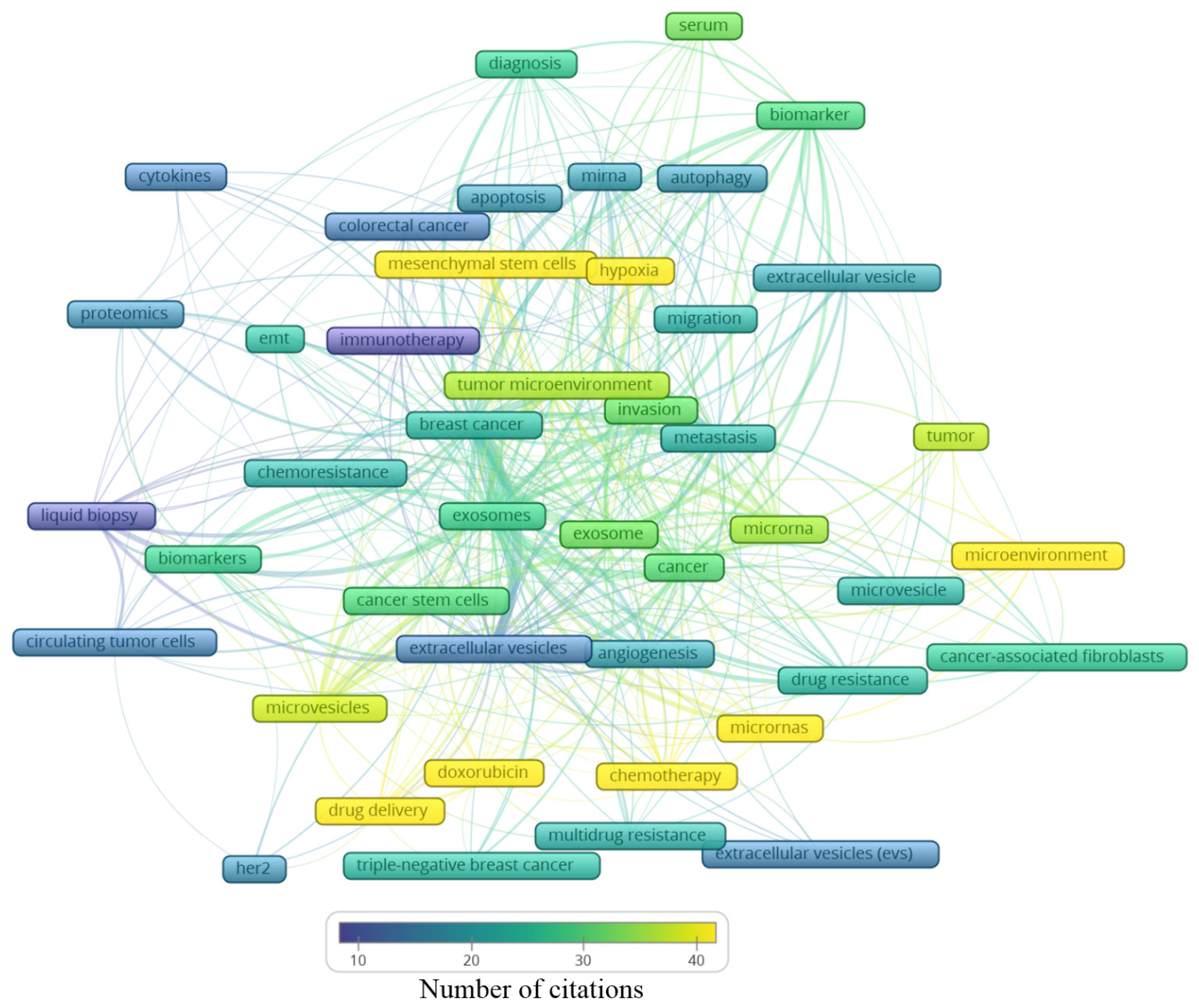
| Author’s keywords | 7924 | |
| Period | 1998–2021 | |
| Average citations per document | 35.75 | |
| Authors | ||
| Authors | 5599 | |
| Author appearances | 8141 | |
| Authors of single-authored documents | 38 | |
| Authors of multi-authored documents | 5561 | |
| Single-authored documents | 43 | |
| Documents per author | 0.206 | |
| Colaborations | ||
| Authors per document | 4.84 | |
| Co-Authors per document | 7.07 | |
| Collaboration index | 5.02 |
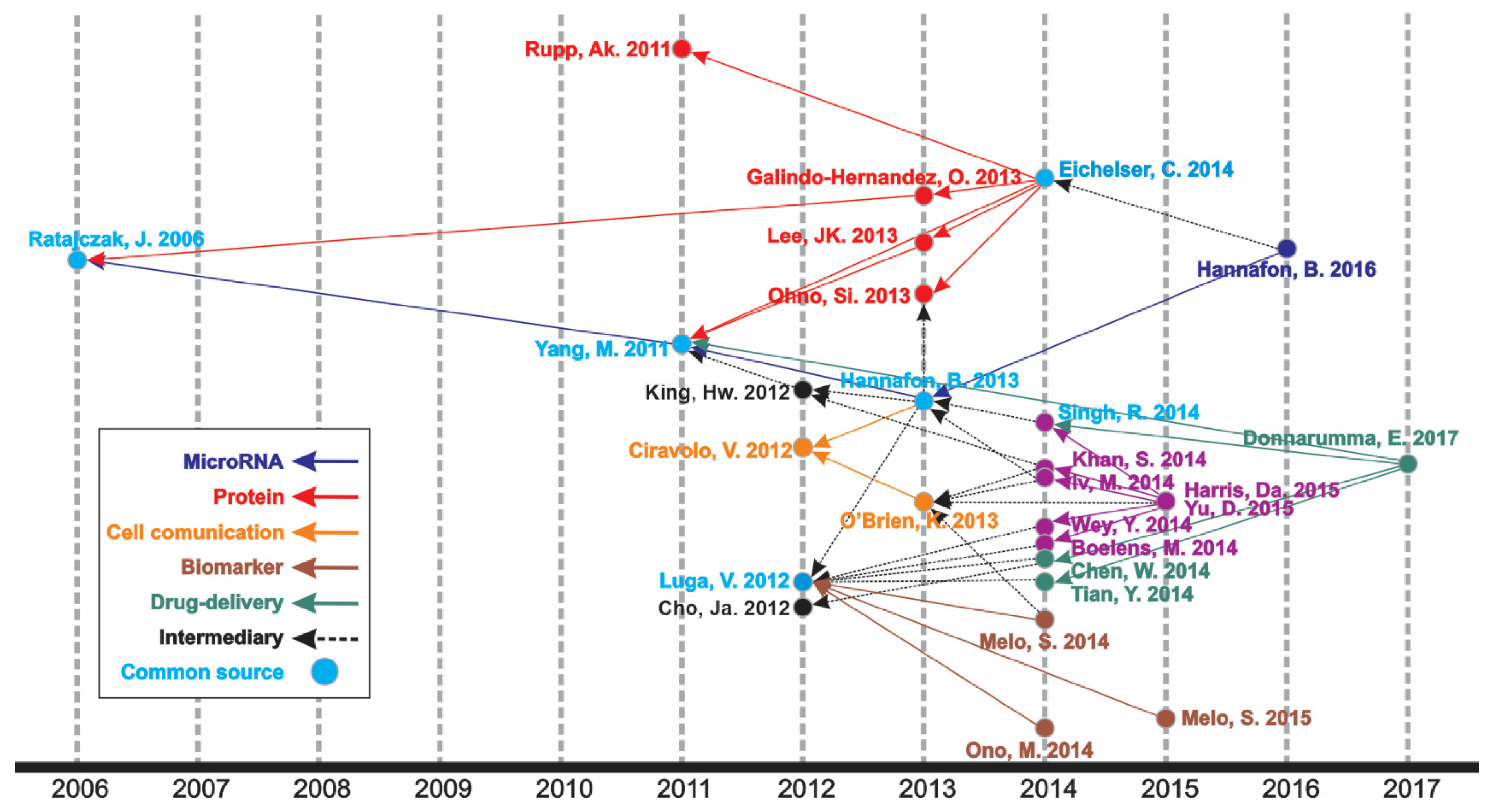
| Author | Article | Year | Total Citations | Ref. |
|---|---|---|---|---|
| MicroRNA | ||||
| Hannafon B.N., Breast Cancer Res. | Plasma exosome microRNAs are indicative of breast cancer | 2016 | 257 | [39] |
| Hannafon B.N., Int. J. Mol. Sci. | Intercellular communication by exosome-derived microRNAs in cancer | 2013 | 375 | [40] |
| Yang M., Mol. Cancer | Microvesicles secreted by macrophages shuttle invasion-potentiating microRNAs into breast cancer cells | 2011 | 603 | [41] |
| Ratajczac J., Leukemia. | Membrane-derived microvesicles: important and underappreciated mediators of cell-to-cell communication. | 2006 | 1336 | [42] |
| Protein | ||||
| Eichelser C., Oncotarget | Increased serum levels of circulating exosomal microRNA-373 in receptor-negative breast cancer patients | 2014 | 251 | [43] |
| Rupp A.K., Gynecol. Oncol. | Loss of EpCAM expression in breast cancer derived serum exosomes: role of proteolytic cleavage | 2011 | 212 | [44] |
| Galindo-Hernandez O., Arch. Med. Res. | Elevated concentration of microvesicles isolated from peripheral blood in breast cancer patients | 2013 | 90 | [45] |
| Lee J.K., PLoS ONE | Exosomes Derived from Mesenchymal Stem Cells Suppress Angiogenesis by Down-Regulating VEGF Expression in Breast Cancer Cells | 2013 | 379 | [46] |
| Ohno S.I., Mol. Ther. | Systemically injected exosomes targeted to EGFR deliver antitumor microRNA to breast cancer cells | 2013 | 981 | [47] |
| Cell comunication | ||||
| Hannafon B.N., Int. J. Mol. Sci. | Intercellular communication by exosome-derived microRNAs in cancer | 2013 | 375 | [40] |
| O’Brien K., Eur. J. Cancer | Exosomes from triple-negative breast cancer cells can transfer phenotypic traits representing their cells of origin to secondary cells | 2013 | 173 | [48] |
| Ciravolo V., J. Cell Physiol. | Potential role of HER2-overexpressing exosomes in countering trastuzumab-based therapy | 2012 | 350 | [49] |
| Biomarker | ||||
| Melo S.A., Nature | Glypican-1 identifies cancer exosomes and detects early pancreatic cancer | 2015 | 1561 | [50] |
| Ono M., Sci. Signal | Exosomes from bone marrow mesenchymal stem cells contain a microRNA that promotes dormancy in metastatic breast cancer cells | 2014 | 433 | [51] |
| Melo S.A., Cancer Cell | Cancer exosomes perform cell-independent microRNA biogenesis and promote tumorigenesis | 2014 | 1074 | [52] |
| Luga H.W., Cell | Exosomes mediate stromal mobilization of autocrine Wnt-PCP signaling in breast cancer cell migration | 2012 | 939 | [53] |
| Drug delivery | ||||
| Donnarumma E., Oncotarget | Cancer-associated fibroblasts release exosomal microRNAs that dictate an aggressive phenotype in breast cancer | 2017 | 165 | [54] |
| Singh R., Mol. Cancer | Exosome-mediated transfer of miR-10b promotes cell invasion in breast cancer. | 2014 | 274 | [55] |
| Chen W.X., Tumor Biol. | Exosomes from docetaxel-resistant breast cancer cells alter chemosensitivity by delivering microRNAs | 2014 | 140 | [56] |
| Tian Y., Biomaterials | A doxorubicin delivery platform using engineered natural membrane vesicle exosomes for targeted tumor therapy | 2014 | 854 | [57] |
| Yang M., Mol. Cancer | Microvesicles secreted by macrophages shuttle invasion-potentiating microRNAs into breast cancer cells | 2011 | 603 | [41] |
| Intermediary | ||||
| Eichelser C., Oncotarget | Increased serum levels of circulating exosomal microRNA-373 in receptor-negative breast cancer patients | 2014 | 251 | [43] |
| Hannafon B.N., Int. J. Mol. Sci. | Intercellular communication by exosome-derived microRNAs in cancer | 2013 | 375 | [40] |
| O’Brien K., Eur. J. Cancer | Exosomes from triple-negative breast cancer cells can transfer phenotypic traits representing their cells of origin to secondary cells | 2013 | 173 | [48] |
| King H.W., BMC Cancer | Hypoxic enhancement of exosome release by breast cancer cells | 2012 | 650 | [58] |
| Cho J.A., Int. J. Oncol. | Exosomes from breast cancer cells can convert adipose tissue-derived mesenchymal stem cells into myofibroblast-like cells | 2012 | 339 | [59] |
| Document Title | Main Finding | Author | Source | Cited By |
|---|---|---|---|---|
| Glypican-1 identifies cancer exosomes and detects early pancreatic cancer [50] | Glypican-1-enriched exosomes as potential specific biomarkers for early detection of pancreatic cancer. | Melo, S.A. et al. | Nature, 2015, 523(7559), pp. 177–182 | 1561 |
| Circulating microRNA in body fluid: A new potential biomarker for cancer diagnosis and prognosis [60] | Review of microRNA as a promissing non-invasive tool for prediction, prognosis, and diagnosis of early cancer. | Kosaka, N., Iguchi, H., Ochiya, T. | Cancer Science, 2010, 101(10), pp. 2087–2092 | 922 |
| Membrane-derived microvesicles: Important and underappreciated mediators of cell-to-cell communication [42] | Insights into different aspects of microvesicle roles in different topics, such as carcinogenesis, coagulation, communication between cells, immune response, and modulation. | Ratajczak, J. et al. | Leukemia, 2006, 20(9), pp. 1487–1495 | 910 |
| Cancer exosomes perform cell-independent microRNA biogenesis and promote tumorigenesis [52] | Cancer exosomes can modulate the cell transcriptome via miRNAs associated with RISC loading complex. They can also process pre-miRNAs into miRNAs independently of cells. | Melo, S.A. et al. | Cancer Cell, 2015, 26(5), pp. 707–721 | 801 |
| Systemically injected exosomes targeted to EGFR deliver antitumor microRNA to breast cancer cells [47] | Engineering exosomes as a potential RNA drug delivery system, addressing exosomes with let-7a miRNA (tumor suppressor) to specifically target EGRP, which is generally high in tumor epithelial cells. | Ohno, S.I. et al. | Molecular Therapy, 2013, 21(1), pp. 185–191 | 779 |
| Exosomes mediate stromal mobilization of autocrine Wnt-PCP signaling in breast cancer cell migration [53] | Exosomes derived from cancer-associated fibroblasts and L-cells have an autocrine influence on Wnt-PCP signaling, a factor that regulates and assists breast cancer cells in the motility and metastasis process. | Luga, V., et al. | Cell, 2012, 151(7), pp. 1542–1556 | 728 |
| A doxorubicin delivery platform using engineered natural membrane vesicle exosomes for targeted tumor therapy [57] | Engineering of the exosome membrane from immature dendritic cells by fusion with the iRGD-targeting peptide for αV integrin, thereby creating a drug delivery system for the chemotherapeutic doxorubicin to tumor tissue. | Tian, Y., et al. | Biomaterials, 2014, 35(7), pp. 2383–2390 | 680 |
| Integrating liquid biopsies into the management of cancer [61] | Insights into various tumor-derived materials that can be the target of liquid biopsies, with the focus on ctDNA, and the potential use of this screening to improve diagnostic performance and the treatment choice. | Siravegna, G., Marsoni, S., Siena, S., Bardelli, A. | Nature Reviews Clinical Oncology, 2017, 14(9), pp. 531–548 | 644 |
| Hypoxic enhancement of exosome release by breast cancer cells [58] | The condition of hypoxia leads to an increase in an exosome that is enriched with miR-210 released by breast cancer cells. This factor could lead to promotion of tumour invasion, progression, angiogenesis, and endothelial activation. | King, H.W., Michael, M.Z., Gleadle, J.M. | BMC Cancer, 2012, 12, 421 | 486 |
| Breast-cancer-secreted miR-122 reprograms glucose metabolism in premetastatic niche to promote metastasis [62] | A higher level of miR-122 secreted by breast cancer mediates a decrease in glucose uptake by healthy normal cells in the premetastatic niche, which favors the uptake of this nutrient by cancer cells during the metastasis process. | Fong, M.Y., et al. | Nature Cell Biology, 2015, 17(2), pp. 183–194 | 477 |
Publisher’s Note: MDPI stays neutral with regard to jurisdictional claims in published maps and institutional affiliations. |
© 2021 by the authors. Licensee MDPI, Basel, Switzerland. This article is an open access article distributed under the terms and conditions of the Creative Commons Attribution (CC BY) license (https://creativecommons.org/licenses/by/4.0/).
Share and Cite
Teles, R.H.G.; Yano, R.S.; Villarinho, N.J.; Yamagata, A.S.; Jaeger, R.G.; Meybohm, P.; Burek, M.; Freitas, V.M. Advances in Breast Cancer Management and Extracellular Vesicle Research, a Bibliometric Analysis. Curr. Oncol. 2021, 28, 4504-4520. https://0-doi-org.brum.beds.ac.uk/10.3390/curroncol28060382
Teles RHG, Yano RS, Villarinho NJ, Yamagata AS, Jaeger RG, Meybohm P, Burek M, Freitas VM. Advances in Breast Cancer Management and Extracellular Vesicle Research, a Bibliometric Analysis. Current Oncology. 2021; 28(6):4504-4520. https://0-doi-org.brum.beds.ac.uk/10.3390/curroncol28060382
Chicago/Turabian StyleTeles, Ramon Handerson Gomes, Rafael Sussumu Yano, Nicolas Jones Villarinho, Ana Sayuri Yamagata, Ruy Gastaldoni Jaeger, Patrick Meybohm, Malgorzata Burek, and Vanessa Morais Freitas. 2021. "Advances in Breast Cancer Management and Extracellular Vesicle Research, a Bibliometric Analysis" Current Oncology 28, no. 6: 4504-4520. https://0-doi-org.brum.beds.ac.uk/10.3390/curroncol28060382