Current Advances in the Management of Adult Craniopharyngiomas

 , , ,
, , ,
Abstract
:1. Introduction
2. Clinical Manifestations
3. Histological Variants
4. Molecular Pathways Involved in Tumor Development
4.1. The Wingless (Wnt)/β-Catenin Pathway
4.2. The Mitogen-Activated Protein Kinases/Extracellular Signal-Regulated Kinase (MAPK/ERK)
5. Current Treatment and Management for Craniopharyngiomas
5.1. Surgical Management
5.2. Radiation Therapy
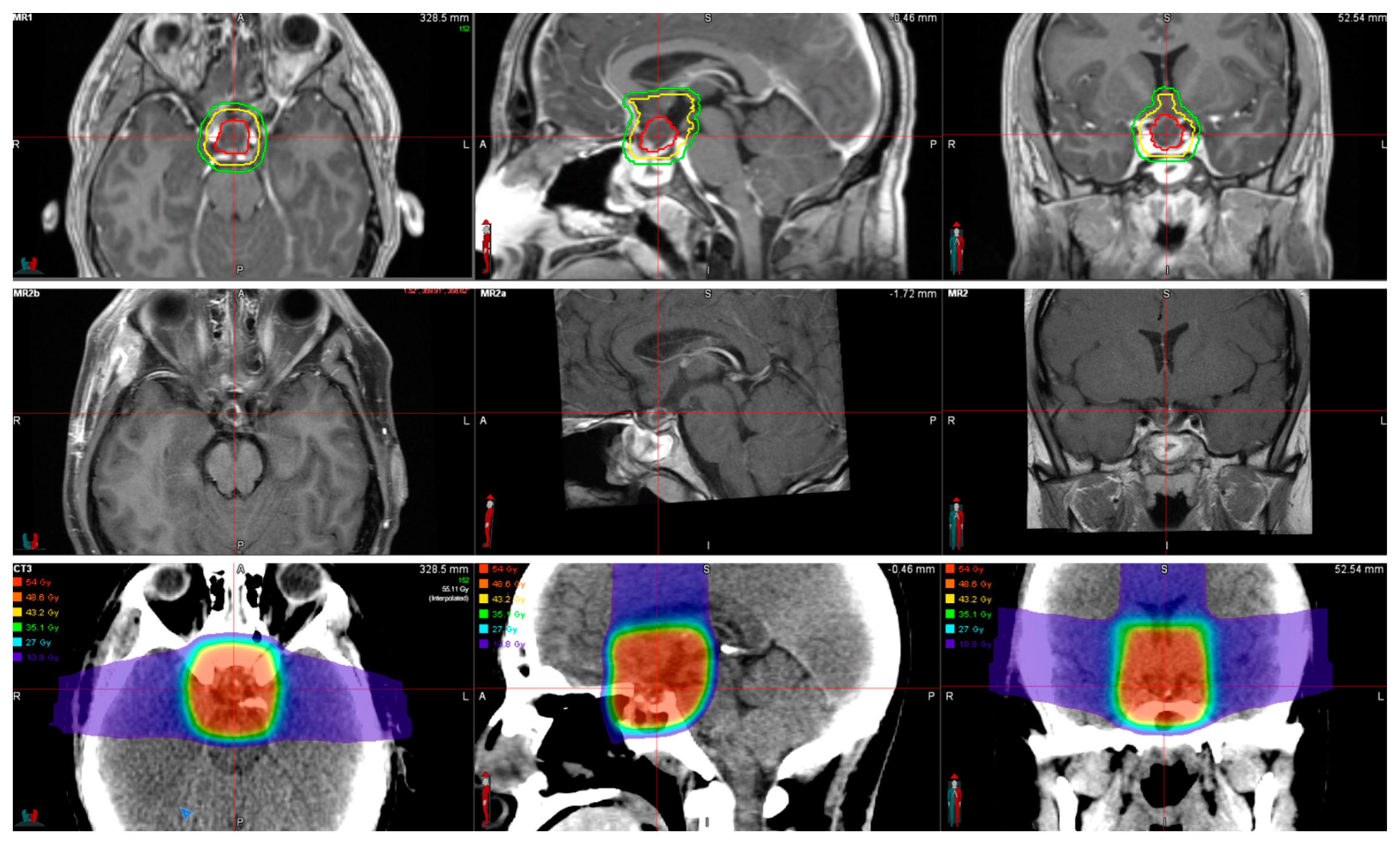
5.3. Stereotactic Radiosurgery
5.4. Fractionated Stereotactic Radiotherapy
5.5. Intensity-Modulated Radiation Therapy
5.6. Proton Beam Therapy
6. Current Trends in the Treatment of Craniopharyngiomas
6.1. Brachytherapy
6.2. Chemotherapy
7. New Landscape in Craniopharyngioma Treatment: Immunotherapy
7.1. The Immune and Inflammatory Components of Craniopharyngioma
7.2. Targeting B7-H3
7.3. Targeting PD-L1
7.4. Targeting CTLA-4
7.5. Targeting VISTA
8. Challenges for the Treatment of Craniopharyngiomas
8.1. Incomplete Resection
8.2. Diabetes Insipidus (DI)
8.3. Metabolism and Hypothalamic Obesity
8.4. Visual Impairment/Loss
8.5. Psychological Complications
9. Conclusions
Author Contributions
Funding
Conflicts of Interest
References
- Bunin, G.R.; Surawicz, T.S.; Witman, P.A.; Preston-Martin, S.; Davis, F.; Bruner, J.M. The descriptive epidemiology of craniopharyngioma. J. Neurosurg. 1998, 89, 547–551. [Google Scholar] [CrossRef] [PubMed]
- Karavitaki, N.; Cudlip, S.; Adams, C.B.; Wass, J.A. Craniopharyngiomas. Endocr. Rev. 2006, 27, 371–397. [Google Scholar] [CrossRef] [PubMed]
- Ostrom, Q.T.; Gittleman, H.; Liao, P.; Vecchione-Koval, T.; Wolinsky, Y.; Kruchko, C.; Barnholtz-Sloan, J.S. CBTRUS Statistical Report: Primary brain and other central nervous system tumors diagnosed in the United States in 2010–2014. Neur. Oncol. 2017, 19, v1–v88. [Google Scholar] [CrossRef] [PubMed] [Green Version]
- Yamini, B.; Narayanan, M. Craniopharyngiomas: An update. Expert Rev. Anticancer Ther. 2006, 6, S85–S92. [Google Scholar] [CrossRef]
- Muller, H.L.; Merchant, T.E.; Warmuth-Metz, M.; Martinez Barbera, J.P.; Puget, S. Craniopharyngioma. Nat. Rev. Dis Primers 2019, 5, 75. [Google Scholar] [CrossRef]
- Ottenhausen, M.; Rumalla, K.; La Corte, E.; Alalade, A.; Nair, P.; Forbes, J.; Ben Nsir, A.; Schwartz, T.H. Treatment strategies for craniopharyngiomas. J. Neurosurg. Sci. 2019, 63, 83–87. [Google Scholar] [CrossRef]
- Zacharia, B.E.; Bruce, S.S.; Goldstein, H.; Malone, H.R.; Neugut, A.I.; Bruce, J.N. Incidence, treatment and survival of patients with craniopharyngioma in the surveillance, epidemiology and end results program. Neuro Oncol. 2012, 14, 1070–1078. [Google Scholar] [CrossRef] [Green Version]
- Hoffmann, A.; Boekhoff, S.; Gebhardt, U.; Sterkenburg, A.S.; Daubenbuchel, A.M.; Eveslage, M.; Muller, H.L. History before diagnosis in childhood craniopharyngioma: Associations with initial presentation and long-term prognosis. Eur. J. Endocrinol. 2015, 173, 853–862. [Google Scholar] [CrossRef]
- Jensterle, M.; Jazbinsek, S.; Bosnjak, R.; Popovic, M.; Zaletel, L.Z.; Vesnaver, T.V.; Kotnik, B.F.; Kotnik, P. Advances in the management of craniopharyngioma in children and adults. Radiol. Oncol. 2019, 53, 388–396. [Google Scholar] [CrossRef] [Green Version]
- Mortini, P.; Losa, M.; Pozzobon, G.; Barzaghi, R.; Riva, M.; Acerno, S.; Angius, D.; Weber, G.; Chiumello, G.; Giovanelli, M. Neurosurgical treatment of craniopharyngioma in adults and children: Early and long-term results in a large case series. J. Neurosurg. 2011, 114, 1350–1359. [Google Scholar] [CrossRef]
- Duff, J.; Meyer, F.B.; Ilstrup, D.M.; Laws, E.R., Jr.; Schleck, C.D.; Scheithauer, B.W. Long-term outcomes for surgically resected craniopharyngiomas. Neurosurgery 2000, 46, 291–302. [Google Scholar] [CrossRef]
- Elliott, R.E.; Hsieh, K.; Hochm, T.; Belitskaya-Levy, I.; Wisoff, J.; Wisoff, J.H. Efficacy and safety of radical resection of primary and recurrent craniopharyngiomas in 86 children. J. Neurosurg. Pediatr. 2010, 5, 30–48. [Google Scholar] [CrossRef] [PubMed] [Green Version]
- Park, H.J.; Dho, Y.-S.; Kim, J.H.; Kim, J.W.; Park, C.-K.; Kim, Y.H. Recurrence Rate and Prognostic Factors for the Adult Craniopharyngiomas in Long-Term Follow-Up. World Neurosurg. 2019, 133, e211–e217. [Google Scholar] [CrossRef] [PubMed]
- Wu, J.; Wu, X.; Yang, Y.Q.; Ding, H.; Yang, L.; Bao, Y.Y.; Zhou, L.; Yang, C.X.; Hong, T. Association of histological subtype with risk of recurrence in craniopharyngioma patients: A systematic review and meta-analysis. Neurosurg. Rev. 2022, 45, 139–150. [Google Scholar] [CrossRef] [PubMed]
- Puget, S.; Garnett, M.; Wray, A.; Grill, J.; Habrand, J.-L.; Bodaert, N.; Zerah, M.; Bezerra, M.; Renier, D.; Pierre-Kahn, A.; et al. Pediatric craniopharyngiomas: Classification and treatment according to the degree of hypothalamic involvement. J. Neurosurg. Pediatr. 2007, 106, 3–12. [Google Scholar] [CrossRef]
- Mortini, P.; Gagliardi, F.; Bailo, M.; Spina, A.; Parlangeli, A.; Falini, A.; Losa, M. Magnetic resonance imaging as predictor of functional outcome in craniopharyngiomas. Endocrine 2016, 51, 148–162. [Google Scholar] [CrossRef] [PubMed]
- Apps, J.R.; Stache, C.; Gonzalez-Meljem, J.M.; Gutteridge, A.; Chalker, J.; Jacques, T.S.; Forshew, T.; Hölsken, A.; Martinez-Barbera, J.P. CTNNB1 mutations are clonal in adamantinomatous craniopharyngioma. Neuropathol. Appl. Neurobiol. 2020, 46, 510–514. [Google Scholar] [CrossRef] [PubMed] [Green Version]
- Desiderio, C.; Rossetti, D.V.; Castagnola, M.; Massimi, L.; Tamburrini, G. Adamantinomatous craniopharyngioma: Advances in proteomic research. Child’s Nerv. Syst. 2021, 37, 789–797. [Google Scholar] [CrossRef]
- Pettorini, B.L.; Inzitari, R.; Massimi, L.; Tamburrini, G.; Caldarelli, M.; Fanali, C.; Cabras, T.; Messana, I.; Castagnola, M.; Di Rocco, C. The role of inflammation in the genesis of the cystic component of craniopharyngiomas. Child’s Nerv. Syst. 2010, 26, 1779–1784. [Google Scholar] [CrossRef]
- Brastianos, P.K.; Santagata, S. ENDOCRINE TUMORS: BRAF V600E mutations in papillary craniopharyngioma. Eur. J. Endocrinol. 2016, 174, R139–R144. [Google Scholar] [CrossRef]
- Gupta, S.; Bi, W.L.; Giantini Larsen, A.; Al-Abdulmohsen, S.; Abedalthagafi, M.; Dunn, I.F. Craniopharyngioma: A roadmap for scientific translation. Neurosurg. Focus. 2018, 44, E12. [Google Scholar] [CrossRef] [PubMed] [Green Version]
- Koni, M.; Pinnaro, V.; Brizzi, M.F. The Wnt Signalling Pathway: A Tailored Target in Cancer. Int. J. Mol. Sci. 2020, 21, 7697. [Google Scholar] [CrossRef] [PubMed]
- Bläker, H.; Hofmann, W.J.; Rieker, R.J.; Penzel, R.; Graf, M.; Otto, H.F. Beta-catenin accumulation and mutation of the CTNNB1 gene in hepatoblastoma. Genes Chromosomes Cancer 1999, 25, 399–402. [Google Scholar] [CrossRef]
- Inukai, T.; Furuuchi, K.; Sugita, K.; Uno, K.; Ooi, A.; Sasaki, F.; Hamada, J.; Moriuchi, T.; Nakazawa, S. Nuclear accumulation of beta-catenin without an additional somatic mutation in coding region of the APC gene in hepatoblastoma from a familial adenomatous polyposis patient. Oncol. Rep. 2004, 11, 121–126. [Google Scholar] [PubMed]
- Segditsas, S.; Tomlinson, I. Colorectal cancer and genetic alterations in the Wnt pathway. Oncogene 2006, 25, 7531–7537. [Google Scholar] [CrossRef] [Green Version]
- Taciak, B.; Pruszynska, I.; Kiraga, L.; Bialasek, M.; Krol, M. Wnt signaling pathway in development and cancer. J. Physiol. Pharmacol. 2018, 69, 185–196. [Google Scholar] [CrossRef]
- Hölsken, A.; Buchfelder, M.; Fahlbusch, R.; Blümcke, I.; Buslei, R. Tumour cell migration in adamantinomatous craniopharyngiomas is promoted by activated Wnt-signalling. Acta Neuropathol. 2010, 119, 631–639. [Google Scholar] [CrossRef]
- Gaston-Massuet, C.; Andoniadou, C.L.; Signore, M.; Jayakody, S.A.; Charolidi, N.; Kyeyune, R.; Vernay, B.; Jacques, T.S.; Taketo, M.M.; Le Tissier, P.; et al. Increased Wingless (Wnt) signaling in pituitary progenitor/stem cells gives rise to pituitary tumors in mice and humans. Proc. Natl. Acad. Sci. USA 2011, 108, 11482–11487. [Google Scholar] [CrossRef] [Green Version]
- Kato, K.; Nakatani, Y.; Kanno, H.; Inayama, Y.; Ijiri, R.; Nagahara, N.; Miyake, T.; Tanaka, M.; Ito, Y.; Aida, N.; et al. Possible linkage between specific histological structures and aberrant reactivation of the Wnt pathway in adamantinomatous craniopharyngioma. J. Pathol. 2004, 203, 814–821. [Google Scholar] [CrossRef]
- Buslei, R.; Nolde, M.; Hofmann, B.; Meissner, S.; Eyupoglu, I.Y.; Siebzehnrübl, F.; Hahnen, E.; Kreutzer, J.; Fahlbusch, R. Common mutations of beta-catenin in adamantinomatous craniopharyngiomas but not in other tumours originating from the sellar region. Acta Neuropathol. 2005, 109, 589–597. [Google Scholar] [CrossRef]
- Cao, J.; Lin, J.P.; Yang, L.X.; Chen, K.; Huang, Z.S. Expression of aberrant beta-catenin and impaired p63 in craniopharyngiomas. Br. J. Neurosurg. 2010, 24, 249–256. [Google Scholar] [CrossRef] [PubMed]
- Sun, Y.; Liu, W.Z.; Liu, T.; Feng, X.; Yang, N.; Zhou, H.F. Signaling pathway of MAPK/ERK in cell proliferation, differentiation, migration, senescence and apoptosis. J. Recept. Signal. Transduct. Res. 2015, 35, 600–604. [Google Scholar] [CrossRef] [PubMed]
- Safa, A.; Abak, A.; Shoorei, H.; Taheri, M.; Ghafouri-Fard, S. MicroRNAs as regulators of ERK/MAPK pathway: A comprehensive review. Biomed. Pharmacother. 2020, 132, 110853. [Google Scholar] [CrossRef] [PubMed]
- Kurtzeborn, K.; Kwon, H.N.; Kuure, S. MAPK/ERK Signaling in Regulation of Renal Differentiation. Int. J. Mol. Sci. 2019, 20, 1779. [Google Scholar] [CrossRef] [Green Version]
- Degirmenci, U.; Wang, M.; Hu, J. Targeting Aberrant RAS/RAF/MEK/ERK Signaling for Cancer Therapy. Cells 2020, 9, 198. [Google Scholar] [CrossRef] [Green Version]
- McCubrey, J.A.; Steelman, L.S.; Chappell, W.H.; Abrams, S.L.; Wong, E.W.; Chang, F.; Lehmann, B.; Terrian, D.M.; Milella, M.; Tafuri, A.; et al. Roles of the Raf/MEK/ERK pathway in cell growth, malignant transformation and drug resistance. Biochim. Biophys Acta. 2007, 1773, 1263–1284. [Google Scholar] [CrossRef] [Green Version]
- Papa, S.; Choy, P.M.; Bubici, C. The ERK and JNK pathways in the regulation of metabolic reprogramming. Oncogene 2019, 38, 2223–2240. [Google Scholar] [CrossRef] [Green Version]
- Santarpia, L.; Lippman, S.M.; El-Naggar, A.K. Targeting the MAPK-RAS-RAF signaling pathway in cancer therapy. Expert Opin. Ther. Targets 2012, 16, 103–119. [Google Scholar] [CrossRef] [Green Version]
- Masliah-Planchon, J.; Garinet, S.; Pasmant, E. RAS-MAPK pathway epigenetic activation in cancer: miRNAs in action. Oncotarget 2016, 7, 38892–38907. [Google Scholar] [CrossRef] [Green Version]
- Zhang, H.; Sun, P.; Wang, Y.L.; Yu, X.F.; Tong, J.J. MiR-214 promotes proliferation and inhibits apoptosis of oral cancer cells through MAPK/ERK signaling pathway. Eur. Rev. Med. Pharmacol. Sci. 2020, 24, 3710–3716. [Google Scholar] [CrossRef]
- Fang, J.Y.; Richardson, B.C. The MAPK signalling pathways and colorectal cancer. Lancet Oncol. 2005, 6, 322–327. [Google Scholar] [CrossRef]
- Sheng, W.; Chen, C.; Dong, M.; Wang, G.; Zhou, J.; Song, H.; Li, Y.; Zhang, J.; Ding, S. Calreticulin promotes EGF-induced EMT in pancreatic cancer cells via Integrin/EGFR-ERK/MAPK signaling pathway. Cell Death Dis. 2017, 8, e3147. [Google Scholar] [CrossRef]
- Lara-Velazquez, M.; Zarco, N.; Carrano, A.; Phillipps, J.; Norton, E.S.; Schiapparelli, P.; Al-Kharboosh, R.; Rincon-Torroella, J.; Jeanneret, S.; Corona, T.; et al. Alpha 1-antichymotrypsin contributes to stem cell characteristics and enhances tumorigenicity of glioblastoma. Neuro Oncol. 2021, 23, 599–610. [Google Scholar] [CrossRef] [PubMed]
- Andoniadou, C.L.; Matsushima, D.; Mousavy Gharavy, S.N.; Signore, M.; Mackintosh, A.I.; Schaeffer, M.; Gaston-Massuet, C.; Mollard, P.; Jacques, T.S.; Le Tissier, P.; et al. Sox2(+) stem/progenitor cells in the adult mouse pituitary support organ homeostasis and have tumor-inducing potential. Cell Stem Cell. 2013, 13, 433–445. [Google Scholar] [CrossRef] [PubMed] [Green Version]
- Davies, H.; Bignell, G.R.; Cox, C.; Stephens, P.; Edkins, S.; Clegg, S.; Teague, J.; Woffendin, H.; Garnett, M.J.; Bottomley, W.; et al. Mutations of the BRAF gene in human cancer. Nature 2002, 417, 949–954. [Google Scholar] [CrossRef] [PubMed]
- Shin, M.H.; Kim, J.; Lim, S.A.; Kim, J.; Lee, K.M. Current Insights into Combination Therapies with MAPK Inhibitors and Immune Checkpoint Blockade. Int. J. Mol. Sci. 2020, 21, 2531. [Google Scholar] [CrossRef] [PubMed] [Green Version]
- Schoenfeld, A.; Pekmezci, M.; Barnes, M.J.; Tihan, T.; Gupta, N.; Lamborn, K.R.; Banerjee, A.; Mueller, S.; Chang, S.; Berger, M.S.; et al. The superiority of conservative resection and adjuvant radiation for craniopharyngiomas. J. Neurooncol. 2012, 108, 133–139. [Google Scholar] [CrossRef] [PubMed] [Green Version]
- Sadashivam, S.; Menon, G.; Abraham, M.; Nair, S.N. Adult craniopharyngioma: The role of extent of resection in tumor recurrence and long-term functional outcome. Clin. Neurol. Neurosurg. 2020, 192, 105711. [Google Scholar] [CrossRef]
- Fitzek, M.M.; Linggood, R.M.; Adams, J.; Munzenrider, J.E. Combined proton and photon irradiation for craniopharyngioma: Long-term results of the early cohort of patients treated at Harvard Cyclotron Laboratory and Massachusetts General Hospital. Int. J. Radiat Oncol. Biol. Phys. 2006, 64, 1348–1354. [Google Scholar] [CrossRef]
- Garnett, M.R.; Puget, S.; Grill, J.; Sainte-Rose, C. Craniopharyngioma. Orphanet J. Rare Dis. 2007, 2, 18. [Google Scholar] [CrossRef] [Green Version]
- Jung, T.Y.; Jung, S.; Choi, J.E.; Moon, K.S.; Kim, I.Y.; Kang, S.S. Adult craniopharyngiomas: Surgical results with a special focus on endocrinological outcomes and recurrence according to pituitary stalk preservation. J. Neurosurg. 2009, 111, 572–577. [Google Scholar] [CrossRef]
- Jung, T.Y.; Jung, S.; Moon, K.S.; Kim, I.Y.; Kang, S.S.; Kim, J.H. Endocrinological outcomes of pediatric craniopharyngiomas with anatomical pituitary stalk preservation: Preliminary study. Pediatr Neurosurg. 2010, 46, 205–212. [Google Scholar] [CrossRef] [PubMed]
- Almeida, J.P.; Kalyvas, A.; Mohan, N.; Oswari, S.; Takami, H.; Velasquez, C.; Asha, M.; Zadeh, G.; Gentili, F. Current Results of Surgical Treatment of Craniopharyngiomas: The Impact of Endoscopic Endonasal Approaches. World Neurosurg. 2020, 142, 582–592. [Google Scholar] [CrossRef]
- Matsuo, T.; Kamada, K.; Izumo, T.; Nagata, I. Indication and limitations of endoscopic extended transsphenoidal surgery for craniopharyngioma. Neurol. Med. Chir. 2014, 54, 974–982. [Google Scholar] [CrossRef] [Green Version]
- Li, P.; Axier, A.; Li, S.; Zhou, K.; Yun, J.; Wang, H.; Zhang, T. The safety and efficacy of endoscopic endonasal approach in the treatment of recurrent craniopharyngioma: A protocol. for systematic review and meta-analysis. Medicine 2020, 99, e22995. [Google Scholar] [CrossRef] [PubMed]
- Marx, S.; Tsavdaridou, I.; Paul, S.; Steveling, A.; Schirmer, C.; Eördögh, M.; Nowak, S.; Matthes, M.; El Refaee, E.; Fleck, S.K.; et al. Quality of life and olfactory function after suprasellar craniopharyngioma surgery-a single-center experience comparing transcranial and endoscopic endonasal approaches. Neurosurg. Rev. 2021, 44, 1569–1582. [Google Scholar] [CrossRef]
- Emanuelli, E.; Zanotti, C.; Munari, S.; Baldovin, M.; Schiavo, G.; Denaro, L. Sellar and parasellar lesions: Multidisciplinary management. Acta Otorhinolaryngol. Ital. 2021, 41, S30–S41. [Google Scholar] [CrossRef]
- Rahmathulla, G.; Barnett, G.H. Minimally invasive management of adult craniopharyngiomas: An analysis of our series and review of literature. Surg. Neurol. Int. 2013, 4, S411–S421. [Google Scholar]
- Simonin, A.; Bangash, O.; Henley, D.; Bala, A. Endonasal endoscopic resection of suprasellar craniopharyngioma: A retrospective single-center case series. J. Clin. Neurosci. 2020, 81, 436–441. [Google Scholar] [CrossRef]
- Algattas, H.; Setty, P.; Goldschmidt, E.; Wang, E.W.; Tyler-Kabara, E.C.; Snyderman, C.H.; Gardner, P.A. Endoscopic Endonasal Approach for Craniopharyngiomas with Intraventricular Extension: Case Series, Long-Term Outcomes, and Review. World Neurosurg. 2020, 144, e447–e459. [Google Scholar] [CrossRef]
- Schelini, J.C.; Cavalheiro, S.; Dastoli, P.A.; Hirai, É.R.; Atallah, C.; Costa, M.; Nicacio, J.; Capellano, A.M.; Silva, N.; Zymberg, S.; et al. Endoscopic endonasal transsphenoidal approach for pediatric craniopharyngiomas: A case series. Int. J. Pediatr. Otorhinolaryngol. 2020, 130, 109786. [Google Scholar] [CrossRef]
- De Oliveira, R.S.; Viana, D.C.; Augusto, L.P.; Santos, M.V.; Machado, H.R. The supraorbital eyebrow approach for removal of craniopharyngioma in children: A case series. Child’s Nerv. Syst. 2018, 34, 547–553. [Google Scholar] [CrossRef]
- La Corte, E.; Younus, I.; Pivari, F.; Selimi, A.; Ottenhausen, M.; Forbes, J.A.; Pisapia, D.J.; Dobri, G.A.; Anand, V.K.; Schwartz, T.H. BRAF V600E mutant papillary craniopharyngiomas: A single-institutional case series. Pituitary 2018, 21, 571–583. [Google Scholar] [CrossRef] [PubMed]
- Yamada, S.; Fukuhara, N.; Yamaguchi-Okada, M.; Nishioka, H.; Takeshita, A.; Takeuchi, Y.; Inoshita, N.; Ito, J. Therapeutic outcomes of transsphenoidal surgery in pediatric patients with craniopharyngiomas: A single-center study. J. Neurosurg. Pediatr. 2018, 21, 549–562. [Google Scholar] [CrossRef] [Green Version]
- Patel, V.S.; Thamboo, A.; Quon, J.; Nayak, J.V.; Hwang, P.H.; Edwards, M.; Patel, Z.M. Outcomes After Endoscopic Endonasal Resection of Craniopharyngiomas in the Pediatric Population. World Neurosurg. 2017, 108, 6–14. [Google Scholar] [CrossRef] [PubMed]
- Jamshidi, A.O.; Beer-Furlan, A.; Prevedello, D.M.; Sahyouni, R.; Elzoghby, M.A.; Safain, M.G.; Carrau, R.L.; Jane, J.A.; Laws, E.R. A modern series of subdiaphragmatic craniopharyngiomas. J. Neurosurg. 2018, 131, 526–531. [Google Scholar] [CrossRef] [PubMed]
- Forbes, J.A.; Ordóñez-Rubiano, E.G.; Tomasiewicz, H.C.; Banu, M.A.; Younus, I.; Dobri, G.A.; Phillips, C.D.; Kacker, A.; Cisse, B.; Anand, V.K.; et al. Endonasal endoscopic transsphenoidal resection of intrinsic third ventricular craniopharyngioma: Surgical results. J. Neurosurg. 2018, 1, 1–11. [Google Scholar] [CrossRef]
- Alalade, A.F.; Ogando-Rivas, E.; Boatey, J.; Souweidane, M.M.; Anand, V.K.; Greenfield, J.P.; Schwartz, T.H. Suprasellar and recurrent pediatric craniopharyngiomas: Expanding indications for the extended endoscopic transsphenoidal approach. J. Neurosurg. Pediatr. 2018, 21, 72–80. [Google Scholar] [CrossRef] [Green Version]
- Rajan, B.; Ashley, S.; Gorman, C.; Jose, C.C.; Horwich, A.; Bloom, H.J.; Marsh, H.; Brada, M. Craniopharyngioma--a long-term results following limited surgery and radiotherapy. Radiother. Oncol. 1993, 26, 1–10. [Google Scholar] [CrossRef]
- Tsang, R.W.; Brierley, J.D.; Panzarella, T.; Gospodarowicz, M.K.; Sutcliffe, S.B.; Simpson, W.J. Radiation therapy for pituitary adenoma: Treatment outcome and prognostic factors. Int. J. Radiat. Oncol. Biol. Phys. 1994, 30, 557–565. [Google Scholar] [CrossRef]
- Bidur, K.C.; Prasad, D.U. Outcome following surgical resection of craniopharyngiomas: A case series. Asian J. Neurosurg. 2017, 12, 514–518. [Google Scholar] [CrossRef] [PubMed] [Green Version]
- Ramanbhavana, V.S.; Vara Prasad, K.S. A Case Series of Craniopharyngioma: Epidemiological Study and Management Analysis at Tertiary Care Center. Asian J. Neurosurg. 2019, 14, 1196–1202. [Google Scholar] [CrossRef] [PubMed]
- Foran, S.J.; Laperriere, N.; Edelstein, K.; Janzen, L.; Tadic, T.; Ramaswamy, V.; Shultz, D.; Gentili, F.; Bouffet, E.; Tsang, D.S. Reirradiation for recurrent craniopharyngioma. Adv. Radiat. Oncol. 2020, 5, 1305–1310. [Google Scholar] [CrossRef]
- Lauretti, L.; Legninda Sop, F.Y.; Pallini, R.; Fernandez, E.; D’Alessandris, Q.G. Neuroendoscopic Treatment of Cystic Craniopharyngiomas: A Case Series with Systematic Review of the Literature. World Neurosurg. 2018, 110, e367–e373. [Google Scholar] [CrossRef]
- Rutenberg, M.S.; Rotondo, R.L.; Rao, D.; Holtzman, A.L.; Indelicato, D.J.; Huh, S.; Morris, C.G.; Mendenhall, W.M. Clinical outcomes following proton therapy for adult craniopharyngioma: A single-institution cohort study. J. Neurooncol. 2020, 147, 387–395. [Google Scholar] [CrossRef] [PubMed]
- Shirane, R.; Ching-Chan, S.; Kusaka, Y.; Jokura, H.; Yoshimoto, T. Surgical outcomes in 31 patients with craniopharyngiomas extending outside the suprasellar cistern: An evaluation of the frontobasal interhemispheric approach. J. Neurosurg. 2002, 96, 704–712. [Google Scholar] [CrossRef]
- Gautier, A.; Godbout, A.; Grosheny, C.; Tejedor, I.; Coudert, M.; Courtillot, C.; Jublanc, C.; De Kerdanet, M.; Poirier, J.Y.; Riffaud, L.; et al. Markers of recurrence and long-term morbidity in craniopharyngioma: A systematic analysis of 171 patients. J. Clin Endocrinol. Metab. 2012, 97, 1258–1267. [Google Scholar] [CrossRef]
- Rath, S.R.; Lee, S.; Kotecha, R.S.; Taylor, M.; Junckerstorff, R.C.; Choong, C.S. Childhood craniopharyngioma: 20-year institutional experience in Western Australia. J. Paediatr. Child Health 2013, 49, 403–408. [Google Scholar] [CrossRef]
- Velnar, T.; Bosnjak, R. Radiosurgical techniques for the treatment of brain neoplasms: A short review. World J. Methodol. 2018, 8, 51–58. [Google Scholar] [CrossRef]
- Loeffler, J.S.; Larson, D.A. Subspecialization in radiation oncology: Impact of stereotactic radiosurgery. Int. J. Radiat Oncol. Biol. Phys. 1992, 24, 885–887. [Google Scholar] [CrossRef]
- Harris, L.; Das, J.M. Stereotactic Radiosurgery; StatPearls: Treasure Island, FL, USA, 2021. [Google Scholar]
- Minniti, G.; Esposito, V.; Amichetti, M.; Enrici, R.M. The role of fractionated radiotherapy and radiosurgery in the management of patients with craniopharyngioma. Neurosurg. Rev. 2009, 32, 125–132; discussion 132. [Google Scholar] [CrossRef] [PubMed]
- Yang, I.; Udawatta, M.; Prashant, G.N.; Lagman, C.; Bloch, O.; Jensen, R.; Sheehan, J.; Kalkanis, S.; Warnick, R. Stereotactic Radiosurgery for Neurosurgical Patients: A Historical Review and Current Perspectives. World Neurosurg. 2019, 122, 522–531. [Google Scholar] [CrossRef] [PubMed]
- Shaw, E.; Scott, C.; Souhami, L.; Dinapoli, R.; Kline, R.; Loeffler, J.; Farnan, N. Single dose radiosurgical treatment of recurrent previously irradiated primary brain tumors and brain metastases: Final report of RTOG protocol. 90-05. Int. J. Radiat Oncol. Biol. Phys. 2000, 47, 291–298. [Google Scholar] [CrossRef]
- Pikis, S.; Mantziaris, G.; Lavezzo, K.; Dabhi, N.; Sheehan, J. Stereotactic radiosurgery for craniopharyngiomas. Acta Neurochir. 2021, 163, 3201–3207. [Google Scholar] [CrossRef]
- Xu, Z.; Yen, C.P.; Schlesinger, D.; Sheehan, J. Outcomes of Gamma Knife surgery for craniopharyngiomas. J. Neurooncol. 2011, 104, 305–313. [Google Scholar] [CrossRef]
- Chiou, S.M.; Lunsford, L.D.; Niranjan, A.; Kondziolka, D.; Flickinger, J.C. Stereotactic radiosurgery of residual or recurrent craniopharyngioma, after surgery, with or without radiation therapy. Neur. Oncol. 2001, 3, 159–166. [Google Scholar] [CrossRef] [Green Version]
- Varlotto, J.; DiMaio, C.; Grassberger, C.; Tangel, M.; Mackley, H.; Pavelic, M.; Specht, C.; Sogge, S.; Nguyen, D.; Glantz, M.; et al. Multi-modality management of craniopharyngioma: A review of various treatments and their outcomes. Neurooncol. Pract. 2016, 3, 173–187. [Google Scholar] [CrossRef] [Green Version]
- Kobayashi, T. Long-term results of gamma knife radiosurgery for 100 consecutive cases of craniopharyngioma and a treatment strategy. Prog Neurol. Surg. 2009, 22, 63–76. [Google Scholar] [CrossRef]
- Griffiths, M.J.; Gama, R. Recurrent dizzy spells: All in the head. Ann. Clin. Biochem. 2005, 42, 308. [Google Scholar] [CrossRef] [Green Version]
- Habrand, J.L.; Ganry, O.; Couanet, D.; Rouxel, V.; Levy-Piedbois, C.; Pierre-Kahn, A.; Kalifa, C. The role of radiation therapy in the management of craniopharyngioma: A 25-year experience and review of the literature. Int. J. Radiat. Oncol. Biol. Phys. 1999, 44, 255–263. [Google Scholar] [CrossRef]
- Lin, L.L.; El Naqa, I.; Leonard, J.R.; Park, T.S.; Hollander, A.S.; Michalski, J.M.; Mansur, D.B. Long-term outcome in children treated for craniopharyngioma with and without radiotherapy. J. Neurosurg. Pediatr. 2008, 1, 126–130. [Google Scholar] [CrossRef] [PubMed]
- Iannalfi, A.; Fragkandrea, I.; Brock, J.; Saran, F. Radiotherapy in craniopharyngiomas. Clin Oncol. 2013, 25, 654–667. [Google Scholar] [CrossRef]
- Jeon, C.; Kim, S.; Shin, H.J.; Nam, D.H.; Lee, J.I.; Park, K.; Kim, J.H.; Jeon, B.; Kong, D.S. The therapeutic efficacy of fractionated radiotherapy and gamma-knife radiosurgery for craniopharyngiomas. J. Clin Neurosci. 2011, 18, 1621–1625. [Google Scholar] [CrossRef] [PubMed]
- Astradsson, A.; Munck Af Rosenschöld, P.; Feldt-Rasmussen, U.; Poulsgaard, L.; Wiencke, A.K.; Ohlhues, L.; Engelholm, S.A.; Broholm, H.; Hansen Møller, E.; Klose, M.; et al. Visual outcome, endocrine function and tumor control. after fractionated stereotactic radiation therapy of craniopharyngiomas in adults: Findings in a prospective cohort. Acta Oncol. 2017, 56, 415–421. [Google Scholar] [CrossRef] [PubMed] [Green Version]
- Fokas, E.; Henzel, M.; Surber, G.; Kleinert, G.; Hamm, K.; Engenhart-Cabillic, R. Stereotactic radiosurgery and fractionated stereotactic radiotherapy: Comparison of efficacy and toxicity in 260 patients with brain metastases. J. Neurooncol. 2012, 109, 91–98. [Google Scholar] [CrossRef]
- Kalapurakal, J.A. Radiation therapy in the management of pediatric craniopharyngiomas--a review. Childs Nerv. Syst. 2005, 21, 808–816. [Google Scholar] [CrossRef]
- Taylor, A.; Powell, M.E. Intensity-modulated radiotherapy—What is it? Cancer Imaging 2004, 4, 68–73. [Google Scholar] [CrossRef] [Green Version]
- Cho, B. Intensity-modulated radiation therapy: A review with a physics perspective. Radiat. Oncol. J. 2018, 36, 1–10. [Google Scholar] [CrossRef] [Green Version]
- Hatano, K.; Tohyama, N.; Kodama, T.; Okabe, N.; Sakai, M.; Konoeda, K. Current status of intensity-modulated radiation therapy for prostate cancer: History, clinical results and future directions. Int. J. Urol. 2019, 26, 775–784. [Google Scholar] [CrossRef] [Green Version]
- Torres, M.A.; Gogineni, K.; Howard, D.H. Intensity-Modulated Radiation Therapy in Breast Cancer Patients Following the Release of a Choosing Wisely Recommendation. J. Natl. Cancer Inst. 2020, 112, 314–317. [Google Scholar] [CrossRef]
- Sato, A.; Omura, M.; Minagawa, Y.; Matsui, K.; Shirata, R.; Hongo, H.; Hashimoto, H.; Misumi, T.; Inoue, T.; Hata, M. Intensity-modulated Radiation Therapy for Lymph Node Oligo-recurrence. In Vivo 2020, 34, 2587–2593. [Google Scholar] [CrossRef] [PubMed]
- Kudkuli, J.; Agrawal, A.; Gurjar, O.P.; Sharma, S.D.; Rekha, P.D.; Manzoor, M.A.P.; Singh, B.; Rao, B.S.; Abdulla, R. Demineralization of tooth enamel following radiation therapy; An in vitro microstructure and microhardness analysis. J. Cancer Res. Ther. 2020, 16, 612–618. [Google Scholar] [CrossRef]
- Greenfield, B.J.; Okcu, M.F.; Baxter, P.A.; Chintagumpala, M.; Teh, B.S.; Dauser, R.C.; Su, J.; Desai, S.S.; Paulino, A.C. Long-term disease control. and toxicity outcomes following surgery and intensity modulated radiation therapy (IMRT) in pediatric craniopharyngioma. Radiother. Oncol. 2015, 114, 224–229. [Google Scholar] [CrossRef]
- Merchant, T.E.; Kun, L.E.; Hua, C.H.; Wu, S.; Xiong, X.; Sanford, R.A.; Boop, F.A. Disease control. after reduced volume conformal and intensity modulated radiation therapy for childhood craniopharyngioma. Int. J. Radiat. Oncol. Biol. Phys. 2013, 85, e187–e192. [Google Scholar] [CrossRef] [PubMed] [Green Version]
- Mohamed Ali, A.; Mathis, T.; Bensadoun, R.J.; Thariat, J. Radiation induced optic neuropathy: Does treatment modality influence the risk? Bull Cancer 2019, 106, 1160–1176. [Google Scholar] [CrossRef]
- Pemberton, L.S.; Dougal, M.; Magee, B.; Gattamaneni, H.R. Experience of external beam radiotherapy given adjuvantly or at relapse following surgery for craniopharyngioma. Radiother. Oncol. 2005, 77, 99–104. [Google Scholar] [CrossRef] [PubMed]
- Kim, J.K.; Leeman, J.E.; Riaz, N.; McBride, S.; Tsai, C.J.; Lee, N.Y. Proton Therapy for Head and Neck Cancer. Curr Treat Options Oncol. 2018, 19, 28. [Google Scholar] [CrossRef] [PubMed]
- Hu, M.; Jiang, L.; Cui, X.; Zhang, J.; Yu, J. Proton beam therapy for cancer in the era of precision medicine. J. Hematol. Oncol. 2018, 11, 136. [Google Scholar] [CrossRef]
- Merchant, T.E.; Hua, C.H.; Shukla, H.; Ying, X.; Nill, S.; Oelfke, U. Proton versus photon radiotherapy for common pediatric brain tumors: Comparison of models of dose characteristics and their relationship to cognitive function. Pediatr. Blood Cancer 2008, 51, 110–117. [Google Scholar] [CrossRef]
- Kooy, H.M.; Grassberger, C. Intensity modulated proton therapy. Br. J. Radiol. 2015, 88, 20150195. [Google Scholar] [CrossRef] [Green Version]
- Luu, Q.T.; Loredo, L.N.; Archambeau, J.O.; Yonemoto, L.T.; Slater, J.M.; Slater, J.D. Fractionated proton radiation treatment for pediatric craniopharyngioma: Preliminary report. Cancer J. 2006, 12, 155–159. [Google Scholar] [PubMed]
- O’Steen, L.; Indelicato, D.J. Advances in the management of craniopharyngioma. F1000Research 2018, 7, 1632. [Google Scholar] [CrossRef] [PubMed]
- Otterlei, O.M.; Indelicato, D.J.; Toussaint, L.; Ytre-Hauge, K.S.; Pilskog, S.; Fjaera, L.F.; Rørvik, E.; Pettersen, H.E.S.; Muren, L.P.; Lassen-Ramshad, Y.; et al. Variation in relative biological effectiveness for cognitive structures in proton therapy of pediatric brain tumors. Acta Oncol. 2021, 60, 267–274. [Google Scholar] [CrossRef] [PubMed]
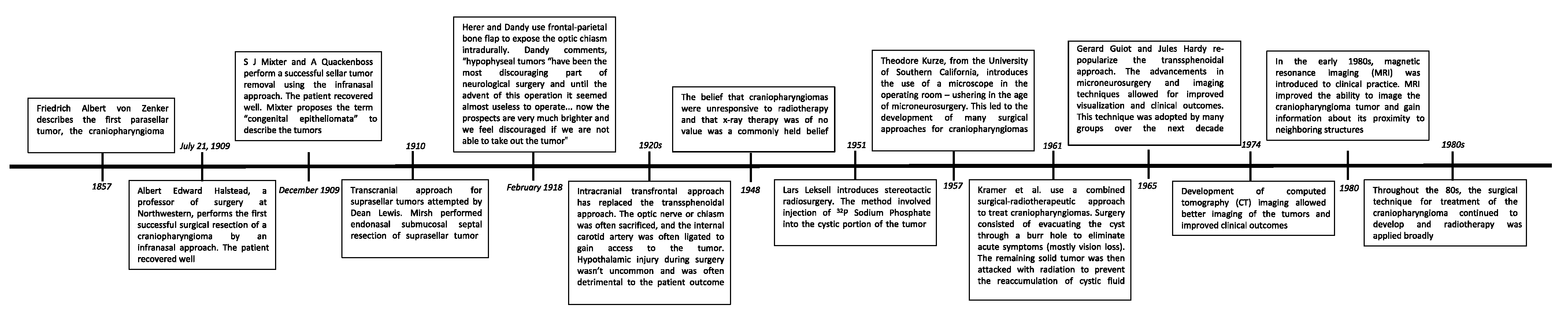
- Barkhoudarian, G.; Laws, E.R. Craniopharyngioma: History. Pituitary. 2013, 16, 1–8. [Google Scholar] [CrossRef] [PubMed]
- Mixter, S.J.; Quackenboss, A. II. Tumor of the Hypophysis (with Infantilism): Operation-Recovery (Preliminary Report). Ann Surg. 1910, 52, 15–22. [Google Scholar] [CrossRef]
- DiPatri, A.J., Jr.; Prabhu, V. A history of the treatment of craniopharyngiomas. Childs Nerv Syst. 2005, 21, 606–621. [Google Scholar] [CrossRef]
- Raimondi, A.J.; Rougerie, J. A critical review of personal experiences with craniopharyngioma: Clinical history, surgical technique and operative results. Pediatr. Neurosurg. 1994, 21, 134–150. [Google Scholar] [CrossRef]
- Seidel, H.; Richter, J.; Kurerov, N.N.; Schajpak, E.J.; Blüthner, R.; Erdmann, U.; Hinz, B. Psychophysical assessment of sinusoidal whole-body vibration in z-axis between 0.6 and 5 Hz combined with different noise levels. Int. Arch. Occup. Env. Health 1989, 61, 413–422. [Google Scholar] [CrossRef]
- Major, T.; Fröhlich, G.; Ágoston, P.; Polgár, C.; Takácsi-Nagy, Z. The value of brachytherapy in the age of advanced external beam radiotherapy: A review of the literature in terms of dosimetry. Strahlenther Onkol. 2022, 198, 93–109. [Google Scholar] [CrossRef]
- Yu, X.; Christ, S.M.; Liu, R.; Wang, Y.; Hu, C.; Feng, B.; Mahadevan, A.; Kasper, E.M. Evaluation of Long-Term Outcomes and Toxicity After Stereotactic Phosphorus-32-Based Intracavitary Brachytherapy in Patients with Cystic Craniopharyngioma. Int. J. Radiat. Oncol. Biol. Phys. 2021, 111, 773–784. [Google Scholar] [CrossRef]
- Ruge, M.I.; Kickingereder, P.; Grau, S.; Treuer, H.; Sturm, V.; Voges, J. Stereotactic iodine-125 brachytherapy for brain tumors: Temporary versus permanent implantation. Radiat. Oncol. 2012, 7, 94. [Google Scholar] [CrossRef] [PubMed] [Green Version]
- Guimarães, M.M.; Cardeal, D.D.; Teixeira, M.J.; Lucio, J.E.D.C.; Sanders, F.H.; Kuromoto, R.K.; Matushita, H. Brachytherapy in paediatric craniopharyngiomas: A systematic review and meta-analysis of recent literature. Child’s Nerv. Syst. 2022, 38, 253–262. [Google Scholar] [CrossRef] [PubMed]
- Silva, J.T.; Daruich de Souza, C.; Angelocci, L.V.; Arcos Rosero, W.A.; Nogueira, B.R.; Correia, R.W.; Zeituni, C.A.; Chuery Martins Rostelato, M.E. New model for an epoxy-based brachytherapy source to be used in spinal cancer treatment. Appl. Radiat. Isot. 2021, 178, 109952. [Google Scholar] [CrossRef] [PubMed]
- Ansari, S.F.; Moore, R.J.; Boaz, J.C.; Fulkerson, D.H. Efficacy of phosphorus-32 brachytherapy without external-beam radiation for long-term tumor control. in patients with craniopharyngioma. J. Neurosurg. Pediatr. 2016, 17, 439–445. [Google Scholar] [CrossRef] [PubMed]
- Hukin, J.; Steinbok, P.; Lafay-Cousin, L.; Hendson, G.; Strother, D.; Mercier, C.; Samson, Y.; Howes, W.; Bouffet, E. Intracystic bleomycin therapy for craniopharyngioma in children: The Canadian experience. Cancer: Interdiscip. Int. J. Am. Cancer Soc. 2007, 109, 2124–2131. [Google Scholar] [CrossRef] [PubMed]
- Dastoli, P.A.; Nicácio, J.M.; Silva, N.S.; Capellano, A.M.; Toledo, S.R.; Ierardi, D.; Cavalheiro, S. Cystic craniopharyngioma: Intratumoral chemotherapy with alpha interferon. Arq. Neuropsiquiatr. 2011, 69, 50–55. [Google Scholar] [CrossRef] [Green Version]
- Cavalheiro, S.; Dastoli, P.A.; Silva, N.S.; Toledo, S.; Lederman, H.; da Silva, M.C. Use of interferon alpha in intratumoral chemotherapy for cystic craniopharyngioma. Child’s Nerv. Syst. 2005, 21, 719–724. [Google Scholar] [CrossRef]
- Hengartner, A.C.; Prince, E.; Vijmasi, T.; Hankinson, T.C. Adamantinomatous craniopharyngioma: Moving toward targeted therapies. Neurosurg. Focus 2020, 48, E7. [Google Scholar] [CrossRef] [Green Version]
- Hargrave, D.R. Does chemotherapy have a role in the management of craniopharyngioma? J. Pediatr. Endocrinol. Metab. 2006, 19, 407–412. [Google Scholar]
- Takahashi, H.; Yamaguchi, F.; Teramoto, A. Long-term outcome and reconsideration of intracystic chemotherapy with bleomycin for craniopharyngioma in children. Child’s Nerv. Syst. 2005, 21, 701–704. [Google Scholar] [CrossRef]
- Lafay-Cousin, L.; Bartels, U.; Raybaud, C.; Kulkarni, A.V.; Guger, S.; Huang, A.; Bouffet, E. Neuroradiological findings of bleomycin leakage in cystic craniopharyngioma. Report of three cases. J. Neurosurg. 2007, 107, 318–323. [Google Scholar] [CrossRef] [PubMed]
- Hader, W.J.; Steinbok, P.; Hukin, J.; Fryer, C. Intratumoral therapy with bleomycin for cystic craniopharyngiomas in children. Pediatr. Neurosurg. 2000, 33, 211–218. [Google Scholar] [CrossRef] [PubMed]
- Grob, S.; Mirsky, D.M.; Donson, A.M.; Dahl, N.; Foreman, N.K.; Hoffman, L.M.; Hankinson, T.C.; Mulcahy Levy, J.M. Targeting IL-6 Is a Potential Treatment for Primary Cystic Craniopharyngioma. Front Oncol. 2019, 9, 791. [Google Scholar] [CrossRef] [PubMed]
- Sharma, J.; Bonfield, C.M.; Singhal, A.; Hukin, J.; Steinbok, P. Intracystic interferon-α treatment leads to neurotoxicity in craniopharyngioma: Case report. J. Neurosurg Pediatr. 2015, 16, 301–304. [Google Scholar] [CrossRef] [PubMed] [Green Version]
- Chen, C.; Wang, Y.; Zhong, K.; Jiang, C.; Wang, L.; Yuan, Z.; Nie, C.; Xu, J.; Guo, G.; Zhou, L.; et al. Frequent B7-H3 overexpression in craniopharyngioma. Biochem. Biophys. Res. Commun. 2019, 514, 379–385. [Google Scholar] [CrossRef]
- Coy, S.; Rashid, R.; Lin, J.R.; Du, Z.; Donson, A.M.; Hankinson, T.C.; Foreman, N.K.; Manley, P.E.; Kieran, M.W.; Reardon, D.A.; et al. Multiplexed immunofluorescence reveals potential PD-1/PD-L1 pathway vulnerabilities in craniopharyngioma. Neuro Oncol. 2018, 20, 1101–1112. [Google Scholar] [CrossRef] [Green Version]
- Wang, Y.; Deng, J.; Wang, L.; Zhou, T.; Yang, J.; Tian, Z.; Yang, J.; Chen, H.; Tang, X.; Zhao, S.; et al. Expression and clinical significance of PD-L1, B7-H3, B7-H4 and VISTA in craniopharyngioma. J. Immunother. Cancer. 2020, 8, e000406. [Google Scholar] [CrossRef]
- Mori, M.; Takeshima, H.; Kuratsu, J. Expression of interleukin-6 in human craniopharyngiomas: A possible inducer of tumor-associated inflammation. Int. J. Mol. Med. 2004, 14, 505–509. [Google Scholar] [CrossRef]
- Apps, J.R.; Carreno, G.; Gonzalez-Meljem, J.M.; Haston, S.; Guiho, R.; Cooper, J.E.; Manshaei, S.; Jani, N.; Hölsken, A.; Pettorini, B.; et al. Tumour compartment transcriptomics demonstrates the activation of inflammatory and odontogenic programmes in human adamantinomatous craniopharyngioma and identifies the MAPK/ERK pathway as a novel therapeutic target. Acta Neuropathol. 2018, 135, 757–777. [Google Scholar] [CrossRef] [Green Version]
- Lin, D.; Wang, Y.; Zhou, Z.; Lin, Z. Immune Microenvironment of Primary and Recurrent Craniopharyngiomas: A Study of the Differences and Clinical Significance. World Neurosurg. 2019, 127, e212–e220. [Google Scholar] [CrossRef]
- Qian, B.Z.; Pollard, J.W. Macrophage diversity enhances tumor progression and metastasis. Cell 2010, 141, 39–51. [Google Scholar] [CrossRef] [PubMed] [Green Version]
- Chanmee, T.; Ontong, P.; Konno, K.; Itano, N. Tumor-associated macrophages as major players in the tumor microenvironment. Cancers 2014, 6, 1670–1690. [Google Scholar] [CrossRef] [PubMed] [Green Version]
- Von Boehmer, H.; Daniel, C. Therapeutic opportunities for manipulating T(Reg) cells in autoimmunity and cancer. Nat. Rev. Drug Discov. 2013, 12, 51–63. [Google Scholar] [CrossRef] [PubMed]
- Witt, D.A.; Donson, A.M.; Amani, V.; Moreira, D.C.; Sanford, B.; Hoffman, L.M.; Handler, M.H.; Levy, J.M.M.; Jones, K.L.; Nellan, A.; et al. Specific expression of PD-L1 in RELA-fusion supratentorial ependymoma: Implications for PD-1-targeted therapy. Pediatr Blood Cancer. 2018, 65, e26960. [Google Scholar] [CrossRef]
- Brastianos, P.K.; Shankar, G.M.; Gill, C.M.; Taylor-Weiner, A.; Nayyar, N.; Panka, D.J.; Sullivan, R.J.; Frederick, D.T.; Abedalthagafi, M.; Jones, P.S.; et al. Dramatic Response of BRAF V600E Mutant Papillary Craniopharyngioma to Targeted Therapy. J. Natl. Cancer Inst. 2015, 108, djv310. [Google Scholar] [CrossRef]
- Hellmann, M.D.; Ciuleanu, T.E.; Pluzanski, A.; Lee, J.S.; Otterson, G.A.; Audigier-Valette, C.; Minenza, E.; Linardou, H.; Burgers, S.; Salman, P.; et al. Nivolumab plus Ipilimumab in Lung Cancer with a High Tumor Mutational Burden. N. Engl. J. Med. 2018, 378, 2093–2104. [Google Scholar] [CrossRef] [PubMed]
- Motzer, R.J.; Tannir, N.M.; McDermott, D.F.; Arén Frontera, O.; Melichar, B.; Choueiri, T.K.; Plimack, E.R.; Barthélémy, P.; Porta, C.; George, S.; et al. CheckMate 214 Investigators. Nivolumab plus Ipilimumab versus Sunitinib in Advanced Renal-Cell Carcinoma. N. Engl. J. Med. 2018, 378, 1277–1290. [Google Scholar] [CrossRef]
- Ji, R.R.; Chasalow, S.D.; Wang, L.; Hamid, O.; Schmidt, H.; Cogswell, J.; Alaparthy, S.; Berman, D.; Jure-Kunkel, M.; Siemers, N.O.; et al. An immune-active tumor microenvironment favors clinical response to ipilimumab. Cancer Immunol. Immunother. 2012, 61, 1019–1031. [Google Scholar] [CrossRef]
- Karpathiou, G.; Hamlat, M.; Dridi, M.; Forest, F.; Papoudou-Bai, A.; Dumollard, J.M.; Peoc’h, M. Autophagy and immune microenvironment in craniopharyngioma and ameloblastoma. Exp. Mol. Pathol. 2021, 123, 104712. [Google Scholar] [CrossRef]
- Harrington, M.H.; Casella, S.J. Pituitary tumors in childhood. Curr. Opin. Endocrinol. Diabetes Obes. 2012, 19, 63–67. [Google Scholar] [CrossRef]
- Fan, J.; Liu, Y.; Pan, J.; Peng, Y.; Peng, J.; Bao, Y.; Nie, J.; Wang, C.; Qiu, B.; Qi, S. Endoscopic endonasal versus transcranial surgery for primary resection of craniopharyngiomas based on a new QST classification system: A comparative series of 315 patients. J. Neurosurg. 2021, 5, 1–12. [Google Scholar] [CrossRef] [PubMed]
- Haroon, S.; Afzal, A.; Zia, S.; Ali, S.J.; Zia, F.; Shamail, F.; Irfan, M.; Hashmi, A.A. Clinicopathological Features of Craniopharyngioma: A 15-Year Study from a Tertiary Care Center in Pakistan. Cureus 2021, 13, e14153. [Google Scholar] [CrossRef] [PubMed]
- Ma, G.; Kang, J.; Qiao, N.; Zhang, B.; Chen, X.; Li, G.; Gao, Z.; Gui, S. Non-Invasive Radiomics Approach Predict Invasiveness of Adamantinomatous Craniopharyngioma Before Surgery. Front. Oncol. 2021, 10, 599888. [Google Scholar] [CrossRef]
- Giese, H.; Haenig, B.; Haenig, A.; Unterberg, A.; Zweckberger, K. Neurological and neuropsychological outcome after resection of craniopharyngiomas. J. Neurosurg. 2019, 132, 1425–1434. [Google Scholar] [CrossRef] [PubMed]
- Cavallo, L.M.; Prevedello, D.M.; Solari, D.; Gardner, P.A.; Esposito, F.; Snyderman, C.H.; Carrau, R.L.; Kassam, A.B.; Cappabianca, P. Extended endoscopic endonasal transsphenoidal approach for residual or recurrent craniopharyngiomas. J. Neurosurg. 2009, 111, 578–589. [Google Scholar] [CrossRef]
- Sabharwal, P.; Panda, N.; Sahni, N.; Sahoo, A.K.; Luthra, A.; Chauhan, R.; Bhagat, H.; Dutta, P. Effect of Perioperative Fluids on Serum Osmolality and Serum Sodium in Patients Undergoing Transcranial Excision of Craniopharyngioma: A Prospective Randomized Controlled Trial. Asian J. Neurosurg. 2021, 16, 126–131. [Google Scholar] [CrossRef]
- Kinoshita, Y.; Taguchi, A.; Tominaga, A.; Sakoguchi, T.; Arita, K.; Yamasaki, F. Predictive factors of postoperative diabetes insipidus in 333 patients undergoing transsphenoidal surgery for non-functioning pituitary adenoma. Pituitary 2022, 25, 100–107. [Google Scholar] [CrossRef]
- Almalki, M.H.; Ahmad, M.M.; Brema, I.; Almehthel, M.; AlDahmani, K.M.; Mahzari, M.; Beshyah, S.A. Management of Diabetes Insipidus following Surgery for Pituitary and Suprasellar Tumours. Sultan Qaboos Univ. Med. J. 2021, 21, 354–364. [Google Scholar] [CrossRef]
- Blair, E.T.; Clemmer, J.S.; Harkey, H.L.; Hester, R.L.; Pruett, W.A. Physiologic Mechanisms of Water and Electrolyte Disturbances After Transsphenoidal Pituitary Surgery. World Neurosurg. 2017, 107, 429–436. [Google Scholar] [CrossRef]
- Hensen, J.; Henig, A.; Fahlbusch, R.; Meyer, M.; Boehnert, M.; Buchfelder, M. Prevalence, predictors and patterns of postoperative polyuria and hyponatraemia in the immediate course after transsphenoidal surgery for pituitary adenomas. Clin Endocrinol. 1999, 50, 431–439. [Google Scholar] [CrossRef]
- Bereket, A. Postoperative and Long-Term Endocrinologic Complications of Craniopharyngioma. Horm. Res. Paediatr. 2020, 93, 497–509. [Google Scholar] [CrossRef] [PubMed]
- Hui, C.; Khan, M.; Radbel, J.M. Diabetes Insipidus; StatPearls: Treasure Island, FL, USA, 2021. [Google Scholar]
- Wu, W.; Sun, Q.; Zhu, X.; Xiang, B.; Zhang, Q.; Miao, Q.; Wang, Y.; Li, Y.; Ye, H. Risk Factors for Hypothalamic Obesity in Patients With Adult-Onset Craniopharyngioma: A Consecutive Series of 120 Cases. Front Endocrinol. 2021, 12, 694213. [Google Scholar] [CrossRef] [PubMed]
- Luquet, S.; Magnan, C. The central nervous system at the core of the regulation of energy homeostasis. Front. Biosci. 2009, 1, 448–465. [Google Scholar] [CrossRef]
- Muller, H.L. Management of Hypothalamic Obesity. Endocrinol. Metab Clin. North. Am. 2020, 49, 533–552. [Google Scholar] [CrossRef] [PubMed]
- Chou, C.L.; Chen, H.H.; Yang, H.C.; Chen, Y.W.; Chen, C.J.; Chen, Y.W.; Wu, H.M.; Guo, W.Y.; Pan, D.H.; Chung, W.Y.; et al. Effects of stereotactic radiosurgery versus conventional radiotherapy on body mass index in patients with craniopharyngioma. J. Neurosurg. Pediatr. 2021, 14, 1–7. [Google Scholar] [CrossRef]
- Van Santen, S.S.; Wolf, P.; Kremenevski, N.; Boguszewski, C.L.; Beiglböck, H.; Fiocco, M.; Wijnen, M.; Wallenius, V.R.; van den Heuvel-Eibrink, M.M.; van der Lely, A.J.; et al. Bariatric Surgery for Hypothalamic Obesity in Craniopharyngioma Patients: A Retrospective, Matched Case-Control. Study. J. Clin. Endocrinol. Metab. 2021, 106, e4734–e4745. [Google Scholar] [CrossRef] [PubMed]
- Eveslage, M.; Calaminus, G.; Warmuth-Metz, M.; Kortmann, R.D.; Pohl, F.; Timmermann, B.; Schuhmann, M.U.; Flitsch, J.; Faldum, A.; Müller, H.L. The Postopera tive Quality of Life in Children and Adolescents with Craniopharyngioma. Dtsch. Arztebl. Int. 2019, 116, 321–328. [Google Scholar] [CrossRef]
- Carnevale, J.A.; Babu, C.S.; Goldberg, J.L.; Fong, R.; Schwartz, T.H. Visual deterioration after endonasal endoscopic skull base surgery: Causes, treatments, and outcomes. J. Neurosurg. 2021, 1, 1–11. [Google Scholar] [CrossRef]
- El Beltagy, M.A.; Atteya, M.M.E. Benefits of endoscope-assisted microsurgery in the management of pediatric brain tumors. Neurosurg. Focus. 2021, 50, E7. [Google Scholar] [CrossRef]
- Fjalldal, S.; Holmer, H.; Rylander, L.; Elfving, M.; Ekman, B.; Osterberg, K.; Erfurth, E.M. Hypothalamic involvement predicts cognitive performance and psychosocial health in long-term survivors of childhood craniopharyngioma. J. Clin Endocrinol. Metab. 2013, 98, 3253–3262. [Google Scholar] [CrossRef] [Green Version]


| Surgical/Endoscopic Recent Studies | Patient Population | Treatment | Summary of Report |
|---|---|---|---|
| Simonin 2020 [59] | 16 patients, mean age = 42.9 | Endonasal endoscopic approach | Endonasal endoscopic approach for the removal of suprasellar craniopharyngioma. Gross total resection was completed on 10/16 patients, with subtotal resection on the rest. Visual symptoms improved on 13/16 patients and remained unchanged for the rest. New endocrinological deficits were the most common complications (9/16), mostly diabetes insipidus. There was one mortality case and the mean follow-up time was 22.05 months, with 3/16 patients having a recurrence during that time. |
| Algattas 2020 [60] | 62 patients, mean age = 41 | Endonasal endoscopic approach | Retrospective analysis (2002–2015) of patients undergoing endonasal endoscopic approach for removal of craniopharyngioma. Gross total resection was initially achieved in 47% of cases, which increased to 77% by 2012. The review demonstrated similar outcomes between the present cohort and a transcranial approach. Although the literature suggests a greater gross total resection rate using a transcranial approach, studies have large variation. In this study, gross total resection and cerebrospinal fluid leak rates improved with time, suggesting there was a learning curve for complex resections in the institution. |
| Schelini 2019 [61] | 20 patients, mean age = 7.5 | Endoscopic endonasal transsphenoidal approach | Retrospective analysis (2007–2017) of patients with craniopharyngiomas. Gross total resection was achieved in 70% of patients and subtotal resection in 25% of patients. CSF leak occurred in 5% of patients and 55% of patients developed panhypopituitarism. Relapse occurred in 3/20 patients. |
| Santos de Oliveira 2017 [62] | 8 patients, mean age = 10 | Supraorbital eyebrow approach | Retrospective analysis (2014–2016) of patients who underwent supraorbital eyebrow approach. Incomplete resection took place in six patients and total resection took place in two patients. The author concludes that the supraorbital eyebrow approach offers sufficient working space for the surgical instruments and minimal surgical complications. |
| La Corte 2018 [63] | 16 patients, mean age = 50 | Endoscopic endonasal approach (n = 14), transcranial approach (n = 2) | Retrospective analysis (2005–2017) of patients with BRAF V600E mutant papillary craniopharyngiomas. A total of 68.7% developed postoperative diabetes insipidus and 56.3% increased their BMI. The authors concluded that patients with distinct BRAF V600E mutant papillary tumors may be treated with chemotherapy initially. However, if surgical intervention is necessary, the endonasal endoscopic technique should be favored over the transcranial approach. |
| Yamada 2018 [64] | 65 patients, mean age = 9.6 | Transsphenoidal approach | Retrospective analysis (1990–2015) of patients with childhood craniopharyngiomas. Gross total resection was achieved in 91% of the cases, and among this group, 12% had tumor recurrence. Vision improved in 62% of patients with pre-operative vision impairment and worsened in 11%. There were also six cases of CSF leak, three cases of meningitis, two cases of memory disturbance, and one case of hydrocephalus. |
| Patel 2017 [65] | 16 patients, mean age = 11.0 | Endoscopic transsphenoidal resection | Retrospective analysis (1995–2016) of patients with craniopharyngiomas. Gross total resection was achieved in 93.8%. A total of 66.7% of patients presented resolution of symptoms; vision improvements/retention were seen in 69.2% of patients. Postoperative complications included new-onset diabetes insipidus (46.7%), hypothalamic obesity (28.6%), panhypopituitarism (63.6%), and CSF leak (18.8%), and one intraventricular hemorrhage occurred. The author concludes that the endoscopic transsphenoidal approach can be used to achieve complete resection, but the hypothalamic-pituitary axis can be disturbed, and the CSF leak is a major postoperative complication. |
| Jamshidi 2018 [66] | 28 patients, mean age = 19.3 | Endoscopic endonasal approach | Retrospective analysis (2005–2017) of patients with craniopharyngiomas originating from the sellar inferior to the diaphragma sellae. Visual improvements were seen in 71% of patients with preoperative visual impairments. However, 21% of patients experienced iatrogenic complications, 7% experienced CSF leakage, and there was a recurrence rate of 18%. The author concluded that the transnasal approach can successfully treat subdiaphragmatic sellar tumors. |
| Forbes 2018 [67] | 10 patients, (26–67 y/o) | Endoscopic endonasal approach | Retrospective analysis (2006–2017) of patients with craniopharyngiomas. Complete anterior pituitary insufficiency was seen in 90% postoperatively and complete posterior pituitary insufficiency was seen in 70% postoperatively. In 6 patients who had preoperative vision impairment, vision was normal in 4/6, postoperatively. |
| Alalade 2018 [68] | 11 patients, mean age = 7.9 | Endonasal endoscopic approach | Retrospective analysis (2007–2016) of patients with craniopharyngiomas in a variety of locations. Gross total resection was achieved in 45% of patients. Near-total resection was achieved in the remaining patients. Complications included anterior pituitary dysfunction (81.8%), diabetes insipidus (63.3%), and increased BMI (18%). Visual improvement was stable or improved in 73% of patients. The author concluded that the transsphenoidal approach is effective in removing craniopharyngiomas because it allows direct visualization of the hypothalamus, avoiding unnecessary injury. |
| SRS and IMRT Recent Studies | Patient Population | Treatment | Summary of Report |
|---|---|---|---|
| Bidur 2017 [71] | 25 patients, mean age = 30.12 | Gross total resection or partial resection followed by radiotherapy (dose not specified). | A total of 21 patients had a gross total resection, with 4 patients having partial resection followed by radiotherapy. Out of the 21 patients who developed diabetes insipidus, 2 had partial resection followed by radiotherapy. In terms of quality of life, 2 patients died and 1 patient was dependent, all of which were part of the gross total resection group. |
| Ramanbhavana 2019 [72] | 41 patients, mean age = 15.9 | Gross total resection or partial resection followed by radiotherapy (dose not specified). | Epidemiological study and management of 41 craniopharyngioma patients. Patients who had surgical resection followed by radiosurgery (17/41) had better outcomes than surgery alone. Patients who were 18 years or older and those without a headache also had a better prognosis, although none of the comparisons were statistically significant. |
| Foran 2020 [73] | 4 patients, ages 4, 14, 14, and 51 | Recurrent craniopharyngioma treated with fractionated radiotherapy: RT1/RT2 dose (Gy)/fractions were 54/30 for three patients and 54/24 for 1 patient. | Retrospective study of 4 patients with recurrent craniopharyngioma, with a median follow-up of 33 months after reirradiation. A total of 3/4 patients had no further recurrences, and 1 patient developed progressive disease. In 3/4 patients, vision remained stable or improved after irradiation. None of the patients experienced new endocrine toxicities. |
| Lauretti 2017 [74] | 10 patients, mean age = 43 | Gross total resection or partial resection followed by radiotherapy (dose not specified). | Case series with systematic literature review of the neuroendoscopic treatment of cystic craniopharyngiomas. Case series yielded a recurrence rate of 20%, median PFS of 57 months, and no significant differences after using adjuvant radiotherapy. Authors suggest reserving radiotherapy for recurrent or progressive cases. |
| Rutenberg 2020 [75] | 14 patients, ≥22 years old | All patients had gross disease at the time of radiotherapy, 54 GyRBE in 1.8 GyRBE/fraction; 9/15 patients had recurrent disease and the rest were de novo. | The three-year local control and survival was 100%. No radiotherapy-induced long-term visual disturbances. Ten patients experienced new endocrine deficits, including seven pan-hypopituitarism and eight diabetes insipidus cases. |
| Radiotherapy Modality | Mechanism |
|---|---|
| Stereotactic radiosurgery [76] | A single, high radiation dose is delivered using multiple, intersecting beams. Head frames or individual body molds are used to minimize movement. |
| Fractionated stereotactic radiotherapy [87] | Utilizes the same mechanism as stereotactic radiosurgery but distributes the radiation dose over multiple sessions to minimize toxicity to surrounding structures. |
| Intensity-modulated radiotherapy [96] | Multiple, intersecting beams are used to irradiate a target, but the intensity of each beam can be adjusted throughout the treatment. |
| Proton-beam therapy [108] | The physical properties of proton beams allow for a sharper dose distribution with minimal scattered radiation to healthy tissue. All previously listed delivery modalities can also be used for proton-beam therapy to further minimize toxicity. |
| Study | Model | Target | Proposed Mechanism | Potential Responders |
|---|---|---|---|---|
| Chen et al. 2019 [136] | Human primary craniopharyngioma cells | B7-H3 | Increased T-cell and decreased IBA1+ (microglial) cell infiltration | ACP and PCP |
| Coy et al. 2018 [137] | Human primary craniopharyngioma cells | PD-L1 | Inhibition of BRAF/MEK leading to increased T-cell infiltration | PCP and recurrent CP |
| N/A | Not directly investigated | CTLA-4 | Increased efficacy when combined with an additional checkpoint inhibitor | ACP and PCP |
| Wang et al. 2020 [138] | Human primary craniopharyngioma cells | VISTA | Increased T-cell activation | PCP |
Publisher’s Note: MDPI stays neutral with regard to jurisdictional claims in published maps and institutional affiliations. |
© 2022 by the authors. Licensee MDPI, Basel, Switzerland. This article is an open access article distributed under the terms and conditions of the Creative Commons Attribution (CC BY) license (https://creativecommons.org/licenses/by/4.0/).
Share and Cite
Lara-Velazquez, M.; Mehkri, Y.; Panther, E.; Hernandez, J.; Rao, D.; Fiester, P.; Makary, R.; Rutenberg, M.; Tavanaiepour, D.; Rahmathulla, G. Current Advances in the Management of Adult Craniopharyngiomas. Curr. Oncol. 2022, 29, 1645-1671. https://0-doi-org.brum.beds.ac.uk/10.3390/curroncol29030138
Lara-Velazquez M, Mehkri Y, Panther E, Hernandez J, Rao D, Fiester P, Makary R, Rutenberg M, Tavanaiepour D, Rahmathulla G. Current Advances in the Management of Adult Craniopharyngiomas. Current Oncology. 2022; 29(3):1645-1671. https://0-doi-org.brum.beds.ac.uk/10.3390/curroncol29030138
Chicago/Turabian StyleLara-Velazquez, Montserrat, Yusuf Mehkri, Eric Panther, Jairo Hernandez, Dinesh Rao, Peter Fiester, Raafat Makary, Michael Rutenberg, Daryoush Tavanaiepour, and Gazanfar Rahmathulla. 2022. "Current Advances in the Management of Adult Craniopharyngiomas" Current Oncology 29, no. 3: 1645-1671. https://0-doi-org.brum.beds.ac.uk/10.3390/curroncol29030138