Article

Analyses of Microstructure and Dynamic Deposition of Cell Wall Components in Xylem Provide Insights into Differences between Two Black Poplar Cultivars

1
National Engineering Laboratory for Tree Breeding, College of Biological Sciences and Technology, Beijing Forestry University, Beijing 100083, China
2
Key Laboratory of Genetics and Breeding in Forest Trees and Ornamental Plants, Ministry of Education, Beijing Forestry University, Beijing 100083, China
3
State Key Laboratory of Tree Genetics and Breeding, Chinese Academy of Forestry, Beijing 100091, China
*
Author to whom correspondence should be addressed.
These authors contributed equally to this work.
Submission received: 12 May 2021 / Revised: 9 July 2021 / Accepted: 20 July 2021 / Published: 22 July 2021
(This article belongs to the Section Wood Science and Forest Products)

Abstract

:
The chemical composition of the cell wall varies between species and even within the same species, and impacts the properties of the cell wall. In this study, the dynamic chemical compositions of the xylem cell walls of two black poplar cultivars, Populus × euramericana ‘Zhonglin46’ and Populus × euramericana ‘Neva,’ were investigated in situ using stimulated Raman scattering microscopy (SRS). Meanwhile, the pectin structural features were examined using immunofluorescence methods. The results showed that Neva displayed faster thickening of the fiber cell walls than Zhonglin46 did, and it had a greater cell wall thickness in mature xylem. A faster deposition speed of lignin and cellulose during xylem maturation was revealed in Neva. Significantly higher lignin contents were found in the mature xylem of Neva compared with those of Zhonglin46, while no obvious differences in cellulose deposition in mature xylem were observed between the two cultivars. The patterns of pectin deposition during xylem maturation were similar in the two cultivars, but more pectin was found in the mature xylem of Neva than in that of Zhonglin46. The chemical deposition patterns account for the anatomical feature differences between the cultivars. These results provide valuable insights into the chemical deposition and anatomical differences between cultivars, and they might be helpful in understanding the wood growth processes and facilitating the utilization of different poplar cultivars.

1. Introduction

Wood, or the secondary xylem of trees, is one of the most important sources of sustainable energy. It originates from cell proliferation of the vascular cambium, followed by cell expansion, primary cell wall biosynthesis, secondary cell wall thickening, and, finally, programmed cell death [1,2]. The nanostructure of wood is determined by the cell wall, which encases the cells and plays many fundamental roles. The cell walls provide mechanical strength, create the vascular tissue needed for water and nutrient transport, determine plant morphogenesis and architecture, provide resistance to biotic and abiotic threats, and contribute the bulk of tree biomass [3,4].
Cell wall composition impacts the cell wall structure and the physical and mechanical properties of plants, thus affecting the potential applications of wood. At the beginning of cell expansion, xylem cells have only an ultrathin primary wall consisting of three main polymers: cellulose, hemicelluloses, and pectin [5]. Once cell expansion is complete, an additional secondary cell wall composed primarily of cellulose, hemicellulose, and lignin is deposited inside the primary cell wall [6]. Cellulose is a load-bearing polymer in the plant cell wall and is an important source of energy [7]. Lignin is essential for the structural integrity of the cell wall and is most frequently associated with hardening. On the other hand, as lignin must be removed while cellulose is retained during the paper-making process, the presence of lignin increases the industrial processing costs for cell wall deconstruction [8]. Pectin is integral to the maintenance of cell wall porosity and cell–cell adhesion, as well as playing crucial roles in primary wall expansion [9]. Generally, the properties of the primary cell wall determine the rate of cell expansion and the growth direction, affecting the xylem fiber length and vessel dimensions [10,11], while secondary cell walls provide strength and rigidity in plant tissues that have ceased growing [5].
The chemical composition of the plant cell wall and its components varies between species, tissue types, and developmental stages [12,13]. To document this variation, major efforts have been made to investigate the content and distribution of the biopolymer components within the xylem cell wall. Studies have shown that lignin displays higher concentrations in the corners of the cells and accumulates in the middle lamella, while cellulose is evenly distributed in primary cell walls and the middle lamella [13,14,15]. As for pectin components, the arabinan-rich pectin was found to be absent from the tangential walls of secondary xylem cells. In addition, rhamnogalacturonan type I pectins (RG-I) were abundant in poplar G-layers [16]. However, most existing research has mainly focused on mature xylem cells. Although a few studies have revealed distribution changes of cell wall components during xylem development, only lignin monomers have been considered [17]. In addition, there could be component distributions and structural differences in the xylem between tree species and even within the same species, but the few studies based on microanalyses of chemical composition have been conducted using different cultivars.
In the past two decades, the composition and structural components of the cell wall have been investigated by optical microscopy [18,19], transmission electron microscopy (TEM), and scanning electron microscopy (SEM) [20,21]. Advances in confocal Raman microscopy and imaging have enabled label-free and noninvasive investigation. However, time-consuming and generally nonquantifiable analysis techniques make it difficult to follow the changes involved in xylem development at high spatiotemporal resolutions. With the development of cell wall imaging techniques, stimulated Raman scattering (SRS) microscopy was used to visualize the major structures and chemical composition of plant cell walls, providing a label-free, fast, and high-specificity method for plant cell wall imaging and composition analysis [13,15,22].
Black poplar is an economically and ecologically important forest tree species, widely used as a wood source in the paper industry and for biofuel production in China [23]. P. euramericana ‘Neva’ and P. euramericana ‘Zhonglin46’ are both important poplar cultivars in China, and previous studies have indicated that there are differences in their growth and wood properties. For example, Zhonglin46 displays better adaptivity than Neva in terms of pulp and paper making [24]. The aim of this work was to study the different topochemical changes in the xylem of the two poplar cultivars, thus elucidating the differences in deposition of cell wall components in the process of the dynamic formation of xylem in these two black poplar cultivars at the microscopic level. Such an analysis provides valuable insight into the anatomy of the two black poplar cultivars, and also provides a scientific basis for further investigations on the developmental control of wood formation.

2. Materials and Methods

2.1. Materials

In this study, two black poplar cultivars, P. euramericana ‘Neva’ and P. euramericana ‘Zhonglin46’, were used as plant material. Poplar trees were grown in Langfang, Hebei Province, China (39°18′32″ N, 116°30′43″ E). Samples were collected from 6–8-year-old trees in July and December 2019. The stems were debarked and blocks measuring 5 × 2 × 2 mm (length × width × depth) and containing phloem, cambium, and xylem were collected from three tree trunks for each cultivar. A schematic overview of the sample preparation is shown in Figure 1A. The samples were immediately fixed in 2.5% glutaraldehyde and 2% paraformaldehyde in 0.1 M of phosphate-buffered saline (PBS, pH 7.2). After vacuum treatment, the blocks were fixed in fresh fixative and preserved at 4 °C for further analysis. In addition, blocks measuring about 30 × 5 × 5 mm (length × width × depth), including phloem, cambium, and xylem, were also collected and maintained at 4 °C for SRS analysis.

2.2. Sample Preparation and Light Microscopy

The chemically fixed blocks were washed four times in 0.1 M of pH 7.2 PBS, dehydrated in an ethanol series (30%, 50%, 60%, 70%, 80%, 90%, and 100%) and acetone, and then embedded in Spurr’s resin (Sigma, MO, USA). Transverse sections 4 µm thick were cut from the Spurr-embedded samples using an ultramicrotome (Leica-RM2265, Wetzlar, Germany), stained with 1% toluidine blue, and observed under light microscopy (Olympus CX31RTSF, Tokyo, Japan).

2.3. Measurement of Cell Wall Thickness and Anatomical Characteristics

The thickness of the radial walls of the fiber cells was measured using the image analysis software Image J. To examine the changes in thickness of the fiber cell wall during cell wall maturation, fiber cells within a distance of 650 µm from the cambial zone, sampled in July, were selected for measurement. Mature xylem fiber cells sampled in December were selected for measurement across a region of 500 µm × 500 µm, and the diameter of fiber cells was also measured. At least three images were analyzed for each sample, each with no fewer than 100 fiber cells. In addition, for the vessels in mature xylem sampled in December, the mean vessel diameter, proportion of vessels, and number of vessels per mm2 were also measured, analyzed, and averaged as described by Liu et al., [25]. The cell wall percentage was determined using the image analysis software Image J following the method described previously [26]. Briefly, images were opened and changed to 8 bit. Then, Image > Adjust > Threshold was used to threshold the image, and the cell wall was colored red. Subsequently, Analyze > Set > Measurements was selected, and the boxes for Area, Area fraction, and Limit to Threshold were clicked. Analyze > Measurement was clicked to determine the percentage of the red region per unit area.

2.4. SRS Microscopy

The blocks maintained at 4 °C were cut into 40 µm thick cross-sections on a sliding microtome (Leica SM2010 R) without any embedding routine. SRS imaging was performed using an SRS imaging system from the Biomedical Pioneering Innovation Center of Peking University equipped with a picosecond laser as the light source. The picosecond laser (APE PicoEmerald, Germany) outputted synchronous pump and Stokes beams. The pump beam consisted of an 80 MHz pulse train with a 2 ps pulse width, and it was tunable from 700 to 990 nm. The Stokes beam was 1031 nm and modulated at 20 MHz by a built-in electro-optic modulator (EOM). The pump and Stokes beams were overlapped and coupled into a vertical laser-scanning microscope (Olympus FVMPE-RS, Japan). A detector and lock-in amplifier module (APE Picoemerald, Germany) were used for SRS imaging. The 25× water-immersion objective imaging was optimized using a near-infrared laser. The wavelengths of the pump beam were set to 885.5 and 926.5 nm to induce the stimulated Raman signal for the 1100 cm−1 cellulose C-O stretching vibration and the 1600 cm−1 lignin aromatic ring vibration, respectively.

2.5. Quantitative Analysis of SRS Image

The obtained SRS images were processed for analysis using Fiji software, as described by Xu et al. [15]. Briefly, images were opened and changed to 8 bit. Then, the images were adjusted with the Bright and Contrast button using the same parameter. After this, most of the pixel values from the cell interior were zero. Subsequently, the ‘Analyze,’ ‘Histogram,’ and ‘List’ functions were used in turn to obtain the intensity values and counts. The total intensity values were divided into 256 parts and the values of zero were excluded. The intensity counts for every 10th value were calculated. Image histograms were created using the sum of 10 counts divided by the total counts to obtain the ratio of the intensity value. Three images were used for analysis. The average intensities were calculated for at least 50 xylem fiber cells for each image.

2.6. Immunolocalization of Pectin

Immunolocalization of pectin was performed as described previously [19,27]. The samples in the fixed solution were dehydrated via the ethanol gradient described in Section 2.2, after which 100% ethanol was penetrated with a LR-White Resin (Sigma Company, MO, USA) gradient and the samples were embedded with LR-White Resin before polymerization at 60 °C for more than 16 h. Transverse sections 4 µm thick were cut from the LR-White Resin-embedded samples using an ultramicrotome (Leica RM2265, Wetzlar, Germany) and then soaked in 0.05 mol/L of Tris-HC1 buffer (TBS), pH 7.4–7.6 for 10 min. Goat serum (Zhongshan Biological Co., Ltd., Beijing, China) was diluted 1:30 (v/v) with TBST (TBS + 0.1% Tween 20) buffer and used to treat the semi-thin sections for 1 h. The pectin monoclonal antibodies JIM5, JIM7, LM5, and LM6 were diluted to 1:4 in TBST Buffer and applied to the sections overnight at 4 °C. Then, the sections were rinsed with TBST, TBS, and ddH2O successively, each for 3 times (5 min each time). The secondary antibody FITC-goat anti-rat Ig-G (Kangweishiji Biological Co., Ltd., Beijing, China) was diluted with TBST (1:100), and then washed again after reaction at room temperature for 1 h. The sections were mounted in glycerol/PBS (50% v/v). The images were captured on a Zeiss LSM 880 confocal system (Zeiss, Oberkochen, Germany) (excitation: 488 nm; emission: 490–535 nm). All images were captured using same acquisition parameters.

3. Results and Discussion

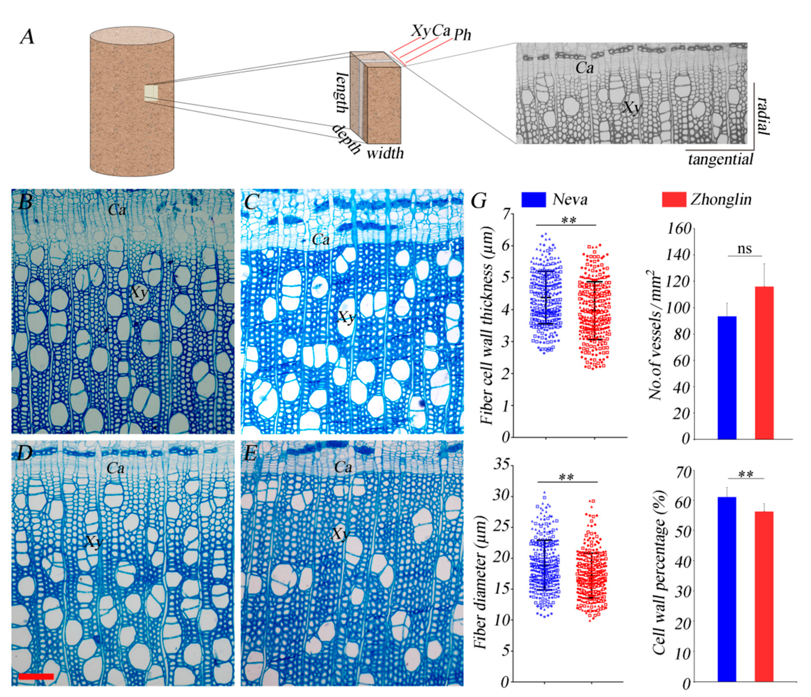
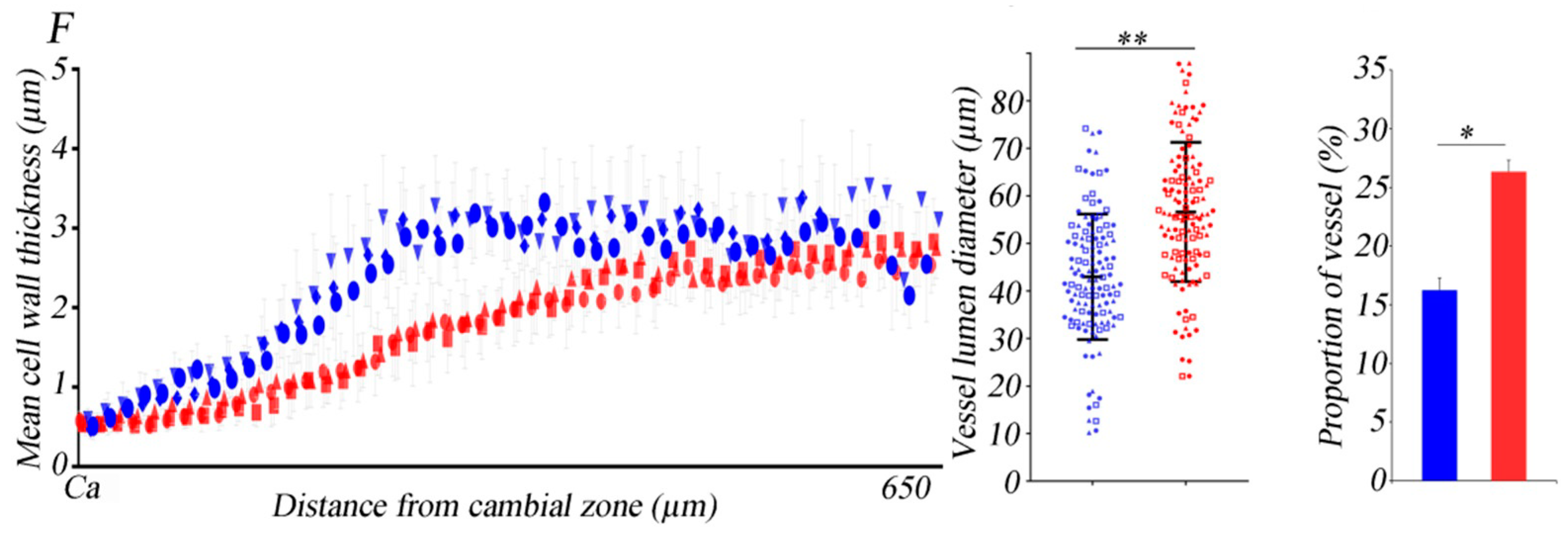
3.1. Differences in Anatomical Characteristics between the Two Black Poplar Cultivars

To analyze the microstructural differences between the two poplar cultivars, stem cross-sections sampled from the two kinds of poplar in July and December were stained with toluidine blue and observed under a microscope. As shown in Figure 1, the schematic overview shows the directions of the sample blocks, as well as the radial and tangential directions of the stem cross-sections (Figure 1A). The cambial region of Neva in July was composed of 8 to 9 cell layers, less than that of Zhonglin46 (10 to 11 cell layers) (Figure 1B,D). In December, the numbers of cambium cell layers were similar in these two poplar cultivars, with both having 8 to 9 cell layers (Figure 1C,E). As shown in Figure 1F, the thickness of the fiber cell wall during xylem development was plotted against the distance from the cambial zone. The gradual thickening of the fiber secondary wall of Neva was faster than that of Zhonglin46.
When measuring the fiber cell wall thickness and fiber diameter in the sections of the two cultivars with matured xylem in December, we found that all these indicators in Neva were significantly higher than those in Zhonglin46. We tallied the number of vessels and the results showed that there were 93 ± 10 vessels per mm2 in the sections of Neva, which was not apparently different to Zhonglin46, which contained about 116 ± 17 vessels. Meanwhile, the analysis of the vessel lumen diameter and proportion of vessels showed that Neva displayed a lower vessel lumen diameter and vessel proportion than Zhonglin46 did, and the difference between the two poplar cultivars was significant. When we counted the percentage of the cell wall, we found that the cell wall percentage in Neva was 61.10% ± 3.17%, remarkably higher than that of Zhonglin46 (56.31% ± 2.59%) (Figure 1G). Previously, cell wall percentage was found to be significantly correlated with wood density, which correlates with mechanical properties and hardness [26,28]. The difference observed between the two cultivars reflects their possible difference in properties.
The analysis of the anatomical structures of stem cross-sections of Neva and Zhonglin46 helped to clarify the differences in the morphological features of the xylem in the two cultivars. From our results, the thickening of the developing xylem was different; the cell walls of Neva thickened faster than those of Zhonglin46, reflecting the accelerated xylem development of Neva compared with Zhonglin46. Poplars are usually used as a source of raw material for paper making. Generally, anatomical characteristics such as cell wall thickness and diameter are important factors in the pulp and paper industry [29,30]. For example, the fiber cell wall thickness is connected with surface characteristics and high tearing strength in paper [31]. The number of vessels per unit area and vessel percentage are inversely correlated with pulp yield and paper strength properties [32]. Here, the differences in the anatomical characteristics of these two black poplars provide the basis for further understanding and utilization of these two different cultivars.

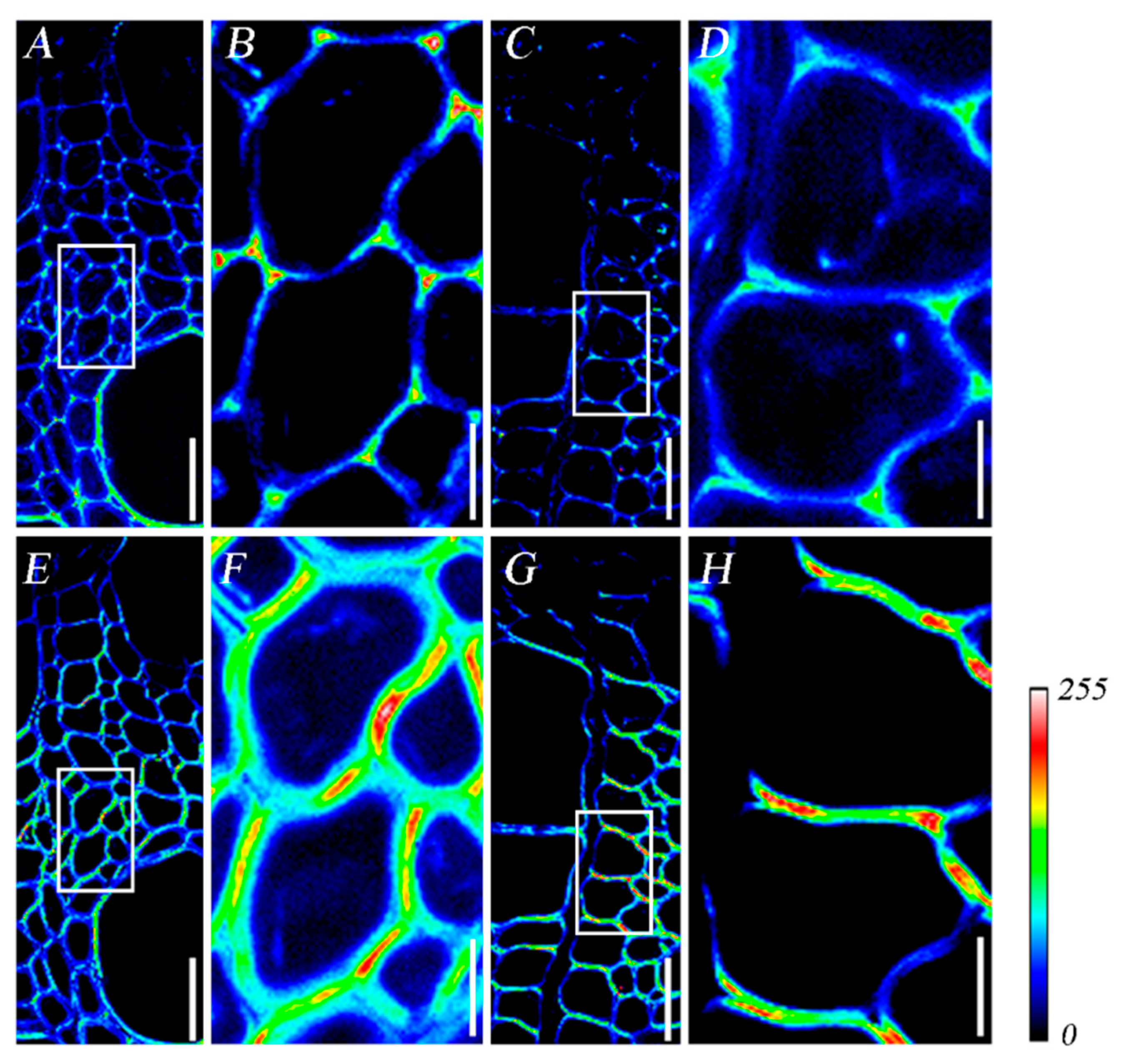
3.2. Heterogeneous Deposition of Lignin and Cellulose during Xylem Development

Regarding the obvious differences in anatomical characteristics between the two poplar cultivars, we further detected a difference in the deposition of lignin and cellulose during xylem development between these two cultivars. For quick and accurate comparison of the dynamic distribution of cell wall components, we used SRS microscopy, a label-free, in situ, and quantitative microanalysis technique based on vibrational contrast [33]. By combination with computational analysis, the chemical imaging results of different poplar cultivars over a large area were acquired rapidly and quantitatively analyzed.
Figure 2 shows the heat map with a color gradient scale indicating low (blue) to high (red) lignin and cellulose content during xylem development. With the maturation of xylem, lignification started at the cell corner and later in the compound middle lamella, which agrees with previous reports [34,35]. Meanwhile, Neva (Figure 2A,B) displayed more lignin content than Zhonglin46 (Figure 2C,D). Cellulose signals were detected more strongly in the primary cell walls and the middle lamella. In Zhonglin46, cellulose distributed more in the tangential cell walls, while in Neva, it was detected more in the radial cell walls (Figure 2C,D,G,H). Hence, the deposition of cellulose during xylem development may not be simultaneous, and it may preferentially deposit first on different cell walls such as radial or tangential cell walls in different cultivars. In addition, the cellulose signal in Neva encased the whole cell earlier than in Zhonglin46. Combined with the higher lignin deposition in Neva, the deposition of cell wall components was well correlated to faster thickening of the fiber cell walls in Neva.

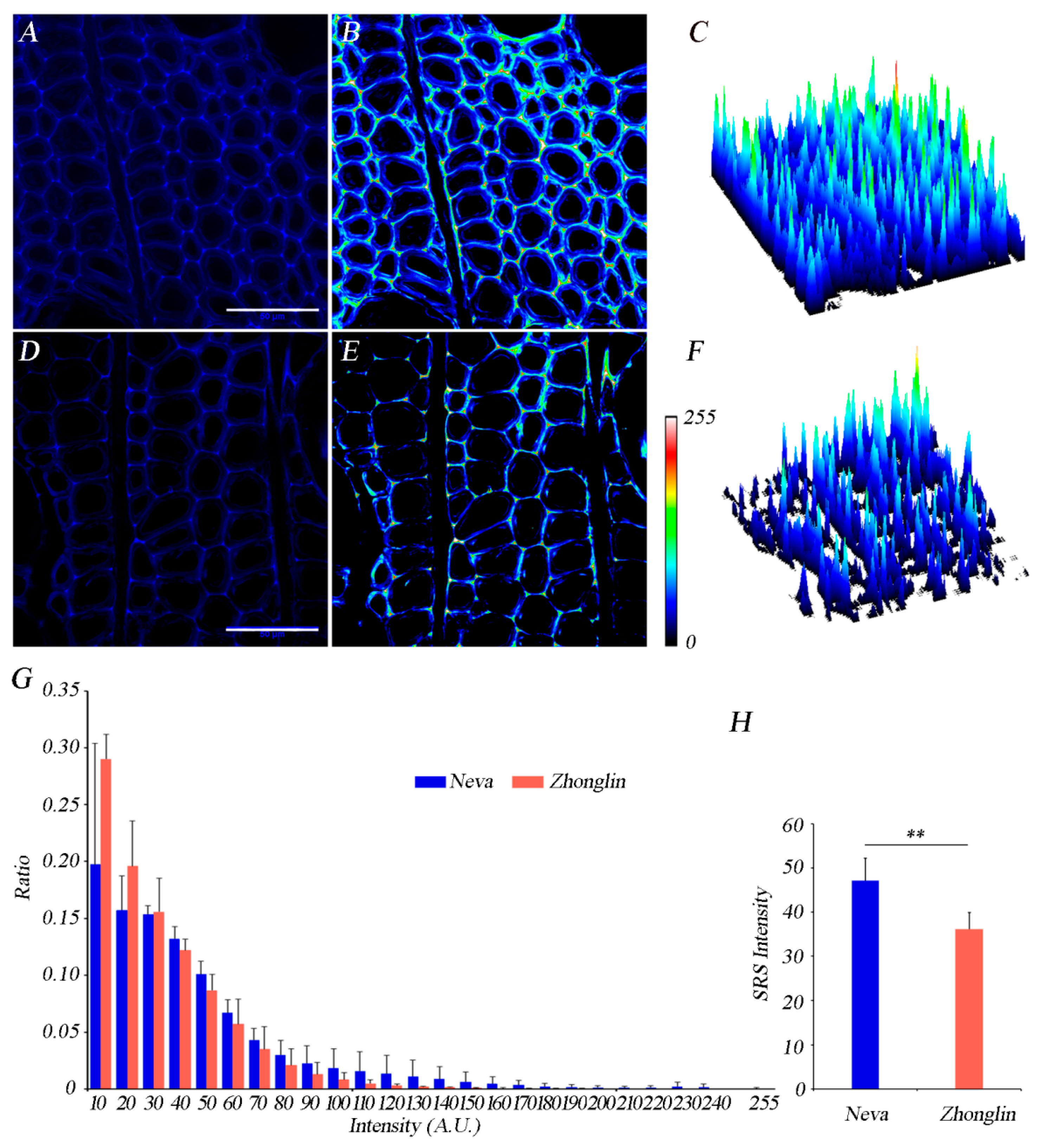
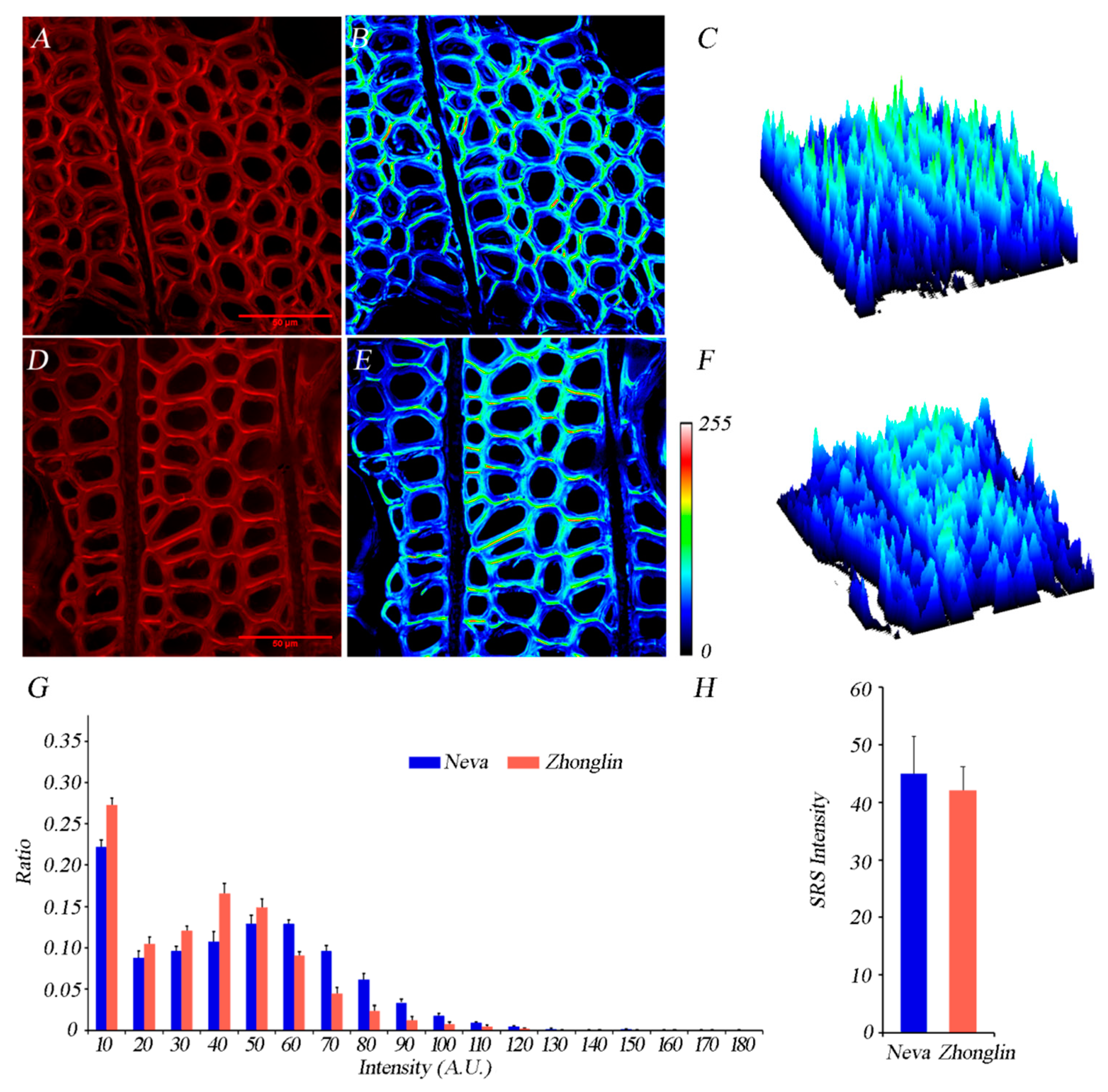
3.3. Differences of Lignin and Cellulose Deposition in Matured Xylem in the Two Cultivars

In order to compare the lignin deposition between the two cultivars, the mature xylems of both cultivars were observed under SRS microscopy and images were acquired at 1600 cm−1. Subsequently, a quantitative and straightforward SRS image analysis was made, as described by Xu et al. [15]. SRS imaging showed that lignin concentrations in the cell corner and compound middle lamella were higher than those in the secondary cell wall. As shown in Figure 3, the fluorescence intensity of the cell wall in the mature xylem in Neva was higher than that in Zhonglin46 (Figure 3A–F). Neva showed an intensity profile between 10 and 240, whereas the intensity profile of Zhonglin46 was between 10 and 180, demonstrating higher lignin deposition in Neva (Figure 3G). We further calculated the average intensity of xylem cells in the two poplar cultivars, and we found that the fluorescence intensity of cell walls in the mature xylem of Neva was significantly higher than that in Zhonglin46 (Figure 3H). Previously, the chemical composition of Zhonglin46 and Neva has been compared and analyzed biochemically, and the results showed that the lignin content in Neva was about 13.04% higher than that in Zhonglin46 [36]. Hence, our SRS-based results are in agreement with previous biochemical measurements.
To further analyze the distribution of cellulose in the xylem cell walls of these two poplar cultivars, we analyzed the intensity of the cellulose signals in samples taken from these two poplar cultivars. The content of cellulose in the cell corner was much lower than that in the secondary wall in both cultivars, which was the opposite result to that observed for the deposition of lignin. As shown in Figure 4, the highest signal intensity of cell walls in the mature xylem was found in Neva (Figure 4A–F). Accordingly, Neva showed an intensity profile between 10 and 130, while Zhonglin46 displayed an intensity profile between 10 and 120. Moreover, there was a greater proportion of high-intensity signals for Neva than for Zhonglin46. In contrast to the developing xylem, a large amount of cellulose deposited in the secondary wall with the maturation of the fiber cells, and the continuous deposition of cellulose formed the thickened secondary wall of the fiber cells, which was consistent with the results of the toluidine blue staining. However, after selecting individual cells and analyzing the average cellulose intensity of xylem cells in the two poplar cultivars, we found that although the fluorescence intensity of cell walls in the mature xylem of Neva was higher than that in Zhonglin46, the difference was not significant (Figure 4H).
Lignin is the main cause of lignocellulosic biomass recalcitrance to enzymatic hydrolysis. The possibility of obtaining low-lignin-content poplars is of significance for forestry applications [37]. Genetic engineering approaches have been used to successfully modify lignin contents in a variety of plants, including poplars. In transgenic poplars, inhibition of the expression of the 4CL gene resulted in a significant reduction in lignin content, accompanied by an obvious decrease in wood strength and a slight decrease in wood density [38]. Downregulations in the expressions of C4H and COMT genes also resulted in decreased lignin content, with no significant morphological changes [39]. Besides the manipulation of lignin content via the production of genetically modified plants, it is also important to choose low-lignin-content clones from among the existing clones. In our study, compared with Neva, Zhonglin46 displayed a lower lignin content, with no significant reduction in cellulose. Based only on the chemical composition, Zhonglin46 may be preferable for use in the paper industry, although other factors should also be taken into consideration. Using the same materials, Song et al. [24] also indicated that Zhonglin46 displays a better adaptivity than Neva in pulp and paper making, which is consistent with our results.

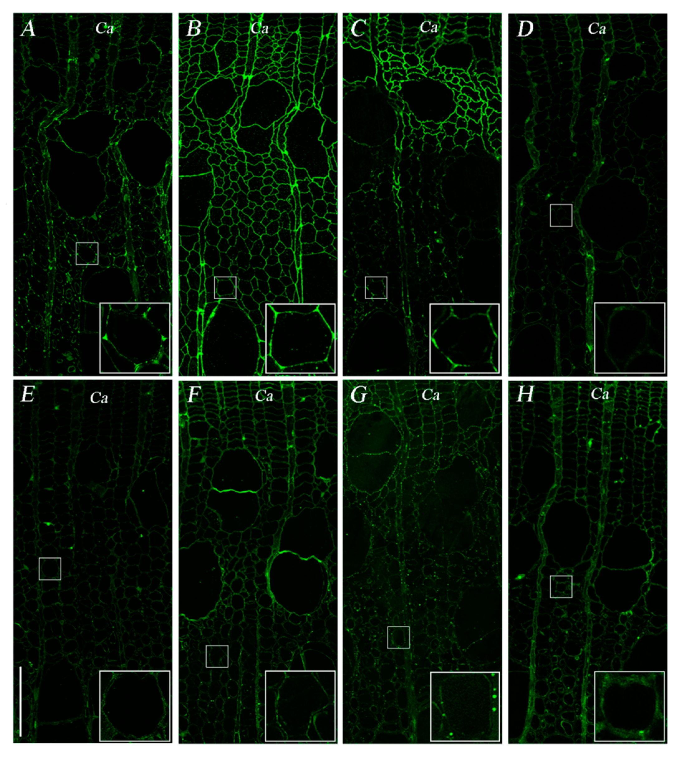
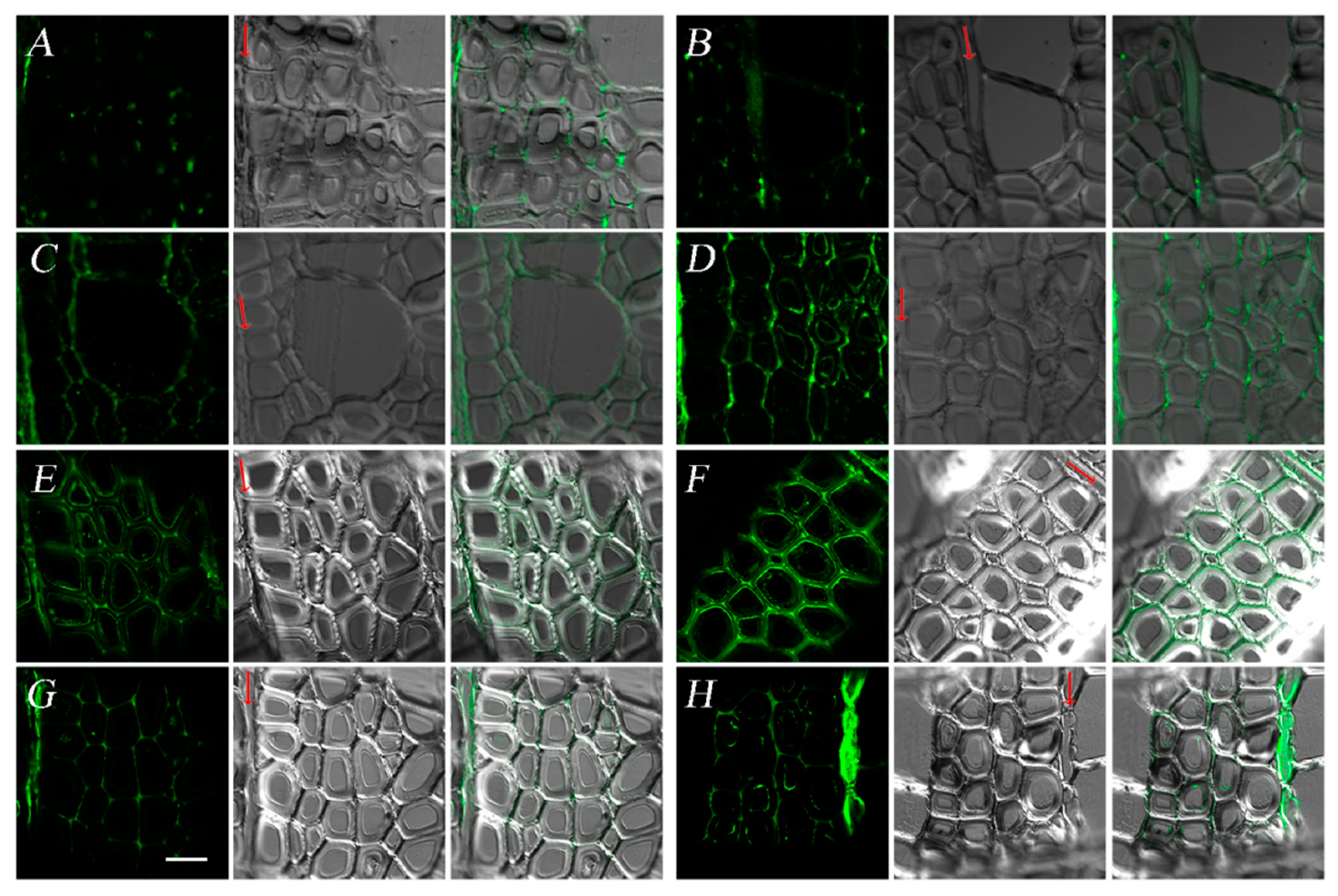
3.4. Difference in Pectin Patterns between the Two Cultivars

Pectin is another major component of plant cell walls, playing important roles in primary wall biosynthesis and modification. It was recently discovered that pectin also influences secondary wall formation and wood properties despite its low abundance in the secondary cell walls. Poplars with significantly decreased pectin content created via genetic engineering showed reduced xylem development and secondary cell wall thickening, probably due to their lower pectin contents [40]. Here, we detected the pectin distributions during xylem maturation in two poplar cultivars using immunofluorescence methods. The most abundant class of pectin is homogalacturonan, which is thought to be synthesized in a form with extensive methylesterification and then selectively demethylesterified [41,42]. Besides homogalacturonan, there are another two types of pectin found in primary cell walls: rhamnogalacturonan-I (RG-I) and rhamnogalacturonan-II (RG-II). Here, we used primary antibodies JIM5 and JIM7 to examine the changes in homogalacturonan with low and high degrees of methylesterification, LM5 and LM6, in order to detect the (1→4)-β-d-galactan and (1→5)-α-l-arabinan, which are both present in RG-I pectins.
As indicated by the JIM5 signals in Figure 5, JIM5-labeled unesterified pectin mainly deposited in the cell corners of the fiber cells, as well as in the radial cell walls. Most JIM7-labeled esterified pectin deposited relatively evenly on the radial cell wall, while there was also a small amount of distribution on the tangential wall. As for RG-I pectins, LM5-labeled pectin deposited obviously in the cambium, displaying faint signals in radial cell walls in developing xylem. LM6-labeled RG-I pectin had relatively strong signals in the developing xylem compared with the cambium, and the deposition increased with the maturation of xylem. In general, the distribution of pectin in the developing xylem of the two cultivars was similar.
In the mature xylem of both cultivars, JIM5-labeled unesterified pectin could only be detected in the corners of xylem cells. JIM7-labeled esterified pectin appeared more in radial cell walls, and the intensity of JIM7 signals was higher in Neva than in Zhonglin46. The LM5 signal was distributed in the primary walls of fiber cells, and the signal in Neva was stronger than that in Zhonglin46. LM6 signals were preferentially observed in the cell corners and the radial cell walls in both cultivars (Figure 6).
As a main polymer of primary cell walls, the structure of pectin also impacts the properties of wood [40]. For example, transgenic aspen with a higher level of methylesterified pectin displayed decreased cellular adhesion and cell wall rigidity [43]. The overexpression of Pectate lyase-like genes PtPL1-18 resulted in reduced xylem development, as well as reduced secondary cell wall thickening, which was probably due to the lower pectin content. Differences in the methylesterification status of HG pectin and the amount of RG-I pectin may elicit structural or solubility changes in nonpectin cell wall components, and subsequently altered lignin deposition [40]. In our study, the deposition of pectin during xylem development in these two black poplar cultivars was similar, while the JIM7-labeled esterified pectin and LM5-labeled RG-I pectin signals were stronger in Neva than in Zhonglin46. On the basis of the present results and previous reports that low pectin content may reduce xylem development and cell wall thickening [40], we speculated that the higher pectin content of Neva corresponded to its faster xylem development and thicker cell walls. Further studies need to be conducted to illustrate the relevance of pectin deposition and the associated anatomical characteristics.

4. Conclusions

In this study, the microanatomical and topochemical variations in the xylem of the two black poplar cultivars at different stages of development were analyzed with the aim of describing the dynamic chemical composition, as well as the differences between the cultivars. There were significant differences in the anatomical characteristics between the two black poplars. P. euramericana ‘Neva’ displayed faster thickening of the fiber cell wall than P. euramericana ‘Zhonglin46’ did, and ultimately displayed a greater cell wall thickness in the mature xylem. SRS microscopy and subsequent quantitative image analysis showed that Neva had a higher lignin content than Zhonglin46 did, and the cellulose signal in Neva encased the whole cell earlier in xylem development than that in Zhonglin46. Moreover, consistent with the previous biochemical results, a significantly higher lignin content was found in the mature xylem of Neva compared with that of Zhonglin46, while no obvious difference in cellulose deposition was found between the two poplar cultivars. The patterns of pectin deposition during xylem maturation were similar in the two cultivars, but JIM7-labeled esterified pectin and LM5-labeled RG-I pectin were higher in the mature xylem of Neva than in that of Zhonglin46. The discrepancy in dynamic deposition of cell wall components in the xylem of the two poplar cultivars was well correlated to their anatomical features. These results provide valuable insights into the chemical deposition and anatomical differences between these cultivars, and they might be helpful in understanding wood growth processes and facilitating the utilization of different poplar cultivars.

Author Contributions

Conceptualization, Y.J.; methodology, N.S., Y.B. and Y.C.; software, N.S.; validation, N.S. and Y.B.; formal analysis, N.S. and Y.B.; investigation, C.P. and X.W.; resources, C.P. and X.W.; data curation, N.S.; writing—original draft preparation, N.S. and Y.B.; writing—review and editing, Y.J.; funding acquisition, Y.J. All authors have read and agreed to the published version of the manuscript.

Funding

This research was funded by the National Key Research and Development Program of China (No. 2017YFD0600201).

Institutional Review Board Statement

Not applicable.

Informed Consent Statement

Not applicable.

Data Availability Statement

All data presented in this study are included in the article.

Acknowledgments

The authors thank Yuanzhen Suo of the School of Life Sciences of Peking University for technical assistance with SRS microscopy. The authors also acknowledge Xiaojie Liu and Junshan Wang at the Langfang Academy of Agriculture and Forestry Sciences for providing tree materials.

Conflicts of Interest

The authors declare no conflict of interest.

References

  1. Samuels, A.L.; Samuels, A.L.; Kaneda, M.; Kaneda, M.; Rensing, K.H.; Rensing, K.H. The cell biology of wood formation: From cambial divisions to mature secondary xylem. Can. J. Bot. 2006, 84, 631–639. [Google Scholar] [CrossRef]
  2. Lucas, W.J.; Groover, A.; Lichtenberger, R.; Furuta, K.; Yadav, S.R.; Helariutta, Y.; He, X.Q.; Fukuda, H.; Kang, J.; Brady, S.M. The plant vascular system: Evolution, development and functions. J. Integr. Plant Biol. 2013, 55, 294–388. [Google Scholar] [CrossRef]
  3. Meents, M.J.; Watanabe, Y.; Samuels, A.L. The cell biology of secondary cell wall biosynthesis. Ann. Bot. 2018, 121, 107–1125. [Google Scholar] [CrossRef] [PubMed] [Green Version]
  4. Lampugnani, E.R.; Khan, G.A.; Somssich, M.; Persson, S. Building a plant cell wall at a glance. J. Cell Sci. 2018, 131, jcs207373. [Google Scholar] [CrossRef] [PubMed] [Green Version]
  5. Cosgrove, D.J.; Jarvis, M.C. Comparative structure and biomechanics of plant primary and secondary cell walls. Front. Plant Sci. 2012, 3, 204. [Google Scholar] [CrossRef] [PubMed] [Green Version]
  6. Höfte, H.; Voxeur, A. Plant cell walls. Curr. Biol. 2017, 27, R865. [Google Scholar] [CrossRef] [PubMed] [Green Version]
  7. Zhang, B.; Gao, Y.; Zhang, L.; Zhou, Y. The plant cell wall: Biosynthesis, construction, and functions. J. Integr. Plant Biol. 2021, 63, 251–272. [Google Scholar] [CrossRef] [PubMed]
  8. Vanholme, R.; Demedts, B.; Morreel, K.; Ralph, J.; Boerjan, W. Lignin biosynthesis and structure. Plant Physiol. 2010, 153, 895–905. [Google Scholar] [CrossRef] [PubMed] [Green Version]
  9. Voxeur, A.; Höfte, H. Cell wall integrity signaling in plants: “To grow or not to grow that’s the question”. Glycobiology 2016, 26, 950–960. [Google Scholar] [CrossRef]
  10. Plomion, C.; Leprovost, G.; Stokes, A. Wood formation in trees. Plant Physiol. 2001, 127, 1513–1523. [Google Scholar] [CrossRef]
  11. Abedi, T.; Castilleux, R.; Nibbering, P.; Niittylä, T. The spatio-temporal distribution of cell wall-associated glycoproteins during wood formation in Populus. Front. Plant Sci. 2020, 11, 611607. [Google Scholar] [CrossRef] [PubMed]
  12. Pauly, M.; Keegstra, K. Plant cell wall polymers as precursors for biofuels. Curr. Opin. Plant Biol. 2010, 13, 305–312. [Google Scholar] [CrossRef]
  13. Zeng, Y.; Himmel, M.E.; Ding, S.Y. Visualizing chemical functionality in plant cell walls. Biotechnol. Biofuels 2017, 10, 263. [Google Scholar] [CrossRef] [PubMed] [Green Version]
  14. Zeng, Y.; Zhao, S.; Yang, S.; Ding, S.Y. Lignin plays a negative role in the biochemical process for producing lignocellulosic biofuels. Curr. Opin. Biotechnol. 2014, 27, 38–45. [Google Scholar] [CrossRef]
  15. Xu, H.; Zhao, Y.; Suo, Y.; Guo, Y.; Man, Y.; Jing, Y.; He, X.; Lin, J. A label-free, fast and high-specificity technique for plant cell wall imaging and composition analysis. Plant Methods 2021, 17, 29. [Google Scholar] [CrossRef] [PubMed]
  16. Guedes, F.T.P.; Laurans, F.; Quemener, B.; Assor, C.; Lainé-Prade, V.; Boizot, N.; Vigouroux, J.; Lesage-Descauses, M.C.; Leplé, J.C.; Déjardin, A.; et al. Non-cellulosic polysaccharide distribution during G-layer formation in poplar tension wood fibers: Abundance of rhamnogalacturonan I and arabinogalactan proteins but no evidence of xyloglucan. Planta 2017, 246, 857–878. [Google Scholar] [CrossRef] [PubMed]
  17. Jin, K.; Liu, X.; Wang, K.; Jiang, Z.; Tian, G.; Yang, S.; Shang, L.; Ma, J. Imaging the dynamic deposition of cell wall polymer in xylem and phloem in Populus× euramericana. Planta 2018, 248, 849–858. [Google Scholar] [CrossRef] [PubMed]
  18. Soukup, A. Selected simple methods of plant cell wall histochemistry and staining for light microscopy. Methods Mol. Biol. 2019, 1992, 27–42. [Google Scholar] [PubMed]
  19. Wu, H.; Xu, H.; Li, H.; Wei, D.; Lin, J.; Li, X. Seasonal development of cambial activity in relation to xylem formation in Chinese fir. J. Plant Physiol. 2016, 195, 23–30. [Google Scholar] [CrossRef] [PubMed]
  20. Fromm, J.; Rockel, B.; Lautner, S.; Windeisen, E.; Wanner, G. Lignin distribution in wood cell walls determined by TEM and backscattered SEM techniques. J. Struct. Biol. 2003, 143, 77–84. [Google Scholar] [CrossRef]
  21. Donaldson, L.A.; Knox, J.P. Localization of cell wall polysaccharides in normal and compression wood of radiata pine: Relationships with lignification and microfibril orientation. Plant Physiol. 2012, 158, 642–653. [Google Scholar] [CrossRef] [PubMed] [Green Version]
  22. Man, Y.; Zhao, Y.Y.; Ye, R.; Lin, J.X.; Jing, Y.P. In vivo cytological and chemical analysis of Casparian strips using stimulated Raman scattering microscopy. J. Plant Physiol. 2018, 220, 136–144. [Google Scholar] [CrossRef]
  23. Zhang, L.; Liu, B.; Zhang, J.; Hu, J. Insights of molecular mechanism of xylem development in five black poplar cultivars. Front. Plant Sci. 2020, 11, 620. [Google Scholar] [CrossRef]
  24. Song, X.L.; Yao, C.L.; Le, W.; Zou, X. Analysis of wood property of four species of Populus euramericanas and studies on their pulping performance. China Puplp Pap. Ind. 2010, 31, 34–38. [Google Scholar]
  25. Liu, Y.; Zhou, L.; Zhu, Y.; Liu, S. Anatomical features and its radial variations among different catalpa bungei clones. Forests 2020, 11, 824. [Google Scholar] [CrossRef]
  26. Istikowati, W.I.; Aiso, H.; Ishiguri, F.; Sutiya, B.; Ohshima, J.; Iizuka, K.; Yokota, S. Study of radial variation in anatomical characteristics of three native fast-growing tree species of a secondary forest in South Kalimantan. Indones. Appita. J. 2016, 69, 49–56. [Google Scholar]
  27. Chen, H.M.; Han, J.J.; Cui, K.M.; He, X.Q. Modification of cambial cell wall architecture during cambium periodicity in Populus tomentosa Carr. Trees 2010, 24, 533–540. [Google Scholar] [CrossRef]
  28. Li, H.; Jiang, X.; Ramaswamy, H.S.; Zhu, S.; Yu, Y. High-pressure treatment effects on density profile, surface roughness, hardness, and abrasion resistance of paulownia wood boards. Trans. ASABE 2018, 61, 1181–1188. [Google Scholar] [CrossRef]
  29. Ververis, C.; Georghiou, K.; Christodoulakis, N.; Santas, P.; Santas, R. Fiber dimensions, lignin, and cellulose content of various plant materials and their suitability for paper production. Ind. Crop. Prod. 2003, 19, 245–254. [Google Scholar] [CrossRef]
  30. Francis, R.C.; Brown, A.F.; Bosshart, R.R.; Balch, S.; Waite, D.M. Four additional hybrid poplars with papermaking properties superior to aspen. TAPPI J. 2005, 4, 13–17. [Google Scholar]
  31. Seth, R.S.; Page, D.H. Fiber properties and tearing resistance. Tappi. J. 1988, 71, 103–107. [Google Scholar]
  32. Ona, T.; Sonoda, T.; Ito, K.; Shibata, M.; Tamai, Y.; Kojima, Y.; Ohshima, J.; Yokota, S.; Yoshizawa, N. Investigation of relationship between cell and pulp properties in Eucalyptus by examination of within-tree property variations. Wood. Sci. Technol. 2001, 35, 229–243. [Google Scholar] [CrossRef]
  33. Freudiger, C.W.; Min, W.; Saar, B.G.; Lu, S.; Holtom, G.R.; He, C.; Tsai, J.C.; Kang, J.X.; Xie, X.S. Label-free biomedical imaging with high sensitivity by stimulated Raman scattering microscopy. Science 2008, 322, 1857–1861. [Google Scholar] [CrossRef] [PubMed] [Green Version]
  34. Zhang, X.; Ma, J.; Zhe, J.; Yang, G.H.; Zhou, X.; Xu, F. Method for automatically identifying spectra of different wood cell wall layers in Raman imaging data set. Microsc. Res. Technol. 2014, 77, 609–618. [Google Scholar] [CrossRef] [PubMed]
  35. Gierlinger, N.; Schwanninger, M. Chemical imaging of poplar wood cell walls by confocal Raman microscopy. Plant Physiol. 2006, 140, 1246–1254. [Google Scholar] [CrossRef] [PubMed] [Green Version]
  36. Wang, L.; Yao, C.L.; Song, X.L.; Zou, X. Infection of the different soils and different tree-ages of chemical compositions in Populus×euramericana Cv.‘74/76′. Pap. Pap. Mak. 2010, 29, 25–28. [Google Scholar]
  37. De Meester, B.; Calderón, B.M.; de Vries, L.; Pollier, J.; Goeminne, G.; Van Doorsselaere, J.; Chen, M.; Ralph, J.; Vanholme, R.; Boerjan, W. Tailoring poplar lignin without yield penalty by combining a null and haploinsufficient CINNAMOYL-CoA REDUCTASE2 allele. Nat. Commun. 2020, 11, 5020. [Google Scholar] [CrossRef]
  38. Hu, J.Q.; Qi, Q.; Zhao, Y.L.; Tian, X.M.; Lu, H.; Gai, Y.; Jiang, X.N. Unraveling the impact of Pto4CL1 regulation on the cell wall components and wood properties of perennial transgenic Populus tomentosa. Plant Physiol. Biochem. 2019, 139, 672–680. [Google Scholar] [CrossRef]
  39. Shafrin, F.; Ferdous, A.S.; Sarkar, S.K.; Ahmed, R.; Amin, A.; Hossain, K.; Sarker, M.; Rencoret, J.; Gutiérrez, A.; Del Rio, J.C.; et al. Modification of monolignol biosynthetic pathway in Jute: Different gene, different consequence. Sci. Rep. 2017, 7, 39984. [Google Scholar] [CrossRef] [PubMed]
  40. Bai, Y.; Wu, D.; Liu, F.; Li, Y.; Chen, P.; Lu, M.; Zheng, B. Characterization and functional analysis of the poplar Pectate Lyase-Like Gene PtPL1-18 reveal its role in the development of vascular tissues. Front. Plant Sci. 2017, 8, 1123. [Google Scholar] [CrossRef] [Green Version]
  41. Willats, W.G.; Gilmartin, P.M.; Mikkelsen, J.D.; Knox, J.P. Cell wall antibodies without immunization: Generation and use of de-esterified homogalacturonan block-specific antibodies from a naive phage display library. Plant J. 1999, 18, 57–65. [Google Scholar] [CrossRef] [PubMed]
  42. Wolf, S.; Mouille, G.; Pelloux, J. Homogalacturonan methyl-esterification and plant development. Mol. Plant 2009, 2, 851–860. [Google Scholar] [CrossRef] [PubMed]
  43. Leśniewska, J.; Öhman, D.; Krzesłowska, M.; Kushwah, S.; Barciszewska-Pacak, M.; Kleczkowski, L.A.; Sundberg, B.; Moritz, T.; Mellerowicz, E.J. Defense responses in aspen with altered pectin methylesterase activity reveal the hormonal inducers of tyloses. Plant Physiol. 2017, 173, 1409–1419. [Google Scholar] [CrossRef] [PubMed]
Figure 1. Microscopic analysis of stems from two black poplar cultivars. (A) A schematic overview showing the directions of the sample blocks and the stem cross-sections. (B,C) Cross-sections of phloem–xylem region from P. euramericana ‘Zhonglin46’ sampled on 4 July (B) and December 19 (C). (D,E) Cross-sections of phloem–xylem region from P. euramericana ‘Neva’ sampled on 4 July (D) and December 19 (E). Scale bar = 100 µm. (F) Changes in thickness of fiber cell wall during cell wall maturation. (G) Anatomical characteristics of the two poplar cultivars. *, p < 0.05; **, p < 0.01; ns, not significant. The triangle, rectangle, and circle represent different biological repetitions. Neva is shown in blue and Zhonglin46 is shown in red. Ph: phloem, Ca: cambium, Xy: xylem.
Forests 12 00972 g001aForests 12 00972 g001b
Figure 2. SRS chemical images of lignin and cellulose in developing xylem of P. euramericana ‘Neva’ and P. euramericana ‘Zhonglin46’. (A,C) SRS images of the developing xylem based on the deconvoluted lignin band at 1600 cm1 in Neva (A) and Zhonglin46 (C). (E,G) SRS images of the developing xylem based on the cellulose band at 1100 cm1 in Neva (E) and Zhonglin46 (G). (B,D,F,H) Magnifications of the white boxes in (A,C,E,G). Scale bar = 50 µm (A,C,E,G). Scale bar = 10 µm (B,D,F,H).
Forests 12 00972 g002
Figure 3. Quantification of lignin in mature xylem from two black poplar cultivars. (A,D) SRS images of P. euramericana ‘Neva’ (A) and P. euramericana ‘Zhonglin46’ (D) acquired at 1600 cm1, showing the lignin distribution. Scale bar = 50 µm. (B,E) Heat map of (A) and (D). (C,F) The 3D surface plots of (A) and (D). (G) Distribution of fluorescence intensity of the SRS images in (A) and (D). (H) The average intensity of lignin in xylem cells from the two black poplar cultivars. **, p < 0.01.
Forests 12 00972 g003
Figure 4. Quantification of cellulose in mature xylem from two black poplar cultivars. (A,D) SRS images of P. euramericana ‘Neva’ (A) and P. euramericana ‘Zhonglin46’ (D) acquired at 1100 cm1, showing the cellulose distribution. Scale bar = 50 µm. (B,E) Heat map of (A) and (D). (C,F) The 3D surface plots of (A) and (D). (G) Distribution of fluorescence intensity of the SRS images in (A) and (D). (H) The average intensity of cellulose in xylem cells from the two black poplar cultivars. ns, not significant.
Forests 12 00972 g004
Figure 5. Pectin immune localization in developing xylem of P. euramericana ‘Zhonglin46’ (AD) and P. euramericana ‘Neva’ (EH). (A,E) JIM5-labeled unesterified pectin. (B,F) JIM7-labeled esterified pectin. (C,G) LM5-labeled RG-I pectin. (D,H) LM6-labeled RG-I pectin. An enlarged view of a fiber cell is shown in the white box. Ca: cambium. Scale bar = 100 µm.
Forests 12 00972 g005
Figure 6. Pectin immune localization in mature xylem of P. euramericana ‘Zhonglin46’ (A,C,E,G) and P. euramericana ‘Neva’ (B,D,F,H). (A,B) JIM5-labeled unesterified pectin. (C,D) JIM7-labeled esterified pectin. (E,F) LM5-labeled RG-I pectin. (G,H) LM6-labeled RG-I pectin. The red arrowhead indicates the direction of the radial wall. Scale bar = 20 µm.
Forests 12 00972 g006
Publisher’s Note: MDPI stays neutral with regard to jurisdictional claims in published maps and institutional affiliations.

