Article

Castanea crenata Ginkbilobin-2-like Recombinant Protein Reveals Potential as an Antimicrobial against Phytophthora cinnamomi, the Causal Agent of Ink Disease in European Chestnut

by
Maria Belén Colavolpe
1,
Fernando Vaz Dias
2,
Susana Serrazina
2,
Rui Malhó
2 and
Rita Lourenço Costa
1,3,*
1
Instituto Nacional de Investigação Agrária e Veterinária I.P., Avenida da República, Quinta do Marquês, 2780-157 Oeiras, Portugal
2
BioISI—Biosystems & Integrative Sciences Institute, Faculdade de Ciências, Universidade de Lisboa, 1749-016 Lisboa, Portugal
3
Centro de Estudos Florestais, Instituto Superior de Agronomia, Universidade de Lisboa, 1349-017 Lisboa, Portugal
*
Author to whom correspondence should be addressed.
Submission received: 26 February 2023 / Revised: 30 March 2023 / Accepted: 5 April 2023 / Published: 11 April 2023
(This article belongs to the Special Issue Application of Plant Biotechnology in Forestry)

Abstract

:
The European chestnut tree (Castanea sativa Mill.) is widely cultivated throughout the world’s temperate regions. In the Mediterranean region, it has a significant economic role mainly because of the high quality of its edible nuts. The Oomycete Phytophthora cinnamomi is one of the most severe pathogens affecting European chestnuts, causing ink disease and significant losses in production. Ginkgobilobin-2 (Gnk2) in Ginkgo biloba is a secreted protein with a plant-specific cysteine-rich motif that functions as a lectin, and its carbohydrate-binding properties are closely related to its antifungal activity. The binding of lectins to mannose residues of the cell wall of Phytophthora species may disturb and disrup the cell wall structure. This work determined that the amino acid sequence has a signal peptide that directs the final protein peptide to the apoplast. The Cast_Gnk2-like expression was performed and optimized, and different in vitro antagonism tests were done against P. cinnamomi using different purified protein concentrations. As a result of one of these assays, Cast_Gnk2-like significantly reduced the mycelia growth of P. cinnamomi in liquid medium as shown by the mycelia weight (g) in control treatments was 377% higher than in the treatments. These insights reveal the potential of Cast_Gnk2-like for agricultural uses and biotechnological developments for the pathosystem chestnut/P. cinnamomi.

1. Introduction

The genus Castanea belongs to the Fagaceae family, which dominates much of the hardwood forests of the Northern Hemisphere. The European chestnut (Castanea sativa Mill.) is considered to be the only native chestnut species in Europe. This species is a multipurpose tree, that is used in the food industry for its high-quality edible fruits (nuts), in the forest industry for timber, and also for ecological and landscaping purposes [1].
Over 100 species have been described within the genus Phytophthora [2]. According to many authors [3,4], most survive in the soil for long periods without a host and affect the parts of the plant in contact with the ground, thus causing a destruction of the root system. The impact of this group of organisms is mirrored in its name, as Phytophthora, which means, in Greek, plant destroyer [1,4].
The rural economy of the northern mountainous regions of Portugal are mainly based on chestnut culture. Unfortunately, the current area and productivity per hectare are much less than the country’s potential, mainly due to root rot caused by Phytophthora cinnamomi Rands, the main threat to chestnut orchards in Europe [5]. Castanea sativa was severely affected and nut production has declined by 251,549 tons from 1961 to 2015 [1,6]. The economy of mountainous areas in the Mediterranean regions of southwest Europe mainly depends on the significant income that chestnut culture represents to the local farmers, which is sometimes the only income they can get from the land. In recent years, the persistent losses of chestnut trees to P. cinnamomi is contributing to a decline in the population of these regions [6].
Phytophthora cinnamomi infects the roots and after reaching the vascular tissues it continues to colonize the roots until it obstructs the xylem vessel, thus restricting root growth and interfering with water and nutrient uptake to the plant shoot. The roots and root collar start to rot, resulting in a progressive decline of the tree. The above-ground symptoms include chlorosis and wilting of leaves, dieback of branches, defoliation, and gradual decline until the tree dies [1]. Phytophthora cinnamomi, has an extensive host range, destroying thousands of plant species worldwide and causing devastating impacts on natural ecosystems, agriculture, and forestry [6].
The coexistence of Asian chestnut species from Japan and China (Castanea crenata Sieb. & Zucc. and Castanea mollissima Blume) with P. cinnamomi (originally from the Asian tropics) lead to the development of resistance against the pathogen during the evolution process. The secretion of antifungal proteins and cell wall reinforcement are part of the constitutive defense barriers to pathogen growth in the plants that may explain the difference in resistance to P. cinnamomi between C. crenata and C. sativa. Although the Asian species have proven resistant to ink disease, these species are not adapted to the Atlantic environmental conditions. Therefore, the resistance of Japanese and Chinese chestnut species to this pathogen led to their introduction in breeding programs over the last years as donors of resistance in controlled crosses [7].
Over time, several approaches have been proposed to deal with root diseases (which affect the xylem of trees), such as the intensive application of pesticides and the selection and production of improved plant material with some disease resistance [8]. Phosphite and metalaxyl have been the most frequently used chemicals against P. cinnamomi. Nevertheless, the continuous use of these two chemicals has led to the development of tolerance within the pathogen [1]. Effective biological control methods have not been developed to date [1]. More common control approaches involve using resistant rootstocks for propagation or planting of resistant hybrids [1]. Nevertheless, the measures and procedures have not yet been successful, mainly due to the easy development and migration of P. cinnamomi zoospores in wet conditions, especially during rainfalls and waterlogging and because of the resistance structures that persist in the soil and are extremely difficult to eradicate [1,9].
A previous study identified a secreted protein with antifungal activity, Ginkbilobin2 (Gnk2), from seeds of the gymnosperm Ginkgo biloba [10]. Gnk2 comprises 108 amino acids as a mature protein with a plant-specific cysteine-rich motif. It functions as a lectin [3,11], and its carbohydrate-binding properties are tightly related to its antifungal activity. It binds with high affinity to D-mannose and with less affinity to D-glucose [11], and both exist in the hyphal cell walls of Phytophthora species [12].
Our group has been studying the C. sativa and C. crenata root transcriptome in response to P. cinnamomi to elucidate chestnut defense mechanisms. In a previous study [13], we identified candidate genes differentially expressed in the roots of the susceptible species to the pathogen (C. sativa), and the resistant one (C. crenata) observed after P. cinnamomi inoculation. The research demonstrated that both species recognize the pathogen attack, but only the resistant species (C. crenata) may involve more genes in the defense response than the susceptible species (C. sativa). RNA-seq analysis further enabled the selection of candidate genes for ink-disease resistance in Castanea [12]. Following the same rationale to elucidate chestnut defense mechanisms against P. cinnamomi, our group selected the Ginkgobilobin-2-like gene (Cast_Gnk2-like) from the transcriptomes of C. crenata, intending to evaluate the early expression of candidate resistance genes in both species (C. sativa and C. crenata). Cast_Gnk2-like was the most expressed gene and the one that best discriminates between susceptible and resistant chestnut genotypes. The highest Cast_Gnk2-like expression registered in non-inoculation conditions suggests that C. crenata root surroundings may be a hostile environment for fungal and fungal-like pathogens, such as P. cinnamomi. On the other hand, C. sativa showed a very low Cast_Gnk2-like expression level, even after pathogen inoculation [7].
The present work is part of an ongoing research program that aims to find solutions to this important problem regarding the Chestnut/P. cinnamomi pathosystem in Europe. It was reported that Gnk2 inhibits the growth of pathogenic fungi such as Fusarium oxysporum, F. culmorum, and Candida albicans; and activates actin-dependent by inducing hypersensitive response (HR-related) plant cell death [10]. The homology of Cast_Gnk2-like with Gnk2 led us to test the encoded protein against P. cinnamomi. The present work aims to express and produce the Cast_Gnk2-like protein for evaluation of its effectiveness on in vitro assays as a possible antagonist of the P. cinnamomi pathogen.

2. Materials and Methods

2.1. Cast_GNK2-like In Silico Analysis

The nucleotide sequence of the C. crenata Cast_GNK2-like transcript was obtained from the sequenced root transcriptome after P. cinnamomi inoculation [13]. It was annotated as a Cysteine-rich repeat secretory protein 38. After a BLASTn at NCBI and a comparison of the sequences with the highest homology, a prediction of the coding sequence and translation to the amino acid sequence was achieved. A BLAST to Cast_GNK2-like was achieved in Uniprot [14] to identify the proteins with higher similarity. Uniprot was also used to predict the protein families and domains of Cast_GNK2-like by comparison with the ones of Ginkgo biloba (GNK2), Arabidopsis thaliana, and C. mollissima. Signal peptides for Cast_GNK2-like were searched using SignalP—6.0 [15] and TargetP—2.0 [
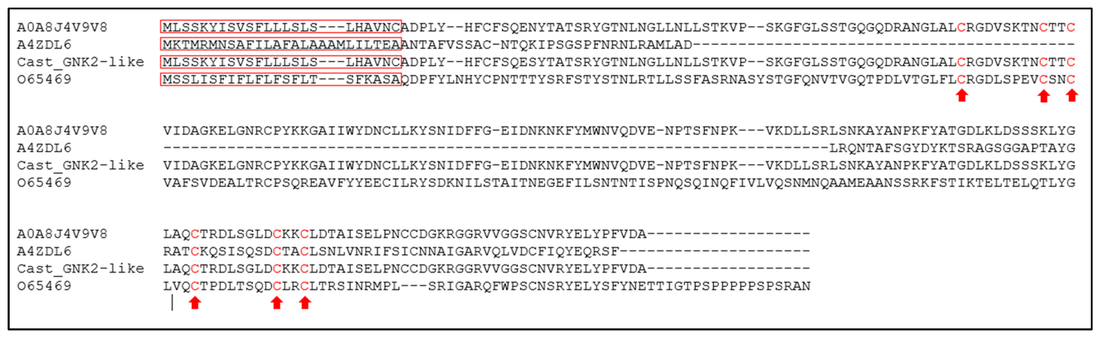
16]. Alignment of the amino acid sequences of Castanea mollissima Cysteine-rich repeat secretory protein 38 (A0A8J4V9V8), Ginkgo biloba Antifungal protein ginkbilobin-2, C. crenata putative Ginkbilobin-2 protein and A. thaliana Putative cysteine-rich receptor-like protein kinase 9, was performed using CLUSTALW [17].

2.2. Bacterial Strains and Growth Condition

Escherichia coli bacteria strains used in this study were TOP10 and BL21 (DE3) pLysS (competent cells from Thermo Fisher Scientific Inc., Waltham, MA, USA). E. coli strains containing recombinant plasmids were cultured, at 37 °C, in LB broth medium supplemented with 100 μg/mL ampicillin or 50 μg/mL kanamycin.

2.3. Phytophthora Cinnamomi Strain

Phytophthora cinnamomi (strain PH107) was isolated at the UTAD (the University of Trás-os-Montes and Alto Douro), from Vila Real (Portugal), and it is preserved at the INIAV I.P. (Instituto Nacional de Investigação Agrária e Veterinária I.P.) in Lisbon. It is routinely cultured at 25 °C on Potato-Dextrose Agar (PDA) in Petri plates and maintained in darkness. For all the assays, a fresh culture of P. cinnamomi was prepared by transferring a small piece of mycelium from a previous culture to a new plate of PDA nutritive medium and incubated in darkness at 25 °C for 4/5 days (or maximum one week).

2.4. Plasmid Construction

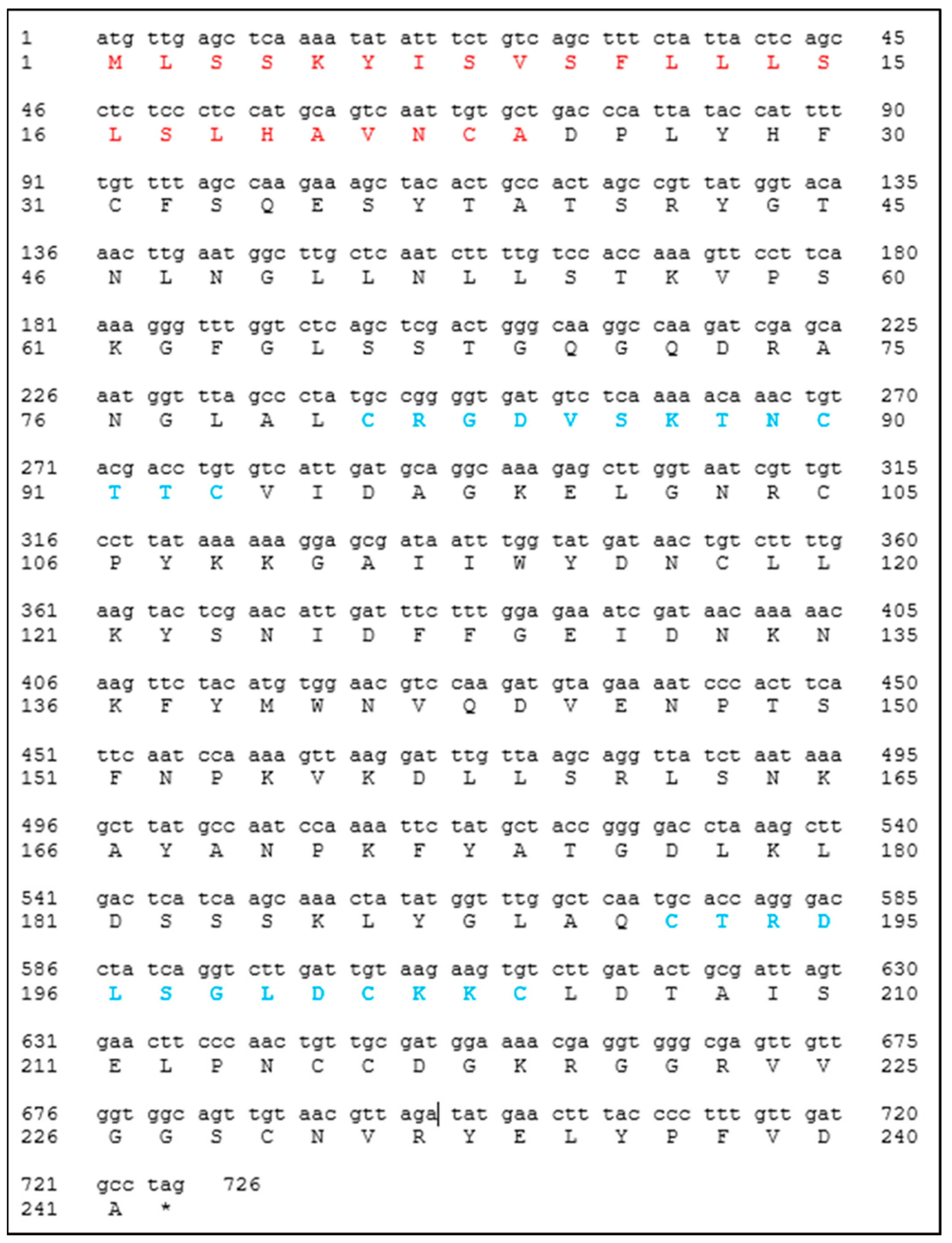
The designed primers for the amplification of the Cast_Gnk2-like coding sequence were 5′–CTCCATATGgctgacccattataccatttttgttttag–3′ and 5′– CACGGATCCctaggcatcaacaaaggggta–3′. The PCR was performed using Phusion™ High-Fidelity DNA Polymerase (2 U/µL) according to the manufacturer protocol. The vectors used for the construction were pET15b and pET28a (Novagen®, Gujarat, India), which carry an N-terminal or an N- and C- terminal oligo-histidine tag (6x His-tags), respectively. The DNA sequence encoding Cast_Gnk2-like was inserted into the vectors between NdeI and BamHI sites. Control plasmids were constructed without the 
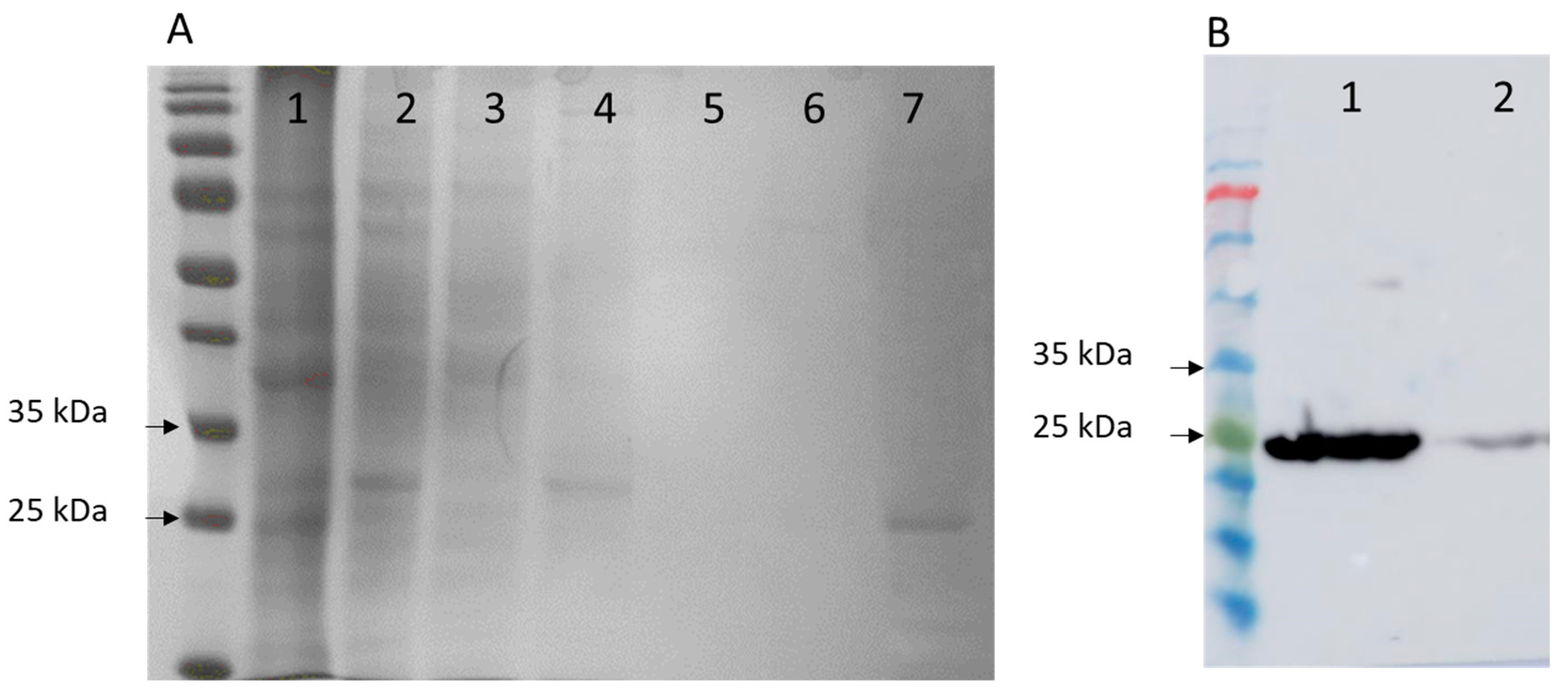
Cast_Gnk2-like fragment. It is expected to observe a protein fraction (a band in the gels) in the region of 25 kDa. The size of the Cast_Gnk2-like coding sequence is represented in Figure 1 (726 bp).

2.5. Protein Expression

2.5.1. Preparation of Bacterial Cellular Lysates

The cell culture was centrifuged at 4000 g for 20 min, and the pellet was resuspended in a 50 mM sodium HEPES buffer (4-(2-hydroxyethyl)-1-piperazineethanesulfonic acid), pH 7.5, containing 1 M NaCl and 10 mM imidazole (1,3-diazaciclopenta-2,4-dieno). Cells were disrupted by sonication. The cell debris was removed by centrifugation, and the supernatant was filtrated (0.45 μm) before the protein purification process.

2.5.2. Protein Purification and Electrophoresis (SDS-PAGE)

The supernatant (cell-free extract) obtained after centrifugation was applied to 5 mL Ni2+ ion chelating immobilised ion affinity chromatography column (HiTrap, GE Healthcare, Chicago, IL, USA). The column was preequilibrated with 50 mM sodium HEPES buffer, pH 7.5, containing 1 M NaCl and 10 mM imidazole. The recombinant protein, Cast_Gnk2-like, was eluted with a linear gradient of imidazole (0–300 mM) in 50 mM sodium HEPES buffer, pH 7.5, containing 1 M NaCl. Protein purity was evaluated by SDS–PAGE (sodium dodecyl sulfate polyacrylamide gel electrophoresis), performed in a Protean II xi cell (Bio-Rad, Hercules, CA, USA) at 480 V and 120 mA. The running buffer contained 25 mM Tris base, 0.2 M glycine, and 0.1% SDS. Gels were stained with Coomassie (Bio-Rad Biosafe, Hercules, CA, USA). The protein concentrations were determined using Bradford reagent (using bright blue Coomassie G-250) from BioRad utilizing bovine serum albumin (BSA) as the standard.

2.6. Western Blot

Identification of antigens recognized by serum antibodies was carried out after the separation of total antigens by SDS-PAGE and Western blotting [18]. Following SDS-PAGE, the separated proteins were transferred to nitrocellulose sheets (AmershamTM Protran® Premium 0.2 µm pore size NC). The sheets were blocked with phosphate-buffered saline, pH 7.2, containing 0.3% Tween-20 (0.3% PBS-T), and washed twice. Then they were incubated with His-Tag Antibody (AD1.1.10) solution for one hour at room temperature, always in soft shaking. After washing them three times with 0.3% PBS-T, the secondary antibody solution (m-IgGκ BP-HRP) was added for one hour in the same conditions. After that, another three washes with 0.3% PBS-T were done.
Immunoreactive spots were detected with a chemiluminescence-based kit (Amersham Pharmacia Biotech, Amersham, UK). Before immunodetection, the nitrocellulose membranes were stained with 0.2% w/v Ponceau S in 3% w/v trichloroacetic acid for 3 min.

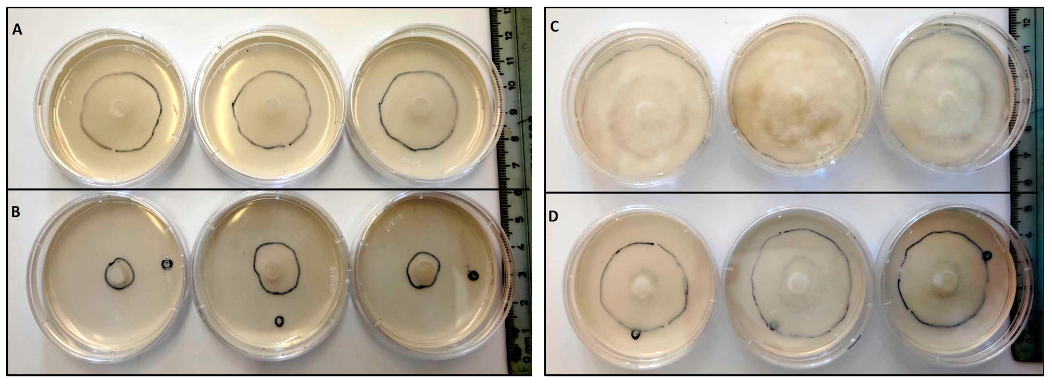
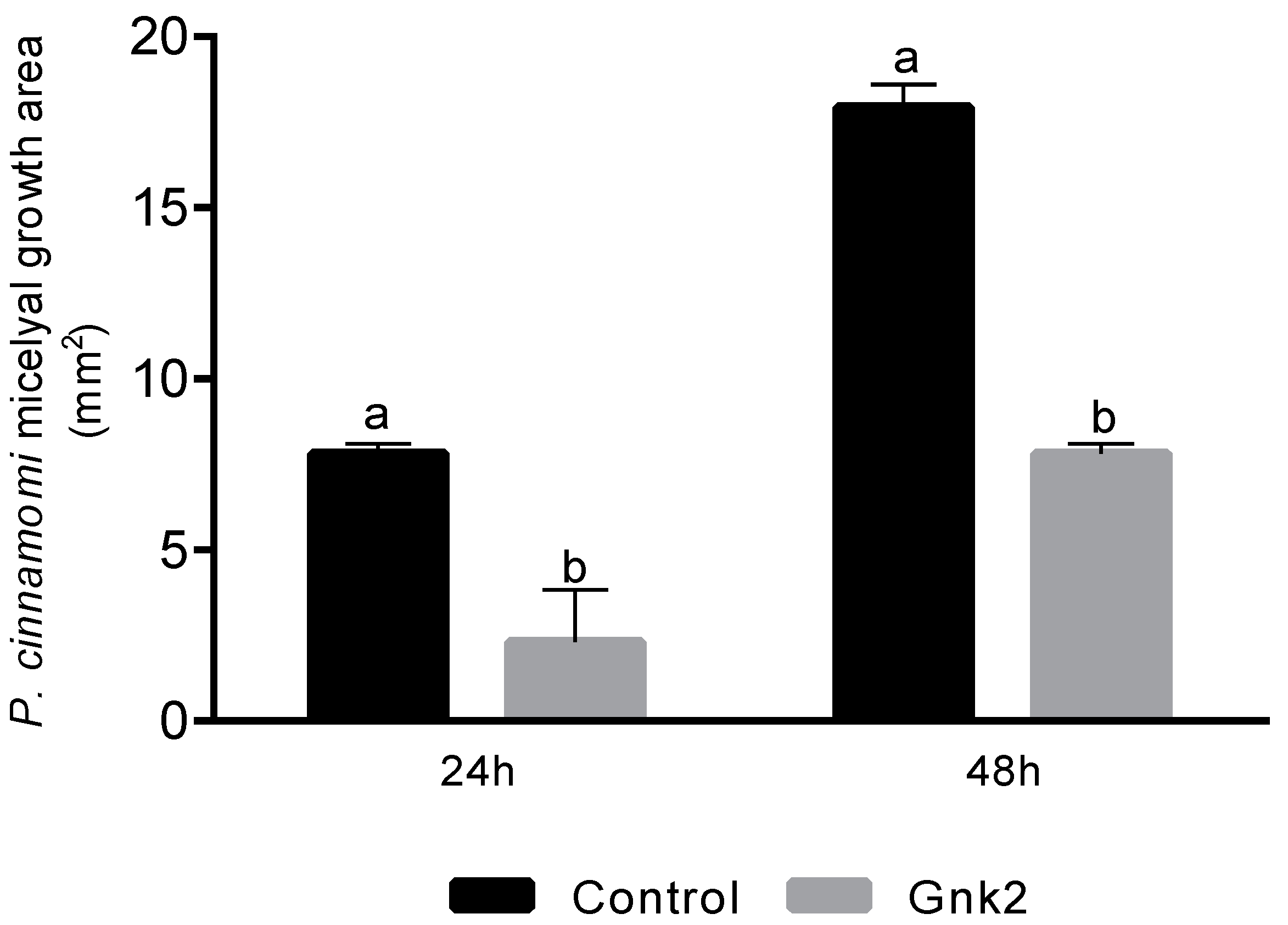
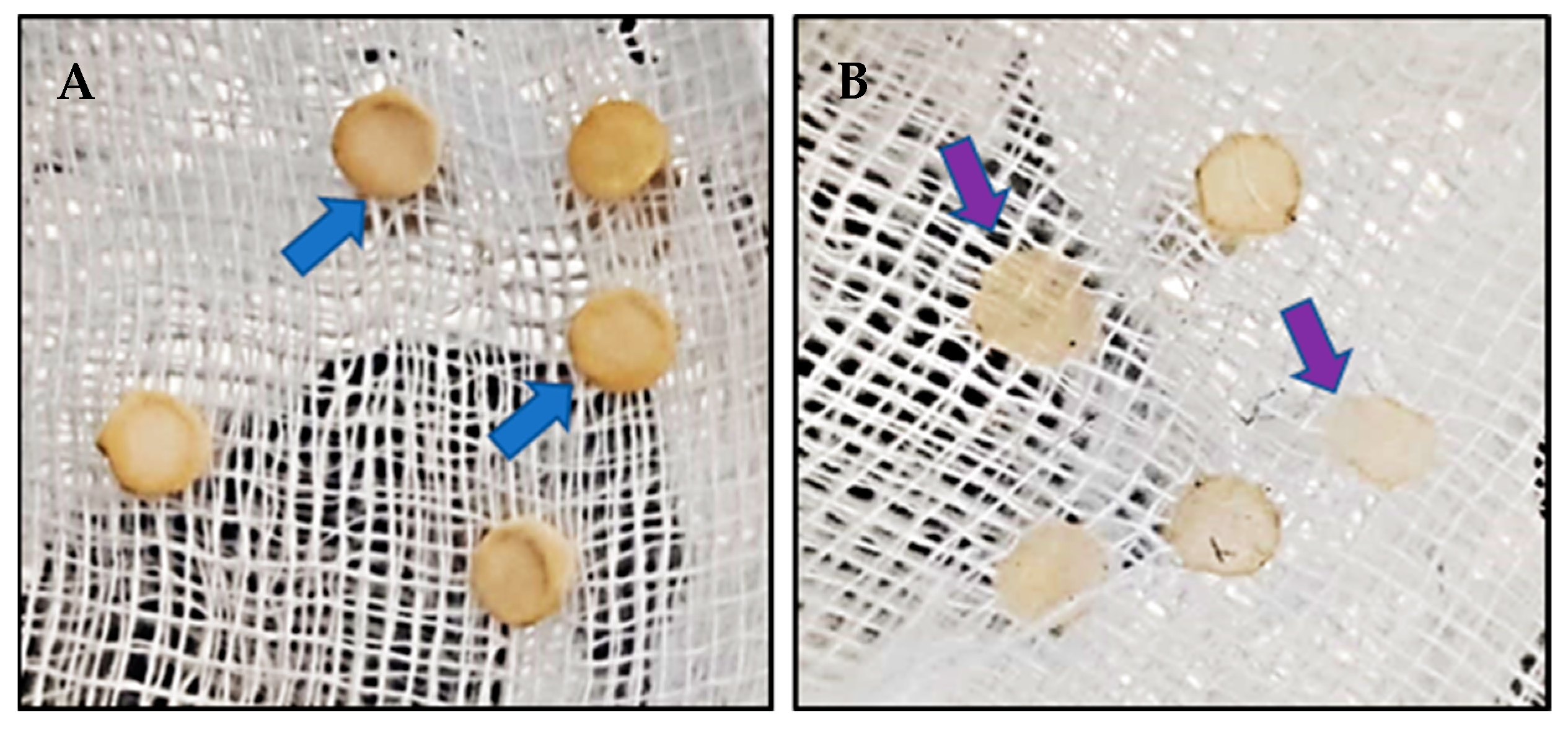
2.7. Cast_Gnk2-like In Vitro Antagonist Activity against P. cinnamomi

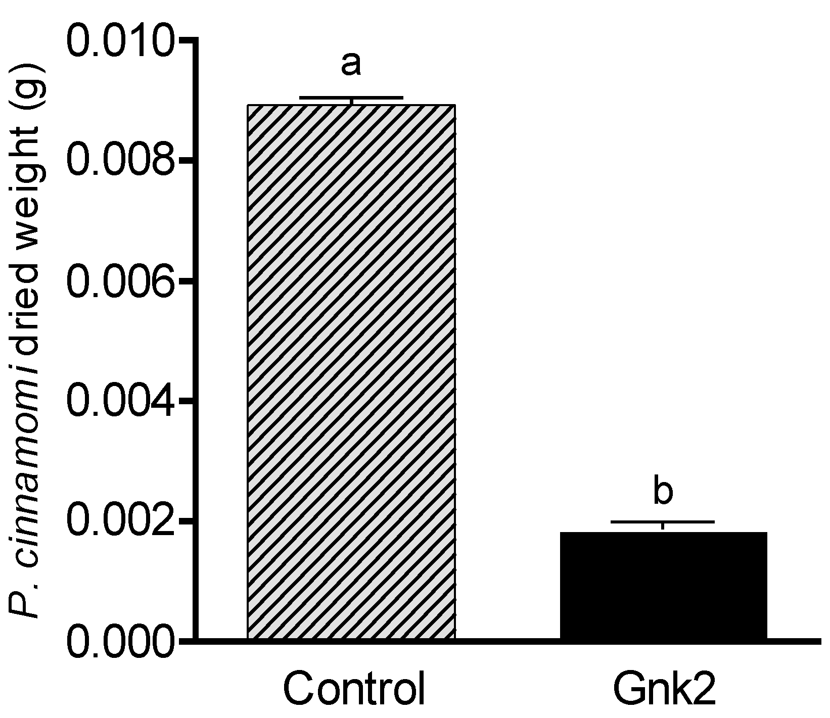
Two types of antagonist assays were performed. One of them was done in 60 mm diameter Petri dishes to evaluate P. cinnamomi mycelium development with different Cast_Gnk2-like pure protein concentrations (0.2 and 0.5 mg/mL). Briefly, in the solid medium assay, mycelial discs of 5-mm diameter were taken from the margins of approx. 1-week-old P. cinnamomi cultures and placed on new PDA medium plates. Different protein concentrations were tested and first spread in all the plates homogeneously, forming a thin film. The Petri plates were left for 25 min inside the flow chamber to dry the liquid above the solid medium before inoculation with P. cinnamomi. Different volumes of pure protein were also tested (100, 250, and 500 µL/plate). Control PDA medium Petri plates contained buffer solution without Cast_Gnk2-like protein. The plates were kept at 25 °C in darkness, and the growth of P. cinnamomi was evaluated during the following days of incubation (mainly 24, 48, and 72 h). Photographs of the mycelial growth were taken and the area determined with Image J software (Version 1.51r, NIH, Rockville, MD, USA). The second assay was performed in a liquid culture using Potato-Dextrose medium (50 mL) in Erlenmeyer flasks. Mycelial discs (10 discs of 5-mm diameter) were added to each flask and growth of P. cinnamomi was evaluated by determining mycelial dry weights (g) after two days of shaking (180 rpm) at 25°C. Controls were buffer solution without Cast_Gnk2-like protein.

2.8. Statistics

For fungal inoculation experiments, at least 10 biological replicates were used for each test, and the experiments were performed 3 times with similar results. The results shown in the pictures are examples from representative samples of n repetitions. The area of P. cinnamomi mycelia growth expressed in mm2 was determined. Graph Path software was used to prepare the figures, and the results are shown as means ± SEM. Data were analyzed by appropriate t-test or ANOVA followed by post hoc comparisons by Tukey or Dunnett’s test.

3. Results

3.1. Cast_GNK2-like Predictably Has Two Sites for Microbial Binding

Ginkbilobin-2 protein from G. biloba (GNK2) was the most thoroughly characterized so far. It has one conserved cysteine motif (C-8X-C-2X-C) related to its antimicrobial action [8]. We detected two cysteine motifs in Cast_GNK2-like (Figure 1). The same occurs with A. thaliana CRK9 and C. mollissima A0A8J4V9V8 (Table 1, Pfam column). As expected, the protein with the highest similarity to Cast_GNK2-like is A0A8J4V9V8 (99.6%, E-value 3.3 × 10−180) from the Chinese chestnut. Additionally, the amino acid sequences of all the proteins start with a signal peptide that guides the final peptide to the apoplast (Table 1). The alignment of the four amino acid sequences also indicates that all the proteins have a signal peptide for the protein to be secreted. Moreover, the alignment shows the existence of one conserved cysteine motif for GNK2 and two motifs for Cast_GNK2-like, as A0A8J4V9V8 and CRK9 (Figure 2).

3.2. Protein Isolation and Purification

The results of the recombinant protein expression, following different conditions to optimize the process, showed (according to the electrophoresis gels, SDS-PAGE) that the best expression was obtained using the E. coli BL21 (DE3) pLysS, in plasmid pET15, for 16 h at 20 °C, using 1 mM IPTG. We could observe a band in the gels (a protein fraction) in the region of 25 kDa (Figure 3A).
The immunoreactive spots in the region of 25 kDa from the Western Blot were detected with a chemiluminescence-based kit (Figure 3B).

3.3. Cast_Gnk2-like In Vitro Activity against P. cinnamomi

Mycelial growth of P. cinnamomi in PDA medium containing 0.5 mg/mL Cast_Gnk2-like pure protein was significantly less than the control treatment (without the protein). Figure 4 shows the growth of P. cinnamomi mycelia with and without Cast_Gnk2-like pure protein after 24 and 48 h at 25 °C in darkness. Cast_Gnk2-like does not inhibit the growth of P. cinnamomi, but it significantly reduces and delays its development.
The mycelia area (cm2) of P. cinnamomi growth in PDA medium under control treatment was 7.8 cm2, while in Cast_Gnk2-like pure protein treatment (0.5 mg/mL), it was 2.3 cm2, after 24 h. Cast_Gnk2-like pure protein treatment reduced the P. cinnamomi growth area by more than 100%. The same pattern was found after 48 h of incubation (Figure 5).
The dried mycelia of P. cinnamomi growing from PDA plugs in potato dextrose liquid medium (after 48 h shaking) under control treatment was visibly higher (Figure 6A) than in Cast_Gnk2-like pure protein treatment, where the mycelia growth was reduced (Figure 6B). The mycelial weight (g) of P. cinnamomi under the Control treatment was significantly higher (0.00892 g) than in Cast_Gnk2-like pure protein treatment (0.00187 g) after 48 h in shaking (Figure 7). The Control treatment had an increment in mycelia weight (g) of 377%.

4. Discussion

This work is part of the ongoing research program that aims to find solutions to the health problem regarding European chestnuts (C. sativa) and the pathogen P. cinnamomi [1]. This program involves an integrated approach using genomics, phenomics (precision phenotyping), transcriptomics, association mapping of traits, and histopathology, to reveal the mechanisms of the resistance of Asian species to infection by P. cinnamomi, and to transfer the knowledge for the improvement of resistance of European chestnut [7,9,13,18,19,20,21,22]. The program is supported by a breeding program initiated in 2006, based on controlled crosses, from which segregated hybrid populations were obtained with the selection of new genotypes with improved resistance to P. cinnamomi, which soon will be disclosed to the market to be used as rootstocks. The research and breeding efforts made in the last decades are having a positive impact since chestnut production in Europe has been increasing since 2015 for the first time in many decades. Nevertheless, more research and techniques are needed to overcome the decline of European chestnut [1].
The secretion of antimicrobial proteins in plants and the reinforcement of the cell wall are part of constitutive defense barriers against pathogens [7,23]. Also, the secretion of compounds toxic to pathogens is an effective chemical defense mechanism in plants. According to Santos et al. (2017) [7], Ginkbilobin-2 (Gnk2) is the most expressed protein, secreted by Ginkgo biloba seeds [24], and it was shown to possess antifungal activity. In a previous study where we used a histological approach to observe the responses exhibited by susceptible and resistant chestnuts under P. cinnamomi infection, we found that the early accumulation of phenolic-like compounds in cell walls was observed 0.5 h after inoculation in C. crenata root tissues which may prevent the spread of hyphae [9]. In the chestnut species resistant to P. cinnamomi, C. crenata, the Ginkgobilobin-2-like gene (isolated from transcriptomes of C. crenata [13] showed relevant expression constitutively and upon P. cinnamomi inoculation when compared with the susceptible C. sativa [7]. These previous studies of our group focused on the resistance of chestnut and its response to the Oomycete, indicating that C. crenata Ginkgobilobin-2-like gene and the encoded protein (Cast_Gnk2-like) might be important in chestnut resistance. In an in vitro tolerance assay with the pathogen P. cinnamomi, Serrazina, et al. (2022) [20], observed that transgenic plants of holm oak (Quercus ilex) with the Cast_Gnk2-like were able to survive longer than non-transgenic ones [20]. Similar results were obtained in resistance tests against F. oxysporum in transgenic cucumber (Cucumis sativus) plants, which showed that the expression of Gnk2-1 conferred antifungal activity against the disease [25]. Currently, to further demonstrate Cast_Gnk2-like action, the group is working with somatic embryos of C. sativa lines genetically transformed with the overexpression of the gene. These are being maintained for further assays (studies are still in process).
Other authors have also reported the antimicrobial effects of this protein against many plant and human pathogens [10,25]. However, Cast_Gnk2-like was never tested against P. cinnamomi pathogen to improve the defense focused in Chestnut/P. cinnamomi pathosystem.
The present work focused on expressing the Cast_Gnk2-like protein and testing its efficacy as a control of P. cinnamomi. The first step was to optimize the protein expression and test its effectiveness against the pathogen. The recombinant protein expression was performed following different conditions to optimize its process, and the best expression was obtained using the E. coli strain BL21 (DE3) pLysS. These results are in correspondence to the previous work of Miyakawa et al. (2007) [26], where the Gnk2 was overexpressed in E. coli BL21(DE3) using pET-26b and pDsbABCD1 vectors [25].
Diseases caused by soil-borne pathogens (such as ink disease) are difficult to eradicate, because of their asymptomatic nature during the initial stages of infection and their complex modes of life and dissemination of the pathogens [27]. The possibility of cure or eradication is very low, and it is, therefore, necessary to develop multidisciplinary strategies to reduce and control them. Farmers depend basically on fungicides to control these diseases. For many years synthetic fungicides were used to control plant production decay. However, public concerns about the harmful effects of chemicals on human health and the environment have caused scientists to search for new alternatives. The incidence of pathogens infections might become more severe in the context of climate change with the possible rise of new strains. For that, it is important to start making more efforts toward these problems [1].
Currently, we are developing studies and tests for the in-silico protein structure prediction, in vitro 7-day antagonist assays with P. cinnamomi, and metabolomics analysis.
This work re-confirmed that the protein studied has significant potential for biotechnological developments that could reduce the destructive impact of these harmful diseases. Moreover, the Cast_Gnk2-like gene is demonstrated to be a valuable candidate for marker-assisted selection of tolerant C. sativa genotypes. Finally, methods and strategies for the production of antimicrobial phytopharmaceuticals considering Cast_Gnk2-like (pure protein) against P. cinnamomi are also being considered for further research.

5. Conclusions

This work is part of the ongoing research program that aims to find solutions for European chestnut decline mainly due to P. cinnamomi, the causal agent of ink disease. The possibility of eradicating this pathogen is extremely low, and it is necessary to develop multi-disciplinary strategies to reduce and control it. Our group is studying new approaches and developing novel techniques for this purpose. As a result of this research, we determined that Cast_Gnk2-like has a signal peptide that guides the final protein peptide to the apoplast. Moreover, Cast_Gnk2-like (pure protein) significantly reduced the mycelia growth of P. cinnamomi in vitro, indicating the potential antagonistic effect on this pathogen. These insights reveal the potential of Cast_Gnk2-like for agricultural uses and biotechnological developments, which are becoming more necessary and critical in managing the threat this pathogen possess to European chestnut under rapidly changing environmental conditions.

Author Contributions

Conceptualization: R.L.C. and M.B.C.; Methodology: F.V.D., S.S. and M.B.C.; Validation: R.L.C., M.B.C., F.V.D. and S.S.; Formal analysis: R.L.C., M.B.C., F.V.D. and S.S.; Investigation: M.B.C., F.V.D. and S.S.; Resources: R.L.C. and R.M.; Data curation: M.B.C., F.V.D. and S.S.; Writing—original draft preparation: M.B.C.; Writing—review and editing: R.L.C., F.V.D., S.S., R.M. and M.B.C.; Visualization: R.L.C., F.V.D., S.S., R.M. and M.B.C.; Supervision: F.V.D. and R.L.C.; Project administration: R.L.C.; Funding acquisition: R.L.C. and R.M. All authors have read and agreed to the published version of the manuscript.

Funding

This research was funded by Fundação para a Ciência e Tecnologia under the project 414103 FCT-Lx-FEDER-28760, DL57/2016/CP [12345/2018]/CT[2477] to S.S., DL57/2016/CP [12345/2018]/CT[2470] to F.V.D., UIDB/04046/2020, UIDP/04046/2020, UIDB/04046/2021 and UIDP/04046/2021 to BioISI.

Data Availability Statement

Data from this paper are not included in any other site or repository.

Acknowledgments

We acknowledge Helena Machado (Instituto Nacional de Investigação Agrária e Veterinária, I.P) for providing Phytophotora cinnamomi culture for inoculations and Alan Phillips (BioISI—BioSystems and Integrative Sciences Institute) for English review and valuable suggestions.

Conflicts of Interest

The authors declare that the research was conducted without any commercial or financial relationships that could be construed as a potential conflict of interest.

References

  1. Fernandes, P.; Colavolpe, M.B.; Serrazina, S.; Costa, R.L. European and American chestnuts: An overview of the main threats and control efforts. Front. Plant Sci. 2022, 13, 844–951. [Google Scholar] [CrossRef]
  2. Thines, M. Taxonomy and phylogeny of Phytophthora and related Oomycetes. In Phytophthora: A Global Perspective; CABI: Wallingford, UK, 2013; pp. 11–18. [Google Scholar]
  3. Erwin, D.C.; Ribeiro, O.K. Phytophthora Diseases Worldwide; American Phytopathological Society (APS Press): St. Paul, MN, USA, 1996. [Google Scholar]
  4. Agrios, G.N. Plant Pathology, 5th ed.; Academic Press: Cambridge, MA, USA, 2005; p. 922. [Google Scholar]
  5. Hardham, A.R. Phytophthora cinnamomi. Mol. Plant Pathol. 2005, 6, 589–604. [Google Scholar] [CrossRef] [PubMed]
  6. Kamoun, S.; Furzer, O.; Jones, J.D.; Judelson, H.S.; Ali, G.S.; Dalio, R.J.; Govers, F. The Top 10 Oomycete pathogens in molecular plant pathology. Mol. Plant Pathol. 2015, 16, 413–434. [Google Scholar] [CrossRef]
  7. Santos, C.; Duarte, S.; Tedesco, S.; Fevereiro, P.; Costa, R. Expression Profiling of Castanea Genes during Resistant and Susceptible Interactions with the Oomycete Pathogen Phytophthora cinnamomi Reveal Possible Mechanisms of Immunity. Front. Plant Sci. 2017, 8, 515. [Google Scholar] [CrossRef] [Green Version]
  8. Niu, F.; Xing, Y.; Jia, N.; Ding, K.; Xie, D.; Chen, H.; Chi, D. Effectiveness of entomopathogenic fungal strains against poplar/willow weevil (Cryptorhynchus lapathi L.) larvae. J. For. Res. 2022, 33, 1691–1702. [Google Scholar] [CrossRef]
  9. Fernandes, P.; Machado, H.; Silva, M.D.C.; Costa, R.L. A Histopathological Study Reveals New Insights Into Responses of Chestnut (Castanea spp.) to Root Infection by Phytophthora cinnamomi. Phytopathology 2021, 111, 345–355. [Google Scholar] [CrossRef] [PubMed]
  10. Mélida, H.; Sandoval-Sierra, J.V.; Diéguez-Uribeondo, J.; Bulone, V. Analyses of Extracellular Carbohydrates in Oomycetes Unveil the Existence of Three Different Cell Wall Types. Eukaryotic. Cell 2013, 12, 194–203. [Google Scholar] [CrossRef] [Green Version]
  11. Miyakawa, T.; Hatano, K.; Miyauchi, Y.; Suwa, Y.; Sawano, Y.; Tanokura, M. A secreted protein with plant-specific cysteine-rich motif functions as a mannose-binding lectin that exhibits antifungal activity. Plant Physiol. 2014, 166, 766–778. [Google Scholar] [CrossRef] [PubMed] [Green Version]
  12. Bartnicki-Garcia, S. Chemistry of hyphal walls of Phytophthora. Microbiology 1966, 42, 57–69. [Google Scholar] [CrossRef] [Green Version]
  13. Serrazina, S.; Santos, C.; Machado, H.; Pesquita, C.; Vicentini, R.; Pais, M.S.; Sebastiana, M.; Costa, R. Castanea root transcriptome in response to Phytophthora cinnamomi challenge. Tree Genet. Genomes 2015, 11, 6. [Google Scholar] [CrossRef]
  14. The UniProt Consortium. UniProt: The Universal Protein Knowledgebase in 2023. Nucleic Acids Res. 2023, 51, D523–D531. [Google Scholar] [CrossRef]
  15. Petersen, T.; Brunak, S.; von Heijne, G. SignalP 4.0: Discriminating signal peptides from transmembrane regions. Nat. Methods 2011, 8, 785–786. [Google Scholar] [CrossRef] [PubMed]
  16. Almagro Armenteros, J.J.; Salvatore, M.; Emanuelsson, O.; Winther, O.; von Heijne, G.; Elofsson, A.; Nielsen, H. Detecting sequence signals in targeting peptides using deep learning. Life Sci. Alliance 2019, 2, e201900429. [Google Scholar] [CrossRef] [PubMed] [Green Version]
  17. Thompson, J.D.; Higgins, D.G.; Gibson, T.J. CLUSTAL W: Improving the sensitivity of progressive multiple sequence alignment through sequence weighting, position-specific gap penalties and weight matrix choice. Nucleic Acids Res. 1994, 22, 4673–4680. [Google Scholar] [CrossRef] [Green Version]
  18. Fernandes, P.; Tedesco, S.; Vieira da Silva, I.; Santos, C.; Machado, H.; Lourenço Costa, R. A New Clonal Propagation Protocol Develops Quality Root Systems in Chestnut. Forests 2020, 11, 826. [Google Scholar] [CrossRef]
  19. Santos, C.; Nelson, C.D.; Zhebentyayeva, T.; Machado, H.; Gomes-Laranjo, J.; Costa, R.L. First interspecific genetic linkage map for Castanea sativa x Castanea crenata revealed QTLs for resistance to Phytophthora cinnamomi. PLoS ONE 2017, 12, e0184381. [Google Scholar] [CrossRef] [PubMed] [Green Version]
  20. Serrazina, S.; Martínez, M.T.; Cano, V.; Malhó, R.; Costa, R.L.; Corredoira, E. Genetic Transformation of Quercus ilex Somatic Embryos with a Gnk2-like Protein That Reveals a Putative Anti-Oomycete Action. Plants 2022, 11, 304. [Google Scholar] [CrossRef] [PubMed]
  21. Santos, C.; Zhebentyayeva, T.; Serrazina, S.; Nelson, C.; Cost, R. Development and characterization of EST-SSR markers for mapping reaction to Phytophthora cinnamomi in Castanea spp. Sci. Hortic. 2015, 194, 181–187. [Google Scholar] [CrossRef]
  22. Santos, C.; Machado, H.; Correia, I.; Gomes, F.; Gomes-Laranjo, J.; Costa, R. Phenotyping Castanea hybrids for Phytophthora cinnamomi resistance. Plant Pathol. 2014, 64, 901–910. [Google Scholar] [CrossRef]
  23. Hardham, A.R.; Blackman, L.M. Molecular cytology of Phytophthora-plant interactions. Australas. Plant Pathol. 2010, 39, 29–35. [Google Scholar] [CrossRef]
  24. Miyakawa, T.; Miyazono, K.; Sawano, Y.; Hatano, K.; Tanokura, M. Crystal structure of ginkbilobin-2 with homology to the extracellular domain of plant cysteine-rich receptor-like kinases. Proteins 2009, 77, 247–251. [Google Scholar] [CrossRef] [PubMed]
  25. Liu, J.; Huali, T.; Yahong, W.; Aiguang, G. Overexpression of a Novel Antifungal Protein Gene GNK2-1 Results in Elevated Resistance of Transgenic Cucumber to Fusarium oxysporum. Chin. Bull. Bot. 2010, 45, 411–418. [Google Scholar] [CrossRef]
  26. Miyakawa, T.; Sawano, Y.; Miyazono, K.; Hatano, K.; Tanokura, M. Crystallization and preliminary X-ray analysis of ginkbilobin-2 from Ginkgo biloba seeds: A novel antifungal protein with homology to the extracellular domain of plant cysteine-rich receptor-like kinases. Acta Crystallogr. Sect. F Struct. Biol. Cryst. Commun. 2007, 63, 737–739. [Google Scholar] [CrossRef] [Green Version]
  27. Nelson, B.D.; Mallik, I.; McEwen, D.; Christianson, T. Pathotypes, distribution, and metalaxyl sensitivity of Phytophthora sojae from North Dakota. Plant Dis. 2008, 92, 1062–1066. [Google Scholar] [CrossRef] [PubMed] [Green Version]
Figure 1. Sequences S1. Cast_GNK2-like coding sequence (up) and respective amino acid sequence (below). In red is the prediction of the signal peptide to the apoplast. In blue, the two conserved cysteine motifs attributed to the antimicrobial action. *: stop codon.
Forests 14 00785 g001
Figure 2. A multiple alignment of C. crenata putative Ginkbilobin-2 protein (Cast_GNK2-like), C. mollissima Cysteine-rich repeat secretory protein 38 (A0A8J4V9V8), Ginkgo biloba Antifungal protein ginkbilobin-2 (A4ZDL6), and A. thaliana Putative cysteine-rich receptor-like protein kinase 9 (O65469), with CLUSTALW [17]. Protein accession numbers are indicated (Uniprot Accession). The signal peptide is surrounded by a red box and the conserved cysteine motif (C-8X-C-2X-C) is indicated in red.
Forests 14 00785 g002
Figure 3. SDS-PAGE gel (14%), panel A, and Western blotting showing the immunodetection of Cast_Gnk2-like protein, panel B. Hyper-expression and purification of Cast_Gnk2-like protein obtained using the E. coli BL21 (DE3) pLysS transformed with pET15b_Cast_Gnk2, lane 1: Cell pellet after sonication; lane 2: cell free extract; lane 3 to 6: fractions of the column washing procedure wash; lane 7: purified recombinant enzyme, 0.5 mg/mL (A). Western blotting showing the immunodetection of Cast_Gnk2-like protein using total cell extract after sonication, lane 1—insoluble fraction, lane 2—soluble fraction (B). The molecular masses (kDa) of protein standards (NZYTech Ltd., Lisbon, Portugal) are indicated.
Forests 14 00785 g003
Figure 4. Mycelia of P. cinnamomi growth in PDA medium under different conditions (3 Petri plates out of n are shown as representative of the assay). (A): Control treatment with buffer solution and P. cinnamomi mycelial growth after 24 h. (B): Cast_Gnk2-like pure protein (250 µL/plate and 0.5 mg/mL) and P. cinnamomi mycelial growth after 24 h. (C): Control treatment with buffer solution and P. cinnamomi mycelial growth after 48 h. (D): Cast_Gnk2-like pure protein (250 µL/plate and 0.5 mg/mL) and P. cinnamomi mycelial growth after 48 h.
Forests 14 00785 g004
Figure 5. Mycelia area (mm2) of P. cinnamomi growth in PDA under control treatment (only with buffer solution) and Cast_Gnk2-like pure protein treatment (250 µL/plate and 0.5 mg/mL) in Petri plates after 24 and 48 h at 25 °C in darkness. Results are shown as means ± SEM. Significant differences are shown as different letters (t-Test, p < 0.05).
Forests 14 00785 g005
Figure 6. Phytophthora cinnamomi mycelia dried (after 24 h at 65 °C) on PDA plugs (5 mm diam.) grown in potato dextrose liquid medium (50 mL) for 48 h in shaking (180 rpm) at 25 °C. Some of n plugs are shown as representative of the assay. (A): Control treatment (only with buffer solution), and (B): Cast_Gnk2-like pure protein treatment (250 µL and 0.5 mg/mL).
Forests 14 00785 g006
Figure 7. Mycelial dry weight (g) of P. cinnamomi growth in potato dextrose liquid medium (50 mL) under control treatment (only with buffer solution) and Cast_Gnk2-like pure protein treatment (250 µL and 0.5 mg/mL) after 48 h in shaking (180 rpm) at 25 °C. Results are shown as means ± SEM. Significant differences are shown as different letters (t-test, p < 0.05).
Forests 14 00785 g007
Table 1. In silico characteristics of the amino acid sequences homologous to Ginkbilobin-2. N.d.: non-determined. Pos: position of the signal peptide.
SpeciesProtein Name (Uniprot)Gene AcronymUniprot AccessionLength (AA)Interpro Family/DomainPfam Family, #HitsSubcellular Localization (Uniprot)SignalPTargetP
Ginkgo bilobaAntifungal protein ginkbilobin-2GNK2A4ZDL6134Gnk2-homologous domain (IPR002902); Gnk2-homologous domain superfamily (IPR038408)Salt stress response/antifungal (PF01657), 1SecretedSecretoryPos 1–26/27
Arabidopsis thalianaPutative cysteine-rich receptor-like protein kinase 9CRK9O65469265Gnk2-homologous domain (IPR002902); Gnk2-homologous domain superfamily (IPR038408)Salt stress response/antifungal (PF01657), 2SecretedSecretoryPos 1–23/24
Castanea mollissimaCysteine-rich repeat secretory protein 38N.d.A0A8J4V9V8241Gnk2-homologous domain (IPR002902); Gnk2-homologous domain superfamily (IPR038408)Salt stress response/antifungal (PF01657), 2N.d.SecretoryPos 1–23/24
Castanea crenataN.d.Cast_GNK2-likeN.d.241N.d.N.d.N.d.SecretoryPos 1–23/24
Disclaimer/Publisher’s Note: The statements, opinions and data contained in all publications are solely those of the individual author(s) and contributor(s) and not of MDPI and/or the editor(s). MDPI and/or the editor(s) disclaim responsibility for any injury to people or property resulting from any ideas, methods, instructions or products referred to in the content.

