Case Report

An Aminoglycoside-Sparing Regimen with Double Beta-Lactam to Successfully Treat Granulicatella adiacens Prosthetic Aortic Valve Endocarditis—Time to Change Paradigm?

1
Infectious Diseases Division, Department of Medicine (DAME), Azienda Sanitaria Universitaria Friuli Centrale (ASUFC), 33100 Udine, Italy
2
Department of Biomedical and Biotechnological Sciences, Section of Microbiology, University of Catania, 95123 Catania, Italy
3
Microbiology Unit, Azienda Sanitaria Universitaria Friuli Centrale (ASUFC), 33100 Udine, Italy
*
Author to whom correspondence should be addressed.
These authors contributed equally to this work and share first authorship.
These authors contributed equally to this work and share senior authorship.
§
These authors share last authorship.
Infect. Dis. Rep. 2024, 16(2), 249-259; https://0-doi-org.brum.beds.ac.uk/10.3390/idr16020020
Submission received: 12 January 2024 / Revised: 26 February 2024 / Accepted: 6 March 2024 / Published: 14 March 2024
(This article belongs to the Section Bacterial Diseases)

Abstract

:
(1) Background: Granulicatella adiacens is a former nutritionally variant streptococci (NVS). NVS infective endocarditis (IE) is generally characterized by a higher rate of morbidity and mortality, partially due to difficulties in choosing the most adequate microbiological culture method and the most effective treatment strategy, and partially due to higher rates of complications, such as heart failure, peripheral septic embolism, and peri-valvular abscess, as well as a higher rate of valve replacement. Depending on the affected valve (native valve endocarditisNVE, or prosthetic valve endocarditisPVE), the American Heart Association (AHA) 2015 treatment guidelines (GLs) suggest penicillin G, ampicillin, or ceftriaxone plus gentamicin (2 weeks for NVE and up to 6 weeks for PVE), while vancomycin alone may be a reasonable alternative in patients who are intolerant of β-lactam therapy. The European Society of Cardiology (ESC) 2023 GLs recommend treating NVE with penicillin G, ceftriaxone, or vancomycin for 6 weeks, suggesting combined with an aminoglycoside (AG) for at least the first 2 weeks only for PVE; likewise, the same recommendations for IE due to Enterococcus faecalis. (2) Methods: Starting from the case of a 51-year-old man with G. adiacens aortic bio-prosthesis IE who was successfully treated with aortic valve replacement combined with double beta-lactams, an AG-sparing regimen, we performed microbiology tests in order to validate this potential treatment change. (3) Results: As for E. faecalis IE, we found that the combination of ampicillin plus cephalosporines (like ceftriaxone or ceftobiprole) showed a synergistic effect in vitro, probably due to wider binding to penicillin-binding proteins (PBPs), thus contributing to enhanced bacterial killing and good clinical outcome, as well as avoiding the risk of nephrotoxicity due to AG association therapy. (4) Conclusions: Further studies are required to confirm this hypothesis, but double beta-lactams and an adequate sourcecontrol could be a choice in treating G. adiacens IE.

1. Introduction

Infective endocarditis (IE) remains a severe disease that is associated with high morbidity and mortality, despite many efforts aimed at improving diagnostic and therapeutic strategies. As reported in the European Infective Endocarditis Registry, the most frequent microorganisms identified remain staphylococci in 44.1%, enterococci in 15.8%, oral streptococci in 12.4%, and Streptococcus gallolyticus in 6.6% [1]. Granulicatella spp., previously known as NVS, are considered (together with Abiotrophia spp.) to be responsible for 5% of all streptococcal endocarditis. This fastidious bacterium has been reported to be a common cause of culture-negative bacterial endocarditis and its role in this scenario could be underestimated [2]. Granulicatella is catalase-negative, oxidase-negative, facultatively anaerobic, Gram-positive coccus, generally found as part of the normal oral, genitourinary, and intestinal tracts’ flora, with the oral cavity being the most probable bacterial entry point for most cases of IE [3,4]. Although its role as a causative agent of IE is known, diagnostic and clinical management are not completely defined. Depending on the affected valve (native or prosthetic), international GLs suggest treating G. adiacens IE with a combination therapy of beta-lactam plus gentamicin for 2 up to 6 weeks (with different classes and/or levels of recommendation), as in enterococcal endocarditis [5,6]. As reported in other experience [7], we tried to confirm that intravenous double beta-lactam combination therapy could result in good outcomes also for IE due to G. adiacens.

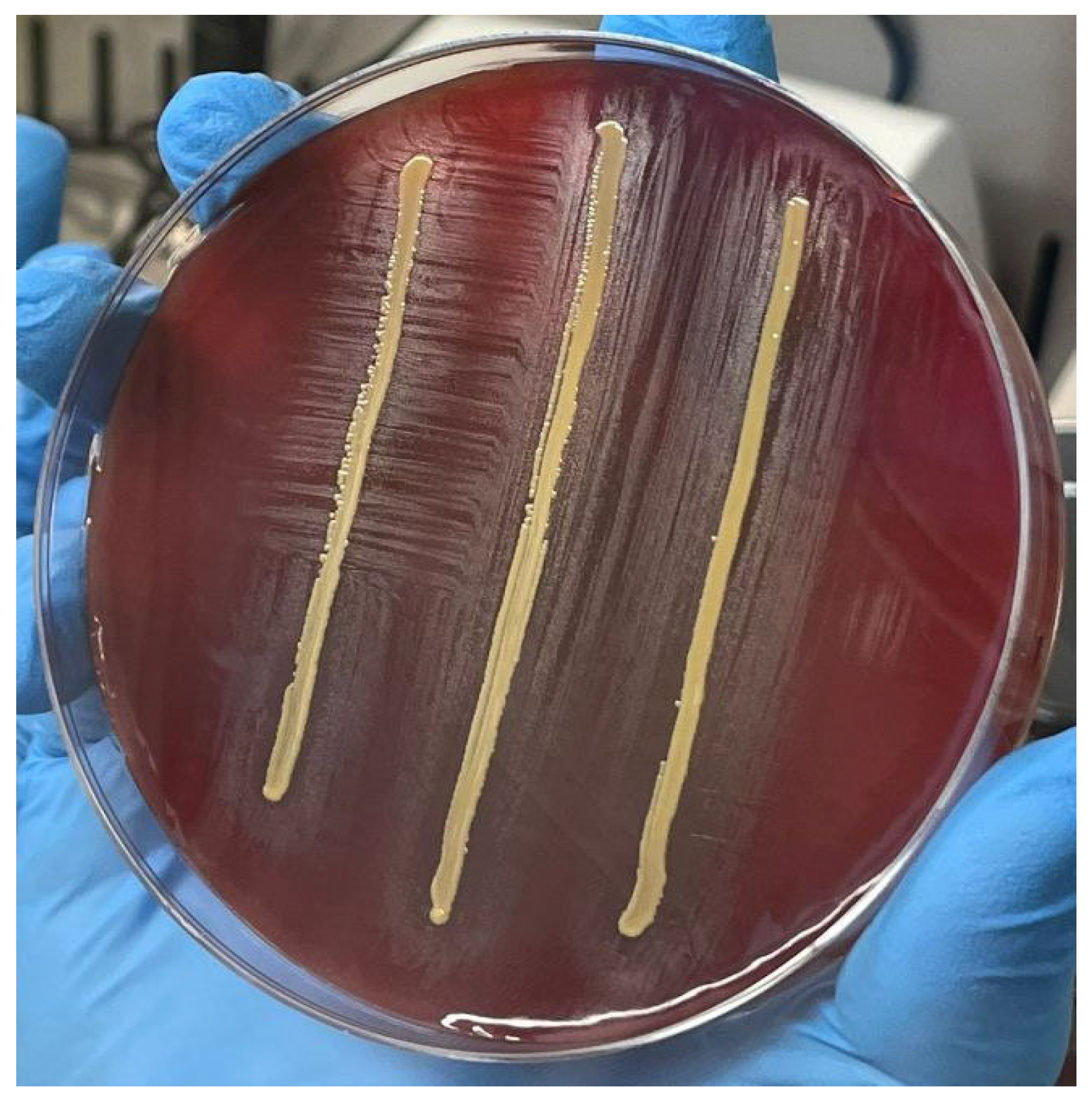
2. Case Presentation
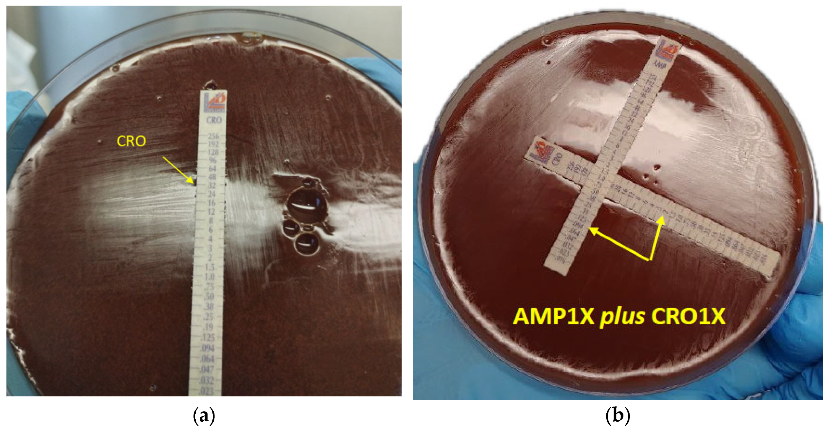
A 51-year-old man with a dual-chamber pacemaker (PM Ensura DDDR) implanted in March 2017 due to a Bruce treadmill test revealing 1° atrio-ventricular block and iso-rhythmic atrio-ventricular dissociation, an aortic valve bio-prosthesis (Carpentier-Edwards Perimount Magna EASE 25 mm), plus an ascending aorta vascular prosthesis (Vascutek Gelweave 28 mm), both implanted in September 2017 due to severe aortic valve stenosis in the bicuspid valve and ascending aorta ectasia, was admitted in August 2022 to the emergency department (ED) of Udine’s Hospital (Friuli Venezia-Giulia Region, Italy), with fever (up to 38 °C) for about 2 weeks and dry cough (lasting for 2 months after COVID-19 infection occurred in June 2022). He also complained of lumbar pain and a history of lumbar disc herniation, with no relief after taking anti-inflammatory and muscle relaxant drugs for at least 2 weeks. He denied any dental procedures and/or urinary tract or abdominal problems in the period before hospital admission. Physical examination showed oxygen blood saturation of 97%, bilateral norm-transmitted vesicular murmur with no other respiratory findings (no rales, no whistles, no crackles), preserved and rhythmic cardiac activity with controlled heart rate, a low-moderate systolic murmur (2/6) at the aortic focus, irradiated to the neck, with good hemodynamic stability (no peripheral edema), nor abdominal, neurological, or cutaneous pathological findings.
Initial laboratory studies revealed a C-reactive protein (CRP) value of 51.12 mg/L (cut-off 0–5 mg/L) and procalcitonin value of 0.12 ng/mL (cut-off < 0.10 ng/mL). The transthoracic echocardiography (TTE) performed in the emergency room revealed a vegetation with a thickening of the aortic bio-prosthesis (not seen in a previous recent control). Blood cultures (four bottles) were taken and, after one day, all samples yielded Gram-positive cocci in chains subsequently identified as Granulicatella adiacens by Bruker MALDI-TOF at the microbiology department of Udine’s Hospital. Antimicrobial susceptibility testing, conducted on the Evo Freedom Tecan workstation using the commercial MICRONAUT-S panel (MERLIN Diagnostika GmbH, Bornheim-Hersel, Germany) by micro-dilution broth method, showed susceptibility to amoxicillin (≤0.016 mg/L); cefotaxime (0.125 mg/L), ceftriaxone (0.094 mg/L), gentamicin (0.38 mg/L), meropenem (0.125 mg/L), and penicillin G (0.125 mg/L), according to standard guidelines.
A total body computed tomography (CT) scan was performed, which did not reveal any secondary embolism (including no sign of spondyliscitis), nor the dental scan any signs of peri-apical radiolucency. A second echocardiography control performed 1 week after hospital admission (both transthoracic and transesophageal) confirmed a vegetation both on the aortic right coronary cusp (14 mm × 6 mm) and on the non-coronary cusp with minimum regurgitation; no apparent peri-valvular abscess, no vegetation on PM electrodeleads, and no other valve infectious involvement was observed.
While waiting for blood culture results, an empiric iv therapy with daptomycin (10 mg/kg/day) and ceftaroline (600 mg tid) was started. After G. adiacens was identified (4 days after blood samples were taken), antibiotic therapy was switched to 4 g ampicillin intravenously every 6 h in continuous infusion (optimal dosage driven by therapeutic drug monitoring) and 2 g ceftriaxone intravenously every 12 h. This regimen was selected in order to avoid AG nephrotoxicity, although the patient’s serum creatinine levels were normal.
The case was carefully discussed with the cardiac surgeon team and, considering the fastidious isolated bacteria, the vegetation dimensions (near 15 mm) with high risk of embolism, the presence of prosthetic devices, and supposed low intra-operative and post-operative risks (young patient with no other comorbidity and with long life expectancy), the patient underwent surgery 2 days after starting the new antibiotic regimen. The intra-operative field revealed large masses attached to the previous implanted bio-prosthesis (almost on the ventricular side) but no abscess cavity; furthermore, the wall of the aortic bulb appeared thinned and fragile, while the valvular anulus was really damaged and discontinuous, especially under the coronary ostia. Therefore, surgeons decided to perform aortic valve bio-prosthesis and ascending aorta prosthesis replacement with a biological stentless root (Medtronic Freestyle 25 mm), an aortic vascular prosthesis (Vascutek Gelweave 28 mm), plus PM and leads removal. Shortly afterward (5 days after major surgery), the patient underwent PM re-implantation (with bi-cameral Medtronic Azure S DR). The standard culture results of the intra-operative samples (vegetation, aortic valve bio-prosthesis, aortic vascular prosthesis) were negative. Unfortunately, PM leads culture and valve histopathology were not performed.
Intravenous antibiotic therapy was continued for overall 6 weeks after first blood culture negativization (this occurred 10 days after first positivization), then the patient was discharged from the hospital with oral therapy (amoxicillin plus cefditoren pivoxil). Far from any GL recommendations about the correct timing of antibiotic treatment in NVS PVE, we decided to continue home treatment in order to avoid any risk of possible relapse considering that Granulicatella spp. are fastidious bacteria that are very hard to eradicate, even in cases where the strain is highly susceptible, the intra-operative field revealed a very complex and fragile situation with the necessity of both aortic valve and ascending aorta replacement, and the patient was fairly young with a history of re-operation.
Three weeks later, during a follow-up ambulatory visit, there was no clinical nor microbiological evidence of relapse, so antibiotics were stopped. The last TTE control performed 10 months after aortic valve replacement revealed no signs of IE relapse.

3. Laboratory Tests

In order to validate the potential synergism by using double beta-lactams in Granulicatella spp. treatment, the bacterial strain was sent to a reference microbiology laboratory at Catania University (Italy).
The identification was carried out by sequencing an internal fragment of the 16S rRNA gene, as already described [8]. Sequence alignment was performed using BLAST (Basic Local Alignment Search Tool; https://blast.ncbi.nlm.nih.gov/Blast.cgi accessed on 4 September 2023), which identified G. adiacens (100% NT identity with G. adiacens ATCC 49175 ID: CP102283.1). The organism was co-cultivated on blood agar with a streak of Staphylococcus aureus to demonstrate satellitism, which is a distinctive trait of this organism (Figure 1).
Antibiotic susceptibility testing was performed using Minimum Inhibitory Concentration (MIC) Test Strips (Liofilchem ®, Roseto degli Abruzzi, Italy) on in-house chocolate agar using Columbia Blood Agar Base (Oxoid, Milan, Italy) supplemented with 5% defibrinated horse blood (Biolife Italiana S.r.l., Milan, Italy) and 0.01% L-cysteine (100 mcg/mL) (Sigma-Aldrich®, Louis, MO, USA). Tests were performed in duplicate. MICs were interpreted using the CLSI M45 breakpoints for Abiotrophia spp. and Granulicatella spp. when available (CLSI 2015. Methods for antimicrobial dilution and disk susceptibility testing of infrequently isolated or fastidious bacteria, 3rd ed, CLSI guideline M45. Clinical and Laboratory Standards Institute, Wayne, PA). For quality control, we used CLSI-recommended Streptococcus pneumoniae ATCC 49619. According to CLSI guidelines, the strain showed resistance to ceftriaxone and cefotaxime, non-susceptibility to penicillin and ampicillin, and susceptibility to imipenem and vancomycin (Table 1).
Synergy testing was performed in duplicate, using the gradient-cross or 90° angle method, with some modification. Briefly, MIC test strips were placed in a cross formation, with a 90° angle at the intersection between the scales at their respective MICs for the organism, and incubated for 18 h at 35 °C. The following combinations were tested: ampicillin + ceftobiprole (AMP + BPR), ampicillin + ceftriaxone (AMP + CRO), ampicillin + gentamicin (AMP + CN); ceftobiprole + gentamicin (BPR + CN); and ceftriaxone + gentamicin (CRO + CN). In some cases, where the combination with the MICs of respective antibiotics did not exhibit a synergistic effect, a modification was made by testing the cross with the 2 × MIC value (Figure 2). The gradient-cross method was performed in duplicate for all combinations. The fractional inhibitory concentration index (FIC index) was used to interpret the gradient-cross method, as follows: synergy, FIC index ≤ 0.5; additive effect, FIC index 0.5–1; indifference, FIC index 1–4; antagonism, FIC index > 4 [9]. Despite their resistance profiles, double beta-lactam combinations showed better synergistic activity. Synergy was demonstrated at their respective MIC values (1 × MIC) when ampicillin was tested in combination with ceftriaxone (Figure 2b) and ceftobiprole, while beta-lactam combinations with gentamicin showed a synergistic effect only with double MICs of ampicillin, ceftriaxone, and ceftobiprole (2 × MIC), as demonstrated by their FICindexes (Table 2).

4. Discussion

NVS are fastidious Gram-positive bacteria, requiring either L-cysteine or pyridoxal-supplemented medium to support growth. Chromosomal DNA-DNA hybridization in 1989, and the more recent 16S rRNA gene sequencing data, led to a reclassification of NVS isolates into two different genera, Abiotrophia and Granulicatella, which in turn comprise four recognized species: Abiotrophia defectiva, Granulicatella adiacens, Granulicatella elegans, and Granulicatella balaenopterae [10]. Penicillin, cephalosporin (ceftriaxone, cefotaxime, cefepime, ceftaroline), carbapenem (meropenem, imipenem), vancomycin, daptomycin, and gentamicin susceptibility consistently varies for G. adiacens strains in different studies, probably due to difficult isolation and identification techniques [11,12]. Recent studies have shown that molecular biology techniques, such as 16S rDNA NGS analysis, have higher sensitivity than traditional microbiological methods for detecting IE (either blood and heart valve cultures) caused by fastidious or difficult-to-culture microorganisms like G. adiacens [13].
Our blood isolate—which was first tested for antimicrobial susceptibility using the micro-dilution broth method—showed good susceptibility to penicillin, ceftriaxone, and gentamicin. However, due to variability in in vitro penicillin susceptibility and the challenges associated with obtaining dependable antimicrobial susceptibility testing (AST) results for these organisms, mono-therapy with penicillin for G. adiacens IE, even at high dose, may result in poor outcomes. As reported by Giuliano et al., such organisms may exhibit a laboratory phenomenon of “penicillin tolerance”: for tolerant strains, it is possible that the minimum bactericidal concentration (MBC) of penicillin exceeds the MIC up to 32-fold [2]. Even rabbit model studies showed the superiority of penicillin plus gentamicin compared to penicillin alone [14].
Another potential explanation of antibiotic treatment failure using penicillin in mono-therapy, thus focusing on the problem of the most proper antimicrobial choice (mono-therapy instead of combination therapy) in treating Granulicatella spp. IE (and in general streptoccocal IE) apart from PEN susceptibility, could be related to biofilm formation. As described in recent studies, growing evidence suggests that the ability to produce biofilms may play a major pathogenetic role in supporting microbial adhesion and persistence while protecting bacteria from antimicrobial drugs, and recent results support the knowledge that native valve IE (not only prosthetic valve IE), even due to streptococci (as well as Staphylococcus aureus and E. faecalis), represents a model of biofilm-related infection which, therefore, could be associated with increased antibiotic tolerance/resistance [15].
Thus, treating susceptible NVS strain (and in general streptococcal strain) IE with beta-lactam mono-therapy solely on the basis of blood culture MIC, without any concern about eventual MBC and/or minimum biofilm inhibitory concentration (MBIC), could lead to microbiological and clinical worsening/failure. Nowadays, there is a lack of data about the diffusion gradient of beta-lactams into the vegetation, and the ratio of antibiotic concentration at steady state between plasma and vegetation cannot be completely determined [16]. Assuming that the ratio of the AUC of antibiotics in tissue to the AUC of the total serum concentration is linked to the ratio of diffusible (supposedly unbound) drug in tissue to the total concentration, most of the currently available studies about antibiotic penetration into heart valves are based upon high performance liquid chromatography (HPLC) methods [17,18]. On the one hand, HPLC has been developed widely in recent years and allows the precise determination of the concentrations of most drugs in various body fluids like blood [19], on the other hand, MALDI MS imaging (MSI) and micro dialysis (MD) techniques could represent better methods for visualizing and quantifying antibiotic penetration into the vegetation [20]. But these tools are still waiting for validation.
Surely, when surgical intervention is considered a feasible and safe option considering the patient’s underlying conditions, surgical disruption and removal of microbial vegetation can largely remove the biofilm structure, thus improving the patient’s healing, as we decided to perform in our case. But surgery cannot lead to complete eradication of infection in peri-valvular cardiac tissues, unless being extremely demolitive, and that is why it would be necessary to continue highperformance antibiotic treatment, depending on the vegetation/valvular culture sample results.
Specifically, our patient underwent a redo operation, as described in a retrospective German study, performed to evaluate the impact of NVE versus PVE on post-operative outcomes and long-term survival and to identify pre-operative risk factors in a large cohort of 4300 patients with IE, in which the evidence suggested that PVE alone should not be a contraindication for redo operations [21].
The 2012 British Society of Antimicrobial Chemotherapy guidelines recommend 4–6 weeks with benzylpenicillin and gentamicin for the treatment of Granulicatella or Abiotrophia endocarditis [22].
The 2015 GLs from the AHA recommend therapy with penicillin G, AMP, or CRO plus gentamicin (2 weeks for NVE and up to 6 weeks for PVE), while vancomycin alone may be a reasonable alternative in patients who are intolerant of β-lactam therapy [5]. The ESC 2023 GLs recommend treating NVE with penicillin G, CRO, or vancomycin for 6 weeks, suggesting combined with an AG for at least the first 2 weeks only in the case of PVE [6].
However, even with these recommended treatment regimens, the rates of surgical therapy (27%) and relapse and death (17–27%) remain high. Additionally, relapses may occur frequently, even in cases where the strain is highly susceptible [23,24].
This mirrors the outcomes and recommendations for treating E. faecalis IE (EFIE). Experimental studies have been successfully carried out as a way to evaluate the synergism of beta-lactam combination against clinical strains of E. faecalis, regardless of their susceptibility to aminoglycosides. Specifically, the mainstay for the synergistic activity of beta-lactam combination could be based on to the differential and complementary saturation of E. faecalis PBPs, thus generating the necessary bactericidal effect [25,26,27].
In 1995, for the first time ever, Mainardi and co-workers yielded a demonstration of the synergism between amoxicillin and cefotaxime, through partial saturation of essential PBPs 4 and 5 (primary role in cell growth of E. faecalis) by amoxicillin (AML), and also total saturation of non-essential PBPs 2 and 3 (probably engaged in cell wall assembly) by cefotaxime [25]. Thereafter, these first pilot test results represented the backbone for clinical studies focused on establishing the true efficacy of this therapeutic approach in humans with E. faecalis IE. In a recent review about the efficacy of AMP plus CRO regimen against EFIE, Marino and colleagues concluded that the epidemiological changes of E. faecalis IE, that is the fact that the population is aging and consequently becoming more fragile, and a possible underestimation of treatment sideeffects, above all the higher risk of nephrotoxicity, should compel an ideal shift in antibiotic choice and that the growing body of literature with the combination AMP and CRO appears promising. But in this context, large and high-quality non-inferiority clinical studies are needed to clearly assess the efficacy and safety of double beta-lactam regimens against E. faecalis IE instead of AG regimens [26].
In a recent retrospective case series by Giuliano at al., 21 patients with a diagnosis of E. faecalis IE or primary or non-primary complicated or uncomplicated bacteremia were treated with AMP plus BPR. The results showed a high clinical success rate of 81% and microbiological cure in 86% of patients. One of the reasons for this treatment strategy was that, unlike CRO, BPR can inhibit non-essential high-molecular-weight enterococcal PBPs and has a higher affinity for the essential PBP4, which is a critical lethal target and the main determinant of beta-lactam susceptibility in E. faecalis [27].
Regarding IE caused by streptococci with a PEN MIC between 0.25 and 2 mg/L, no prospective comparative study has ever assessed the advantages of adding AG in this setting. This topic was recently confirmed in a retrospective French study (414 patients with streptococcal IE, mostly viridans group streptoccoci), in which the authors observed that streptococcal IE with AML MIC between 0.25 and 2 mg/L had a higher mortality rate than that with lower MIC, but combination with AG was not associated with better outcome [28]. From the microbiological point of view, even if a PEN MIC of 0.25 to 2 mg/L is similar to the PEN MIC for E. faecalis, bacterial tolerance to PEN is uncommon in streptococci (10–20%) as compared to enterococci (75%) [29]. Therefore, in a recent review by D. Lebeaux et al., the authors suggested that AG may not be necessary for streptococcal IE with AML MIC between 0.25 and 0.5 mg/L, and their use should be restricted to streptococcal IE with AML MIC > 0.5 mg/L [30].
As recently described by Khan et al., it is possible that using AMP plus CRO therapy for G. adiacens IE, similarly to E. faecalis, may result in synergy, increased bacterial killing, and better clinical outcome, without the risk of nephrotoxicity due to AG use. In their study, the authors performed susceptibility testing using E-tests for AMP and CRO alone and in combination at standard concentrations, which demonstrated synergy (based on their FICindex) [7].
In order to achieve a better understanding of the potential synergism of using double beta-lactam treatment for Granulicatella spp. infections, we sent our bacterial strain to a reference microbiology laboratory at Catania University. After identifying the strain as G. adiacens through 16S rRNA gene sequencing, it was difficult to perform antimicrobial susceptibility by the recommended BMD method due to growth issues [31]. As there are no commercially available methods with FDA clearance for AST of these fastidious organisms, we opted to use the gradient-test (GT) in Columbia Blood Agar as an alternative.
In similar fashion to previous studies by Michael O. Alberti et al., where they compared categorical agreement (CA) and essential MIC agreement (EA) between the CLSI BMD reference method and E-tests using a commercially available Chocolate Mueller Hinton Agar (CMHA) plate for vancomycin, PEN, and CRO [32,33], we also observed poor agreement between the two methods. In our case, this was seen to a lesser extent for PEN (BMD MIC 0.125 mg/L vs. GT MIC 0.38–0.5 mg/L) but more significantly for CRO (BMD MIC 0.094 mg/L vs. GT MIC 32 mg/L) and cefotaxime (BMD MIC 0.125 mg/L vs. GT MIC 8–12 mg/L).
In our experience, using the gradient-cross method to test for synergy (through FIC index), we observed a clear synergistic effect with double beta-lactam treatment, specifically with AMP plus BPR and AMP plus CRO at their MICs (despite CRO being non-susceptible by GT). We hypothesize that the basis for this synergistic activity may be due to the differential and complementary saturation of PBPs (similarly to E. faecalis), resulting in a necessary bactericidal effect. In order to confirm this topic, we have already started to study the possible G. adiacens PBP BPR inhibition, finding promising results concerning interaction with PBP2 and PBP1 family.
Interestingly, our strain showed daptomycin (DAP) nonsusceptibility (DNS) (MIC ≥ 2 mg/L) and internal colonies displayed a high-level DAP resistance (HLDR) profile (MIC ≥ 256 mg/L) based on existing CLSI breakpoints for viridans group streptococci. In a recent Spanish study evaluating DAP susceptibility among nine G. adiacens isolates, baseline MICs varied between 1 and 16 mg/L, of which only one (1/9–11.1%) showed a DAP MIC of 1 mg/L. After being incubated with inhibitory concentrations of DAP, all strains rapidly increased the baseline MIC of daptomycin and showed resistance with either a DNS or HLDR profile. Even combination therapy did not prevent the development of DAP resistance with AMP (2/3 strains), CN (2/3 strains), CRO (2/3 strains), or ceftaroline (2/3 strains), thus rising further doubts about the optimal antibiotic treatment for these species [34].

5. Conclusions

While G. adiacens IE accounts for less than 5% of all streptococci IE cases, it is important to note that, as part of the NVS group, this type of endocarditis has historically been associated with higher rates of morbidity and mortality. This is in part attributed to challenges in culturing and susceptibility testing, which can lead to diagnostic delays and uncertainties in treatment decisions.
Even if, in our case, the surgical intervention was performed very early after the double beta-lactam treatment was started, thus surely leading to better source control and final good clinical outcome, it is equally important to point out that the surgery, as much as it tries to be demolitive, cannot totally eradicate all of the microbiological burden around and into the peri-valvular cardiac tissue.
Our findings highlight the importance of considering double beta-lactam treatment as a potential option for G. adiacens IE, especially in cases where aminoglycosides are contraindicated or not tolerated. It is plausible to consider that the broader saturation of PBPs may contribute to a favorable synergy, enhanced killing, and good clinical outcome, similar to E. faecalis, although this theory is still speculative and requires further confirmation.

Author Contributions

Conceptualization, A.P., F.C. and C.T.; methodology, A.P., F.C. and C.T.; validation, A.P., F.C. and C.T.; formal analysis, F.C. and P.C.; investigation, A.P., F.C. and P.C.; data curation, A.P., F.C. and P.C.; writing—original draft preparation, A.P. and F.C.; writing—review and editing, A.P., F.C., P.C., F.P., P.D.S., S.F., S.G., L.M., D.P., A.S., M.S., T.S. and C.T.; supervision, F.C. and C.T.; funding acquisition, F.C. All authors have read and agreed to the published version of the manuscript.

Funding

This research was partially funded by MUR PNRR Extended Partnership Initiative on Emerging Infectious Diseases (project no. PE00000007, INF-ACT).

Institutional Review Board Statement

We declare that the manuscript has not been previously published nor is it being considered for publication elsewhere. The study was conducted according to the guidelines of the Declaration of Helsinki, and approved by the Institutional Review Board of Department of Medicine (protocol code 45/2024, date of approval 7 February 2024).

Informed Consent Statement

Written informed consent has been obtained from the patient to publish this paper.

Data Availability Statement

The original contributions presented in the study are included in the article, further inquiries can be directed to the corresponding author.

Conflicts of Interest

The authors declare no conflicts of interest.

List of Abbreviations

NVSNutritionally variant streptococci
IEInfective endocarditis
NVENative valve endocarditis
PVEProsthetic valve endocarditis
AHAAmerican Heart Association
ESCEuropean Society of Cardiology
GLGuidelines
PENPenicillin
AMPAmpicillin
BRPCeftobiprole
CROCeftriaxone
AMLAmoxicillin
CNGentamicin
AGAminoglycoside
PBPsPenicillin-binding proteins
PMPacemaker
EDEmergency department
CRPC-reactive protein
CTComputed tomography
TTETransthoracic echocardiography
MICMinimum inhibitory concentration
MBCMinimum bactericidal concentration
MBICMinimum biofilm inhibitory concentration
BMDBroth micro-dilution
FIC indexFractional inhibitory concentration index
ASTAntimicrobial susceptibility testing
EFIEEnterococcus faecalis infective endocarditis
CMHAChocolate Mueller Hinton Agar

References

  1. Habib, G.; Erba, P.A.; Iung, B.; Donal, E.; Cosyns, B.; Laroche, C.; Popescu, B.A.; Prendergast, B.; Tornos, P.; Sadeghpour, A.; et al. Clinical presentation, aetiology and outcome of infective endocarditis. Results of the ESC-EORP EURO-ENDO (European infective endocarditis) registry: A prospective cohort study. Eur. Heart J. 2019, 40, 3222–3233. [Google Scholar] [CrossRef]
  2. Giuliano, S.; Caccese, R.; Carfagna, P.; Vena, A.; Falcone, M.; Venditti, M. Endocarditis caused by nutritionally variant streptococci: A case report and literature review. Infez. Med. 2012, 2, 67–74. [Google Scholar]
  3. Garibyan, V.; Shaw, D. Bivalvular endocarditis due to Granulicatella adiacens. Am. J. Case Rep. 2013, 14, 435–438. [Google Scholar] [PubMed]
  4. Shaik, A.; Roeuth, D.; Azmeen, A.; Thanikonda, V.; Guevara-Pineda, D.; Alamnajam, M.; Yamase, H.; Haider, L. Granulicatella Causing Infective Endocarditis and Glomerulonephritis. IDCases 2020, 21, e00792. [Google Scholar] [CrossRef] [PubMed]
  5. Baddour, L.M.; Wilson, W.R.; Bayer, A.S.; Fowler, V.G., Jr.; Tleyjeh, I.M.; Rybak, M.J.; Barsic, B.; Lockhart, P.B.; Gewitz, M.H.; Levison, M.E.; et al. Infective endocarditis in adults: Diagnosis, antimicrobial therapy, and management of complications. Circulation 2015, 132, 1435–1486. [Google Scholar] [CrossRef] [PubMed]
  6. ESC Scientific Document Group. 2023 ESC Guidelines for the management of endocarditis. Developed by the task force on the management of endocarditis of the European Society of Cardiology (ESC). Endorsed by the European Association for Cardio-Thoracic Surgery (EACTS) and the European Association of Nuclear Medicine (EANM). Eur. Heart J. 2023, 44, 3948–4042. [Google Scholar] [CrossRef]
  7. Khan, S.; Urban, C.; Singh, V.; Liu, D.; Segal-Maurer, S.; Parmar, Y.; Yoon, J. Novel double beta-lactam therapy for Granulicatella adiacens infective endocarditis. IDCases 2021, 24, e01082. [Google Scholar] [CrossRef] [PubMed]
  8. Muyzer, G.; De Waal, E.C.; Uitterlinden, A.G. Profiling of Complex Microbial Populations by Denaturing Gradient Gel Electrophoresis Analysis of Polymerase Chain Reaction-Amplified Genes Coding for 16S rRNA. Appl. Environ. Microbiol. 1993, 59, 695–700. [Google Scholar] [CrossRef] [PubMed]
  9. White, R.L.; Burgess, D.S.; Manduru, M.; Bosso, J.A. Comparison of three different in vitro methods of detecting synergy: Time-kill, checkerboard, and E test. Antimicrob. Agents Chemother. 1996, 40, 1914–1918. [Google Scholar] [CrossRef]
  10. Alberti, M.O.; Hindler, J.A.; Humphries, R.M. Antimicrobial Susceptibilities of Abiotrophia defectiva, Granulicatella adiacens, and Granulicatella elegans. Antimicrob. Agents Chemother. 2016, 60, 1411–1420. [Google Scholar] [CrossRef]
  11. Mushtaq, A.; Greenwood-Quaintance, K.E.; Cole, N.C.; Kohner, P.C.; Ihde, S.M.; Strand, G.J.; Harper, L.W.; Virk, A.; Patel, R. Differential antimicrobial susceptibilities of Granulicatella adiacens and Abiotrophia defectiva. Antimicrob. Agents Chemother. 2016, 60, 5036–5039. [Google Scholar] [CrossRef]
  12. Kanamoto, T.; Terakubo, S.; Nakashima, H. Antimicrobial Susceptibilities of Oral Isolates of Abiotrophia and Granulicatella According to the Consensus Guidelines for Fastidious Bacteria. Medicines 2018, 5, 129. [Google Scholar] [CrossRef] [PubMed]
  13. Podgórska, A.; Kordybach-Prokopiuk, M.; Jaworska-Wilczyńska, M.; Hoffman, P.; Biernacka, K.; Kuśmierski, K.; Kuthan, R.; Hryniewiecki, T.; Lutyńska, A. The First Case of Granulicatella adiacens Identified from a Resected Heart Valve by Next Generation Sequencing (NGS) in Poland. Pathogens 2022, 11, 295. [Google Scholar] [CrossRef] [PubMed]
  14. Bouvet, A. Human endocarditis due to nutritionally variant streptococci: Streptococcus adjacens and Streptococus defectivus. Eur. Heart J. 1995, 16, 24–27. [Google Scholar] [CrossRef] [PubMed]
  15. Di Domenico, E.G.; Rimoldi, S.G.; Cavallo, I.; D’Agosto, G.; Trento, E.; Cagnoni, G.; Palazzin, A.; Pagani, C.; Romeri, F.; De Vecchi, E.; et al. Microbial biofilm correlates with an increased antibiotic tolerance and poor therapeutic outcome in infective endocarditis. BMC Microbiol. 2019, 19, 228. [Google Scholar] [CrossRef]
  16. Flateau, C.; Riazi, A.; Cassard, B.; Camus, M.; Diamantis, S. Streptococcal and enterococcal endocarditis: Time for individualized antibiotherapy? J. Antimicrob. Chemother. 2021, 76, 3073–3076. [Google Scholar] [CrossRef]
  17. Lalanne, S.; Guérin, F.; Flécher, E.; Cattoir, V.; Nesseler, N.; Revest, M.; Verdier, M.-C. Diffusion of amoxicillin into heart valves from infective endocarditis patients. J. Antimicrob. Chemother. 2023, 78, 232–237. [Google Scholar] [CrossRef]
  18. Martin, C.; Viviand, X.; Alaya, M.; Lokiec, F.; Ennabli, K.; Said, R.; Pecking, M. Penetration of ceftriaxone (1 or 2 grams intravenously) into mediastinal and cardiac tissues in humans. Antimicrob. Agents Chemother. 1996, 40, 812–815. [Google Scholar] [CrossRef]
  19. Tattevin, P.; Tribut, O.; Arvieux, C.; Dupont, M.; Flicoteaux, R.; Desbordes, L.; Le Tulzo, Y.; Michelet, C. Use of high-performance liquid chromatography (HPLC) to monitor beta-lactam plasma concentrations during the treatment of endocarditis. Clin. Microbiol. Infect. 2005, 11, 76–79. [Google Scholar] [CrossRef]
  20. Giuliano, S.; Angelini, J.; Flammini, S.; Tascini, C. Comment on: Streptococcal and enterococcal endocarditis: Time for individualized antibiotherapy? J. Antimicrob. Chemother. 2022, 77, 2044–2045. [Google Scholar] [CrossRef]
  21. Weber, C.; Petrov, G.; Luehr, M.; Aubin, H.; Tugtekin, S.M.; Borger, M.A.; Akhyari, P.; Wahlers, T.; Hagl, C.; Matschke, K.; et al. Surgical results for prosthetic versus native valve endocarditis: A multicenter analysis. J. Thorac. Cardiovasc. Surg. 2021, 161, 609–619. [Google Scholar] [CrossRef]
  22. Gould, F.K.; Denning, D.W.; Elliott, T.S.; Foweraker, J.; Perry, J.D.; Prendergast, B.D.; Sandoe, J.A.; Spry, M.J.; Watkin, R.W.; Working Party of the British Society for Antimicrobial Chemotherapy. Guidelines for the diagnosis and antibiotic treatment of endocarditis in adults: A report of the Working Party of the British Society for Antimicrobial Chemotherapy. J. Antimicrob. Chemother. 2012, 67, 269–289. [Google Scholar] [CrossRef]
  23. Sinner, S.W.; Tunkel, A.R. Viridans Streptococci, Nutritionally Variant Streptococci, Groups C and G Streptococci, and Other Related Organisms. In Mandell, Douglas, and Bennett’s Principles and Practice of Infectious Diseases, 8th ed.; WB Saunders: Philadelphia, PA, USA, 2015. [Google Scholar]
  24. Shailaja, T.S.; Sathiavathy, K.A.; Unni, G. Infective endocarditis caused by Granulicatella adiacens. Indian Heart J. 2013, 65, 447–449. [Google Scholar] [CrossRef]
  25. Mainardi, J.L.; Gutmann, L.; Acar, J.F.; Goldstein, F.W. Synergistic effect of amoxicillin and cefotaxime against Enterococcus faecalis. Antimicrob. Agents Chemother. 1995, 39, 1984–1987. [Google Scholar] [CrossRef] [PubMed]
  26. Marino, A.; Munafò, A.; Zagami, A.; Ceccarelli, M.; Di Mauro, R.; Cantarella, G.; Bernardini, R.; Nunnari, G.; Cacopardo, B. Ampicillin Plus Ceftriaxone Regimen against Enterococcus faecalis Endocarditis: A Literature Review. J. Clin. Med. 2021, 10, 4594. [Google Scholar] [CrossRef] [PubMed]
  27. Giuliano, S.; Angelini, J.; D’Elia, D.; Geminiani, M.; Barison, R.D.; Giacinta, A.; Sartor, A.; Campanile, F.; Curcio, F.; Cotta, M.O.; et al. Ampicillin and Ceftobiprole Combination for the Treatment of Enterococcus faecalis Invasive Infections: “The Times They Are A-Changin”. Antibiotics 2023, 12, 879. [Google Scholar] [CrossRef] [PubMed]
  28. Pilmis, B.; Lourtet-Hascoët, J.; Barraud, O.; Piau, C.; Isnard, C.; Hery-Arnaud, G.; Amara, M.; Merens, A.; Farfour, E.; Thomas, E.; et al. Be careful about MICs to amoxicillin for patients with Streptococci-related infective endocarditis. Int. J. Antimicrob. Agents 2019, 53, 850–854. [Google Scholar] [CrossRef] [PubMed]
  29. Van der Meer, J.T.M.; Van Vianen, W.; Hu, E.; Van Leeuwen, W.B.; Valkenburg, H.A.; Thompson, J.; Michel, M.F. Distribution, antibiotic susceptibility and tolerance of bacterial isolates in culture-positive cases of endocarditis in The Netherlands. Eur. J. Clin. Microbiol. Infect. Dis. 1991, 10, 728–734. [Google Scholar] [CrossRef] [PubMed]
  30. Lebeaux, D.; Fernández-Hidalgo, N.; Pilmis, B.; Tattevin, P.; Mainardi, J.L. Aminoglycosides for infective endocarditis: Time to say goodbye? Clin. Microbiol. Infect. 2020, 26, 723–728. [Google Scholar] [CrossRef] [PubMed]
  31. Clinical and Laboratory Standards Institute (CLSI). Methods for Antimicrobial Dilution and Disk Susceptibility Testing of Infrequently Isolated or Fastidious Bacteria, 3rd ed.; CLSI Guideline M45; Clinical and Laboratory Standards Institute: Wayne, PA, USA, 2015. [Google Scholar]
  32. Alberti, M.O.; Hindler, J.A.; Humphries, R.M. Comparison of Antimicrobial Susceptibility Testing by Broth Microdilution and E-Test for Abiotrophia defectiva and Granulicatella spp. Am. J. Clin. Pathol. 2015, 143, A035. [Google Scholar] [CrossRef]
  33. Alberti, M.O.; Hindler, J.A.; Humphries, R.M. Performance of Etest for Antimicrobial Susceptibility Testing of Abiotrophia defectiva and Granulicatella Species. J. Clin. Microbiol. 2016, 54, 2194–2196. [Google Scholar] [CrossRef] [PubMed]
  34. Cañas, M.A.; Téllez, A.; García de la Mària, C.; Dahl, A.; García-González, J.; Hernández-Meneses, M.; Almela, M.; Ambrosioni, J.; Falces, C.; Quintana, E.; et al. Development of High-Level Daptomycin Resistance in Abiotrophia and Granulicatella Species Isolates from Patients with Infective Endocarditis. Antimicrob. Agents Chemother. 2021, 65, e02522-20. [Google Scholar] [CrossRef] [PubMed]
Figure 1. Satellite growth of Granulicatella adiacens in proximity to the staphylococcal streak.
Idr 16 00020 g001
Figure 2. Antimicrobial susceptibility testing and synergistic combination of ceftriaxone (CRO) with ampicillin (AMP) by gradient-cross method. Arrowheads indicate the MIC values of CRO alone (a) and in combination (b) with AMP (AMP1X plus CRO1X).
Idr 16 00020 g002
Table 1. Granulicatella adiacens MIC Values by E-test (mg/L).
PENAMPAMLBPRCPTCROCTXIMILNZCNSTRTEIVANDAPTGCDAL
NSNSSNANARRSNANANANASNANANA
Granulicatella0.50.50.1250.1923280.25241114 *0.0940.125
S: Susceptible; NS: Non-Susceptible; R: Resistant; NA: Not applicable. PEN: Penicillin; AMP: Ampicillin; AML: Amoxicillin; BPR: Ceftobiprole; CPT: Ceftaroline; CRO: Ceftriaxone: CTX: Cefotaxime: IMI: Imipenem; LNZ: Linezolid; CN: Gentamicin; STR: Streptomycine; TEI: Teicoplanin; VAN: Vancomycin; DAP: Daptomycin; TGC: Tigecycline; DAL: Dalbavancyn. * Internal colonies MIC > 256 mg/L.
Table 2. Gradient-Cross Method.
DRUG ADRUG BMIC
Drug A
MIC
Drug B
MIC Drug A
in Combination
MIC Drug B
in Combination
FIC
Drug A
FIC
Drug B
FIC IndexINTERPRETATION
AMP 1×BPR 1×0.50.190.1250.0470.250.2470.49SYN
AMP 1×BPR 2×0.50.380.1250.0230.250.060.31SYN
AMP 1×CRO 1×0.5320.09420.1880.0620.25SYN
AMP 1×CRO 2×0.5640.04780.0940.1250.21SYN
AMP 1×CN 1×0.540.2530.50.751.25IND
AMP 2×CN 1×140.250.750.250.1880.4SYN
BPR 1×CN 1×0.1940.12530.6580.751.4IND
BPR 2×CN 1×0.3840.09410.2470.250.4SYN
CRO 1×CN 1×3241620.50.51ADD
CRO 2×CN 1×64440.750.06250.1870.25SYN
AMP: Ampicillin; BPR: Ceftobiprole; CRO: Ceftriaxone; CN: Gentamicin. SYN: Synergy; ADD: Additive effect; IND: Indifferent.
Disclaimer/Publisher’s Note: The statements, opinions and data contained in all publications are solely those of the individual author(s) and contributor(s) and not of MDPI and/or the editor(s). MDPI and/or the editor(s) disclaim responsibility for any injury to people or property resulting from any ideas, methods, instructions or products referred to in the content.

