Congenital Hypofibrinogenemia in a Neonate with a Novel Mutation in the FGB Gene


Abstract
:1. Introduction
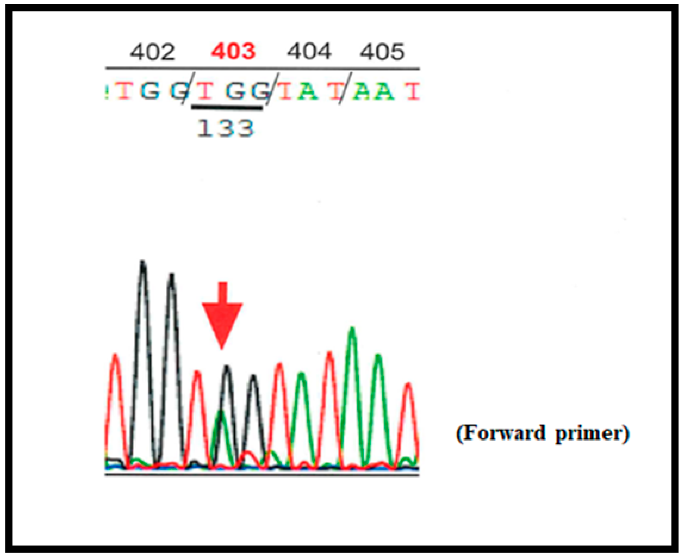
2. Case Report
3. Discussion
Author Contributions
Funding
Institutional Review Board Statement
Informed Consent Statement
Data Availability Statement
Conflicts of Interest
References
- Woods, C.W.; Bradshaw, W.T.; Woods, A.G. Hemophagocytic lymphohistiocytosis in the premature neonate. Adv. Neonatal Care 2009, 9, 265–273. [Google Scholar] [CrossRef] [PubMed]
- Schmidt, B.K.; Vegh, P.; Andrew, M.; Johnston, M. Coagulation screening tests in high risk neonates: A prospective cohort study. Arch. Dis. Child. 1992, 67, 1196–1197. [Google Scholar] [CrossRef] [PubMed] [Green Version]
- de Moerloose, P.; Casini, A.; Neerman-Arbez, M. Congenital fibrinogen disorders: An update. Semin. Thromb. Hemost. 2013, 39, 585–595. [Google Scholar] [PubMed] [Green Version]
- Zhang, Y.; Zuo, X.; Teng, Y. Women with Congenital Hypofibrinogenemia/Afibrinogenemia: From Birth to Death. Clin. Appl. Thromb. Hemost. 2020, 26. [Google Scholar] [CrossRef] [PubMed] [Green Version]
- Casini, A.; Neerman-Arbez, M.; Ariëns, R.A.; de Moerloose, P. Dysfibrinogenemia: From molecular anomalies to clinical manifestations and management. J. Thromb. Haemost. 2015, 13, 909–919. [Google Scholar] [CrossRef] [PubMed]
- Casini, A.; Lukowski, S.; Quintard, V.L.; Crutu, A.; Zak, M.; Regazzoni, S.; De Moerloose, P.; Neerman-Arbez, M. FGB mutations leading to congenital quantitative fibrinogen deficiencies: An update and report of four novel mutations. Thromb. Res. 2014, 133, 868–874. [Google Scholar] [CrossRef] [PubMed]
- Vu, D.; Neerman-Arbez, M. Molecular mechanisms accounting for fibrinogen deficiency: From large deletions to intracellular retention of misfolded proteins. J. Thromb. Haemost. 2007, 5 (Suppl. 1), 125–131. [Google Scholar] [CrossRef] [PubMed]
- Human Fibrinogen Database. Available online: http://site.geht.org/base-de-donnees-fibrinogene/ (accessed on 21 February 2021).
- Peyvandi, F.; Haertel, S.; Knaub, S.; Mannucci, P.M. Incidence of bleeding symptoms in 100 patients with inherited afibrinogenemia or hypofibrinogenemia. J. Thromb. Haemost. 2006, 4, 1634–1637. [Google Scholar] [CrossRef] [PubMed]
- Chinni, E.; Tiscia, G.; Favuzzi, G.; Cappucci, F.; Malcangi, G.; Bagna, R.; Izzi, C.; Rizzi, D.; De Stefano, V.; Grandone, E. Identification of novel mutations in patients with fibrinogen disorders and genotype/phenotype correlations. Blood Transfus. 2019, 17, 247–254. [Google Scholar] [PubMed]
- Zhou, W.; He, Y.; Li, Q.; Li, Y.; Su, Y.; Yan, L. Clinical Characteristics of Hospitalized Neonates with Hypofibrinogenemia: A Retrospective Cohort Study. Front. Pediatr. 2020, 8, 589. [Google Scholar] [CrossRef] [PubMed]
- Vlietman, J.J.; Verhage, J.; Vos, H.L.; Van Wijk, R.; Remijn, J.A.; Van Solinge, W.W.; Brus, F. Congenital afibrinogenaemia in a newborn infant due to a novel mutation in the fibrinogen alpha gene. Br. J. Haematol. 2002, 119, 282–283. [Google Scholar] [CrossRef] [PubMed]
- Ataoglu, E.; Duru, N.S.; Celkan, T.; Civilibal, M.; Yavuz, S.C.; Elevli, M.; Ayta, S. Spontaneous intracranial bleeding in a neonate with congenital afibrinogenemia. Blood Coagul. Fibrinolysis 2010, 21, 592–594. [Google Scholar] [CrossRef] [PubMed]
- Hariharan, G.; Ramachandran, S.; Parapurath, R. Congenital Afibrinogenemia presenting as antenatal intracranial bleed: A case report. Ital. J. Pediatr. 2010, 36, 1. [Google Scholar] [CrossRef] [PubMed] [Green Version]
- Kaur, M.; Kumar, N.; Bose, S.K.; Rajendran, A.; Trehan, A.; Ahluwalia, J. Congenital afibrinogenemia in a new born: A rare cause for bleeding. Blood Coagul. Fibrinolysis 2014, 25, 527–529. [Google Scholar] [CrossRef] [PubMed]
- Abolghasemi, H.; Shahverdi, E. Umbilical bleeding: A presenting feature for congenital afibrinogenemia. Blood Coagul. Fibrinolysis 2015, 26, 834–835. [Google Scholar] [CrossRef] [PubMed]
- Yapıcıoğlu, H.; Narlı, N.; Satar, M.; Antmen, A.B. Congenital Hypofibrinogenemia: A Newborn Infant with Cord Bleeding. Turk. J. Haematol. 2000, 17, 217–219. [Google Scholar] [PubMed]
- Breen, C.M.; Riazat, M.I.; McCallion, N.; Boyle, M.A. Congenital hypofibrinogenaemia: A presymptomatic detection of an extremely rare bleeding disorder in preterm twins. BMJ Case Rep. 2017, 2017. [Google Scholar] [CrossRef] [PubMed]
- Cai, H.; Liang, M.; Yang, J.; Zhang, X. Congenital hypofibrinogenemia in pregnancy: A report of 11 cases. Blood Coagul. Fibrinolysis 2018, 29, 155–159. [Google Scholar] [CrossRef] [PubMed] [Green Version]
- Bornikova, L.; Peyvandi, F.; Allen, G.; Bernstein, J.; Manco-Johnson, M.J. Fibrinogen replacement therapy for congenital fibrinogen deficiency. J. Thromb. Haemost. 2011, 9, 1687–1704. [Google Scholar] [CrossRef] [PubMed]
- Kozek-Langenecker, S.; Sørensen, B.; Hess, J.R.; Spahn, D.R. Clinical effectiveness of fresh frozen plasma compared with fibrinogen concentrate: A systematic review. Crit. Care 2011, 15, R239. [Google Scholar] [CrossRef] [PubMed] [Green Version]


| CBC | Coagulation Data | ||
| WBC (3000–8500)/µL | 12,500 | PT activity (80–100)% | 42.8 |
| Hb (11–16) g/dL | 18.9 | PT-INR (0.9–1.1) | 1.62 |
| MCV (83–100) fL | 111.4 | APTT (control) s | 66.4 (27.3) |
| Reticulocyte (3–11)‰ | 53 | Fibrinogen (200–400) mg/dL | <50 |
| Platelet count (150 K–360 K)/µL | 197 K | D-dimer (<1.0) µg/mL | 2.7 |
| Biochemical data | |||
| CRP (<0.29) mg/dL | <0.01 | Albumin (4.1–5.2) g/dL | 3.1 |
| AST (13–37) U/L | 36 | BUN (7.8–18.9) mg/dL | 11.3 |
| ALT (8–45) U/L | 5 | Creatinine (0.45–0.82) md/dL | 0.63 |
| LDH (122–228) U/L | 427 | Na (138–146) mmol/L | 139 |
| Total bilirubin (0.3–1.3) mg/dL | 1.95 | K (3.6–5.1) mmol/L | 4.1 |
| Total protein (6.7–8.3) g/dL | 4.8 | Cl (99–108) mmol/L | 106 |
| Family Members | Age (Years) | Fibrinogen (mg/dL) | Sequencing (FGB Bβ Gene; exon 8) | Hemorrhagic Symptoms | ||
|---|---|---|---|---|---|---|
| (Activity) | (Antigen) | |||||
| Father | 35 | 99 | 97 | p.403Try > stop | none | |
| Mother | 35 | 297 | - | - | - | |
| Elder sister | 2 | 67 | NT | NT | none | |
| Proband (newborn) | 0 | Day 0 | <50 | NT | p.403Try > stop | none |
| Day 8 * | 61 | 67 | ||||
Publisher’s Note: MDPI stays neutral with regard to jurisdictional claims in published maps and institutional affiliations. |
© 2021 by the authors. Licensee MDPI, Basel, Switzerland. This article is an open access article distributed under the terms and conditions of the Creative Commons Attribution (CC BY) license (http://creativecommons.org/licenses/by/4.0/).
Share and Cite
Shinozuka, J.; Okumura, N.; Nagasawa, M.; Nishikado, M.; Kadowaki, S.; Katsuda, I.; Imashuku, S. Congenital Hypofibrinogenemia in a Neonate with a Novel Mutation in the FGB Gene. Pediatr. Rep. 2021, 13, 113-117. https://0-doi-org.brum.beds.ac.uk/10.3390/pediatric13010016
Shinozuka J, Okumura N, Nagasawa M, Nishikado M, Kadowaki S, Katsuda I, Imashuku S. Congenital Hypofibrinogenemia in a Neonate with a Novel Mutation in the FGB Gene. Pediatric Reports. 2021; 13(1):113-117. https://0-doi-org.brum.beds.ac.uk/10.3390/pediatric13010016
Chicago/Turabian StyleShinozuka, Jun, Nobuo Okumura, Mayumi Nagasawa, Motokazu Nishikado, Sayaka Kadowaki, Itsuro Katsuda, and Shinsaku Imashuku. 2021. "Congenital Hypofibrinogenemia in a Neonate with a Novel Mutation in the FGB Gene" Pediatric Reports 13, no. 1: 113-117. https://0-doi-org.brum.beds.ac.uk/10.3390/pediatric13010016