Article

Deciphering the Factors for Nodulation and Symbiosis of Mesorhizobium Associated with Cicer arietinum in Northwest India

by
Raghvendra Pratap Singh
1,2,3,*,
Geetanjali Manchanda
4,
Yingjie Yang
5,
Dipti Singh
6,
Alok Kumar Srivastava
2,
Ramesh Chandra Dubey
1 and
Chengsheng Zhang
5,*
1
Department of Botany and Microbiology, Gurukul Kangri University, Haridwar, Uttarakhand 249404, India
2
National Bureau of Agriculturally Important Microorganisms, ICAR, Kushmaur, Kaithauli, Maunath Bhanjan, Uttar Pradesh 275101, India
3
Department of Research & Development, Biotechnology, Uttaranchal University, Uttarakhand 248007, India
4
Department of Botany and Environmental Studies, DAV University, Jalandhar, Punjab 144001, India
5
Marine Agriculture Research Center, Tobacco Research Institute of Chinese Academy of Agricultural Sciences, Keyuanjingsilu 11, Qingdao 266101, China
6
Department of Microbiology, V.B.S. Purvanchal University, Jaunpur 222003, India
*
Authors to whom correspondence should be addressed.
Sustainability 2019, 11(24), 7216; https://0-doi-org.brum.beds.ac.uk/10.3390/su11247216
Submission received: 9 October 2019 / Revised: 9 December 2019 / Accepted: 9 December 2019 / Published: 16 December 2019
(This article belongs to the Section Sustainable Agriculture)

Abstract

:
The compatibility between rhizobia and legumes for nitrogen-fixing nodules and the stages of root hair curling, formation of infection thread, and nodulation initiation have been vitally studied, but the factors for the sustainable root surface colonization and efficient symbiosis within chickpea and rhizobia have been poorly investigated. Hence, we aimed to analyze phenotypic properties and phylogenetic relationships of root-nodule bacteria associated with chickpea (Cicer arietinum) in the north-west Indo Gangetic Plains (NW-IGP) region of Uttar Pradesh, India. In this study, 54 isolates were recovered from five agricultural locations. Strains exhibited high exopolysaccharide production and were capable of survival at 15–42 °C. Assays for phosphate solubilization, catalase, oxidase, Indole acetic acid (IAA) production, and 1-aminocyclopropane-1-carboxylic acid (ACC) deaminase activity revealed that all the tested isolates possessed plant growth-promoting potential. Metabolic profiling using Biolog plates indicated that patterns of substrate utilization differed considerably among isolates. A biofilm formation assay showed that isolates displayed a nearly four-fold range in their capacity for biofilm development. Inoculation experiments indicated that all isolates formed nodules on chickpea, but they exhibited more than a two-fold range in symbiotic efficiency. No nodules were observed on four other legumes (Phaseolus vulgaris, Pisum sativum, Lens culinaris, and Vigna mungo). Concatenated sequences from six loci (gap, edD, glnD, gnD, rpoB, and nodC) supported the assignment of all isolates to the species Mesorhizobium ciceri, with strain M. ciceri Ca181 as their closest relative.

1. Introduction

For years, plant-associated bacteria have been speculated to be the organisms capable of promoting growth and/or suppressing diseases when present in the rhizosphere and as endophytes within healthy plant tissues [1,2]. Leguminous plants have the capability to associate with rhizobia by the nodulation process and fix atmospheric nitrogen (N) in the root nodule. This can help plants in soil that is low in nitrogen (N) and condition the soil itself, if other factors are favorable for growth. Bacteria have typically been associated with or adhere around the legumes plant roots and act as a plant growth promoter by several secretory or substrate capabilities, such as phosphate, ACC deaminase, siderophore, IAA, catalase, oxidase, and NH3 production [3,4]. Moreover, they also act as endophytic or nodule-forming bacteria (found within the tissues of the plant) as well as saprophytic bacteria (found free-living in the soil).
Rhizobia may colonize soil that envelops the roots [5] or adhere to the root surface [6], which is directly influenced by their attachments on their desired surface. Biofilm formation by bacteria offers conductive sites for an appropriate, effective, and reproductive environment for the bacteria to adhere to the surface. Biofilm thus provides the space for growth, a protective degree of homeostasis, and allows the bacteria to overcome biotic and abiotic stresses—this is done by a complex extrapolymeric substance (EPS) matrix sheath [7,8,9]. During infection, thread formation, and root colonization, EPS plays an important role in the infection and the active nodules [8]. Bacteria adheres to the legume roots with the help of EPS and can fix the nitrogen for plant growth, act as biocontrol against pathogens, produce phytohormone, and mobilize the nutrients to enriched the soil and the environment [10,11]. Rhizobia have been used in agricultural practices mainly for nitrogen fixation and plant growth promotion (PGP) [12,13,14] due to their wide distribution. The first criterion for a Rhizobium strain to be used in legume inocula is that it must be highly effective when fixing nitrogen [15].
Chickpea is one of the major pulse crops throughout the world and ranks second (The Food and Agriculture Organization of United Nation) amongst food legumes in terms of world production and is cultivated on a large scale in arid and semi-arid environments [16,17]. Chickpea is the most important legume in the Indo Gangetic Plain (IGP), and its cultivation is completely dependent on rhizobia, which have an impact on agriculture as well as the environment. Currently, chickpea rhizobia are included in the genus Mesorhizobium (gram-negative bacterium), with two species that form functional nodules after interacting with chickpea roots [18,19]. These species are described as specific microsymbionts—M. ciceri [20] and M. mediterraneum [21]. When not in symbiosis, they are part of the habitat and behave as saprophytes. M. ciceri cells sense the root exudates in the soil and invade after adhering to the root hairs of C. arietinum, which are dependent on the exopolysaccharides of rhizobia. Which in turn, act as signal molecules for the establishment of symbiosis between rhizobia and legumes [22,23]. After invading the root, the formation of specific root organs, called nodules, takes place. During the maturation of the nodules, invading rhizobial cells differentiate into endo-symbiotic structures called bacteroides. Fixed nitrogen (NH4+) is provided to plants by bacteroides. It is hypothesized that, in return, the plants deliver organic acids and several carbon and energy sources to the bacteroides as root exudates [24,25,26]. The overall procedure is completely based on saprophytic survival and adherence during chickpea cropping.
The absence of a high population of potential nitrogen-fixing rhizobia in the soil and their sustainability in symbiosis has been one of the main limiting factors for legume production. Plant growth promotion, resistance to stress, and utilization of various carbon sources support the rhizobia in their saprophytic survival (while they are in a free-living state), effective nodulation with legumes, and symbiotic efficiency. Moreover, the ability to survive against stress and the formation of biofilm can shape the abundance and efficiency of rhizobia. Hence, we aimed to investigate the major factors required for efficient symbiosis and PGP for mesorhizobia, associated symbiotically with C. arietinum. Further, sequencing of six gene loci was done to characterize the diversity and relationships of Mesorhizobium strains associated with chickpea in the IGP region of India.

2. Materials and Methods

2.1. Sampling Sites, Isolation, and Culture Condition

This study was carried out on chickpea nodules from 32 different representatives from five semiarid or sub-humid alkaline field sites of the NW-IGP region Uttar Pradesh, India (Figure S1). Isolation and purification of rhizobia from root nodules was done on yeast extract-mannitol agar (YMA) by using the standard procedure [27]. All procured rhizobia were incubated on YMA slants at 28 °C and maintained at 4 °C for routine use and in 20% (w/v) glycerol at −80 °C for long-term storage [28]. Standard culture of M. ciceri strains IC-2058 and IC-2018 were taken from the microbial genomics laboratory of Indian council of Agriculture research-National Bureau of Agriculturally Important Microorganisms (ICAR-NBAIM) Mau, India.

2.2. Validation, Nodulation, and Host-Specificity Test of Chickpea Nodule Rhizobia

Studied isolates were validated as rhizobia by testing the ability to induce root nodules on chickpea according to previous reported methods [29]. Briefly, 100 µL log phase culture of tested isolates was taken and inoculated on to pre-surface sterilized chickpea seeds (Avarodhi variety). Further, the sterilized clay pots (30 cm high 20 cm diameter) were filled with equal amount of autoclaved soil (approx. 4.0 kg). After that, inoculated seeds were sown in clay pots for growth. The ideal condition for plant growth was adjusted (28/19 °C (day/night) with a 12-h photoperiod). After 40 days, plants were uprooted gently from the pots for observation of various parameters such as nodule number (NN), shoot dry weight (SDW), chlorophyll content, and symbiotic efficiency (SE) according to Gibson A.H. [30]. Nitrogen supplemented (T2) plant was treated as a control while without N (T1) acted as a negative control. The whole experiment was performed in 5 replicates.
Host specificity was examined by cross nodulation test as described by Nandasena et al. [31]. Briefly, all the tested rhizobia were inoculated in Phaseolus vulgaris, Pisum sativum, Lens culinaris, and Vigna mungo separately in pots, as described for nodulation assay in this study. During the flowering stage, plants were uprooted gently and the root nodules were examined.

2.3. PGP Traits

Phosphate solubilization index (S.I.) of the tested strains was determined using Pikovaskya agar plates spot method [32], calculated as the ratio of halo zone diameter to colony diameter for positive isolates [33]. Qualitative and quantitative estimation of IAA production was estimated as described by Khanmna et al. [34]. Ammonia (NH3) production of each isolate was evaluated in peptone water. Briefly, 100 µL of log phase cultures (106 cells mL−1) were inoculated in 15 ml nutrient broth (NB from Hi-Media Co.) and incubated at 125 rpm and 28 ± 2 °C in rotator shaker for 4–5 days. Development of deep yellow to brown color by addition of Nessler’s reagent (Hi-Media Co.) (0.5 mL) was noted as a positive test for NH3 production [34]. Catalase and oxidase production tests were carried out according to Smibert et al. [35]. For ACC deaminase, minimal medium (ACC as sole source of nitrogen) was inoculated with 100 µL log phase culture, and growth was observed at different time intervals through the absorbance measurement (at 600 nm), as well as viable count by serial dilution–spread plate method. Multiplication capabilities of inoculated strains were considered as ACC deaminase positive. Quantification of ACC deaminase was carried out according to Glick et al. [36].
All morpho-phenotypic and PGP tests were performed in five replicates at three different time intervals for their reproducibility and consistency.

2.4. Biofilm Assay

Qualitative and quantitative estimation of biofilm formation ability of all the tested rhizobia were evaluated as described by Mirani J. A. and Jamil N. [37] with partial modification. Briefly, log phase culture of isolates was pelleted out, washed 2 times with ddH2O, and resuspended into sterile ddH2O to adjust the optical density (OD) 0.2 at A600 with spectrophotometer. Further, 100 µL of dissolved culture was inoculated into the small size culture vial that contained 250 µL of M63 minimal medium supplemented with 0.2% glucose (Hi Media Co. In,), 1 mM magnesium sulfate (Hi Media Co. In.), and 0.5% casamino acids (Hi Media Co. In.). Further, the cover slip was placed vertically and then incubated for 48 h at 28 °C. After that, the cover slip was put carefully in 0.1% crystal violet (CV) for 30 min to stain the developed biofilm. Then, it was solubilized in 1 ml 30% glacial acetic acid and the absorbance was recorded (Shimadzu Co. Ltd. In.) at 563 nm wave length by using at least five replicates for each assay to avoid the deviation and error.

2.5. Biolog Carbon Source Utilization and Numerical Analysis

Based on the SE test, morpho-phenotypic characteristics, and biofilm assay, 16 most prominent isolates were selected for further characterization. Isolates were cultivated separately in YEM broth, harvested at log phase, washed twice with autoclave ddH2O, and maintained the OD600 1.0 in autoclave ddH2O for Biolog plate inoculation. Individual strains were tested for sole carbon source utilization on Biolog GN microtiter plates (Microlog2, Version4.2, Biolog Inc., Hayward, CA, USA). GN plate wells were inoculated with 150 µl of cell suspension of selected isolates and incubated at 28 °C for 24 h. BIOLOG GN microplates used in this study contained 95 wells each with a separate sole C source, and a control well without a C source. Complete information regarding the plates has been given by Garland and Mills [38]. The average well color development (AWCD) was calculated for each microplate. AWCD analysis allows a comparison of plates that have achieved the same degree of color development regardless of differences in cell inoculum density that may cause differences in rates of color development [39]. OD readings were first truncated to lie in the range {0, 2}, since values below 0 are clearly erroneous and values above 2 have been shown to be dominated by measurement error.
The AWCD for each microplate was calculated by subtracting the control well optical density from the substrate well OD (blanked substrate wells), setting any resultant blanked substrate wells with negative values to zero and taking the mean of the 95 blanked substrate wells. The mean of the AWCD for each set of triplicate plates was calculated. If OD represents the corrected OD for well, i of replicate j at time t, then the AWCD for replicate j at time t is given as:
AWCD j t =   1 95 i = 1 95 OD i j t .
The standardized OD values are then given by:
OD ¯ i j t   = OD i j t AWCD j t .
The readings obtained from the selected Biolog microplates were analyzed and compared by principal component analysis (PCA), using SPSS v16. Interpretation of the principal components was based on significant factor loading of the individual substrates on each of the principal components [39].

2.6. Molecular Identification, Sequence Typing, Genetic Differentiation, and Gene Flow of (gap, edD, gnD, glnD, rpoB, and nodC)

Genomic DNA of selected strains were isolated through Wizard® Genomic DNA Purification Kit (Promega) and quality was measured in NanoDrop microvolume spectrophotometers. Gene amplicons of 16S rRNA, glyceraldehyde3 –phosphate dehydrogenase (gap), phosphogluconate dehydratase (edD), 6-phosphogluconate dehydrogenase (gnD) and protein-PII uridylyl transferase (glnD) genes, ribosomal polymerase B subunit (rpoB), and nodulation gene (nodC) were amplified by primer pairs in in an ABI. PCR system. Primer sequences, locus tag and locus location of selected primers were mentioned in Supplementary Table S2. also [40] Amplified genes were examined by horizontal electrophoresis in 1.5% agarose with 1 µL aliquots of PCR product and further purified by gel elution method (Nucleopore gel elution kit). Amplified gene products were sequenced in automated sequencer (model ABI 3130xl), using ABI cycle sequencing kit version 3.1. Further, BLAST searches were done to identify similar sequences in the NCBI database. Phylogeny, robustness of the tree topology, and distance calculation of 16S rRNA, gap, edD, gnD, glnD, rpoB, and nodC gene sequences were carried out by using the minimum evolution bootstrap phylogeny method and bootstrapping algorithms confined (1000 replication) in the MEGA version 6.0 [41]. Further, sequences were concatenated and aligned using ClustalW with the manually concatenated sequences of the same housekeeping genes from type strains of the defined mesorhizobium species obtained from the NCBI database.
The genetic differentiation and gene flows among the tested strains from different MLSA clades and the 9 representatives of mesorhizobial type strains sequence (M. ciceri Ca181, M. ciceri bio. Bis. WSM1271, M. alhagi CCNWXJ12-2, M. loti MAFF 303099, M. huakuii 7653R, M. muleiense CGMCC, M. amorphae CCNNGS0123, M. australicum WSM2073, and M. opportunistum WSM2075) were predicted by using the DnaSP 5 software [42]. The parameter used for nucleotide sequence was set as DNA, genomic state was haploid, and chromosomal location was prokaryotic. Further, the concatenated sequence tree of gap, edD, gnD, glnD, rpoB, and nodC was constructed as described previously [43,44]. Briefly, the concatenation of sequences of the same gene of all strains were aligned using MEGA 6.0 [41] and saved as “fasta”, then merged using online Fasta alignment joiner tool [45] to form a matrix of concatenated gene sequences.

2.6.1. Nucleotide Sequence Accession Number

The obtained 16S rRNA, gap, edD, gnD, glnD, rpoB, and nodC gene sequences were deposited in the NCBI database and their accession numbers were KM678282, JX868849, JX868850, KM678283, KM678284, KM678285, JX868854, JX868855, JX868856, KM678286, JX868858, JX868859, JX868860, JX868861, JX868862, JX868863, KF214880-KF214895, KF214864-KF214879, KF246069-KF246084, and KF214896-KF214911, KF214848-KF214863, KF049156-KF049171, respectively.

2.6.2. Statistical Analysis

Pearson correlation test is performed by BioVinci 1.1.5 software, for PGP traits and symbiotic efficiency test. Mean values were compared by Duncan’s multiple-range test [46].

3. Results

3.1. Morph-Phenotypic Characters

Phenotypically, all the studied 54 isolates were mucilaginous, round with convex colony, and failed to absorb congo red in the medium. Most of the isolates were moderately fast growing with a generation time (GT) between 4 and 6 h, however, CPN3 and CPN16 were fast growing with GT lesser than 3 h. The bromothymol blue (BTB) test displayed the yellow color (acid production) in most of the isolates, after incubation at 28 °C on YEMA incorporated with BTB while CPN4, CPN13, CPN37, CPN38, and CPN44 produced blue color colonies. In this study, all of the tested symbiont strains showed growth between temperatures 15 to 42 °C. However, only CPN29, CPN32, and CPN34 displayed the average growth at 45 °C.
The P-solubilization index value (PSI) of tested isolates revealed it positive and were found between 2.87 mm to 4.33 mm with respect to reference strain (Table 1) which proved them as good P-solubilizers. In addition, the PSI value of strain CPN8, CPN9, CPN29, CPN32, CPN34, and CPN52 were signified as strong P-solubilizer. The catalase, oxidase, NH3, and IAA tests revealed the potentiality of isolates as PGP enzyme producers (Table 1). IAA production of all the isolates varied from 79.31 to 107.41, 100 µg mL−1 after 72 h incubation (in the presence of tryptophan). All the tested rhizobia were found catalase and NH3 positive. Meanwhile, CPN5, CPN11, CPN13, and CPN14 were found negative to oxidase (Table 1). The Pearson correlation test of PGP traits (PSI, IAA, ACC-Deaminase, and Biofilm) and SE of tested strains revealed the positive correlation among them. Biofilm efficiency was corelated positively with SE while ACC deaminase is somewhat positive to SE. PSI and IAA have partial or negatively corelated with SE (Figure 1).

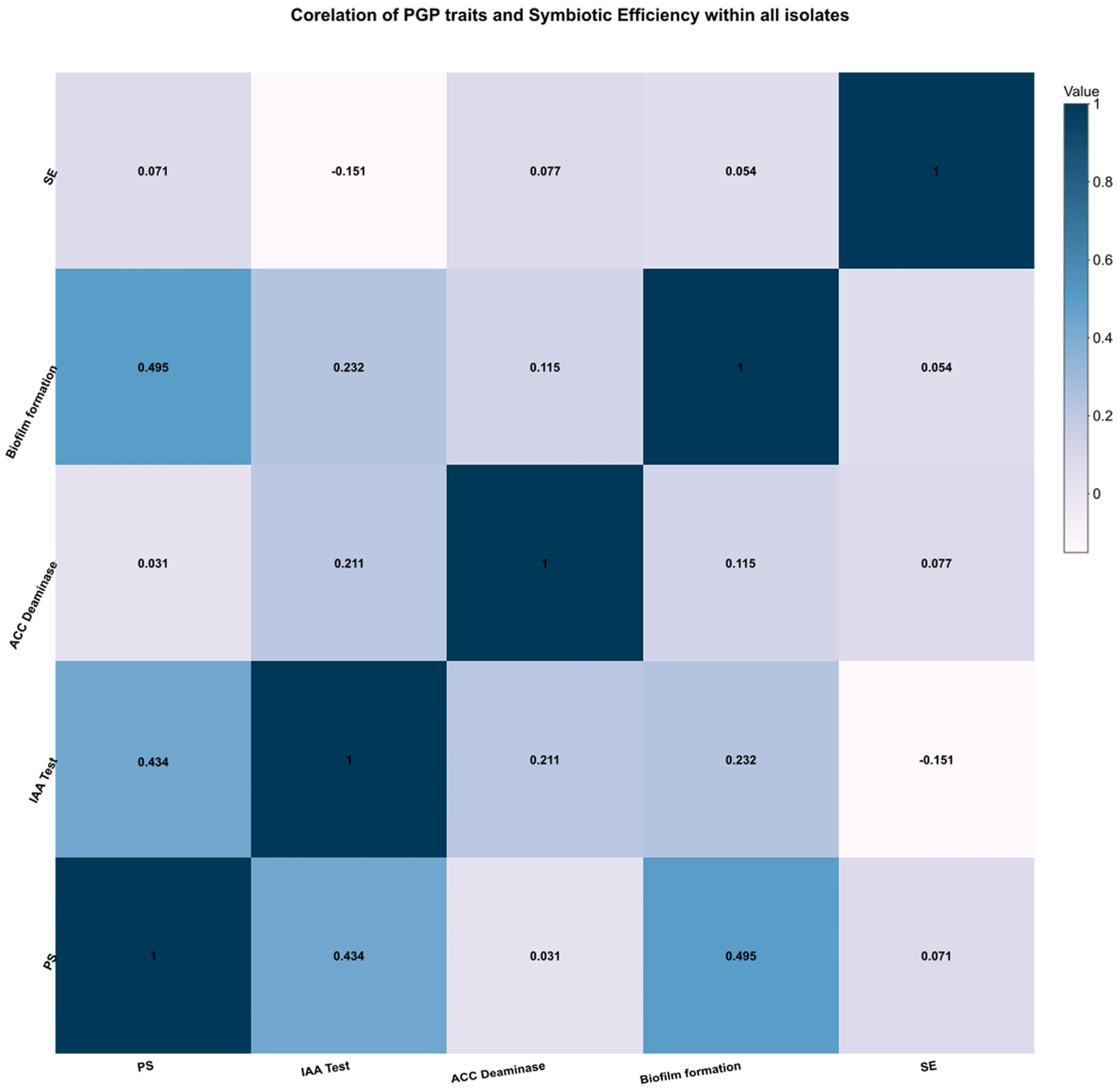
3.2. Biofilm Efficiency

Biofilm formation may have comprehensive ecological benefits to rhizobia such as assisting bacterial attachment to surfaces, nutrient acquisition, and providing protection from abiotic stresses [46]. All the tested strains were found positive to biofilm formation in which CPN1, CPN3, CPN6, CPN7, CPN8, CPN9, CPN10, CPN16, CPN21, CPN22, CPN29, CPN31, CPN32, CPN34, CPN35, CPN39, CPN45, and CPN52 were profound, while the rest of the 37 isolates were found moderate or weak in quantitative estimation of biofilm (Table 1).

3.3. Nodulation and Symbiotic Efficiency Test

Pot study of tested isolates revealed that all of the 54 strains were capable of nodulation in Cicer arietinum. The SE of all the validated nodule forming isolates were calculated and found maximum (31.01%) in CPN32, while lowest (13.91%) in CPN53, when compared with the T2 (Table 2). The shoot dry biomass of CPN32 (1.301 ± 0.165 g) and CPN53 were also proportional to SE value (0.587 g). Based on the SE and SDW results, CPN1, CPN3, CPN6, CPN7, CPN8, CPN10, CPN16, CPN21, CPN22, CPN29, CPN31, CPN32, CPN34, CPN35, CPN39, CPN45, and CPN52 formed most promising to nodulation and SE (Table 2).
The cross-nodulation results of all 54 rhizobial isolates were strictly considered as monospecific to Cicer arietinum due to failure of nodulation in Phaseolus vulgaris, Pisum sativum, Lens culinaris, and in Vigna mungo.

3.4. Carbon Utilization

Supplementary Table S1 shows the loading scores of 95 carbon sources in Biolog GN plates in the first two principal components. The principal component analysis (PCA) showed that first and the second principal component (PC1 and PC2) accounted for 20.2% and 9.6% of data variance respectively, the 2 principal component factors explained 29.9% of total variance. The most important carbon sources in differentiating among our isolates were defined as those that had high variance explained by PC1 or PC2 (>0.70). It was reported that samples located in different spaces were relevant to the ability of carbon substrates utilization. The higher the loading scores; the larger were the effects of carbon source on the principal components. Taking the microbial metabolic pathway of three major nutrients as the basic delineation standards, the carbon sources in Biolog-GN plates were divided into following types: carbohydrates and their derivatives, amino acid and their derivatives, carboxylic acids, amino acids, and miscellaneous. As mentioned in Table S1, 15 carbon sources had an impact on the first principal components (PC1) and 19 carbon sources had impacts on the second principal component. PCA analysis aggregated the isolates primarily into 2 areas that very roughly corresponded to the areas of sampling and alkalotolerance. The variability between the two groups was accounted for by the utilization of different carbon sources (Figure 2). All the isolates from the Agathuwa region distributed along the PC1 axis, and those from Darshan Nagar and Maznai distributed along the PC2 axis. Most of the isolates, showing very high alkalotolerant properties, correlated with PC1. Group 1 was formed mainly by CPN29, CPN34, CPN35 (from Agathuwa); CPN3, CPN7 (from Mau), and CPN21 (from Raunahi), which shared similar overall substrate utilization patterns, but differed from the isolates CPN6, CPN16, and CPN32 along PC 1. These stations showed a higher utilization of xylitol, L-fucose, D-psicose, hydroxy-L-proline, phenyethyl-amine, 2-aminoethanol, and L-pyroglutamic than the others. Group 2 was formed by isolates from CPN39, CPN45 (Darshan Nagar), CPN52 (Maznai), CPN1 (Mau), and CPN22 (Raunahi), which had similar carbon utilization patterns, but different from CPN10 (Mau) and CPN16 (Raunahi), along the PC2. Most of them were grouped due to their D, L-lactic acid, D-saccharic acid, turanose, bromosuccinic acid, D-galactonic acid lactone, D-gluconic acid and D, L-lactic acid utilization. Mostly all the highly alkalotolerant isolates distributed along PC1, indicating their similar carbon utilization patterns.

3.5. Molecular Identification and Sequence Typing

16S rRNA gene amplification and sequencing of the best SE strains was done. Results designating them as genus Mesorhizobium sp. ciceri with the 98% to 100% identical report, after the annotation and Blast analysis at the NCBI database. Minimum evolution bootstrap phylogeny of the homogenous gene sequences displayed the common clustering among the tested as well as type strains (Figure 3).
However, CPN16 (JX868854) was clustered as subclade with the Mesorhizobium sp. ORS 1080 (AJ295082).
Moreover, the sequences for four genes coding for gap, edD, gnD, glnD, rpoB, and nodC were simply provided for the further understanding of studied strains relationship (Figures S2, S3, S4, S4, S6, and S7). Repeated attempts to amplify and sequence the tested gene of screened isolates of chickpea-nodulating rhizobia were successful. The blast report of the above amplified gene fragments found 98% to 100% sequence similarity with genus Mesorhizobium. The gap gene sequences of all the selected strains of M. ciceri revealed great variation in their alignment. The constructed phylogenetic tree was clustered into three groups which differed from 16S rRNA phylogeny. Strains CPN21, CPN29, CPN34, and CPN39 were clustered with Mesorhizobium sp. B7, while the rest of the 12 isolates were clustered with designated Mesorhizobium species (Figure 3). Moreover, other representative rhizobium sp., such as R. undicola MG11875, R. gallicum R.4387, R. giardinni R-4385, R. tropici LGM9503, and R. leguminosarum WSM2304 was separately aligned in the tree. A similar approach was used in the edD gene phylogeny which revealed the comprehensive divergence with the various type strains of Mesorhizobium.
Moreover, the concatenated phylogenetic tree of six different genes of studied isolates and representative type strains reflected the precise diversification (Figure 4). Only M. ciceri Ca 181 closely associated with all 16 isolates, whereas other type strains (M. ciceri bio. Bis. WSM1271, M. alhagi CCNWXJ12-2, M. loti MAFF 303099, M. huakuii 7653R, M. muleiense CGMCC, M. amorphae CCNNGS0123, M. australicum WSM2073, and M. opportunistum WSM2075) were grouped into two other clades separate from the NW-IGP isolates.

4. Discussion

Bio-inoculation of C. arietinum with rhizobia is now indispensable for agriculture and environment due to its high nutritional value as well as nitrogen fixation and is entirely based on the symbiotic relationship with rhizobia which can be disturbed by several exogenous as well as indigenous factors. Hence, we investigated the actual influencing factors that are required for establishing the symbiotic relationship between C. arietinum and rhizobia. The results obtained from Gram staining, growth on YMA-congo red (CR), and YMA (BTB) as utilization of CR, and from moderately yellow to deep yellow due to their acid producing ability which is a common characteristic of fast growing Mesorhizobium species. Koskey et al. [47] reported similar observations in terms of reaction on the YMA (BTB). Indigenous soybean root-nodulating bacteria were also categorized by the use of YMA-BTB medium in Japan [48]. In light of this characteristic feature, the tested strains were further evaluated for physiological and biochemical factors. The optimum temperature range for ideal growth and activity of rhizobia is between 25 to 31 °C, which is in agreement with previous studies in chickpea rhizobia and other species [49,50]. However, some reports indicate that the maximum growth temperature is 40 °C for both M. ciceri and M. mediterraneum [51,52]. PGP attributes of test strains indicated their competence to increase the soil fertility nutritional value, in which, essentiality of phosphorus (P) as a key element for plant growth and development has been reported widely [52,53]. Phosphorous is one of the most limiting nutrients in tropical soil and only 0.1% of the total P present is available to the plants because of its chemical bonding and low solubility [52]. Interestingly, all the tested strains were able to solubilized the phosphate which proved that rhizobia had the potential to accelerate the solubilization of unavailable soil phosphate and thus could account for high efficiency of phosphorus usage. Similar research was reported previously [54,55]. It could be revealed that Mesorhizobium is useful as a reducing agent of the negative effects of soil calcification on soil P nutrition. The ability to produce IAA by tested strains can have direct effects on production of phytohormones, enzymatic activities, nitrogen accumulation in shoots and seeds, and plant biomass [56,57,58,59]. The isolated strains showed high catalase activity, similar to the observations made by Nakayama et al. [60], which indicated that these isolates could possibly be resistant to environmental, mechanical, and chemical stress. Downie A. J. [61] has observed that high activity oxidase points towards the enhanced ability of strains to effect nodulation. However, CPN11, CPM13, and CPN1 gave negative results for oxidase activity. Each isolate was found positive in NH3 production that might be responsible for the facilitation of limited bacterial growth within the nodules which is controlled by the legumes by diverse ways. Hence, NH3 worked as a major factor for controlled growth of bacteroides inside the nodule [60]. It is established well that plant associated-bacteria lowers the ethylene levels of plants through the ACC deaminase activity and helps to grow under biotic and abiotic stresses [61]. In this study, all the strains were found positive for ACC deaminase, but it was lower than previous reports of ACC-D production by Pseudomonas fluorescens and Streptomyces djakartensis TB4 [61,62,63].
Mono-specificity of tested strains were established by cross-nodulation assay against Cicer arietinum, Pisum sativum, Lens culinaris, and in Vigna mungo. The results revealed that the studied strains nodulated only C. arietinum and no cross-nodulation compatibility was observed between chickpea nodule rhizobia and other mesorhizobia leguminous plants. Thiswas in congruence with previous reports of cross nodulation tests [31,64]. Biofilm formation ability of all 54 tested strains can be considered as the key factor for symbiotic efficiency and it supports the possibility for their commercial use in stress condition [65]. Among all, 16 isolates (CPN1, CPN3, CPN6, CPN7, CPN8, CPN9, CPN10, CPN16, CPN21, CPN22, CPN29, CPN31, CPN32, CPN34, CPN35, CPN39, CPN45, and CPN52) were efficient in biofilm formation, while the rest of the 37 isolates were found moderate or weak in quantitative estimation of biofilm (Table 1). This ability to form biofilms can be positively correlated to symbiotic efficiency test of 16 most promising isolates selected in previous studies [66,67]. Wang et al. [68] also indicated that M. huakuii and M. tianshanense are efficient to biofilm formation and were reviewed by Rinaudi L.V. and Giordano W. [10]. Hence, it can be speculated that biofilm is an important factor of an efficient nodulation in chickpea.
Correlation map among the PGP traits (phosphate solubilization, IAA, ACC-deaminase production), biofilm formation, and SE revealed that biofilm is the positive factor efficient nodulation, while ACC-D is partially corelated to SE. Phosphate solubilization and IAA might be useful for the bacterial survival in saprophytic survival.
Based on the above and previous results, CPN1, CPN3, CPN6, CPN7, CPN8, CPN10, CPN16, CPN21, CPN22, CPN29, CPN32, CPN34, CPN35, CPN39, CPN45, and CPN52 were used to study the carbon utilization pattern and functional genes by Biolog and gene sequencing, respectively. Community-level carbon source utilization (Biolog) profiles have recently been introduced as means of classifying microbial communities on the basis of heterotrophic metabolism. Such a classification system might allow microbial ecologists to compare the metabolic roles of microbial communities from different environments without involving tedious isolation and identification of community members. The consumption of carbon substrates present in the Biolog system is a sensitive indicator of short-term changes in the microbial functional diversity [69]. The use of carbon substrates present in the Biolog plate was sensitive enough to detect short-term changes in the microbial functional diversity. Biolog analyses of the sixteen tested isolates revealed differences between soil microbial functional diversity from the region. Prior results showed the rhizosphere to have its own unique soil microbial community capable of producing distinctive metabolic diversity patterns of different composition, and suggested that differences in carbon source utilization can be linked to differences in carbon source availability [70]. There seems to have been no previous studies of metabolic diversity patterns in soil microbial communities in alkaline soils of the IGP region. However, a perusal of our data indicates that the driving environmental factor causing shifts in the metabolic diversity patterns seemed to be the varying amount of alkalinity in the soil.
The accurate identification is mandatory for the strains for further implication and utilization. In this study, the 16S rRNA gene was studied to identify the tested strains. Results revealed that tested strains were identical to Mesorhizobium sp. ciceri and hence, it was proposed that the NW-IGP region of India have positive abundance of C. arietinum nodulating rhizobia. Though, 16S rRNA genes are highly similar within the genus which might be a constrain for their accurate differentiation and identification [71,72]. The usefulness of housekeeping gene sequences for taxonomy and phylogeny analysis has been demonstrated widely [73]. The report displayed the currently published 30 validly species of genus Mesorhizobium in which about 50% were added in the last ten years and has been supported mainly by recA (homologous recombination protein A), atpD (ATP synthase Beta-subunit), and glnII (glutamine synthetase II) phylogenies [19]. Hence, gap, edD, gnD, glnD, rpoB, and nodC gene sequences have been amplified, sequenced, and characterized for the most stable classification. In our study, screened rhizobia were designated as Mesorhizobium on the basis of the 16S rRNA gene, while the species definition and molecular correlation among them was based on the polyphasic functional gene study. This apparent link between gap, edD, gnD, glnD, rpoB, and nodC gene sequences of tested strains and field site may be at least in part due to adaptation of the bacteria to local conditions outside the host plant, and this warrants further study.

5. Conclusions

In conclusion, the plant growth promotion and symbiotic performances of the isolates revealed the causes and highlighted the factors for efficient nodulation in chickpea. Further, carbon utilization pattern and their biofilm formation ability validated them as best bioprospecting strains in saprophytic as well as endophytic environment. The cross nodulation test has confirmed the strain specificity for chickpea as the restricted host in the NW-IGP region of India and its molecular typing support them as genus Mesorhizobium, which has been revalidated well by the concatenation approached phylogeny.

Supplementary Materials

The following are available online at https://0-www-mdpi-com.brum.beds.ac.uk/2071-1050/11/24/7216/s1, Figure S1: Geographical location of sampling sites of North west IGP Region of India. Figure S2. Phylogeny showing the relationships between Mesorhizobia and related rhizobia-based aligned gap gene sequences. Scale bars represents 0.1 substitutions per site. Significant bootstrap probability values are indicated at the branching points (only values greater than 80% over 1000 replicates are shown). Figure S3. Phylogenetic tree based on the edD gene sequences, showing the relationships between chickpea mesorhizobia isolates from the NW-IGP region and type strains. Percentage bootstrap is indicated on internal branches (1000 replicates). Scale bars represent the average number of substitutions per site. Figure S4. Phylogenetic trees based on gnD gene sequences, showing the relationships between chickpea mesorhizobia isolates from the NW-IGP region and type strains. Percentage bootstrap is indicated on internal branches (1000 replicates). Scale bars represent the average number of substitutions per site. Figure S5. Phylogenetic tree based on the glnD sequences, showing the relationships between chickpea mesorhizobia isolates from the NW-IGP region and type strains. Percentage bootstrap is indicated on internal branches (1000 replicates). Scale bars represent the average number of substitutions per site. Figure S6. Phylogenetic trees based on rpoB gene sequences. Percentage bootstrap is indicated on internal branches (1000 replicates). Scale bars represent the average number of substitutions per site. Figure S7. Phylogenetic trees based on nodC gene sequences. Percentage bootstrap is indicated on internal branches (1000 replicates). Scale bars represent the average number of substitutions per site. Table S1: Carbon substrates utilized by selected isolates in Biolog plate, significantly correlated to PC1 and PC2 (R > 0.70). Table S2: List of Primers.

Author Contributions

R.P.S., A.K.S. and R.C.D. designed the work. R.P.S. performed the experiments, R.P.S., G.M., Y.Y., D.S. and C.Z. analyzed the data, R.P.S., G.M., D.S. and C.Z. wrote the manuscript

Funding

Authors thankfully acknowledge the SERB-DST India (Grant No.: PDF/2015/000801).

Conflicts of Interest

The authors declare no conflict of interest.

References

  1. Singh, R.P.; Manchanda, G.; Maurya, I.K.; Tiwari, P.K.; Maheshwari, N.K.; Rai, A.R. Streptomyces from rotten wheat straw endowed the high Plant growth potential traits and agro-active compounds. Biocatal. Agri. Biotechnol. 2019, 17, 507–513. [Google Scholar] [CrossRef]
  2. Pinski, A.; Betekhtin, A.; Hupert-Kocurek, K.; Luis, A.J.M.; Hasterok, R. Defining the Genetic Basis of Plant-Endophytic Bacteria Interactions. Int. J. Mol. Sci. 2019, 20, 1947. [Google Scholar] [CrossRef] [PubMed] [Green Version]
  3. Gage, D.J. Infection and Invasion of Roots by Symbiotic, Nitrogen-Fixing Rhizobia during Nodulation of Temperate Legumes. Microbiol. Mol. Biol. Rev. 2004, 68, 280–300. [Google Scholar] [CrossRef] [PubMed] [Green Version]
  4. Santoyo, G.; Moreno-Hagelsieb, G.; del Carmen Orozco-Mosqueda, M.; Glick, B.R. Plant growth-promoting bacterial endophytes. Microbiol. Res. 2016, 183, 92–99. [Google Scholar] [CrossRef] [PubMed]
  5. Rovira, A.D. Rhizosphere research, 85 years of progress and frustration. In The Rhizosphere and Plant Growth; Keister, D.L., Cregan, P.B., Eds.; Kluwer Academic Publisher: Dordrecht, The Netherlands, 1991; pp. 3–13. [Google Scholar]
  6. Andrews, J.H.; Harris, R.F. The ecology and biogeography of microorganisms of plant surfaces. Annual. Rev. Phytopathol. 2000, 38, 145–180. [Google Scholar] [CrossRef]
  7. Karatan, E.; Watnick, P. Signals, regulatory networks, and materials that build and break bacterial biofilms. Microbiol. Mol. Biol. Rev. 2009, 73, 310–347. [Google Scholar] [CrossRef] [Green Version]
  8. Davey, M.E.; O’Toole, G.A. Microbial biofilms: From ecology to molecular genetics. Microbiol. Mol. Biol. Rev. 2000, 64, 847–867. [Google Scholar] [CrossRef] [Green Version]
  9. Nocelli, N.; Bogino, P.C.; Banchio, E.; Giordano, W. Roles of Extracellular Polysaccharides and Biofilm Formation in Heavy Metal Resistance of Rhizobia. Materials 2016, 9, 418. [Google Scholar] [CrossRef] [Green Version]
  10. Rinaudi, L.V.; Giordano, W. An integrated view of biofilm formation in rhizobia. FEMS Microbiol. Lett. 2010, 304, 1–11. [Google Scholar] [CrossRef] [Green Version]
  11. Yuan, H.L.; Wang, E.T.; Jiao, Y.S.; Tian, C.F.; Wang, L.; Wang, Z.J.; Guan, J.J.; Singh, R.P.; Chen, W.X.; Che, W.F. Symbiotic characteristics of Bradyrhizobium diazoefficiens USDA 110 mutants associated with shrubby sophora (Sophora flavescens) and soybean (Glycine max). Microbiol. Res. 2018, 214, 19–27. [Google Scholar]
  12. Sturz, A.; Christie, B.; Nowak, J. Bacterial endophytes, potential role in developing sustainable systems of crop production. CRC Crit. Rev. Plant Sci. 2000, 19, 1–30. [Google Scholar] [CrossRef]
  13. Weyens, N.; Van-der, L.D.; Taghavi, S.; Vangronsveld, J. Phytoremediation, plant endophyte partnerships take the challenge. Curr. Opin. Biotechnol. 2009, 20, 248–254. [Google Scholar] [CrossRef] [PubMed]
  14. Brígido, C.; Singh, S.; Menéndez, E.; Tavares, M.J.; Glick, B.R.; Félix, M.R.; Oliveira, S.; Carvalho, M. Diversity and Functionality of Culturable Endophytic Bacterial Communities in Chickpea Plants. Plants 2019, 8, 42. [Google Scholar] [CrossRef] [PubMed] [Green Version]
  15. Matos, I.; Schröder, E.C. Strain selection for pigeon pea Rhizobium under greenhouse conditions. Plant Soil. 1989, 116, 19–22. [Google Scholar] [CrossRef]
  16. FAOSTAT. FAO (Food and Agricultural Organization of the United Nations). Available online: http://faostat.fao.org (accessed on 11 December 2014).
  17. Upadhyaya, H.D.; Kashiwagi, J.; Varshney, R.K.; Gaur, P.M.; Saxena, K.B.; Krishnamurthy, L.; Gowda, C.L.L.; Pundir, R.P.S.; Chaturvedi, S.K.; Basu, P.S.; et al. Phenotyping chickpeas and pigeonpeas for adaptation to drought. Front. Physiol. 2012, 3, 179. [Google Scholar] [CrossRef] [Green Version]
  18. Dudeja, S.S.; Nidhi. Molecular diversity of rhizobial and non rhizobial bacteria from nodules of cool season legumes. In Biotechnology: Prospects and Applications; Salar, R.K., Gahlawat, S.K., Siwach, P., Duhan, J.S., Eds.; Springer: Berlin/Heidelberg, Germany, 2014. [Google Scholar] [CrossRef]
  19. Pérez-Yépez, J.; Armas-Capote, N.; Velázquez, E.; Pérez-Galdona, R.; Rivas, R.; León-Barrios, M. Evaluation of seven housekeeping genes for multilocus sequence analysis of the genus Mesorhizobium: Resolving the taxonomic affiliation of the Cicer canariense rhizobia. Syst. Appl. Microbiol. 2014, 37, 553–559. [Google Scholar] [CrossRef]
  20. Nour, S.H.; Fernandez, M.P.; Normand, P.; Cleyet-Marel, J.C. Rhizobium ciceri sp. nov., consisting of strains that nodulate chickpeas (Cicer arietinum L.). Inter. J. Syst. Bacteriol. 1994, 44, 511–522. [Google Scholar] [CrossRef] [Green Version]
  21. Nour, S.M.; Cleyet-Marel, J.; Normand, C.P.; Fernandez, M.P. Genomic heterogeneity of strains nodulating chickpeas (Cicer arietinum L.) and description of Rhizobium mediterraneum sp. nov. Inter. J. Syst. Bacteriol. 1995, 45, 640–648. [Google Scholar] [CrossRef] [Green Version]
  22. Zgadzaj, R.; James, E.K.; Kelly, S.; Kawaharada, Y.; de Jonge, N.; Jensen, D.B.; Madsen, L.H.; Radutoiu, S. A Legume Genetic Framework Controls Infection of Nodules by Symbiotic and Endophytic Bacteria. PLoS Genet. 2015, 11, 1–21. [Google Scholar] [CrossRef] [Green Version]
  23. Andrews, M.; Andrews, M.E. Specificity in Legume-Rhizobia Symbioses. Int. J. Mol. Sci. 2017, 18, 705. [Google Scholar] [CrossRef] [Green Version]
  24. Geurts, R.; Franssen, H. Signal transduction in Rhizobium induced nodule formation. Plant Physiol. 1996, 112, 447–453. [Google Scholar] [CrossRef] [PubMed] [Green Version]
  25. Cabanes, D.; Boistard, P.; Batut, J. Identification of Sinorhizobium meliloti genes regulated during symbiosis. J. Bacteriol. 2000, 182, 3632–3637. [Google Scholar] [CrossRef] [PubMed] [Green Version]
  26. Flores-Félix, J.D.; Carro, L.; Velázquez, E.; Valverde, A.; Cerda-Castillo, E.; García-Fraile, P.; Rivas, R. Phyllobacterium endophyticum sp. nov., isolated from nodules of Phaseolus vulgaris. Int. J. Syst. Evol. Microbiol. 2012, 63, 821–826. [Google Scholar] [CrossRef] [PubMed] [Green Version]
  27. Vincent, J.M. A Manual for the Practical Study of Root-Nodule Bacteria; IBP Handbook 15; Blackwell: Oxford, UK, 1970. [Google Scholar]
  28. Zhang, J.J.; Yang, X.; Chen, G.; de Lajudie, P.; Singh, R.P.; Wang, E.T.; Chen, W.F. Mesorhizobium muleiense and Mesorhizobium gsp. nov. are symbionts of Cicer arietinum L. in alkaline soils of Gansu, Northwest China. Plant Soil 2016, 410, 103–112. [Google Scholar] [CrossRef]
  29. Zhang, J.J.; Jing, X.Y.; de Lajudie, P.; Ma, C.; He, P.X.; Singh, R.P.; Chen, W.F.; Wang, E.T. Association of white clover (Trifolium repens L.) with rhizobia of sv. trifolii belonging to three genomic species in alkaline soils in North and East China. Plant Soil 2016, 407, 417–427. [Google Scholar]
  30. Gibson, A.H. Evaluation of nitrogen fixation by legumes in the greenhouse and growth chamber. In Symbiotic Nitrogen Fixation Technology; Gibson, A.H., Ed.; Marcel Dekker: New York, NY, USA, 1987; pp. 321–363. [Google Scholar]
  31. Nandasena, K.G.; O’Hara, G.W.; Tiwari, R.P.; Yates, R.J.; Kishinevsky, B.D.; Howieson, J.G. Symbiotic relationships and root nodule ultrastructure of the pasture legume Biserrula pelecinus L.; a new legume in agriculture. Soil Biol. Biochem. 2004, 36, 1309–1317. [Google Scholar] [CrossRef]
  32. Pikovskya, R.I. Mobilization of phosphorus in soil in connection with the vital activity of some microbial species. Microbiology 1948, 17, 362–370. [Google Scholar]
  33. Vazquez, P.; Holguin, G.; Puente, M.E.; Lopez-Cortes, A.; Bashan, Y. Phosphate solubilizing microorganisms associated with the rhizosphere of Mangroves growing in a semiarid coastal lagoon. Biol. Fertil. Soils 2000, 30, 460–468. [Google Scholar] [CrossRef]
  34. Khanmna, S.; Yokota, A.; Lumyong, S. Actinomycetes isolated from medicinal plant rhizosphere soils: Diversity and screening of antifungal compounds, indole-3-acetic acid and siderophore production. World J. Microbiol. Biotechnol. 2009, 25, 649–655. [Google Scholar] [CrossRef]
  35. Smibert, R.M.; Krieg, N.R. Phenotypic characterization. In Methods for General and Molecular Bacteriology; Gerhardt, P., Murray, R.G.E., Wood, W.A., Krieg, N.R., Eds.; American Society for Microbiology: Washington, DC, USA, 1994; pp. 607–654. [Google Scholar]
  36. Glick, B.R. Bacteria with ACC deaminase can promote plant growth and help to feed the world. Microbiol. Res. 2014, 169, 30–39. [Google Scholar] [CrossRef]
  37. Mirani, Z.A.; Jamil, N. Effect of sub-lethal doses of vancomycin and oxacillin on biofilm formation by vancomycin intermediate resistant Staphylococcus aureus. J. Basic Microbiol. 2011, 51, 191–195. [Google Scholar] [CrossRef] [PubMed]
  38. Garland, J.L.; Mills, A.L. Classification and characterization of heterotrophic microbial communities on the basis of patterns of community-level sole-carbon-source utilization. Appl. Environ. Microbiol. 1991, 57, 2351–2359. [Google Scholar] [PubMed]
  39. Garland, J.L. Analytical approaches to the characterization of samples of microbial communities using patterns of potential C source utilization. Soil Biol. Biochem. 1996, 28, 213–221. [Google Scholar] [CrossRef]
  40. Van Berkum, P.; Badri, Y.; Elia, P.; Aouani, M.E.; Eardly, B.D. Chromosomal and symbiotic relationships of rhizobia nodulating Medicago truncatula and M. laciniata. Appl. Environ. Microbiol. 2007, 73, 7597–7604. [Google Scholar] [CrossRef] [PubMed] [Green Version]
  41. Tamura, K.; Stecher, G.; Peterson, D.; Filipski, A.; Kumar, S. MEGA6, molecular evolutionary genetics analysis version 6.0. Mol. Biol. Evol. 2013, 30, 2725–2729. [Google Scholar] [CrossRef] [PubMed] [Green Version]
  42. Librado, P.; Rozas, J. DnaSP v5: A software for comprehensive analysis of DNA polymorphism data. Bioinformatics 2009, 25, 1451–1452. [Google Scholar] [CrossRef] [PubMed] [Green Version]
  43. Tan, H.W.; Heenan, P.B.; De, M.; Sofie, E.; Willems, A.; Andrews, M. Diverse novel mesorhizobia nodulate New Zealand native Sophora species. Syst. Appl. Microbiol. 2015, 38, 91–98. [Google Scholar] [CrossRef] [Green Version]
  44. Degefu, T.; Wolde-meskel, E.; Woliy, K.; Frostegård, A. Phylogenetically diverse groups of Bradyrhizobium isolated from nodules of tree and annual legume species growing in Ethiopia. Syst. Appl. Microbiol. 2017, 40, 205–214. [Google Scholar] [CrossRef]
  45. Villesen, P. FaBox, an online toolbox for Fasta sequences. Mol. Ecol. Res. 2007, 7, 965–968. [Google Scholar] [CrossRef]
  46. Duncan, D.B. Multiple range and multiple F-tests. Biometrics 1955, 11, 1–42. [Google Scholar] [CrossRef]
  47. Koskey, G.; Mburu, S.W.; Kimiti, J.M.; Ombori, O.; Maingi, J.M.; Njeru, E.M. Genetic Characterization and Diversity of Rhizobium Isolated from Root Nodules of Mid-Altitude Climbing Bean (Phaseolus vulgaris L.) Varieties. Front. Microbiol. 2018, 9, 968. [Google Scholar] [CrossRef] [PubMed] [Green Version]
  48. Saeki, Y.; Aimi, N.; Tsukamoto, S.; Yamakawa, T.; Nagatomo, Y.; Akao, S. Diversity and geographical distribution of indigenous soybean-nodulating bradyrhizobia in Japan. Soil Sci. Plant Nutri. 2006, 52, 418–426. [Google Scholar] [CrossRef]
  49. Somasegaran, P.; Hoben, H.J. Hand Book for Rhizobia; Springer: New York, NY, USA, 1994. [Google Scholar]
  50. Rodrigues, C.; Laranjo, M.; Oliveira, S. Effect of heat and pH stress in the growth of chickpea mesorhizobia. Curr. Microbiol. 2006, 53, 1–7. [Google Scholar] [CrossRef] [PubMed] [Green Version]
  51. Nour, S.M.; Cleyet-Marel, J.B.; Effosse, C.D.A.; Fernandez, M.P. Genotypic and phenotypic diversity of Rhizobium isolated from chickpea (Cicer arietinum L.). Can. J. Microbiol. 1994, 40, 345–354. [Google Scholar] [CrossRef] [PubMed]
  52. Peix, A.; Rivas-Boyero, A.A.; Mateos, P.F.; Rodriguez-Barrueco, C.; Martınez-Molina, E.; Velazquez, E. Growth promotion of chickpea and barley by a phosphate solubilizing strain of Mesorhizobium mediterraneum under growth. Soil Biol. Biochem. 2001, 33, 103–110. [Google Scholar] [CrossRef]
  53. Chen, L.; Luo, S.; Xiao, X.; Guo, H.; Chen, J.; Wan, Y.; Li, B.; Xu, T.; Xi, Q.; Rao, C.; et al. Application of plant growth-promoting endophytes (PGPE) isolated from Solanum nigrum L. for phytoextraction of Cd-polluted soils. Appl. Soil Ecol. 2010, 46, 383–389. [Google Scholar] [CrossRef]
  54. Pravin, V.; Rosazlin, A.; Tumirah, K.; Salmah, I.; Amru, N.B. Role of Plant Growth Promoting Rhizobacteria in Agricultural Sustainability—A Review. Molecules 2016, 21, 573. [Google Scholar]
  55. Chandra, S.; Choure, K.; Dubey, R.C.; Maheshwari, D.K. Rhizosphere competent Mesorhizobium loti MP6 induces root hair curling, inhibits Sclerotinia sclerotiorum and enhances growth of Indian mustard (Brassica campestris). Braz. J. Microbiol. 2007, 38, 124–130. [Google Scholar] [CrossRef]
  56. Adnan, M.; Shah, Z.; Fahad, S.; Arif, M.; Alam, M.; Khan, I.A.; Mian, I.A.; Basir, A.; Ullah, H.; Arshad, M.; et al. Phosphate-Solubilizing Bacteria Nullify the Antagonistic Effect of Soil Calcifcation on Bioavailability of Phosphorus in Alkaline Soils. Sci. Rep. 2017, 7, 16131. [Google Scholar] [CrossRef]
  57. Gupta, C.P.; Dubey, R.C.; Maheshwari, D.K. Plant growth enhancement and suppression of Macrophomina phaseolina causing charcoal rot of peanut by fluorescent Pseudomonas. Biol. Fertil. Soil. 2002, 35, 399–405. [Google Scholar]
  58. María, P.F.I.; Valentine, A. Enhanced Plant Performance in Cicer arietinum L. Due to the Addition of a Combination of Plant Growth-Promoting Bacteria. Agriculture 2017, 7, 40. [Google Scholar]
  59. Nakayama, M.; Nakajima-Kambe, T.; Katayama, H.; Higuchi, K.; Kawasaki, Y.; Fuji, R. High catalase production by Rhizobium radiobacter strain 2-1. J. Biosci. Bioeng. 2008, 106, 554–558. [Google Scholar] [CrossRef] [PubMed] [Green Version]
  60. Downie, A.J. Legume nodulation. Curr. Biol. 2014, 24, R184–R190. [Google Scholar] [CrossRef] [PubMed] [Green Version]
  61. Glick, B.R.; Todorovic, B.; Czarny, J.; Cheng, Z.; Duan, J.; Mc-Conkey, B. Promotion of plant growth by bacterial ACC deaminase. Crit. Rev. Plant Sci. 2007, 26, 227–242. [Google Scholar] [CrossRef]
  62. Anwar, S.; Ali, B.; Sajid, I. Screening of Rhizospheric Actinomycetes for Various In-vitro and In-vivo Plant Growth Promoting (PGP) Traits and for Agroactive Compounds. Front. Microbiol. 2016, 7, 1334. [Google Scholar] [CrossRef] [Green Version]
  63. Mahmood, A.; Athar, M. Cross inoculation studies: Response of Vigna mungo to inoculation with rhizobia from tree legumes growing under arid Environment. Int. J. Environ. Sci. Technol. 2008, 5, 135–139. [Google Scholar] [CrossRef] [Green Version]
  64. Jiao, Y.S.; Liu, Y.H.; Yan, H.; Wang, E.T.; Tian, C.F.; Chen, W.X.; Guo, B.L.; Chen, W.F. Rhizobial diversity and nodulation characteristics of the extremely promiscuous legume Sophora flavescens. Mol. Plant-Microbe Interact. 2015, 28, 1338–1352. [Google Scholar] [CrossRef] [Green Version]
  65. Singh, R.P.; Manchanda, G.; Singh, R.N.; Srivastava, A.K.; Dubey, R.C. Selection of alkalotolerant and symbiotically efficient chickpea nodulating rhizobia from North-West Indo Gangetic Plains. J. Basic Microbiol. 2016, 56, 14–25. [Google Scholar] [CrossRef]
  66. Fujishige, N.A.; Kapadia, N.N.; De Hoff, P.L.; Hirsch, A.M. Investigations of Rhizobium biofilm formation. FEMS Microbiol. Ecol. 2006, 56, 195–206. [Google Scholar] [CrossRef] [Green Version]
  67. Wang, H.; Zhong, Z.; Cai, T.; Li, S.; Zhu, J. Heterologous overexpression of quorum-sensing regulators to study cell density-dependent phenotypes in a symbiotic plant bacterium Mesorhizobium huakui. Arch. Microbiol. 2004, 182, 520–525. [Google Scholar] [CrossRef]
  68. Wang, D.; Xu, A.; Elmerich, C.; Ma, L.Z. Biofilm formation enables free-living nitrogen-fixing rhizobacteria to fix nitrogen under aerobic conditions. ISME J. 2017, 11, 1602–1613. [Google Scholar] [CrossRef] [PubMed] [Green Version]
  69. Govaerts, B.; Mezzalama, M.; Unno, Y.; Sayre, K.D.; Luna-Guido, M.; Vanherck, K.; Dendooven, L.; Deckers, J. Influence of tillage, residue management, and crop rotation on soil microbial biomass and catabolic diversity. Appl. Soil Ecol. 2007, 37, 18–30. [Google Scholar] [CrossRef]
  70. Grayston, S.J.; Wang, S.Q.; Campbell, C.D.; Edwards, A.C. Selective influence of plant species on microbial diversity in the rhizosphere. Soil Biol. Biochem. 1998, 30, 369–378. [Google Scholar] [CrossRef]
  71. Dighe, A.S.; Jangid, K.; González, J.M.; Pidiyar, V.J.; Patole, M.S.; Ranade, D.R.; Shouche, Y.S. Comparison of 16S rRNA gene sequences of genus Methanobrevibacter. BMC Microbiol. 2004, 4, 20. [Google Scholar] [CrossRef] [Green Version]
  72. Sato, M.; Miyazaki, K. Phylogenetic Network Analysis Revealed the Occurrence of Horizontal Gene Transfer of 16S rRNA in the Genus Enterobacter. Front. Microbiol. 2017, 8, 2225. [Google Scholar] [CrossRef]
  73. Elbannaa, K.; Elbadryb, M.; Gamal-Eldina, H. Genotypic and phenotypic characterization of rhizobia that nodulate snap bean (Phaseolus vulgaris L.) in Egyptian soils. Syst. Appl. Microbiol. 2009, 32, 522–530. [Google Scholar] [CrossRef]
Figure 1. Correlation map of PGP traits and symbiotic efficiency within all isolates.
Sustainability 11 07216 g001
Figure 2. PC1 plotted against PC2 for Biolog data collected.
Sustainability 11 07216 g002
Figure 3. 16S rRNA gene phylogeny.
Sustainability 11 07216 g003
Figure 4. Concatenated phylogeny of gap, edD, gnD, glnD, rpoB, and nodC gene.
Sustainability 11 07216 g004
Table 1. Eco-physiological, biochemical, and plant growth-promoting factors of chickpea nodule rhizobia.
IsolatesGrowth ConditionPS (S.I. Value)IAA TestOxidase TestCatalase TestAmmonia ProductionACC Deaminase (mmol−l)Biofilm Formation (OD)
IC-2058MS3.92 ± 0.0499.54 ± 0.07+++0.91 ± 0.080.487 ± 0.04
IC-2018MS3.86 ± 0.0791.71 ± 0.06+++1.03 ± 0.110.565 ± 0.02
CPN1MS3.63 ± 0.01100.24 ± 0.09+++1.57 ± 0.040.678 ± 0.05
CPN2MS3.12 ± 0.0379.31 ± 0.08+++0.84 ± 0.060.405 ± 0.05
CPN3MF3.33 ± 0.0480.22 ± 0.07+++1.04 ± 0.020.733 ± 0.04
CPN4MS3.09 ± 0.0494.12 ± 0.05+++0.87 ± 0.120.532 ± 0.06
CPN5MS2.92 ± 0.0098.61 ± 0.07-++1.02 ± 0.040.219 ± 0.04
CPN6MS3.41 ± 0.01103.90 ± 0.06+++1.12 ± 0.060.374 ± 0.07
CPN7MS3.29 ± 0.0186.31 ± 0.04+++1.05 ± 0.020.752 ± 0.04
CPN8MS4.17 ± 0.07104.52 ± 0.09+++1.24 ± 0.120.502 ± 0.02
CPN9MS3.99 ± 0.0597.24 ± 0.11+++1.32 ± 0.050.785 ± 0.05
CPN10MS3.56 ± 0.0593.61 ± 0.09+++1.16 ± 0.050.692 ± 0.02
CPN11MS3.68 ± 0.0191.76 ± 0.07-++1.27 ± 0.020.775 ± 0.03
CPN12MS3.14 ± 0.0386.35 ± 0.11+++1.33 ± 0.080.523 ± 0.04
CPN13MS3.51 ± 0.0797.58 ± 0.06-++1.21 ± 0.060.293 ± 0.01
CPN14MS2.97 ± 0.0193.41 ± 0.12-++1.31 ± 0.050.465 ± 0.04
CPN15MS2.88 ± 0.0795.24 ± 0.07+++1.42 ± 0.140.504 ± 0.08
CPN16MF3.69 ± 0.0899.47 ± 0.05+++0.88 ± 0.040.802 ± 0.06
CPN17MS3.05 ± 0.0498.66 ± 0.06+++0.67 ± 0.020.514 ± 0.06
CPN18MS3.19 ± 0.0390.05 ± 0.06+++1.09 ± 0.020.308 ± 0.02
CPN19MS3.25 ± 0.0897.11 ± 0.09+++1.18 ± 0.060.347 ± 0.03
CPN20MS3.07 ± 0.0582.41 ± 0.08+++1.19 ± 0.070.289 ± 0.03
CPN21MS3.61 ± 0.00102.57 ± 0.08+++0.98 ± 0.050.872 ± 0.05
CPN22MS3.77 ± 0.02105.38 ± 0.07+++1.13 ± 0.030.759 ± 0.05
CPN23MS3.47 ± 0.0799.84 ± 0.05+++1.28 ± 0.050.384 ± 0.04
CPN24MS3.33 ± 0.0284.61 ± 0.05+++1.09 ± 0.040.405 ± 0.02
CPN25MS3.43 ± 0.0289.73 ± 0.07+++1.09 ± 0.050.372 ± 0.02
CPN26MS3.28 ± 0.0490.51 ± 0.07+++1.22 ± 0.040.325 ± 0.04
CPN27MS3.16 ± 0.07100.82 ± 0.05+++1.28 ± 0.060.217 ± 0.05
CPN28MS3.84 ± 0.03100.25 ± 0.11+++0.86 ± 0.020.374 ± 0.05
CPN29MS4.01 ± 0.0698.67 ± 0.12+++1.34 ± 0.050.805 ± 0.08
CPN30MS3.87 ± 0.0499.58 ± 0.11+++1.09 ± 0.020.541 ± 0.04
CPN31MS3.61 ± 0.0681.64 ± 0.07+++0.96 ± 0.040.581 ± 0.03
CPN32MS4.08 ± 0.06107.41 ± 0.09+++1.08 ± 0.0.60.791 ± 0.05
CPN33MS3.63 ± 0.0589.33 ± 0.05+++0.84 ± 0.060.537 ± 0.04
CPN34MS4.13 ± 0.0692.56 ± 0.06+++1.06 ± 0.020.861 ± 0.06
CPN35MS3.5 ± 0.0691.63 ± 0.06+++1.18 ± 0.080.837 ± 0.02
CPN36MS3.25 ± 0.0187.64 ± 0.05+++1.12 ± 0.060.465 ± 0.03
CPN37MS3.61 ± 0.0690.35 ± 0.08+++1.04 ± 0.060.438 ± 0.06
CPN38MS3.17 ± 0.0388.62 ± 0.06+++0.87 ± 0.040.483 ± 0.05
CPN39MS3.34 ± 0.03100.71 ± 0.09+++1.34 ± 0.070.806 ± 0.04
CPN40MS3.01 ± 0.0185.61 ± 0.06+++0.78 ± 0.020.308 ± 0.09
CPN41MS2.95 ± 0.0387.24 ± 0.05+++1.03 ± 0.060.355 ± 0.07
CPN42MS3.62 ± 0.05101.13 ± 0.06+++1.05 ± 0.080.428 ± 0.09
CPN43MS3.19 ± 0.0283.61 ± 0.07+++1.05 ± 0.120.611 ± 0.06
CPN44MS3.55 ± 0.0189.34 ± 0.05+++0.93 ± 0.060.489 ± 0.05
CPN45MS3.29 ± 0.0099.65 ± 0.06+++1.02 ± 0.080.785 ± 0.07
CPN46MS2.87 ± 0.0392.04 ± 0.08+++1.14 ± 0.060.523 ± 0.06
CPN47MS2.96 ± 0.0393.84 ± 0.12+++1.07 ± 0.050.429 ± 0.05
CPN48MS3.41 ± 0.0288.06 ± 0.09+++0.86 ± 0.030.367 ± 0.03
CPN49MS3.64 ± 0.0190.71 ± 0.05+++1.22 ± 0.090.356 ± 0.04
CPN50MS3.06 ± 0.0487.42 ± 0.06+++1.14 ± 0.120.503 ± 0.04
CPN51MS3.88 ± 0.0590.21 ± 0.08+++0.96 ± 0.050.486 ± 0.06
CPN52MS4.33 ± 0.03101.08 ± 0.09+++1.01 ± 0.050.806 ± 0.05
CPN53MS3.44 ± 0.0097.33 ± 0.05+++0.97 ± 0.020.539 ± 0.07
CPN54MS3.21 ± 0.0191.82 ± 0.11+++1.04 ± 0.050.211 ± 0.04
Characters are scored as, +: all positive; −: all negative; MF: moderately fast; MS: moderately slow; PS: phosphate solubilization; IAA: indole acetic acid; OD: optical density. Each value is the mean of five replicates.
Table 2. Plant chlorophyll content (mg g−1 F.W. of leaves), nodule number (NN), shoot dry weight (SDW, g plant−1), and symbiotic effectiveness (SE, %) of all the isolates.
S.No.IsolatesChlorophyll Content (mg g−1 F.W. of Leaves)NNSDW (g Plant-1)SE (%)
1.T1 (Control)0.806 ± 0.4440.419 ± 1.528
2.T2 (Control) N-Supplemented1.111 ± 0.1214.217 ± 0.639100.00
3.IC-20581.047 ± 0.45915.31 ± 1.2241.127 ± 0.55826.72
4.IC-20180.986 ± 0.19412.21 ± 1.3210.927 ± 0.16121.98
5.CPN11.240 ± 0.15222.81 ± 0.4231.203 ± 0.18328.52
6.CPN20.958 ± 0.4087.37 ± 1.7290.603 ± 0.19914.29
7.CPN31.121 ± 0.05711.66 ± 2.8541.186 ± 0.24128.12
8.CPN40.919 ± 0.1586.24 ± 1.8520.884 ± 0.11420.96
9.CPN50.911 ± 0.0086.51 ± 2.0100.653 ± 0.083815.48
10.CPN61.126 ± 0.05817.38 ± 0.8741.119 ± 0.29226.53
11.CPN71.199 ± 0.12213.57 ± 0.9731.288 ± 0.54130.54
12.CPN81.201 ± 0.00814.91 ± 1.2331.303 ± 0.34630.89
13.CPN90.906 ± 0.0027.61 ± 0.9350.971 ± 0.15423.02
14.CPN101.128 ± 0.11618.51 ± 1.9001.266 ± 0.57330.02
15.CPN110.891 ± 0.0489.22 ± 1.3120.639 ± 0.11315.15
16.CPN120.931 ± 0.04312.42 ± 09410.997 ± 0.15723.64
17.CPN130.933 ± 0.03211.61 ± 2.0780.999 ± 0.20223.68
18.CPN140.975 ± 0.0388.33 ± 2.3331.107 ± 0.24426.25
19.CPN150.897 ± 0.0559.33 ± 0.3340.769 ± 0.11818.23
20.CPN161.217 ± 0.33016.00 ± 1.1541.176 ± 0.42127.88
21.CPN170.968 ± 0.39311.66 ± 0.8810.894 ± 0.05021.19
22.CPN180.922 ± 0.05113.24 ± 1.6220.931 ± 0.15722.07
23.CPN190.938 ± 0.19615.29 ± 2.2991.091 ± 0.47225.87
24.CPN200.871 ± 0.0556.33 ± 1.7630.646 ± 0.56315.31
25.CPN211.211 ± 0.20219.88 ± 3.4891.207 ± 0.20028.62
26.CPN221.113 ± 0.28917.79 ± 1.3991.198 ± 0.43528.40
27.CPN230.839 ± 0.0856.33 ± 1.7630.693 ± 0.20116.43
28.CPN240.854 ± 0.09610.41 ± 1.6910.707 ± 0.06416.76
29.CPN250.819 ± 0.0917.33 ± 0.8810.697 ± 0.11216.52
30.CPN260.824 ± 0.0696.33 ± 1.3340.655 ± 0.22715.53
31.CPN270.971 ± 0.02814.66 ± 1.3331.009 ± 0.05623.92
32.CPN280.873 ± 0.0437.66 ± 1.8550.687 ± 0.04716.29
33.CPN291.218 ± 0.39118.33 ± 1.7631.200 ± 0.22428.45
34.CPN300.872 ± 0.1437.66 ± 1.3340.683 ± 0.09816.19
35.CPN310.962 ± 0.32410.67 ± 1.4520.916 ± 0.14021.72
36.CPN321.298 ± 0.16627.65 ± 1.3321.301 ± 0.16531.01
37.CPN330.888 ± 0.0729.34 ± 1.2010.982 ± 0.37023.28
38.CPN341.271 ± 0.17317.66 ± 1.8561.281 ± 0.21430.37
39.CPN351.259 ± 0.06216.00 ± 1.1541.266 ± 0.29930.02
40.CPN360.963 ± 0.36111.34 ± 2.9050.916 ± 0.18521.72
41.CPN370.847 ± 0.0838.00 ± 1.5270.729 ± 0.10017.28
42.CPN380.855 ± 0.02310.67 ± 0.3340.773 ± 0.09318.33
43.CPN391.115 ± 0.10513.50 ± 1.7561.181 ± 0.37128.00
44.CPN400.908 ± 0.21512.67 ± 1.2010.975 ± 0.19623.12
45.CPN410.914 ± 0.02910.34 ± 0.6670.862 ± 0.22320.44
46.CPN420.897 ± 0.05110.34 ± 2.6030.904 ± 0.18221.43
47.CPN430.895 ± 0.05310.34 ± 2.3340.899 ± 0.12321.43
48.CPN440.866 ± 0.0609.67 ± 0.8810.788 ± 0.20518.68
49.CPN451.171 ± 0.05013.65 ± 1.4521.152 ± 0.41327.31
50.CPN460.937 ± 0.0267.67 ± 1.4520.996 ± 0.35123.61
51.CPN470.841 ± 0.0819.32 ± 1.4530.711 ± 0.14516.86
52.CPN480.819 ± 0.0346.65 ± 1.8560.591 ± 0.09914.01
53.CPN490.842 ± 0.2116.40 ± 1.7630.603 ± 0.22714.29
54.CPN500.837 ± 0.0227.34 ± 0.6670.597 ± 0.24614.15
55.CPN510.869 ± 0.0229.66 ± 1.3340.759 ± 0.14117.99
56.CPN521.118 ± 0.05813.34 ± 2.4031.163 ± 0.35427.57
57.CPN530.803 ± 0.0925.34 ± 1.4520.587 ± 0.20213.91
58.CPN540.972 ± 0.05313.67 ± 3.1790.913 ± 0.30321.65

