Article

Valorization of Banana and Red Beetroot Peels: Determination of Basic Macrocomponent Composition, Application of Novel Extraction Methodology and Assessment of Biological Activity In Vitro

1
Faculty of Food Technology and Biotechnology, University in Zagreb, Pierottijeva 6, 10 000 Zagreb, Croatia
2
Faculty of Food Technology, Josip Juraj Strossmayer University of Osijek, Franje Kuhača 20, 31 000 Osijek, Croatia
3
Institute for Medical Research and Occupational Health, Ksaverska Cesta 2, 10 000, Zagreb, Croatia
*
Author to whom correspondence should be addressed.
Sustainability 2020, 12(11), 4539; https://0-doi-org.brum.beds.ac.uk/10.3390/su12114539
Submission received: 10 May 2020 / Revised: 31 May 2020 / Accepted: 1 June 2020 / Published: 3 June 2020
(This article belongs to the Special Issue Sustainable Food Processing)

Abstract

:
The nutritional and bioactive content of banana and red beetroot peels was investigated. The basic macrocomponent composition was determined using standard AOAC (Association of Official Analytical Chemists) methods, while the recovery efficiency of bioactive compounds was investigated using conventional and innovative extraction techniques (subcritical water extraction, ultrasound- and microwave-assisted extraction). Extracts were analyzed for biological effects in vitro on human hepatic, tongue and colon cancer cell lines. A macrocomponent analysis revealed a notable amount of dietary fiber in banana and beetroot peels (39.0 and 33.6% dmb) and a relatively high content of protein in beetroot peel (18.3% dmb). Regarding the micronutrients-minerals, banana and beetroot peels were shown to be a very good source of potassium (75.06 and 41.86 mg g−1 dmb). Both extracts of banana and beetroot peels obtained by conventional extraction - decoction (100 °C, 20 min) exhibited the highest total phenolic content and antioxidant capacity. Additionally, in banana peel, these extracts were the richest in dopamine content (12.63 mg g−1 dmb). Extraction by infusion (80 °C, 30 min) yielded a beetroot peel extract with the highest total betacyanin content (9.80 mg g−1 dmb). Biological effects in vitro were dose- and time-dependent, as well as influenced by the presence of polysaccharides.

1. Introduction

For the period between 2009 and 2050, the world population is predicted to grow by a third, which will also result in a substantial increase in food demand [1]. Therefore, the agro-industrial sectors, besides providing food to the whole population, are searching for the new eco-friendly and sustainable forms of food production, including agro-industrial waste reutilization. Agro-industrial waste includes stalks, stems, leaves, roots, molasses, husks, peels, etc., that are known to contain high-value ingredients, such as nutrients and bioactive compounds [2], pointing to wider possibilities of application in the food industry.
Banana (Musaceae) is a tropical fruit available throughout the whole year and, after tomato, the most consumed fruit in the world [3], with an annual production of 115 million tons in 2018 [4]. Since approximately 30% of banana fruit is comprised of inedible peel [5], it is obvious that, on a global scale, a lot of peel waste is generated annually in fruit industries, as well as in households. Regarding the chemical composition, carbohydrates and crude fibers make up most of the banana peel’s dry matter, but a significant amount of proteins, potassium, essential amino acids and polyunsaturated fatty acids can also be found [6,7,8]. Banana peel also represents a great source of phenolic compounds, like gallocatechin, and catecholamines, especially dopamine, whose content was found to be much higher in the peel compared to the banana pulp [9,10].
Red beetroot (Beta vulgaris) has been listed in the top 10 vegetables with the highest antioxidant activity [11], largely due to the nitrogen red-violet colored pigments called betacyanins, and especially betanin [12]. Since the application of betanin as a food colorant has been approved by EFSA and FDA [13], and due to the rise in demand for natural food colorants, the global consumption of beetroot extracts has increased [14]. The quantity of betanin and phenolic compounds in the beetroot is higher in the peel than in the flesh and crown [15], indicating a possibility for the peel’s reutilization. Red beetroot peel is also known for a high content of ferulic acid, which is characteristic of many betalain-bearing species [16].
To accomplish the maximum recovery of target bioactive compounds from plant material, the key step is to choose the most adequate extraction technique. To overcome the limitations of conventional extraction techniques, that are often long-lasting, result in the degradation of thermolabile compounds and require organic solvents, some of them harsh, innovative methods of extraction have been introduced, such as enzyme-, ultrasound- and microwave-assisted extraction, pulsed electric field extraction, pressurized liquid extraction, etc. [17]. Ultrasound-assisted extraction (UAE) has been investigated for many years and has found application in the industry in the form of an ultrasonic reactor for the extraction and preparation of tinctures from different herbs, that was registered as a patent [18]. Although microwave-assisted extraction (MAE) and subcritical water extraction (SWE) have been introduced later than UAE, the advantages of their are currently well known. MAE is characterized by a special heating system that allows for homogeneous internal heating throughout the whole volume of the material, resulting in an increase of pressure inside the plant cells followed by their disruption and the release of the compounds of interest [19]. SWE is based on maintaining the water in a liquid state at temperatures higher than its boiling point, using high pressure and thus improving the physical and chemical properties of water as a solvent, by changing the dielectric constant, viscosity, surface tension, etc. [20]. MAE has been applied for the extraction of bioactive compounds from different types of agro-industrial wastes, such as longan peel [21], potato peel [22] and black rice husk [23]. SWE has been used for cocoa shell [24], orange peel [25] and potato peel [26].
In this study, conventional (infusion, decoction and maceration) and innovative (subcritical water extraction, ultrasound- and microwave-assisted extraction) techniques of extraction were applied in order to assess their extraction efficiency regarding the bioactive constituents of banana and beetroot peels, possibly contributing to their valorization. The present study is the first reporting the use of innovative techniques of extraction on red beetroot peel. Additionally, the obtained extracts were evaluated for their biological activity (cytotoxicity and antioxidative/prooxidative effects) in vitro test systems (human hepatic, tongue and colon cancer cell lines), to estimate a safe intake level corresponding to the potential usage of these extracts as functional food ingredients, since these cells come into contact with bioactive compounds during consumption, digestion and metabolism.

2. Materials and Methods

2.1. Materials and Chemicals

Banana (var. Cavendish, produced by Costanza Organic Bananas, Ecuador) and red beetroot (grown at a local agricultural farm) were purchased in a local store. Hydrochloric acid (37%), bromcresol green and methyl red indicators, boric acid and Folin–Ciocalteu’s reagent were supplied from Kemika (Zagreb, Croatia). Kjeldahl tablets were purchased from CarlRoth (Karsruhe, Germany). An integrated total dietary fiber assay kit was purchased from Megazyme (Wicklow, Ireland). Dopamine hydrochloride, (S)-6-Methoxy-2,5,7,8-tetramethylchromane-2-carboxylic acid (Trolox), 2,2-Diphenyl-1-picrylhydrazyl (DPPH), 2,2′-Azino-bis(3-ethylbenzothiazoline-6-sulfonic acid) diammonium salt (ABTS), gallic acid, neutral red and dichlorofluorescein diacetate (DCFDA) were purchased from Sigma-Aldrich (St. Louis, USA). Standards of D-glucose, D-fructose and sucrose were purchased from Fluka (Taufkirchen, Germany). Methanol was supplied from Panreac (Barcelona, Spain) and ethanol (96%), glacial acetic acid (85%) and acetonitrile from Carlo Erba (Val de Reuil, France). All chemicals used for experimental procedures were of analytical grade.

2.2. Methods

2.2.1. Preparation of Banana and Red Beetroot Peel Powder

The maturity stage of bananas was determined following the standard color chart of Tapre & Jain [27], by which they exhibited a value of 6 (full yellow). Bananas were peeled, and the collected whole peels were submerged in boiling water and blanched for 7 min. Afterward, the peels were dried using paper towels, cut into small pieces and freeze-dried (Alpha 1-2 LD plus freeze-dryer, Martin Christ, Germany). Red beetroots were peeled, and the peels were left to air-dry at room temperature for 48 h. The dried banana and red beetroot peels were milled into powder and sieved through a screen with pores of 450 µm, to obtain fractions to be used in the analyses.

2.2.2. Determination of Macrocomponent Composition

The dry matter was determined according to the AOAC 930.15 method [28], by drying the sample until constant mass. The crude protein content was determined according to the AOAC 976.05 method [29], by the Kjeldahl protocol (6.25 was used as a conversion factor). The crude fat content was determined according to the AOAC 920.39 method [30], using the Soxhlet apparatus, and the crude mineral content following the AOAC 942.05 method [31]. The high molecular weight insoluble and soluble fiber content was determined using the Integrated Total Dietary Fiber Assay kit according to the AOAC 2011.25 method [32]. The analysis of fatty acid composition, using the EN ISO 5509 method [33] and an Agilent Gas Chromatography 6890 series equipped with an Agilent 5973 Inert Mass Selective Detector (Agilent Technologies, Santa Clara, CA, USA), was carried out only for banana peel, since it contained a significant lipid fraction. The HPLC analysis of soluble sugars (glucose, fructose and sucrose) was conducted on an Agilent Series 1200 chromatographic system (Agilent Technologies, Santa Clara, CA, USA) coupled with RI detector (Agilent Technologies, Santa Clara, CA, USA) and by using the Phenomenex Luna NH2 column (Phenomenex, Santa Clara, CA, USA) in HILIC mode, following the method described by Vojvodić et al. [34]. The content of micro- and macroelements was analyzed using an inductively coupled plasma mass spectrometer (Agilent 7500cx, Agilent Technologies, Tokyo, Japan).

2.2.3. Extraction

Conventional Extraction Techniques

Infusion (INF), decoction (DEC) and maceration (MAC) were performed using distilled water as a solvent, with the same ratio sample/solvent (1:20, w/v). The parameters for infusion, decoction and maceration were as follows: 80 °C for 30 min, 100 °C for 20 min and room temperature for 48 h, respectively. The extractions were followed by centrifugation (9500 rpm, 20 min) and filtration (Whatman® filter papers 4). The extracts were stored at +4 °C until further analyses.

Innovative Extraction Techniques

All techniques of extraction were performed using distilled water as a solvent, with the same ratio sample/solvent (1:20, w/v). Ultrasound-assisted extraction (UAE) was performed in the ultrasound bath (Elmasonic 2 120, Elma, Singen, Germany) with a nominal power of 200 W and a frequency of 37 kHz during 30 (U30) and 60 (U60) min. Subcritical water extraction (SWE) was performed in an appropriate system, previously described by Jokić et al. [35]. SWE was performed during 5 min at a temperature of 150 °C with an extraction pressure of 30 bar. Microwave-assisted extraction (MAE) was carried out in the Micro SYNTH platform (Milestone, Sorisole, Italy) at a temperature 50 °C during 5 min. All extractions were followed by centrifugation (9500 rpm, 20 min) and filtration (Whatman® filter papers 4). The extracts were stored at +4 °C until further analyses.

2.2.4. Characterization of the Bioactive Content of the Obtained Extracts

Determination of Total Phenolic Content (TPC) and Antioxidant Capacity (AC)

TPC in prepared extracts was determined spectrophotometrically (Genesys 10S UV-VIS Spectrophotometer, Thermo Fisher Scientific, Waltham, MA, USA), according to the method of Singleton & Rossi [36]. The standard calibration curve was constructed with solutions of gallic acid (25–200 µg L−1). The results were expressed as mg gallic acid equivalents/g of the sample’s dry matter (mg GAE g−1 dmb). The AC of the prepared extracts was determined using the DPPH and ABTS radical cation decolorization assays by Brand-Williams et al. [37] and Re et al. [38], respectively. Solutions of Trolox (25–200 µg L−1) where used for the construction of the standard calibration curve, and the results were expressed as mg Trolox equivalent/g of the sample’s dry matter (mg Trolox g−1 dmb). All measurements were performed in triplicate.

HPLC Determination of Dopamine in Banana Peel Extracts

The HPLC analysis was performed on an Agilent Series 1200 chromatographic system (Agilent Technologies, Santa Clara, CA, USA) using a Zorbax Extend C18 (4.6 × 250 mm, 5 μm i.d.) chromatographic column (Agilent Technologies, USA) and coupled with a Photodiode Array Detector (Agilent Technologies, Santa Clara, CA, USA). The elution was performed in a gradient with a three-component mobile phase consisting of (A) 100% acetonitrile, (B) 2% (v/v) formic acid solution in methanol and (C) 2% (v/v) formic acid solution in water, according to the following regimen: 0 min – 0% A, 3% B, 97% C; 5 min – 0% A, 3% B, 97% C; 10. min – 0% A, 5% B, 95% C; 30 min – 30% A, 30% B, 40% C; 35 min – 30% A, 30% B, 40% C; 45 min – 0% A, 3% B, 97% C; 60 min – 0% A, 3% B, 97% C. The flow was 0.7 mL min−1, the injection volume 20 μL and the column temperature 25 °C. The chromatograms were recorded at 280 nm. Dopamine identification was performed by comparing the retention time and characteristic absorption spectrum (190–400 nm) with a commercially available standard. Quantification was enabled by establishing a dopamine calibration curve (25–300 μg mL−1). The analysis was performed in duplicate. All samples were filtered through a 0.45 µm membrane filter (Nylon Membranes, Supelco, Bellefonte, PA, USA) prior to the analysis.

Determination of Total Betalain Content in Red Beetroot Peel Extracts

The total betalain content was determined spectrophotometrically (Genesys 10S UV-VIS Spectrophotometer, Thermo Fisher Scientific, Waltham, MA, USA), as described previously [39]. The extracts were briefly diluted with distilled water without pH adjustment, so that the measured absorption values, at 538 nm for betacyanins and 476 nm for betaxanthins, were in the range 0.8–1.0. The total betacyanin content was expressed as mg equivalents of betanin/g of sample’s dry matter (mg betanin g−1 dmb), and the total betaxanthin content as equivalents of vulgaxanthin I (mg vulgaxanthin I g−1 dmb). The total betacyanin and betaxanthin contents were calculated using the Equation (1):
BC = [(A × Df × Mw × 1000)/(e × l)
where A is the measured absorbance at 538 or 476 nm, Df the dilution factor, Mw the molecular weight of betanin or vulgaxanthin I, e the molar extinction coefficients of betanin or vulgaxanthin I and l the pathlength of the cuvette. The measurements were performed in triplicate.

2.2.5. Biological Effect of Banana and Red Beetroot Peel Extract

Preparation of Extracts for Biological Effects In Vitro

The extracts of banana and red beetroot peels with the highest bioactive content were further analyzed for biological effects in vitro. Additionally, extracts with removed polysaccharides were also prepared and analyzed. Polysaccharides in extracts were removed by ethanol precipitation (4-fold volume of 96% ethanol). The precipitation lasted for 1 h, and the precipitated polysaccharides were removed by filtration (Whatman® filter papers 4). Both extracts, with and without polysaccharides, were evaporated under vacuum (Buchi Rotavapor R124, Flawil, Switzerland) and freeze-dried (Christ, Alpha 1-2 LD plus, Osterode, Germany) to obtain powdered extracts that were subjected to further analysis in vitro. Based on the known polyphenol concentration in both extracts, working solutions were prepared in a range of 0.014–1 mg mL−1. The lowest concentrations (0.014 mg mL−1 and 0.2 mg mL−1) corresponded to the prescribed daily intake of polyphenols and were expressed per volume of body weight (70 kg) and blood volume (5 L), respectively. The other concentrations, 1 mg mL−1 and 10 mg mL−1, were 5x and 10x higher than the prescribed ones.

Cytotoxicity Assay

The cytotoxicity of banana and red beetroot peel extracts was determined by a neutral red (NR) assay, as described previously [40]. Human hepatic (HepG2), tongue (CAL 27) and colon (Caco-2) cancer cell lines were briefly seeded in a 96-well plate and grown to confluency. Afterward, the cells were treated with different concentrations of banana and red beetroot peels extracts (final concentrations to which the cells were exposed: 0.014, 0.1, 1 and 10 mg mL −1) for 0.5, 1 and 2 h. The time of incubation was chosen according to the stability of polyphenols in banana and red beetroot peel extracts under previously determined incubation conditions. After incubation, NR solution was added into each well after 45 min. The intensity of absorbance was measured at 540 nm in a microtiter reader (Cecil Instruments Ltd., Cambridge, UK). Each extract concentration was tested in quadruplicate. The cell viability was calculated using the Equation (2):
% cell viability = Absorbancesample/Absorbancecontrol × 100%
where Absorbancesample is the absorbance of cells treated with extracts, and Absorbancecontrol is the absorbance of the growth medium with 0.1% DMSO.

Reactive Oxygen Species Determination

The reactive oxygen species (ROS) formation in the cells (human hepatic (HepG2), tongue (CAL 27) and colon (Caco-2) cancer cell lines) after the treatment with banana and red beetroot peel extract was determined by the dichlorohydrofluorescein (DCF) assay using a microplate reader, as described previously [41,42]. After seeding the cells and obtaining confluency, they were treated with different concentrations of banana and red beetroot peel extracts (final concentrations to which the cells were exposed to: 0.014, 0.2, 1 and 10 mg mL−1) for 0.5, 1 and 2 hours. Each extract concentration was tested in quadruplicate. The fluorescence (Cecil Instruments Ltd., Cambridge, United Kingdom) of the cells was measured with an excitation wavelength of 485 nm and an emission wavelength of 520 nm. The measured fluorescence was compared to the fluorescence of control, and the results were expressed a as percentage of ROS induction (Equation (3)):
% of fluorescence of samples = Fluorescencesample/% cell survivalsample/Fluorescence control/% of cell survivalcontrol)
where Fluorescencesample is the fluorescence of cells treated with extracts, and Fluorescencecontrol is the fluorescence of non-treated cells.

2.2.6. Statistical Analysis

The results were statistically analyzed using the SPSS Statistics 17.0 program (IBM, Armonk, New York, USA). The analysis of variance (ANOVA) and post-hoc analysis (Tukey’s HSD test) were performed with a significance level of α = 0.05%.

3. Results and Discussion

The aim of this study was to present a valorization possibility of banana peel and red beetroot peel by means of their macro- and micronutrient composition, as well as the recovery of bioactive compounds by employing different extraction strategies. The macrocomponent composition was determined using standard AOAC methods. The total phenolic content and antioxidant capacity were determined spectrophotometrically, as well as the total betalain content in the case of red beetroot peel. The dopamine content in banana peel was determined using the HPLC-DAD methodology. The extracts exhibiting the highest bioactive content were further analyzed in vitro.

3.1. Macrocomponent Composition and Content of Macro- and Microelemenents

The macrocomponent composition and the content of macro- and microelements of banana peel and red beetroot peel are presented in Table 1 and Table 2, respectively.
Insoluble dietary fibers made up most of the banana peel’s dry matter (38.1% dmb), followed by soluble sugars (19.1% dmb), crude minerals (14.0% dmb), crude protein (9.2% dmb), crude fat (7.5% dmb) and soluble dietary fiber content (0.9% dmb) (Table 1). Regarding soluble sugars, they were evaluated as fructose, glucose and sucrose content, and fructose was the main sugar found in banana peel, making up 67% (12.7% dmb) of the total soluble sugar content, while the rest was sucrose (6.4% dmb). A high content of insoluble dietary fibers in agro-industrial wastes correlates with an abundance in cellulose, hemicellulose and lignin [43,44]. Anhwange [6] also reported a high content of crude fiber for banana peel (31.7%), while Emaga et al. [8] also reported an increase of fiber content in banana peel during maturation. Further, the results on sugar content presented in this study are consistent with the study of Tapre & Jain [27], who reported a sugar content of 18.48% in banana peel in maturity stage 7. Moreover, the lipid fraction we have determined is in accordance to previously reported values: 7.0 and 7.8% dmb in peels of Pelipita and CRBP039 banana varieties in maturity stage 7 [8]. Additionally, the collected lipid fraction in this study was further analyzed for fatty acid composition, using the GC method. Linolenic acid, an omega-3 fatty acid, was found to be the predominant fatty acid in the lipid fraction of banana peel, comprising 37.8% of all presented fatty acids. Among other fatty acids, lauric, palmitic and stearic acids were also identified, representing relative contents in total fatty acids of 28.9%, 28.6% and 4.7%, respectively. In the study of Morais et al. [45], a different profile of fatty acids in freeze-dried banana peel was determined, with palmitic acid being dominant (3.59 mg g−1 dmb), followed by linoleic (3.35 mg g−1 dmb) and linolenic (2.42 mg g−1 dmb) acids. Regarding the macroelement composition (Table 2), banana peel showed to be a rich source of potassium (75.06 mg g−1 dmb), as also reported by Anhwange [6], who stated that banana peel consumption could contribute to the regulation of body fluids, the maintenance of normal blood pressure and the mitigation of respiratory, kidney or heart problems due to a significant amount of potassium. The content of other macroelements, including sodium, magnesium, calcium, iron and phosphorus, was notably lower (Table 2). Among the microelements, the highest contents were determined for nickel, chrome and molybdenum, as follows: 0.40, 0.17 and 0.14 µg g−1 dmb, respectively.
A relatively high content of insoluble dietary fiber was also noted in the beetroot peel’s dry matter (26.6% dmb), and a higher content of soluble dietary fiber (6.9% dmb) than in banana peel. The contents of crude protein, crude minerals and crude fat were 18.3% dmb, 12.1% dmb and 0.6% dmb, respectively. Among soluble sugars, only sucrose was identified (12.5% dmb). A high content of sucrose is characteristic of beetroot, since it is a root vegetable where energy is stored in the form of sucrose [46]. The chemical composition of beetroot peel is not well covered in the available literature, so it is not possible to make comparisons. Nevertheless, the determined protein content is close to the one reported by Costa et al. [47] for beetroot waste containing peels, stalks and parings, in the range of 12.64%–12.68% dmb. Further, potassium was the main macroelement (41.86 mg g−1 dmb) found in beetroot peel, followed by a much lower quantity of phosphorus and magnesium (7.81 and 6.57 mg g−1 dmb, respectively), while among microelements, the quantity of nickel (1.37 µg g−1 dmb) deviated considerably from the others (Table 2).

3.2. Bioactive Content of Obtained Extracts

3.2.1. Characterization of Banana Peel Extracts

The bioactive content—including TPC, antioxidant capacity and dopamine content—of the obtained banana peel extracts is presented in Table 3.
The conventional extraction techniques INF and DEC resulted in the extracts with the highest TPC (25.59 and 25.38 mg GAE g−1 dmb, respectively) and the highest antioxidant capacity, determined by the DPPH (0.156 and 0.163 mmol Trolox g−1 dmb, respectively) and ABTS (0.160 and 0.195 mmol Trolox g−1 dmb, respectively) assays, while the extract obtained by MAE was characterized by the lowest TPC and antioxidant capacity (3.46 mg GAE g−1 dmb; 0.021 and 0.020 mmol Trolox g−1 dmb) (Table 3). High correlations between TPC and DPPH (R2 = 0.99), as well as for ABTS (R2 = 0.95), were observed. It is noteworthy to point out the similar values (p > 0.05) of TPC between MAC (18.73 mg GAE g−1 dmb) and U30 (18.26 mg GAE g−1 dmb) extracts, implying the increased efficiency of UAE, which has reached the effect of 48 h maceration in just 30 min. The mechanism of UAE has been extensively reviewed by highlighting ultrasonically induced cavitation as the most influential phenomenon in the extraction enhancement of UAE [48,49,50]. According to Vu et al. [51], optimal UAE parameters using the ultrasonic bath for the recovery of phenolic compounds from Musa Cavendish banana peel, with a 60% aqueous solution of acetone as solvent, are a temperature of 30 °C, a time period of 5 min and an ultrasonic power of 150 W. Under these conditions, the authors [51] obtained a banana peel extract with a total phenolic content of 23.49 mg GAE g−1 dmb and an antioxidant capacity of 47.09 and 48.38 mg Trolox g−1 dmb, measured with the DPPH and ABTS assays, respectively, which is in good agreement with the present study. Among the other available literature, the TPC of banana peel was reported to be 17.89 mg GAE g−1 dmb in an 80% aqueous solution of methanol [52], 5.85 and 6.85 mg GAE g−1 dmb for ripe and green Cavendish peel, respectively, and 0.91 and 1.6 mg GAE g−1 dmb for ripe and green Dream banana peel, respectively, also measured in 80% methanolic extracts [53]. Further, MAE, as already mentioned, resulted in the extract with the lowest content of TPC and an antioxidant capacity which is inconsistent with studies reporting a high recovery of bioactive compounds [21,22,23,54]. Kaderides et al. [54] reported the increased efficiency of UAE and MAE for the extraction of phenolics and punicalagin from pomegranate peels under specific operating conditions, as well as more intensive creases and ruptures on the surface of the sample caused by MAE. Further, regarding the SWE in the present study, it has shown a moderate extraction efficiency, resulting in a TPC value of 16.06 mg g−1 dmb and an antioxidant capacity of 0.110 and 0.097 mmol Trolox g−1 dmb, determined by the DPPH and ABTS assays, respectively. Ishak et al. [55] studied the SWE of phenolic compounds from Pisang Tanduk and Pisang Cavendish banana peel varying the extraction temperature and time at 100 bar and obtained a highest recovery of 69.51 mg GAE g−1 dmb (90 min) and 151.40 mg GAE g−1 dmb (120 min), respectively, at 200 °C. The authors [55] studied banana peels at maturity stage 3 (more green than yellow), which could explain the higher results in comparison to the ones obtained here, since it has been reported that the phenolic content in banana peel decreases with maturity [53].
The antioxidant capacity of the obtained banana peel extracts was mostly attributed to the presence of dopamine, a strong water-soluble antioxidant from the group of catecholamines [10], since it was the predominant bioactive compound detected by the HPLC. Additionally, high a correlation between dopamine content and the DPPH and ABTS assays was observed (R2 = 0.80 and 0.83, respectively), and the same was found in the case of TPC and antioxidant capacity. The DEC extract exhibited the highest dopamine content (12.63 mg g−1 dmb) and the MAE extract the lowest (5.14 mg g−1 dmb). INF (10.29 mg g−1 dmb) and U30 (10.33 mg g−1 dmb), both performed for 30 min, resulted in the extracts with similar dopamine content (p > 0.05). Among the available literature reports, González-Montelongo et al. [5] studied the effect of extraction time, temperature and different solvents on dopamine content in banana peel extracts and reported the highest content (3.81 mg g−1 dmb) in methanolic extract obtained at 55 °C during 120 min for the Granda Naine cultivar and at 25 °C during 12 min for the Gruesa cultivar (3.42 mg g−1 dmb).

3.2.2. Characterization of Red Beetroot Peel Extracts

The bioactive content—including TPC, antioxidant capacity, total betacyanin and betaxanthin content—of the red beetroot peel extracts is presented in Table 4.
Among the obtained red beetroot peel extracts, TPC ranged between 39.72 mg GAE g−1 dmb, measured in the extract obtained by MAE, and 66.30 mg GAE g−1 dmb in the DEC extract. A high correlation was observed between TPC and antioxidant capacity in both the DPPH and ABTS assays (R2 = 0.80), so the lowest antioxidant capacity was noted in the MAE extract (0.040 and 0.132 mmol Trolox g−1 dmb) and the highest in the DEC extract (0.098 and 0.295 mmol Trolox g−1 dmb). Further, the applied innovative techniques U30 (44.07 mg GAE g−1 dmb), U60 (47.99 mg GAE g−1 dmb) and SWE (45.77 mg GAE g−1 dmb) exhibited an extraction efficiency, measured as TPC, similar (p > 0.05) to that of the INF (45.03 mg GAE g−1 dmb) and MAC (44.75 mg GAE g−1 dmb) conventional techniques. It is noteworthy to point out that U30, INF, U60 and MAC required 30, 60 min and 48 h, respectively, to reach a similar recovery of phenolics (p > 0.05), while SWE reached the same recovery in only 5 min, thus standing out in terms of time saving. Additionally, the extract obtained by SWE was characterized by an antioxidant capacity 1.3–1.5 times higher than that of the U30 and U60 extracts, and 1.2–1.3 higher than that of INF and MAC—with the exception of the ABTS assay, where SWE and MAC exhibited similar values (p > 0.05). The extraction of phenolics from beetroot peel is not extensively studied. Among the few available studies, Kujala et al. [16] reported that the extraction method and type of solvent have a noticeable effect on the extraction efficiency of phenolics from red beetroot peel. The authors determined the highest TPC of 24.1 mg GAE g−1 dmb in methanolic extract.
The highest total betacyanin content (9.81 mg betanin g−1 dmb) was observed in the extract obtained by the INF and the highest betaxanthin content in the U30 extract (8.61 mg vulgaxanthin I g−1 dmb). It can be concluded that the temperature and time of extraction had a notable effect on the betalain content. A temperature up to 80 °C during 30 min, as applied in INF, enabled a higher betalain content, while with the DEC parameters (100 °C, 20 min), the betalain content in the extract decreased 0.63 and 0.77 times for betacyanins and betaxanthins, respectively, in comparison to INF. The results are consistent with the study by Wong and Siow [56], who reported the stability of betacyanins in red-fleshed dragon fruit juice for a temperature range of 65 °C–80 °C during heating times of 10, 20 and 30 min, and their further decrease while increasing the temperature up to 95 °C. Further, the results obtained in the present study correspond to the previously reported betacyanin and betaxanthin content in red beetroot peel (12.79 mg betanin g−1 dmb and 4.46 mg vulgaxanthin I g−1 dmb, respectively) when extracted with a mixture of water/methanol/formic acid [57]. Regarding the applied innovative techniques, U30 resulted in the highest recovery of total betacyanins (3.87 mg betanin g−1 dmb) and betaxanthins (8.61 mg vulgaxanthin I g−1 dmb). Laqui-Vilca et al. [58] studied the UAE of betalains from colored quinoa hulls using sonotrode and reported an optimum extraction of betacyanins (0.965 mg g−1 fresh weight of sample) at an amplitude of 70%, a 0.6 cycle and an extraction time of 9.2 s, while the maximum recovery of betaxanthins (2.010 mg g−1 fresh weight of sample) was achieved with an amplitude of 90%, a 0.7 cycle and an extraction time of 40 s. Although SWE showed a good extraction efficiency of phenolics, similar to that od conventional and innovative techniques, the contents of betalains in this extract were the lowest of all beetroot peel extracts (betacyanin 0.03 mg betanin g−1 dmb and betaxanthin 0.19 mg vulgaxanthin I g−1 dmb). One of the explanations could be the changed behavior of water as a solvent in its subcritical region, exhibiting properties similar to those of organic solvents [20], and thus not effective for betalain extraction. In addition, the applied high temperature (150 °C) during SWE probably had a negative effect on the stability of betalains, as they are known to be thermosensitive [56]. During heat processing, betanin, the predominant betacyanin in red beetroot, may be degraded by isomerization, decarboxylation or cleavage followed by the reduction of the red coloring, and eventually by the appearance of a light brown color [59]. As far as the application of MAE, it resulted in 3.08 mg g−1 dmb of betanin and 1.74 mg g−1 dmb of vulgaxanthin I extracted from the red beetroot peels, which are 0.31 and 0.21 times lower, respectively, than the values obtained for INF. The results obtained for MAE in the present study are inconsistent with those of Cardoso-Ugarte [60], who reported a higher extraction yield of betalains from red beets using MAE compared to the conventional aqueous ethanolic extraction. The authors reported optimal MAE parameters of 400 W, 100% duty cycle and a duration of 90–120 s for betacyanins, and 140–150 s for betaxanthins. A further increase of the extraction time resulted in a decrease of both betacyanins and betaxanthins. This observation could explain the low extraction efficiency of MAE with respect to betalains in the present study, since the extraction time was set to 5 min.

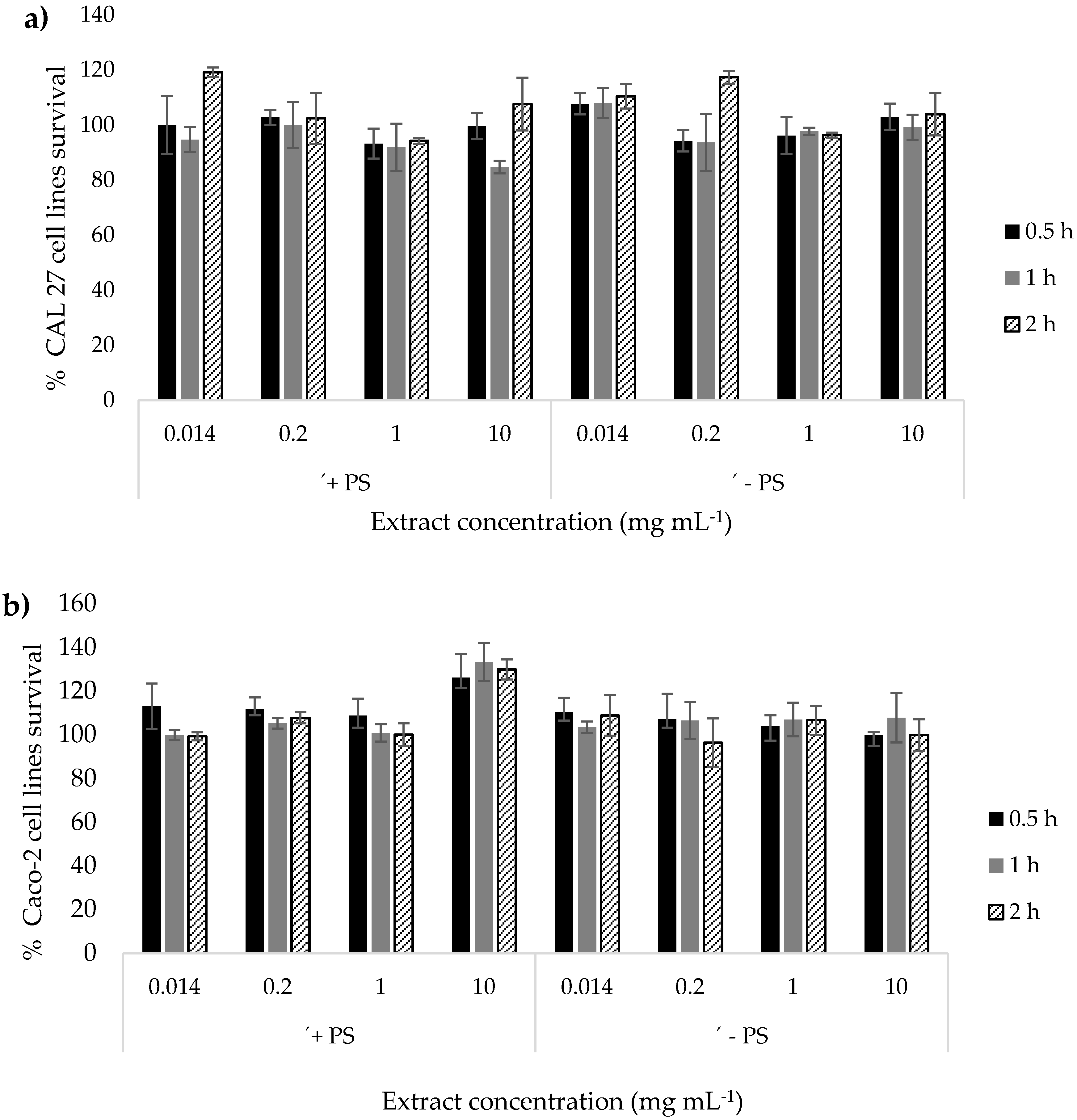
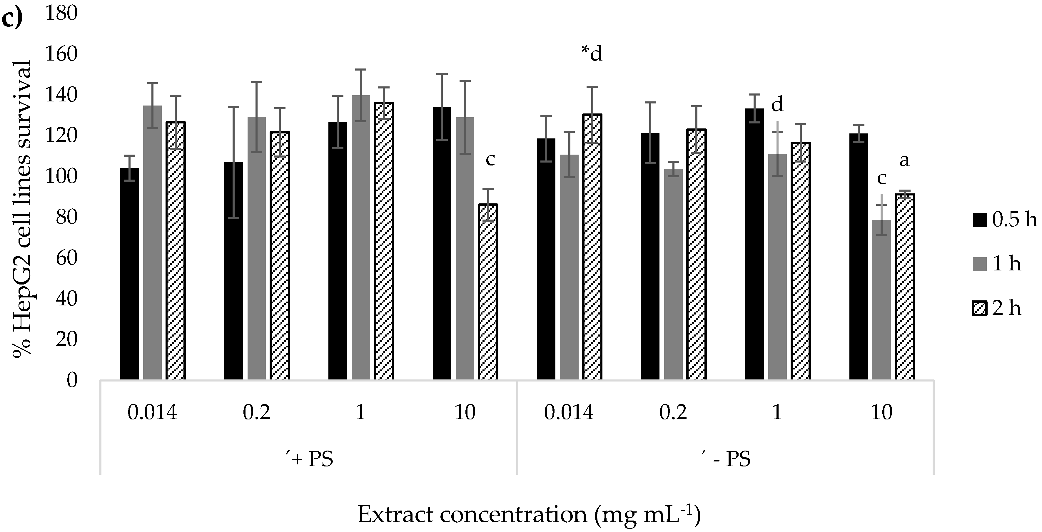
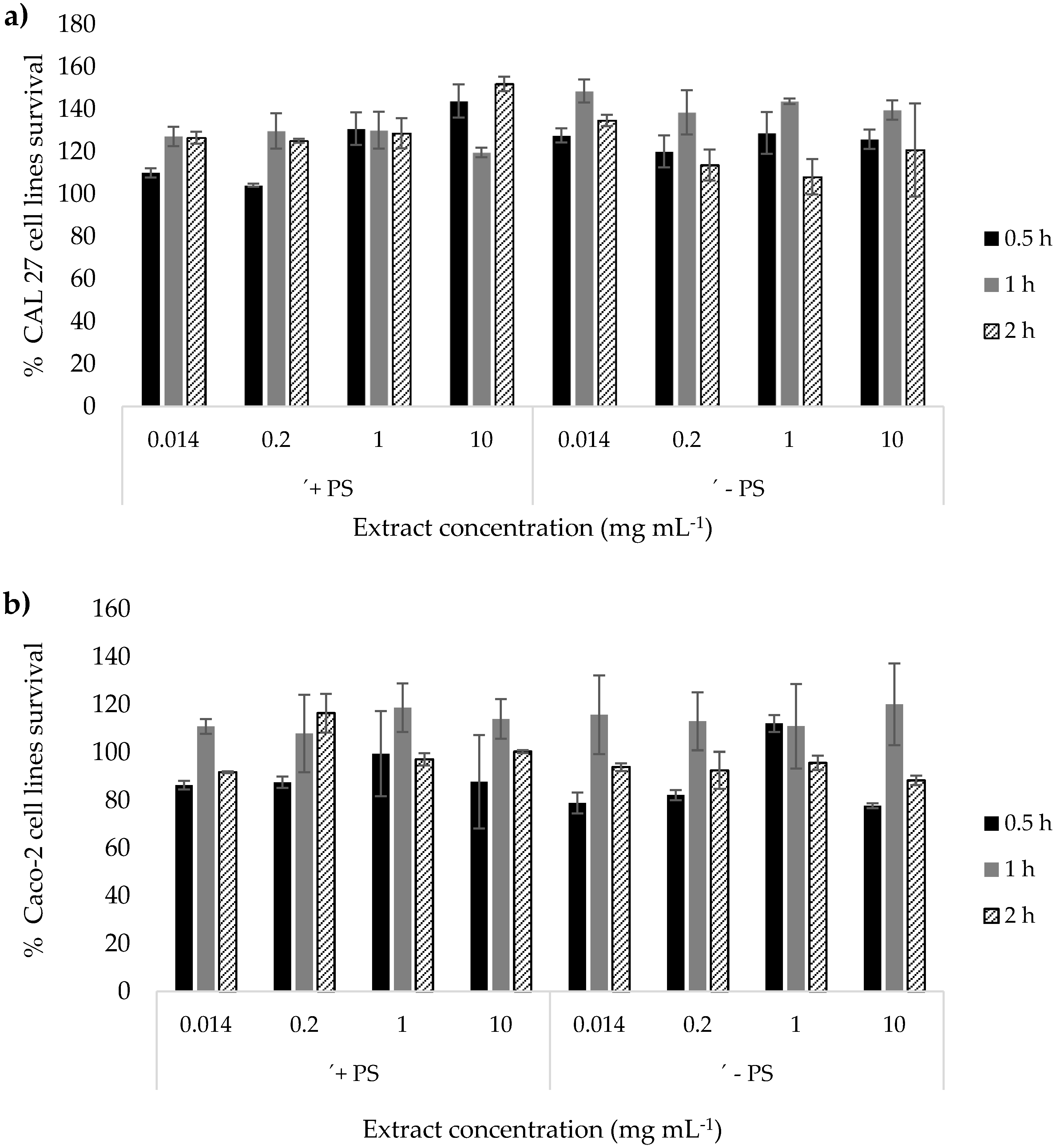
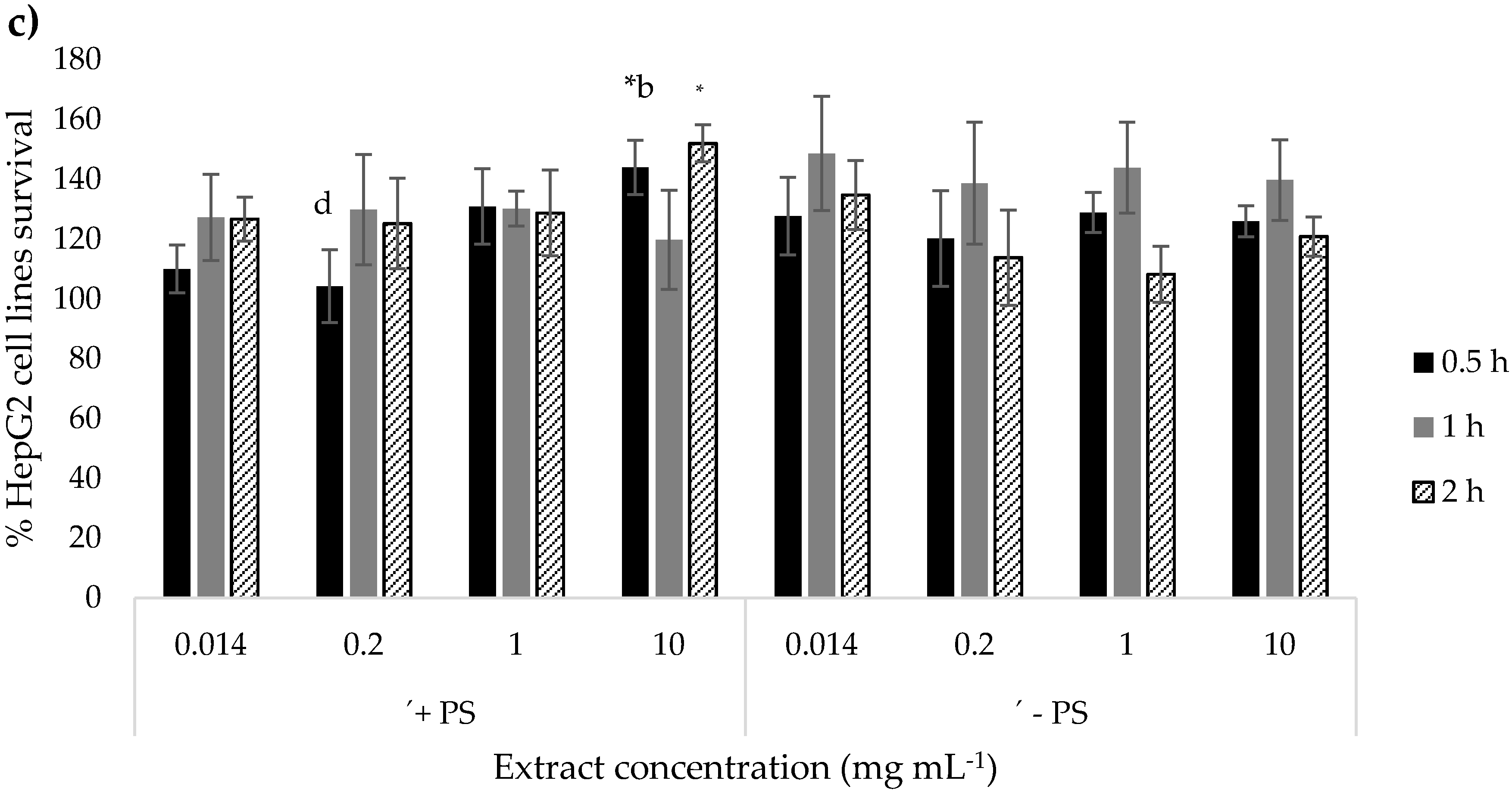
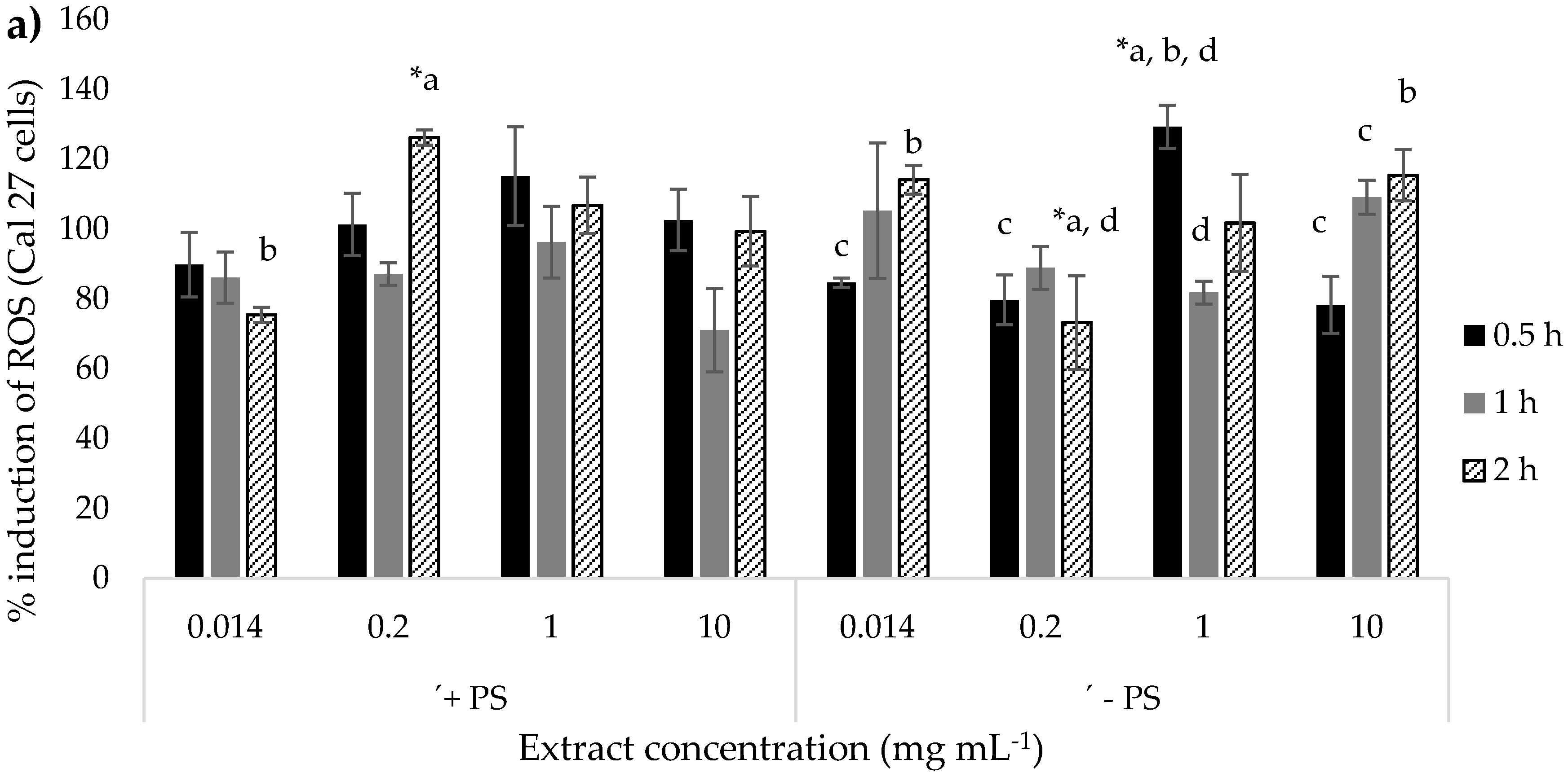
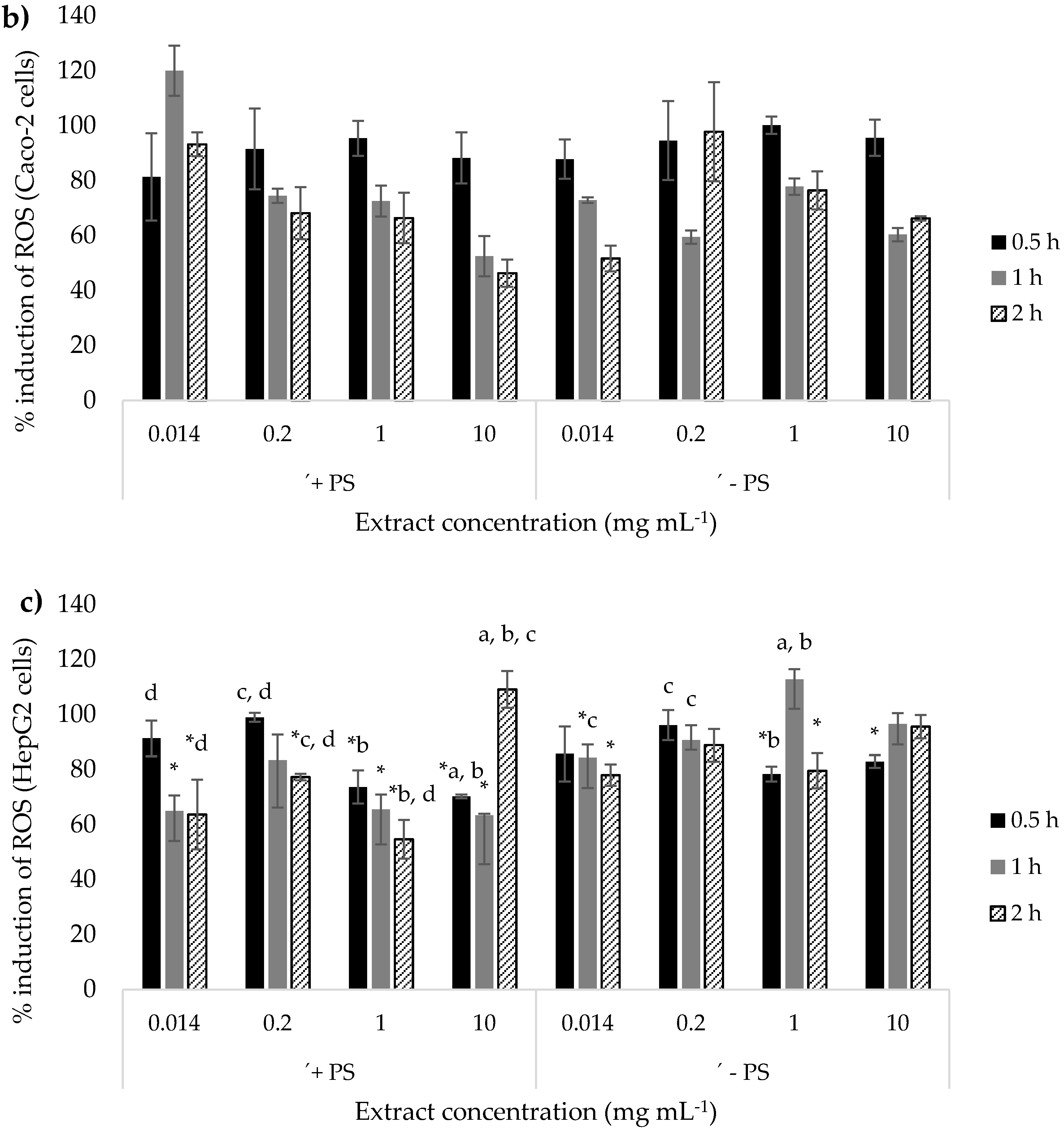
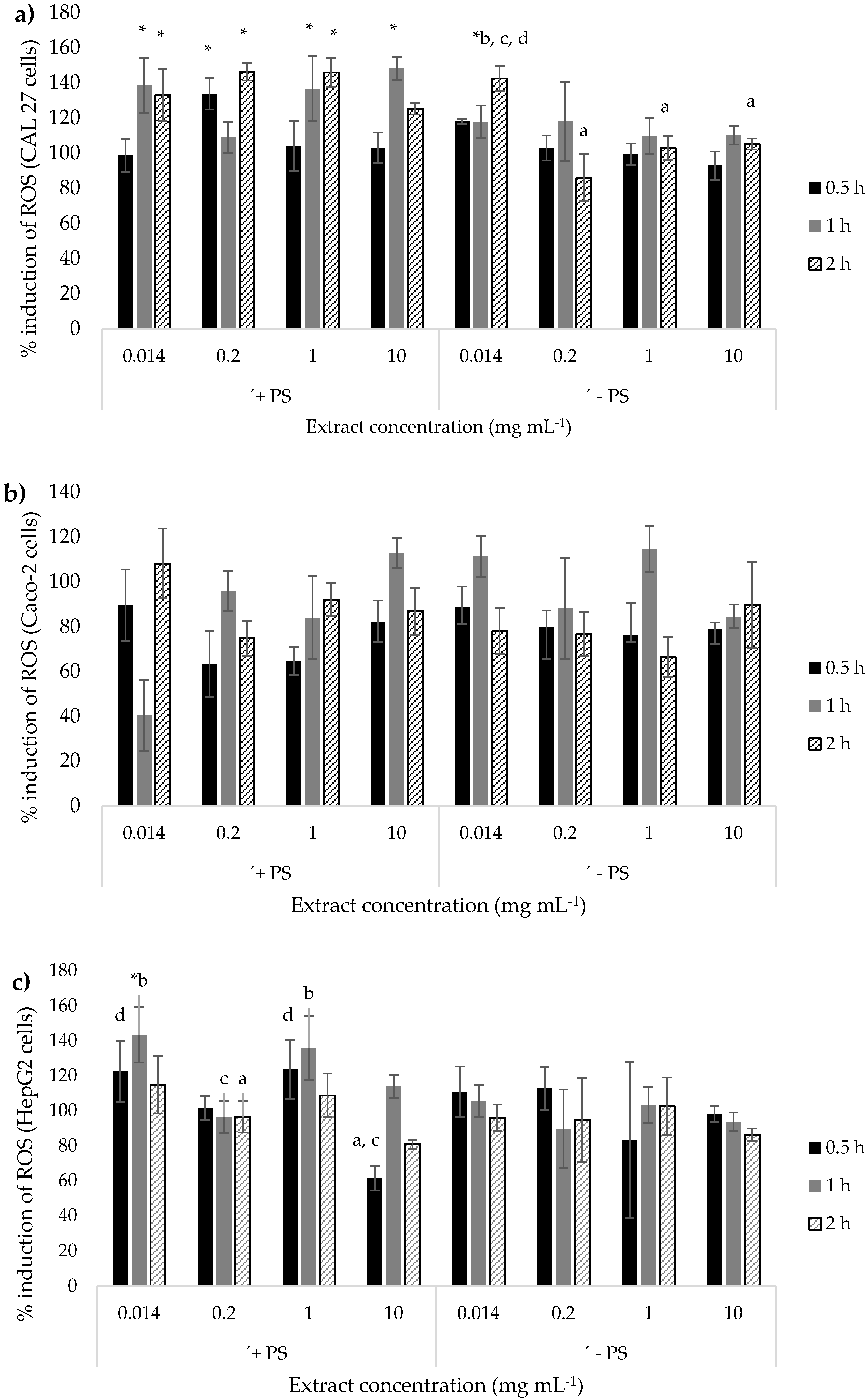
3.3. Biological Effect of Banana and Red Beetroot Peel Extracts

The extracts with the highest bioactive content were analyzed for biological effects in vitro. In the case of banana peel, it was the DEC extract, and for the red beetroot peel, it was the INF extract. Three different human cell lines were used to determine the cytotoxic and antioxidant/prooxidant effects of banana and red beetroot peel extracts: human squamous cell carcinoma (CAL 27), human colon adenocarcinoma (Caco-2) and human hepatocellular carcinoma cells (HepG2). At the time of treatment with the extracts to test the cytotoxic and antioxidant/prooxidative effects, the cell lines were subconfluent, and the treatment time was 0.5, 1 and 2 h. Results in the present study (data not shown) showed that after 2 h of incubation, the stability of polyphenols in both extracts decreased significantly. Consequently, an incubation longer than 2 h would not be representative of the effect of the polyphenols from the extract. During the treatment with the tested extracts, conditions of 37 °C and 5% CO2 were ensured. The extracts that were used for the treatment contained 0.014, 0.2, 1 and 10 mg mL1 of polyphenols and were prepared from freeze-dried extracts in which the concentrations of polyphenols were previously determined. These concentrations were determined given that the recommended dose of polyphenols for adults is 1 g per day. Therefore, a concentration of 0.014 mg mL−1 represents the recommended daily dose of polyphenols for an adult body weight of 70 kg. A concentration of 0.2 mg mL−1 is the concentration corresponding to the recommended daily dose of polyphenol per 5 L of blood held by an adult. The remaining two concentrations (1 and 10 mg mL−1) are 5x and 50x higher than the daily recommended dose of polyphenols. Extracts with and without polysaccharides were used to determine whether the polysaccharides in the extracts had any effect on any of the properties tested.
The cytotoxic effect of the extracts was evaluated according to the standard "neutral red" (NR) method [40]. The principle of the method is that a weak cationic dye enters the living cells by non-ionic diffusion and binds to the lysosomal matrix. Color can only be bound within viable cells. Accordingly, the amount of bound and then released color is proportional to the amount of cells that have survived the treatment with a particular xenobiotic and have no damaged membrane. The cytotoxic effect test results were expressed as a percentage of control survival, which was set as 100% survival. The cytotoxicity results of banana and red beetroot peel extracts are presented in Figure 1 and Figure 2, respectively.
The cytotoxicity results of banana peel extract (Figure 1) indicate that there was no cytotoxic effect on the cells of tongue epithelium (CAL 27). The survival rate of these cells did not differ with respect to different concentrations or treatment times. Moreover, the polysaccharides in the extract did not make a difference in the cytotoxic effect. In the case of colon epithelial cells (Caco-2), only the concentration of 10 mg mL−1 of the original extract showed a slight proliferative effect in all three treatment times (0.5, 1, and 2 h). Other concentrations showed neither cytotoxic nor proliferative effects on cell survival relative to control. As regards liver cells (HepG2), banana peel extract with polysaccharides showed no cytotoxic/proliferative effect at any of the tested concentrations or treatment times. Only the polysaccharide-free banana peel extract at the concentration of the recommended daily dose of polyphenols (0.014 mg mL−1) during a 2 h treatment showed a mild proliferative effect on liver cells. It can be concluded that none of the cell lines tested showed any significant sensitivity to the cytotoxic effects of banana peel extract. In contrast, certain concentrations showed a proliferative effect. If we compare the results of other studies, Dahham et al. [61] found that banana peel extract obtained by extraction with hexane had cytotoxic effects on the human colorectal cancer cell line HCT-116 and human umbilical cord endothelial cells HUVEC (inhibition of 62.04 and 61.21%). On the other hand, aqueous and ethanol extracts of banana peel showed low antiproliferative effects against HCT-116 and MCF-7, which is consistent with the present results of aqueous banana peel extract. The mild antiproliferative effects of aqueous and ethanol extract may have been the result of a much longer cell treatment (48 h), as opposed to our experiment (0.5, 1, and 2 h).
The beetroot peel extract showed a proliferative effect on the CAL 27 cell-line only for purified extracts with precipitated polysaccharides after a 1 h treatment. For Caco-2 cells, the original beetroot peel extract at a concentration of 0.2 mg mL−1, after a 2 h treatment, showed a mild proliferative effect. In the case of liver cells, the original beetroot peel extract at the highest concentration (10 mg mL−1) and after treatments for 0.5 h and 2 h exhibited a proliferative effect. None of the beetroot peel extracts resulted in the cytotoxic effect. Kapadia et al. [62] found that the cytotoxic effect of beetroot pulp extract was dose-dependent (0.29 - 290 µg mL−1) in all four stable lines used (human androgen-independent prostate cancer cells (PC-3), human breast cancer cells (MCF) -7) normal skin (FC) and liver (HC) cell lines) after 3 days of treatment. The concentrations used were consistent with those used in the present experiment (daily recommended dose of polyphenols: 0.014–0.2 mg mL−1), but the treatment time was much longer (3 days), unlike in the present experiment (0.5, 1 and 2 h), so the proven cytotoxic activity should be explained by this difference. In the work of Lee et al. [63], the results showed that betanin obtained from beetroot had anticancer activity (49% inhibition of proliferation), especially in HepG2 cells, at a relatively high concentration of betanin (200 µg mL−1) after 48 h of treatment. Unfortunately, the authors that have treated cells for several days did not provide neither information concerning the stability of the examined extracts nor the conditions under which they achieved a stability of polyphenols for such a long time.
The determination of the Reactive Oxygen Species (ROS) is based on the conversion of the non-fluorescent compound 2′, 7′-dichlorofluorescein diacetate (DCFH-DA) into the fluorescent derivative of fluorescein. Specifically, non-ionic and non-polar DCFH-DA passes through the cell membrane where, due to the action of cellular esterases, it is hydrolyzed to 2′,7′-dichlorofluorescein (DCFH), which is polar and thus remains in the cell and in contact with reactive oxygen groups (ROS) of fluorescent compound fluorescein. In this way, the amount of reactive oxygen groups in the cell can be determined by the fluorescence intensity [41,42]. One of the results of excessive ROS levels is a change in the structure and function of cellular proteins and lipids, leading to cellular dysfunction, including impaired energy metabolism, altered cell signaling and cell cycle control, impaired cellular transport mechanisms and overall dysfunctional biological activity, immunoactivation and inflammation. Oxidative stress is manifested by elevated lipid peroxidation products, protein carbonylation and decreased antioxidant status. The results of the antioxidant/prooxidative effect of banana and red beetroot peel extracts are presented in Figure 3 and Figure 4, respectively.
The results for banana peel extract show a prooxidative effect of the original extract containing polysaccharides at concentration 0.2 mg mL−1 for the CAL 27 cell-line after 2 h of treatment. The same concentration of the purified extract with precipitated polysaccharides exhibited an antioxidative effect for the same treatment duration. From these results, it can be concluded that the pro/antioxidative effect of the examined extracts strongly depends upon concentration and the environment to which extract is exposed to. There is no clear dose-response effect that can help in the prediction of the overall effect of banana peel extract. What is also true is that polysaccharides play a role in the overall antioxidative effect. The prooxidative effects on the CAL 27 cell-line were also observed for 1 mg mL−1 of purified extract after a 0.5 h treatment. In the case of colon cells (Caco-2), the lowest concentration of banana peel with polysaccharides (0.014 mg mL−1) showed a prooxidative effect after 1 h, while other concentrations (0.2 - 10 mg mL−1) showed antioxidant effect only after 2 h of treatment. Moreover, in the case of banana peel extract with polysaccharides, a positive dose-dependent effect was observed during the longest treatment of the Caco-2 cell-line. As regards the purified banana peel extract effect on Caco-2 cells, only the lowest and highest concentrations showed antioxidant effect after 2 h treatment. Regarding liver cells (HepG2), the antioxidative effect was observed for lower concentrations (0.014 and 0.2 mg mL−1) after a longer treatment time (2 h), as well as for higher concentrations (1 and 10 mg mL−1) after a shorter treatment time (0.5 and 1 h). These results indicate the positive dependence of the length of treatment and extract concentration on the antioxidant effect of the original banana extract. The same effect could also be observed for purified extracts with precipitated polysaccharides. It can be concluded that only the banana peel extract containing polysaccharides exhibited notable antioxidant activity in the case of colon (Caco-2) and liver (HepG2) cells. There is a positive dose dependence as well as the effect of exposure time. Ortiz et al. [64] did not observe clear effects of banana peel extract on the ability to inhibit lipid peroxidation, which is the final result of an increased level of free radicals in the cell. The results of Sathya [65] confirmed those of the present study and showed that aqueous banana peel extract with polysaccharides had a significant percentage of inhibition of lipid peroxidation, indicating that the extract effectively bound free radicals, so that further damage to cellular macromolecules was prevented. The oxidation of unsaturated fatty acids in biological membranes leads to the formation and propagation of lipid radicals, oxygen uptake, rearrangement of double bonds in unsaturated lipids and the eventual destruction of membrane lipids by the production of breakdown products. In addition, the results of the study by Baldi et al. [66] showed significant free radical scavenging activity by an alcoholic extract of banana peel marked by a significant decrease in lipid peroxidation level.
The results of the antioxidant/prooxidative effect of the red beetroot peel extract indicate that, in the case of tongue epithelial cells (CAL 27), only the red beetroot peel extract containing polysaccharides in all the tested concentration range (0.014 - 10 mg mL−1) exhibited prooxidative effect over a prolonged period of treatment (1 and 2 h). In colon cells (Caco-2), neither prooxidative nor antioxidant effects of beetroot peel extract with or without polysaccharide at all treatment times were demonstrated. In liver cells (HepG2), prooxidative/antioxidative effect was not detected. It can be concluded that, contrary to the effect of banana peel extract, the beetroot peel extract has no antioxidant effect on any of the cell lines tested. In contrast, the original beetroot peel extract showed a statistically significant pro-oxidative effect on cells of the tongue epithelium.

Author Contributions

Conceptualization, D.K.; methodology, D.Š., K.D., A.V.C. and A.H.; formal analysis, D.Š., A.V.C., A.H., K.D., S.J., J.J. and A.M.; writing—original draft preparation, D.K., D.Š. and K.D.; writing—review and editing, A.V.C, A.M. and A.H., visualization, D.Š.; supervision, D.K. and K.D.; All authors have read and agreed to the published version of the manuscript.

Funding

This research was funded by the Croatian Science Foundation (Project “Sustainable production of biochemicals from waste lignocellulose containing feedstock”, OPB-SLS, 9717).

Conflicts of Interest

The authors declare no conflicts of interest.

References

  1. FAO: How to Feed the World. 2050. Available online: http://www.fao.org/fileadmin/templates/wsfs/docs/Issues_papers/HLEF2050_Global_Agriculture.pdf (accessed on 19 March 2020).
  2. Sadh, P.K.; Duhan, S.; Duhan, J.S. Agro-industrial wastes and their utilization using solid state fermentation: A review. Bioresour. Bioprocess. 2018, 5, 1–15. [Google Scholar] [CrossRef] [Green Version]
  3. World Atlas: The Most Popular Fruit in The World. Available online: https://www.worldatlas.com/articles/the-most-popular-fruit-in-the-world.html (accessed on 19 March 2020).
  4. Knoema: World–Bananas Production Quantity. Available online: https://knoema.com/atlas/World/topics/Agriculture/Crops-Production-Quantity-tonnes/Bananas-production (accessed on 24 March 2020).
  5. González-Montelongo, R.; Lobo, M.G.; González, M. Antioxidant activity in banana peel extracts: Testing extraction conditions and related bioactive compounds. Food Chem. 2010, 119, 1030–1039. [Google Scholar] [CrossRef]
  6. Anhwange, B.A. Chemical Composition of Musa sapientum (Banana) Peels. J. Food Technol. 2008, 6, 263–266. [Google Scholar]
  7. Abou-Arab, A.A.; Abu-Salem, F.M. Nutritional and Anti-Nutrtional Composition of Banana Peels as Influenced by Microwave Drying Methods. Int. J. Nutr. Food. Sci. 2017, 11, 845–852. [Google Scholar]
  8. Emaga, T.H.; Andrianaivo, R.H.; Wathelet, B.; Tchango, J.T.; Paquot, M. Effects of the stage of maturation and varieties on the chemical composition of banana and plantain peels. Food Chem. 2007, 103, 590–600. [Google Scholar] [CrossRef]
  9. Someya, S.; Yoshiki, Y.; Okubo, K. Antioxidant compounds from bananas (Musa Cavendish). Food Chem. 2002, 79, 351–354. [Google Scholar] [CrossRef]
  10. Kanazawa, K.; Sakakibara, H. High content of dopamine, a strong antioxidant, in cavendish banana. J. Agric. Food Chem. 2000, 48, 844–848. [Google Scholar] [CrossRef]
  11. Vinson, J.A.; Hao, Y.; Su, X.; Zubik, L. Phenol Antioxidant Quantity and Quality in Foods: Vegetables. J. Agric. Food Chem. 1998, 46, 3630–3634. [Google Scholar] [CrossRef]
  12. da Silva, D.V.T.; dos Santos Baião, D.; de Oliveira Silva, F.; Alves, G.; Perrone, D.; Del Aguila, E.M.; Paschoalin, V.M.F. Betanin, a Natural Food Additive: Stability, Bioavailability, Antioxidant and Preservative Ability Assessments. Molecules 2019, 24, 458. [Google Scholar] [CrossRef] [Green Version]
  13. Lόpez, N.; Puértolas, E.; Condόn, S.; Raso, J.; Alvarez, I. Enhancement of the extraction of betanine from red beetroot by pulsed electric fields. J. Food Eng. 2009, 90, 60–66. [Google Scholar] [CrossRef]
  14. Transparency Market Research: Beet Root Extract Market–Global Industry Analysis, Size, Share, Growth, Trend and Forecast 2018–2026. Available online: https://www.transparencymarketresearch.com/beet-root-extract-market.html (accessed on 19 March 2020).
  15. Kujala, T.S.; Loponen, J.M.; Klika, K.D.; Pihlaja, K. Phenolics and Betacyanins in Red Beetroot (Beta vulgaris) Root: Distribution and Effect of Cold Storage on the Content of Total Phenolics and Three Individual Compounds. J. Agric. Food Chem. 2000, 48, 5338–5342. [Google Scholar] [CrossRef] [PubMed]
  16. Kujala, T.; Loponen, J.; Pihlaja, K. Betalains and Phenolics in Red Beetroot (Beta vulgaris) Peel Extracts: Extraction and Characterisation. Zeitschrift für Naturforschung C 2001, 56, 343–348. [Google Scholar] [CrossRef] [PubMed]
  17. Azmir, J.; Zaidul, I.S.M.; Rahman, M.M.; Sharif, K.M.; Mohamed, A.; Sahena, F.; Jahurul, M.H.A.; Ghafoor, K.; Norulaini, N.A.N.; Omar, A.K.M. Techniques for extraction of bioactive compounds from plant materials: A review. J. Food Eng. 2013, 117, 426–436. [Google Scholar] [CrossRef]
  18. Vinatoru, M. An overview of the ultrasonically assisted extraction of bioactive principles from herbs. Ultrason. Sonochem. 2001, 8, 303–313. [Google Scholar] [CrossRef]
  19. Vinatoru, M.; Mason, T.J.; Calinescu, I. Ultrasonically Assisted Extraction (UAE) and Microwave Assisted Extraction (MAE) of Functional Compounds from Plants Materials. Trac-Trend. Anal. Chem. 2017, 97, 159–178. [Google Scholar] [CrossRef]
  20. Zakaria, S.M.; Kamal, S.M. Subcritical Water Extraction of Bioactive Compounds from Plants and Algae: Applications in Pharmaceutical and Food Ingredients. Food Eng. Rev. 2016, 8, 23–34. [Google Scholar] [CrossRef]
  21. Pan, Y.; Wang, K.; Huang, S.; Wang, H.; Mu, X.; He, C.; Ji, X.; Zhang, J.; Huang, F. Antioxidant activity of microwave-assisted extract of longan (Dimocarpus Longan Lour.) peel. Food Chem. 2008, 106, 1264–1270. [Google Scholar] [CrossRef]
  22. Singh, A.; Sabally, K.; Kubow, S.; Donnelly, D.J.; Gariepy, Y.; Orsat, V.; Raghavan, G.S.V. Microwave-Assisted Extraction of Phenolic Antioxidants from Potato Peels. Molecules 2011, 16, 2218–2232. [Google Scholar] [CrossRef] [Green Version]
  23. Jha, P.; Das, A.J.; Deka, S.C. Optimization of utrasound and microwave assisted extractions of polyphenols from black rice (Oryza sativa cv. Poireton) husk. J. Food Sci. Techol. 2017, 54, 3847–3858. [Google Scholar] [CrossRef]
  24. Jokić, S.; Gagić, T.; Knez, Ž.; Šubarić, D.; Škerget, M. Separation of active compounds from food by-product (cocoa shell) using subcritical water extraction. Molecules 2018, 23, 1408. [Google Scholar] [CrossRef] [Green Version]
  25. Lachos-Perez, D.; Baseggio, A.M.; Mayanga-Torres, P.C.; Maróstica Junior, M.R.; Rostagno, M.A.; Martínez, J.; Forster-Carneiro, T. Subcritical water extraction of flavanones from defatted orange peel. J. Supercrit. Fluid. 2018, 138, 7–16. [Google Scholar] [CrossRef]
  26. Singh, P.P.; Saldana, M.D.A. Subcritical water extraction of phenolic compounds from potato peel. Food Res. Int. 2011, 44, 2452–2458. [Google Scholar] [CrossRef]
  27. Tapre, A.R.; Jain, R.K. Study of Advanced Maturity Stages of Banana. Int. J. Adv. Eng. Res. Stud. 2012, 1, 272–274. [Google Scholar]
  28. Padmore, J.M. Animal feed - AOAC official method 930.15 - Moisture in animal feed. In Official Methods of Analysis, 15th ed.; Helrich, K., Ed.; AOAC International: Arlington, VA, USA, 1990; Volume 1, pp. 69–70. [Google Scholar]
  29. Padmore, J.M. Animal feed - AOAC official method 976.05 – Protein (crude) in animal feed, automated Kjeldahl method. In Official Methods of Analysis, 15th ed.; Helrich, K., Ed.; AOAC International: Arlington, VA, USA, 1990; Volume 1, p. 72. [Google Scholar]
  30. Padmore, J.M. Animal feed-AOAC official method 920.39 – Fat (crude) or ether extract in animal feed. In Official Methods of Analysis, 15th ed.; Helrich, K., Ed.; AOAC International: Arlington, VA, USA, 1990; Volume 1, p. 79. [Google Scholar]
  31. Padmore, J.M. Animal feed-AOAC official method 942.05–Ash of animal feed. In Official Methods of Analysis, 15th ed.; Helrich, K., Ed.; AOAC International: Arlington, VA, USA, 1990; Volume 1, p. 70. [Google Scholar]
  32. McCleary, B.V.; DeVries, J.W.; Rader, J.I.; Cohen, G.; Prosky, L.; Mugford, D.C.; Okuma, K. Determination of insoluble, soluble, and total dietary fibre (CODEX definition) by enzymatic-gravimetric method and liquid chromatography: Collaborative study. J. AOAC Int. 2012, 95, 824–844. [Google Scholar] [CrossRef] [PubMed]
  33. ISO 5509. Animal and Vegetable Fats and Oils–Preparation of Methyl Esters of Fatty Acids; International Organization for Standardization: Geneva, Switzerland, 2000. [Google Scholar]
  34. Vojvodić, A.; Komes, D.; Vovk, I.; Belščak-Cvitanović, A.; Bušić, A. Compositional evaluation of selected agro-industrial wastes as valuable sources for the recovery of complex carbohydrates. Food Res. Int. 2016, 89, 565–573. [Google Scholar] [CrossRef]
  35. Jokić, S.; Aladić, K.; Šubarić, D. Subcritical water extraction laboratory plant design and application. Annu. Croat. Acad. Eng. 2018, 21, 247–258. [Google Scholar]
  36. Singleton, V.L.; Rossi, J.A. Colorimetry of total phenolics with phosphotungstic acid reagents. Am. J. Enol. Viticult. 1965, 16, 144–158. [Google Scholar]
  37. Brand-Williams, W.; Cuvelier, M.E.; Berset, C. Use of a free radical method to evaluate antioxidant activity. Lebensm.-Wiss. Technol. 1995, 28, 25–30. [Google Scholar] [CrossRef]
  38. Re, R.; Pellegrini, N.; Proteggente, A.; Pannala, A.; Yang, M.; Rice-Evans, C. Antioxidant activity applying an improved ABTS radical cation decolorisation assay. Free Radic. Biol. Med. 1999, 26, 1231–1237. [Google Scholar] [CrossRef]
  39. Stintzing, F.; Schieber, A.; Carle, R. Evaluation of colour properties and chemical quality parameters of cactus juice. Eur. Food Res. Technol. 2003, 216, 303–311. [Google Scholar] [CrossRef]
  40. Babich, H.; Borenfreund, E. Cytotoxicity of T-2 toxin and its metabolites determined with the neutral red cell viability assay. Appl. Environ. Microbiol. 1991, 57, 2101–2103. [Google Scholar] [CrossRef] [PubMed] [Green Version]
  41. Wang, H.; Joseph, J.A. Quantifying cellular oxidative stress by dichlorofluorescein assay using microplate reader. Free Radic. Biol. Med. 1999, 27, 612–616. [Google Scholar] [CrossRef]
  42. Hempel, S.L.; Buettner, G.R.; O’Malley, Y.Q.; Wessels, D.A.; Flaherty, D.M. Dihydrofluorescein diacetate is superior for detecting intracellular oxidants: Comparison with 2’,7’-dichlorodihydrofluorescein diacetate, 5(and 6)-carboxy-2’,7’-dichlorodihydrofluorescein diacetate, and dihydrorhodamine. Free Radic. Biol. Med. 1999, 27, 146–159. [Google Scholar] [CrossRef]
  43. Abdolali, A.; Guo, W.S.; Ngo, H.H.; Chen, S.S.; Nguyen, N.C.; Tung, K.L. Typical lignocellulosic wastes and by-products for biosorption process in water and wastewater treatment: A critical review. Bioresour. Technol. 2014, 160, 57–66. [Google Scholar] [CrossRef] [PubMed]
  44. Okeke, B.C.; Obi, S.K.C. Lignocellulose and Sugar Composition of Some Agro-waste Materials. Bioresour. Technol. 1994, 47, 283–284. [Google Scholar] [CrossRef]
  45. Morais, D.R.; Rotta, E.M.; Sargi, S.C.; Bonafe, E.G.; Suzuki, R.M.; Souza, N.E.; Matsushita, M.; Visentainer, J.V. Proximate Composition, Mineral Contents and Fatty Acid Composition of the Different Parts and Dried Peels of Tropical Fruits Cultivated in Brazil. J. Braz. Chem. Soc. 2017, 28, 308–318. [Google Scholar] [CrossRef]
  46. Vasconcellos, J.; Conte-Junior, C.; Silva, D.; Pierucci, A.P.; Paschoalin, V.; Alvares, T.S. Comparison of Total Antioxidant Potential, and Total Phenolic, Nitrate, Sugar, and Organic Acid Contents in Beetroot Juice, Chips, Powder, and Cooked Beetroot. Food Sci. Biotechnol. 2016, 25, 79–84. [Google Scholar] [CrossRef]
  47. Costa, A.P.D.; Hermes, V.S.; Rios, A.O.; Flôres, S.H. Minimally processed beetroot waste as an alternative souce to obtain functional ingredients. J. Food Sci. Technol. 2017, 54, 2050–2058. [Google Scholar] [CrossRef]
  48. Shirsath, S.R.; Sonawane, S.H.; Gogate, P.R. Intensification of extraction of natural products using ultrasonic irradiations—A review of current status. Chem. Eng. Process. 2012, 53, 10–23. [Google Scholar] [CrossRef]
  49. Vilkhu, K.; Mawson, R.; Simons, L.; Bates, D. Applications and opportunities for ultrasound assisted extraction in the food industry. Innov. Food Sci. Emerg. 2008, 9, 161–169. [Google Scholar] [CrossRef]
  50. Chemat, F.; Rombaut, N.; Sicaire, A.G.; Meullemiestre, A.; Fabiano-Tixier, A.S.; Abert-Vian, M. Ultrasound assisted extraction of food and natural products. Mechanisms, techniques, combinations, protocols and applications. A review. Ultrason. Sonochem. 2017, 34, 540–560. [Google Scholar] [CrossRef] [PubMed]
  51. Vu, H.T.; Scarlett, C.J.; Vuong, Q.V. Optimization of ultrasound-assisted extraction conditions for recovery of phenolic compounds and antioxidant capacity from banana (Musa cavendish) peel. J. Food Process. Pres. 2017, 41, 1–14. [Google Scholar] [CrossRef]
  52. Aboul-Enein, A.M.; Salama, Z.A.; Gaafar, A.A.; Aly, H.F.; A bou-Elella, F.; Ahmed, H.A. Identification of phenolic compounds from banana peel (Musa paradaisica L.) as antioxidant and antimicrobial agents. J. Chem. Pharm. Res. 2016, 8, 46–55. [Google Scholar]
  53. Fatemeh, S.R.; Saifullah, R.; Abbas, F.M.A.; Azhar, M.E. Total phenolics, flavonoids and antioxidant activity of banana pulp and peel flours: Influence of variety and stage of ripeness. Int. Food Res. J. 2012, 19, 1041–1046. [Google Scholar]
  54. Kaderides, K.; Papaoikonomou, L.; Serafim, M.; Goula, A.M. Microwave-assisted extraction of phenolics from pomegranate peels: Optimization, kinetics, and comparison with ultrasounds extraction. Chem. Eng. Process. 2019, 137, 1–11. [Google Scholar] [CrossRef]
  55. Ishak, N.A.; Abdul Rashid, M.Y.; Zabidi, N.A. Local Banana Peels Extract by Using Subcritical Water Extraction Technique. Ulum Islamiyyah 2019, 26, 19–30. [Google Scholar]
  56. Wong, Y.M.; Siow, L.F. Effects on heat, pH, antioxidant, agitation and light on betacyanin stability using red-fleshed dragon fruit (Hylocereus polyrhizus) juice and concentrate as models. J. Food Sci. Technol. 2015, 52, 3086–3092. [Google Scholar] [CrossRef] [Green Version]
  57. Sawicki, T.; Bączek, N.; Wiczkowski, W. Betalain profile, content and antioxidant capacity of red beetroot dependent on the genotype and root part. J. Funct. Food. 2016, 27, 249–261. [Google Scholar] [CrossRef]
  58. Laqui-Vilca, C.; Aguilar-Tuesta, S.; Mamani-Navarro, W.; Montaño-Bustamante, J.; Condezo-Hoyos, L. Ultrasound-assisted optimal extraction and thermal stability of betalains from colored quinoa (Chenopodium quinoa Willd) hulls. Ind. Crop. Prod. 2018, 111, 606–614. [Google Scholar] [CrossRef]
  59. Azeredo, H.M.C. Betalains: Properties, sources, applications, and stability–a review. Int. J. Food Sci. Technol. 2009, 44, 2365–2376. [Google Scholar] [CrossRef] [Green Version]
  60. Cardoso-Ugarte, G.A.; Sosa-Morales, M.E.; Ballard, T.; Liceaga, A.; San Martín-González, M.F. Microwave-assisted extraction of betalains from red beet (Beta vulgaris). LWT-Food Sci. Technol. 2014, 59, 276–282. [Google Scholar] [CrossRef]
  61. Dahham, S.S.; Agha, M.T.; Tabana, Y.M.; Malik, A.; Abdul, S. Antioxidant Activities and Anticancer Screening of Extracts from Banana Fruit (Musa sapientum). Acad. J. Cancer Res. 2015, 8, 28–34. [Google Scholar]
  62. Kapadia, G.J.; Azuine, M.A.; Rao, G.S.; Arai, T.; Iida, A.; Tokuda, H. Cytotoxic Effect of the Red Beetroot (Beta vulgaris L.) Extract Compared to Doxorubicin (Adriamycin) in the Human Prostate (PC-3) and Breast (MCF-7) Cancer Cell Lines. Anticancer Agents Med. Chem. 2011, 11, 280–284. [Google Scholar] [CrossRef] [PubMed]
  63. Lee, E.J.; An, D.; Nguyen, C.T.T.; Patil, B.S.; Kim, J.; Yoo, K.S. Betalain and Betaine Composition of Greenhouse- or Field-Produced Beetroot (Beta vulgaris L.) and Inhibition of HepG2 Cell Proliferation. J. Agric. Food Chem. 2014, 62, 1324–1331. [Google Scholar] [CrossRef]
  64. Ortiz, L.; Dorta, E.; Gloria Lobo, M.; González-Mendoza, L.A.; Díaz, C.; González, M. Use of Banana (Musa acuminata Colla AAA) Peel Extract as an Antioxidant Source in Orage Juices. Plant Food Hum. Nutr. 2017, 72, 60–66. [Google Scholar] [CrossRef]
  65. Sathya, M. Assaying the Antioxidant Activity of Banana Peel. Am. J. Biochem. Mol. Biol. 2014, 4, 122–129. [Google Scholar] [CrossRef]
  66. Baldi, A.; Pandit, M.; Ranka, P. Amelioration of in-vivo Antioxidant Activity by Banana Extracts. Int. J. Pharm. Biol. Arch. 2012, 3, 157–161. [Google Scholar]
Figure 1. Cell survival (% of control) of (a) CAL 27 cells, (b) Caco-2 cells, (c) HepG2 cells determined with the neutral red assay after 0.5, 1 and 2 h of exposure to different concentrations of banana peel extract (0.014, 0.2, 1 and 10 mg mL−1) with (+PS) and without polysaccharides (-PS); Statistically significant difference (p < 0.05) between different concentrations of the extract and the control: *-control; a-0.014 mg mL−1; b-0.2 mg mL−1; c-1 mg mL−1; d-10 mg mL−1.
Sustainability 12 04539 g001aSustainability 12 04539 g001b
Figure 2. Cell survival (% of control) of (a) CAL 27 cells, (b) Caco-2 cells, (c) HepG2 cells determined with the neutral red assay after 0.5, 1 and 2 h of exposure to different concentrations of red beetroot peel extract (0.014, 0.2, 1 and 10 mg mL−1) with (+PS) and without polysaccharides (-PS), Statistically significant difference (p < 0.05) between different concentrations of the extract and the control: *-control; a-0.014 mg mL−1; b-0.2 mg mL−1; c-1 mg mL−1; d-10 mg mL−1.
Sustainability 12 04539 g002aSustainability 12 04539 g002b
Figure 3. Antioxidative/prooxidative effects of different concentrations of banana peel extract (0.014, 0.2, 1 and 10 mg mL−1) with (+PS) and without polysaccharides (-PS) on (a) CAL 27 cells (b) Caco-2 cells (c) HepG2 cells during 0.5, 1 and 2 h of exposure, Statistically significant difference (p < 0.05) between different concentrations of the extract and control: *-control; a-0.014 mg mL−1; b-0.2 mg mL−1; c-1 mg mL−1; d-10 mg mL−1.
Sustainability 12 04539 g003aSustainability 12 04539 g003b
Figure 4. Antioxidative/prooxidative effects of different concentrations of red beetroot peel extract (0.014, 0.2, 1 and 10 mg mL−1) with (+PS) and without polysaccharides (-PS) on (a) CAL 27 cells, (b) Caco-2 cells, (c) HepG2 cells during 0.5, 1 and 2 h of exposure; Statistically significant difference (p < 0.05) between different concentrations of the extract and the control: *-control; a-0.014 mg mL−1; b-0.2 mg mL−1; c-1 mg mL−1; d-10 mg mL−1.
Sustainability 12 04539 g004
Table 1. Macrocomponent composition of banana peel and red beetroot peel.
Banana PeelRed Beetroot Peel
Dry matter (%)88.1 ± 0.190.8 ± 0.0
Crude protein content (% dmb*)9.2 ± 0.318.3 ± 0.2
Crude fat content (% dmb)7.5 ± 0.10.6 ± 0.0
● Lauric acid C12:0 (% fa*)28.9 ± 0.2np
● Palmitic acid C16:0 (% fa)28.6 ± 0.1np
● Stearic acid C18:1 (% fa)4.7 ± 0.0np
● Linolenic acid C18:3n3 (% fa)37.8 ± 0.2np
Crude mineral content (% dmb)14.0 ± 0.312.1 ± 0.1
Total dietary fibre (% dmb)39.0 ± 2.733.6 ± 1.0
● Insoluble dietary fibre (% dmb)38.1 ± 2.926.6 ± 0.5
● Soluble dietary fibre (% dmb)0.9 ± 0.36.9 ± 0.5
Soluble sugar content (% dmb)19.1 ± 0.112.5 ± 0.2
● Fructose (% dmb)12.7 ± 0.2nd
● Glucose (% dmb)n.d.nd
● Sucrose (% dmb)6.4 ± 0.112.5 ± 0.2
dmb—dry matter basis of the sample; fa—fatty acids; np—not performed; nd—not detected.
Table 2. Macro- and microelement content in banana peel and red beetroot peel.
Banana PeelRed Beetroot Peel
Macroelements (mg kg−1 dmb*)
Na38 ± 04380 ± 189
Mg150 ± 146570 ± 356
K75061 ± 56341854 ± 1366
Ca2698 ± 32851 ± 153
Fe33 ± 4159 ± 10
P2900 ± 317811 ± 393
Microelements (µg kg−1 dmb)
V17 ± 1308 ± 21
Mn63 ± 052 ± 3
Cr172 ± 31343 ± 21
Co35 ± 0200 ± 7
Ni391 ± 191370 ± 76
Cu6 ± 019 ± 1
Zn14 ± 049 ± 2
As8 ± 091 ± 6
Se13 ± 026 ± 1
Mo135 ± 3580 ± 11
Cd11 ± 7421 ± 8
Sn23 ± 117 ± 1
Sb8 ± 19 ± 1
Hg49 ± 236 ± 1
dmb—dry matter basis of the sample.
Table 3. Bioactive content of differently prepared extracts of banana peel.
SampleTPC
(mg GAE g−1 dmb)
Antioxidant CapacityDopamine
(mg g−1 dmb)
DPPH
(mmol Trolox g−1 dmb)
ABTS
(mmol Trolox g−1 dmb)
INF25.59 ± 0.38 a0.156 ± 0.000.160 ± 0.0010.29 ± 0.00 a
DEC25.38 ± 0.53 a0.163 ± 0.000.195 ± 0.0012.63 ± 0.00
MAC18.73 ± 0.48 bc0.124 ± 0.000.125 ± 0.009.94 ± 0.02
U3018.26 ± 0.59 bd0.117 ± 0.000.123 ± 0.0010.33 ± 0.01 a
U6017.96 ± 0.13 cd0.112 ± 0.000.132± 0.0011.31 ± 0.02
SWE16.06 ± 0.060.110 ± 0.010.097 ± 0.0010.21 ± 0.03
MAE3.46 ± 0.050.021 ± 0.000.020 ± 0.005.14 ± 0.01
dmb—dry matter basis of the sample; INF—infusion; DEC—decoction; MAC—maceration; U30 and U60—ultrasound bath for 30 and 60 min, respectively; SWE—subcritical water extraction; MAE—microwave-assisted extraction; Means in the same column denoted with the same superscript letters (a,b,c,d) are not significantly different (p > 0.05).
Table 4. Bioactive content of differently prepared extracts of red beetroot peel.
SampleTPC
(mg GAE g−1 dmb)
Antioxidant CapacityTotal Betalain Content
DPPH
(mmol Troloxg−1 dmb)
ABTS
(mmol Trolox g−1 dmb)
Total Betacyanin Content
(mg betanin g−1 dmb)
Total Betaxanthin Content
(mg vulgaxanthin I g−1 dmb)
INF45.03 ± 0.99 abcd0.056 ± 0.000.164 ± 0.009.80 ± 0.148.41 ± 0.03 a
DEC66.30 ± 0.410.098 ± 0.000.295 ± 0.006.15± 0.016.50 ± 0.06
MAC44.75 ± 2.36 aef0.066 ± 0.00 a0.188 ± 0.000.85 ± 0.013.99 ± 0.02
U3044.07 ± 0.22 beg0.042 ± 0.00 b0.140 ± 0.00 a3.87 ± 0.03 a8.61 ± 0.08 a
U6047.99 ± 0.98 ch0.048 ± 0.000.142 ± 0.00 a3.84 ± 0.02 a6.98 ± 0.06
SWE45.77 ± 0.05 dfgh0.064 ± 0.00 a0.210 ± 0.000.03 ± 0.000.19 ± 0.04
MAE39.72 ± 0.760.040 ± 0.00 b0.132 ± 0.003.08 ± 0.021.74 ± 0.08
dmb—dry matter basis of the sample; INF—infusion; DEC—decoction; MAC—maceration; U30 and U60—ultrasound bath for 30 and 60 min, respectively; SWE—subcritical water extraction; MAE—microwave-assisted extraction; Means in the same column denoted with the same superscript letters (a,b,c,d,e,f,g,h) are not significantly different (p > 0.05).

