Article

Equol: A Microbiota Metabolite Able to Alleviate the Negative Effects of Zearalenone during In Vitro Culture of Ovine Preantral Follicles

by
Talyne Emilia Santos Silva
1,
Danielle Cristina Calado de Brito
1,
Naiza Arcângelo Ribeiro de Sá
1,
Renato Felix da Silva
1,
Anna Clara Accioly Ferreira
1,
José Ytalo Gomes da Silva
2,
Maria Izabel Florindo Guedes
2,
Ana Paula Ribeiro Rodrigues
1,
Regiane Rodrigues dos Santos
3,* and
José Ricardo de Figueiredo
1
1
Laboratory of Manipulation of Oocytes and Ovarian Pre-Antral Follicles (LAMOFOPA), Faculty of Veterinary Medicine, State University of Ceará, Fortaleza 60714-903, CE, Brazil
2
Laboratory of Biotechnology and Molecular Biology (LBBM), State University of Ceará, Fortaleza 60714-903, CE, Brazil
3
Schothorst Feed Research, 8218 NA Lelystad, The Netherlands
*
Author to whom correspondence should be addressed.
Submission received: 20 October 2019 / Revised: 3 November 2019 / Accepted: 5 November 2019 / Published: 9 November 2019
(This article belongs to the Section Mycotoxins)

Abstract

:
The impact of zearalenone (ZEN) on female reproduction remains an issue, since its effects may differ among exposed cell types. Besides the use of decontaminants in animal diet, other approaches should be considered to minimise ZEN effects after exposure. Since the first organ in contact with ZEN is the gastrointestinal tract, we hypothesise that products of microbiota metabolism may play a role in ZEN detoxification. We aimed to evaluate the effect of 1 µmol/L ZEN and 1 µmol/L equol (a microbial metabolite), alone or in combination, on the survival and morphology of in vitro cultured ovarian preantral follicles. Ovaries from 12 sheep were collected at a local abattoir and fragmented, and the ovarian pieces were submitted to in vitro culture for three days in the presence or absence of the test compounds. The follicular morphology was impaired by ZEN, but equol could alleviate the observed degeneration rates. While ZEN decreased cell proliferation in primary and secondary follicles, as well as induced DNA double-strand breaks in primordial follicles, all these observations disappeared when equol was added to a culture medium containing ZEN. In the present culture conditions, equol was able to counteract the negative effects of ZEN on ovarian preantral follicles.
Key Contribution: This study evaluates the ability of a microbiota metabolite (equol) to minimise the negative effects of zearalenone (ZEN) on the initial development of ovine preantral follicles. The improved follicular survival when equol is added to a culture medium containing ZEN suggests that this phytoestrogen plays a role in counteracting the toxicity of ZEN. Although equol should not be claimed as a mycotoxin detoxifier; microbiota modulation to increase the synthesis of this phytoestrogen might alleviate the impact of ZEN on early staged female gametes.

1. Introduction

Zearalenone (ZEN) is one of the most commonly found mycotoxins in food and feed, especially those based on corn, beet pulp, wheat, rice, barley, oats, and soybeans. A 10-year survey based on the analysis of 74,821 commodities from 100 countries demonstrated that 45% of the samples were contaminated with ZEN, at levels ranging from 1.53 ppm in rice up to 23.3 ppm in wheat [1]. This nonsteroidal oestrogenic mycotoxin is produced by the fungi of the genus Fusarium to control its reproduction. Due to structural and functional similarity to oestrogens, ZEN can also interact with animal cells and tissue structures, acting as an endocrine-disrupting chemical [2].
The negative impact of ZEN on fertility is well documented in humans [3,4] and farm animals, especially pigs and ruminants [5,6,7,8,9]. Most ZEN studies have focused on the action of this mycotoxin on cell lines [10,11], spermatozoa [8,12], or mature oocytes [6,13]. In a transgenerational study, Schoevers et al. [7] showed that immature oocytes, yet enclosed in preantral follicles, were sensitive to ZEN exposure, which affected follicular assembly, resulting in premature exhaustion of this follicle pool. Besides ZEN, diets usually contain phytoestrogens, which are plant-derived compounds with a structure similar to 17-β-oestradiol (E2), enabling them to induce (anti) oestrogenic effects depending on the dosage [14]. These phytoestrogens are divided into isoflavones, prenylflavonoids, coumestans, and lignans. Soybeans, alfalfa, and red clover are isoflavone-rich ingredients present in the diets of farm animals. Based on the fact that soybeans may also be contaminated with ZEN, its interaction with phytoestrogens should not be neglected. A biomonitoring study already showed the concomitant presence of the isoflavones genistein, daidzein, equol, and ZEN in serum and urine from pregnant women [15]. Unfortunately, these latter authors did not evaluate the possible interactions among these substances. It was recently demonstrated that genistein interacts with ZEN in vitro and, depending on the concentration range of both substances, the oestrogenic effect can be potentiated of inhibited [16]. Although, interaction studies between other phytoestrogens with mycotoxins are still lacking, one must bear in mind that ingested phytoestrogens are metabolised by reductase enzymes produced by the host microbiota. For example, soybeans and other legumes like alfalfa and red clover are rich in daidzein, which is converted to equol depending on the intestinal bacterial population of the animal [17]. Compared with its precursor daidzein, equol is more stable and more easily absorbable, and no other isoflavones shows stronger oestrogenic activity than equol [17]. Therefore, the interaction of ZEN with a microbiota product like equol should not be neglected in animals’ daily fed diets containing phytoestrogenic sources.
It has already been demonstrated that equol can be produced in several animal species, such as monkey [18,19], rat [18,19], pig [20,21], sheep [22], and human [19,23]. Equol also has a great affinity with oestrogen receptors, but depending on the dietary concentration, it may bring many beneficial health effects due to its antioxidant, antitumour, and anti-inflammatory properties [24]. Importantly, although both ZEN and equol are xenoestrogens and are usually originated from the same feedstuffs, they act differently. For instance, (i) equol preferentially binds oestrogen receptor (ER)-β, while ZEN has more affinity to ER-α; (ii) equol is a co-substrate to prostaglandin H synthase (PHS)-peroxidase stimulating PHS cyclooxygenase, while ZEN is an inhibitor [25]; (iii) equol inhibits the expression of the multidrug resistance protein ATP-binding cassette, subfamily G, member 2 (ABCG2 or BCRP [breast cancer resistance protein]) [26], while ZEN is an ABCG2 substrate [27]; and (iv) equol is not an antioxidant itself, but triggers cell signalling pathways to induce the synthesis of antioxidant enzymes [17], while ZEN induces oxidative stress [28]. Although these compounds are not often competing for the same oestrogen receptors, we hypothesise that the antioxidant and anti-inflammatory effects of equol may minimise the toxic effect of ZEN. Therefore, ovine ovarian fragments were in vitro cultured in the presence of ZEN, equol, or both, with the aim to evaluate the effect of equol on follicular morphology, development, and function.

2. Results

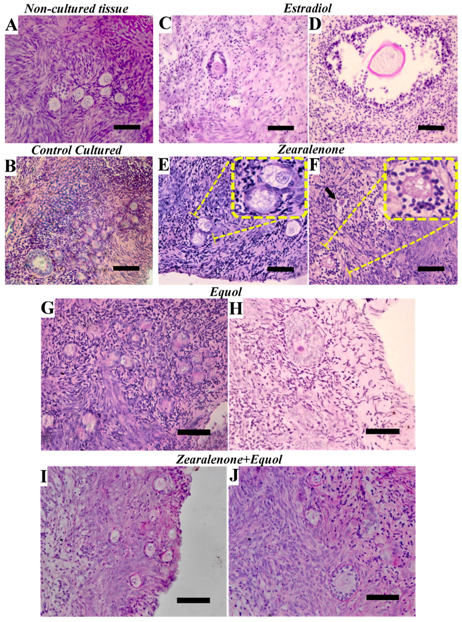
2.1. Morphology and Density of Preantral Follicles

During in vitro preantral follicle culture, morphological changes are observed according to the follicular development (e.g., primordial, primary, or secondary), and atresia can be detected by histological analysis. Ovarian pieces were cultured in vitro for three days to determine the effect of ZEN and equol, alone or in combination, on follicular development. Table 1 depicts the results obtained after morphological analysis. In vitro culture did not affect the percentage of morphologically normal primordial follicles when compared to non-cultured control.
Regarding primary and secondary follicles, their morphology was significantly impaired in all in vitro culture treatments, but with a prominently negative effect observed when ovarian fragments were exposed to E2. Although no differences were observed between ZEN and equol alone, a combination of ZEN with equol increased the percentage of morphologically normal secondary preantral follicles, being similar to untreated cultured tissue. Representative images of ovarian preantral follicles per class are given in Figure 1. It was remarkable that preantral follicles exposed to ZEN usually presented ooplasm vacuolisation.
During in vitro culture, it is expected that the pool of primordial follicles is activated for further development into primary and, subsequently, secondary follicles. The decrease in the density of primordial follicles with a concomitant increase in the density of secondary follicles was significant in the ovarian fragments cultured in non-supplemented medium, or in medium containing equol or ZEN + equol. The density of primordial follicles also decreased significantly during culture in the presence of E2, but without resulting in a significant increase in the density of primary or secondary follicles. When ovarian fragments were exposed to ZEN, a significant decrease of primordial follicle density was observed when compared to non-cultured control, followed by a significant increase in the density of primary follicles only (Table 2).

2.2. Nitrite and Malondialdehyde Levels in Culture Medium

None of the treatments affected nitrite and malondialdehyde (MDA) levels in the culture medium.

2.3. Oestradiol Levels in Culture Medium

Although oestradiol levels in the medium supplemented with 3.12 μmol/L E2 were significantly the highest when compared to all other treatments, no effect of the ZEN or equol was observed (p = 0.323). Oestradiol levels were measured as 35.3, 68.4, 48.5, and 27.1 ng/mL in the non-supplemented, ZEN, equol, and ZEN + equol group, respectively.

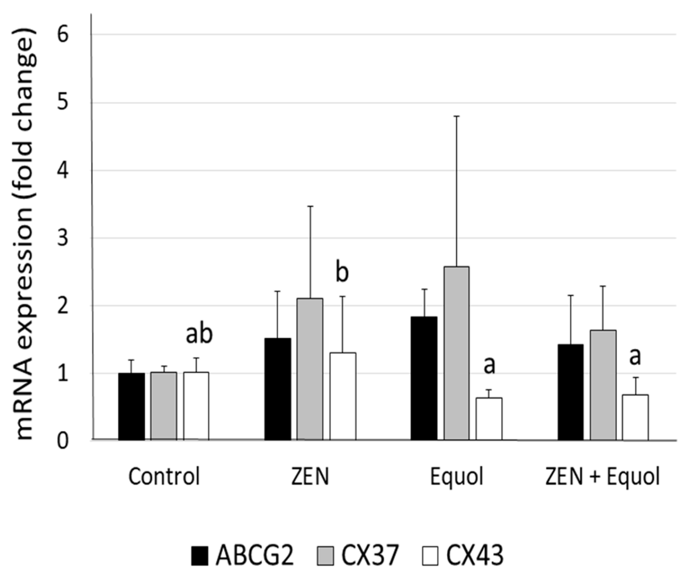
2.4. Quantitative RT-PCR

No significant differences were observed in the mRNA expression of ABCG2, CX37, and CX43 when ZEN, equol, or their combinations were compared to the control group. However, a significant decrease in CX43 expression was observed in ovarian tissue exposed to equol or ZEN + equol when compared with the tissues exposed to ZEN only (Figure 2).

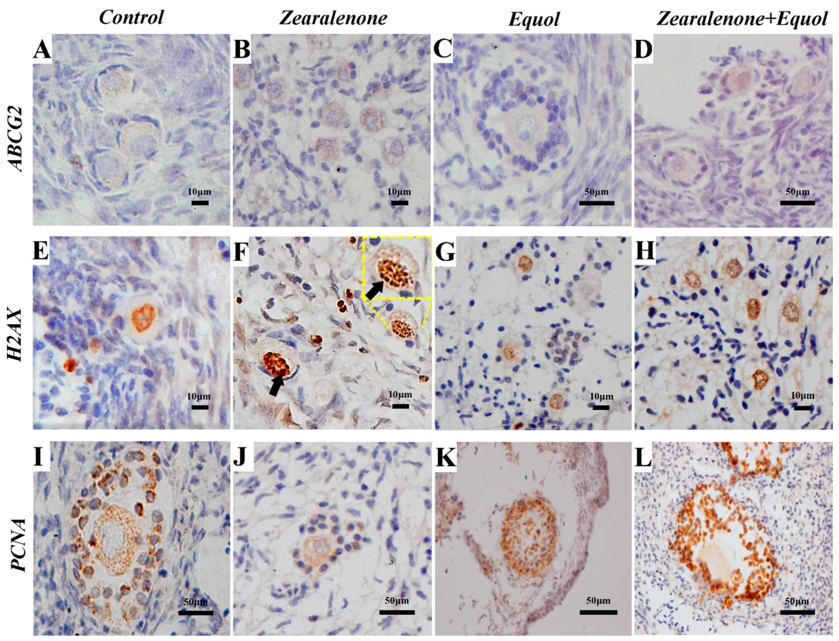
2.5. Immunohistochemistry

The percentages of follicles positively stained for ABCG2, H2AX, and PCNA at the different developmental stages (primordial, primary, and secondary) are detailed in Table 3. Representative images are given in Figure 3. All negative controls failed to show 3,3’-diaminobenzidine in chromogenic solution (DAB)-positive immunostaining, demonstrating the specificity of the markers.

2.5.1. ABCG2 Protein Expression

A total of 1669 follicles (1258 primordial, 334 primary, and 77 secondary follicles) were investigated, and no differences were observed among the groups.

2.5.2. H2AX Protein Expression

A total of 1609 follicles (921 primordial, 631 primary, and 57 secondary follicles) were investigated, and the results of immunohistochemistry showed that H2AX protein expression significantly increased in primordial follicles exposed to ZEN. No differences were observed between the control and equol groups. Furthermore, when ZEN was combined with equol, H2AX expression remained significantly higher than that observed in primordial follicles from the control group, but significantly lower than the rates of immunolabelled primordial follicles after exposure to ZEN.

2.5.3. PCNA Protein Expression

A total of 1151 follicles (903 primordial, 215 primary, and 33 secondary follicles) were investigated, and the results of immunohistochemistry showed that proliferation of granulosa cells, as labelled by PCNA protein expression, was similar in primordial follicles, regardless of the treatment. However, exposure to ZEN significantly decreased the rate of granulosa cell proliferation in primary and secondary follicles, and this negative effect was counteracted when ZEN was combined with equol in the culture medium.

3. Discussion

In the present study, we demonstrated that even at low levels (1 µmol/L), ZEN can impair the in vitro development of preantral follicles, but this negative impact is minimised when these follicles are simultaneously exposed to ZEN and equol. Previous in vitro studies have showed that ZEN inhibits testosterone production by AM-10 Leydig cells (8 µmol/L) [29], increases the apoptosis rate in rat Sertoli cells (5 µmol/L) [30], affects gene expression in granulosa cells from mouse (30 µmol/L) and sows (10 µmol/L) [31], and impairs bovine oocyte maturation (1 mmol/L) [32] and porcine blastocyst rates (5 µmol/L) [33]. Although most of these concentrations are far higher than those tested in the present study, comparisons cannot be performed due to the differences of cell types and animal species. This also explains why we could not find any indication of oxidative stress or E2 modulation due to ZEN. Furthermore, chronic exposure cannot always be mimicked in vitro, since the period of culture may also affect cell viability, and in the present study the main focus was on the effect of ZEN in combination with equol.
ZEN is rapidly metabolised by the gastrointestinal tract after ingestion, producing some metabolites that are even more toxic, such as α-zearalenol [34]. Unfortunately, it is not possible to draw a parallel between in vitro and in vivo exposure, since there are no reference values for toxicological ZEN levels in biological matrices [35]. Levels of ZEN in plasma were determined in horses as 0.008 µmol/L–2.54 µg/L 24 h after being fed a diet containing 1 ppm ZEN [36], in cattle as 0.5 nmol/L–0.15 µg/L after being fed 0.66 ppm ZEN [37], and in women at levels up to 0.6 µmol/L–182.88 µg/L [38]. Concentrations of porcine follicular fluid were measured in a range of 15.2–54.8 µg/L (0.05–0.17 µmol/L) [39]. However, this last exposure level cannot be translated to preantral follicles because their oocytes are not yet surrounded by the antral cavity containing the follicular fluid. Importantly, it is known that preantral follicles seem to be more sensitive than antral ones when exposed to ZEN.
In a transgenerational study, Schoevers et al. [7] showed that exposure to ZEN via placenta and during lactation negatively affected the preantral population in porcine ovaries, and degeneration was mostly characterised by oocyte vacuolisation. At similar concentrations to those used in the present study, ZEN (25 µg/L; 0.08 µmol/L) also caused vacuolisation in canine granulosa cells [40]. Such vacuolisation was also remarkable in the present study after exposure to ZEN, but this process was minimised when ZEN was combined with equol in the culture medium. Vacuolisation has been linked to autophagy in oocytes [7] and hepatocytes [41] as a result of ZEN exposure. Autophagy was induced in IPEC-J2 cells via activation of the p38/MAPK activation in response to ZEN [42]. In Sertoli cells, ZEN induces autophagy by the inhibition of the PI3K/Akt/mTOR signalling pathway [43]. On the other hand, equol leads to the activation of this pathway in MCF-7 cells [44]. This could be one of the pathways involved in the interaction of these two xenoestrogens. Nevertheless, considering the complexity of the mode of action of such compounds and the different studied cell types, it is still not possible to determine all the factors involved in their interaction.
Equol is a metabolite of the host microbiota after degradation of daidzein, an isoflavone found in different feedstuffs like soybeans and other legumes like alfalfa. Mahalingam et al. [45] demonstrated that equol at a concentration of 100 μmol/L inhibits growth and induces atresia in mice antral follicle cultures in vitro. These authors also showed minor endocrine effects when follicles were cultured in the presence of 6 or 36 μmol/L of equol. At a concentration of 20 μmol/L, equol is able to induce apoptosis via caspase-3 cleavage [46]. However, when evaluating the effects of this metabolite, plasma levels should be considered. For instance, equol was found at levels of 0.5 mg/L (circa 2 µmol/L) in plasma from sheep fed red clover [22], 2.4 µmol/L in rats [18], 0.4 µmol/L in monkeys [18], 0.005 µmol/L in weaned piglets [20], and 0.05 µmol/L in sows [21]. In humans, equol plasma levels range from 0.4 to 1.2 μmol [23]. Importantly, plasma levels of this isoflavone will depend on dietary composition and intake, gender, age, and microbiota composition [23,47].
It was previously described that rats and pigs fed ZEN at concentrations of 250 and 10 mg/kg diet, respectively, could overcome ZEN toxicity when simultaneously fed a fibre-rich alfalfa meal [48]. These authors could not determine the exact compound counteracting the effect of ZEN, and they did not measure the levels of important phytoestrogens like equol. Most recently, it was shown that the flavonoid chrysin is capable of protecting mouse spermatozoa against the deleterious effects caused by dietary exposure (via gavage) to ZEN (40 mg/kg) [49]. Like equol, chrysin has antioxidant and anti-inflammatory properties. Although in the present study we could not find evidence of oxidative stress in the culture medium, in further studies we should evaluate the tissue itself together with markers of antioxidant capacity. Besides this, other pathways involving equol activity should be considered. Although ZEN has a higher affinity to ERα, it can also bind to ERβ, and when binding to ERα, ZEN leads to a stronger effect than via ERβ [50]. In preantral follicles, ERβ is expressed in granulosa cells, while the expression of ERα occurs in granulosa cells from antral follicles [51,52], indicating that the binding site of ZEN was preferably ERβ. The toxic effect of ZEN on normal prostate cells can be increased by blocking the activity of ERβ, indicating that this receptor may play a protective role against this mycotoxin [53]. Furthermore, ERβ is crucial for the subsequent differentiation of granulosa cells and follicular development [54]. Phytoestrogens like genistein [55] and resveratrol [56] are known to induce ERβ, and the same effect is expected for equol. This may explain the positive effect of equol on the proliferation of primary and secondary follicles, even when exposed to ZEN. ZEN can also induce cell cycle arrest, as observed by the decrease in PCNA labelling of proliferating rates of granulosa cells from primary and secondary follicles. Again, this effect was counteracted by equol. Although the present study was performed with reproductive cells, it should be pointed out that ERβ is also found in the intestines, the first organ exposed after oral contamination with ZEN via diet.
Only in primordial follicles, ZEN exposure increased the expression of the H2AX, a protein involved in DNA restoration during DNA double-strand breaks. DNA double-strand breaks can induce oocyte death or activate a programme of DNA repair [57]. It appears that in the present study, developing follicles (primary and secondary) were not selected for DNA integrity restoration, but only the pool of reserve (i.e., the primordial ones). The presence of equol in the culture medium, regardless of the presence of ZEN, did not induce the expression of the H2AX protein, indicating DNA protection. Except for the two-fold difference in the expression of CX37 when comparing ZEN with equol, no other difference was observed in gene regulation. Furthermore, such low fold change should not be considered biologically significant. This can be explained by the short-term in vitro culture for these types of cells [58], as well as the tested concentrations of the substances. For instance, ABCG2 is usually upregulated by ZEN [27] and downregulated by equol [26], but no differences were observed in the present study.
In conclusion, equol may alleviate the toxic effect of ZEN by counteracting oocyte vacuolisation and supporting granulosa cell proliferation and subsequent follicular development. It seems that different pathways play a role, and this needs to be understood. Equol production will depend on the host microbiota, meaning that complex and well-functioning microbiota will help the host defense against ZEN. The microbial population may vary among individuals, which may explain the individual variation on the effects of ZEN toxicity within different animal species. Daidzein-rich diets as a source of substrate for microbial equol production might not be sufficient, and gut modulation with probiotics able to degrade daidzein into equol would improve gut health and, subsequently, animal reproduction. The present study was performed in vitro at two fixed concentrations of both xenoestrogens as a proof of principle. To confirm the present findings, in vivo studies are still needed.

4. Materials and Methods

4.1. Test Compounds

Unless mentioned otherwise, substances used in the present experiment were purchased from Chem Cruz (Santa Cruz Biotechnology, Inc., Dallas, TX, USA) and stock solutions were prepared using dimethyl sulfoxide (DMSO) as a solvent. The final DMSO concentration added to the culture medium was 0.1%. The culture medium and 17β-estradiol were purchased from Sigma Chemical Co. (St. Louis, MO, USA).

4.2. Animal Ethics Statement

This study was approved and conducted according to the Animal Management and Ethical Regulation Committee of the State University of Ceará (N° 9433833/2018). Ovarian tissues were collected in a commercial abattoir.

4.3. Ovarian Tissue Collection

Sheep ovarian pairs (n = 12) were collected from 12 adult sheep in a local abattoir immediately after slaughter. After collection, ovaries were washed one time in alcohol 70% for 10 s, followed by two washes in Minimum Essential Medium (MEM) supplemented with N-2-hydroxyethylpiperazine-N-ethanesulfonic acid (HEPES) (MEM-HEPES), penicillin (100 μg/mL) and streptomycin (100 μg/mL). Then, the ovaries were stored in sterile tubes with the same medium and transported to the laboratory at 4 °C within one hour.

4.4. Experimental Design

In an initial trial, each of nine sheep ovarian pairs was divided into six fragments of 3 × 3 × 1 mm, and randomly assigned to the different experimental conditions. Of these, one fragment was randomly selected as fresh control, and the remaining eight fragments were in vitro cultured in non-treated culture medium, or in culture medium supplemented with estradiol (E2), zearalenone (ZEN), equol, or combinations of ZEN and equol (ZEN + equol). All supplements were used at a concentration of 1 µm/L, except for the E2 that was added at a concentration of 3.12 μmol/L. The exposure to E2 during in vitro culture was based on its structural similarity with phytoestrogens, and the high tested concentration was chosen due to its known negative effect in oocytes [59]. Concentrations of the other test compounds were selected based on plasma levels of equol encountered in ovine (1 μmol/L) [22] and the previously tested level of ZEN in combination with xenoestrogen (1 μmol/L) [16]. After in vitro culture, ovarian fragments were fixed for histological analysis (morphology, follicular development and stromal density) and culture media were separately analysed for E2 levels, as well as for oxidative stress using nitrite and malondialdehyde (MDA) as markers.
Simultaneously, three pairs of sheep ovaries were divided into 14 fragments of 3 × 3 × 1 mm each, and randomly assigned to the different experimental conditions. The 14 fragments were in vitro cultured in non-treated culture medium, or in culture medium supplemented with ZEN, equol, or ZEN + equol (two fragments per treatment). All supplements were used at a concentration of 1 µm/L. After in vitro culture, the fragments were assessed via immunohistochemistry and quantitative real-time polymerase chain reaction (qRT-PCR).

4.5. In Vitro Culture

Fragments of ovarian cortex were individually in vitro cultured in 24-well plates containing 1 mL of culture medium each. Culture medium consisted of α-MEM supplemented with 10 μg/mL insulin, 5.5 μg/mL transferrin, 5 ng/mL selenium, 2 mg/L glutamine, 2 mg/L hypoxanthine, and 1.25 mg/mL bovine serum albumin (BSA). According the experimental group, ovarian fragments were cultured in the absence or presence of the test compounds, as mentioned above. Dimethyl sulfoxide (DMSO) at a concentration of 0.1% was used as dilution vehicle and was added as well in the non-supplemented culture medium. The fragments were then cultured for three days at 38 °C in humidified air with a 5% CO2 atmosphere.

4.6. Histological Analysis

The ovarian fragments were fixed in 4% paraformaldehyde and then dehydrated at increasing ethanol concentrations. Subsequently, these fragments were embedded in paraffin and cut into 7 μm sections, mounted under glass slides and stained with Periodic Acid-Schiff (PAS) and haematoxylin. For morphological evaluation, slides were examined under an optical microscope (Nikon, Tokyo, Japan) at 400× magnification. The analysed preantral follicles were classified into primordial (oocyte surrounded by a layer of squamous granulosa cells), primary (oocyte surrounded by a layer of cubic granulosa cells), or secondary (oocyte surrounded by two or more layers of cubic granulosa cells) [60]. In order to avoid double counting, follicles were counted only in sections where their oocyte nucleus was observed. Follicles were classified as morphologically normal or degenerated considering the following characteristics: absence or presence of pycnotic bodies, cytoplasmic retraction and granulosa cell organization, as described by Santos et al. [61].

4.7. Density of Preantral Follicles

The density of preantral follicles, per development group or as total, was calculated as the total number of follicles, per class, divided by the total tissue volume and expressed as the number of follicles/mm3 of ovarian tissue [62]. The volume of the analysed ovarian sections was calculated by summing the number of sections of area multiplied by the thickness of each section.

4.8. Nitrite and Malondialdehyde Quantification in Culture Medium

Exposure to ZEN may result in oxidative stress and cell membrane damage, which can be characterized by an increase in nitric oxide production and lipid peroxidation [63]. Such an effect can be determined in cell culture suspensions, where an increase in nitrite levels are indicative of oxidative stress and membrane lipid peroxidation will result in increased malondialdehyde (MDA) levels. For the evaluation of Nitrite and MDA levels, 200 μL of culture medium from each treatment were collected and stored in a freezer −80 °C for later measurement. For Nitrite levels, a protocol already established by Green et al. [64] was applied. In summary, 100μL of Griess reactive (1% sulfanylamide, 0.1% N-(1-naphthyl)-ethylenediamine hydrochloride, H3PO4 in 1%, 1:1:1:1 distilled water) was added to 100 μL of the culture medium. Then the samples were incubated at room temperature for 10 min. The standard curve was elaborated with various sodium Nitrite concentrations (ranging from 0.25 to 2.378 μM), and the samples were evaluated at a wavelength of 560 nm using a microplate reader (Metertech Inc., Taipei, Taiwan). The blank (negative control) was prepared by adding 100 μL of Griess reactive to 100 μL of the base medium.
To assess MDA levels, the TBARS (thiobarbituric acid reactive substances) method was used. For this, 100 μL of the culture medium was used and incubated in a water bath at 37 °C for 1 h. Subsequently, 80 μL of 35% perchloric acid was added and centrifuged at 14,000 rpm at 4 °C for 10 min. Thereafter, 50 μL of thiobarbituric acid was added to the supernatant and the mixture was incubated at 100 °C for 30 min [65]. After cooling to room temperature, the measurement was performed by absorbance at a wavelength of 532 nm in a microplate reader (Metertech Inc., Taipei, Taiwan).

4.9. Estradiol Dosage

To measure estradiol levels in the culture media, the enzyme-linked fluorescent assay (ELFA) was applied according to the manufacturer’s instructions (VIDAS® Estradiol II, refª 30431 bioMérieux SA, 376 Chemin de L’Orme, 69280 Marcy-l’Etoile, France). The fluorescence intensity emission was recorded at 450 nm, and the fluorescence signal values were inversely proportional to the antigen concentration present in the samples. The coefficient of variation was of 7.5% and the lower limit of detection was of 9 pg/mL.

4.10. Immunohistochemistry

It was previously reported that expression of the multidrug resistance protein ATP-binding cassette, subfamily G, member 2 (ABCG2 or BCRP [breast cancer resistance protein]) can be modulated by both equol and ZEN [26,27]. Signs of DNA repair due stress were detected by H2A histone family member X (H2AX) labelling [57]. Proliferation rate of granulosa cells per follicle was determined by proliferating cell nuclear antigen (PCNA) labelling. Paraffin sections were deparaffinized with xylene (Xylene, Sigma-Aldrich, St. Louis, MO, USA) and rehydrated in alcohol series. The endogenous peroxidase activity was blocked with 3% H2O2 diluted in methanol. A demasking step was performed for 75 min at 98 °C with citrate buffer and 20% Triton X100 before sections were subjected to antigen retrieval step. Antigen retrieval steps, antibody dilutions and incubation conditions are summarized in Table 1. In brief, slides were incubated for 30 min at room temperature with anti-PCNA rabbit polyclonal primary antibody (Abcam Inc., Cambridge, MA, USA), anti-H2AX (Abcam Inc.), or anti-ABCG2 (Novus Biologicals, Centennial, CO, USA). Subsequently, slides were incubated for 30 min in the Goat anti-rabbit IgG secondary antibody (1: 200 dilution; Abcam) (Table 4). The slides were then incubated for 30 min with the avidin-biotin enzyme complex (ABC; Vector laboratories, Burlingame, CA, USA) to react with 3,3’-diaminobenzidine in chromogenic solution (DAB; Dako Inc., Santa Clara, CA, USA). Hematoxylin and Scott’s solution were used for counterstaining. Negative controls were performed under the same conditions, but in the absence of the primary antibody. The following positive controls were used: spleen for PCNA and poisoned heart for H2AX. For quantitative analysis, follicles were considered PCNA-positive when at least one granulosa cell was stained and were considered as belonging to the growing pool (primary or secondary follicles), H2AX-positive when the oocyte was immunostained; ABCG2-positive when oocytes and or granulosa cells were immunostained.

4.11. Ribonucleic Acid (RNA) Extraction and Quantitative Real-Time PCR

Besides ABCG2 modulation by ZEN, this mycotoxin may also impair cellular gap junctions [66]. Therefore, mRNA expression of ABCG2 and gap junction proteins present in ovarian follicles, e.g., Connexin 37 (CX37) and -43 (CX43) was determined. For evaluation of gene expression levels for ABCG2, CX37 and CX43, the ovarian fragments were submitted to RNA extraction with TRIzol® Plus RNA Purification Kit (Invitrogen, São Paulo, SP, Brazil). Isolated RNA preparations were treated with DNase I and Pure Link ™ Mini RNA Kit (Invitrogen, Sao Paulo, SP, Brazil). Complementary DNA (cDNA) was synthesized from RNA isolated using the SuperscriptTM II RNase H Reverse Transcriptase (Invitrogen, Sao Paulo, Brazil). The qPCR reactions had a final volume of 20 μL and contained the following components: 1 μL cDNA as template in 7.5 μL SYBR Green Master Mix (PE Applied Biosystems, Foster City, CA, USA), 5.5 μL of ultrapure water and 0.5 μL of each primer. Primers were designed to amplify ABCG2, CX37 and CX43 mRNA levels (Table 5). The reference genes used as endogenous control for normalization, gene expression evaluation and gene expression stability in all samples were glyceraldehyde-3-phosphate dehydrogenase (GAPDH) and β-actin (ACTB). Primer specificity and amplification efficiency were verified for each gene. Expression stability of these genes was analysed using BestKeeper software. The thermal cycle profile for the first qRT-PCR cycle was as follows: initial denaturation and polymerase activation for 15 min at 94 °C, followed by 40 cycles of 15 s at 94 °C, 30s at 60 °C and 45 s at 72 °C. The final extension was 10 min at 72 °C. All reactions were performed on a real-time PCR Mastercycler (Eppendorf, Germany, Hamburg, Germany, USA). Data were analysed using the efficiency-corrected Delta-Delta-Ct method [67]. The fold-change values of the genes of interest were normalized using the geometric mean of the fold-change values of two housekeeping genes.

4.12. Statistical Analysis

The ovarian fragments were randomly assigned to treatment conditions prior each experiment. Statistical analysis was conducted with the GenStat statistical software (GenStat for Windows 18th Edition, VSN International, Hemel, Hempstead, UK; https://www.vsni.co.uk/downloads/genstat/). Data were compared with ANOVA. The null hypothesis was that there was no treatment effect on the response parameter. Treatment means were compared according Fishers’ LSD (for a two-sided test and p ≤ 0.05) after a significant treatment effect was confirmed by ANOVA. Data are presented as mean ± SEM. The p-value of the statistical model is given per response parameter. Effects with p ≤ 0.05 were considered to be statistically significant.

Author Contributions

Conceptualization: R.R.d.S., D.C.C.d.B. and J.R.d.F.; Funding acquisition: J.R.d.F., A.P.R.R., R.R.d.S. and M.I.F.G.; Investigation: T.E.S.S., D.C.C.d.B., N.A.R.d.S., R.F.d.S., A.C.A.F. and J.Y.G.S.; Project administration: R.R.d.S., D.C.C.d.B. and J.R.d.F.; Validation: T.E.S.S., D.C.C.d.B., A.C.A.F., J.Y.G.d.S., A.P.R.R., R.R.d.S., D.C.C.d.B. and J.R.d.F.; Writing original draft: T.E.S.S., D.C.C.d.B. and R.R.d.S.; Writing review & editing: D.C.C.d.B., R.R.d.S. and J.R.d.F.; Supervision: D.C.C.d.B., R.R.d.S. and J.R.d.F.

Funding

This study was supported by PRONEX/FUNCAP/CNPq grant (number-PR2-0101-00049.01.00/15). The authors would like to thank the CNPq for supporting the project.

Conflicts of Interest

The authors declare no conflict of interest.

Abbreviations

ABCG2/BCRPATP-binding cassette, subfamily G, member 2/breast cancer resistance protein
ACTBBeta actin
ANOVAAnalysis of variance
BSABovine serum albumin
cDNAComplementary deoxyriboNucleic Acid
CX37Connexin 37
CX43Connexin 43
DAB3,3’-diaminobenzidine in chromogenic solution
DMSODimethyl sulfoxide
DNADeoxyriboNucleic Acid
E217-β-oestradiol
ELFAEnzyme-linked fluorescent assay
ER-αOestrogen receptor alpha
ER-βOestrogen receptor beta
GAPDHGlyceraldehyde 3-phosphate dehydrogenase
H2AXH2A histone family member X
HEPESN-2-hydroxyethylpiperazine-N-ethanesulfonic acid
H2O2Hydrogen peroxide
H3PO4Phosphoric acid
IgGImmunoglobulin G
IPEC-J2Intestinal Porcine Epithelial Cell line-J2
LSDLeast significant difference
MA-10 Leydig cellsMouse Leydig tumor cell line
MCF-7Breast cancer cells (Michigan Cancer Foundation-7)
MDAMalondialdehyde
MEMMinimum essential medium
PASPeriodic Acid-Schiff
PCNAProliferating cell nuclear antigen
PHSProstaglandin H synthase
PI3K/Akt/mTORPhosphatidylinositol 3-kinase/serine-threonine protein kinase/mammalian target of rapamycin
P38/MAPKp38 mitogen-activated protein kinase
qRT-PCRQuantitative real-time polymerase chain reaction
RNARibonucleic acid
TBARSThiobarbituric acid reactive substances
ZENZearalenone

References

  1. Gruber-Dorninger, C.; Jenkins, T.; Schatzmayr, G. Global mycotoxin occurrence in feed: A ten-year survey. Toxins 2019, 11, 375. [Google Scholar] [CrossRef] [PubMed]
  2. Zielonka, L.; Waskiewicz, A.; Beszterda, M.; Kostecki, M.; Dabrowksi, M.; Obremski, K.; Golinski, P.; Gajecki, M. Zearalenone in the intestinal tissues of immature gilts exposed per os to mycotoxins. Toxins 2015, 7, 3210–3223. [Google Scholar] [CrossRef] [PubMed]
  3. EFSA Panel on Contaminants in the Food Chain (CONTAM). Scientific Opinion on the risks for public health related to the presence of zearalenone in food. EFSA J. 2011, 9, 2197. [Google Scholar] [CrossRef]
  4. Mauro, T.; Hao, L.; Pop, L.C.; Buckley, B.; Schneider, S.H.; Bandera, E.V.; Shapses, S.A. Circulating zearalenone and its metabolites differ in women due to body mass index and food intake. Food Chem. Toxicol. 2018, 116, 227–232. [Google Scholar] [CrossRef] [PubMed]
  5. Minervini, F.; Dell’Aquila, M.E. Zearalenone and reproductive function in farm animals. Int. J. Mol. Sci. 2008, 9, 2570–2584. [Google Scholar] [CrossRef] [PubMed]
  6. Sambuu, R.; Takagi, M.; Namula, Z.; Otoi, T.; Shiga, S.; Rodrigues dos Santos, R.; Fink-Gremmels, J. Effects of exposure to zearalenone on porcine oocytes and sperm during maturation and fertilization in vitro. J. Reprod. Dev. 2011, 57, 547–550. [Google Scholar] [CrossRef] [PubMed]
  7. Schoevers, E.J.; Santos, R.R.; Colenbrander, B.; Fink-Gremmels, J.; Roelen, B.A. Transgenerational toxicity of zearalenone in pigs. Reprod. Toxicol. 2012, 34, 110–119. [Google Scholar] [CrossRef]
  8. Sambuu, R.; Takagi, M.; Namula, Z.; Nii, M.; Taniguchi, M.; Uno, S.; Kokushi, E.; Tshering, C.; dos Santos, R.R.; Fink-Gremmels, J.; et al. Effects of long-term in vitro exposure of ejaculated boar sperm to zearalenone and α-zearalenol in sperm liquid storage medium. Anim. Sci. J. 2013, 84, 28–34. [Google Scholar] [CrossRef]
  9. Gallo, A.; Giuberti, G.; Frisvad, J.C.; Bertuzzi, T.; Nielsen, K.F. Review on mycotoxin issues in ruminants: Occurrence in forages, effects of mycotoxin ingestion on health status and animal performance and practical strategies to counteract their negative effects. Toxins 2015, 7, 3057–3111. [Google Scholar] [CrossRef]
  10. Santos, R.R.; Schoevers, E.J.; Roelen, B.A.J.; Fink-Gremmels, J. Mycotoxins and female reproduction: In vitro approaches. World Mycot. J. 2013, 6, 245–253. [Google Scholar] [CrossRef]
  11. Zheng, W.; Wang, B.; Li, X.; Wang, T.; Zou, H.; Gu, J.; Yuan, Y.; Liu, X.; Bai, J.; Bian, J.; et al. Zearalenone promotes cell proliferation or causes cell death? Toxins 2018, 10, 184. [Google Scholar] [CrossRef] [PubMed]
  12. Filannino, A.; Sout, T.A.E.; Gadella, B.M.; Sostaric, E.; Pizzi, F.; Colenbrander, B.; Dell’Aquila, M.E.; Minervini, F. Dose-response effects of estrogenic mycotoxins (zearalenone, alpha- and beta-zearalenol) on motility, hyperactivation and the acrosome reaction of stallion sperm. Reprod. Biol. Endocrinol. 2001, 9, 134. [Google Scholar] [CrossRef] [PubMed]
  13. Malekinejad, H.; Schoevers, E.J.; Daemen, I.J.; Zijlstra, C.; Colenbrander, B.; Fink-Gremmels, J.; Roelen, B.A.J. Exposure of oocytes to the Fusarium toxins zearalenone and deoxynivalenol causes aneuploidy and abnormal embryo development in pigs. Biol. Reprod. 2007, 77, 840–847. [Google Scholar] [CrossRef] [PubMed]
  14. Rietjens, I.M.C.M.; Louisse, J.; Beekmann, K. The potential health effects of dietary phytoestrogens. Br. J. Phasmacol. 2017, 174, 1263–1280. [Google Scholar] [CrossRef]
  15. Fleck, S.C.; Churchwell, M.I.; Doerge, D.R.; Teeguarden, J.G. Urine and serum biomonitoring of exposure to environmental estrogen II: Soy isoflavones and zearalenone in pregnant women. Food Chem. Toxicol. 2016, 95, 19–27. [Google Scholar] [CrossRef]
  16. Vejdovszky, K.; Schmidt, V.; Warth, B.; Marko, D. Combinatory estrogenic effects between the isoflavones genistein and the mycotoxins zearalenone and alternariol in vitro. Mol. Nutry. Food Res. 2017, 61, 1600526. [Google Scholar] [CrossRef]
  17. Mayo, B.; Vasquez, L.; Florez, A.B. Equol: A bacterial metabolite from the daidzein isoflavone and its presumed beneficial health effects. Nutrients 2019, 11, 2231. [Google Scholar] [CrossRef]
  18. Gu, L.; House, S.E.; Prior, R.L.; Fang, N.; Ronis, M.J.; Clarkson, T.B.; Wilson, M.E.; Badger, T.M. Metabolic phenotype of isoflavonces differ among female rats, pigs, monkeys, and women. J. Nutr. 2006, 136, 1215–1221. [Google Scholar] [CrossRef]
  19. Schwen, R.J.; Nguyen, L.; Jackson, R.L. Elucidation of the metabolic pathway of S-equol in rat, monkey and man. Food Chem. Toxicol. 2012, 50, 2074–2083. [Google Scholar] [CrossRef]
  20. Kuhn, G.; Hennig, U.; Kalbe, C.; Rehfeldt, C.; Ren, M.Q.; Moors, S.; Degen, G.H. Growth performance, carcass characteristics and bioavailability of isoflavones in pigs fed soy bean based diets. Arch. Anim. Nutr. 2004, 58, 265–276. [Google Scholar] [CrossRef]
  21. Farmer, C.; Robertson, P.; Xiao, C.; Rehfeldt, C.; Kalbe, C. Exogenous genistein in late gestation: Effects on fetal development and sow and piglet performance. Animal 2016, 10, 1423–1430. [Google Scholar] [CrossRef] [PubMed]
  22. Schutt, D.A.; Braden, W.H. The significance of equol in relation to the oestrogenic responses in sheep ingesting clover with a high formononetin content. Aust. J. Agric. Res. 1968, 19, 545–553. [Google Scholar] [CrossRef]
  23. Van der Velpen, V.; Hollman, P.C.; van Nielen, M.; Schouten, E.G.; Mensink, M.; Van’t Veer, P.; Geelen, A. Large inter-individual variation in isoflavone plasma concentration limits use of isoflavone intake data for risk assessment. Eur. J. Clin. Nutr. 2014, 68, 1141–1147. [Google Scholar] [CrossRef] [PubMed] [Green Version]
  24. Pazourekova, S.; Lucova, M.; Nosal, R.; Drabikova, K.; Harmatha, J.; Smidrkal, J.; Jancinova, V. Equol effectively inhibity toxic activity of human neutrophils without influencing their viability. Pharmacology 2016, 97, 138–145. [Google Scholar] [CrossRef]
  25. Degen, G.H.; Blaich, G.; Metzler, M. Multiple pathways for the oxitadive metabolism of estrogens in the Syrian hamster and rabbit kidney. J. Biochem. Toxicol. 1990, 5, 91–97. [Google Scholar] [CrossRef] [PubMed]
  26. Rigalli, J.P.; Scholz, P.N.; Tocchetti, G.N.; Ruiz, M.L.; Weiss, J. The phytoestrogens daidzein and equol inhibit the drug transporter BCRP/ABCG2 in breast cancer cells: Potential chemosensitizing effect. Eur. J. Nutr. 2019, 58, 139–150. [Google Scholar] [CrossRef]
  27. Xiao, J.; Wang, Q.; Bircsak, K.M.; Wen, X.; Aleksunes, L.M. In vitro screening of environmental chemicals identifies zearalenone as a novel substrate of the plancental BCRP/ABCG2 transporter. Toxicol. Res. 2015, 4, 695–706. [Google Scholar] [CrossRef]
  28. Cao, H.; Zhi, Y.; Xu, H.; Fang, H.; Jia, X. Zearalenone causes embryotoxicity and induces oxidative stress and apoptosis in differentiated human embryonic stem cells. Toxicol. In Vitro 2019, 54, 243–250. [Google Scholar] [CrossRef]
  29. Eze, U.A.; Huntriss, J.D.; Routledge, M.N.; Gong, Y.Y. In vitro effects of single and binary mixtures of regulated mycotoxins and persistent organochloride pesticides on steroid hormone production in MA-10 Leydig cell line. Toxicol. In Vitro 2019, 60, 272–280. [Google Scholar] [CrossRef]
  30. Cai, G.; Si, M.; Li, X.; Zou, H.; Gu, J.; Yuan, Y.; Liu, X.; Liu, Z.; Bian, J. Zearalenone induces apoptosis of rat Sertoli cells through Fas-Fas ligand and mitochondrial pathway. Environ. Toxicol. 2019, 34, 424–433. [Google Scholar] [CrossRef]
  31. Zhang, G.L.; Song, J.L.; Zhou, Y.; Zhang, R.Q.; Cheng, S.F.; Sun, X.F.; Qin, G.Q.; Shen, W.; Li, L. Differentiation of sow and mouse ovarian granulosa cells exposed to zearalenone in vitro using RNA-seq gene expression. Toxicol. Appl. Pharmacol. 2018, 350, 78–90. [Google Scholar] [CrossRef] [PubMed]
  32. Takagi, M.; Mukai, S.; Kurivagawa, T.; Takagaki, K.; Uno, S.; Kokushi, E.; Otoi, T.; Budivanto, A.; Shirasuna, K.; Miyamoto, A.; et al. Detection of zearalenone and its metabolites in naturally contaminated follicular fluids by using LC/MS/MS and in in vitro effects of zearalenone on oocyte maturation in cattle. Reprod. Toxicol. 2008, 26, 164–169. [Google Scholar] [CrossRef] [PubMed]
  33. Xu, Y.; Zhang, K.H.; Sun, M.H.; Lan, M.; Wan, X.; Zhang, Y.; Sun, S.C. Protective effects of melatonin against zearalenone toxicity on porcine embryos in vitro. Front. Pharmacol. 2019, 10, 327. [Google Scholar] [CrossRef] [PubMed]
  34. Pajewska, M.; Lojko, M.; Cendrowski, K.; Sawicki, W.; Kowalkowski, T.; Buszewski, B.; Gadzala-Kopciuch, R. The determination of zearalenone and ist major metabolites in endometrial cancer tissues. Anal. Bioanal. Chem. 2018, 410, 1571–1582. [Google Scholar] [CrossRef]
  35. Hennig-Pauka, S.; Koch, F.J.; Schaumberger, S.; Woechtl, B.; Novak, J.; Sulyok, M.; Nagl, V. Current challenges in the diagnosis of zearalenone toxicosis as illustrated by a field case of hyperestrogenism in suckling piglets. Porc. Health Manag. 2018, 4, 18. [Google Scholar] [CrossRef]
  36. Songsermsakul, P.; Bohm, J.; Aurich, C.; Zentek, J.; Razzazi-Fazeli, E. The levels of zearalenone and its metabolites in plasma, urine and faeces of horses fed with naturally, Fusarium toxin-contaminated oats. J. Anim. Physiol. Anim. Nutr. 2013, 97, 155–161. [Google Scholar] [CrossRef]
  37. Winkler, J.; Kersten, S.; Meyer, U.; Engelhardt, U.; Danicke, S. Residues of zearalenone (ZEN), deoxynivalenol (DON) and their metabolites in plasma of dairy cows fed Fusarium contaminated maize and their relationships to performance parameters. Food Chem. Toxicol. 2014, 65, 196–204. [Google Scholar] [CrossRef]
  38. Kuciel-Lisieska, G.; Obremski, K.; Stelmachow, J.; Gajecka, M.; Zielonka, U.; Jakimiuky, E.; Gajecki, M. Presence of zearalenone in blood plasma in women with neoplastic lesions in the mammary gland. Bull. Vet. Inst. Pulawy 2008, 52, 671–674. [Google Scholar]
  39. Sambuu, R.; Takagi, M.; Shiga, S.; Uno, S.; Kokushi, E.; Namula, Z.; Otoi, T.; Miyamoto, A.; Deguchi, E.; Fink-Gremmels, J. Detection of zearalenone and its metabolites in naturally contaminated porcine follicular fluid by using liquid chromatography-tandem mass spectrometry. J. Reprod. Dev. 2011, 57, 303–306. [Google Scholar] [CrossRef]
  40. Skorska-Wyszynska, E.; Jakimiuk, E.; Gajecka, M.; Mlynarczuk, J.; Obremski, K.; Gajecki, M. Preliminary evaluation of influence of zearalenone on cocultures of granulosa and internal theca cells of ovarian follicles in bitches in in vitro culture. Pol. J. Vet. Sci. 2004, 7, 305–309. [Google Scholar]
  41. Tiemann, U.; Brüssow, K.; Küchenmeister, U.; Jonas, L.; Kohlschein, P.; Pöhland, R.; Dänicke, S. Influence of diets with cereal grains contaminated by graded levels of two Fusarium toxins on selected enzymatic and histological parameters of liver in gilts. Food Chem. Toxicol. 2006, 44, 1228–1235. [Google Scholar] [CrossRef] [PubMed]
  42. Shen, T.; Miao, Y.; Ding, C.; Fan, W.; Liu, S.; Lv, Y.; Gao, X.; De Boevre, M.; Yan, L.; Okoth, S.; et al. Activation of the p38/MAPK pathway regulates autophagy in response to the CYPOR-dependent oxidative stress induced by zearalenone in porcine intestinal epithelial cells. Food Chem. Toxicol. 2019, 131, 110527. [Google Scholar] [CrossRef] [PubMed]
  43. Wang, B.J.; Zheng, W.I.; Feng, N.N.; Wang, T.; Zou, H.; Gu, J.H.; Yuan, Y.; Liu, X.Z.; Liu, Z.P.; Bian, J.C. The effects of autophagy and pi3k/akt/m-tor signaling pathway on the cell-cycle arrest of rats primary sertoli cells induced by zearalenone. Toxins 2018, 10, 398. [Google Scholar] [CrossRef] [PubMed]
  44. Liu, H.; Hu, C.; Wu, X.; Li, Z. Equol elicits estrogenic activities via PI3K/akt pathway in the estrogen receptor-positive MCF-7 cells. Mol. Cell. Toxicol. 2014, 10, 285–291. [Google Scholar] [CrossRef]
  45. Mahalingam, S.; Gao, L.; Gonnering, M.; Helferich, W.; Flaws, J.A. Equol inhibits growth, induces atresia, and inhibits steroidogenesis of mouse antral follicles in vitro. Toxicol. Appl. Pharmacol. 2016, 295, 47–55. [Google Scholar] [CrossRef] [Green Version]
  46. Yang, Z.; Zhao, Y.; Yao, Y.; Li, J.; Wang, W.; Wu, X. Equol Induces Mitochondria-Dependent Apoptosis in Human Gastric Cancer Cells via the Sustained Activation of ERK1/2 Pathway. Mol. Cells 2016, 39, 742–749. [Google Scholar] [CrossRef] [Green Version]
  47. Zheng, W.; Zhang, X.; Yao, W. Individual difference in faecal and urine equol excretion and their correlation with intestinal microbiota in large white sows. Anim. Prod. Sci. 2016, 57, 262–270. [Google Scholar] [CrossRef]
  48. Stangroom, K.E.; Smith, T.K. Effect of whole and fractioniated dietary alfalfa meal on zearalenone toxicosis and metabolism in rats and swine. Can. J. Physiol. Pharmacol. 1984, 62, 1219–1224. [Google Scholar] [CrossRef]
  49. Del Fabbro, L.; Jesse, C.R.; de Gomes, M.G.; Borges Filho, C.; Donato, F.; Souza, L.C.; Goes, A.R.; Furian, A.F.; Boeira, S.P. The flavonoid chrysin protects against zearalenone induced reproductive toxicity in male mice. Toxicon 2019, 165, 13–21. [Google Scholar] [CrossRef]
  50. Kuiper, G.G.J.M.; Lemmen, J.G.; Carlsson, B.; Corton, J.C.; Safe, S.H.; van der Saag, P.T.; Van der Burg, B.; Gastafsson, J.A. Interaction of estrogenic chemicals and phytoestrogens with estrogen receptor beta. Endocrinology 1998, 139, 4252–4563. [Google Scholar] [CrossRef]
  51. Saunders, P.T.K.; Millar, M.R.; Williams, K.; Macpherson, S.; Harkiss, D.; Anderson, R.A.; Orr, B.; Groome, N.P.; Scobie, G.; Fraser, H.M. Differential expression of estrogen receptor-a and -b and androgen receptor in the ovaries of marmosets and humans. Biol. Reprod. 2000, 63, 1098–1105. [Google Scholar] [CrossRef] [PubMed]
  52. Lenie, S.; Smitz, J. Estrogen receptor subtypes localization shifts in cultured mouse ovarian follicles. Histochem. Cell Biol. 2008, 129, 827–840. [Google Scholar] [CrossRef] [PubMed]
  53. Kowalska, K.; Habrowska-Gorcznska, D.E.; Urbanek, K.A.; Dominska, K.; Sakowicz, A.; Piastowska-Ciesjelska, A.W. Estrogen receptor β plays a protective role in zearalenone-induced oxidative stress in normal prostate epithelial cells. Ecotoxicol. Environ. Saf. 2019, 172, 504–513. [Google Scholar] [CrossRef] [PubMed]
  54. Couse, J.F.; Yates, M.M.; Deroo, B.J.; Korach, K.S. Estrogen receptor-beta is critical to granulosa cell differentiation and the ovulatory response to gonadotropins. Endocrinology 2005, 146, 3247–3262. [Google Scholar] [CrossRef] [PubMed]
  55. Drummond, A.E.; Fuller, P.J. The importance of ERb signalling in the ovary. J. Endocrinol. 2010, 205, 15–23. [Google Scholar] [CrossRef] [PubMed]
  56. Henry, L.A.; Witt, D.M. Resveratrol: Phytoestrogen effects on reproductive physiology and behavior in female rats. Horm. Behav. 2002, 41, 220–228. [Google Scholar] [CrossRef] [PubMed]
  57. Winship, A.L.; Stringer, J.M.; Liew, S.H.; Hutt, K.J. The importance of DNA repair for maintaining oocyte quality in response to anti-cancer treatments, environmental toxins and maternal ageing. Hum. Reprod. 2018, 24, 119–134. [Google Scholar] [CrossRef] [Green Version]
  58. Figueiredo, J.R.; Rodrigues, A.P.R.; Silva, J.R.V.; Santos, R.R. Cryopreservation and in vitro culture of caprine preantral follicles. Reprod. Fertil. Dev. 2011, 23, 40–47. [Google Scholar] [CrossRef]
  59. Solak, K.A.; Santos, R.R.; van den Berg, M.; Blaauboer, B.J.; Roelen, B.A.; van Duursen, M.B. Naringenin (NAR) and 8-prenylnaringenin (8-PN) reduce the developmental competence of porcine oocytes in vitro. Reprod. Toxicol. 2014, 49, 1–11. [Google Scholar] [CrossRef]
  60. Sales, A.D.; Duarte, A.B.; Santos, R.R.; Alves, K.A.; Lima, L.F.; Rodrigues, G.Q.; Brito, I.R.; Lobo, C.H.; Bruno, J.B.; Locatelli, Y.; et al. Modulation of aquaporins 3 and 9 after exposure of ovine ovarian tissue to cryoprotectants followed by in vitro culture. Cell Tissue Res. 2016, 365, 415–424. [Google Scholar] [CrossRef]
  61. Santos, R.R.; Rodrigues, A.P.; Costa, S.H.; Silva, J.R.; Matos, M.H.; Lucci, C.M.; Bao, S.N.; van den Hurk, R.; Figueiredo, J.R. Histological and ultrastructural analysis of cryopreserved sheep preantral follicles. Anim. Reprod. Sci. 2006, 91, 249–263. [Google Scholar] [CrossRef] [PubMed]
  62. Asadi-Azarbaijani, B.; Santos, R.R.; Jahnukainen, K.; Braber, S.; van Duursen, M.B.M.; Toppari, J.; Saugstad, O.D.; Nurmio, M.; Oskam, I.C. Developmentatl effects of imatinib mesylate on follicle assembly and early activation of primordial follicle pool in postnatal rat ovary. Reprod. Biol. 2017, 17, 25–33. [Google Scholar] [CrossRef] [PubMed]
  63. Barbasz, A.; Rudolphi-Skorska, E.; Filek, M.; Janeczko, A. Exposure of human lymphoma cells (U-937) to the action of a single mycotoxin as well as in mixtures with the potential protectors 24-epibrassinolide and selenium ions. Mycotoxin Res. 2019, 35, 89–98. [Google Scholar] [CrossRef]
  64. Green, L.C.; Tannenbaum, S.R.; Goldman, P. Nitrite synthesis in the germfree and conventional rat. Science 1981, 212, 56–58. [Google Scholar] [CrossRef] [PubMed]
  65. Draper, H.H.; Hadley, M. Malondialdehyde determination as index of lipid peroxidation. Methods Enzymol. 1990, 186, 421–431. [Google Scholar]
  66. Liu, M.; Gao, R.; Meng, Q.; Zhang, Y.; Bi, C.; Shan, A. Toxic effects of maternal zearalenone exposure on intestinal oxidative stress, barrier function, immunological and morphological changes in rats. PLoS ONE 2014, 9, e106412. [Google Scholar] [CrossRef] [PubMed]
  67. Pfaffl, M.W. A new mathematical model for relative quantification in real-time RT-PCR. Nucleic Acids Res. 2001, 29, e45. [Google Scholar] [CrossRef]
Figure 1. Representative images showing histological aspects of the preantral follicles before culture (A) primordial follicles, and after culture in the absence of xenoestrogens (B) follicles at different developmental stages, or in the presence of 3.12 μmol/L E2 (C,D) secondary follicles, 1 μmol/L ZEN (E) primordial follicles, (F) secondary follicle, 1 μmol/L Equol (G) primordial follicles, (H) secondary follicle), 1 μmol/L ZEN + 1 μmol/L Equol (I) primordial follicles, (J) secondary follicle with a primordial one). In panels E and F, inserts are given to show oocyte vacuolization in primordial and secondary follicles, respectively, after exposure to ZEN. Bar 50 µm. Staining: Haematoxylin-eosin.
Toxins 11 00652 g001
Figure 2. Mean (± SEM) levels of ABCG2, CX37 and CX43 in ovarian tissue cultured in the absence of xenoestrogens (control), or in the presence of 1 μmol/L ZEN, 1 μmol/L Equol or 1 μmol/L ZEN + 1 μmol/L Equol. a,b, different letters indicate significant difference among treatments (p ≤ 0.05).
Toxins 11 00652 g002
Figure 3. Representative images showing immunohistochemical staining for ABCG2 (AD), H2AX (EH), and PCNA (IL) in preantral follicles at different stages of development. The oolema of preantral follicles, regardless of the developmental stage and treatment, was similarly stained for ABCG2 (AD). The immunostaining of H2AX in the nucleus of preantral follicles is visible in panel F, where 3 or more foci per oocyte nucleus (see arrow). Decreased immunostaining was observed in granulosa cells from secondary follicles after culture in the presence of ZEN (J).
Toxins 11 00652 g003
Table 1. Mean (±SEM) percentages of morphologically normal preantral follicles cultured or not in the presence of zearalenone alone or combined with equol.
TreatmentsMorphologically Normal Preantral Follicles (%)
PrimordialPrimarySecondary
Non cultured control76.3 ± 12.6b78.0 ± 11.6c97.1 ± 6.39d
Cultured control64.4 ± 10.4b58.3 ± 16.5b74.7 ± 22.5c
Estradiol52.0 ± 12.3a36.2 ± 15.7a13.6 ± 19.3a
ZEN51.8 ± 11.2a45.1 ± 19.4ab50.6 ± 23.7b
Equol65.6 ± 14.6b48.1 ± 19.8ab53.6 ± 15.9bc
ZEN+Equol66.6 ± 14.2b60.2 ± 20.9b65.7 ± 25.4bc
p-values0.001 <0.001 <0.001
a–d different letters indicate significant difference among treatments (p ≤ 0.05).
Table 2. Mean (±SEM) density of preantral follicles cultured or not in the presence of zearalenone alone or combined with equol.
TreatmentsDensity of Preantral Follicles (Follicles/mm3)
PrimordialPrimarySecondary
Non cultured control237 ± 53b53 ± 18a3.2 ± 2.1a
Cultured control154 ± 47a69 ± 40a10.9 ± 3.1bc
Estradiol93 ± 48a55 ± 27a7.6 ± 6.7ab
ZEN151 ± 98a127 ± 59b8.5 ± 7.7ab
Equol115 ± 78a58 ± 33a18.6 ± 10.8c
ZEN+Equol81 ± 48a81 ± 33a12.1 ± 8.3bc
p-values0.001 0.002 0.042
a–c different letters indicate significant difference among treatments (p ≤ 0.05).
Table 3. Summary of the expression of ABCG2, H2AX, and PCNA protein in ovarian tissue.
FactorsControl
n (% stained)
ZEN
n (% stained)
Equol
n (% stained)
ZEN + Equol
n (mean % ± SEM)
p-Values
ABCG2
Primordial follicles251/319365/436232/309162/194
Oocytes (cytoplasm)76.3 ± 6.0 83.2 ± 1.074.8 ± 1.083.3 ± 3.20.056
Primary follicles80/10196/11046/5852/65
Oocytes (cytoplasm)79.3 ± 3.784.0 ± 5.477.5 ± 8.780.6 ± 3.10.661
Secondary follicles26/2916/1814/1513/15
Oocytes (cytoplasm) 86.0 ± 10.090.3 ± 5.888.9 ± 11.086.1 ± 7.40.937
H2AX
Primordial follicles132/321141/22872/162108/210
Oocytes40.7 ± 5.5 a62.7 ± 4.2 c44.4 ± 1.2 a51.5 ± 9.4 bp < 0.001
Primary follicles89/22375/17153/12343/114
Oocytes38.4 ± 0.942.8 ± 3.438.5 ± 4.642.5 ± 6.30.514
Secondary follicles6/226/185/145/12
Oocytes31.3 ± 3.346.7 ± 3.533.3 ± 2.933.3 ± 3.40.477
PCNA
Primordial follicles97/235144/33669/20458/168
Granulosa cells/follicle 19.3 ± 1.97.6 ± 1.813.1 ± 5.38.6 ± 3.40.248
Primary follicles66/8653/7357/8352/63
Granulosa cells/follicle 137.4 ± 6.4 a18.2 ± 1.8 b28.4 ± 13.2 ab36.2 ± 3.5 ap < 0.01
Secondary follicles17/1715/1518/1813/13
Granulosa cells/follicle 175.5 ± 4.8 a55.1 ± 9.3 b81.7 ± 4.5 a77.2 ± 7.0 ap < 0.001
1 Mean percentage of positive granulosa cells per follicle. a–c different letters indicate significant difference among treatments (p ≤ 0.05).
Table 4. Antibodies dilutions used in the present study.
AntigenAntibodyDilutionIncubation
ABCG2Rabbit monoclonal primary antibody1:504° C O/N
H2AXMouse monoclonal gamma primary antibody1:1004° C O/N
PCNARabbit polyclonal primary antibody1:254° C O/N
Table 5. Primers used for the quantification of genes of interest and housekeeping gene expression.
GenesGenBank Accession no.PrimerSequence
ACTBXM_018039831.1Forward
Reverse
CTTCCTTCTTGGGTATGGA
ACGGATGTCAACGTCACACT
GAPDHXM_018049688.1 Forward
Reverse
ATGCCTCCTGCACCACCA
AGTCCCTCCCACGATGCCA
ABCG2XM_018049131
Forward
Reverse
CGGCATTCCAGAGACAACCT
CX37AY745977.1
Forward
Reverse
CGACGAGCAGTCGGATTT
AGATGACATGGCCCAGGTAG
CX43AY074716.1
Forward
Reverse
ATGAGCAGTCTGCCTTTCGT
TCTGCTTCAAGTGCATGTCC

