Article

Daphnia magna Exudates Impact Physiological and Metabolic Changes in Microcystis aeruginosa

1
Univ Rennes, CNRS, ECOBIO-UMR 6553, F-35000 Rennes, France
2
School of Pharmacy and Life Sciences, Robert Gordon University, Aberdeen AB10 7GJ, UK
3
IFREMER, Phycotoxins Laboratory, F-44311 Nantes, France
*
Author to whom correspondence should be addressed.
Submission received: 21 May 2019 / Revised: 16 July 2019 / Accepted: 17 July 2019 / Published: 19 July 2019
(This article belongs to the Special Issue Environmental Drivers of Algal and Cyanobacterial Toxin Dynamics)

Abstract

:
While the intracellular function of many toxic and bioactive cyanobacterial metabolites is not yet known, microcystins have been suggested to have a protective role in the cyanobacterial metabolism, giving advantage to toxic over nontoxic strains under stress conditions. The zooplankton grazer Daphnia reduce cyanobacterial dominance until a certain density, which may be supported by Daphnia exudates, affecting the cyanobacterial physiological state and metabolites’ production. Therefore, we hypothesized that D. magna spent medium will impact the production of cyanobacterial bioactive metabolites and affect cyanobacterial photosynthetic activity in the nontoxic, but not the toxic strain. Microcystin (MC-LR and des-MC-LR) producing M. aeruginosa PCC7806 and its non-microcystin producing mutant were exposed to spent media of different D. magna densities and culture durations. D. magna spent medium of the highest density (200/L) cultivated for the shortest time (24 h) provoked the strongest effect. D.magna spent medium negatively impacted the photosynthetic activity of M. aeruginosa PCC7806, as well as the dynamics of intracellular and extracellular cyanobacterial metabolites, while its mutant was unaffected. In the presence of Daphnia medium, microcystin does not appear to have a protective role for the strain. On the contrary, extracellular cyanopeptolin A increased in M. aeruginosa PCC7806 although the potential anti-grazing role of this compound would require further studies.
Key Contribution: Spent medium from D. magna cultures provoked stronger negative physiological and metabolic impact in Microcystis aeruginosa PCC 7806 than in its non-MC-producing mutant. The associated responses increased with the density of D. magna but decreased with the time of their cultivation. In the presence of Daphnia medium; microcystin does not appear to have a protective role for the strain.

1. Introduction

Cyanobacterial blooms can occur in freshwater ecosystems due to eutrophication, often producing toxic and bioactive secondary metabolites that can affect the life traits of all organisms of the ecosystem [1]. At the same time, zooplankton graze on phytoplankton, including cyanobacteria, and as such it has been suggested that Daphnia are able to reduce cyanobacterial dominance in aquatic ecosystems up to a certain density and toxicity [2,3,4]. Cyanobacteria can impair planktivorous zooplankton by several mechanisms including the action of toxic metabolites or compounds inhibiting digestive enzymes, their nutritional inadequacy, or simply the mechanical interference during feeding [5,6]. Consequences are reduced growth, survival, and reproduction [7,8,9]. Microcystis is a widespread genus of freshwater cyanobacteria, that produces microcystins (MCs), the most commonly investigated cyanotoxins, with over 250 variants [10]. The toxicity of MC is reflected in a wide range of organisms including mammals, plants, and zooplankton through the inhibition of serine-threonine protein phosphatase typ 1 and 2A [11,12]. Blocking the protein phosphatases typ 1 and 2A moves the critical balance between phosphorylated and dephosphorylated proteins towards the hyperphosphorylated state with consequences for cellular metabolism including energy allocation, gene expression and translation, and cytoskeletal components [13]. Oxidative stress is another mechanism, which impairs the cellular function on protein, lipid including membranes, and the nucleic acid level [14]. If not detoxified, damages occur from cellular to organism level, with death as the ultimate consequence [15]. Cyanobacteria produce a wide range of secondary metabolites such as non-ribosomal peptides, polyketides, ribosomal peptides, alkaloids, and isoprenoids [16]. The complexity and dynamics of biotic and abiotic factors (availability of nutrients, light, pH, temperature) may influence cyanobacterial growth and production of cyanobacterial secondary metabolites (cyanotoxins and other compounds) [1,17]. Additionally, the biosynthesis of secondary metabolites occurs at the expense of energy [18], therefore, the understanding of metabolites or pathways that trigger cyanobacterial secondary metabolites production is of high importance for their management in aquatic environments.
It has been suggested that secondary metabolites are produced as a response to rapid changes in the environment and may have a role as a competitive advantage [1]. Potential functions of microcystin such as an “iron-scavenging” molecule [19,20], intracellular protection [21,22,23], anti-grazing role [5,24,25,26,27] have been proposed, however, understanding their ecological role is still challenging [28]. MC-producing species have been assumed to have a higher chance of survival compared to nontoxic species [1,29,30]. Recent research raises the possibility that microcystins may play a role in enhancing the ability of surviving oxidative stress [1,31]. The increased production of intracellular MC under conditions of stress [32] allowed MC-producing strains to cope better with oxidative stress over non-toxic strain [21,30,33]. However, other findings show opposite results: The nontoxic M. aeruginosa PCC7806 mutant was favored over its toxic wild type when exposed to prolonged oxidative stress caused by hydrogen peroxide [34].
Most reported studies on MC production explore how abiotic factors influence toxin production, while only a few studies aim to understand the impact of biotic factors (mainly phytoplankton competition and zooplankton grazing) as MCs triggers in Microcystis [26,35,36,37]. The role of cyanobacterial metabolites could be a defense mechanism against predators [38], where toxic species have a higher chance of survival compared to nontoxic species [1,39]. Conversely, molecular evidence clearly shows that genes responsible for toxin production dates back to before the reported existence of more complex organisms [40]. Therefore, cyanobacterial secondary metabolites might have evolved their role over time as a chemical defense against predators [29,41]. However, triggers for the production of cyanobacterial metabolites in the presence of grazers and their role is still scarce [42]. While some studies show that Daphnia via infochemicals induce MC production [25,43] others suggest that Daphnia infochemicals have a weak influence on MC dynamics [44], or even inhibit its production [45]. Although it has been suggested that Daphnia via infochemicals can impact cyanobacterial metabolites dynamics [25,38,46,47], their chemical structures remain unknown, due to challenging detection and isolation [48].
In addition to MCs, Microcystis produces a range of peptides, such as aeruginosins, anabaenopeptins, cyanobactins, cyanopeptolins, microginins, and microviridins [49], out of which several negatively influence zooplankton population [50,51,52,53,54]. The growth of Daphnia magna was impaired when exposed to nontoxic mutant of M. aeruginosa PCC7806 mcy- in comparison with the wild-type strain, suggesting the involvement of other secondary metabolites that have negative impact on Daphnia feeding [55]. Sadler and von Elert (2014) suggested that cyanopeptolins and aerucyclamides could have a potential role against D. magna grazing, as their productions were elevated in the presence of D. magna [26]. However, the role of metabolites, as well as the triggers for their production remain unclear [36]. There is no data addressing microviridins and aeruginosins production induced by the presence of Daphnia and despite their trypsin inhibition capacity [56], it has yet to be understood, if their role could involve active anti-grazing activities.
This study investigates whether the presence of D. magna infochemicals (via spent medium (DM)) affects secondary metabolites production, the physiological state in the M. aeruginosa PCC7806 (MC+), and its mutant M. aeruginosa PCC7806 mcy- (MC-). We hypothesize that the presence of D. magna infochemicals will: (a) Induce the production of microcystins and other cyanobacterial bioactive compounds in both strains and (b) have negative impact on photosynthetic activity in M. aeruginosa PCC7806 mutant, but not M. aeruginosa PCC7806.

2. Results

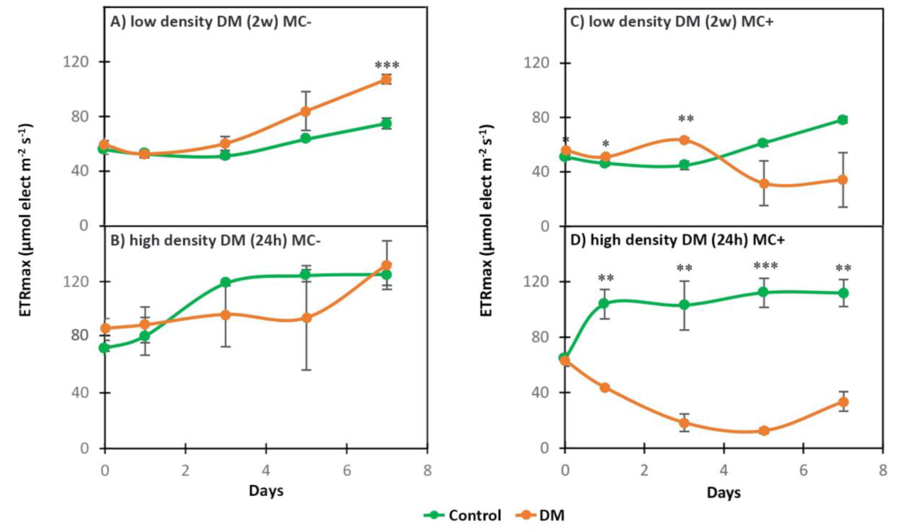
2.1. Photosynthetic Activity

The photosynthetic activity of the strains was measured through the maximum electron transport rate ETRmax. When exposed to spent medium from 75 all age individuals of D. magna /L raised for two weeks (low density DM (2w)), MC- was not negatively affected, on the contrary, it remained in good physiological state during seven days of exposure (Figure 1A, Table 1). However, in the MC+ strain the photosynthetic activity showed a non-statistically significant decrease after being initially significantly higher during the first three days of exposure and did not recover compared to the control (Figure 1C, Table 1). Then, both strains were exposed to a medium in which a higher density of Daphnia (200 all age individuals of D. magna /L) was cultured for 24 h (high density DM (24 h)). This exposure had contrasting effects on the strains: The photosynthetic activity of the MC- was high during the entire experiment in the exposed cells as well as in the control (Figure 1B, Table 1), whereas the MC+ strain suffered from a statistically significant decrease of photosynthetic activity during the whole experiment compared to the control (Figure 1D, Table 1).
Photosynthetic activity was also compared between the MC+ and MC- strains in those exposures showing differences between treatment and control (low density DM (2w) and high density DM (24 h), Table 1). In the low density DM (2w) exposure, control MC+ and control MC- showed no significant difference during the experiment, except on the first day. Photosynthetic activity of the low density DM (2w) treatments was not significantly different between the strains during the first three days of the experiment, but it significantly decreased in MC+ on day 5 and 7 compared MC- exposed to low density DM (2w) (Table 1). In the high density DM (24 h) exposure, there was no statistically significant difference between control MC+ and control MC-. However, photosynthetic activity in MC+ exposed to high density DM (24 h) significantly decreased during the whole experiment (except on day 5, owing to a high variability in MC-) compared to MC- exposed to high density DM (24 h) (Table 1).

2.2. Dynamics of Intracellular and Extracellular Metabolites in MC+ and MC-

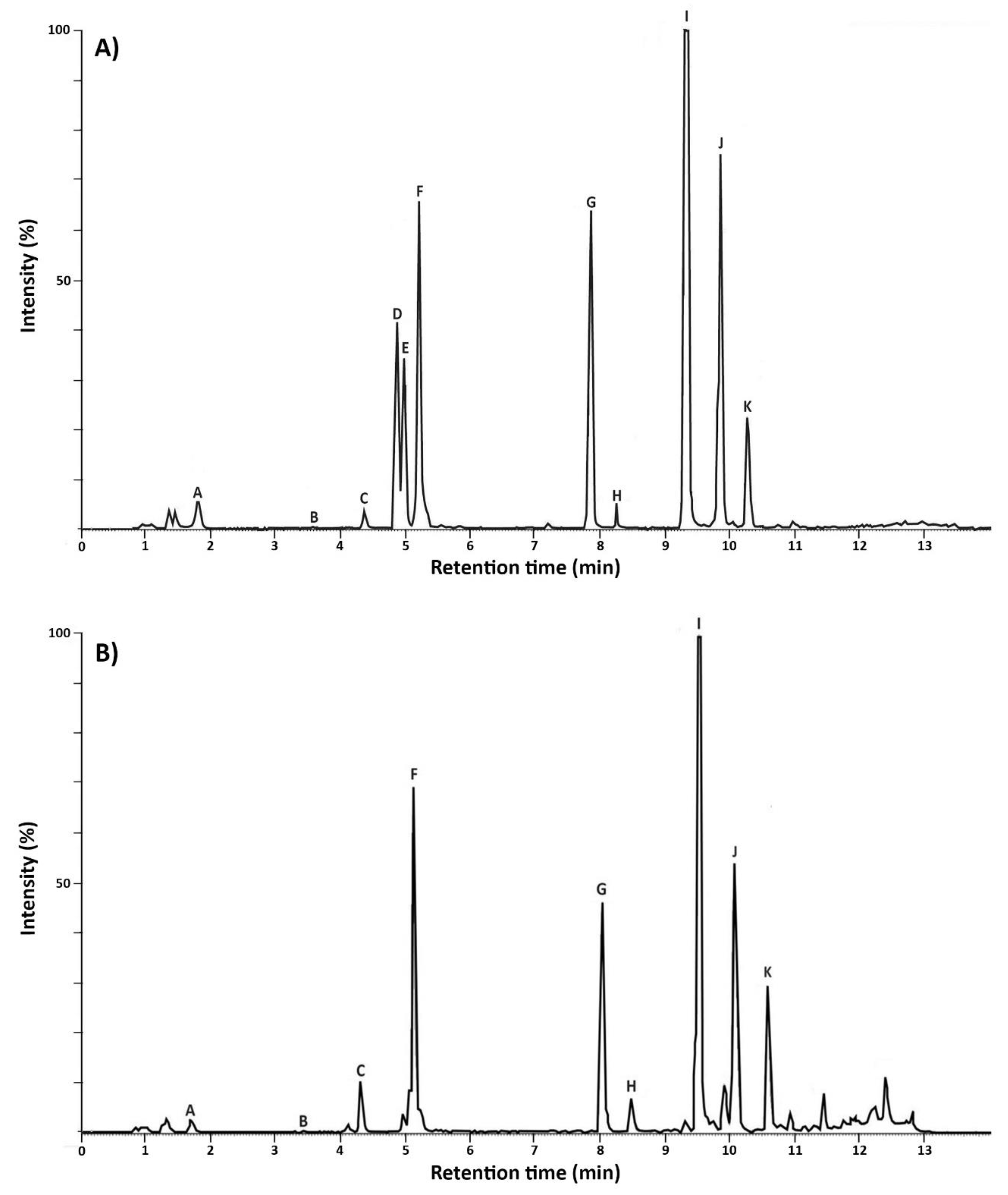
2.2.1. Metabolic Profiles of MC+ and MC–

Eleven metabolites produced by MC+ (Figure 2A) were detected and nine metabolites produced by MC-(Figure 2B). MC+ produced microcystins (LR and des-MC-LR), cyanopeptolins (963A, A, and B), aerucyclamides (A, B, C, and D) and aeruginosins (684 and 602) (Figure 2A). MC- produced cyanopeptolins (963A, A and B), aerucyclamides (A, B, C, and D) and aeruginosins (684 and 602) (Figure 2B).

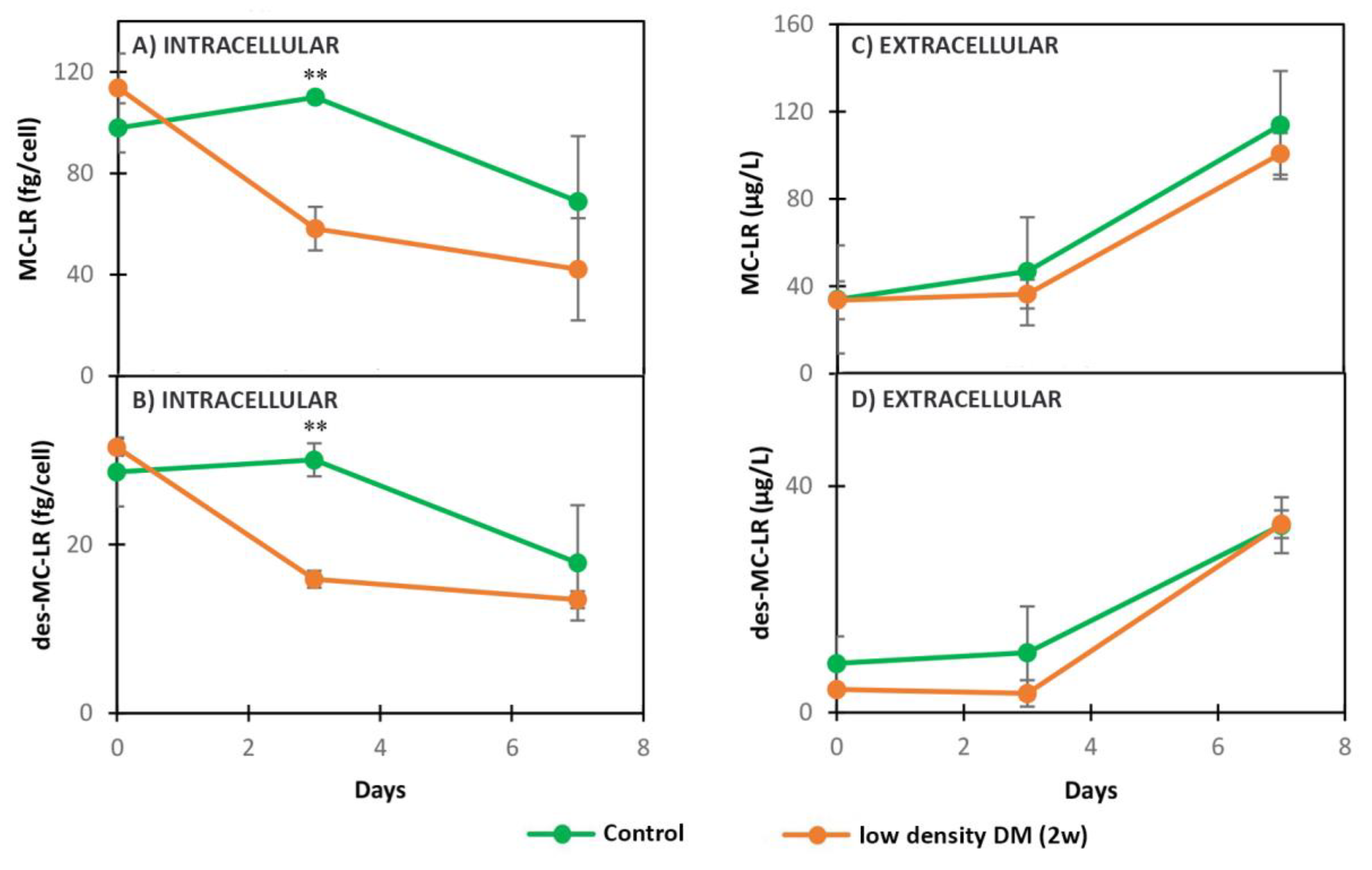
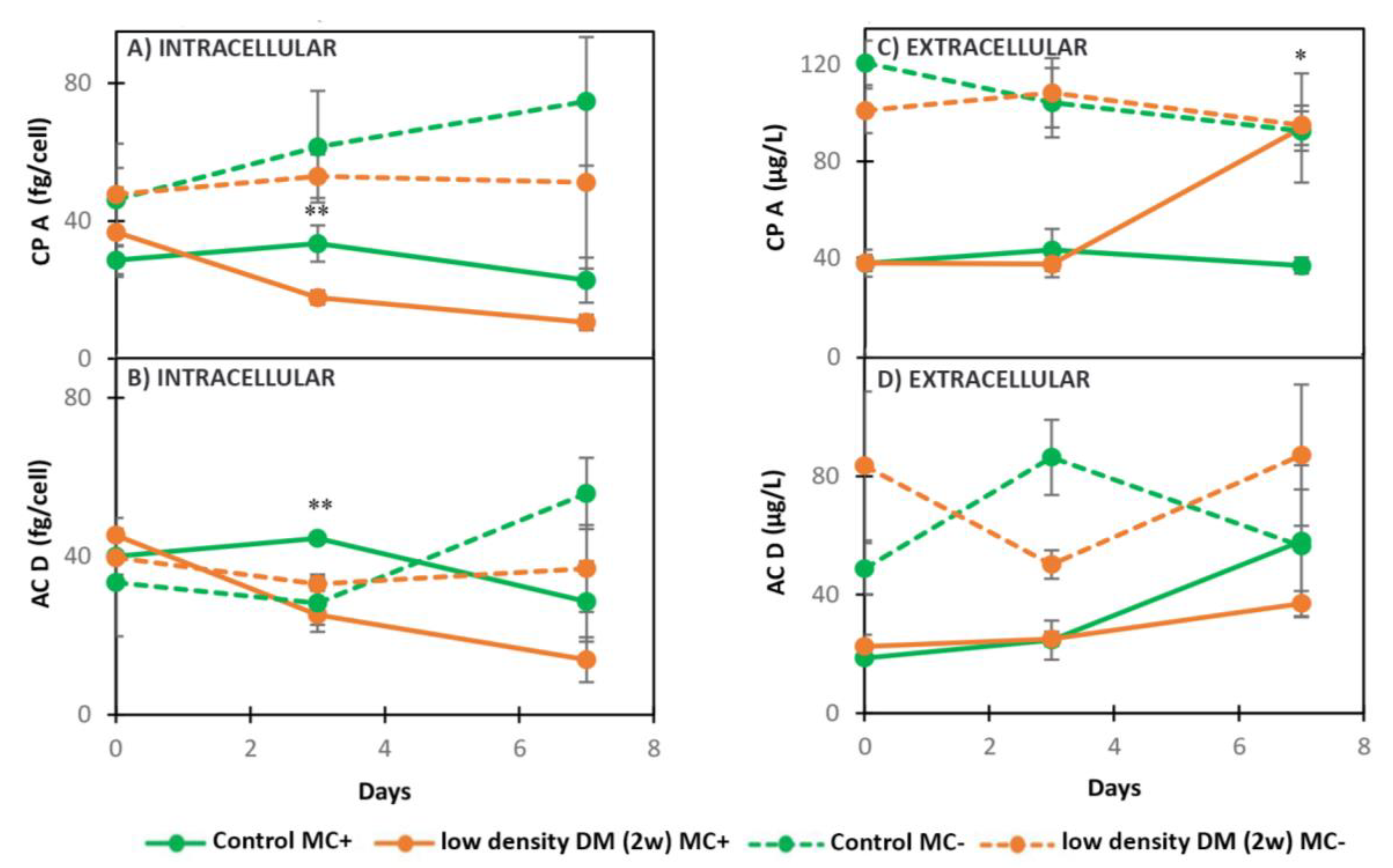
2.2.2. Dynamics of Intracellular and Extracellular Metabolites in MC+ and MC- When Exposed to Low Density DM (2w)

Low density DM (2w) significantly decreased the concentration of intracellular MC-LR and des-MC-LR at the second day of exposure in comparison with the control (Figure 3A,B, Table 2). The dynamics of extracellular MC-LR and des-MC-LR were not significantly different between the control and treated cells during the experiment in MC+ (Figure 3C,D, Table 2). Intracellular AC D and CP A produced by MC+ followed a decreasing trend when exposed to low density DM (2w), with significant difference on day 3 in comparison with the control (Figure 4A,B, Table 2). While the export of AC D produced by MC+ did not change significantly in the treatment compared with the control (Figure 4D, Table 2), CP A was excreted in significantly higher amounts in the presence of low density DM (2w) in MC+ on day 7 of the experiment in comparison with the control (Figure 4C, Table 2). Neither the intracellular nor extracellular dynamics of CP A and AC D produced in MC- were statistically different when exposed to low density DM (2w) compared to the control (Figure 4A–D, Table 2).
The concentration of intracellular and extracellular CP A and AC D was compared between MC+ and MC- in the low density DM (2w) exposure experiment (Table 2). The intracellular concentration of CP A and AC D was significantly higher in the control MC- when compared with the control MC+ on day 3 and 7. The concentration of intracellular CP A was significantly higher in low density DM (2w) MC- compared to low density DM (2w) MC+ on day 3, but not on day 0 and 7. There was no significant difference in AC D intracellular concentration in low density DM (2w) MC+ compared to low density DM (2w) MC- during the whole experiment. The concentration of extracellular CP A significantly increased in control MC- compared to control MC+ on day 3 and 7. The amount of extracellular AC D was significantly higher on day 0 and 3 in control MC- compared to control MC+, while there was no difference on day 7. When MC+ and MC- were exposed to low density DM (2w), the concentration of extracellular CP A was significantly lower in low density DM (2w) MC+ compared to low density DM (2w) MC- on day 0 and 3 of the experiment, while there was no statistical significance on day 7. The concentration of extracellular AC D was significantly lower in low density DM (2w) MC+ compared with low density DM (2w) MC- on day 3, while on day 0 and 7 there was no statistically significant difference (Table 2).

2.2.3. Dynamics of Intracellular and Extracellular Metabolites in MC+ When Exposed to High Density DM (24 h)

When exposed to high density DM (24 h), the concentrations of intracellular MC-LR and des-MC-LR by MC+ were reduced at three days of exposure, compared with the control, and did not recover during the experiment (Figure 5A,B, Table 3). Reduction of intracellular metabolites was followed by a non-statistically significant lower excretion of MC-LR and significantly lower excretion of des-MC-LR in treatment compared to the control on day 7 (Figure 5C,D, Table 3). CP A and AC D were detected in both strains in control and 24 h DM treatment, however they were below the limit of quantification. Hence, unfortunately, no results on the CP A and AC D concentrations are available for this exposure.

3. Discussion

MCs are by far the most commonly investigated toxins in cyanobacteria [57], but their role and regulation still remain unclear. While abiotic factors have been extensively studied, biotic factors including responses to grazing have received less attention. In this study, we compared the responses of both a MC-producing strain and its nontoxic mutant to Daphnia magna exudates via spent medium exposures in order to test the potential role of MC and/or other cyanopeptides as a response to the presence of grazers.
It is believed that most cyanobacterial species retained this pathway due to the potential protective role of MCs against oxidative stress, favoring the proliferation of MC-producing strains over nontoxic ones under stressful conditions [33]. Under growth limiting conditions of light, temperature, and nitrogen, MC-producing Planktothrix agardhii strains showed better fitness than nontoxic strains [30]. Similarly under growth limiting conditions of light and nitrogen, MC-producing M. aeruginosa showed better fitness than nontoxic strains [58]. A proteomic study showed that MC-producing M. aeruginosa species coped better with environmental changes that induced production of ROS than the nontoxic strain, hypothesizing possible functions of MC as a scavenger of free radicals [21]. Zilliges et al. (2011) revealed that if MC binds to proteins, it increases their stability, suggesting an important protective role of MC against stress [22].
The possibility of stress in cyanobacteria induced by grazers, such as Daphnia sp, has gained scientific interest. Most of the studies looked at the indirect effect of Daphnia on cyanobacteria used media in which Daphnia were cultivated for short periods of time, usually 24 h–48 h [25,26,38,46,59]. Effects of aged zooplankton medium on cyanobacteria are scarce in the literature, although the culturing of Daphnia for a longer duration, allowing the accumulation of D. magna metabolites over time, would be environmentally more relevant. As the spent medium of low density DM (1w) seemed to have no effect on both strains in this study, D. magna were cultivated for one more week in order to accumulate more metabolites that would potentially evoke an effect on cyanobacterial cell physiology. The photosynthetic activity (as ETRmax) is often measured as proxy of the physiological state of algal cells, as in this study. That parameter as a precursor of growth [58,60], indicates a poor physiological state from which the strain does not recover when ETRmax values are below 30, while values reaching 100 and above are associated with a healthy physiological state [58]. Contrary to our expectations, the toxic strain decreased its photosynthetic activity (ETRmax < 30), coping poorly with stress caused by low density DM (2w), while the mutant strain stayed in good physiological state during the entire experiment (seven days). These findings are supported by a recent study using the same strains, also discovering higher sensitivity of the toxic strain, when comparing responses of both strains to prolonged oxidative stress caused by hydrogen peroxide: Only the mutant was able to recover and regain initial photosynthetic yields [34]. As cyanobacterial reactions could be strain specific, different strain’s reactions are important to consider: When MC-producing M. aeruginosa strains Ch10 and UTEX LB2385 were exposed to five-day old D.magna spent medium, the content of chlorophyll also decreased, similar to our results [47].
In environments that promote oxidative stress MC binds to proteins [22,61], amongst others to the same target protein as thioredoxin [62] and possibly peroxiredoxin [34]. Binding of MC to these proteins will render them inactive, and upon loosing these antioxidant activities, the MC producing cells become more sensitive to oxidative stress [34]. Furthermore, the higher expression of genes regulating anti-oxidative enzymes in the nontoxic mutant, may favor the nontoxic mutant over the toxic strain when it comes to coping with high levels of oxidative stress [34].
Initial cyanobacterial density and concentration of Daphnia infochemicals influence the interaction between Daphnia and cyanobacteria [63]. In order to address this and avoid possible bias by a bacterial degradation of Daphnia metabolites, a culture of lower cyanobacterial cell density was exposed to high density DM (24 h). This experiment confirmed tendencies observed in the previous exposures by a drastically decreased photosynthetic activity in the toxic strain (ETRmax < 30), while the mutant stayed in good physiological state during the whole experiment. This suggests that the nontoxic mutant was coping better, compared with the toxic strain, when exposed to high density D. magna spent medium.
Depending on the density of D. magna and cultivation time, concentrations of intra- and extracellular MC-LR and des-MC-LR differed in the exposures: Whereas lower D. magna densities caused increasing tendencies of the toxins, higher D. magna densities and in particular those with short time for bacterial degradation of infochemicals caused a decrease. Similarly, M. aeruginosa increased MC production when exposed to increasing D. magna spent medium concentrations, suggesting an anti-grazer role triggered by zooplankton infochemicals [23], when only 50 adult D. magna were grown for four days per L of dechlorinated water of which 10–50% were mixed with the cyanobacterial culture. In addition, the presence of large cladocerans such as D. cucullata and D. longispina in the Sulejow Reservoir (Poland) positively correlated with the increase of MCs content in Microcystis [35]. Similarly, in the presence of D. pulex, the production of MC was significantly higher in Microcystis spp in all tested cyanobacterial (0.5–4.5 × 10⁶ cells/mL) and D. pulex (100–500 individuals/L) densities [27]. Different developmental stages of Daphnia can promote different production of MCs [25,35] because the same number of adults can possibly produce more, or other infochemicals than neonates, inducing higher MC production [43]. Although these studies support the hypothesis that a possible role of MC could be as anti-grazer defense [25,27,35], molecular evidence clearly shows that genes responsible for toxin production are much older than the existence of more complex organisms [40]. Furthermore, Kurmayer et al. 2016 [64] pointed out the presence and global distribution of both toxic and nontoxic cyanobacterial genotypes, suggesting their co-existence through evolution, as none of them were strongly favored by natural selection. Therefore, the potential role of cyanobacterial secondary metabolites as a defense mechanism against grazers, might be an adaptation they acquired over time [39,41]. However, not all studies support these results (e.g., [38,45]. Van Gremberghe et al. (2009) reported that Daphnia infochemicals (300 D. magna individuals/L grown for 24 h in the WC medium) generally had a weak influence on MC production in several Microcystis strains [38], while Becker (2010) demonstrated that when exposed to Daphnia medium (165 D. magna individuals/L grown in 24 h WC medium), Microcystis stopped producing MC [45]. In exposure to 2wDM our results showed similar findings: The concentration of intracellular MC-LR and des-MC-LR decreased significantly in MC+ compared with the control. Supposedly, in the high density DM (24 h) exposure, the concentration of both MC-LR and des-MC-LR decreased, owing to a lower cyanobacterial density being exposed to a higher number of D. magna producing more metabolites, which were not degraded during the short cultivation time. However, MC decrease could be due to its potential ability to bind to proteins (such as thioredoxin [62] and peroxiredoxin [34]) under stressful conditions [34], thus escaping detection. Further studies including either the expression of the genes involved in MC production or the detection of the protein-bound MC fraction are needed to clarify this.
Hence, an overall decrease in the cyanobacterial physiology and the negative effect this Daphnia medium had on the toxic strain was demonstrated.
To note, all our controls exhibited similar trends in dynamics where intracellular MC-LR increased on the third day and then decreased in the seven-day experiment. Similar results were observed by Becker 2010 [45], suggesting that this performance might not be caused by zooplankton. The transcription of mcyHIJ and mcyB genes increased in M. aeruginosa in 50% direct grazing treatments and in 90% of Daphnia infochemicals treatments after 24 h exposure, while transcription of mcyACDEFG was only expressed in 8% of the exposures [59]. The synthesis of MC, however, requires the expression of the whole MC synthetase cassette (mcyABCDEFG), therefore in that study zooplankton presence did not trigger MC production, however, it is yet to be clarified whether genes for these secondary metabolites synthesis need more time to respond than has been considered in this study [59]. mcy A was expressed in the first five days of D. magna exposure in the M. aeruginosa UTEX LB2385 strain, while in the Ch10 strain this occurred after 10 days, demonstrating a different response between strains [47]. As gene expression after stress vary in time depending on the particular gene and the strain, and moreover may or may not lead to MC production, we favored the analysis of the metabolite by chemical analysis as applied in this study over gene expression by RT qPCR. Additionally, it has been documented that Microcystis response to Daphnia medium is strongly strain specific [38], leaving the role of MC as anti-grazer compound arguable [28].
In addition to MC, Microcystis produces a range of peptides, such as cyanopeptolins, aeruginosins, and aerucyclamides that negatively influence digestive enzymes in zooplankton [56]. Metabolic pathways and triggers for production of these group of metabolites, as well as their functional role still needs more research [65]. Production of MC is energetically costly [66], allowing strains lacking MC to allocate energy in possible alternative mechanisms of defense or production of other secondary metabolites [34,58]. Our results show that the overall concentrations of CP A and AC D was higher in MC- then in MC+ in controls as well as the treatments. Interestingly, in monocultures, higher concentrations of cyanopeptolins, aerucyclamides, and aeruginosins were produced by the non-MC-producing mutant in comparison with the wild type [36]. When exposed to 1 wDM, concentrations of CP A and AC D did not change significantly in either toxic or nontoxic strain in our seven-day experiment. Similar results were observed in low density DM (2w) exposure in the nontoxic strain. The toxic strain, however, increased the export of CP A as a response to Daphnia medium, which might be a potential defensive mechanism. Cyanopeptolins are known as protease inhibitors [67] produced similarly to MC, through non-ribosomal pathways [49] with negative impact on zooplankton via inhibiting their trypsin-like digestion enzymes [6,56,68]. Thus, one of their potential roles could be grazing avoidance. If or not CP A has an anti-grazing effect or role will be proven in future studies. In order to understand fate and possible triggers for cyanopeptolins and aerucyclamides production, Sadler and von Elert (2014) [26] found increased production of CP B in M. aeruginosa PCC7806 against D. magna grazing, suggesting an induced defense mechanism, while relative and total amounts of CP A and C were not affected by D. magna presence. The transcription of genes encoding cyanopeptolins, microviridins, and aeruginosins in direct and indirect exposure to D. magna and D. pulex was mostly unaffected [59]. However, it is yet to be clarified whether genes for the synthesis of these secondary metabolites need more time to respond than was considered in this study (24 h) [59].
Different responses to D. magna spent medium in our experiment were strain dependent. MC- used in this study is a genetically engineered strain and its physiology as well as the response to D. magna spent medium might differ from MC non-producing strains that naturally occur in aquatic environments. Therefore, more studies are needed to show how D. magna spent medium would affect other MC producing and MC non-producing cyanobacterial species.

4. Conclusions

To the best of our knowledge, this is the first study showing a lower physiological state of a MC+ strain when exposed to DM than its MC- mutant. D. magna spent medium of the highest D. magna density cultivated for the shortest time (high density DM (24 h)) provoked the strongest effect, which can be attributed to both high zooplankton density and a short time for bacterial degradation of the exudates.
Photosynthetic activity as a proxy of physiological state of the MC+ strain was negatively affected by low density DM (2w) and high density DM (24 h). The mutant strain remained in good physiological state in all exposure scenarios, suggesting that it coped better with the stress induced by DM. The production of intracellular MC-LR, as well as other secondary metabolites in MC+ was decreased, as a consequence of the affected cell physiology, while the dynamics of the quantified metabolites were not affected in the mutant strain (cyanopeptolin A and aeruginosin D being higher in the MC- compared with MC+).
Extracellular cyanopeptolin A concentrations, however, increased in MC+ in the presence of 2wDM, suggesting a potential anti-grazing role, which is in line with its previously evidenced inhibition of trypsin-like digestion enzymes in Daphnia [52]. Furthermore, to the best of our knowledge, this is the first study showing that the concentration of extracellular CP A increased in MC+ when exposed to DM. The MC- strain did not show this increase in the treatment, compared to the control, possibly because the concentration was already initially that high. Therefore, more research is needed to confirm this role. In the presence of the tested Daphnia media, MC-LR and des-MC-LR do not appear to have a protective role for the strain, as their intracellular concentration decreased while the extracellular remained unchanged.
This study evidenced negative impact of D. magna spent medium on the MC+ strain, that was dependent on densities of the organisms in the seven days of exposure.

5. Materials and Methods

5.1. Culture Conditions

5.1.1. Cyanobacterial Cultures

Two axenic strains were used in the experiments: MC+ and its mutant MC-, both obtained from the Pasteur Culture collection of Cyanobacteria [69]. MC- is MC+ transformed by inserting a chloramphenicol resistance cartridge into the mcyB gene involved in microcystin biosynthesis. This transformation led to the inability of derived mutant cells to produce any variant of microcystin [70], making MC- a useful tool for exploring the precise effects of microcystins on cyanobacterial physiology and metabolism, when compared to MC+ [70]. Before the start of the experiments, strains were cultivated in BG11 medium (SIGMA), with the addition of 5 µg chloramphenicol mL−1 for the MC-, to maintain the selection of the mutant cells. However, to exclude any impact of the chloramphenicol on DM, the experiments were performed without an antibiotic in the medium, as well as in the control medium. Both strains were grown under a 14 h:10 h light:dark regime using daylight white fluorescent tubes (Toshiba, 15 W, FL15D) with 20 μmol photons m−2 s−1 illumination at a constant temperature of 20 ± 1 °C (Sanyo incubator). Cultures were maintained in exponential growth phase by repeated dilution every 3 weeks in fresh culture medium, while axenicity was regularly evaluated as described in Reference [58].

5.1.2. D. Magna

The D. magna clone (originating from the INERIS) was provided by D. Azam and M. Coke from the PEARL INRA 1036 U3E system (The National Infrastructure in Biology and Health, France). D. magna were slowly acclimatised during three weeks to the cyanobacterial BG11 medium that was used in all the experiments. The green algae Scenedesmus communis (isolated in our laboratory, originating from the lake of Grand Lieu) was provided by Bertrand Le Rouzic (University of Rennes 1). The animals were fed every second day with Scenedesmus communis (reaching max ≈ 2 × 10⁴ cells/mL in the aquarium at feeding time) grown in BG11 under the same conditions as the cyanobacteria. Some published studies showed that Scenedesmus has a negative impact on Microcystis through allopathic interactions [71,72]. In our experimental setup however, Scenedesmus have been used as Daphnia food during the medium preparations; therefore, Daphnia medium could contain Scenedesmus metabolites that could affect cyanobacteria [71,72]. However, Scenedesmus cell densities were very low when given to the Daphnia compared to those used in the above mentioned studies showing effects (strains mixed together using equal cell densities of 5 × 10⁵ cells/mL (S. huji with the M. aeruginosa PCC 7005) [72] and 1 × 10⁵ cells/mL (S. acuminatus with the M. panniformis BCCUSP200) [71]). In our experimental setup, Scenedesmus was given as the only food source, it was consumed by Daphnia, leaving not many, if any, Scenedesmus cells that could produce allopathic compounds affecting cyanobacteria. Noteworthy, when used in the experiment, Daphnia spent medium was mixed with cyanobacterial culture in 4:1 ration, therefore diluting even more possible Scenedesmus compounds. Hence it seems unlikely that the Microcystis strains were affected by Scenedesmus in this experimental setup. D. magna were cultivated at the constant temperature of 20 °C, light intensity of 15 μmol photons m−2 s−1 and a day/night cycle of 14 h/10 h (Sanyo incubator).

5.2. Experimental Design

Experiments were performed in the three different exposure scenarios (Table 4), where MC+ and its mutant MC- were exposed to D. magna spent medium.
The preparation of the media for the experiments: In order to test environmentally relevant scenarios where Daphnia infochemicals are accumulated over time, we cultivated 75 neonatal individuals of D. magna/L for 1 week (low density DM (1w)). However, we observed no significant difference between control and treatment in this exposure in the photosynthetic activity (Figure S1) and metabolites dynamics (Figures S2 and S3), therefore 75 all age individuals of D. magna /L were cultivated for 2 weeks (low density DM (2w)). Moreover, we wanted to exclude potential degradation of Daphnia infochemicals, caused by bacteria coexisting in the D. magna medium during the 2-week cultivation period, as well as provoke a stronger effect in cyanobacteria. Therefore, we cultivated a high number of Daphnia (75 all age individuals of D. magna /L) for 24 h (high density DM (24 h)). One batch of spent medium was prepared per Daphnia treatment group. D. magna were cultivated in BG11 in aquariums and medium was filtered through 0.2 µm filter to remove bacteria and other particles and then stored at −20 °C. Before being used in experiments, exponentially growing cyanobacteria were centrifuged and cells were introduced in fresh BG11 medium and grown for one week to reach a cyanobacterial cell density of 1 × 10⁷ cells/mL for low density DM (1w) and low density DM (2w) exposures (pre-culture). Relative densities between cyanobacteria and low density DM (1w) and low density DM (2w) exposures did not provoke strong effects, therefore, the cyanobacterial density was lowered for the 24 h DM exposure, reaching 2x10⁶ cells/mL after one week. Filtered D. magna and fresh BG11 media for the treatments and for the controls, respectively, were mixed with the one week old cyanobacterial pre-cultures in 4:1 ratio in 2 L Erlenmeyer flasks. Cyanobacterial density at the beginning of the experiment was 2 × 10⁶ cells/mL for low density DM (1w and 2w) exposures and 5 × 10⁵ cells/mL for high density DM (24 h) exposure.
The initial nutrients (PO4 and NO3) and pH were measured for the low density DM (2w) exposure to ensure similar conditions. Dissolved nutrient concentrations were measured from filtered (GF/F 0.7 µm) water using common colorimetric methods [73]. Nitrate was measured after reduction to nitrite on a cadmium-copper column [74]. Phosphate was measured following the method of Murphy and Riley (1962) [75]. Data presented in Table S1 show no significant difference between control and treatment (p > 0.05) for PO4 measurements in both strains, and for NO3 in the toxic strain. NO3 was significantly increased by 8% in the control of the MC- strain, which is still within the range of optimal growth conditions. We also measured pH that was not significantly different between control and treatment (p > 0.05) in all exposure scenarios (Table S2). Overall, there was no nutrient of pH differences that could affect the cyanobacteria physiology.
The procedure was the same for both strains, MC+ and its mutant MC-. The control and the treatment were done simultaneously. Experiments were done in triplicate, cultivated in 1 L flasks, and mixed every day for seven days.

5.3. Photosynthetic Activity

To characterize the physiological state of the cyanobacteria, the electron transport rate (ETR) was measured every two days with a pulse-amplitude-modulated fluorescence monitoring system (PhytoPAM, Walz, Germany), following Reference [76] and [60]. The PhytoPAM is equivalent to four separate PAM-fluorometers using light-emitting diodes (LED) with 10 s light pulses at four different excitation wavelengths (470, 520, 645 and 665 nm), with the 645 nm specific to cyanobacteria (due to phycocyanin and allophycocyanin absorption). The PhytoPAM was used with only one channel, corresponding to the cyanobacteria. The reference excitation spectrum measured at the factory was used, as it was not significantly different from reference excitation spectra performed on our cyanobacterial cultures. After being dark adapted for 15 min, Ft (the instantaneous steady state fluorescence at every successive step of actinic irradiance) and Fm (the maximum fluorescence) were measured during a 20 increments of actinic irradiance from 1 to 1864 µmol photons m−2 s−1, with a 10 s time interval between successive steps. The ETR was calculated as follows: (Fm-Ft)/Fm x 0.42 x PAR (PAR: photosynthetic radiation) [60,76]. The ETRmax was inferred using the regression model of [77] to fit the ETR irradiance curves.

5.4. Cyanobacterial Cell Density

Cyanobacterial cell density was followed by measuring the optical density on a spectrophotometer (UVIKONxs SECOMAN) at a corresponding absorbance at 750 nm. Culture medium BG11 was used as reference (blank). The optical density was used to calculate the cyanobacterial cell density (cell number/mL) [58] in order to establish a relationship between the optical density at 750 nm and the cell density, calibration curves with Microcystis cell number/mL vs corresponding absorbance were established for both strains [60]. The counting of the cells was carried out using a Nageotte cell and observed under the Olympus BX50 microscope (objective 40).

5.5. Analysis of Cyanobacterial Secondary Metabolites

The dynamics of intracellular and extracellular cyanobacterial peptides of MC+ and MC- in response to different D. magna media were analyzed using a Waters Acquity Ultra-High Performance Liquid Chromatography coupled to a photo diode array detector (PDA) and Xevo quadrupole time of flight mass spectrometer in series. Samples were centrifuged to separate cells (for intracellular metabolites) from supernatant (for extracellular metabolites) and lyophilized. Freeze-dried material was extracted in 0.5 ml 50% aqueous methanol. Extracts were separated on a CORTECS C18 column (0.2 mm ID × 100 mm long), which was maintained at 40 °C. The mobile phase was Milli-Q Water plus 0.1% formic acid (A) and acetonitrile plus 0.1% formic acid (B). Separation was achieved using a gradient increasing from 20% B to 70% B over 10 min, followed by a 100% B wash step and re-equilibration. Autosampler was maintained at 6 °C at all times. UV data was acquired from 200 to 400 nm. Mass data was acquired in positive ion electrospray scanning from m/z 50 to 2000 with a scan time of 2 s and inter-scan delay of 0.1 s. Ion source parameters, i.e., a capillary and sampling cone, were 2.9 V and 25 V respectively; desolvation temperature, 300 °C; and source temperature, 80 °C. Cone gas and desolvation gas flows were 50 L h−1 and 400 L h−1 respectively. Sodium iodide (2 μg μL−1 211 in 50/50 Propan-2-ol/H2O) was used as the calibrant with leucine-enkephalin (0.5 mg mL−1 212 in 50/50 methanol/Milli-Q) as the lock spray. Instrument control, data acquisition (centroid) and processing were achieved using MassLynx v4.1 software (Waters, Milford, MA, USA). We monitored secondary metabolites produced by Microcystis that were observed to be changing due to abiotic or biotic factors [36,50,78] (Table 5). Cyanobacterial peptides were detected using extracted ion chromatograms for the respective specific masses of the different compounds (Table 5).
MC-LR, des-MC-LR, cyanopeptolin A, and aerucyclamide D were quantified in our strains. Quantification was achieved using linear relationship between peak area (MC-LR and des-MC-LR at 238 nm, cyanopeptolin A at 220 nm, and aerucyclamide D at 240 nm) and known concentrations of the toxin standards. The microcystin-LR standard was purified as previously described [84]. Cyanopeptolin A standard and aerucyclamide D standard were purified using preparative HPLC (Biotage Parallex Flex, Cardiff, UK) and Flex V3 software for instrument control and data acquisition. The separation was performed on Atlantis Prep C18 column (5 µm particle size, 19 mm ID × 300 mm long; Waters, Elstree, UK) using a 30-min linear gradient from 60% to 100% methanol in MilliQ water. Absorbance was monitored at 210 nm and 280 nm. The flow rate was 20 mL/min and 4 mL fractions were collected. MassLynx v4.1 was used for both detection and quantification of the cyanobacterial peptides.

5.6. Statistics

R Core Team (2013) was used to access statistical analysis of the obtained data. All data are presented as mean ± standard deviation. Significant differences were determined at p < 0.05. We performed t-tests to determine the difference between photosynthetic activity of control and treatment for both strains (exposed to different DM (low density DM (2w) and high density DM (24 h)). T-test was also used to compare concentration of intracellular and extracellular metabolites in control and treatment for each strain. Additionally, one-way ANOVAs were used for the comparison between strains in the experiments. Prior to the ANOVAs, Shapiro–Wilk-Normality tests was performed. Where p value was >0.05, Tukey post-hoc test for pairwise comparison was performed (Control MC+/Control MC- and DM MC+/DM MC-). Where p value was <0.05 (Shapiro–Wilk-Normality tests), Kruskal–Wallis ANOVA was done, followed by t-tests for pairwise comparison (Control MC+/Control MC- and DM MC+/DM MC-).

Supplementary Materials

The following are available online at https://0-www-mdpi-com.brum.beds.ac.uk/2072-6651/11/7/421/s1, Figure S1: Photosynthetic activity of MC- and MC+ when cultured in D. magna spent medium (DM) and cultured in BG11 (Control): (A) low density DM (1w) MC- (B) low density DM (1w) MC-, Figure S2: Dynamics of intracellular and extracellular metabolites in MC+ when exposed to low density DM (1w) compared to when grown in BG11 (Control): (A) intracellular MC-LR, (B) intracellular des-MC-LR, (C) extracellular MC-LR, (D) extracellular des-MC-LR. (p > 0.05) t-test, Figure S3: Dynamics of intracellular and extracellular metabolites in MC+ and MC- when exposed to low density DM (1w): M. aeruginosa PCC7806 (low density DM (1w) MC+) and M. aeruginosa PCC7806 mcy- (low density DM (1w) MC-) and when grown in BG11: M. aeruginosa PCC7806 (Control MC+) and M. aeruginosa PCC7806 mcy- (Control MC-). (A) intracellular CP A, (B) intracellular AC D, (C) extracellular CP A, (D) extracellular AC D. (p > 0.05) t-test, Table S1: Concentration of PO4 (mg/L) and NO3 (mg/L) on day 0 in the 2wDM experiment, Table S2: pH on day 0 in the 1wDM, 2wDM and 24 h DM.

Author Contributions

Conceptualization, M.B., C.W., L.L., E.B., and G.B.S; methodology, M.B., C.W., L.L., and C.E.; software, C.E. and G.B.S.; formal analysis, G.B.S. and C.E.; investigation, G.B.S; resources, M.B., C.W., and L.L..; data curation, G.B.S.; writing—original draft preparation, G.B.S.; writing—review & editing, M.B., C.W., E.B., C.E., and L.L.; visualization, G.B.S.; supervision, M.B., C.W., and L.L.; funding acquisition, M.B., C.W., and L.L.

Funding

Gorenka Bojadzija Savic was supported by a Presidential scholarship from the University of Rennes 1, the project benefited from funding from the Centre National de la Recherche Scientifique (CNRS) Initiative Structurante Ecosphère continentale et côtière (EC2CO) under the Interactions métaboliques entre cyanobactéries et daphnies (MICYDA) project. Gorenka Bojadzija Savic was granted a mobility grant from the Doctoral School to visit the Robert Gordon University in Aberdeen for metabolites analyses.

Acknowledgments

We are grateful to D. Azam and M. Coke from the PEARL INRA 1036 U3E system (The National Infrastructure in Biology and Health, France) for providing the Daphnia magna strain (originating from l’INERIS) and Bertrand Le Rouzic (University of Rennes 1) for providing us with the Scenedesmus communis culture (originating from the lake of Grand Lieu). The help during the experiments of master student Naoual Bendahhane is greatly acknowledged. We would like to thank Len Montgomery (Robert Gordon University) for for proofreading the manuscript.

Conflicts of Interest

The authors declare that there are no conflicts of interest.

Abbreviations

MC+M. aeruginosa PCC7806
MC-M. aeruginosa PCC7806 mcy-
DMDaphnia medium
low density DM (1w)spent medium in which 75 neonatal individuals of D. magna/L were cultured for 1 week
low density DM (2w)spent medium in which 75 all age individuals of D. magna/L were cultured for 2 weeks
high density DM (24 h)spent medium in which 200 all age individuals of D.magna/L were cultured for 24 h
MC-LRmicrocystin LR
des-MC-LRdes microcystin LR
CPcyanopeptolin
ACaerucyclamide
CP Acyanopeptolin A
AC Daerucyclamide D
Control MC+M. aeruginosa PCC7806 cultivated in BG11
Control MC-M. aeruginosa PCC7806 mcy- cultivated in BG11

References

  1. Holland, A.; Kinnear, S. Interpreting the Possible Ecological Role(s) of Cyanotoxins: Compounds for Competitive Advantage and/or Physiological Aide? Mar. Drugs 2013, 11, 2239–2258. [Google Scholar] [CrossRef] [PubMed] [Green Version]
  2. Asselman, J.; Hochmuth, J.D.; De Schamphelaere, K.A.C. A comparison of the sensitivities of Daphnia magna and Daphnia pulex to six different cyanobacteria. Harmful Algae 2014, 39, 1–7. [Google Scholar] [CrossRef]
  3. Ekvall, M.K.; Urrutia-Cordero, P.; Hansson, L.A. Linking cascading effects of fish predation and zooplankton grazing to reduced cyanobacterial biomass and toxin levels following biomanipulation. PLoS ONE 2014, 9, e112956. [Google Scholar] [CrossRef] [PubMed]
  4. Urrutia-Cordero, P.; Ekvall, M.K.; Hansson, L.-A. Controlling Harmful Cyanobacteria: Taxa-Specific Responses of Cyanobacteria to Grazing by Large-Bodied Daphnia in a Biomanipulation Scenario. PLoS ONE 2016, 11, e0153032. [Google Scholar] [CrossRef] [PubMed]
  5. Rohrlack, T.; Dittmann, E.; Börner, T.; Christoffersen, K. Effects of Cell-Bound Microcystins on Survival and Feeding of Daphnia spp. Appl. Environ. Microbiol. 2001, 67, 3523–3529. [Google Scholar] [CrossRef] [PubMed]
  6. von Elert, E.; Zitt, A.; Schwarzenberger, A. Inducible tolerance to dietary protease inhibitors in Daphnia magna. J. Exp. Biol. 2012, 215, 2051–2059. [Google Scholar] [CrossRef] [PubMed]
  7. Lampert, W. Inhibitory and Toxic Effects of Blue green Algae on Daphnia. Int. Rev. Gesamten Hydrobiol. Hydrogr. 1981, 66, 285–298. [Google Scholar] [CrossRef]
  8. Demott, W.R.; Zhang, Q.-X.; Carmichael, W.W. Effects of toxic cyanobacteria and purified toxins on the survival and feeding of a copepod and three species of Daphnia. Limnol. Oceanogr. 1991, 36, 1346–1357. [Google Scholar] [CrossRef]
  9. Dao, T.S.; Do-Hong, L.C.; Wiegand, C. Chronic effects of cyanobacterial toxins on Daphnia magna and their offspring. Toxicon 2010, 55, 1244–1254. [Google Scholar] [CrossRef]
  10. Mowe, M.A.D.; Mitrovic, S.M.; Lim, R.P.; Furey, A.; Yeo, D.C.J. Tropical cyanobacterial blooms: A review of prevalence, problem taxa, toxins and influencing environmental factors. J. Limnol. 2015, 74, 205–224. [Google Scholar] [CrossRef]
  11. MacKintosh, C.; Beattie, K.A.; Klumpp, S.; Cohen, P.; Codd, G.A. Cyanobacterial microcystin-LR is a potent and specific inhibitor of protein phosphatases 1 and 2A from both mammals and higher plants. FEBS Lett. 1990, 264, 187–192. [Google Scholar] [CrossRef] [Green Version]
  12. DeMott, W.R.; Dhawale, S. Inhibition of in vitro protein phosphatase activity in three zooplankton species by microcystin-LR, a toxin from cyanobacteria. Arch. Hydrobiol. 1995, 134, 417–424. [Google Scholar]
  13. Zurawell, R.W.; Chen, H.; Burke, J.M.; Prepas, E.E. Hepatotoxic cyanobacteria: A review of the biological importance of microcystins in freshwater environments. J. Toxicol. Environ. Health B 2005, 8, 1–37. [Google Scholar] [CrossRef] [PubMed]
  14. Amado, L.L.; Monserrat, J.M. Oxidative stress generation by microcystins in aquatic animals: Why and how. Environ. Int. 2010, 36, 226–235. [Google Scholar] [CrossRef] [PubMed]
  15. Wiegand, C.; Pflugmacher, S. Ecotoxicological effects of selected cyanobacterial secondary metabolites: A short review. Toxicol. Appl. Pharmacol. 2005, 203, 201–218. [Google Scholar] [CrossRef] [PubMed]
  16. Kultschar, B.; Llewellyn, C. Secondary Metabolites in Cyanobacteria. In Secondary Metabolites—Sources and Applications; InTech: London, UK, 2018. [Google Scholar] [Green Version]
  17. Neilan, B.A.; Pearson, L.A.; Muenchhoff, J.; Moffitt, M.C.; Dittmann, E. Environmental conditions that influence toxin biosynthesis in cyanobacteria. Environ. Microbiol. 2013, 15, 1239–1253. [Google Scholar] [CrossRef]
  18. Herms, D.A.; Mattson, W.J. The Dilemma of Plants: To Grow or Defend. Q. Rev. Biol. 1992, 67, 283–335. [Google Scholar] [CrossRef] [Green Version]
  19. Kaebernick, M.; Neilan, B.A. Ecological and molecular investigations of cyanotoxin production. FEMS Microbiol. Ecol. 2001, 35, 1–9. [Google Scholar] [CrossRef]
  20. Alexova, R.; Fujii, M.; Birch, D.; Cheng, J.; Waite, T.D.; Ferrari, B.C.; Neilan, B.A. Iron uptake and toxin synthesis in the bloom-forming Microcystis aeruginosa under iron limitation. Environ. Microbiol. 2011, 13, 1064–1077. [Google Scholar] [CrossRef]
  21. Dziallas, C.; Grossart, H.P. Increasing oxygen radicals and water temperature select for toxic Microcystis sp. PLoS ONE 2011, 6, e25569. [Google Scholar] [CrossRef]
  22. Zilliges, Y.; Kehr, J.C.; Meissner, S.; Ishida, K.; Mikkat, S.; Hagemann, M.; Kaplan, A.; Börner, T.; Dittmann, E. The cyanobacterial hepatotoxin microcystin binds to proteins and increases the fitness of Microcystis under oxidative stress conditions. PLoS ONE 2011, 6, e17615. [Google Scholar] [CrossRef]
  23. Kaplan, A.; Harel, M.; Kaplan-Levy, R.N.; Hadas, O.; Sukenik, A.; Dittmann, E. The languages spoken in the water body (or the biological role of cyanobacterial toxins). Front. Microbiol. 2012, 3, 138. [Google Scholar] [CrossRef]
  24. Jang, M.-H.; Ha, K.; Lucas, M.C.; Joo, G.-J.; Takamura, N. Changes in microcystin production by Microcystis aeruginosa exposed to phytoplanktivorous and omnivorous fish. Aquat. Toxicol. 2004, 68, 51–59. [Google Scholar] [CrossRef]
  25. Jang, M.H.; Jung, J.M.; Takamura, N. Changes in microcystin production in cyanobacteria exposed to zooplankton at different population densities and infochemical concentrations. Limnol. Oceanogr. 2007, 52, 1454–1466. [Google Scholar] [CrossRef]
  26. Sadler, T.; von Elert, E. Physiological interaction of Daphnia and Microcystis with regard to cyanobacterial secondary metabolites. Aquat. Toxicol. 2014, 156, 96–105. [Google Scholar] [CrossRef]
  27. Pérez-morales, A.; Sarma, S.S.S.; Nandini, S. Producción de microcistinas en Microcystis inducida por Daphnia pulex (Cladocera) y Brachionus calyciflorus (Rotifera). Hidrobiológica 2015, 25, 411–415. [Google Scholar]
  28. Omidi, A.; Esterhuizen-Londt, M.; Pflugmacher, S. Still challenging: The ecological function of the cyanobacterial toxin microcystin—What we know so far. Toxin Rev. 2018, 37, 87–105. [Google Scholar] [CrossRef]
  29. Chislock, M.F.; Doster, E.; Zitomer, R.A.; Wilson, A.E. Eutrophication: Causes, Consequences, and Controls in Aquatic Ecosystems. Nat. Educ. Knowl. 2013, 4, 10. [Google Scholar]
  30. Briand, E.; Yéprémian, C.; Humbert, J.F.; Quiblier, C. Competition between microcystin- and non-microcystin-producing Planktothrix agardhii (cyanobacteria) strains under different environmental conditions. Environ. Microbiol. 2008, 10, 3337–3348. [Google Scholar] [CrossRef]
  31. Van de Waal, D.B.; Verspagen, J.M.H.; Finke, J.F.; Vournazou, V.; Immers, A.K.; Kardinaal, W.E.A.; Tonk, L.; Becker, S.; Van Donk, E.; Visser, P.M.; et al. Reversal in competitive dominance of a toxic versus non-toxic cyanobacterium in response to rising CO2. ISME J. 2011, 5, 1438–1450. [Google Scholar] [CrossRef]
  32. Yeung, A.C.Y.; D’Agostino, P.M.; Poljak, A.; McDonald, J.; Bligh, M.W.; Waite, T.D.; Neilan, B.A. Physiological and proteomic responses of continuous cultures of Microcystis aeruginosa PCC 7806 to changes in iron bioavailability and growth rate. Appl. Environ. Microbiol. 2016, 82, 5918–5929. [Google Scholar] [CrossRef]
  33. Paerl, H.W.; Otten, T.G. Blooms bite the hand that feeds them. Science 2013, 342, 433–434. [Google Scholar] [CrossRef]
  34. Schuurmans, J.M.; Brinkmann, B.W.; Makower, A.K.; Dittmann, E.; Huisman, J.; Matthijs, H.C.P. Microcystin interferes with defense against high oxidative stress in harmful cyanobacteria. Harmful Algae 2018, 78, 47–55. [Google Scholar] [CrossRef]
  35. Izydorczyk, K.; Jurczak, T.; Wojtal-frankiewicz, A.; Skowron, A.; Mankiewicz-Boczek, J.; Tarczyńska, M. Influence of abiotic and biotic factors on microcystin content in Microcystis aeruginosa cells in a eutrophic temperate reservoir. J. Plankton Res. 2008, 30, 393–400. [Google Scholar] [CrossRef]
  36. Briand, E.; Bormans, M.; Gugger, M.; Dorrestein, P.C.; Gerwick, W.H. Changes in secondary metabolic profiles of Microcystis aeruginosa strains in response to intraspecific interactions. Environ. Microbiol. 2016, 18, 384–400. [Google Scholar] [CrossRef]
  37. Briand, E.; Reubrecht, S.; Mondeguer, F.; Sibat, M.; Hess, P.; Amzil, Z.; Bormans, M. Chemically mediated interactions between Microcystis and Planktothrix: Impact on their growth, morphology and metabolic profiles. Environ. Microbiol. 2019, 21, 1552–1566. [Google Scholar] [CrossRef]
  38. van Gremberghe, I.; Vanormelingen, P.; Van der Gucht, K.; Mancheva, A.; D’hondt, S.; De Meester, L.; Vyverman, W. Influence of Daphnia infochemicals on functional traits of Microcystis strains (cyanobacteria). Hydrobiologia 2009, 635, 147–155. [Google Scholar] [CrossRef]
  39. Chislock, M.F.; Sarnelle, O.; Jernigan, L.M.; Wilson, A.E. Do high concentrations of microcystin prevent Daphnia control of phytoplankton? Water Res. 2013, 47, 1961–1970. [Google Scholar] [CrossRef]
  40. Rantala, A.; Fewer, D.P.; Hisbergues, M.; Rouhiainen, L.; Vaitomaa, J.; Börner, T.; Sivonen, K. Phylogenetic evidence for the early evolution of microcystin synthesis. Proc. Natl. Acad. Sci. USA 2004, 101, 568–573. [Google Scholar] [CrossRef]
  41. Wilken, S.; Wiezer, S.; Huisman, J.; Van Donk, E. Microcystins do not provide anti-herbivore defence against mixotrophic flagellates. Aquat. Microb. Ecol. 2010, 59, 207–216. [Google Scholar] [CrossRef]
  42. Ger, K.A.; Urrutia-Cordero, P.; Frost, P.C.; Hansson, L.A.; Sarnelle, O.; Wilson, A.E.; Lürling, M. The interaction between cyanobacteria and zooplankton in a more eutrophic world. Harmful Algae 2016, 54, 128–144. [Google Scholar] [CrossRef]
  43. Jang, M.H.; Ha, K.; Takamura, N. Microcystin production by Microcystis aeruginosa exposed to different stages of herbivorous zooplankton. Toxicon 2008, 51, 882–889. [Google Scholar] [CrossRef]
  44. Van Gremberghe, I.; Vanormelingen, P.; Vanelslander, B.; Van Der Gucht, K.; D’Hondt, S.; De Meester, L.; Vyverman, W. Genotype-dependent interactions among sympatric Microcystis strains mediated by Daphnia grazing. Oikos 2009, 118, 1647–1658. [Google Scholar] [CrossRef]
  45. Becker, S. Biotic factors in induced defence revisited: Cell aggregate formation in the toxic cyanobacterium Microcystis aeruginosa PCC 7806 is triggered by spent Daphnia medium and disrupted cells. Hydrobiologia 2010, 644, 159–168. [Google Scholar] [CrossRef]
  46. Jang, M.-H.; Ha, K.; Joo, G.-J.; Takamura, N. Toxin production of cyanobacteria is increased by exposure to zooplankton. Freshw. Biol. 2003, 48, 1540–1550. [Google Scholar] [CrossRef]
  47. Pineda-Mendoza, R.M.; Zúñiga, G.; Martínez-Jerónimo, F. Infochemicals released by Daphnia magna fed on Microcystis aeruginosa affect mcyA gene expression. Toxicon 2014, 80, 78–86. [Google Scholar] [CrossRef]
  48. Van Donk, E. Chemical information transfer in freshwater plankton. Ecol. Inform. 2007, 2, 112–120. [Google Scholar] [CrossRef]
  49. Welker, M.; Von Döhren, H. Cyanobacterial peptides—Nature’s own combinatorial biosynthesis. FEMS Microbiol. Rev. 2006, 30, 530–563. [Google Scholar] [CrossRef]
  50. Rohrlack, T.; Christoffersen, K.; Kaebernick, M.; Neilan, B.A. Cyanobacterial protease inhibitor microviridin J causes a lethal molting disruption in Daphnia pulicaria. Appl. Environ. Microbiol. 2004, 70, 5047–5050. [Google Scholar] [CrossRef]
  51. Bister, B.; Keller, S.; Baumann, H.I.; Nicholson, G.; Weist, S.; Jung, G.; Süssmuth, R.D.; Jüttner, F. Cyanopeptolin 963A, a chymotrypsin inhibitor of Microcystis PCC 7806. J. Nat. Prod. 2004, 67, 1755–1757. [Google Scholar] [CrossRef]
  52. Von Elert, E.; Oberer, L.; Merkel, P.; Huhn, T.; Blom, J.F. Cyanopeptolin 954, a chlorine-containing chymotrypsin inhibitor of Microcystis aeruginosa NIVA Cya 43. J. Nat. Prod. 2005, 68, 1324–1327. [Google Scholar] [CrossRef]
  53. Ishida, K.; Christiansen, G.; Yoshida, W.Y.; Kurmayer, R.; Welker, M.; Valls, N.; Bonjoch, J.; Hertweck, C.; Börner, T.; Hemscheidt, T.; et al. Biosynthesis and Structure of Aeruginoside 126A and 126B, Cyanobacterial Peptide Glycosides Bearing a 2-Carboxy-6-Hydroxyoctahydroindole Moiety. Chem. Biol. 2007, 14, 565–576. [Google Scholar] [CrossRef] [Green Version]
  54. Elkobi-Peer, S.; Singh, R.K.; Mohapatra, T.M.; Tiwari, S.P.; Carmeli, S. Aeruginosins from a microcystis sp. bloom material collected in Varanasi, India. J. Nat. Prod. 2013, 76, 1187–1190. [Google Scholar] [CrossRef]
  55. Lürling, M. Daphnia growth on microcystin-producing and microcystin-free Microcystis aeruginosa in different mixtures with the green alga Scenedesmus obliquus. Limnol. Oceanogr. 2003, 48, 2214–2220. [Google Scholar] [CrossRef]
  56. Czarnecki, O.; Henning, M.; Lippert, I.; Welker, M. Identification of peptide metabolites of Microcystis (Cyanobacteria) that inhibit trypsin-like activity in planktonic herbivorous Daphnia (Cladocera). Environ. Microbiol. 2006, 8, 77–87. [Google Scholar] [CrossRef]
  57. O’Neil, J.M.; Davis, T.W.; Burford, M.A.; Gobler, C.J. The rise of harmful cyanobacteria blooms: The potential roles of eutrophication and climate change. Harmful Algae 2012, 14, 313–334. [Google Scholar] [CrossRef]
  58. Briand, E.; Bormans, M.; Quiblier, C.; Salençon, M.J.; Humbert, J.F. Evidence of the cost of the production of microcystins by Microcystis aeruginosa under differing light and nitrate environmental conditions. PLoS ONE 2012, 7, e29981. [Google Scholar] [CrossRef]
  59. Harke, M.J.; Jankowiak, J.G.; Morrell, B.K.; Gobler, C.J. Transcriptomic responses in the bloom-forming cyanobacterium Microcystis induced during exposure to zooplankton. Appl. Environ. Microbiol. 2017, 83, e02832-16. [Google Scholar] [CrossRef]
  60. Pannard, A.; Pédrono, J.; Bormans, M.; Briand, E.; Claquin, P.; Lagadeuc, Y. Production of exopolymers (EPS) by cyanobacteria: Impact on the carbon-to-nutrient ratio of the particulate organic matter. Aquat. Ecol. 2016, 50, 29–44. [Google Scholar] [CrossRef]
  61. Miles, C.O.; Sandvik, M.; Nonga, H.E.; Ballot, A.; Wilkins, A.L.; Rise, F.; Jaabaek, J.A.H.; Loader, J.I. Conjugation of Microcystins with Thiols Is Reversible: Base-Catalyzed Deconjugation for Chemical Analysis. Chem. Res. Toxicol. 2016, 29, 860–870. [Google Scholar] [CrossRef]
  62. Alexova, R.; Dang, T.C.; Fujii, M.; Raftery, M.J.; Waite, T.D.; Ferrari, B.C.; Neilan, B.A. Specific global responses to N and Fe nutrition in toxic and non-toxic Microcystis aeruginosa. Environ. Microbiol. 2016, 18, 401–413. [Google Scholar] [CrossRef]
  63. Sarnelle, O. Initial conditions mediate the interaction between Daphnia and bloom-forming cyanobacteria. Limnol. Oceanogr. 2007, 52, 2120–2127. [Google Scholar] [CrossRef]
  64. Kurmayer, R.; Deng, L.; Entfellner, E. Role of toxic and bioactive secondary metabolites in colonization and bloom formation by filamentous cyanobacteria Planktothrix. Harmful Algae 2016, 54, 69–86. [Google Scholar] [CrossRef]
  65. Janssen, E.M.-L. Cyanobacterial peptides beyond microcystins—A review on co-occurrence, toxicity, and challenges for risk assessment. Water Res. 2019, 151, 488–499. [Google Scholar] [CrossRef]
  66. Dittmann, E.; Fewer, D.P.; Neilan, B.A. Cyanobacterial toxins: Biosynthetic routes and evolutionary roots. FEMS Microbiol. Rev. 2013, 37, 23–43. [Google Scholar] [CrossRef]
  67. Weckesser, J.; Martin, C.; Jakobi, C. Cyanopeptolins, depsipeptides from cyanobacteria. Syst. Appl. Microbiol. 1996, 19, 133–138. [Google Scholar] [CrossRef]
  68. Agrawal, M.K.; Zitt, A.; Bagchi, D.; Weckesser, J.; Bagchi, S.N.; Von Elert, E. Characterization of proteases in guts of Daphnia magna and their inhibition by Microcystis aeruginosa PCC 7806. Environ. Toxicol. 2005, 20, 314–322. [Google Scholar] [CrossRef]
  69. Available online: https://research.pasteur.fr/en/team/collection-of-cyanobacteria/ (accessed on 17 July 2019).
  70. Dittmann, E.; Neilan, B.A.; Erhard, M.; Von Döhren, H.; Börner, T. Insertional mutagenesis of a peptide synthetase gene that is responsible for hepatotoxin production in the cyanobacterium Microcystis aeruginosa PCC 7806. Mol. Microbiol. 2003, 26, 779–787. [Google Scholar] [CrossRef]
  71. Bittencourt-Oliveira, M.; do, C.; Chia, M.A.; de Oliveira, H.S.B.; Cordeiro Araújo, M.K.; Molica, R.J.R.; Dias, C.T.S. Allelopathic interactions between microcystin-producing and non-microcystin-producing cyanobacteria and green microalgae: Implications for microcystins production. J. Appl. Phycol. 2014, 27, 275–284. [Google Scholar] [CrossRef]
  72. Harel, M.; Weiss, G.; Lieman-Hurwitz, J.; Gun, J.; Lev, O.; Lebendiker, M.; Temper, V.; Block, C.; Sukenik, A.; Zohary, T.; et al. Interactions between Scenedesmus and Microcystis may be used to clarify the role of secondary metabolites. Environ. Microbiol. Rep. 2013, 5, 97–104. [Google Scholar] [CrossRef]
  73. Aminot, A.; Chaussepied, M. Manuel de Analyses Chimiques en Milieu Marin; Centre National Pour L’Exploitation des Océans: Paris, France, 1983; p. 395. [Google Scholar]
  74. Henriksen, P.; Carmichael, W.W.; An, J.; Moestrup, O. Detection of an anatoxin-a(s)-like anticholinesterase in natural blooms and cultures of cyanobacteria/blue-green algae from Danish lakes and in the stomach contents of poisoned birds. Toxicon 1997, 35, 901–913. [Google Scholar] [CrossRef]
  75. Murphy, J.; Riley, J.P. A modified single solution method for the determination of phosphate in natural waters. Anal. Chim. Acta. 1962, 27, 31–36. [Google Scholar] [CrossRef]
  76. Schreiber, U.; Bilger, W.; Hormann, H.; Neubauer, C. Chlorophyll fluorescence as diagnostic tool: Basics and some aspects of practical relevance. In Photosynthesis: A Comprehensive Treatise; Raghavendra, A.S., Ed.; Cambridge University Press: Cambridge, UK, 1998; pp. 320–336. [Google Scholar]
  77. Eilers, P.H.C.; Peeters, J.C.H. A model for the relationship between light intensity and the rate of photosynthesis in phytoplankton. Ecol. Model. 1988, 42, 199–215. [Google Scholar] [CrossRef]
  78. Sadler, T.; von Elert, E. Dietary exposure of Daphnia to microcystins: No in vivo relevance of biotransformation. Aquat. Toxicol. 2014, 150, 73–82. [Google Scholar] [CrossRef]
  79. Mayumi, T.; Kato, H.; Imanishi, S.; Kawasaki, Y.; Hasegawa, M.; Harada, K.I. Structural characterization of microcystins by LC/MS/MS under ion trap conditions. J. Antibiot. 2006, 59, 710–719. [Google Scholar] [CrossRef]
  80. Martin, C.; Oberer, L.; Buschdtt, M.; Weckesser, J. Cyanopeptolins, new depsipeptides from the cyanobacterium Microcystis sp. PCC 7806. J. Antibiot. 1993, 46, 1550–1556. [Google Scholar] [CrossRef]
  81. Portmann, C.; Blom, J.F.; Kaiser, M.; Brun, R.; Jüttner, F.; Gademann, K.; Jüttner, F.; Gademann, K. Isolation of aerucyclamides C and D and structure revision of microcyclamide 7806A: Heterocyclic ribosomal peptides from Microcystis aeruginosa PCC 7806 and their antiparasite evaluation. J. Nat. Prod. 2008, 71, 1891–1896. [Google Scholar] [CrossRef]
  82. Portmann, C.; Blom, J.F.; Gademann, K.; Jüttner, F.; Jüttner, F.; Jüttner, F. Aerucyclamides A and B: Isolation and synthesis of toxic ribosomal heterocyclic peptides from the cyanobacterium Microcystis aeruginosa PCC 7806. J. Nat. Prod. 2008, 71, 1193–1196. [Google Scholar] [CrossRef]
  83. Fastner, J.; Erhard, M.; Von Döhren, H. Determination of Oligopeptide Diversity within a Natural Population of Microcystis spp. (Cyanobacteria) by Typing Single Colonies by Matrix-Assisted Laser Desorption Ionization-Time of Flight Mass Spectrometry. Appl. Environ. Microbiol. 2001, 67, 5069–5076. [Google Scholar] [CrossRef]
  84. Simonian, M.H.; Capp, M.W. Purification of poly(A)-messenger ribonucleic acid by reversed-phase high-performance liquid chromatography. J. Chromatogr. A 1983, 266, 351–358. [Google Scholar] [CrossRef]
Figure 1. Photosynthetic activity of MC- and MC+ when cultured in different D. magna spent medium (DM) and cultured in BG11 (Control): (A) low density DM (2w) MC− (B) high density DM (24 h) MC- (C) low density DM (2w) MC+ (D) high density DM (24 h) MC+. * (p < 0.05), ** (p < 0.01), *** (p < 0.001) t-test.
Toxins 11 00421 g001
Figure 2. Secondary metabolites detected in MC+ (A) and MC- (B). A aeruginosin 684, B cyanopeptolin B, C aeruginosin 602, D des-MC-LR, E MC-LR, F cyanopeptolin A, G aerucyclamide D, H cyanopeptolin 963A, I aerucyclamide A, J aerucyclamide C, K aerucyclamide B.
Toxins 11 00421 g002
Figure 3. Dynamics of intracellular and extracellular metabolites in MC+ when exposed to low density DM (2w) and grown in BG11 (Control): (A) intracellular MC-LR, (B) intracellular des-MC-LR, (C) extracellular MC-LR, (D) extracellular des-MC-LR. ** (p < 0.01) t-test.
Toxins 11 00421 g003
Figure 4. Dynamics of intracellular and extracellular metabolites in MC+ and MC- when exposed to low density DM (2w): M. aeruginosa PCC7806 (low density DM (2w) MC+) and M. aeruginosa PCC7806 mcy- (low density DM (2w) MC-) and when grown in BG11: M. aeruginosa PCC7806 (Control MC+) and M. aeruginosa PCC7806 mcy- (Control MC-). (A) intracellular CP A, (B) intracellular AC D, (C) extracellular CP A, (D) extracellular AC D. * (p < 0.05), ** (p < 0.01) t-test.
Toxins 11 00421 g004
Figure 5. Dynamics of intracellular and extracellular metabolites in MC+ when exposed to high density DM (24 h) and grown in BG11 (Control): (A) Intracellular MC-LR, (B) intracellular des-MC-LR, (C) extracellular MC-LR, (D) extracellular des-MC-LR. * (p < 0.05), ** (p < 0.01) t-test.
Toxins 11 00421 g005
Table 1. Statistical comparison of photosynthetic activity between MC+ and MC- strains.
Day 0Day 1Day 3Day 5Day 7
Low Density DM (2w) Experiment
Control MC+/Control MC-NS*NSNSNS
low density DM (2w) MC+/low density DM (2w) MC-NSNSNS*****
Control MC+/low density DM (2w) MC+++++NSNS
Control MC-/low density DM (2w) MC-NSNSNSNS***
High Density DM (24 h) Experiment
Control MC+/Control MC-NSNSNSNSNS
high density DM (24 h) MC+/high density DM (24 h) MC-*******NS¤¤
Control MC+/low density DM (24 h) MC+NS+++++++++
Control MC-/low density DM (24 h) MC-NSNSNSNSNS
* (p < 0.05), ** (p < 0.01), *** (p < 0.001) with Tukey post-hoc test after one way ANOVA; ¤¤ (p < 0.01) with t-test after Kruskal–Wallis; + (p < 0.05), ++ (p < 0.01), +++ (p < 0.001) with t-test; statistically non-significant (NS) (p > 0.05).
Table 2. Statistical comparison of intracellular and extracellular MC+ and MC- metabolites in low density DM (2w) exposure.
Low Density DM (2w) ExperimentDay 0Day 3Day 7Day 0Day 3Day 7
CP A IntracellularAC D Intracellular
Control MC+/Control MC-NS**NS***
low density DM (2w) MC+/ low density DM (2w) MC-NS**NSNSNSNS
Control MC+/ low density DM (2w) MC+NS++NSNS++NS
Control MC-/ low density DM (2w) MC-NSNSNSNSNSNS
CP A ExtracellularAC D Extracellular
Control MC+/Control MC-NS****¤**NS
low density DM (2w) MC+/ low density DM (2w) MC-****NSNS***NS
Control MC+/ low density DM (2w) MC+NSNS+NSNSNS
Control MC-/ low density DM (2w) MC-NSNSNSNSNSNS
MC-LR Intracellulardes-MC-LR Intracellular
Control MC+/ low density DM (2w) MC+NS++NSNS++NS
MC-LR Extracellulardes-MC-LR Extracellular
Control MC+/ low density DM (2w) MC+NSNSNSNSNSNS
* (p < 0.05), ** (p < 0.01), *** (p < 0.001) with Tukey post-hoc test after ANOVA; ¤ (p < 0.05) with t-test after Kruskal-Wallis; + (p < 0.05), ++ (p < 0.01) with t-test; NS (p > 0.05).
Table 3. Statistical comparison of intracellular and extracellular metabolites in low density DM (24 h) exposure.
High Density DM (24 h) ExperimentDay 0Day 3Day 7Day 0Day 3Day 7
MC-LR Intracellulardes-MC-LR Intracellular
Control MC+/ low density DM (24 h) MC+NS++NSNS++++
MC-LR extracellulardes-MC-LR Extracellular
Control MC+/ low density DM (24 h) MC+NSNSNSNSNS+
+ (p < 0.05), ++ (p < 0.01) with t-test; NS (p > 0.05).
Table 4. D. magna spent medium characteristics and cyanobacterial cell densities used in the experiments.
D. magna Spent Medium Characteristics (One Batch of DM per Daphnia Treatment Group)Cyanobacterial Cell Density After 1 Week Pre-Cultivation for MC+ and MC-Cyanobacterial Cell Density at the Beginning of the Experiment for MC+ and MC-
75 neonatal individuals of D. magna/L cultivated for 1 week
(low density DM (1w))
1 × 10⁷ cells/mL2 × 106 cells/mL
75 all age individuals of D. magna/L cultivated for 2 weeks
(low density DM (2w))
1 × 10⁷ cells/mL2 × 106 cells/mL
200 all age individuals of D. magna/L cultivated for 24 h
(high density DM (24 h))
2 × 10⁶ cells/mL5 × 105 cells/mL
Table 5. Secondary metabolites produced in M. aeruginosa PCC7806.
Peptide ClassPeptide Sub-Classm/z [M+H]+References
Microcystins
MC-LR996[79]
Des-MCLR982[79]
Cyanopeptolins (CP)
CP 963A947 [51]
CP A958[80]
CP B930[80]
Aerucyclamides (AC)
AC C517[81]
AC B533[82]
AC A535[82]
AC D587[81]
Aeruginosins
Aeruginosin 684685[36]
Aeruginosin 602603[83]

