Article

Carbon Catabolite Repression Gene AoCreA Regulates Morphological Development and Ochratoxin A Biosynthesis Responding to Carbon Sources in Aspergillus ochraceus

1
Key Laboratory of Agro-Products Quality and Safety Control in Storage and Transport Process, Ministry of Agriculture and Rural Affairs, Institute of Food Science and Technology, Chinese Academy of Agricultural Sciences, Beijing 100193, China
2
School of Food Science and Engineering, Foshan University, Foshan 528231, China
*
Authors to whom correspondence should be addressed.
Equally contributed as the first author.
Submission received: 4 August 2020 / Revised: 23 October 2020 / Accepted: 25 October 2020 / Published: 3 November 2020
(This article belongs to the Special Issue Mycotoxins and Its Gene Regulation)

Abstract

:
Carbon is one of the most important nutrients for the development and secondary metabolism in fungi. CreA is the major transcriptional factor mediating carbon catabolite repression, which is employed in the utilization of carbon sources. Aspergillus ochraceus contaminates various food and feed containing different carbon sources by producing ochratoxin A (OTA). However, little is known about the function of AoCreA in regulating the morphology and OTA production of A. ochraceus. To give an insight into the mechanism of the carbon sources regulating development of A. ochraceus and OTA production, we have identified AoCreA in A. ochraceus. The homologous recombination strategy was used to generate the AoCreA deletion mutant (ΔAoCreA). We have investigated the morphology and OTA production of the wild type (WT) and ΔAoCreA of A. ochraceus with media containing different carbon sources (glucose, fructose, maltose, D-xylose, D-mannose, acetate, D-galactose, D-mannitol and lactose). ΔAoCreA showed a significant growth and conidiation defect on all media as compared with WT. Glucose and maltose were the most inducing media for OTA production by A. ochraceus, followed by sucrose and the nutrient-rich Yeast Extract Sucrose (YES) and Potato Dextrose Agar (PDA). The deletion of AoCreA led to a drastic reduction of OTA production on all kinds of media except PDA, which was supported by the expression profile of OTA biosynthetic genes. Furthermore, infection studies of ΔAoCreA on oats and pears showed the involvement of AoCreA in the pathogenicity of A. ochraceus. Thus, these results suggest that AoCreA regulates morphological development and OTA biosynthesis in response to carbon sources in A. ochraceus.
Key Contribution: Aspergillus ochraceus has a high capability of producing ochratoxin A. This is the first study demonstrating that the growth, development, mycotoxin production and pathogenicity of A. ochraceus were affected by carbon sources and modulated by AoCreA.

Graphical Abstract

1. Introduction

Ochratoxin A (OTA) is a secondary metabolite produced by different filamentous fungi belonging to the Aspergillus and Penicillium genera, and A. ochraceus is one of the main producer species [1]. OTA contamination of plant products such as barley, wheat, bread, beer, wine and coffee beans has become a serious health hazard worldwide [2]. It was also proved to have nephrotoxic, immunosuppressive, teratogenic and carcinogenic properties in animals and humans [2,3]. Several metabolites in the ochratoxin family were identified including OTA, OTB, OTC, OTα and OTβ [4,5,6], of which OTA has been shown to be the most toxic, and for this reason, it is widely studied. In 1993, OTA was listed as a possible carcinogenic substance (group 2B) by the International Agency for Research on Cancer [7]. Based on a comprehensive understanding of the dangers of OTA, researchers have paid more attention to the biosynthetic and regulatory mechanism of the toxin in the producer species [8,9,10,11,12,13].
Carbon is one of the most important nutrients for the growth and development of fungi; it also affects the production of secondary metabolites [14,15]. This leads to differences in mycotoxin production among different carbon source components. The food substrate containing the preferred carbon source for the fungal species will most likely be contaminated by mycotoxin. In the utilization of carbon sources by fungi, a mechanism known as carbon catabolite repression (CCR) is used to regulate the utilization of carbon sources. It results in the repression of enzymes required for the utilization of less favored carbon sources when more favored carbon sources are present in the medium [16,17,18]. In some Aspergillus species, D-glucose, sucrose, D-xylose and acetate are the most favored carbon sources and have the maximum inhibition ability against other carbon sources, while D-mannose, D-fructose, D-galactose and maltose are medium favored carbon sources, and lactose and L-arabinose are not favored carbon sources [19].
The CCR mechanism is regulated by a group of genes conserved in filamentous fungi including CreA, CreB and CreC [20]. In fungi, CreA is a transcription repressor containing two zinc fingers; it recognizes and binds to the 5′-SYGGRG-3′ (S = C/G, Y = C/T, R = A/G) on the promoter region of related genes [21]. The regulatory function of CreA on morphology and secondary metabolism was identified in many fungal species by constructing the gene deletion mutants. In A. flavus, the absence of CreA led to the inhibition of aflatoxin production on the complete medium, and ΔCreA could not effectively produce aerial hyphae on a minimal medium supplemented with different carbon sources [21]. In A. niger, CreA deletion mutants showed decreased growth rate and reduced spore production [19]. In A. oryzae, the production of α-amylase was improved after the deletion of genes CreA and CreB [22]. In A. nidulans, CreA has been shown to be functionally involved in the regulation of proline, ethanol, xylan and arabinan utilization [19]. CreA homologue genes were identified in other fungi species, such as Mig1p in Saccharomyces cerevisiae and CaMIG1 in Candida albicans [23,24]. This protein has been implicated in the glucose repression of several genes in S. cerevisiae. However, disruption of the CaMIG1 in C. albicans had no effect on filamentation or infectivity. It was also reported that in Fusarium proliferatum, the FUM gene cluster regulated the production of fumonisins in response to different carbon sources [25], suggesting that a possible role of CreA is also in this fungal species. Therefore, CreA plays important roles in fungi in response to carbon sources.
Although the role of the CreA gene regulating carbon catabolism in fungi has been extensively studied, little is known about the function of the CreA gene in A. ochraceus. In this study, we elucidate the functional role of AoCreA gene in the development, OTA production and pathogenicity in A. ochraceus.

2. Results

2.1. Identification of AoCreA in A. ochraceus

The major transcription repressor gene AoCreA of A. ochraceus, encoding of 427 amino acids, was identified based on protein BLAST with the CreA homologous proteins of S. cerevisiae (AJR77073.1) and A. nidulans (AN6195). A total of 24 homologous proteins from fungal species (15 Aspergillus species, 8 Penicillium species and 1 Saccharomyces species) were used to construct the phylogenetic tree. As shown in Figure 1, CreA from A. steynii (XP_024703200.1) was most related to AoCreA from A. ochraceus, with the identity of 97.4%. The length of the CreA protein was similar in different fungal species, which ranged from 400 to 500 amino acids, and the location of the C2H2 function domains were also similar. Furthermore, CreA proteins from OTA-producing fungi were not clustered in one branch, and CreA proteins from the same genus spread across different branches.

2.2. Identification of AoCreA Gene Disruption in A. ochraceus

The AoCreA gene in A. ochraceus was replaced by hygR through the homologous recombination strategy (Figure 2A). A total of 64 mutant strains were first screened by a Potato Dextrose Agar (PDA) medium supplemented with 70 μg/mL of hygromycin, in which ΔAoCreA mutants could grow and WT could not. A. ochraceus WT and ΔAoCreA mutants were verified by diagnostic PCR. Finally, five transformants were verified to be positive, and the gene replacement efficiency by homologous recombination obtained was 7.8%. As shown in Figure 2B, the AoCreA amplification product (AAP) was detected in WT, which could not be detected in the three ΔAoCreA mutants. To identify whether the replacement of hygR was inserted in the right loci, the fragments UP and DOWN (Figure 2A) were both amplified. The results showed that UP and DOWN could be detected in ΔAoCreA mutants while not being detected in WT. In order to determinate the copy numbers of hygR construct that were integrated in the genome, real-time genomic PCR analyses were carried out. As a result (Figure 2C), both deletion mutants (ΔAoCreA-1, ΔAoCreA-2 and ΔAoCreA-3) and WT have one copy of ef1a (reference gene). The WT has one copy of gene AoCreA, while the mutants do not have AoCreA. It was also found the three mutants have one hygR copy integrated within their genomes. Thus, no hygR construct ectopic insertions affect the ΔAoCreA phenotypes. The three mutants (ΔAoCreA-1, ΔAoCreA-2 and ΔAoCreA-3) were regarded as three biological replicates of ΔAoCreA in the subsequent experiments.

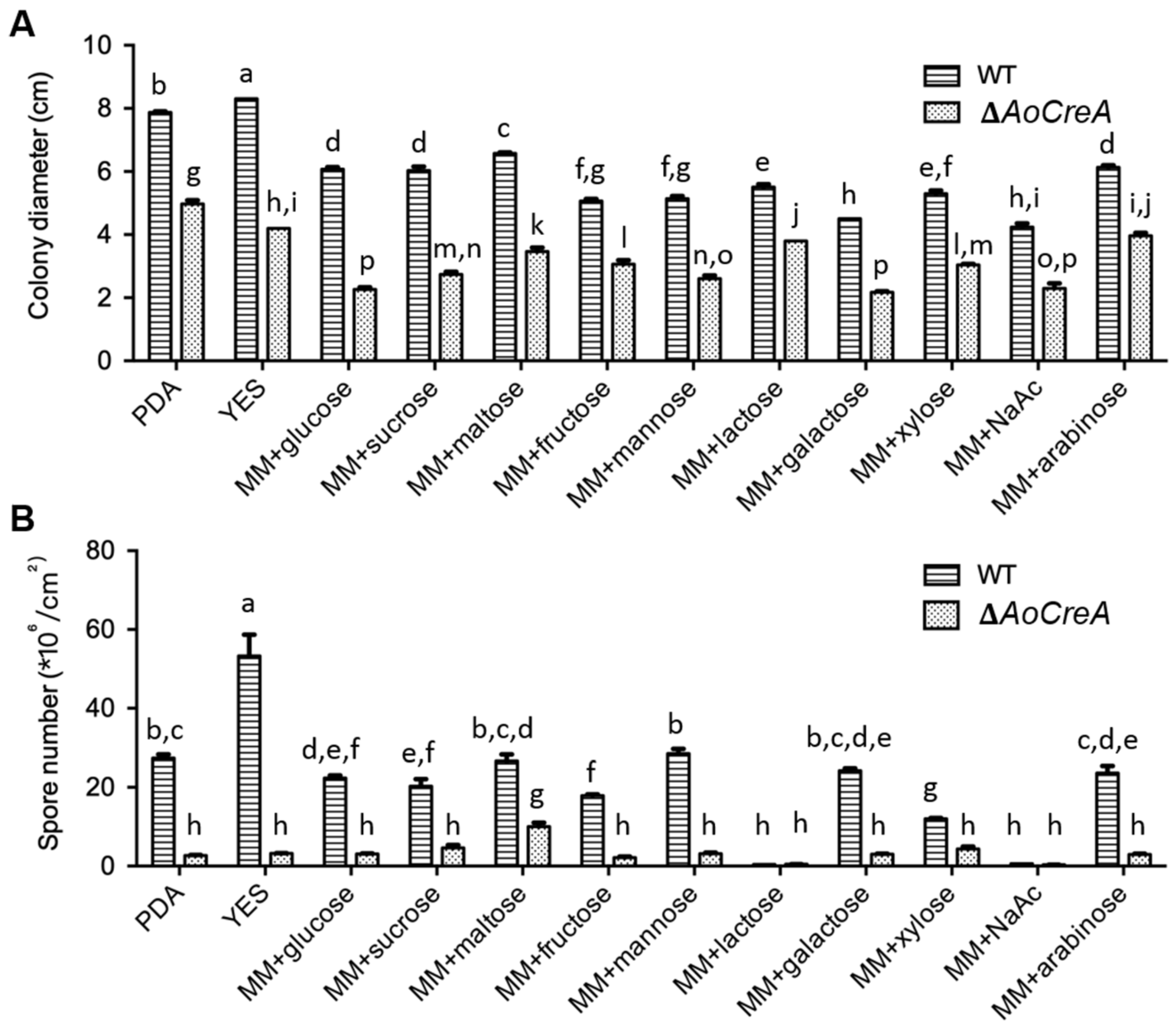
2.3. Growth and Conidiospore Production Were Affected by Carbon Sources and Modulated by AoCreA

A series of morphological changes were observed between A. ochraceus WT and three ΔAoCreA mutants when grown in different carbon sources (Figure 3). In PDA and Yeast Extract Sucrose (YES) media, conidiospores were largely produced in WT but not in ΔAoCreA. In the YES medium, as expected, wrinkles appeared on the surface of WT colony, while they disappeared in ΔAoCreA. When glucose, sucrose and maltose were used as carbon sources, the A. ochraceus WT and ΔAoCreA mutants showed almost the same morphology. When arabinose, NaAc and lactose were used as carbon sources, the colonies were transparent as little hypha was grown on the medium. The growth rate of A. ochraceus WT and ΔAoCreA grown in a single carbon source medium was slower than that grown on the YES and PDA media (Figure 3 and Figure 4A). The deletion of the AoCreA gene has a significant impact on the growth rate in all kinds of culture media (Figure 4A). In the media containing a single carbon source, glucose, sucrose, maltose and arabinose promote the growth of A. ochraceus, but the hypha were sparse in arabinose (Figure 3). The deletion of the AoCreA gene in A. ochraceus had a significant impact on the production of conidiospores. The deletion of AoCreA decreased the number of conidiospores on all kinds of media (Figure 4B). With lactose and NaAc as carbon sources, hardly any conidiospores were observed in WT and ΔAoCreA.

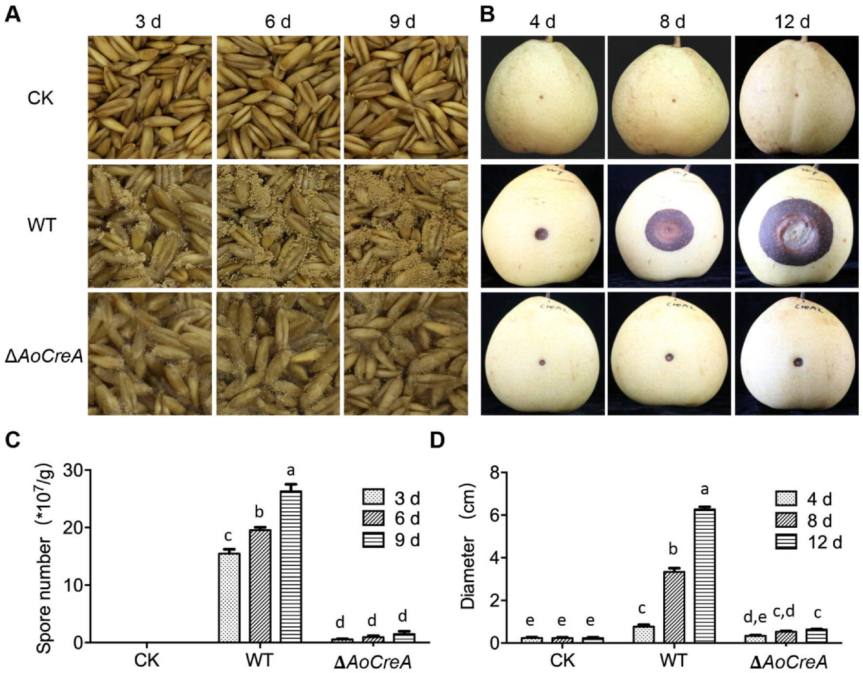
2.4. AoCreA Was Involved in OTA Biosynthesis

To investigate whether AoCreA is linked to the biosynthesis of OTA in A. ochraceus, OTA production was analyzed by High-Performance Liquid Chromatography (HPLC). The samples were collected after 12 days of cultivation at 28 °C under dark conditions and minimal media (MM) containing different carbon sources; PDA and YES were used as the cultural media. The results showed that glucose, sucrose and maltose contribute more to the biosynthesis of OTA. The concentration of OTA reached around 40 μg/cm2 for the MM containing glucose and maltose, around 20 μg/cm2 for the YES medium and MM containing sucrose and around 4 μg/cm2 for the PDA medium. Conversely, in other carbon sources, little or no OTA was detected (Figure 5A). These results demonstrated that glucose and maltose were more suitable for A. ochraceus to produce OTA, followed by sucrose and the nutrient-rich YES and PDA. OTA production was suppressed by D-xylose, D-mannose, acetate, D-galactose, D-mannitol, lactose and L-arabinose. In ΔAoCreA mutants, the production of OTA was drastically reduced compared with WT on all the media except PDA.
Seven genes (otaA, otaB, otaC, otaD, otaE, otaR1 and otaR2) consisting of a gene cluster were involved in the biosynthesis of OTA in A. ochraceus, as reported previously [9]. The relative expression level of these genes in A. ochraceus grown on media containing OTA-induced carbon sources (glucose, sucrose and maltose) and an OTA-suppressed carbon source (fructose) was examined (Figure 5B). The expression level of five genes (otaA, otaB, otaC, otaD and otaR1) increased drastically when A. ochraceus was cultured on the media containing OTA-induced carbon sources compared with being cultured on the OTA-suppressed carbon source. The expression level changes were not obvious for the genes otaE and otaR2. The gene expression level ratio of WT to ΔAoCreA was also calculated as shown in Figure 5C. The expression of five genes (otaA, otaB, otaC, otaD and otaR1) were drastically regulated by AoCreA when A. ochraceus was cultured on all the media tested. In more detail, the deletion of the AoCreA gene led to a 300-fold reduction of expression of otaA when A. ochraceus was cultured on a medium containing glucose. In particular, the expression ratio of the seven genes showed a similar profile when WT and ΔAoCreA were cultured on the OTA-suppressed medium containing fructose.

2.5. AoCreA in A. ochraceus Is Required for Fungal Infection

The role of the AoCreA gene of A. ochraceus in the pathogenicity was studied through infection experiments on oats and pears. The phenotype of oats was measured at 3, 6 and 9 days after inoculation (Figure 6A). In detail, it was observed that lots of conidiospores on the surface of oats were inoculated with WT with respect to those inoculated with ΔAoCreA. Additionally, the number of conidiospores on oats infected by WT increased over time. When oats were infected by ΔAoCreA, the conidiospore number was maintained at a low level (Figure 6C). In pear infections, the phenotype of pears was measured at 4, 8 and 12 days after inoculation (Figure 6B,D). The lesion, as a consequence of WT infection, increased over time, while that of ΔAoCreA was limited and was without expansion over time.

3. Discussion

Studies about the ochratoxin A (OTA) contamination in food safety and its biosynthetic process had been well studied in OTA producing fungi, but little was known about AoCreA regulated carbon source utilization in the biosynthesis of OTA in A. ochraceus. Carbon source is a kind of basic energy source in the growth and development of fungi, and it also plays an important role in the biosynthesis of secondary metabolites [15]. The contamination of mycotoxin in food and feed depends mostly on the types of nutrients in food substrates. Thus, most fungi have their preference during infection. This is also reflected in different stages of crop development, as the preferred carbon source may be massively synthesized during one stage. For example, maize is vulnerable to aflatoxin contamination during late development [26]. From this point of view, mycotoxin can be controlled by modifying the components of the carbon source in crops through cultivating new types of crop varieties. Many studies showed that many kinds of physical phenotypes such as growth rate, conidia production and secondary metabolite production were influenced by the utilization of a carbon source. CreA was an important regulator in this process. Furthermore, few studies have explored how CreA is regulated at the level of transcription and post-translation. Phosphorylation was demonstrated to be a post-translational pathway for regulation of CreA in S. cerevisiae and Trichoderma reesei through the involvement of the protein kinase Snf1p in the process [27,28]. CreA was also found to be regulated by ubiquitination. In fact, when cell signal was sensed by the CreB-CreC de-ubiquitination complex (DUB), CreA was de-ubiquitinated and activated, followed by CreA activating as a transcription factor in carbon catabolite repression (CCR) [29].
In our study, phylogenetic analysis indicated the function of CreA was conserved during evolution. The results illustrated that the utilization of different carbon sources was not the same in A. ochraceus; the AoCreA gene played an important role in the growth and conidiation in A. ochraceus. The AoCreA gene also played an important role in the biosynthesis of OTA in A. ochraceus, and the regulatory role of AoCreA responded to carbon sources. Furthermore, the deletion of AoCreA led to the weakened pathogenic ability of A. ochraceus. A. ochraceus has the maximum capacity for growth, conidiation and OTA production on the MM containing glucose, sucrose or maltose, which are the most abundant in the natural environment. This may be the result of adaptation of the environment during evolution. A. ochraceus infected pear and oat with severe symptoms under natural conditions, while the pathogenicity greatly decreased in the AoCreA deletion mutant. Glucose was abundant in the pear, and the starch in the oat would be digested to glucose by amylase in A. ochraceus [30]. These results illustrate that AoCreA plays an important role in the development of A. ochraceus, and many metabolic pathways were regulated by the gene. The genome of A. ochraceus was published in 2018 and consensus of the OTA biosynthetic pathway was found by a comparative genomic analysis of six OTA-producing fungi [9]. Additionally, five genes representing a putative OTA-gene cluster (otaA, otaB, otaC, otaD and otaR1) showed a consistent expression pattern. For the genes otaE and otaR2 located close to the OTA-putative gene cluster, the expression pattern under different carbon sources was different. This result could be an evidence for the unnecessary role of the two genes in OTA biosynthesis, confirming the results obtained for A. carbonarius, another main OTA-producing fungus and other OTA-producing fungi [11,12].

4. Conclusions

In conclusion, the AoCreA gene regulates morphological development and OTA biosynthesis responding to carbon sources in A. ochraceus. As far as we know, this is the first report about AoCreA in A. ochraceus. As an important global regulatory factor, there are still more functions to be explored, and how AoCreA functions in the CCR process is still unknown. This study provides some evidence about AoCreA in the development of A. ochraceus, which may provide some theoretical basis in the prevention of A. ochraceus.

5. Materials and Methods

5.1. Strains and Culture Conditions

The WT A. ochraceus used in this study was stored in Institute of Food Science and Technology, Chinese Academy of Agricultural Sciences [9]. The fungal strains used in this study were cultured at 28 °C in dark conditions. Potato Dextrose Agar (PDA, Becton, Dickinson and Company, Franklin Lakes, NJ, USA) and Yeast Extract Sucrose (YES, 5% yeast extract, 40% sucrose and 10% agar) were routinely used to culture A. ochraceus strains. Minimal media (MM, 6.0 g NaNO3, 0.52 g KCl, 0.52 g MgSO4·7H2O, 1.52 g KH2PO4, 1 mL trace elements, 10 g glucose, 12.5 g agar, pH 6.5, in 1 L distilled water) [31], supported with different carbon sources at the concentration of 2%, were used to detect the response of A. ochraceus. The test for the evaluation of different carbon sources included the adding to the MM of glucose, fructose, maltose, D-xylose, D-mannose, acetate, D-galactose, D-mannitol, lactose and L-arabinose. Each experiment was performed three times as biological replicates.

5.2. AoCreA Identification and Phylogenetic Analysis

CreA amino acid sequences from S. cerevisiae (AJR77073.1) and A. nidulans (AN6195) were used as queries, and the Basic Local Alignment Search Tool (BLAST, NCBI, Bethesda, MD, USA) algorithm was used to search AoCreA from the genome of A. ochraceus. Other CreA sequences were downloaded from the National Center for Biotechnology Information resources (NCBI). The amino acid sequences of CreA were aligned by ClustalW, and a maximum likelihood phylogeny was constructed by MEGA 5.1 software [32] using 1000 bootstrap replicates.

5.3. Construction of Gene Deletion Mutants

The construction of the A. ochraceus AoCreA deletion mutant was performed using overlap PCR procedures, which were previously described [9]. A total of 776 bp from the whole AoCreA gene (1284 bp) in the A. ochraceus genome was designed to be replaced by hygR (hygromycin B phosphotransferase). Primers used in this study are listed in Table S1. The fusion PCR products with the hygR marker were then transformed into the protoplasts of A. ochraceus. Positive transformants were verified by diagnostic PCR [33]. And real-time genomic PCR amplification was used to analyze the copy number of hygR constructs, as previously described [10,34]. Briefly, genomic DNA from each of the strains was extracted and diluted to 10 ng/μL. qPCR reactions were performed using SYBR Green supermix (Takara, Kyoto, Japan) on a Applied Biosystems 7500 Real Time PCR system (Life Technologies, Waltham, MA, USA). The number of gene copies was calculated depending on the Ct (Cycle threshold) value, with an elongation factor 1α (ef1a) as the single copy reference gene [34]. The primers used are listed in Table S1.

5.4. Phenotypic Studies

In order to study the effects of AoCreA deletion on vegetative growth and asexual sporulation, WT and deletion mutants were inoculated on PDA, YES and MM supplies for different carbon sources (glucose, fructose, maltose, D-xylose, D-mannose, acetate, D-galactose, D-mannitol, lactose and L-arabinose). Plates were inoculated with 1 μL of conidiospore suspension (106 cfu/mL) and cultured at 28 °C in darkness for 12 days. The growth rate was measured by checking the diameter of each colony. For the sporulation study, every medium was washed with 10 mL of 0.1% Tween-80 solution, and the number of conidiospores was counted under a microscope with a hemocytometer.

5.5. DNA and RNA Extraction

To extract genomic DNA, the mycelium of A. ochraceus was harvested from a shaking culture in the PDB medium, 28 °C, 180 rpm/min for 3 days. The DNA was extracted using a DNeasy kit (Qiagen, Hilden, Germany), according to the manufacturer’s protocol. For RNA extraction, the mycelium was cultured on a solid culture medium for 12 days under dark conditions. RNA was extracted using the EASYspin Plus Plant RNA Kit (Aidlab, Beijing, China) following the manufacturer’s protocol. A total of 500 ng RNA were used for the synthesis of cDNA. The removal of gDNA and synthesis of first strand cDNA were performed using the FastQuant RT Kit (TIANGEN, Beijing, China).

5.6. OTA Extraction and Production Analysis

To detect the production of OTA, each medium of the A. ochraceus strain was cultured in 28 °C under dark conditions for 12 days. A quarter of the medium was carved and cut up to small pieces. The medium pieces were steeped with 20 mL of methanol for 1 h. Ultrasonic vibration and vortexing were used to make sure all the toxins had been dissolved into methanol. After 30 min of ultrasonic vibration and 3 min of vortexing, 1 mL of the supernatant solution was filtered through a 0.22 μm filter into a vial. For OTA detection, HPLC analysis was performed on an Agilent HPLC system (Agilent Technologies, Santa Clara, CA, USA) with a mobile phase of acetonitrile/water/acetic acid (99:99:2, v/v); each sample was running for 20 min, as previously described [9]. For each plate, the total amount of OTA divided by the colony area (μg/cm2) was used to express OTA concentration.

5.7. Gene Expression Analysis

RTqPCR amplification was performed using the SYBR Green supermix on the Applied Biosystems 7500 Real Time PCR system. The obtained results were normalized against the expression of the GADPH gene of A. ochraceus. The relative expression level was calculated using the 2−ΔΔct method [35]. Primers used for gene expression are listed in Table S1.

5.8. Pathogenicity Assay

As A. ochraceus usually occurs in cereals and fruits, oats and pears were used as infection hosts to study the pathogenicity of A. ochraceus strains in this experiment. In the oat infection experiment, 20 g of oats were weighed and the water activity was adjusted to 0.95. Each bottle of oats was inoculated with 1 mL of 106 cfu/mL conidiospores and incubated at 28 °C. At 3, 6 and 9 days after inoculation, the conidiospores around the oats were washed with 0.1% Tween-80 solution and counted by a hemocytometer. In the pear infection experiment, the surface of each pear was disinfected with 75% alcohol. After the alcohol was volatilized, a hole of 3 mm in depth was punctured in the center of the pear with a sterilized toothpick. A measure of 1 μL of 106 cfu/mL conidiospores were injected and incubated at 28 °C.

5.9. Statistical Analysis

All the data were analyzed with IBM SPSS statistics version 20 and presented with the means and +/− SEM of three biological replicates samples. Different letters were used to indicate statistically significant differences analyzed by ordinary one-way analysis of variance (ANOVA) Fisher’s LSD test (p < 0.05).

Supplementary Materials

The following are available online at https://0-www-mdpi-com.brum.beds.ac.uk/2072-6651/12/11/697/s1, Table S1: Primers used in this study.

Author Contributions

Formal analysis, B.Y. and H.Z.; Methodology, G.W., Y.W. and C.Z.; Writing—original draft, G.W.; Writing—review & editing, F.X. and Y.L. All authors have read and agreed to the published version of the manuscript.

Funding

This work was supported by the Beijing Natural Science Foundation (6191001), National Key R&D Program of China (2020YFE0200206) and National Agricultural Science and Technology Innovation Program (CAAS-ASTIP-2020-IFST-03).

Conflicts of Interest

The authors declare no conflict of interest.

References

  1. Ruprich, J.; Ostry, V. Health risk assessment of the mycotoxin ochratoxin A to humans: Czech Republic--Brno--1991/92. Cent. Eur. J. Public Health 1993, 1, 86–93. [Google Scholar]
  2. Bayman, P.; Baker, J.L.; Doster, M.A.; Michailides, T.J.; Mahoney, N.E. Ochratoxin production by the Aspergillus ochraceus group and Aspergillus alliaceus. Appl. Environ. Microbiol. 2002, 68, 2326–2329. [Google Scholar] [CrossRef] [Green Version]
  3. Varga, J.; Kevei, E.; Rinyu, E.; Teren, J.; Kozakiewicz, Z. Ochratoxin production by Aspergillus species. Appl. Environ. Microbiol. 1996, 62, 4461–4464. [Google Scholar] [CrossRef] [Green Version]
  4. Harris, J.P.; Mantle, P.G. Biosynthesis of ochratoxins by Aspergillus ochraceus. Phytochemistry 2001, 58, 709–716. [Google Scholar] [CrossRef]
  5. Van der Merwe, K.J.; Steyn, P.S.; Fourie, L. Mycotoxins. II. The constitution of ochratoxins A, B, and C, metabolites of Aspergillus ochraceus Wilh. J. Chem. Soc. 1965, 1, 7083–7088. [Google Scholar] [CrossRef]
  6. Wu, Q.; Dohnal, V.; Huang, L.; Kuca, K.; Wang, X.; Chen, G.; Yuan, Z. Metabolic pathways of ochratoxin A. Curr. Drug Metab. 2011, 12, 1–10. [Google Scholar] [CrossRef]
  7. IARC. Some naturally occurring substances: Food items and constituents, heterocyclic aromatic amines and mycotoxins. IARC Monogr. Eval. Carcinog. Risks Hum. 1993, 56, 245–395. [Google Scholar]
  8. Wang, G.; Zhang, H.; Wang, Y.; Liu, F.; Li, E.; Ma, J.; Yang, B.; Zhang, C.; Li, L.; Liu, Y. Requirement of LaeA, VeA, and VelB on asexual development, ochratoxin A biosynthesis, and fungal virulence in Aspergillus ochraceus. Front. Microbiol. 2019, 10, 2759. [Google Scholar] [CrossRef] [Green Version]
  9. Wang, Y.; Wang, L.; Wu, F.; Liu, F.; Wang, Q.; Zhang, X.; Selvaraj, J.N.; Zhao, Y.; Xing, F.; Yin, W.B.; et al. A consensus ochratoxin A biosynthetic pathway: Insights from the genome sequence of Aspergillus ochraceus and a comparative genomic analysis. Appl. Environ. Microbiol. 2018, 84, e01009-18. [Google Scholar] [CrossRef] [Green Version]
  10. Crespo-Sempere, A.; Marin, S.; Sanchis, V.; Ramos, A.J. VeA and LaeA transcriptional factors regulate ochratoxin A biosynthesis in Aspergillus carbonarius. Int. J. Food Microbiol. 2013, 166, 479–486. [Google Scholar] [CrossRef]
  11. Gerin, D.; De Miccolis Angelini, R.M.; Pollastro, S.; Faretra, F. RNA-Seq reveals OTA-related gene transcriptional changes in Aspergillus carbonarius. PLoS ONE 2016, 11, e0147089. [Google Scholar]
  12. Castellá, G.; Bragulat, M.R.; Cigliano, R.A.; Cabañes, F.J. Transcriptome analysis of non-ochratoxigenic Aspergillus carbonarius strains and interactions between some black aspergilli species. Int. J. Food Microbiol. 2020, 317, 108498. [Google Scholar] [CrossRef]
  13. Gallo, A.; Ferrara, M.; Perrone, G. Recent advances on the molecular aspects of ochratoxin A biosynthesis. Curr. Opin. Food Sci. 2017, 17, 49–56. [Google Scholar]
  14. Luchese, R.H.; Harrigan, W.F. Biosynthesis of aflatoxin-the role of nutritional factors. J. Appl. Bacteriol. 1993, 74, 5–14. [Google Scholar]
  15. Szilágyi, M.; Miskei, M.; Karanyi, Z.; Lenkey, B.; Pocsi, I.; Emri, T. Transcriptome changes initiated by carbon starvation in Aspergillus nidulans. Microbiology 2013, 159, 176–190. [Google Scholar]
  16. Bailey, C.; Arst, H.N., Jr. Carbon catabolite repression in Aspergillos nidulans. Eur. J. Biochem. 1975, 51, 573–577. [Google Scholar]
  17. Dowzer, C.E.; Kelly, J.M. Analysis of the creA gene, a regulator of carbon catabolite repression in Aspergillus nidulans. Mol. Cell Biol. 1991, 11, 5701–5709. [Google Scholar]
  18. Hynes, M.J.; Kelly, J.M. Pleiotropic mutants of Aspergillus nidulans altered in carbon metabolism. Mol. Gen. Genet. 1977, 150, 193–204. [Google Scholar]
  19. Ruijter, G.J.; Visser, J. Carbon repression in Aspergilli. FEMS Microbiol. Lett. 1997, 151, 103–114. [Google Scholar]
  20. Kelly, J.M. The regulation of carbon metabolism in filamentous fungi. In Biochemistry and Molecular Biology, the Mycota; Brambl, R., Marzluf, G., Eds.; Springer: Berlin, Germany, 2016; Volume 3, pp. 385–401. [Google Scholar]
  21. Fasoyin, O.E.; Wang, B.; Qiu, M.; Han, X.; Chung, K.R.; Wang, S. Carbon catabolite repression gene creA regulates morphology, aflatoxin biosynthesis and virulence in Aspergillus flavus. Fungal Genet. Biol. 2018, 115, 41–51. [Google Scholar]
  22. Ichinose, S.; Tanaka, M.; Shintani, T.; Gomi, K. Improved α-amylase production by Aspergillus oryzae after a double deletion of genes involved in carbon catabolite repression. Appl. Microbiol. Biot. 2014, 98, 335–343. [Google Scholar] [CrossRef]
  23. Jin, C.; Barrientos, A.; Epstein, C.B.; Butow, R.A.; Tzagoloff, A. SIT4 regulation of Mig1p-mediated catabolite repression in Saccharomyces cerevisiae. FEBS Lett. 2007, 581, 5658–5663. [Google Scholar] [CrossRef] [Green Version]
  24. Zaragoza, O.; Rodriguez, C.; Gancedo, C. Isolation of the MIG1 gene from Candida albicans and effects of its disruption on catabolite repression. J. Bacteriol. 2000, 182, 320–326. [Google Scholar] [CrossRef] [Green Version]
  25. Wu, Y.; Li, T.; Gong, L.; Wang, Y.; Jiang, Y. Effects of different carbon sources on fumonisin production and FUM gene expression by Fusarium proliferatum. Toxins 2019, 11, 289. [Google Scholar] [CrossRef] [Green Version]
  26. Lillehoj, E.B.; Hesseltine, C.W. Aflatoxin control during plant growth and harvest of corn. In Mycotoxins in Human and Animal Health; Rodricks, J.V., Hesseltine, C.W., Eds.; Pathotox: Park Forest South, IL, USA, 1977; pp. 107–119. [Google Scholar]
  27. Brown, N.A.; Ries, L.N.A.; Goldman, G.H. How nutritional status signalling coordinates metabolism and lignocellulolytic enzyme secretion. Fungal Genet. Biol. 2014, 72, 48–63. [Google Scholar] [CrossRef]
  28. Cziferszky, A.; Mach, R.L.; Kubicek, C.P. Phosphorylation positively regulates DNA binding of the carbon catabolite repressor Cre1 of Hypocrea jecorina (Trichoderma reesei). J. Biol. Chem. 2002, 277, 14688–14694. [Google Scholar] [CrossRef] [Green Version]
  29. Kubicek, C.P.; Mikus, M.; Schuster, A.; Schmoll, M.; Seiboth, B. Metabolic engineering strategies for the improvement of cellulase production by Hypocrea jecorina. Biotechnol. Biofuels 2009, 2, 19. [Google Scholar] [CrossRef] [Green Version]
  30. Nahas, E.; Waldemarin, M.M. Control of amylase production and growth characteristics of Aspergillus ochraceus. Rev. Latinoam. Microbiol. 2002, 44, 5–10. [Google Scholar]
  31. Shimizu, K.; Keller, N.P. Genetic involvement of a cAMP-dependent protein kinase in a G protein signaling pathway regulating morphological and chemical transitions in Aspergillus nidulans. Genetics 2001, 157, 591–600. [Google Scholar]
  32. Tamura, K.; Peterson, D.; Peterson, N.; Stecher, G.; Nei, M.; Kumar, S. MEGA 5: Molecular evolutionary genetics analysis using maximumlikelihood, evolutionary distance, and maximum parsimony methods. Mol. Biol. Evol. 2011, 28, 2731–2739. [Google Scholar] [CrossRef] [Green Version]
  33. Wang, G.; Liu, Z.; Lin, R.; Li, E.; Mao, Z.; Ling, J.; Yang, Y.; Yin, W.; Xie, B. Biosynthesis of antibiotic leucinostatins and their inhibition on Phytophthora on bio-control Purpureocillium lilacinum. PLoS Pathog. 2016, 12, e1005685. [Google Scholar] [CrossRef] [Green Version]
  34. Solomon, P.S.; Ipcho, S.V.S.; Hane, J.K.; Tan, K.C.; Oliver, R.P. A quantitative PCR approach to determine gene copy number. Fungal Genet. Rep. 2008, 55, 5–8. [Google Scholar] [CrossRef] [Green Version]
  35. Livak, K.J.; Schmittgen, T.D. Analysis of relative gene expression data using real-time quantitative PCR and the 2−ΔΔCT method. Methods 2001, 25, 402–408. [Google Scholar] [CrossRef]
Figure 1. Phylogenetic relationship and structural domain analysis of the AoCreA homologs from different fungal species. The ochratoxin A (OTA)-producing fungi were marked in red. The blue boxes represent the C2H2 functional domain.
Toxins 12 00697 g001
Figure 2. Obtainment of A. ochraceus ΔAoCreA strains. (A) Strategy for deletion of AoCreA in A. ochraceus. (B) PCR identification of A. ochraceus WT and ΔAoCreA mutants. Each primer pairs corresponds to one WT (1) and three ΔAoCreA mutants (2, 3 and 4). (C) Real-time genomic PCR analyses determine the gene copy numbers.
Toxins 12 00697 g002
Figure 3. Colony view of A. ochraceus WT and ΔAoCreA grown on media containing different carbon sources. Both WT and ΔAoCreA had three biological replicates, and each strain was cultured on four plates as technical replicates.
Toxins 12 00697 g003
Figure 4. Effect of AoCreA deletion on the growth rate and conidiation of A. ochraceus. (A) Colony diameter of A. ochraceus WT and ΔAoCreA on different carbon sources. (B) Conidiospore production in A. ochraceus WT and ΔAoCreA. The error bars represent mean +/− SEM. Different letters indicate a significant difference between the corresponding values (p < 0.05) with three biological replicates.
Toxins 12 00697 g004
Figure 5. OTA biosynthesis in WT and ΔAoCreA strains of A. ochraceus. (A) OTA production of WT and ΔAoCreA on media containing different carbon sources. Different letters indicate a significant difference between the corresponding values (p < 0.05) with three biological replicates. (B) Relative expression level of OTA biosynthetic genes of WT on media containing glucose, sucrose, maltose and fructose analyzed by RTqPCR. (C) The expression ratio (WT/ΔAoCreA) of OTA biosynthetic genes on media containing glucose, sucrose, maltose and fructose. The ratios for different conditions demonstrated an extensive range, so the breakpoint was inserted into the Y axis. The error bars represent mean +/− SEM.
Toxins 12 00697 g005
Figure 6. Pathogenicity assay for WT and ΔAoCreA of A. ochraceus on oats and pears. (A) Infection of A. ochraceus WT and ΔAoCreA on oats. (B) Infection of A. ochraceus WT and ΔAoCreA on pears. (C) Conidiospore production of A. ochraceus grown on oats. (D) Diameter of lesion on pears. Different letters indicate a significant difference between the corresponding values (p < 0.05) with three biological replicates. The error bars represent mean +/− SEM.
Toxins 12 00697 g006
Publisher’s Note: MDPI stays neutral with regard to jurisdictional claims in published maps and institutional affiliations.

