Article

Persistent Exposure to Environmental Levels of Microcystin-LR Disturbs Cortisol Production via Hypothalamic-Pituitary-Interrenal (HPI) Axis and Subsequently Liver Glucose Metabolism in Adult Male Zebrafish (Danio rerio)

1
College of Fisheries, Huazhong Agricultural University, Wuhan 430070, China
2
Hubei Provincial Engineering Laboratory for Pond Aquaculture (Huazhong Agricultural University), Wuhan 430070, China
3
National Demonstration Center for Experimental Aquaculture Education (Huazhong Agricultural University), Wuhan 430070, China
*
Author to whom correspondence should be addressed.
Submission received: 25 March 2020 / Revised: 16 April 2020 / Accepted: 17 April 2020 / Published: 28 April 2020
(This article belongs to the Special Issue Health Risk Assessment Related to Cyanotoxins Exposure)

Abstract

:
There is growing evidence that microcystin-LR (MC-LR) is a new endocrine disruptor, whereas the impacts of persistent exposure to MC-LR on the hypothalamic-pituitary-interrenal (HPI) axis and health hazards thereafter have not been investigated. In this work, adult male zebrafish (Danio rerio) were immersed into MC-LR solutions at concentrations of 0, 1, 5 and 25 μg/L for 30 d, respectively. The results showed that persistent MC-LR exposure caused an extensive upregulation of HPI-axis genes but an inhibition of brain nuclear receptors (gr and mr), which finally increased serum cortisol levels. Furthermore, the decreased expression of hepatic gr might partly be responsible for the strong inhibition on the expression of downstream genes involved in glucose metabolic enzymes, including gluconeogenesis-related genes (pepck, fbp1a, g6pca), glycogenolysis-related gene (pyg), glycolysis-related genes (gk, pfk1b, pk) and glycogenesis-related gene (gys2). These findings are in accordance with the decline in serum glucose, indicating that long-term MC-LR exposure caused a lower production of glucose relative to glucose lysis. Our above results firstly establish the link between persistent MC-LR exposure and impaired glucose metabolism, suggesting that long-term MC-LR-mediated stress might threaten fish’s health.
Key Contribution: We found that persistent microcystin-LR (MC-LR) exposure caused the elevation of serum cortisol levels through modulating the expression of HPI-axis genes; which subsequently resulted in a disruption glucose hemostasis. For the first time, we establish the link between persistent MC-LR exposure and impaired glucose metabolism.

1. Introduction

Due to water eutrophication and global warming, the high frequencies of cyanobacterial blooms have attracted growing interest in recent decades [1,2,3]. Microcystins (MCs), a group of cyclic peptides with seven amino acids, are the most commonly detected cyanotoxins in freshwater blooms. At present, 279 structurally related analogues of MCs have been recognized [4]. Microcystin-LR (MC-LR) is the most widely distributed, abundant and toxic among these congeners [5]. The level of 1 μg/L MC-LR is recommended as the standard concentration in drinking water by the World Health Organization [6]. However, the real concentration of dissolved MC-LR in natural water is in the range of 0.1–10 μg/L, which generally oversteps the recommended level and causes potential harm to aquatic animals and human beings [7,8].
Exposure to MCs has been shown to result in multiorgan toxicity, such as hepatotoxicity [9], nephrotoxicity [1], cardiac toxicity [10], splenic toxicity [11] and neurotoxicity [12]. Recent studies have shown that MC-LR can affect the reproductive endocrine functions of fish, which are characterized with altered sex hormone levels and transcriptional profiles of genes, along the hypothalamic-pituitary-gonad (HPG) axis [13,14,15,16]. Hou et al. [17] revealed that MC-LR could elicit estrogenic responses via disrupting steroidogenesis. It has been found that MC-LR can induce thyroid dysfunction in fish through the regulation of the hypothalamic-pituitary-thyroid (HPT) axis [18,19]. These prior studies suggested that MC-LR could serve on an endocrine disruptor. In fish, the endocrine system consists of three main pathways, including the HPG axis, HPT axis and hypothalamic-pituitary-interrenal (HPI) axis. The effects of chemicals on an endocrine axis pathway could indirectly influence the other endocrine axes of fish [20]. This is corroborated by a prior study result of Bury et al. [21], who reported that acute exposure to lysed Microcystis aeruginosa cells caused an increase in the level of plasma cortisol, one of the HPI-axis final effectors, in brown trout Salmo trutta. Li et al. [22] also demonstrated that plasma cortisol levels were significantly elevated in crucian carp, injected intraperitoneally with Microcystis extract corresponding to 50 and 200 μg/kg body weight of puried MC-LR, implying that MC-LR might be capable of disrupting the adrenal endocrine system. Nevertheless, the present information on the impacts of MC-LR on the HPI axis is very limited in contrast to that of the HPG and HPT axes.
Cortisol is the most abundant and active corticosteroid in teleost fish, which plays crucial roles in controlling whole body homeostasis and response to the stress [23]. By coping with stress, cortisol levels are often elevated, which subsequently interferes with carbohydrate homeostasis, so as to meet higher energy demand [23,24,25]. In previous documents, MCs have been shown to result in disturbances of plasma glucose levels in fish [21,26,27,28]. Several omics investigations also show a positive association between MCs and abnormal carbohydrate metabolism [29,30]. It is noteworthy that teleost fish are glucose intolerant in general. Impaired glucose metabolism therefore elicits persistent hyperglycemia or subsequent physiological disturbances [31]. However, how cortisol is involved in the modulation of hepatic glucose metabolism disorder induced by MC-LR in fish and the underlying mechanism is still unclear.
Zebrafish, a kind of model organism, has been widely used to investigate chemical toxicity for its short life cycle, rapid development and growth, and the fact that it is easy to culture. In addition, prior MC-LR studies have focused primarily on high-dose acute treatments, while longer-term lower doses of MCs exposure appear to be more common and realistic than acute intoxications. The aim of the present study was therefore, through a persistent exposure experiment where adult zebrafish were immersed in MC-LR solutions at environmental equivalent levels of 0, 1, 5, 25 μg/L for 30 d, to evaluate the toxic effects of MC-LR on corticosteroid production via HPI axis and to discuss the underlying mechanism of corticosteroid influencing MC-LR-induced abnormal liver glucose metabolism.

2. Results

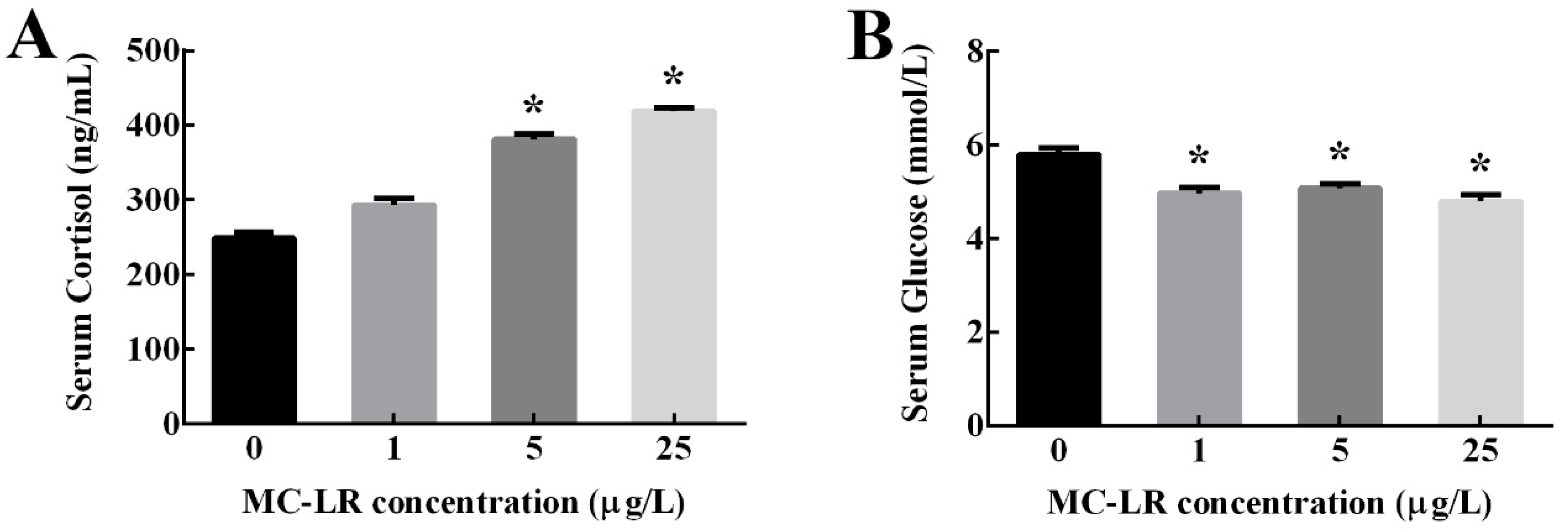
2.1. Serum Cortisol and Glucose Levels

As shown in Figure 1, serum cortisol levels were significantly increased in 5 and 25 μg/L MC-LR treatment groups compared with the control group (p < 0.05). Conversely, serum glucose levels were significantly decreased in three MC-LR treatment groups (p < 0.05).

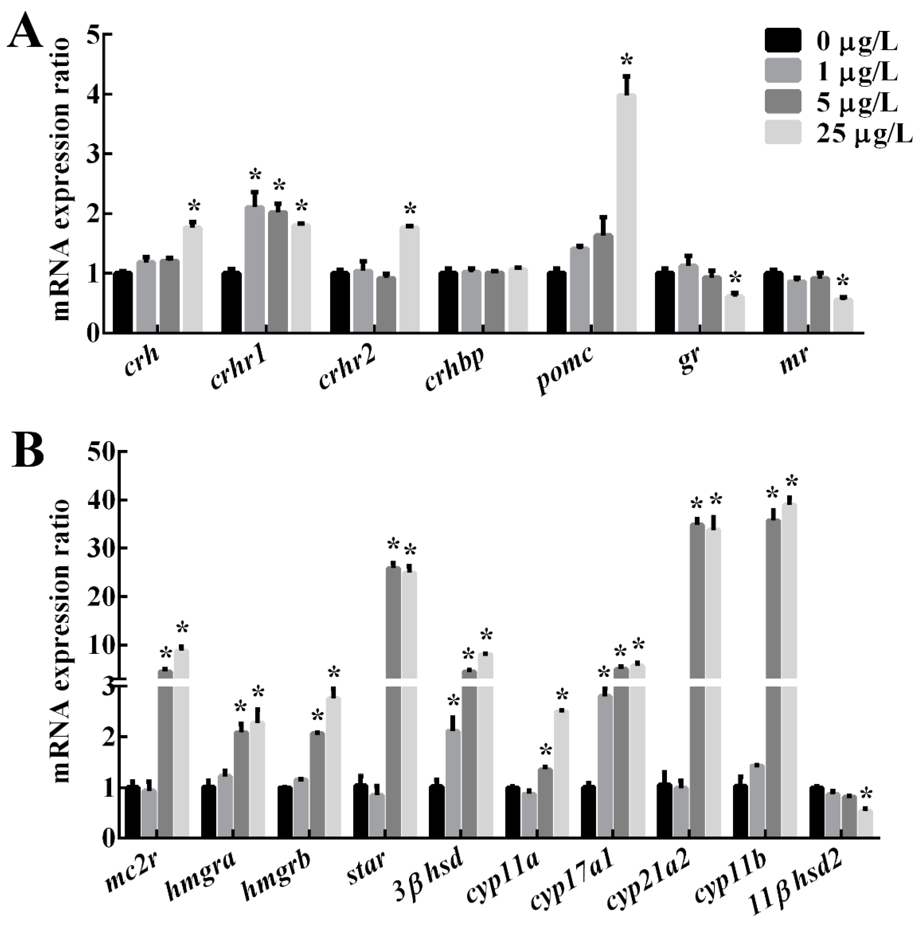
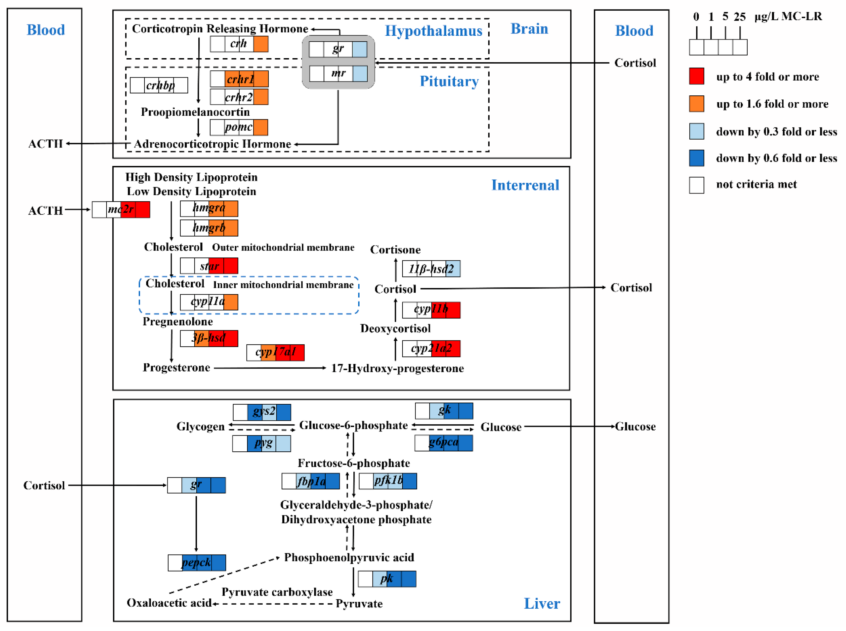
2.2. The Expression of Genes Related to HPI Axis

The effects of MC-LR treatment on the relative expression of genes involved in the HPI axis were shown in Figure 2 and Table 1. In the brain, mRNA levels of crh, crhr2 and pomc were significantly upregulated in the 25 μg/L MC-LR group, and crhr1 mRNA were significantly increased in all MC-LR treatment groups (p < 0.05). Additionally, the expression of crhbp was not affected significantly in any treatment group. In contrast, mRNA levels of gr and mr were significantly down-regulated in 25 μg/L MC-LR groups (p < 0.05).
In the head kidney, the expression of mc2r, star, cyp11a, cyp21a2 and cyp11b was significantly elevated in 5 and 25 μg/L MC-LR groups (p < 0.05). Moreover, the expression levels of 3βhsd and cyp17a1 were significantly up-regulated by all MC-LR treatments (p < 0.05), while the expression of 11βhsd2 was significantly down-regulated in the 5 and 25 μg/L MC-LR groups (p < 0.05).

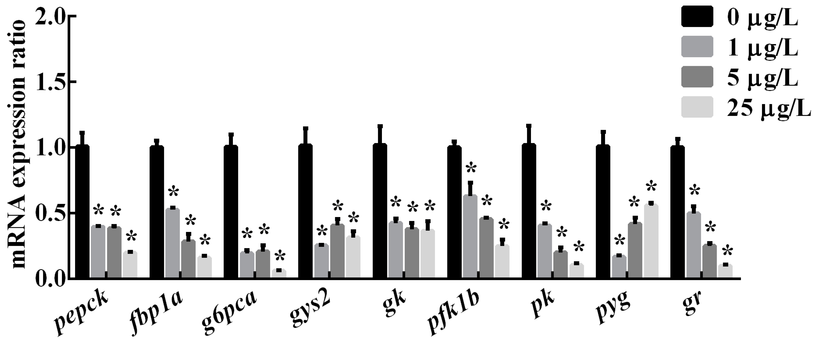
2.3. The Expression of Genes Related to Hepatic Glucose Metabolic Enzymes

In the liver, mRNA levels of glycolysis-related genes (gk, pfk1b, pk) and the glycogenolysis-related gene (pyg) were significantly downregulated in three MC-LR treatment groups (Figure 3 and Figure 4). Similarly, gluconeogenesis-related genes (pepck, fbp1a, g6pca) and the glycogenesis-related gene (gys2) were significantly decreased by all MC-LR treatments (p < 0.05). The expression of gene gr in the liver of male zebrafish was also down-regulated significantly in the 1, 5 and 25 μg/L MC-LR groups (p < 0.05).

3. Discussion

Recently, the disruption of MCs on the endocrine system of fish has been the focus of the risk assessment of the ecological environment harm of toxic cyanobacterial blooms [17,19], whereas the impacts of chronic exposure to MCs on the HPI axis and thereafter health hazards have not been investigated. This present study firstly revealed that persistent exposure to environmental levels of MC-LR interferes with interrenal cortisol production via modulating the expression of HPI-axis genes, which subsequently impaired hepatic glucose metabolism and posed a threat to fish’s health.
Experimental data have shown that toxicants can elicit a stress response in teleosts, where HPI axis is activated and stress hormones are produced in response to the disturbance and restoring homeostasis [32,33,34]. In general, the activation of the HPI axis begins with the release of corticotropin-releasing hormone (CRH) from the hypothalamus, which thereafter triggers the secretion of the adrenocorticotropic hormone (ACTH) in the pituitary. Meanwhile, the CRH signal is modulated by receptors (CRHR1, CRHR2) and a binding protein (CRHBP) [35]. Circulating ACTH binds to the melanocortin type 2 receptor (MC2R) in the interrenal cells of head kidney and stimulates the conversion from high-density lipoprotein and/or low-density lipoprotein to cholesterol. After crossing the outer mitochondrial membrane, cholesterol is then transported to the inner mitochondrial membrane via the steroidogenic acute regulatory protein (StAR). Through a series of enzymatic reactions, cortisol is synthesized in the interrenal tissue of the head kidney and released into the circulating blood. Thus, the level of circulating cortisol, as a final product of the HPI axis, is usually used as an important indicator of the degree of stress response in teleost fish. Previous studies involving short-term exposure to MCs have showed that such exposures lead to disturbed physiology in fish, which are characterized by increased blood cortisol [21,22]. In this study, after 30 d of exposure, zebrafish treated with 5 and 25 μg/L MC-LR showed elevated serum cortisol levels relative to the controls, suggesting that fish suffered a continued physiological stress. In accordance with increased circulating cortisol levels, the transcription levels of HPI-axis genes except brain gr and mr as well as interrenal 11βhsd2 were significantly increased in zebrafish, following exposure to MC-LR. Several studies have documented that MC-LR can be accumulated in the brains of fish and cause neuronal histological damage and development toxicity [2,19,36]. It is then not surprising to detect the alterations of relevant HPI-axis genes in the zebrafish brain induced by MC-LR since the brain is the center of neuroendocrinology. Marked up-regulation of the primary neurohormone, CRH, was also reported in zebrafish larvae exposed to 500 μg/L MC-LR for 96 h [37] and in juvenile zebrafish after exposure to 25 μg/L MC-LR for 7, 14, 21 or 28 d [38]. Similar adverse effects on brain CRH contents were observed in studies on other chemicals exposure such as prochloraz [20] and metyrapone [34]. It was indicated by our finding, together with prior studies, that MC-LR might directly disrupt the structure and function of brain tissue and lead to subsequent corticosteroid signaling disruption. To our knowledge, the interrenal gland of teleosts is the site involved in the production of cortisol. STAR, as the rate-limiting enzyme, modulates the transportation of cholesterol [32]. CYP11A and CYP11B regulate the first and end steps in the corticosteroido-genesis pathway, respectively [39,40]. In the present study, these three key enzymes genes (star, cyp11a and cyp11b) in the interrenal tissue were significantly up-regulated in fish treated with 5 and 25 μg/L MC-LR, indicating that persistent exposure to MC-LR affected the synthesis of interrenal cholesterol. The similar up-regulation of star and cyp11a were reported in zebrafish gonad [17], where environmental concentrations of MC-LR were proven to increase the synthesis of sex steroid hormone via the regulation of the HPG-axis. In fact, in vertebrates, the brain and adrenal/interrenal tissue, as well as the gonad, share parts of steroidogenesis-related genes like star, cyp11a and cyp17 [41]. Apparently, the results of our study further revealed the cause of MC-LR-induced endocrine disruption through endocrine axes is to interfere with steroidogenesis, including cholesterol, cortisol and estrogen. Moreover, our previous study found that exposure to MC-LR caused sex-differential gonadal impairments through disrupting the HPG-axis and growth hormone/insulin-like growth factors (GH/IGFs) systems, and the gonadal development of males was more vulnerable than that of female to MC-LR [2]. Given this information, we focused this study on male zebrafish to avoid the effects of gender differences. A further study might be needed to explore gender differences in HPI-axis activity when fish are exposed to MC-LR.
In teleost fish, the negative feedback of the HPI axis is one key mechanism that allows fish to maintain a stable status when faced with various physiological or environmental stresses [42]. Glucocorticoid receptor (GR) and mineralocorticoid receptor (MR) are the specific intracellular receptors, by which corticosteroids exert their effects [43,44]. Chen et al. [45] detected a significant down-regulation of gr but up-regulation of mr in larvae zebrafish after acute exposure to 300 μg/L MC-LR, which suggested that MC-LR acted on the expression of gr and mr and then influenced the HPI axis. Marked down-regulations of gr were documented in the brain of female zebrafish after exposure to 10 and 100 μg/L monocrotophos for 21 d [40]. Studies reported that mutant zebrafish with non-functional GRs displayed a loss of negative feedback when suffering from physiological stress [46,47]. Hence, in this study, the notable down-regulation of gr and mr in the brain of zebrafish treated with 25 μg/L MC-LR might suggest an inhibition on the negative feedback caused by MC-LR, which finally resulted in the elevation of cortisol. In fact, the persistent high concentration of cortisol can become a detrimental agent to the organism, referred to as a “homeostatic overload”, where cortisol itself damages organisms’ physiological stability [48]. It was reported that increased cortisol caused by toxins could affect multiple physiological processes, such as energy metabolism, growth and development, and reproduction and immunity [23,39]. Therefore, our data showed that persistent MC-LR exposure cause an extensive upregulation of HPI-axis genes but an inhibition of nuclear receptors (gr and mr), which finally increased serum cortisol levels.
Blood glucose is also used as an indirect indicator of stress in fish, since circulating glucose levels can be influenced by multiple hormones like cortisol [23,24]. For higher energy demand, the increase of cortisol could induce an elevation of the blood glucose level through the mediation of GR [23,25]. However, in the present study, the expression levels of hepatic gr were significantly down-regulated in all MC-LR treatment groups, which might cause the problems of downstream gene recognition and activation and lead to decreased glucose levels. Our result that serum glucose levels of fish were significantly down-regulated in all MC-LR treatments supported this hypothesis. Similarly, Woźny et al. [27] observed a significant reduction in the plasma glucose level of whitefish at 14 d after intraperitoneal injection with 100 μg/kg MC-LR of body mass. In fact, hepatic glucose hemostasis is dependent on the regulation of key metabolic enzymes and genes involved in glycogenolysis, gluconeogenesis, glycolysis and glycogenesis. Glycogenolysis and gluconeogenesis speed up the conversion of glycogen and other non-sugar substances to glucose. Among these, glycogen phosphorylase (GP) catalyzes the rate limited step in glycogen depletion, while glucose-6-phosphatase (G6Pase), fructose-1, 6-bisphosphatase (FBPase) and phosphoenolpyruvate carboxykinase (PEPCK) cause de novo glucose synthesis via gluconeogenesis. Conversely, glycolysis and glycogenesis promoted glucose oxidation and storage, which are catalyzed by glucokinase (GK), phosphofructokinase (PFK) and pyruvate kinase (PK), as well as glycogen synthase (GS) [25,49]. In the present study, along with decreased serum glucose, we detected the remarkable down-regulation of gluconeogenesis-related genes (pepck, fbp1a, g6pca), glycogenolysis-related gene (pyg), glycolysis-related genes (gk, pfk1b, pk) and glycogenesis-related gene (gys2) in the liver of fish, following long-term exposure to MC-LR, which suggested a strong suppression in hepatic glucose produce, oxygenolysis and storage. It thus appeared that the liver glucose produced was less than glucose lysis in this study, indicating that persistent MC-LR exposure caused elevated energy expenditure. Chen et al. [50] reported that exposure to 10 μg/L MC-LR for 90 d significantly decreased the expression of several genes (pfk, pkl) involved in energy metabolism in the liver of zebrafish. Zhao et al. [30] also reported a remarkable decline in the expression of gluconeogenesis rate-limiting enzyme genes, including phosphoenolpyruvate carboxykinase and glucose-6-phosphatase in mice, following the 20 μg/kg MC-LR treatment for four weeks, concomitant with the decrease of plasma glucose content. Some results on acute toxicity of MC-LR have shown that MC-LR caused damage to mitochondria and the depletion of glycogen in the liver of fish [9,28,51,52]. Mitochondria are the places that produce energy, and glycogen works as energy reserves in fish liver, so their impairments can reduce the damage repair ability of cells. Thus, the broadly restraining influences on hepatic glucose metabolism in our study revealed that long-term MC-LR exposure impaired the energy generating system, which posed a potential threat to fish. Overall, persistent exposure to MC-LR downregulated hepatic gr, resulting in the inhibitions of downstream genes involved in glucose metabolic enzymes and decreases in serum glucose levels. In addition, as suggested by Malbrouck et al. [51], glycogen dyshomeostasis might be the result of protein phosphatase inhibition caused by MC-LR, because glycogen synthase as a key enzyme in the glycogen synthesis pathway can be regulated by the phosphorylated form of glycogen phosphorylase in hepatocytes. Further research is required to be sure whether the decrease of circulating glucose concentration induced by MC-LR is exclusively involved with cortisol-mediated glucose metabolism abnormality or also as a result of protein phosphatase inhibition.

4. Conclusions

This study demonstrated that persistent MC-LR exposure caused the elevation of serum cortisol levels through modulating the expression of HPI-axis genes, which subsequently resulted in a disruption glucose hemostasis. These findings firstly established the link between persistent MC-LR exposure and impaired glucose metabolism, suggesting that long-term MC-LR-mediated stress might threaten fish’s health.

5. Materials and Methods

5.1. Chemicals and Reagents

MC-LR (purity ≥ 95%) was acquired from Express Technology Co., Ltd. (Beijing, China), the purity of which was identified by high performance liquid chromatography (HPLC). The toxin was dissolved in MilliQ water to obtain a stock solution of 0.5 mg/mL for the experiment. All of the other chemicals utilized in the experiment were of analytical grade.

5.2. Animal Maintenance and Experimental Design

Healthy adult male zebrafish (AB strain, 3-months-old) were purchased from the Institute of Hydrobiology, Chinese Academy of Sciences. Before the experiment, zebrafish were cultured in a closed flow-through system with 28 ± 0.5 °C and a 12 h light: 12 h dark cycle condition for a 14-d acclimation. Fish were fed with freshly hatched Artemia nauplii three times daily to ensure adequate nutrition. After acclimation, fish were respectively immersed into MC-LR solutions at concentrations of 0, 1, 5, and 25 μg/L for 30 d. Three replicates were performed for each concentration and each replicate was assigned randomly to 30 male adult zebrafish. Exposure concentrations were chosen, since it is relevant to the environmental concentration of MC-LR in nature water bodies [8,17,53]. In order to maintain relatively constant MC-LR concentration during exposure, 1/3 exposure solution was replaced every 3 d with fresh water, containing the corresponding MC-LR level or no MC-LR. Actual MC-LR concentrations in exposure solutions were determined every 3 d using the ELISA kit for MC-LR (Beacon Analytical System, Inc., Saco, ME, USA), with a minimum detection limit of 0.1 μg/L (data not shown).
After a 30-d exposure, zebrafish were first anesthetized with tricaine methanesulfonate (MS-222). In each exposure group, the blood samples of 10 fish were immediately collected as one replicate to separate the serum for the measurement of cortisol and glucose, and then brains, head kidneys and livers of 5 fish were sampled as one replicate for the determination of gene expression. The experiment was approved by the guidelines of Institutional Animal and Care and Use Committees (IACUC) of Huazhong Agricultural University (permission number: HZAUFI-2018-013, date of approval: 11 August 2018), Wuhan in China.

5.3. Serum Cortisol and Glucose Analysis

Serum cortisol was determined as described in Rance and Baker [54] using Iodine [125I] Cortisol Radioimmunoassay (RIA) Kit from Beijing North Institute of Biological Technology (Beijing, China). According to Trinder [55], serum glucose was tested by the GOD-POD (glucose oxidase-peroxidase) method, using a commercial kit purchased from the Shanghai Mingdian Institute of Bioengineering, Inc. (Shanghai, China).

5.4. Quantitative Real-Time PCR

Total RNA was respectively isolated from brain, head kidney and liver samples using RNAiso Plus reagent (TaKaRa, Dalian, China). The concentration and purity of total RNA were determined by a NanoDrop ND-2000 spectrophotometer (Thermo Scientific, Wilmington, DE, USA). According to the manufacturer’s instruction, 1 μg of total RNA was reverse transcribed for each sample by using a PrimeScript RT reagent Kit with gDNA Eraser (TaKaRa, Dalian, China). Using SYBR Green Kits (Takara, Dalian, China), quantitative real-time PCR was conducted on LightCycler® 480 Real-Time PCR Detection System (Roche, Basel, Switzerland). Gene specific primers designed by Primer Premier 5.0 (Premier Biosoft International, Palo Alto, CA, USA) were listed in Table 2. The amplification protocol was set according to Lin et al. [11] In the light of our previous study [14], the housekeeping gene gapdh was used as an internal control for normalization. The relative expression ratio (R) was calculated using the 2−ΔΔCt method [56]. All experiments were carried in six biological replicates.

5.5. Statistical Analysis

The statistical analysis was performed using SPSS 20.0 (SPSS Inc., Chicago, IL, USA) for Windows. All values were expressed as mean ± standard error (S.E.) The significant differences were evaluated by one-way analysis of variance (ANOVA), followed by Duncan’s test. Normality and variance uniformity have been verified. Statistical significance was established at p < 0.05.

Author Contributions

L.W. wrote this manuscript text. L.L., D.L. and R.T. designed the experiments. L.W., W.L., Q.Z., H.G., D.Z. and L.Y. carried out the experiments and analyzed the data. All authors reviewed the manuscript. All authors have read and agreed to the published version of the manuscript.

Funding

This research was jointly funded by the National Natural Science Foundation of China (grant number: No. 31670521), the Fundamental Research Funds for the Central Universities (grant number: No. 2662015PY119), the National Training Program of Innovation and Entrepreneurship for Undergraduates (grant number: No. 201810504093) and the Earmarked Fund for China Agriculture Research System (grant number: No. CARS-45).

Acknowledgments

This work was jointly supported by the National Natural Science Foundation of China (No. 31670521), the Fundamental Research Funds for the Central Universities (No. 2662015PY119), the National Training Program of Innovation and Entrepreneurship for Undergraduates (No. 201810504093) and the Earmarked Fund for China Agriculture Research System (No. CARS-45).

Conflicts of Interest

The authors declare that there are no conflicts of interest in the present experiment.

References

  1. Li, L.; Xie, P.; Lei, H.; Zhang, X. Renal accumulation and effects of intraperitoneal injection of extracted microcystins in omnivorous crucian carp (Carassius auratus). Toxicon 2013, 70, 62–69. [Google Scholar] [CrossRef] [PubMed]
  2. Hou, J.; Su, Y.; Lin, W.; Guo, H.; Xie, P.; Chen, J.; Gu, Z.; Li, L. Microcystin-LR retards gonadal maturation through disrupting the growth hormone/insulin-like growth factors system in zebrafish. Ecotoxicol. Environ. Saf. 2017, 139, 27–35. [Google Scholar] [CrossRef] [PubMed]
  3. Rezaitabar, S.; Sari, A.E.; Bahramifar, N.; Ramezanpour, Z. Transfer, tissue distribution and bioaccumulation of microcystin-LR in the phytoplanktivorous and carnivorous fish in Anzali wetland, with potential health risks to humans. Sci. Totol. Environ. 2017, 575, 1130–1138. [Google Scholar] [CrossRef]
  4. Bouaïcha, N.; Miles, C.O.; Beach, D.G.; Labidi, Z.; Djabri, A.; Benayache, N.Y.; Nguyen-Quang, T. Structural diversity, characterization and toxicology of microcystins. Toxins 2019, 11, 714. [Google Scholar] [CrossRef] [Green Version]
  5. Buratti, F.M.; Manganelli, M.; Vichi, S.; Stefanelli, M.; Scardala, S.; Testai, E.; Funari, E. Cyanotoxins: Producing organisms, occurrence, toxicity, mechanism of action and human health toxicological risk evaluation. Arch. Toxicol. 2017, 91, 1049–1130. [Google Scholar] [CrossRef]
  6. World Health Organization. Health Organization. Health criteria and other supporting information: Addendum. In Guidelines for Drinking-Water Quality; No. WHO/EOS/98.1; World Health Organization: Geneva, Switzerland, 1998; Volume 2. [Google Scholar]
  7. Chen, J.; Xie, P.; Li, L.; Xu, J. First identification of the hepatotoxic microcystins in the serum of a chronically exposed human population together with indication of hepatocellular damage. Toxicol. Sci. 2009, 108, 81–89. [Google Scholar] [CrossRef]
  8. Wang, Q.; Niu, Y.; Xie, P.; Chen, J.; Ma, Z.; Tao, M.; Qi, M.; Wu, L.; Guo, L. Factors affecting temporal and spatial variations of microcystins in Gonghu Bay of Lake Taihu, with potential risk of microcystin contamination to human health. Sci. World J. 2010, 10, 1795–1809. [Google Scholar] [CrossRef] [Green Version]
  9. Li, L.; Xie, P.; Chen, J. In vivo studies on toxin accumulation in liver and ultrastructural changes of hepatocytes of the phytoplanktivorous bighead carp ip-injected with extracted microcystins. Toxicon 2005, 46, 533–545. [Google Scholar] [CrossRef]
  10. Li, D.; Xie, P.; Zhang, X.; Zhao, Y. Intraperitoneal injection of extracted microcystins results in hypovolemia and hypotension in crucian carp (Carassius auratus). Toxicon 2009, 53, 638–644. [Google Scholar] [CrossRef]
  11. Lin, W.; Hou, J.; Guo, H.; Qiu, Y.; Li, L.; Li, D.; Tang, R. Dualistic immunomodulation of sub-chronic microcystin-LR exposure on the innate-immune defense system in male zebrafish. Chemosphere 2017, 183, 315–322. [Google Scholar] [CrossRef]
  12. Li, G.; Cai, F.; Yan, W.; Li, C.; Wang, J. A proteomic analysis of MCLR-induced neurotoxicity: Implications for Alzheimer’s disease. Toxicol. Sci. 2012, 127, 485–495. [Google Scholar] [CrossRef] [PubMed] [Green Version]
  13. Zhao, Y.; Xie, L.; Yan, Y. Microcystin-LR impairs zebrafish reproduction by affecting oogenesis and endocrine system. Chemosphere 2015, 120, 115–122. [Google Scholar] [CrossRef] [PubMed]
  14. Hou, J.; Li, L.; Wu, N.; Su, Y.; Lin, W.; Li, G.; Gu, Z. Reproduction impairment and endocrine disruption in female zebrafish after long-term exposure to MC-LR: A life cycle assessment. Environ. Pollut. 2016, 208, 477–485. [Google Scholar] [CrossRef] [PubMed]
  15. Liu, W.; Chen, C.; Chen, L.; Wang, L.; Li, J.; Chen, Y.; Jin, J.; Kawan, A.; Zhang, X. Sex-dependent effects of microcystin-LR on hypothalamic-pituitary-gonad axis and gametogenesis of adult zebrafish. Sci. Rep. 2016, 6, 22819. [Google Scholar] [CrossRef] [PubMed]
  16. Su, Y.; Li, L.; Hou, J.; Wu, N.; Lin, W.; Li, G. Life-cycle exposure to microcystin-LR interferes with the reproductive endocrine system of male zebrafish. Aquat. Toxicol. 2016, 175, 205–212. [Google Scholar] [CrossRef] [PubMed]
  17. Hou, J.; Su, Y.; Lin, W.; Guo, H.; Li, L.; Anderson, D.M.; Li, D.; Tang, R.; Chi, W.; Zhang, X. Estrogenic potency of MC-LR is induced via stimulating steroidogenesis: In vitro and in vivo evidence. Environ. Pollut. 2018, 240, 615–622. [Google Scholar] [CrossRef] [PubMed]
  18. Liu, Z.; Li, D.; Hu, Q.; Tang, R.; Li, L. Effects of exposure to microcystin-LR at environmentally relevant concentrations on the metabolism of thyroid hormones in adult zebrafish (Danio rerio). Toxicon 2016, 124, 15–25. [Google Scholar] [CrossRef]
  19. Cheng, H.; Yan, W.; Wu, Q.; Liu, C.; Gong, X.; Hung, T.C.; Li, G. Parental exposure to microcystin-LR induced thyroid endocrine disruption in zebrafish offspring, a transgenerational toxicity. Environ. Pollut. 2017, 230, 981–988. [Google Scholar] [CrossRef]
  20. Liu, C.; Zhang, X.; Deng, J.; Hecker, M.; Al-Khedhairy, A.; Giesy, J.P.; Zhou, B. Effects of prochloraz or propylthiouracil on the cross-talk between the HPG, HPA, and HPT axes in zebrafish. Environ. Sci. Technol. 2011, 45, 769–775. [Google Scholar] [CrossRef]
  21. Bury, N.R.; Eddy, F.B.; Codd, G.A. Stress responses of brown trout, Salmo trutta L., to the cyanobacterium, Microcystis aeruginosa. Environ. Toxicol. Water Qual. 1996, 11, 187–193. [Google Scholar] [CrossRef]
  22. Li, D.; Xie, P.; Zhang, X. Changes in plasma thyroid hormones and cortisol levels in crucian carp (Carassius auratus) exposed to the extracted microcystins. Chemosphere 2008, 74, 13–18. [Google Scholar] [CrossRef] [PubMed]
  23. Mommsen, T.P.; Vijayan, M.M.; Moon, T.W. Cortisol in teleosts: Dynamics, mechanisms of action, and metabolic regulation. Rev. Fish Biol. Fish. 1999, 9, 211–268. [Google Scholar] [CrossRef]
  24. Trenzado, C.E.; Carrick, T.R.; Pottinger, T.G. Divergence of endocrine and metabolic responses to stress in two rainbow trout lines selected for differing cortisol responsiveness to stress. Gen. Comp. Endocrinol. 2003, 133, 332–340. [Google Scholar] [CrossRef] [Green Version]
  25. López-Patiño, M.A.; Hernández-Pérez, J.; Gesto, M.; Librán-Pérez, M.; Míguez, J.M.; Soengas, J.L. Short-term time course of liver metabolic response to acute handling stress in rainbow trout, Oncorhynchus mykiss. Comp. Biochem. Phys. A 2014, 168, 40–49. [Google Scholar] [CrossRef]
  26. Ernst, B.; Hoeger, S.J.; O’Brien, E.; Dietrich, D.R. Oral toxicity of the microcystin-containing cyanobacterium Planktothrix rubescens in European whitefish (Coregonus lavaretus). Aqua. Toxicol. 2006, 79, 31–40. [Google Scholar] [CrossRef] [Green Version]
  27. Woźny, M.; Lewczuk, B.; Ziółkowska, N.; Gomułka, P.; Dobosz, S.; Łakomiak, A.; Florczyk, M.; Brzuzan, P. Intraperitoneal exposure of whitefish to microcystin-LR induces rapid liver injury followed by regeneration and resilience to subsequent exposures. Toxicol. Appl. Pharm. 2016, 313, 68–87. [Google Scholar] [CrossRef]
  28. Paulino, M.G.; Rossi, P.A.; Venturini, F.P.; Tavares, D.; da Silva Souza, N.E.; Sakuragui, M.M.; Moraes, G.; Terezan, A.P.; Fernandes, J.B.; Giani, A.; et al. Hepatotoxicity and metabolic effects of cellular extract of cyanobacterium Radiocystis fernandoi containing microcystins RR and YR on neotropical fish (Hoplias malabaricus). Chemosphere 2017, 175, 431–439. [Google Scholar] [CrossRef]
  29. Mezhoud, K.; Praseuth, D.; Puiseux-Dao, S.; Francois, J.C. Global quantitative analysis of protein expression and phosphorylation status in the liver of the medaka fish (Oryzias latipes) exposed to microcystin-LR, Balneation study. Aquat. Toxicol. 2008, 86, 166–175. [Google Scholar] [CrossRef]
  30. Zhao, S.; Li, G.; Chen, J. A proteomic analysis of prenatal transfer of microcystin-LR induced neurotoxicity in rat offspring. J. Proteom. 2015, 114, 197–213. [Google Scholar] [CrossRef]
  31. Moon, T.W. Glucose intolerance in teleost fish: Fact or fiction? Comp. Biochem. Phys. B 2001, 129, 243–249. [Google Scholar] [CrossRef]
  32. Alsop, D.; Vijayan, M.M. Molecular programming of the corticosteroid stress axis during zebrafish development. Comp. Biochem. Phys. A 2009, 153, 49–54. [Google Scholar] [CrossRef] [PubMed]
  33. Alsop, D.; Vijayan, M. The zebrafish stress axis: Molecular fallout from the teleost-specific genome duplication event. Gen. Comp. Endocrinol. 2009, 161, 62–66. [Google Scholar] [CrossRef] [PubMed]
  34. Liu, C.; Yu, H.; Zhang, X. Zebrafish embryos/larvae for rapid determination of effects on hypothalamic-pituitary-thyroid (HPT) and hypothalamic-pituitary-interrenal (HPI) axis: mRNA expression. Chemosphere 2013, 93, 2327–2332. [Google Scholar] [CrossRef] [PubMed]
  35. Alderman, S.L.; Bernier, N.J. Ontogeny of the corticotropin-releasing factor system in zebrafish. Gen. Comp. Endocrinol. 2009, 164, 61–69. [Google Scholar] [CrossRef] [PubMed]
  36. Wu, Q.; Yan, W.; Liu, C.S.; Li, L.; Yu, L.Q.; Zhao, S.J.; Li, G.Y. Microcystin-LR exposure induces developmental neurotoxicity in zebrafish embryo. Environ. Pollut. 2016, 213, 793–800. [Google Scholar] [CrossRef]
  37. Yan, W.; Zhou, Y.; Yang, J.; Li, S.; Hu, D.; Wang, J.; Chen, J.; Li, G. Waterborne exposure to microcystin-LR alters thyroid hormone levels and gene transcription in the hypothalamic–pituitary–thyroid axis in zebrafish larvae. Chemosphere 2012, 87, 1301–1307. [Google Scholar] [CrossRef]
  38. Liu, Z.; Tang, R.; Li, D.; Hu, Q.; Wang, Y. Subacute microcystin-LR exposure alters the metabolism of thyroid hormones in juvenile zebrafish (Danio Rerio). Toxins 2015, 7, 337–352. [Google Scholar] [CrossRef] [Green Version]
  39. Fuzzen, M.L.M.; Van Der Kraak, G.; Bernier, N.J. Stirring up new ideas about the regulation of the hypothalamic-pituitary-interrenal axis in zebrafish (Danio rerio). Zebrafish 2010, 7, 349–358. [Google Scholar] [CrossRef]
  40. Zhang, X.; Zhong, Y.; Tian, H.; Wang, W.; Ru, S. Impairment of the cortisol stress response mediated by the hypothalamus–pituitary–interrenal (HPI) axis in zebrafish (Danio rerio) exposed to monocrotophos pesticide. Comp. Biochem. Phys. C 2015, 176, 10–16. [Google Scholar] [CrossRef]
  41. Arukwe, A. Modulation of barin steroidogenesis by affecting transcriptional changes of steroidogenic acute regulatory (StAR) protein and cholesterol side chain cleavage (P450scc) in juvenile Atlantic salmon (Salmo salar) is a novel aspect of nonylphenol toxicity. Environ. Sci. Technol. 2005, 39, 9791–9798. [Google Scholar] [CrossRef]
  42. Nichols, J.W.; Breen, M.; Denver, R.J.; DiStefano, J.J.; Edwards, J.S.; Hoke, R.A.; Volz, D.C.; Zhang, X. Predicting chemical impacts on vertebrate endocrine systems. Environ. Toxicol. Chem. 2011, 30, 39–51. [Google Scholar] [CrossRef] [PubMed] [Green Version]
  43. Bury, N.R.; Sturm, A. Evolution of the corticosteroid receptor signaling pathway in fish. Gen. Comp. Endocrinol. 2007, 153, 47–56. [Google Scholar] [CrossRef] [PubMed]
  44. Schaaf, M.J.M.; Chatzopoulou, A.; Spaink, H.P. The zebrafish as a model system for glucocorticoid receptor research. Comp. Biochem. Phys. A 2009, 153, 75–82. [Google Scholar] [CrossRef] [PubMed]
  45. Chen, L.; Wang, Y.; Giesy, J.P.; Chen, F.; Shi, T.; Chen, J.; Xie, P. Microcystin-LR affects the hypothalamic-pituitary-inter-renal (HPI) axis in early life stages (embryos and larvae) of zebrafish. Environ. Pollut. 2018, 241, 540–548. [Google Scholar] [CrossRef]
  46. Griffiths, B.; Schoonheim, P.J.; Ziv, L.; Voelker, L.; Baier, H.; Gahtan, E. A zebrafish model of glucocorticoid resistance shows serotonergic modulation of the stress response. Front. Behav. Neurosci. 2012, 6, 68. [Google Scholar] [CrossRef] [Green Version]
  47. Ziv, L.; Muto, A.; Schoonheim, P.J.; Meijsing, S.H.; Strasser, D.; Ingraham, H.A.; Schaaf, M.J.M.; Yamamoto, K.R.; Baier, H. An affective disorder in zebrafish with mutation of the glucocorticoid receptor. Mol. Psychiatry 2013, 18, 681–691. [Google Scholar] [CrossRef]
  48. Romero, L.M.; Dickens, M.J.; Cyr, N.E. The reactive scope model—A new model integrating homeostasis, allostasis, and stress. Horm. Behav. 2009, 55, 375–389. [Google Scholar] [CrossRef]
  49. Zhao, F.; Jiang, G.; Wei, P.; Wang, H.; Ru, S. Bisphenol S exposure impairs glucose homeostasis in male zebrafish (Danio rerio). Ecotoxicol. Environ. Saf. 2018, 147, 794–802. [Google Scholar] [CrossRef]
  50. Chen, L.; Hu, Y.; He, J.; Chen, J.; Giesy, J.P.; Xie, P. Responses of the proteome and metabolome in livers of zebrafish exposed chronically to environmentally relevant concentrations of microcystin-LR. Environ. Sci. Technol. 2016, 51, 596–607. [Google Scholar] [CrossRef]
  51. Malbrouck, C.; Trausch, G.; Devos, P.; Kestemont, P. Effect of microcystin-LR on protein phosphatase activity and glycogen content in isolated hepatocytes of fed and fasted juvenile goldfish Carassius auratus L. Toxicon 2004, 44, 927–932. [Google Scholar] [CrossRef]
  52. Hou, J.; Li, L.; Xue, T.; Long, M.; Su, Y.; Wu, N. Hepatic positive and negative antioxidant responses in zebrafish after intraperitoneal administration of toxic microcystin-LR. Chemosphere 2015, 120, 729–736. [Google Scholar] [CrossRef] [PubMed]
  53. Lin, W.; Hou, J.; Guo, H.; Li, L.; Wang, L.; Zhang, D.; Li, D.; Tang, R. The synergistic effects of waterborne microcystin-LR and nitrite on hepatic pathological damage, lipid peroxidation and antioxidant responses of male zebrafish. Environ. Pollut. 2018, 235, 197–206. [Google Scholar] [CrossRef] [PubMed]
  54. Rance, T.A.; Baker, B.I. The in vitro response of the trout interrenal to various fragments of ACTH. Gen. Comp. Endocrinol. 1981, 45, 497–503. [Google Scholar] [CrossRef]
  55. Trinder, P. Determination of glucose in blood using glucose oxidase with an alternative oxygen acceptor. Ann. Clin. Biochem. 1969, 6, 24–27. [Google Scholar] [CrossRef]
  56. Livak, K.J.; Schmittgen, T.D. Analysis of relative gene expression data using real-time quantitative PCR and the 2−ΔΔCT method. Methods 2001, 25, 402–408. [Google Scholar] [CrossRef]
Figure 1. The contents of serum cortisol (A) and glucose (B) in male zebrafish after a 30-d exposure to microcystin-LR (MC-LR) at different concentrations of 0, 1, 5 and 25 μg/L, respectively. Data are shown as mean ± SE (n = 3). Asterisk (*) indicates significant differences at p < 0.05 between MC-LR treatments and the control.
Toxins 12 00282 g001
Figure 2. Relative mRNA expression of genes related to hypothalamic-pituitary-interrenal (HPI) axis in the brain (A) and interrenal (B) of adult male zebrafish after a 30-d exposure to MC-LR at different concentrations of 0, 1, 5 and 25 μg/L, respectively. Values are reported as mean ± S.E. (n = 6). Asterisk (*) indicates significant differences at p < 0.05 between MC-LR treatments and the control.
Toxins 12 00282 g002
Figure 3. The relative mRNA expression of genes related to hepatic glucose metabolic enzymes in adult male zebrafish after a 30-d exposure to MC-LR at different concentrations of 0, 1, 5 and 25 μg/L, respectively. Values are reported as (mean ± S.E., n = 6). Asterisk (*) indicates significant differences at P< 0.05 between MC-LR treatments and the control.
Toxins 12 00282 g003
Figure 4. Graphical summary of the impacts of MC-LR on the HPI axis and thereafter hepatic glucose metabolism disorder in male zebrafish.
Toxins 12 00282 g004
Table 1. Gene list for HPI-axis signaling and hepatic glucose metabolic pathways in male zebrafish.
AbbreviationGene Name
crhcorticotropin releasing hormone
crhr1corticotropin releasing hormone receptor 1
crhr2corticotropin releasing hormone receptor 2
crhbpcorticotropin releasing hormone binding protein
pomcproopiomelanocortin
grglucocorticoid receptor
mrmineralocorticoid receptor
mc2rmelanocortin 2 receptor
hmgrahydroxy-methylglutaryl-CoA reductase a
hmgrbhydroxy-methylglutaryl-CoA reductase b
starsteroidogenic acute regulatory protein
cyp11a1cytochrome P450, family 11, subfamily A, polypeptide 1
-hsd3-beta-hydroxysteroid dehydrogenase
cyp17a1cytochrome P450, family 17, subfamily A, polypeptide 1
cyp21a2cytochrome P450, family 21, subfamily A, polypeptide 2
cyp11bcytochrome P450, family 11, subfamily B
11β-hsd211-beta-hydroxysteroid dehydrogenase-type 3
pepckphosphoenolpyruvate carboxykinase
fbp1afructose-1,6-bisphosphatase 1a
g6pcaglucose-6-phosphatase
gkglucokinase
pfk1bphosphofructokinase 1b
pkpyruvate kinase
pygphosphorylase, glycogen
gys2glycogen synthase 2
Table 2. Primer sequences for genes related to HPI axis and hepatic glucose metabolism of male zebrafish.
GeneSequence of the Primers (5′-3′)Accession No.Product Length (bp)Amplification Efficiency (%)
crhF: TTCGGGAAGTAACCACAAGCNM_001007379.1161109.37%
R: CTGCACTCTATTCGCCTTCC
crhr1F: TCGGTGTTGAGTCTCATGCCXM_691254.618693.89%
R: CGCGTGCGGGTAAAATGTAG
crhr2F: TGAACTGTTGCTCCGAGTCCNM_001113644.181101.76%
R: GGAGAAACACGTCGCCGTTA
crhbpF: AGAGGTCCATTCTTGGCTGCNM_001003459.117098.52%
R: GCTCGACACCATTCTGACCA
pomcF: AGGTCGACTATCCGCAAGAAAY15800323297.13%
R: CAACCTCTCCCCCTTAAAGC
grF: TTCTACGTTGCTGACGATGCEF56711223995.48%
R: CCGGTGTTCTCCTGTTTGAT
mrF: TGCCACTACGGGGTTGTTACEF567113181102.77%
R: GTGCCCCAAGATTCATCCCA
mc2rF: AAAGGGGTATTCGTGGCCTGNM_18097115096.54%
R: TGCCGGATCAATAACAGCGT
hmgraF: GGAACATGCACTAAGCAGGCNM_001079977.214393.24%
R: AGAGAAGAAGGGATCGGTTGC
hmgrbF: CCGCTTCTATTGGACGGGAANM_001014292.220090.61%
R: AACCAGCCCTTAACCTCTGC
starF: TTTCTGGCTGGGATGTCCACNM_131663105104.15%
R: ATCTGCACTTGGTCGCATGA
cyp11aF: TCCCGAAACCAGAGCAATACNM_15295320497.45%
R: GCTCAAACTTGCTCCTGACC
3βhsdF: CGCACATCGTCTCAGGACATAY279108.1122106.82%
R: GTAGAGCGCTGCGTTGAAAG
cyp17a1F: AGACCCACCACAGACCTTTACNM_212806.39692.59%
R: TGAGCACAATCGGCCACTTA
cyp21a2F: AATGGTCTGACTTTGCTGGGAXM_021474355.122295.38%
R: CAGCCTTTCCACTGTAGTCTCG
cyp11bF: TGTATCCTCTGGGTCGTTCCDQ65071022899.68%
R: GCTCTTCTGTGGACGAAACC
11β-hsd2F: ATCAGAATCCATCCAGCCTTAGNM_21272011392.76%
R: ATTAGCATCCACACCAATACATC
pepckF: TGCCTGGATGAAATTTGACANM_213192.110698.67%
R: GGCATGAGGGTTGGTTTTTA
fbp1aF: CATCTGTATGGGATTGCTGGNM_19994217393.12%
R: TTACCCCGTCTATCTGGCTC
g6pcaF: GGCTGAACCTCGTCCTAAAGTBC07644620393.11%
R: GATTGAAAGCAACGCTGTGAT
gkF: TGAGGATGAAGAGCGAGGCBC12235917891.9%
R: AGAGAAGGTGAATCCCAGCG
pfk1bF: GCATGTGTGTCATTCCTGCCNM_001328389.1194104.5%
R: GCCTGTAGTGGTTGCCAGAT
pkF: AGAAACAGCCAAAGGACABC152219253103.16%
R: ACGAGGACGATAACGAGA
pygF: GAAAGGTTTCAACCGGCACCNM_001008538.18890.8%
R: GTGCGACAGCGCGAAATAAT
gys2F: GTGCGATGCGACTATCCAGANM_001018679.1188106.6%
R: TTCACCCCATTCGTCCACAG
gapdhF: CTGGTGACCCGTGCTGCTTNM_00111511415098.2%
R: TTTGCCGCCTTCTGCCTTA

