Microcystin Incidence in the Drinking Water of Mozambique: Challenges for Public Health Protection


1
CIIMAR/CIMAR—Interdisciplinary Center of Marine and Environmental Research, University of Porto, Terminal de Cruzeiros do Porto, Avenida General Norton de Matos, 4450-238 Matosinhos, Portugal
2
Institute of Biomedical Science Abel Salazar, University of Porto, R. Jorge de Viterbo Ferreira 228, 4050-313 Porto, Portugal
3
Department of Chemistry, Faculty of Sciences, Eduardo Mondlane University, Av. Julius Nyerere, n 3453, Campus Principal, Maputo 257, Mozambique
4
Faculty of Science, University of Porto, Rua do Campo Alegre, 4069-007 Porto, Portugal
*
Author to whom correspondence should be addressed.
Toxins 2020, 12(6), 368; https://0-doi-org.brum.beds.ac.uk/10.3390/toxins12060368
Submission received: 6 May 2020
/
Revised: 29 May 2020
/
Accepted: 31 May 2020
/
Published: 2 June 2020
(This article belongs to the Special Issue Freshwater Algal Toxins: Monitoring and Toxicity Profile)
Abstract
:Microcystins (MCs) are cyanotoxins produced mainly by freshwater cyanobacteria, which constitute a threat to public health due to their negative effects on humans, such as gastroenteritis and related diseases, including death. In Mozambique, where only 50% of the people have access to safe drinking water, this hepatotoxin is not monitored, and consequently, the population may be exposed to MCs. The few studies done in Maputo and Gaza provinces indicated the occurrence of MC-LR, -YR, and -RR at a concentration ranging from 6.83 to 7.78 µg·L−1, which are very high, around 7 times above than the maximum limit (1 µg·L−1) recommended by WHO. The potential MCs-producing in the studied sites are mainly Microcystis species. These data from Mozambique and from surrounding countries (South Africa, Lesotho, Botswana, Malawi, Zambia, and Tanzania) evidence the need to implement an operational monitoring program of MCs in order to reduce or avoid the possible cases of intoxications since the drinking water quality control tests recommended by the Ministry of Health do not include an MC test. To date, no data of water poisoning episodes recorded were associated with MCs presence in the water. However, this might be underestimated due to a lack of monitoring facilities and/or a lack of public health staff trained for recognizing symptoms of MCs intoxication since the presence of high MCs concentration was reported in Maputo and Gaza provinces.
Key Contribution: This review will contribute to the implementation of an operational monitoring program of MCs in order to reduce or avoid the possible cases of intoxications since the drinking water quality monitoring protocol recommended by the Ministry of Health does not include MC tests.
1. Introduction
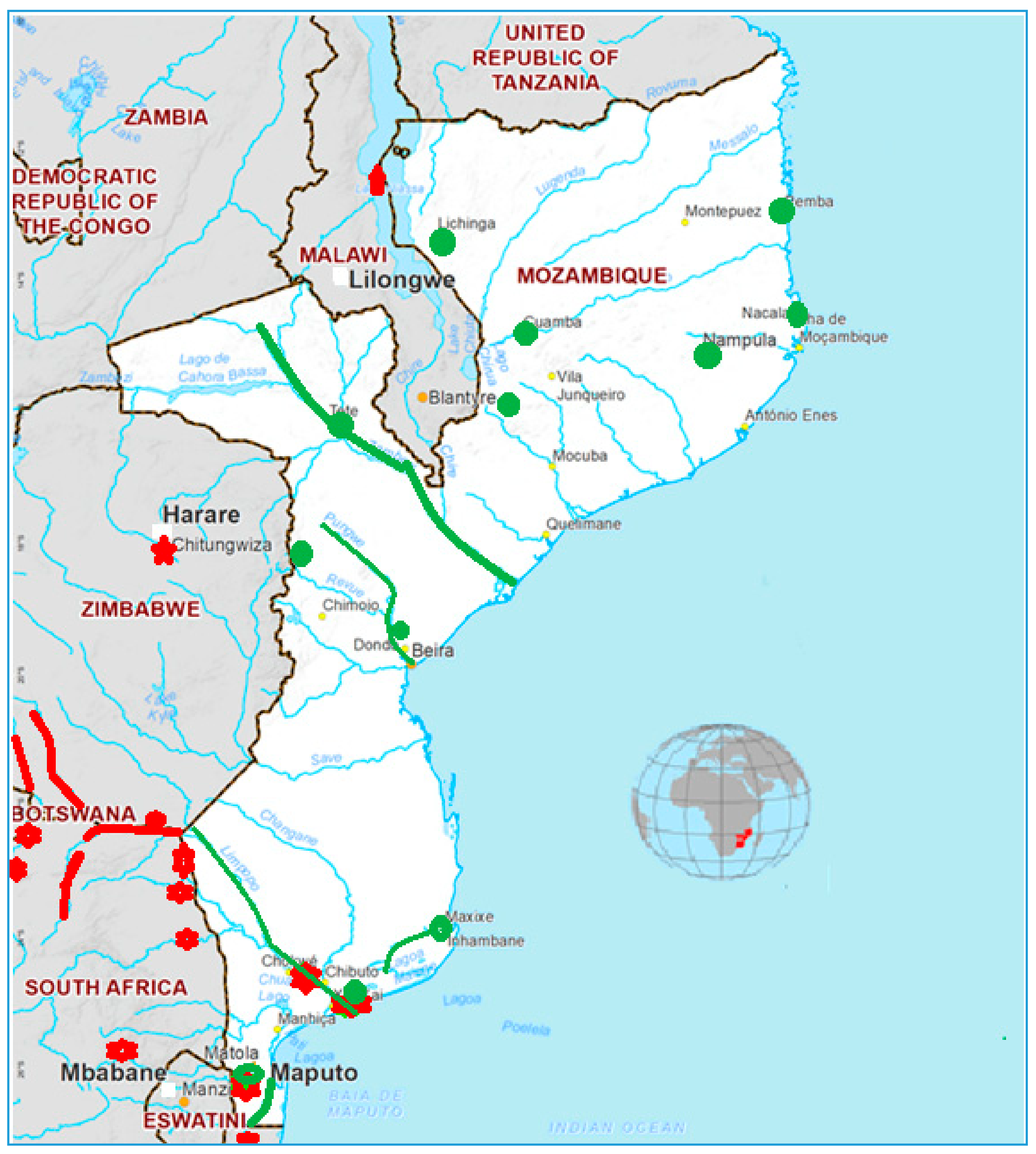
Mozambique (Figure 1) is a country located in southeastern Africa (10°30′–26°52′ S and 40°50′–30°31′ E) covering a total land area of 800,000 km2. It is bathed by the Indian Ocean in the east and makes borders with Tanzania in the north; Malawi and Zambia in the northwest; Zimbabwe in the west and Swaziland and South Africa in the southwest.
According to IV populational census carried out in 2017, this country has 29.67 million habitants distributed in 11 provinces [1]. The climate of Mozambique varies from subtropical climates (north and center) to dry arid (south) [2]. Like many African countries, Mozambique is highly vulnerable to climate variability and extreme weather events (droughts, floods, and tropical cyclones) [3]. Droughts are the most frequent natural disaster that have a negative impact on the population that reside in these rural areas [4]. The location of Mozambique in the coastal area makes it vulnerable to floods since many transnational river basins end [2]. Unfortunately, only 50% of the population has access to “safe drinking water”. Urban areas are the most favored, with 80%, while rural and most of the population have only 35% coverage and consume untreated water daily from rivers, lakes, and small puddles that form after or during the rain [5,6,7], putting at risk public health.
Eutrophication of freshwater resources may lead to the occurrence of cyanobacterial blooms and the presence of cyanotoxins, being microcystins (MC), the most common toxins worldwide [8]. The presence of MC in untreated drinking water is a major threat to public health because this potent cyanotoxin causes hepatotoxicity in humans. Thus, this review evaluates the incidence of MC and its producers in drinking water bodies of Mozambique, based on reported and available data and the estimated human illness case numbers and associated economic damage caused by Microcystin both overall globally, and in Mozambique—Africa. Recommendations for routine control and monitoring of MC will also be done since this hepatotoxin is not included in water control tests data in Mozambique.
2. Microcystin-Producing Species and Toxicology
2.1. Microcystin-Producing Species
MCs are secondary metabolites produced by cyanobacteria species that occur naturally (but it can be increased severely by human activities) in freshwater environments. The most reported cyanobacteria species, which produce MCs are listed in Table 1 and include species of the families Microcystaceae, Nostocaceae, Microcoleaceae, Oscillatoriaceae, Pseudanabaenaceae (Table 1). The occurrence and development of a particular genus and species of cyanobacteria and cyanotoxins production worldwide seem to be conditioned to water chemistry and climate conditions [8]. In a temperate climate, Microcystis and Anabaena blooms occur widely while Cylindrospermopsis develops in tropical regions [9]. There are toxic and non-toxic cyanobacteria of the same species, which may be found together [8,10,11]. Toxic cyanobacteria can produce several toxins with different toxicity making it uncertain to assess the overall toxicity of bloom due to the variations of toxins concentration spatially and seasonally [12]. To distinguish toxic and non-toxic cyanobacteria species is very complicated, and consequently, the methods used are also complex. It implicates that the prevention of cyanobacteria bloom development is a suitable way to control toxic blooms [13,14].
The factors that promote the MC synthesis are not yet clearly understood, however, the optimal growth of MC-producing species and toxicity seem influenced by light intensity, nutrients, and temperature, among other factors. For example, the higher toxicity of M. aeruginosa extracts was verified in extreme pH values [39,40], and heavy metals such as Zinc and Iron did not influence the M. aeruginosa toxicity [41]. The content of nitrogen and phosphorus influenced the toxicity of M. aeruginosa extracts. Low nitrogen content reduces the M. aeruginosa toxicity, while low phosphorous increased the toxicity in the natural population [42,43] and reduced in lab experiments [16,21,44,45]. Another lab conclusion was the correlation of colony size and content of toxic cyclic heptapeptide of the non-axenic strain of M. viridis and axenic M. viridis was also verified [20,46,47]. In general, the optimal temperature for which MC-producing species produce MC ranged from 20 to 25 °C [21,40,48,49]. This range of optimal temperature suggests that cyanobacteria blooms are most toxic during periods with warm weather and in areas with warm climates [8].
2.2. Toxicology
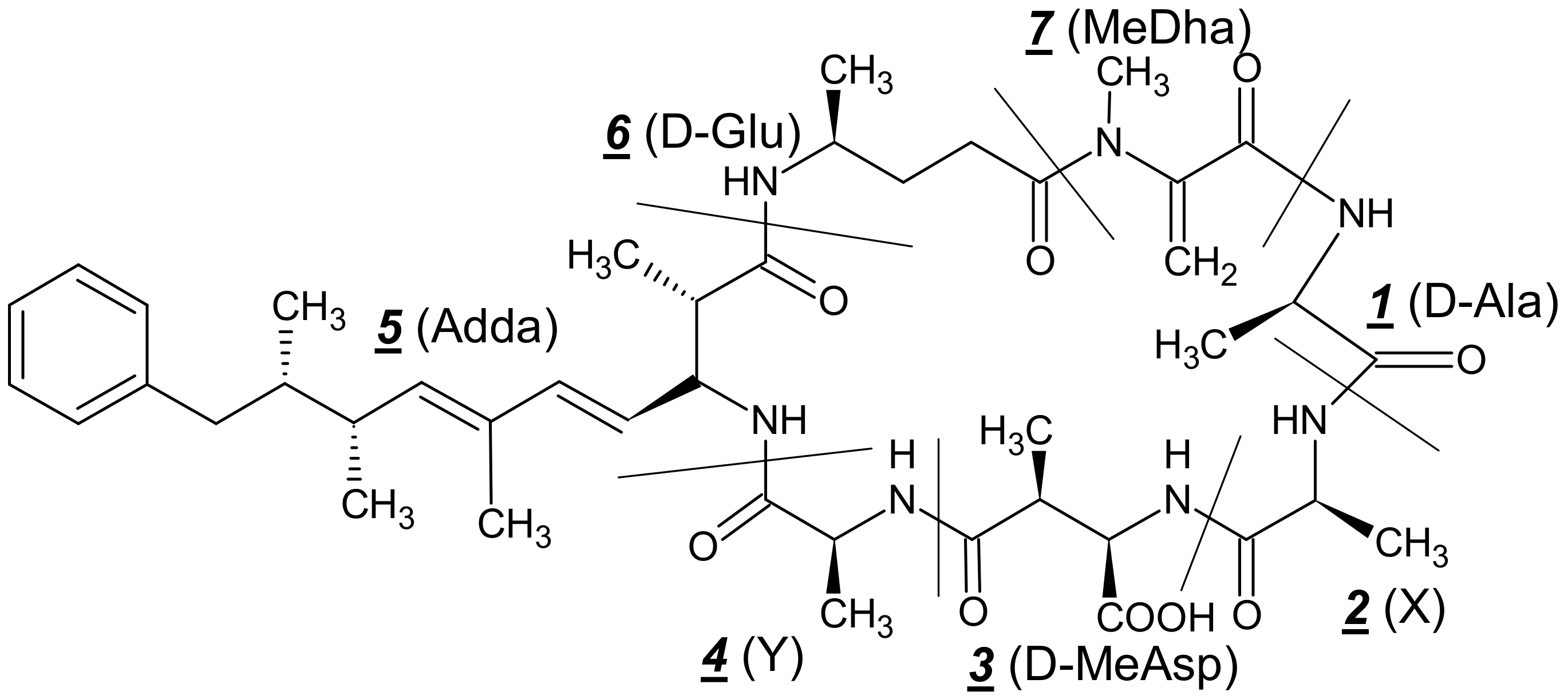
Microcystins (Figure 2) are the largest diverse group of cyanobacterial toxins, and to date, more than 240 MCs analogs are known, and they vary structurally in terms of the degree of methylation, hydroxylation, epimerization, peptide sequence, and consequently in their toxic effects [50,51,52]. Chemically, MC is a group of monocyclic heptapeptides (numbered in Figure 2) containing both D- and L-amino acids plus N-methyldehydroalanine (Mdha) and a unique β-amino acid side-group, 3-amino-9-methoxy-2-6,8-trymethyl-10-phenyldeca-4,6-dienoic acid (Adda) and their analogs differ among them, at the two L-amino acids and on the methyl groups on D-erythro-β-methylaspartic acid (D-MeAsp) and Mdha with molecular weight varying from 900 to 1100 Daltons. MC-LR, MC-RR, and MC-YR are common MC variants, the letters L, R, and Y represent the aminoacids leucine, arginine, and tyrosine, which appear on the MC molecule in different combinations [50,53,54,55,56,57,58] being MC-LR the most studied. The biosynthesis of this group of cyanotoxin is regulated by non-ribosomal peptide synthetase and polyketide synthase domains, being MCyS the gene cluster, which has been sequenced and partially characterized in several cyanobacterial species of the family Microcystaceae, Nostocaceae, Microcoleaceae, Oscillatoriaceae, Merismopediacea, and Pseudanabaenacea [16,17,18,19,20,21,22,23,24,25,26,27,28,29,30,31,32,33,34,35,36,37,38,59,60]
The mechanism of MCs toxicity seems to be well understood. They bind to serine/threonine-specific protein phosphatases (PPs) such as PP1 and PP2A, inhibiting their activity [61,62,63]. Adda moiety (Figure 1) plays an important role in the MC toxicity group since its isomerization and/or oxidation reduces the toxicity [64,65]. The inhibition of PP1and PP2A as a result of MC acute exposure causes excessive protein phosphorylation, alterations in the cytoskeleton, loss of cell shape, and consequently destruction of liver cells leading to intrahepatic hemorrhage or hepatic insufficiency [58]. Oxidative stress increasing in cells and consequent apoptosis, which can cause tumor promotion, is another mechanism of MC toxicity [66,67,68].
3. Effects of Microcystin in Humans, Symptoms, and Treatment
Microcystin effects in humans depend on the time of exposure and concentration ingested [69], and the studies are based on epidemiologic data but the reported studies on laboratory animals. Human health problems are mostly caused by chronic exposure by consumption of contaminated water or food, dermal exposure, or inhalation [57]. MC human poisoning episodes were reported in different parts of the world after the consumption of contaminated water or during sport or recreational activities [59,70,71,72]. Some examples of MC human poisoning cases are described; America—in 1996, an episode of human intoxications by MC was reported in Brazil with more than 76 deaths of patients at two dialysis centers in Caruaru. The municipal water supplied to the dialysis centers was the source of MC [71,73,74,75]. In Argentina, a human poisoning caused by MC involving a young man after immersion in an intense bloom Microcystis sp lake during sport and recreational activities were recorded. Four hours after exposure, the patient showed nausea, abdominal pain, and fever, and 48.6 µg·L−1 of microcystin-LR was detected in the water samples [76]. Other cases were recorded in Uruguay (January 2015) involving a 20-month-old child and her family during recreational activities. These victims were admitted to the hospital with diarrhea, vomiting, fatigue, and jaundice and the analysis confirmed the presence of MC-LR (2.4 ng·g−1 tissue) and [D-Leu1]MC-LR (75.4 ng·g−1 tissue) explanted liver [77]. Africa—toxic cyanobacteria suspected intoxication cases were reported in Zimbabwe involving children that were hospitalized in the Hospital of Harare with gastroenteritis symptoms [78]. In Europe—121 people presented abdominal pain, nausea, vomiting, diarrhea, fever, headaches, and muscle pain after consumption of untreated water from the River Kavlingean in Sweden. In this case a bloom of MC—producing such as Planktothrix agardhii and Microcystis spp. was observed in the river [79]. The most affected human organ is the liver [57]. However, in vivo and in vitro studies indicated that the kidney and colon are also affected [80,81,82,83,84,85]. The symptoms generally reported in humans due to the MC intoxication include gastroenteritis and related diseases, allergic and irritation reactions, liver diseases, tumors, and primary liver cancer and colorectal cancers, and massive hepatic hemorrhage. MC human poisoning treatment is very complicated due to the rapid, irreversible, and severe liver damage [86], however, gastric lavage [87], administration of monoclonal antibodies against MC-LR [88], immunosupressant Cyclosporine A, antibiotic rifampin [89], and membrane-active antioxidant vitamin E, taken as a dietary supplement [90] are recommended.
4. Microcystin Detection and Monitoring in Freshwater
According to the World Health Organization (WHO) guideline, the permitted limit of MC-LR for drinking water is 1.0 µg·L−1, and the tolerable daily intake is 0.04 µgKg−1 [91]. There are several MC detection methods, the most reported are listed in Table 2. Immunoassays (IA) are suitable methods of MC detection in Mozambique because they do not require sophisticated laboratory equipment and have a limit of detection below the maximum limit (1 µg·L−1). Additionally, IA can be used in both laboratory and field studies.
5. The Occurrence of Microcystin in Mozambican Drinking Water
5.1. The Drinking Water Scenario in Mozambique
The drinking water supply scenario in Mozambique still faces major challenges because a majority of the population still consumes untreated drinking water and consequently is exposed to many water-borne diseases. Only 50% of the population has access to “safe drinking water”. Urban areas are the most favored, with 80%, while rural and most of the population have only 35% coverage and consume untreated water daily from rivers, lakes, and small puddles that form after or during the raining season [5,7]. The low water supply cover in Mozambique is inconceivable due to several reasons, among them, the existence of natural water cover (rivers) in the whole country and the presence of excessive fragmentation of governmental organisms for water management (Figure 1). The water management is led by the Ministry of Public Works, Habitation and Hydric Resources (MOPHRH), which operates, among others, with the National Direction of Water Supply and Sanitation, National Direction of Hydric Resource Management, Water Regional Administrations, Sanitation, and Water Supply Infra-structure Administration, Water Regulation Council, Fund for Investment and Patrimony of Water Supply, and other private institutions, which provide goods and services. In order to improve the water management and expand the coverage, different projects funded by the Mozambican government and non-governmental organizations such as Plataforma Moçambicana de Água [6], Greater Maputo Water Supply Expansion Project [110], Integrated Water Supply and Sanitation Project for the provinces of Niassa and Nampula [111], National Rural Water Supply and Sanitation Program (PRONASAR) in Nampula and Zambezia Provinces [112], Inhambane Rural Water Supply and Sanitation Program [113], and others were implemented involving all the MOPHRH, civil society, and private organisms. However, still to date, the national water supply does not cover enough, with the population still consuming untreated water.
The water policy in Mozambique was approved in 1995, revised in 2007 and 2016, which, in the scope of water supply and sanitation, has the following relevant goals [114]:
- Achieve the sustainable development goals, universal access to water supply, and sanitation.
- Meeting of the basic needs of the poorest population, to reduce poverty, always looking for a sustainability situation.
- Water valuing, not only as a social and environmental asset but also with the economic value it holds.
- Government’s concentration on the definition of priorities, standards, regulation, and promotion of the private sector.
- Development of an institutional framework that contributes to the management of water as a resource and provision of decentralized and autonomous water supply and sanitation, where the private sector is called upon to participate.
The Ministry of Health (MH) is the legal organism responsible for water quality control and follows the regulation of the WHO, which sets the parameters of the quality of water intended for human consumption and the methods of carrying out their checks in order to protect human health. The water quality control recommended by WHO include MC among other biological parameter and the provisional guideline value is 1 µg·L−1 for drinking water [115]. The challenge is enormous in Mozambique for control or monitoring of this hepatotoxin due to the lack of adequate laboratories for the detection of MC in drinking water, even for 50% of the population that consumes treated water. This scenario shows clearly that all the Mozambican population is very vulnerable to MC exposure.
5.2. Microcystin in Mozambican Drinking Water
The drinking water is supplied by private (autonomous systems) and governmental operators. In Table 3, are listed the main sources of drinking water in Mozambique and includes underground and water river. The drinking water treatment is performed mainly by disinfection with chlorine, but is some regions such as Pemba and Niassa, the water treatment system includes the removal of iron by aeration. Not only is there no drinking water treatment for MC removal, but also MC incidence data in Mozambique are very limited. However, according to the WHO, more than 500,000 cases of diarrhea were reported, which 100 and 7 cases correspond to dysentery and cholera, respectively, and others are unknown [116].
These data indicate that many people of Mozambique consume food and water unsafely. Few studies (Figure 1) were done by Pedro et al. [117,118,119] and Bojcevska and Jergil [120] in Pequenos Libombos dam, Nhambavale lake, Chòkwé irrigation channels and Chidenguele sites in the South of Mozambique during 2003, 2008, and 2009 and their studies indicated the occurrence of MC-LR, -YR, and -RR produced by Microcystis sp. (M. novacekii, botrys and other) and Cylindrospermopsis raciborskii [117,118,119,120] (Table 4). MC concentration varies from less than 0.01 (below quantification levels) to 0.02 in Pequenos Libombos dam, less to 0.01 to 0.68 in Chòkwé irrigation channels, 0.86 to 7.82 in Nhambavale lake and 0.57 to 6.83 µg·L−1 in Chidenguele. Higher MC concentration values than the maximum limit ranging from 6.83 to 7.78 µg·L−1 (around 7 times above) were found in the Nhambavale lake and Chidenguele sites. These data highlight (suggest) the need to implement an operational monitoring program of MCs since the tests recommended by MH do not include the MC test [121]. Neighboring countries published other data, which support the need for MC monitoring in Mozambique (Figure 1), namely:
- South Africa: MC Producers: Synechocystis sp. Microcystis aeruginosa, Microcystis panniformis, Nostoc sp., Planktothrix sp., Phormidium sp., in the Limpopo river basin [122,123,124,125,126], Hartbeespoort dam [127,128,129,130,131], Kruger National Park [132], Sand, Mawoni, Lephalale, Mokolo, Crocodile, Nzhelele ivers [126] MC -YR, -LR, -FR, -YA, -LA, -LAba (0.156–0.270, 0.059–0.18, 0.09, 0.02–0.044, 0.051–0.241, 0.080 mg.g−1) in Natal and Transvaal dams [133], 8.6 µg·L−1 in Hartbeespoort dam [134], 12,300 µg·L−1 in Hartbeespoort lake [135].
- Tanzania: MC-LR and -RR in different tissues of dead flamingos (Phoeniconaias minor) from Empakai Crater, Lake Natron and Lake Manyara (0.165–1.16 ng.g−1) [136,137,138], MC-RR (0.4–13 µgL−1) in Victoria lake [139,140], MC producers: Aphanocapsa sp., Anabaena sp., Microcystis sp. in Victoria lake [139,140].
- Malawi: MC producers: Anabaena sp. in Malawi lake [143].
For example, the drinking water in the Xai Xai district (Gaza province) (Figure 1) is supplied from the Limpopo river. This river contains different MC producers such as Synechocystis sp. Microcystis aeruginosa, Microcystis panniformis, Nostoc sp., Planktothrix sp., Phormidium sp., which were detected in South Africa areas [122,123,124,125,126]. The presence of a potentially toxic algae is not an indication of MC production but is an indication of the need for MC screening in order to confirm the MC presence.
5.3. Removal of Microscystin from Drinking Water in Mozambique
MCs can be removed from drinking water using several rapid and low-cost. The most are reported in laboratory studies, and they are not adaptable to economic conditions in Mozambique [144,145,146,147,148,149,150]. However, the following techniques seem to be useful in Mozambique and can be implemented in both rural and urban zones: Photolysis at 254 and 185 nm [147], use of wood-based and coconut-based activated carbons [148], use of bamboo-based charcoal adsorbent modified with chitosan [149], hydrophyte filter bed [150], biological activated carbon process [151], aquatic vegetable bed [152], and activated carbon from the seed husks of the pan-tropical tree, Moringa oleifera [153], among others.
6. Final Considerations and Recommendations
The drinking water supply scenario in Mozambique still faces major challenges because the majority of the population still consumes untreated drink water (from rivers, lakes, and small puddles that form after or during the raining season) and consequently exposed to many water diseases [5,7] including, for example, gastroenteritis, which is caused by hepatotoxins MCs. To date, no data of water poisoning episodes recorded were associated with MCs presence in the water. However, this might be underestimated due to a lack of monitoring facilities and/or a lack of public health staff trained for recognizing symptoms of MCs intoxication since the presence of high MCs concentration was reported in Maputo and Gaza provinces. Few studies done in Maputo and Gaza provinces indicated the occurrence of MC-LR, -YR, and -RR at a concentration ranging from 6.83 to 7.78 µg·L−1 [117,118,119,120], which are very high, around 7 times above the maximum limit (1 µg·L−1) recommended by WHO [59]. The potential MC-producing in the studied sites is mostly Microcystis sp. [117,118,119,120]. However, MC distribution in Mozambique is unknown, and a monitoring program would help to understand the dimension of the problem. To date, no water MC poisoning episodes data recorded in Mozambique. The absence of MC intoxication episodes might be underestimated due to the absence of MC monitoring plan and/or a lack of public health staff trained in recognizing symptoms of MC intoxication. MC monitoring may be implemented according to recommendations of WHO [59] (1 µg·L−1), and the respective MC analysis can be done in the existing water treatment centers in each province (Table 3). Rapid tests for MC detection, such as ELISA, can be used in each center. In the case of higher MCs content, some suitable techniques for MC removal may be used. The recommended techniques include photolysis at 254 and 185 [147], the use of wood-based and coconut-based activated carbons [148], use of bamboo-based charcoal adsorbent modified with chitosan [149], hydrophyte Filter Bed [150], Biological Activated Carbon Process [151], and aquatic vegetable bed [152], among others. These techniques can be used in both rural and urban areas due to their low-cost implementation and local access.
Author Contributions
Conceptualization, I.J.T.; introduction, I.J.T.; microcystin-producing species and toxicology, I.J.T.; effects of microcystin in humans, symptoms, and treatment, I.J.T.; microcystin detection and monitoring in freshwater, I.J.T.; the occurrence of Microcystin in Mozambican drinking water, I.J.T.; orientation, supervision and corrections, V.V. All authors have read and agreed to the published version of the manuscript.
Funding
This research was funded by Fundação para a Ciência e a Tecnologia (FCT) projects UIDB/04423/2020 and UIDP/04423/2020.
Acknowledgments
The authors acknowledge the Fundação Calouste Gulbenkian for the partial scholarship of Isidro José Tamele and the project EMERTOX [grant 734748], funded by H2020-MSCA-RISE 2016.
Conflicts of Interest
The authors declare no conflict of interest
References
- Moçambique INE Destaques. IV Censo 2017—Instituto Nacional de Estatistica—INE-Moçambique. Available online: http://www.ine.gov.mz/ (accessed on 26 November 2019).
- Arndt, C.; Strzepeck, K.; Tarp, F.; Thurlow, J.; Fant, C.; Wright, L. Adapting to climate change: An integrated biophysical and economic assessment for Mozambique. Sustain. Sci. 2011, 6, 7–20. [Google Scholar] [CrossRef] [Green Version]
- Strzepek, K.; Arndt, C.; Chinowsky, P.; Kuriakose, A.; Neumann, J.; Nicholls, R.; Thurlow, J.; Wright, L. Economics of Adaptation to Climate Change: Mozambique; World Bank: Washington, DC, USA, 2010. [Google Scholar]
- Arndt, C.; Benfica, R.; Maximiano, N.; Nucifora, A.M.; Thurlow, J.T. Higher fuel and food prices: Impacts and responses for Mozambique. Agric. Econ. 2008, 39, 497–511. Available online: https://www.unicef.org/evaldatabase/files/283729_B85_IOB_360_BW_WEB_Mozambique-Final.pdf (accessed on 22 May 2020). [CrossRef] [Green Version]
- UNICEF; Government of the Netherlands Partnership for Water Supply S.a.H. Impact Evaluation of Drinking Water Supply and Sanitation Interventions in Rural Mozambique: More than Water; Printed Report; UNICEF: New York, NY, USA; Government of the Netherlands Partnership for Water Supply: The Hague, The Netherlands, 2011; Available online: https://www.unicef.org/evaldatabase/files/283729_B85_IOB_360_BW_WEB_Mozambique-Final.pdf (accessed on 22 May 2020).
- Ministério das Obras Públicas, Habitação e Recursos Hídricos. O Sector de Água em Moçambique. PLASMA-PLATAFORMA MOCAMBICANA DA AGUA; Ministério das Obras Públicas, Habitação e Recursos Hídricos: Maputo, Moçambique, 2016. Available online: https://plama.org.mz/index.php (accessed on 1 June 2020).
- UNICEF. Situação da Água, Saneamento e Higiene em Moçambique; UNICEF: Maputo, Mozambique, 2017; Available online: https://www.unicef.org/mozambique/%C3%A1gua-saneamento-e-higiene (accessed on 1 June 2020).
- World Health Organization. Cyanobacterial Toxins: Microcystin-LR in Drinking-Water: Background Document for Development of WHO Guidelines for Drinking-Water Quality; World Health Organization: Geneva, Switzerland, 2003. [Google Scholar]
- Yoo, R.S. Cyanobacterial (Blue-Green Algal) Toxins: A Resource Guide; American Water Works Association: Denver, CO, USA, 1995. [Google Scholar]
- Lawton, L.A.; Codd, G. Cyanobacterial (blue-green algal) toxins and their significance in UK and European waters. Water Environ. J. 1991, 5, 460–465. [Google Scholar] [CrossRef]
- Skulberg, O.M.; Carmichael, W.W.; Codd, G.A.; Skulberg, R. Taxonomy of toxic Cyanophyceae (cyanobacteria). In Algal Toxins in Seafood and Drinking Water; Academic Press: New York, NY, USA, 1993; pp. 145–164. [Google Scholar]
- Falconer, I.R.; Humpage, A.R. Health risk assessment of cyanobacterial (blue-green algal) toxins in drinking water. Int. J. Environ. Res. Public Health 2005, 2, 43–50. [Google Scholar] [CrossRef] [Green Version]
- FerrÃo-Filho, A.S.; Azevedo, S.M.; DeMott, W.R. Effects of toxic and non-toxic cyanobacteria on the life history of tropical and temperate cladocerans. Freshw. Biol. 2000, 45, 1–19. [Google Scholar] [CrossRef]
- Neilan, B.A.; Jacobs, D.; Blackall, L.L.; Hawkins, P.R.; Cox, P.T.; Goodman, A.E. rRNA sequences and evolutionary relationships among toxic and nontoxic cyanobacteria of the genus Microcystis. Int. J. Syst. Evol. Microbiol. 1997, 47, 693–697. [Google Scholar] [CrossRef]
- Vaitomaa, J.; Rantala, A.; Halinen, K.; Rouhiainen, L.; Tallberg, P.; Mokelke, L.; Sivonen, K. Quantitative real-time PCR for determination of microcystin synthetase E copy numbers for Microcystis and Anabaena in lakes. Appl. Environ. Microbiol. 2003, 69, 7289–7297. [Google Scholar] [CrossRef] [Green Version]
- Oh, H.-M.; Lee, S.J.; Jang, M.-H.; Yoon, B.-D. Microcystin production by Microcystis aeruginosa in a phosphorus-limited chemostat. Appl. Environ. Microbiol. 2000, 66, 176–179. [Google Scholar] [CrossRef] [Green Version]
- Kotak, B.G.; Lam, A.K.Y.; Prepas, E.E.; Kenefick, S.L.; Hrudey, S.E. Variability of the hepatotoxin microcystin - LR in hypereutrophic drinking water lakes 1. J. Phycol. 1995, 31, 248–263. [Google Scholar] [CrossRef]
- Zhao, G.; Wu, D.; Cao, S.; Du, W.; Yi, Y.; Gu, H. Efects of CeO2 Nanoparticles on Microcystis aeruginosa Growth and Microcystin Production. Bull. Environ. Contam. Toxicol. 2020, 104, 834–839. [Google Scholar] [CrossRef] [PubMed]
- Utkilen, H.; Gjølme, N. Toxin production by Microcystis aeruginosa as a function of light in continuous cultures and its ecological significance. Appl. Environ. Microbiol. 1992, 58, 1321–1325. [Google Scholar] [CrossRef] [Green Version]
- Watanabe, M.F.; Harada, K.-I.; Matsuura, K.; Watanabe, M.; Suzuki, M. Heptapeptide toxin production during the batch culture of two Microcystis species (Cyanobacteria). J. Appl. Phycol. 1989, 1, 161–165. [Google Scholar] [CrossRef]
- Watanabe, M.F.; Oishi, S. Effects of environmental factors on toxicity of a cyanobacterium (Microcystis aeruginosa) under culture conditions. Appl. Environ. Microbiol. 1985, 49, 1342–1344. [Google Scholar] [CrossRef] [PubMed] [Green Version]
- Wicks, R.J.; Thiel, P.G. Environmental factors affecting the production of peptide toxins in floating scums of the cyanobacterium Microcystis aeruginosa in a hypertrophic African reservoir. Environ. Sci. Technol. 1990, 24, 1413–1418. [Google Scholar] [CrossRef]
- Eynard, F.; Mez, K.; Walther, J.-L. Risk of cyanobacterial toxins in Riga waters (Latvia). Water Res. 2000, 34, 2979–2988. [Google Scholar] [CrossRef]
- Willén, E.; Ahlgren, G.; Söderhielm, A.-C. Toxic cyanophytes in three Swedish lakes. Int. Ver. Für Theor. Und Angew. Limnol. Verh. 2000, 27, 560–564. [Google Scholar] [CrossRef]
- Oudra, B.; Loudiki, M.; Vasconcelos, V.; Sabour, B.; Sbiyyaa, B.; Oufdou, K.; Mezrioui, N. Detection and quantification of microcystins from cyanobacteria strains isolated from reservoirs and ponds in Morocco. Environ. Toxicol. Int. J. 2002, 17, 32–39. [Google Scholar] [CrossRef]
- Mankiewicz-Boczek, J.; Gągała, I.; Jurczak, T.; Urbaniak, M.; Negussie, Y.Z.; Zalewski, M. Incidence of microcystin-producing cyanobacteria in Lake Tana, the largest waterbody in Ethiopia. Afr. J. Ecol. 2015, 53, 54–63. [Google Scholar] [CrossRef]
- Vasconcelos, V.; Sivonen, K.; Evans, W.; Carmichael, W.; Namikoshi, M. Hepatotoxic microcystin diversity in cyanobacterial blooms collected in Portuguese freshwaters. Water Res. 1996, 30, 2377–2384. [Google Scholar] [CrossRef]
- Frank, C.A. Microcystin-producing cyanobacteria in recreational waters in southwestern Germany. Environ. Toxicol. Int. J. 2002, 17, 361–366. [Google Scholar] [CrossRef] [PubMed]
- Henriksen, P. Toxic freshwater cyanobacteria in Denmark. In Cyanotoxins; Springer: Berlin/Heidelberg, Germany, 2001; pp. 49–56. [Google Scholar]
- Rapala, J.; Sivonen, K. Assessment of environmental conditions that favor hepatotoxic and neurotoxic Anabaena spp. strains cultured under light limitation at different temperatures. Microb. Ecol. 1998, 36, 181–192. [Google Scholar] [CrossRef] [PubMed]
- Rapala, J.; Sivonen, K.; Lyra, C.; Niemelä, S.I. Variation of microcystins, cyanobacterial hepatotoxins, in Anabaena spp. as a function of growth stimuli. Appl. Environ. Microbiol. 1997, 63, 2206–2212. [Google Scholar] [CrossRef] [Green Version]
- Mohamed, Z.A.; El-Sharouny, H.M.; Ali, W.S. Microcystin production in benthic mats of cyanobacteria in the Nile River and irrigation canals, Egypt. Toxicon 2006, 47, 584–590. [Google Scholar] [CrossRef] [PubMed]
- Maatouk, I.; Bouaïcha, N.; Fontan, D.; Levi, Y. Seasonal variation of microcystin concentrations in the Saint-Caprais reservoir (France) and their removal in a small full-scale treatment plant. Water Res. 2002, 36, 2891–2897. [Google Scholar] [CrossRef]
- Vezie, C.; Brient, L.; Sivonen, K.; Bertru, G.; Lefeuvre, J.-C.; Salkinoja-Salonen, M. Variation of microcystin content of cyanobacterial blooms and isolated strains in Lake Grand-Lieu (France). Microb. Ecol. 1998, 35, 126–135. [Google Scholar] [CrossRef]
- Sivonen, K. Effects of light, temperature, nitrate, orthophosphate, and bacteria on growth of and hepatotoxin production by Oscillatoria agardhii strains. Appl. Environ. Microbiol. 1990, 56, 2658–2666. [Google Scholar] [CrossRef] [Green Version]
- Mez, K.; Beattie, K.A.; Codd, G.A.; Hanselmann, K.; Hauser, B.; Naegeli, H.; Preisig, H.R. Identification of a microcystin in benthic cyanobacteria linked to cattle deaths on alpine pastures in Switzerland. Eur. J. Phycol. 1997, 32, 111–117. [Google Scholar] [CrossRef]
- Nascimento, S.M.; De Oliveira e Azevedo, S.M.F. Changes in cellular components in a cyanobacterium (Synechocystis aquatilis f. salina) subjected to different N/P ratios—An ecophysiological study. Environ. Toxicol. Int. J. 1999, 14, 37–44. [Google Scholar] [CrossRef]
- Domingos, P.; Rubim, T.; Molica, R.; Azevedo, S.; Carmichael, W. First report of microcystin production by picoplanktonic cyanobacteria isolated from a northeast Brazilian drinking water supply. Environ. Toxicol. Int. J. 1999, 14, 31–35. [Google Scholar] [CrossRef]
- Carmichael, W. The Water Environment: Algal Toxins and Health; Springer Science & Business Media: Berlin/Heidelberg, Germany, 2013. [Google Scholar]
- Song, L.; Sano, T.; Li, R.; Watanabe, M.M.; Liu, Y.; Kaya, K. Microcystin production of Microcystis viridis (cyanobacteria) under different culture conditions. Phycol. Res. 1998, 46, 19–23. [Google Scholar] [CrossRef]
- Lukač, M.; Aegerter, R. Influence of trace metals on growth and toxin production of Microcystis aeruginosa. Toxicon 1993, 31, 293–305. [Google Scholar] [CrossRef]
- Rinta-Kanto, J.M.; Konopko, E.A.; DeBruyn, J.M.; Bourbonniere, R.A.; Boyer, G.L.; Wilhelm, S.W. Lake Erie Microcystis: Relationship between microcystin production, dynamics of genotypes and environmental parameters in a large lake. Harmful Algae 2009, 8, 665–673. [Google Scholar] [CrossRef]
- Te, S.H.; Gin, K.Y.-H. The dynamics of cyanobacteria and microcystin production in a tropical reservoir of Singapore. Harmful Algae 2011, 10, 319–329. [Google Scholar] [CrossRef]
- Codd, G. Cyanobacterial toxins. In Biochemistry of the Algae and Cyanobacteria; Oxford University Press: Oxford, UK, 1988; pp. 283–296. [Google Scholar]
- Orr, P.T.; Jones, G.J. Relationship between microcystin production and cell division rates in nitrogen-limited Microcystis aeruginosa cultures. Limnol. Oceanogr. 1998, 43, 1604–1614. [Google Scholar] [CrossRef]
- Jungmann, D.; Ludwichowski, K.U.; Faltin, V.; Benndorf, J. A field study to investigate environmental factors that could effect microcystin synthesis of a Microcystis population in the Bautzen reservoir. Ienternationale Rev. Der Gesamten Hydrobiol. Und Hydrogr. 1996, 81, 493–501. [Google Scholar] [CrossRef]
- Kaya, K.; Watanabe, M.M. Microcystin composition of an axenic clonal strain of Microcystis viridis and Microcystis viridis-containing water blooms in Japanese freshwaters. J. Appl. Phycol. 1990, 2, 173–178. [Google Scholar] [CrossRef]
- Van der Westhuizen, A.; Eloff, J. Effect of temperature and light on the toxicity and growth of the blue-green alga Microcystis aeruginosa (UV-006). Planta 1985, 163, 55–59. [Google Scholar] [CrossRef] [PubMed]
- Gorham, P.R. Toxic algae. In Algae and Man; Springer: Berlin/Heidelberg, Germany, 1964; pp. 307–336. [Google Scholar]
- Bartram, J.; Chorus, I. Toxic Cyanobacteria in Water: A Guide to Their Public Health Consequences, Monitoring and Management; CRC Press: Boca Raton, FL, USA, 1999. [Google Scholar]
- Welker, M.; Von Döhren, H. Cyanobacterial peptides—nature’s own combinatorial biosynthesis. FEMS Microbiol. Rev. 2006, 30, 530–563. [Google Scholar] [CrossRef] [PubMed] [Green Version]
- Svirčev, Z.; Drobac, D.; Tokodi, N.; Mijović, B.; Codd, G.A.; Meriluoto, J. Toxicology of microcystins with reference to cases of human intoxications and epidemiological investigations of exposures to cyanobacteria and cyanotoxins. Arch. Toxicol. 2017, 91, 621–650. [Google Scholar] [CrossRef] [PubMed]
- Botes, D.P.; Wessels, P.L.; Kruger, H.; Runnegar, M.T.; Santikarn, S.; Smith, R.J.; Barna, J.C.; Williams, D.H. Perkin Transactions 1. Structural studies on cyanoginosins-LR,-YR,-YA, and-YM, peptide toxins from Microcystis aeruginosa. J. Chem. Soc. Perkin Trans. 1 1985, 2747–2748. [Google Scholar] [CrossRef]
- Namikoshi, M.; Yuan, M.; Sivonen, K.; Carmichael, W.W.; Rinehart, K.L.; Rouhiainen, L.; Sun, F.; Brittain, S.; Otsuki, A. Seven new microcystins possessing two L-glutamic acid units, isolated from Anabaena sp. strain 186. Chem. Res. Toxicol. 1998, 11, 143–149. [Google Scholar] [CrossRef]
- Rinehart, K.L.; Namikoshi, M.; Choi, B.W. Structure and biosynthesis of toxins from blue-green algae (cyanobacteria). J. Appl. Phycol. 1994, 6, 159–176. [Google Scholar] [CrossRef]
- Sivonen, K. Cyanobacterial toxins and toxin production. Phycologia 1996, 35, 12–24. [Google Scholar] [CrossRef]
- Campos, A.; Vasconcelos, V. Molecular mechanisms of microcystin toxicity in animal cells. Int. J. Mol. Sci. 2010, 11, 268–287. [Google Scholar] [CrossRef] [Green Version]
- Prieto, A.I.; Jos, A.; Pichardo, S.; Moreno, I.; de Sotomayor, M.Á.; Moyano, R.; Blanco, A.; Cameán, A.M. Time-dependent protective efficacy of Trolox (vitamin E analog) against microcystin-induced toxicity in tilapia (Oreochromis niloticus). Environ. Toxicol. Int. J. 2009, 24, 563–579. [Google Scholar] [CrossRef]
- World Health Organization. Guidelines for Drinking-Water Quality [Electronic Resource]: Incorporating First Addendum. Vol. 1, Recommendations; Springer: Geneva, Switzerland, 2006. [Google Scholar]
- Tillett, D.; Dittmann, E.; Erhard, M.; Von Döhren, H.; Börner, T.; Neilan, B.A. Structural organization of microcystin biosynthesis in Microcystis aeruginosa PCC7806: An integrated peptide–polyketide synthetase system. Chem. Biol. 2000, 7, 753–764. [Google Scholar] [CrossRef] [Green Version]
- Van Apeldoorn, M.E.; Van Egmond, H.P.; Speijers, G.J.; Bakker, G.J. Toxins of cyanobacteria. Mol. Nutr. Food Res. 2007, 51, 7–60. [Google Scholar] [CrossRef]
- MacKintosh, C.; Beattie, K.A.; Klumpp, S.; Cohen, P.; Codd, G.A. Cyanobacterial microcystin-LR is a potent and specific inhibitor of protein phosphatases 1 and 2A from both mammals and higher plants. FEBS Lett. 1990, 264, 187–192. [Google Scholar] [CrossRef] [Green Version]
- Gulledge, B.; Aggen, J.; Huang, H.; Nairn, A.; Chamberlin, A. The microcystins and nodularins: Cyclic polypeptide inhibitors of PP1 and PP2A. Curr. Med. Chem. 2002, 9, 1991–2003. [Google Scholar] [CrossRef] [PubMed]
- Song, W.; De La Cruz, A.A.; Rein, K.; O’Shea, K.E. Ultrasonically induced degradation of microcystin-LR and-RR: Identification of products, effect of pH, formation and destruction of peroxides. Environ. Sci. Technol. 2006, 40, 3941–3946. [Google Scholar] [CrossRef] [PubMed] [Green Version]
- Tsuji, K.; Naito, S.; Kondo, F.; Ishikawa, N.; Watanabe, M.F.; Suzuki, M.; Harada, K.-I. Stability of microcystins from cyanobacteria: Effect of light on decomposition and isomerization. Environ. Sci. Technol. 1994, 28, 173–177. [Google Scholar] [CrossRef] [PubMed]
- Zhang, H.; Zhang, J.; Chen, Y.; Zhu, Y. biochemistry. Microcystin-RR induces apoptosis in fish lymphocytes by generating reactive oxygen species and causing mitochondrial damage. Fish Physiol. 2008, 34, 307–312. [Google Scholar]
- Fujiki, H.; Suganuma, M. Carcinogenic aspects of protein phosphatase 1 and 2A inhibitors. In Marine Toxins as Research Tools; Springer: Berlin/Heidelberg, Germany, 2009; pp. 221–254. [Google Scholar]
- Nishiwaki-Matsushima, R.; Ohta, T.; Nishiwaki, S.; Suganuma, M.; Kohyama, K.; Ishikawa, T.; Carmichael, W.W.; Fujiki, H. Liver tumor promotion by the cyanobacterial cyclic peptide toxin microcystin-LR. J. Cancer Res. Clin. Oncol. 1992, 118, 420–424. [Google Scholar] [CrossRef]
- Herfindal, L.; Selheim, F. Microcystin produces disparate effects on liver cells in a dose dependent manner. Mini Rev. Med. Chem. 2006, 6, 279–285. [Google Scholar] [CrossRef]
- Carmichael, W.W.; Azevedo, S.M.; An, J.S.; Molica, R.J.; Jochimsen, E.M.; Lau, S.; Rinehart, K.L.; Shaw, G.R.; Eaglesham, G.K. Human fatalities from cyanobacteria: chemical and biological evidence for cyanotoxins. Environ. Health Perspect. 2001, 109, 663–668. [Google Scholar] [CrossRef]
- Yuan, M.; Carmichael, W.W.; Hilborn, E.D. Microcystin analysis in human sera and liver from human fatalities in Caruaru, Brazil 1996. Toxicon 2006, 48, 627–640. [Google Scholar] [CrossRef]
- Texeira, M.D.G.L.C.; Costa, M.D.C.N.; Carvalho, V.L.P.D.; Pereira, M.D.S.; Hage, E. Gastroenteritis epidemic in the area of the Itaparica Dam, Bahia, Brazil. Bull. Paho 1993, 27, 1993. [Google Scholar]
- Jochimsen, E.M.; Carmichael, W.W.; An, J.; Cardo, D.M.; Cookson, S.T.; Holmes, C.E.; Antunes, M.B.; de Melo Filho, D.A.; Lyra, T.M.; Barreto, V.S.T. Liver failure and death after exposure to microcystins at a hemodialysis center in Brazil. N. Engl. J. Med. 1998, 338, 873–878. [Google Scholar] [CrossRef]
- Pouria, S.; de Andrade, A.; Barbosa, J.; Cavalcanti, R.; Barreto, V.; Ward, C.; Preiser, W.; Poon, G.K.; Neild, G.; Codd, G. Fatal microcystin intoxication in haemodialysis unit in Caruaru, Brazil. Lancet 1998, 352, 21–26. [Google Scholar] [CrossRef]
- Azevedo, S.M.; Carmichael, W.W.; Jochimsen, E.M.; Rinehart, K.L.; Lau, S.; Shaw, G.R.; Eaglesham, G.K. Human intoxication by microcystins during renal dialysis treatment in Caruaru—Brazil. Toxicology 2002, 181, 441–446. [Google Scholar] [CrossRef]
- Giannuzzi, L.; Sedan, D.; Echenique, R.; Andrinolo, D. An acute case of intoxication with cyanobacteria and cyanotoxins in recreational water in Salto Grande Dam, Argentina. Mar. Drugs 2011, 9, 2164–2175. [Google Scholar] [CrossRef] [Green Version]
- Vidal, F.; Sedan, D.; D’Agostino, D.; Cavalieri, M.L.; Mullen, E.; Parot Varela, M.M.; Flores, C.; Caixach, J.; Andrinolo, D. Recreational exposure during algal bloom in Carrasco Beach, Uruguay: A liver failure case report. Toxins 2017, 9, 267. [Google Scholar] [CrossRef] [PubMed] [Green Version]
- Zilberg, B. Gastroenteritis in Salisbury European children-a five-year study. Cent. Afr. J. Med. 1966, 12, 164–168. [Google Scholar] [PubMed]
- Annadotter, H.; Cronberg, G.; Lawton, L.; Hansson, H.-B.; Göthe, U.; Skulberg, O. An extensive outbreak of gastroenteritis associated with the toxic cyanobacterium Planktothrix agardhii (Oscillatoriales, Cyanophyceae) in Scania, South Sweden. In Cyanotoxins; Springer: Berlin, Germany, 2001; pp. 200–208. [Google Scholar]
- Milutinović, A.; Živin, M.; Zorc-Pleskovič, R.; Sedmak, B.; Šuput, D. Nephrotoxic effects of chronic administration of microcystins-LR and-YR. Toxicon 2003, 42, 281–288. [Google Scholar] [CrossRef]
- Milutinović, A.; Sedmark, B.; Horvat-Žnidaršić, I.; Šuput, D. Renal injuries induced by chronic intoxication with microcystins. Cell Mol. Biol. Lett. 2002, 7, 139–141. [Google Scholar]
- Nobre, A.; Jorge, M.; Menezes, D.; Fonteles, M.; Monteiro, H. Effects of microcystin-LR in isolated perfused rat kidney. Braz. J. Med. Biol. Res. 1999, 32, 985–988. [Google Scholar] [CrossRef] [Green Version]
- Botha, N.; van de Venter, M.; Downing, T.G.; Shephard, E.G.; Gehringer, M.M. The effect of intraperitoneally administered microcystin-LR on the gastrointestinal tract of Balb/c mice. Toxicon 2004, 43, 251–254. [Google Scholar] [CrossRef]
- Žegura, B.; Volčič, M.; Lah, T.T.; Filipič, M. Different sensitivities of human colon adenocarcinoma (CaCo-2), astrocytoma (IPDDC-A2) and lymphoblastoid (NCNC) cell lines to microcystin-LR induced reactive oxygen species and DNA damage. Toxicon 2008, 52, 518–525. [Google Scholar] [CrossRef]
- Dias, E.; Andrade, M.; Alverca, E.; Pereira, P.; Batoréu, M.; Jordan, P.; Silva, M.J. Comparative study of the cytotoxic effect of microcistin-LR and purified extracts from Microcystis aeruginosa on a kidney cell line. Toxicon 2009, 53, 487–495. [Google Scholar] [CrossRef] [PubMed]
- De Figueiredo, D.R.; Azeiteiro, U.M.; Esteves, S.M.; Gonçalves, F.J.; Pereira, M.J. Microcystin-producing blooms—a serious global public health issue. Ecotoxicol. Environ. Saf. 2004, 59, 151–163. [Google Scholar] [CrossRef] [PubMed]
- Gorham, P.; Carmichael, W. Hazards of freshwater blue-green algae (cyanobacteria). In Algae and Human Affairs; Lembi, C.A., Waaland, J.R., Eds.; Cambridge University Press: Cambridge, UK, 1988; Sponsored by the Phycological Society of America, Inc. [Google Scholar]
- Nagata, S.; Soutome, H.; Tsutsumi, T.; Hasegawa, A.; Sekijima, M.; Sugamata, M.; Harada, K.I.; Suganuma, M.; Ueno, Y. Novel monoclonal antibodies against microcystin and their protective activity for hepatotoxicity. Nat. Toxins 1995, 3, 78–86. [Google Scholar] [CrossRef]
- Dawson, R. The toxicology of microcystins. Toxicon 1998, 36, 953–962. [Google Scholar] [CrossRef]
- Gehringer, M.M.; Govender, S.; Shah, M.; Downing, T.G. An investigation of the role of vitamin E in the protection of mice against microcystin toxicity. Environ. Toxicol. Int. J. 2003, 18, 142–148. [Google Scholar] [CrossRef]
- World Health Organization. Guidelines for Drinking-Water Quality. Volume 2, Health Criteria and Other Supporting Information: Addendum; World Health Organization: Geneva, Switzerland, 1998; Available online: https://apps.who.int/iris/bitstream/handle/10665/63844/WHO_EOS_98.1.pdf?sequence=1&isAllowed=y (accessed on 22 May 2020).
- Metcalf, J.; Bell, S.; Codd, G. Production of novel polyclonal antibodies against the cyanobacterial toxin microcystin-LR and their application for the detection and quantification of microcystins and nodularin. Water Res. 2000, 34, 2761–2769. [Google Scholar] [CrossRef]
- Zweigenbaum, J.; Henion, J.; Beattie, K.; Codd, G.; Poon, G. Direct analysis of microcystins by microbore liquid chromatography electrospray ionization ion-trap tandem mass spectrometry. J. Pharm. Biomed. Anal. 2000, 23, 723–733. [Google Scholar] [CrossRef]
- Ward, C.J.; Beattie, K.A.; Lee, E.Y.; Codd, G.A. Colorimetric protein phosphatase inhibition assay of laboratory strains and natural blooms of cyanobacteria: Comparisons with high-performance liquid chromatographic analysis for microcystins. FEMS Microbiol. Lett. 1997, 153, 465–473. [Google Scholar] [CrossRef]
- Carmichael, W.W.; An, J.J. Using an enzyme linked immunosorbent assay (ELISA) and a protein phosphatase inhibition assay (PPIA) for the detection of microcystins and nodularins. Nat. Toxins 1999, 7, 377–385. [Google Scholar] [CrossRef]
- Ueno, Y.; Nagata, S.; Tsutsumi, T.; Hasegawa, A.; Yoshida, F.; Suttajit, M.; Mebs, D.; Pütsch, M.; Vasconcelos, V. Survey of microcystins in environmental water by a highly sensitive immunoassay based on monoclonal antibody. Nat. Toxins 1996, 4, 271–276. [Google Scholar] [CrossRef]
- Zeck, A.; Weller, M.G.; Bursill, D.; Niessner, R. Generic microcystin immunoassay based on monoclonal antibodies against Adda. Analyst 2001, 126, 2002–2007. [Google Scholar] [CrossRef] [PubMed]
- Lindner, P.; Molz, R.; Yacoub-George, E.; Dürkop, A.; Wolf, H. Development of a highly sensitive inhibition immunoassay for microcystin-LR. Anal. Chim. Acta 2004, 521, 37–44. [Google Scholar] [CrossRef]
- Lindner, P.; Molz, R.; Yacoub-George, E.; Wolf, H. Rapid chemiluminescence biosensing of microcystin-LR. Anal. Chim. Acta 2009, 636, 218–223. [Google Scholar] [CrossRef]
- Fontal, O.; Vieytes, M.; de Sousa, J.B.; Louzao, M.; Botana, L.J.A.b. A fluorescent microplate assay for microcystin-LR. Anal. Biochem. 1999, 269, 289–296. [Google Scholar] [CrossRef]
- Tippkötter, N.; Stückmann, H.; Kroll, S.; Winkelmann, G.; Noack, U.; Scheper, T.; Ulber, R. A semi-quantitative dipstick assay for microcystin. Anal. Bioanal. Chem. 2009, 394, 863–869. [Google Scholar] [CrossRef]
- Spoof, L.; Vesterkvist, P.; Lindholm, T.; Meriluoto, J. Screening for cyanobacterial hepatotoxins, microcystins and nodularin in environmental water samples by reversed-phase liquid chromatography–electrospray ionisation mass spectrometry. J. Chromatogr. A 2003, 1020, 105–119. [Google Scholar] [CrossRef]
- Lawton, L.A.; Edwards, C.; Codd, G.A. Extraction and high-performance liquid chromatographic method for the determination of microcystins in raw and treated waters. Analyst 1994, 119, 1525–1530. [Google Scholar] [CrossRef]
- Douma, M.; Ouahid, Y.; Del Campo, F.; Loudiki, M.; Mouhri, K.; Oudra, B. Identification and quantification of cyanobacterial toxins (microcystins) in two Moroccan drinking-water reservoirs (Mansour Eddahbi, Almassira). Environ. Monit. Assess. 2010, 160, 439. [Google Scholar] [CrossRef]
- Neffling, M.-R.; Spoof, L.; Meriluoto, J. Rapid LC–MS detection of cyanobacterial hepatotoxins microcystins and nodularins—Comparison of columns. Anal. Chim. Acta 2009, 653, 234–241. [Google Scholar] [CrossRef]
- Barco, M.; Rivera, J.; Caixach, J. Analysis of cyanobacterial hepatotoxins in water samples by microbore reversed-phase liquid chromatography–electrospray ionisation mass spectrometry. J. Chromatogr. A 2002, 959, 103–111. [Google Scholar] [CrossRef]
- Cong, L.; Huang, B.; Chen, Q.; Lu, B.; Zhang, J.; Ren, Y. Determination of trace amount of microcystins in water samples using liquid chromatography coupled with triple quadrupole mass spectrometry. Anal. Chim. Acta 2006, 569, 157–168. [Google Scholar] [CrossRef]
- Mekebri, A.; Blondina, G.; Crane, D. Method validation of microcystins in water and tissue by enhanced liquid chromatography tandem mass spectrometry. J. Chromatogr. A 2009, 1216, 3147–3155. [Google Scholar] [CrossRef] [PubMed]
- Via-Ordorika, L.; Fastner, J.; Kurmayer, R.; Hisbergues, M.; Dittmann, E.; Komarek, J.; Erhard, M.; Chorus, I. Distribution of microcystin-producing and non-microcystin-producing Microcystis sp. in European freshwater bodies: Detection of microcystins and microcystin genes in individual colonies. Syst. Appl. Microbiol. 2004, 27, 592–602. [Google Scholar] [CrossRef] [Green Version]
- Miguel, M. Mozambique - AFRICA- P125120- Greater Maputo Water Supply Expansion Project - Procurement Plan (English); World Bank: Washington, DC, USA, 2019. [Google Scholar]
- Republic, M. Integrated Water Supply and Sanitation Project for the Provinces of Niassa and Nampula-MOZ/PWWS/2000/01; African Development Fund: Maputo, Mozambique, 2000. [Google Scholar]
- Housing, M.o.P.W.a. National Rural Water Supply and Sanitation Program (PRONASAR) in Nampula and Zambezia Provinces; African Development Bank Group: Maputo, Mozambique, 2010. [Google Scholar]
- Canada, G.o.C.-G.A. Inhambane Rural Water Supply and Sanitation Program; Global Affairs Canada: Maputo, Mozambique, 2017. [Google Scholar]
- Uandela, A. Mecanismos e Instrumentos de Planificação e Orçamentação no Sector de águas em Moçambique. Folheto Informativo Moç. M03. 2012, pp. 1–11. Available online: https://www.ircwash.org/sites/default/files/m03_mecanismos_e_instrumentos_de_planificacao_e_oramentacao_no_sector_de_aguas_em_mocambique.pdf (accessed on 22 May 2020).
- Water Sanitation; World Health Organization. Guidelines for Drinking-Water Quality [Electronic Resource]: Incorporating First Addendum. Vol. 1, Recommendations; ONU News, Maputo, Mozambique 2006. Available online: https://news.un.org/pt/story/2019/06/1675251 (accessed on 22 May 2020).
- Pota, O. Em Moçambique, mais de 500 mil pessoas tiveram doenças causadas por consumo de alimentos inseguros. ONU News, 7 June 2019. [Google Scholar]
- Pedro, O.; Rundberget, T.; Lie, E.; Correia, D.; Skaare, J.U.; Berdal, K.G.; Neves, L.; Sandvik, M. Occurrence of microcystins in freshwater bodies in Southern Mozambique. J. Res. Environ. Sci. Toxicol. 2012, 1, 58–65. [Google Scholar]
- Pedro, O.; Correia, D.; Lie, E.; Skåre, J.U.; Leão, J.; Neves, L.; Sandvik, M.; Berdal, K.G. Polymerase chain reaction (PCR) detection of the predominant microcystin-producing genotype of cyanobacteria in Mozambican lakes. Afr. J. Biotechnol. 2011, 10, 19299–19308. [Google Scholar]
- Pedro, O.; Lie, E.; Correia, D.; Neves, L.; Skaare, J.U.; Sandvik, M.; Berdal, K.G. Quantification of microcystin-producing microcystis in freshwater bodies in the Southern Mozambique using quantitative real time polymerase chain reaction. Afr. J. Biotechnol. 2013, 12. [Google Scholar] [CrossRef] [Green Version]
- Bojcevska, H.; Jergil, E. Removal of cyanobacterial toxins (LPS endotoxin and microcystin) in drinking-water using the BioSand household water filter. Minor Field Study 2003, 91, 1–44. [Google Scholar]
- MD. SAUDE. Regulamento sobre a Qualidade da Água para o Consumo Humano. In Diploma Ministerial 180/2004 de 15 de Setembro; Boletim da Republica, I serie, numero 37; MD. SAUDE: Maputo, Mozambique, 2004; Volume Diploma Ministerial 180/2004. [Google Scholar]
- Magonono, M.; Oberholster, P.J.; Shonhai, A.; Makumire, S.; Gumbo, J.R. The presence of toxic and non-toxic Cyanobacteria in the sediments of the Limpopo River Basin: Implications for human health. Toxins 2018, 10, 269. [Google Scholar] [CrossRef] [Green Version]
- Fosso-Kankeu, E.; Jagals, P.; Du Preez, H. Exposure of rural households to toxic cyanobacteria in container-stored water. Water SA 2008, 34, 631–636. [Google Scholar] [CrossRef] [Green Version]
- Falconer, I.R.; Runnegar, M.T.; Beresford, A.M. Evidence of liver damage by toxin from a bloom of the blue-green alga, Microcystis aeruginosa. Med J. Aust. 1983, 1, 511–514. [Google Scholar] [CrossRef] [PubMed]
- Kirumba, W.; Shushu, D.; Masundire, H.; Oyaro, N. Diversity of Algae and Potentially Toxic Cyanobacteria in a River Receiving Treated Sewage Effluent: A case of Notwane River (Gaborone, Botswana). Int. Res. J. Environ. Sci. 2014. [Google Scholar]
- Mbukwa, E.A.; Boussiba, S.; Wepener, V.; Leu, S.; Kaye, Y.; Msagati, T.A.; Mamba, B.B. PCR amplification and DNA sequence of mcyA gene: The distribution profile of a toxigenic Microcystis aeruginosa in the Hartbeespoort Dam, South Africa. J. Water Health 2013, 11, 563–572. [Google Scholar] [CrossRef]
- Robarts, R.; Zohary, T. Microcystis aeruginosa and underwater light attenuation in a hypertrophic lake (Hartbeespoort Dam, South Africa). J. Ecol. 1984, 72, 1001–1017. [Google Scholar] [CrossRef]
- Jarvis, A.C.; Hart, R.C.; Combrink, S. Zooplankton feeding on size fractionated Microcystis colonies and Chlorella in a hypertrophic lake (Hartbeespoort Dam, South Africa): Implications to resource utilization and zooplankton succession. J. Plankton Res. 1987, 9, 1231–1249. [Google Scholar] [CrossRef]
- Zohary, T. Hyperscums of the cyanobacterium Microcystis aeruginosa in a hypertrophic lake (Hartbeespoort Dam, South Africa). J. Plankton Res. 1985, 7, 399–409. [Google Scholar] [CrossRef]
- Scott, W.E.; Barlow, D.J.; Hauman, J.H. Studies on the ecology, growth and physiology of toxic Microcystis aeruginosa in South Africa. In The Water Environment; Springer: Berlin/Heidelberg, Germany, 1981; pp. 49–69. [Google Scholar]
- Ballot, A.; Sandvik, M.; Rundberget, T.; Botha, C.J.; Miles, C. Diversity of cyanobacteria and cyanotoxins in Hartbeespoort Dam, South Africa. Mar. Freshw. Res. 2014, 65, 175–189. [Google Scholar] [CrossRef] [Green Version]
- Oberholster, P.J.; Myburgh, J.G.; Govender, D.; Bengis, R.; Botha, A.-M. Identification of toxigenic Microcystis strains after incidents of wild animal mortalities in the Kruger National Park, South Africa. Ecotoxicol. Environ. Saf. 2009, 72, 1177–1182. [Google Scholar] [CrossRef] [Green Version]
- Scott, W. Occurrence and significance of toxic cyanobacteria in Southern Africa. Water Sci. Technol. 1991, 23, 175–180. [Google Scholar] [CrossRef]
- Mokoena, M.; Mukhola, M.; Okonkwo, O. Hazard assessment of microcystins from the household’s drinking water. Appl. Ecol. Environ. Res. 2016, 14, 695–710. [Google Scholar] [CrossRef]
- Oberholster, P.; Cloete, T.; van Ginkel, C.; Botha, A.; Ashton, P. The use of remote sensing and molecular markers as early warning indicators of the development of cyanobacterial hyperscum crust and microcystin producing genotypes in the hypertrophic Lake Hartebeespoort, South Africa. Pretoria Counc. Sci. Ind. Res. 2008. Available online: https://pdfs.semanticscholar.org/5ff3/c3915fbb680ddcd6a9f5dff1fd247892d541.pdf (accessed on 22 May 2020).
- Nonga, H.; Sandvik, M.; Miles, C.; Lie, E.; Mdegela, R.; Mwamengele, G.; Semuguruka, W.; Skaare, J. Possible involvement of microcystins in the unexplained mass mortalities of Lesser Flamingo (Phoeniconaias minor Geoffroy) at Lake Manyara in Tanzania. Hydrobiologia 2011, 678, 167–178. [Google Scholar] [CrossRef]
- Fyumagwa, R.D.; Bugwesa, Z.; Mwita, M.; Kihwele, E.S.; Nyaki, A.; Mdegela, R.H.; Mpanduji, D.G. Cyanobacterial toxins and bacterial infections are the possible causes of mass mortality of lesser flamingos in Soda lakes in northern Tanzania. Res. Opin. Anim. Vet. Sci. 2013, 3, 1–6. [Google Scholar]
- Lugomela, C.; Pratap, H.B.; Mgaya, Y.D. Cyanobacteria blooms—a possible cause of mass mortality of Lesser Flamingos in Lake Manyara and Lake Big Momela, Tanzania. Harmful Algae 2006, 5, 534–541. [Google Scholar] [CrossRef]
- Sekadende, B.C.; Lyimo, T.J.; Kurmayer, R. Microcystin production by cyanobacteria in the Mwanza Gulf (Lake Victoria, Tanzania). Hydrobiologia 2005, 543, 299–304. [Google Scholar] [CrossRef] [Green Version]
- Mbonde, A.S.; Sitoki, L.; Kurmayer, R. Phytoplankton composition and microcystin concentrations in open and closed bays of Lake Victoria, Tanzania. Aquat. Ecosyst. Health Manag. 2015, 18, 212–220. [Google Scholar] [CrossRef] [Green Version]
- Ndebele, M.R.; Magadza, C.H. The occurrence of microcystin-LR in Lake Chivero, Zimbabwe. Lakes Reserv. Res. Manag. 2006, 11, 57–62. [Google Scholar] [CrossRef]
- Mhlanga, L.; Day, J.; Cronberg, G.; Chimbari, M.; Siziba, N.; Annadotter, H. Cyanobacteria and cyanotoxins in the source water from Lake Chivero, Harare, Zimbabwe, and the presence of cyanotoxins in drinking water. Afr. J. Aquat. Sci. 2006, 31, 165–173. [Google Scholar] [CrossRef]
- Gondwe, M.J.; Guildford, S.J.; Hecky, R.E. Planktonic nitrogen fixation in Lake Malawi/Nyasa. Hydrobiologia 2008, 596, 251–267. [Google Scholar] [CrossRef]
- Liu, I.; Lawton, L.A.; Cornish, B.; Robertson, P.K. Mechanistic and toxicity studies of the photocatalytic oxidation of microcystin-LR. J. Photochem. Photobiol. A Chem. 2002, 148, 349–354. [Google Scholar] [CrossRef]
- Yuan, B.; Li, Y.; Huang, X.; Liu, H.; Qu, J. Fe (VI)-assisted photocatalytic degradating of microcystin-LR using titanium dioxide. J. Photochem. Photobiol. A Chem. 2006, 178, 106–111. [Google Scholar] [CrossRef]
- Pavagadhi, S.; Tang, A.L.L.; Sathishkumar, M.; Loh, K.P.; Balasubramanian, R. Removal of microcystin-LR and microcystin-RR by graphene oxide: Adsorption and kinetic experiments. Water Res. 2013, 47, 4621–4629. [Google Scholar] [CrossRef]
- Chintalapati, P.; Mohseni, M. Degradation of cyanotoxin microcystin-LR in synthetic and natural waters by chemical-free UV/VUV radiation. J. Hazard. Mater. 2020, 381, 120921. [Google Scholar] [CrossRef] [PubMed]
- Pendleton, P.; Schumann, R.; Wong, S.H. Microcystin-LR adsorption by activated carbon. J. Colloid Interface Sci. 2001, 240, 1–8. [Google Scholar] [CrossRef] [PubMed]
- Zhang, H.; Zhu, G.; Jia, X.; Ding, Y.; Zhang, M.; Gao, Q.; Hu, C.; Xu, S. Removal of microcystin-LR from drinking water using a bamboo-based charcoal adsorbent modified with chitosan. J. Environ. Sci. 2011, 23, 1983–1988. [Google Scholar] [CrossRef]
- Song, H.-L.; Lv, X.-L.; Li, X.-L. Safety Improvement for Drinking Water in Rural Region by Hydrophyte Filter Bed. China Water Wastewater 2006, 22, 17–20. [Google Scholar]
- Guang-can, Z.; Xi-wu, L. Removal of Microcystins by Biological Activated Carbon Process. China Water Wastewater 2005, 2, 14–17. [Google Scholar]
- Song, H.-L.; Li, X.-N.; Lu, X.-W.; Inamori, Y. Investigation of microcystin removal from eutrophic surface water by aquatic vegetable bed. Ecol. Eng. 2009, 35, 1589–1598. [Google Scholar] [CrossRef]
- Warhurst, A.; Raggett, S.; McConnachie, G.; Pollard, S.; Chipofya, V.; Codd, G. Adsorption of the cyanobacterial hepatotoxin microcystin-LR by a low-cost activated carbon from the seed husks of the pan-tropical tree, Moringa oleifera. Sci. Total Environ. 1997, 207, 207–211. [Google Scholar] [CrossRef]
Figure 1.
Map of Mozambique. Red points or lines indicate the sites where Microcystin (MC) or MC producers were detected in Mozambique, and in near sites or in the shared rivers with Mozambique, Green points indicate the water sources or water treatment centers.
Figure 1.
Map of Mozambique. Red points or lines indicate the sites where Microcystin (MC) or MC producers were detected in Mozambique, and in near sites or in the shared rivers with Mozambique, Green points indicate the water sources or water treatment centers.

Figure 2.
General chemical structure of microcystins. The common MC variant is MC-LR when X and Y correspond to L-Leu and L-Arg.
Figure 2.
General chemical structure of microcystins. The common MC variant is MC-LR when X and Y correspond to L-Leu and L-Arg.


Table 1.
Microcystin-producing species detected in freshwater bodies.
| Order | Family | Species |
|---|---|---|
| Chroococcales | Microcystaceae | Microcystis sp. [15], M. aeruginosa [16,17,18,19,20,21,22,23,24,25,26], M. viridis [20,24], M. wesenbergii [25,27], M. spp. [28,29], M. ichthyoblabe [25] and Synechocystis sp. [25] |
| Nostocales | Nostocaceae | Anabaena spp. [29,30], A. flos-aquae [23,27], A. sp. [15,31], A. subcylindrica [32], A, variables [32], Nostoc sp. [27], Aphanizomenon flos-aquae [23,29,33] and A. circinalis [34] |
| Oscillatoriales | Microcoleaceae | Planktothrix prolifica [24] and P. agardhii [29] |
| Oscillatoriaceae | Oscillatoria agardhii [35], O. limosa [36], O. chlorina [25], Phormidium konstantinosum (O. tenuis) [36], P. corium [32] and Plectonema boryanum [32] | |
| Synechococcales | Merismopediaceae | Synechocystis aquatilisf. salina [37] and Aphanocapsa cumulus [38] |
| Pseudanabaenaceae | Pseudanabaena mucicola [25] and P. galeata [25] |
Table 2.
MC detection methods in drinking water. IA—immunoassays, HPLC—high-performance liquid chromatographic, PAD—photodiode-array detector, LC—liquid chromatography, MS—mass spectroscopy, MALDI-TOF MS—matrix-assisted laser desorption/ionization time-of-flight mass spectrometry, UV—ultraviolet detector.
| MC Variant | Detection | LOD | LOQ | Reference |
|---|---|---|---|---|
| -LR: -LY: -LW: -LF: -LA: Asp3(Z)-Dhb7-HtyR: -DAsp3-RR | IA | 50–20,000 pg·mL−1 | [88,92,93,94,95,96,97,98,99,100,101,102] | |
| -RR: -LR: -LY: -LF | HPLC-UV | [102] | ||
| -RR: -LR: -LY: -LW: -LF: -FR; -WR | HPLC-PAD | 5 ng | [103,104] | |
| 3-demethyl-MC-LR: -LR: -LY: -LA: -LW: -LF: 3-demethyl-MC-RR: -RR: 3-demethyl-MC-YR: -YR | LC–MS (/MS) | 0.2 pg–2057 pg | 1pg–15 µg·L−1 | [93,102,104,105,106,107,108] |
| D-MC-LR; -LR: D-MC-RR: D-MC-YR: -RR: -YR: [H4]MC-YR: -WR | MALDI-TOF MS | [109] |
Table 3.
Treatment and drinking water supply in Mozambique. Gov—Government system. HTH—High test hypochlorite [7,114].
| Province | Water System | Water Treatment Center | Capacity, m3·dia−1 | Water Source | Supplied Sites |
|---|---|---|---|---|---|
| Maputo | Gov-Umbeluzi | Umbeluzi | 240,000 | Umbeluzi river and Pequenos Limbobo Dam | Maputo, Matola and Boane |
| Ka Tembe Autonomous | Ka Tembe | 760 | Underground – Ka Tembe | Ka Tembe | |
| Vila Olimpia Autonomous | - | Underground - Maputo | Vila Olimpia | ||
| The Small | 6500 | Underground -Maputo | Zona Verde, Kongolote, Matola Gare na Matola, Magoanine and Albazine | ||
| Gaza | Gov-Xai-Xai | Xai-Xai | 22,790 | Limpopo river | Bairro 11, Bairro 13, Hospital, Patrice Lumumba, Inhamissa 6, CFPP, Marieny Gouaby, Chinuguine and Praia |
| Gov-Limpopo | |||||
| Gov-Chongoene | |||||
| Xai-Xai Autonomous | Underground – Xai-Xai | Chicumbane, Julius Nyerere, Muahetane e Chongoene | |||
| Gov-Chókwè | 10,056 | Limpopo river and underground - Chokwe | Lionde, Conhane, Massavassa, Nwachicoluane, Xilembene, Hókwe, Mapapa | ||
| Chókwè Autonomous | 6816 | ||||
| Gov-Guija | Underground - Guija | vila-sede do distrito de Guijá | |||
| Inhambane | Gov-Inhambane | 11,176 | Inhambane City, Salela, Nhamua e Josina Machel | ||
| Gov-Maxixi | 9120 | Inhanombe river | Chambone, Rumbana, Nhambiho, Bato, Habana, Malalane, Macupula, Macuamene, Maquetela, Eduardo Mandlane, Nhamaxaxa, Matadouro, Mabil, Barrane and Bembe | ||
| Mangapana and Mabil Autonomous | Mangapana and Mabil | ||||
| Sofala | Beira and Dondo | Mutua | 50,000 | Pungué river | Beira and Dondo |
| Manica | Gov-Manica | Chicamba | 38,600 | Manica, Chimoio and Gondola and Messica and Bandula village | |
| Tete | Gov-Tete | Tete: Aeration through a cascade, followed by two decantation tanks and then filtration and finally disinfection with granular chlorine | 38,495 | Zambeze river | Tete city |
| The Degué small | Degué | ||||
| Zambezia | Gov-Zambezia | Licuar: Disinfection with HTH | 19,512 | Underground - Licuar | Quelimane, Nicoadala and Licuar |
| Nampula | Gov-Nampula | Nampula: Pre-chlorination, flocculation, decanting and filtration | 20,000 | Monapo dam | Nampula city |
| Gov-Nacala | Nacala: A mixture of flocculation, decantation, filtration, and disinfection | 6000 | Nacala dam | Nacala city | |
| Cabo Delgado | Gov-Pemba | Pemba: Removal of iron by aeration and filtration | 12,000 | Underground-Metuge | Pemba city |
| Gov-Angoche | Angoche: Disinfection with HTH | 1800 | Underground-Malatane | Angoche | |
| Niassa | Gov-Lichinga | Locumué | 2400 | Locumué dam | Lichinga |
| Chiuaula Autonomous | Underground - Chiuala | Chiuaula | |||
| Cuamba | Cuamba: Disinfection with HTH | 960 | Mpopole dam | Cuamba |
Table 4.
The Incidence of Microcystin and its producers in the aquatic environments of Mozambique. PL—Pequenos Libombos dam, NL—Nhambavale lake, CH—Chòkwé irrigation channels, RFLP—restriction fragment length polymorphism, MC—microcystins, ELISA—enzyme-linked immunosorbent assay, CG—Chidengule, LM—light microscope, PCR— polymerase chain reaction, ML—Malawi lake, NL— Niassa lake.
| Local | Date | Producer | MC | Reference | |||
|---|---|---|---|---|---|---|---|
| Species | Detection | MC Variant | Detection | Conc. | |||
| PL | 2008–2009 | Microcystis sp. | PC gene | LR and YR | LC-MS | 3.9 ng·g−1 | [118] |
| Microcystis sp. | MCyA-MISY gene | ||||||
| Microcystis sp. | MCyB gene | ||||||
| Microcystis sp. | RFLP | ||||||
| NL | Microcystis sp. | PC gene | LR, YR and RR | 159.4 ng·g−1 | |||
| Microcystis sp. | MCyA-MISY gene | ||||||
| Microcystis sp. | MCyB gene | ||||||
| Microcystis sp. | RFLP | ||||||
| CH | Microcystis sp. | PC gene | LR | 2.7 ng·g−1 | |||
| negative | MCyA-MISY gene | ||||||
| negative | MCyB gene | ||||||
| negative | RFLP | ||||||
| PL | 2002 | LM | MC | ELISA | 0.22 µg·L−1 | [120] | |
| CH | Cylindrospermopsis raciborskii | < 0.01 µg·L−1 | |||||
| CG | Microcystis novacekii and M. botrys | 6.83 µg·L−1 | |||||
| PL | 2008–2009 | LR | LC-MS | < 0.01 µg·L−1 | [117] | ||
| YR | 0.01 µg·L−1 | ||||||
| CH | LR | 0.68 µg·L−1 | |||||
| YR | 0.06 µg·L−1 | ||||||
| NL | LR | 7.78 µg·L−1 | |||||
| YR | 0.07 µg·L−1 | ||||||
| RR | < 0.01 µg·L−1 | ||||||
| PL | 2008–2009 | Microcystis aeruginosa | PC gene | [119] | |||
| MCyB-Taq-Nuclease assay | |||||||
| NL | PC gene | ||||||
| MCyB-Taq-Nuclease assay | |||||||
| CH | PC gene | ||||||
| MCyB-Taq-Nuclease assay | |||||||
| ML/NL | 2002 | Anabaena sp. | LM | [143] | |||
| NKP | 2007 | Microcystis aeruginosa | PCR | LR | ELISA | 23718 μg·L−1 | [132] |
© 2020 by the authors. Licensee MDPI, Basel, Switzerland. This article is an open access article distributed under the terms and conditions of the Creative Commons Attribution (CC BY) license (http://creativecommons.org/licenses/by/4.0/).
Share and Cite
MDPI and ACS Style
Tamele, I.J.; Vasconcelos, V. Microcystin Incidence in the Drinking Water of Mozambique: Challenges for Public Health Protection. Toxins 2020, 12, 368. https://0-doi-org.brum.beds.ac.uk/10.3390/toxins12060368
AMA Style
Tamele IJ, Vasconcelos V. Microcystin Incidence in the Drinking Water of Mozambique: Challenges for Public Health Protection. Toxins. 2020; 12(6):368. https://0-doi-org.brum.beds.ac.uk/10.3390/toxins12060368
Chicago/Turabian StyleTamele, Isidro José, and Vitor Vasconcelos. 2020. "Microcystin Incidence in the Drinking Water of Mozambique: Challenges for Public Health Protection" Toxins 12, no. 6: 368. https://0-doi-org.brum.beds.ac.uk/10.3390/toxins12060368
Note that from the first issue of 2016, this journal uses article numbers instead of page numbers. See further details here.