Article

Bisdemethoxycurcumin Reduces Methicillin-Resistant Staphylococcus aureus Expression of Virulence-Related Exoproteins and Inhibits the Biofilm Formation

Department of Oriental Pharmacy, College of Pharmacy and Wonkwang Oriental Medicines Research Institute, Wonkwang University, Iksan 54538, Jeonbuk, Korea
*
Authors to whom correspondence should be addressed.
Submission received: 14 October 2021 / Revised: 11 November 2021 / Accepted: 13 November 2021 / Published: 15 November 2021
(This article belongs to the Special Issue Antimicrobial Resistance and Bacterial Toxins)

Abstract

:
Methicillin-resistant Staphylococcus aureus (MRSA) is a major pathogen of nosocomial infection, which is resistant to most antibiotics. Presently, anti-virulence therapy and anti-biofilm therapy are considered to be promising alternatives. In the current work, we investigated the influence of bisdemethoxycurcumin (BDMC) on the virulence-related exoproteins and the biofilm formation using a reference strain and clinic isolated strains. Western blotting, quantitative RT-PCR, and tumor necrosis factor (TNF) release assay were performed to assess the efficacy of BDMC in reducing the expression of Staphylococcus enterotoxin-related exoproteins (enterotoxin A, enterotoxin B) and α-toxin in MRSA. The anti-biofilm activity of BDMC was evaluated through a biofilm inhibition assay. The study suggests that sub-inhibitory concentrations of BDMC significantly inhibited the expression of sea, seb, and hla at the mRNA level in MRSA. Moreover, the expression of virulence-related exoproteins was significantly decreased by down-regulating accessory gene regulator agr, and the inhibition of biofilms formation was demonstrated by BDMC at sub-inhibitory concentrations. Consequently, the study suggests that BDMC may be a potential natural antibacterial agent to release the pressure brought by antibiotic resistance.
Key Contribution: In this work, we suggested for the first time that bisdemethoxycurcumin (BDMC), at sub-inhibitory concentrations, significantly decreased the expression of enterotoxin A, enterotoxin B, and α-toxin by down-regulating related genes as well as inhibiting the biofilm formation in MRSA. BDMC might be a promising natural drug against methicillin-resistant Staphylococcus aureus (MRSA).

1. Introduction

Methicillin-resistant Staphylococcus aureus (MRSA) is a group of Gram-positive bacteria causing infections in communities and medical institutions worldwide, with a high prevalence and severe drug resistance [1,2]. MRSA causes skin and tissue infections to patients, which, in severe cases, can cause endocarditis, sepsis and necrotizing pneumonia [3]. Unfortunately, drug-resistant strains have been reported through the use of vancomycin and linezolid, which are the current guideline-advised treatments for MRSA [4]. Hence, it is necessary to continuously develop novel therapies, including the use of antimicrobial adjuvants, anti-virulence, and anti-biofilm antibacterial [5].
Staphylococcus aureus (S. aureus) produces a variety of toxins during the growing process, including hemolysins, enterotoxins, leukocidin and toxic shock syndrome toxin [6]. α-Toxin (α-hemolysin) is an exocrine protein encoded by the hla gene, which is cytotoxic to a variety of host cells and is also a toxin that causes cell damage and death [7]. Moreover, several studies have confirmed α-toxin production correlated significantly with MRSA biofilm formation [8]. Biofilm is a community of microorganisms attached to biotic and abiotic surfaces [9], exhibiting drug resistance to broad-spectrum antibiotics. This biofilm is recalcitrant in efforts to be eradicated by anti-bacterial agents, further limiting the efficacy of currently available antibiotics [10]. Biofilm formation is regulated by the quorum-sensing (QS) system [11], and the hld gene is regarded as the most effective molecule in the Staphylococcal QS system [12]. The biofilm formation increases MRSA resistance to antibiotics by 10–10,000 times and is also the cause of many chronic MRSA infections [13]. Therefore, ideal antibacterial agents could inhibit biofilm formation.
Staphylococcal enterotoxins (SEs) are a highly toxic family of proteins, which could cause food poisoning and even toxic shock syndrome (TSS). The first enterotoxin SEA was isolated in 1959, and so far, 25 SEs (SEA-SElZ) have been described [14]. These peptides are classified as pyrogenic toxin superantigens (SAg), which can mobilize a large number of T cells [15], resulting in the release of a large number of pro-inflammatory cytokines (such as IL-6 and TNF-α), leading to toxic shock syndrome (TSS) [16]. Characterized by high fever, rash and hypertension, it can rapidly progress to multiple organ failure and death, among which, Staphylococcal enterotoxin B (SEB) is a typical enterotoxin. It was developed as a biological weapon in the 20th century because of its high toxicity and stability [17].
Turmeric, which originated in Asia, has been used as a spice and traditional medicine for centuries [18]. The main biologically active component of turmeric is curcumin, including the structurally related analogs, desmethoxycurcumin, and bisdemethoxycurcumin (BDMC), of which BDMC is a polyphenolic curcuminoid, with a variety of pharmacological activities, such as anti-inflammatory, antioxidant and anti-cancer effects [19,20]. However, currently, the antibacterial activity of BDMC is rarely studied. In our previous study, the antimicrobial activity of BDMC against MRSA has been proved with a MIC value of as low as 7.8 µg/mL, which was resistant to the reference strain ATCC 33591 and clinically isolated strains [21]. The antibacterial mechanism of MRSA was further studied in the current report. The inhibitory effect of sub-inhibitory concentrations of BDMC on the expression of α-toxin and enterotoxin protein as well as the regulatory genes hla, agrA, sea and seb [22] have been studied. Thus, the inhibitory effect of BDMC on biofilm has been studied in the present work.

2. Results

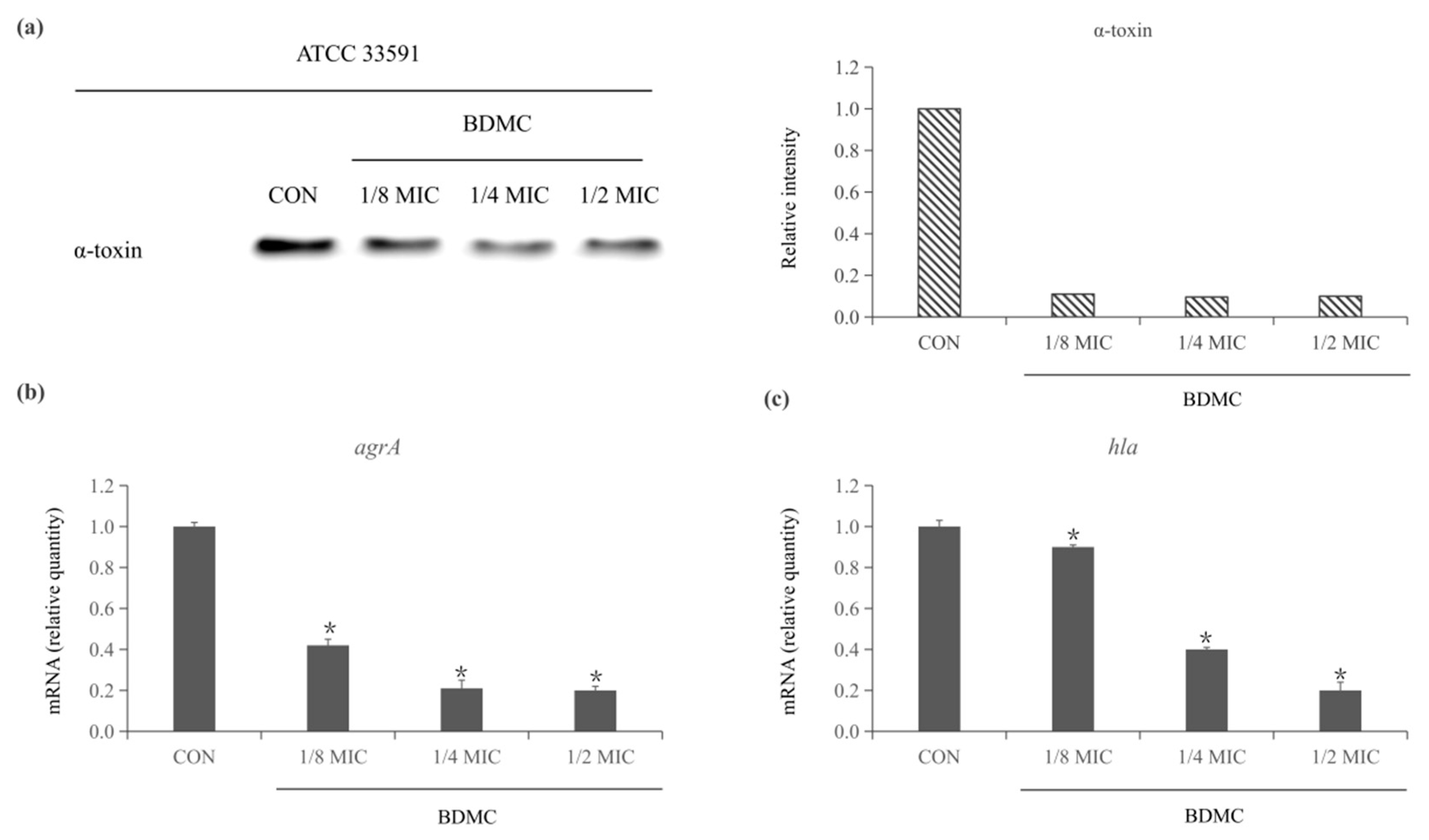
2.1. BDMC Reduces the Expression of α-Toxin and Inhibits the Expression of agrA and hla in S. aureus

Western blot was examined to evaluate the level of expression of α-toxin treated or untreated with BDMC of sub-inhibitory concentrations. After treatment for 4 h, BDMC production of α-toxin was determined to be significantly suppressed. In the presence of 1/8 MIC of BDMC, the protein level of α-toxin was only detected at 11%. However, as the concentration of BMDC increases, protein levels no longer decrease significantly (Figure 1a). The transcriptional level of the α-toxin gene hla and agrA was quantified by quantitative RT-PCR (qRT-PCR). The concentration of BDMC significantly downregulated the expression of agrA and hla as low as 0.9 µg/mL (1/8 MIC) (Figure 1b,c).

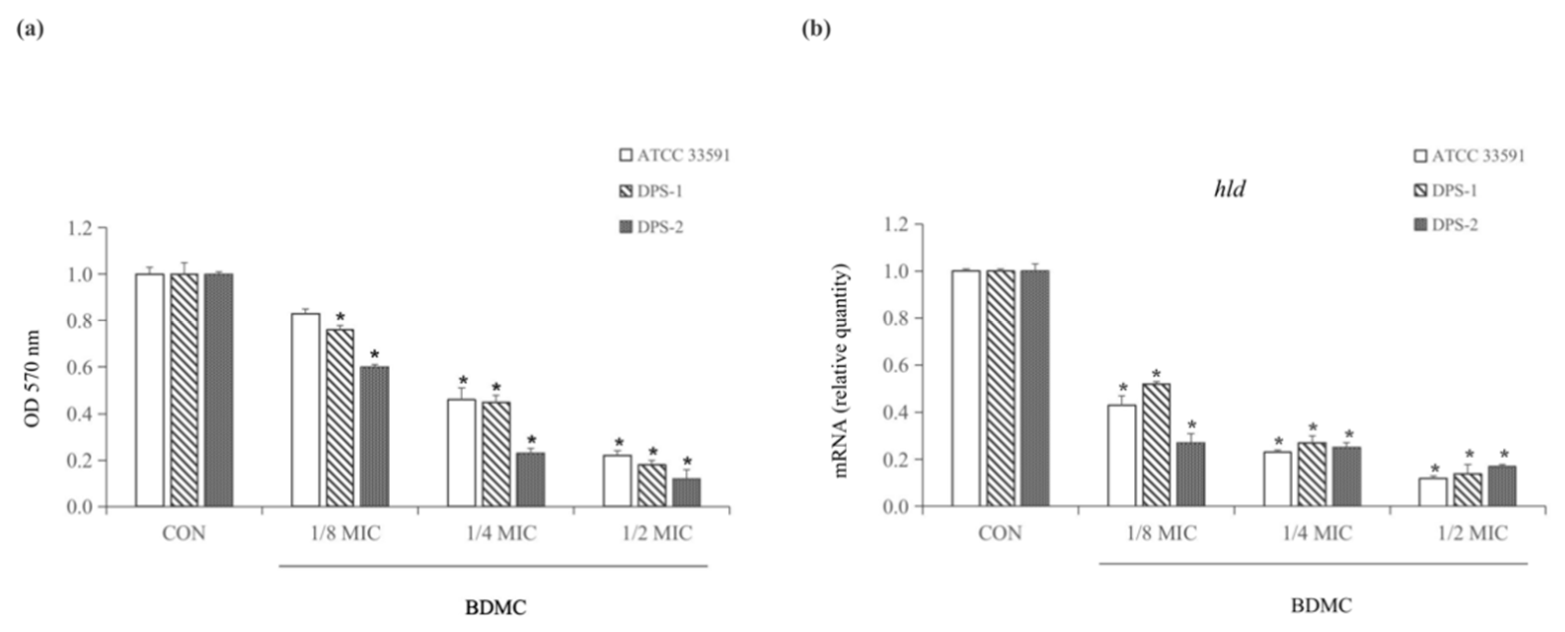
2.2. BDMC Inhibits Biofilm Formation and Downregulated the Expression of hld

A crystal violet biofilm assay has been performed to evaluate the inhibition of BDMC in biofilm formation at sub-inhibitory concentrations (1/8 MIC, 1/4 MIC, and 1/2 MIC). Dose-dependent BDMC inhibited biofilm formation significantly at sub-inhibitory concentrations. Specifically, at 1/2 MIC, BDMC inhibited biofilm formation of ATCC 33591, DPS-1, and DPS-2 by 78%, 82%, and 98%, respectively (Figure 2a). The results indicated that the production of α-toxin was positively correlated with the formation of MRSA biofilm. Additionally, the transcriptional level of the biofilm-related gene hld was quantified by qRT-PCR. BDMC at 3.9 µg/mL (1/2 MIC) significantly downregulated the expressions of hld (Figure 2b).

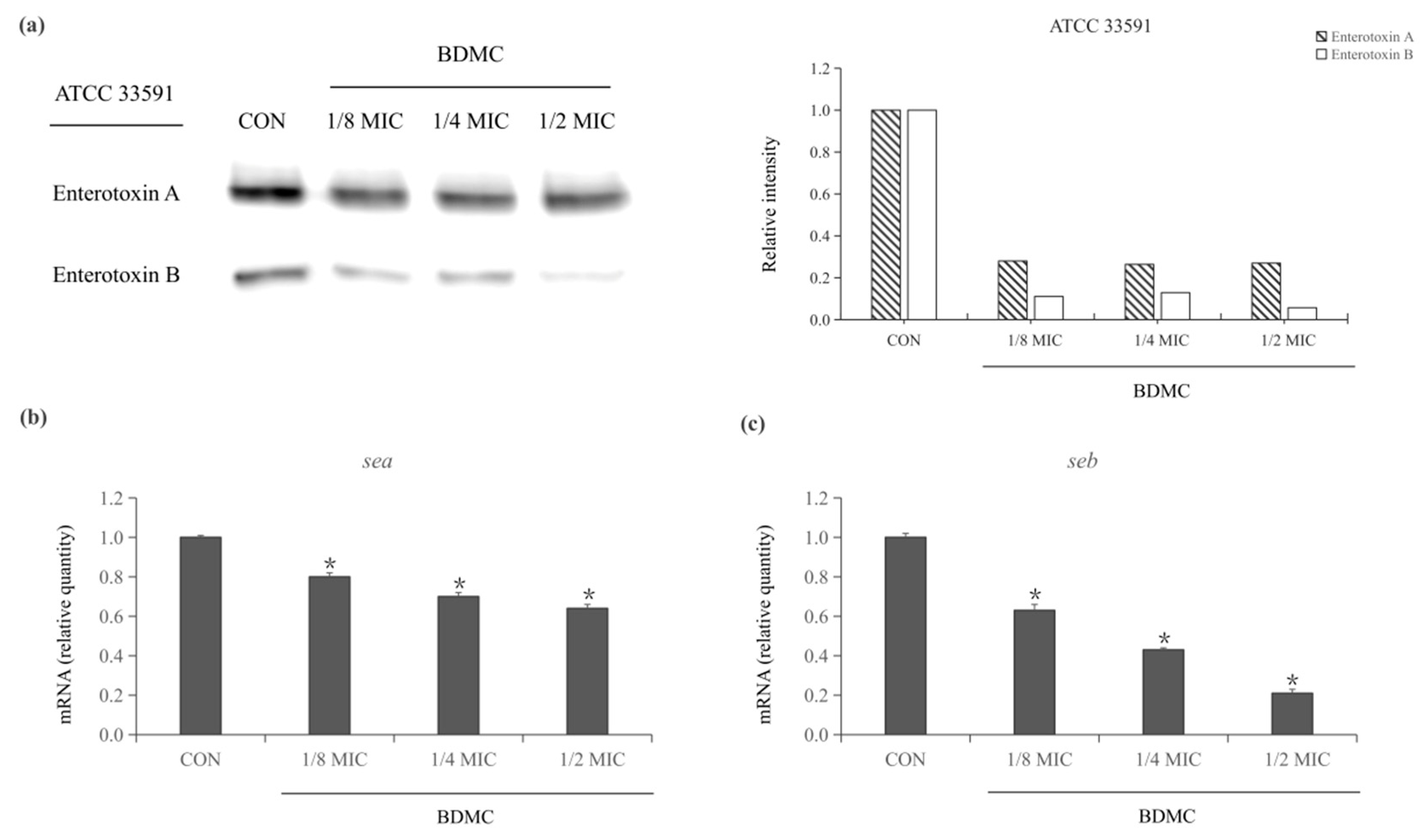
2.3. Expression of Staphylococcus Enterotoxin-Related Exoproteins in MRSA Treated with BDMC

Western blot was performed to evaluate the level of expression of Staphylococcus enterotoxin-related exoproteins (enterotoxin A and enterotoxin B) treated or untreated with BDMC. After exposure to treatment for 4 h, both exotoxins were inhibited by treating with sub-inhibitory concentrations of BDMC. The result showed that enterotoxin A and enterotoxin B were induced after being treated with 1/8 MIC of BDMC. In the presence of 1/4 MIC of BDMC, enterotoxin A was slightly inhibited, and enterotoxin B was significantly inhibited. Moreover, the production of enterotoxin B production in S. aureus ATCC 33591 was almost undetectable in the presence of 1/2 MIC (3.9 µg/mL) of BDMC (Figure 3a).

2.4. BDMC Downregulated the Expression of sea and seb in MRSA

A qRT-PCR was performed to investigate the expression of the Staphylococcus enterotoxin genes (sea and seb) after treating with BDMC at sub-inhibitory concentrations. The expression of sea and seb was significantly down-regulated in S. aureus ATCC 33591 in a dose-dependent manner. The best inhibitory effect was shown after exposure to 1/2 MIC (3.9 µg/mL) of BDMC. Meanwhile, the transcriptional level of sea and seb were decreased by 1.7-fold and 5-fold, respectively (Figure 3b,c).

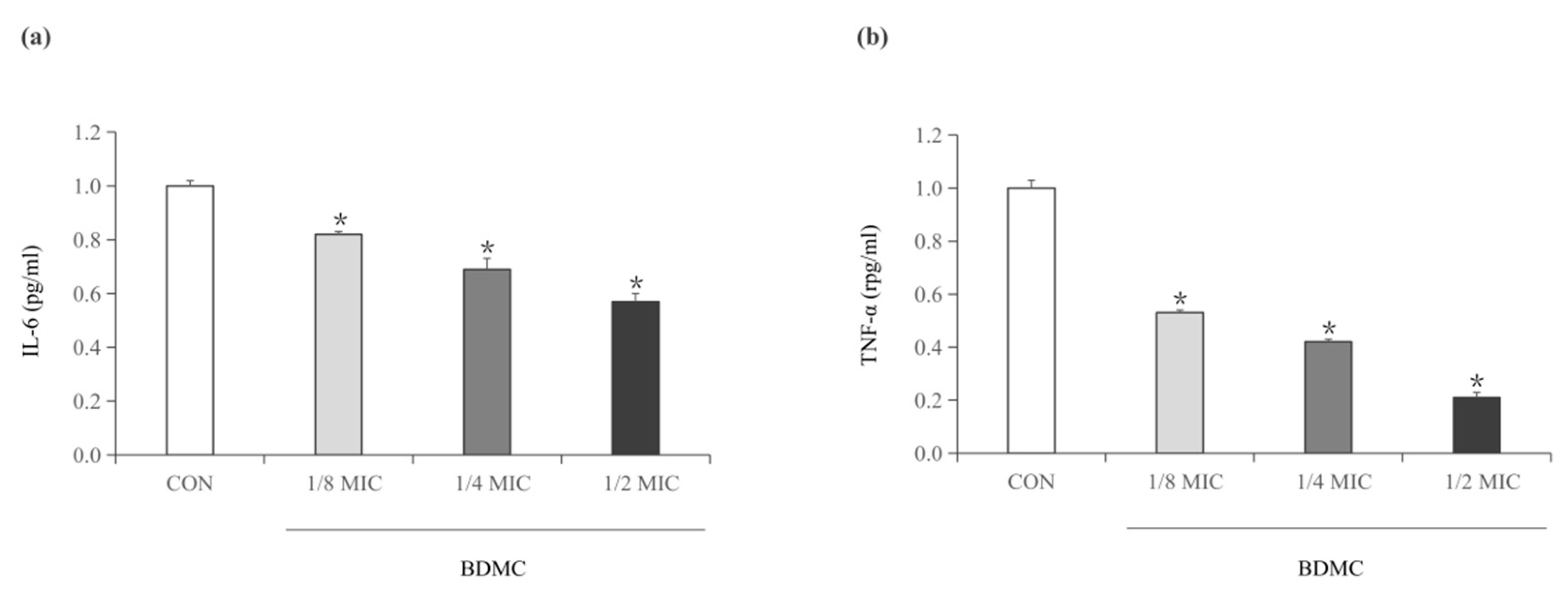
2.5. BDMC Reduces the Activity of IL-6 and TNF-α

As mentioned above, enterotoxin A and enterotoxin B, as superantigens, were produced by S. aureus, which could trigger an immune response, leading to proinflammatory cytokines. Enzyme-linked immunosorbent assay (ELISA) was used to determine the proinflammatory cytokines TNF-α and IL-6 levels in enterotoxin-stimulated RAW 264.7 cells, which grew in the culture supernatants of S. aureus that was treated with sub-inhibitory concentrations of BDMC. The result indicated that the expression of IL-6 and TNF-α decreased in a dose-dependent manner. Moreover, the expression of TNF-α was significantly reduced (Figure 4b), and that the expression of IL-6 was slightly reduced (Figure 4a).

3. Discussion

With the widespread appearance of virulent and multidrug-resistant MRSA strains, the morbidity and mortality caused by S. aureus infection had increased [23]. On the one hand, S. aureus produces a significant quantity of secreted toxins, which is suspected to be a potential mechanism for S. aureus to establish a chronic infection [24]. In the current study, we investigated the inhibitory effect of BDMC on α-toxin, enterotoxine A and enterotoxin B of MRSA. The results indicated that sub-inhibitory concentrations of BDMC significantly inhibited the expression of hla at the mRNA level in MRSA and significantly reduced the α-toxin production of S. aureus. Moreover, BDMC reduced the expression of Staphylococcal accessory gene regulator agrA. Therefore, we speculated that BDMC inhibits α-toxin production by down-regulating the agrA gene. The results of qRT-PCR showed that sub-inhibitory concentrations of BDMC down-regulated the expression of enterotoxin regulatory genes sea and seb, which was consistent with the results of inhibition of enterotoxin proteins indicated by Western blotting. The dose-dependent decrease in the level of pro-inflammatory cytokines TNF-α and IL-6 in RAW 264.7 cells grown in S. aureus treated with sub-inhibitory concentrations of BDMC, which strengthened the evidence of the inhibitory effect of BDMC on enterotoxin. According to the above results, we speculated that reducing the expression level of enterotoxin protein by down-regulating related genes was one of the reasons why BDMC could inhibit enterotoxin. In addition, it has been reported that enterotoxins could bind to polyphenols [25], and BDMC is a polyphenolic compound with two phenolic hydroxyl groups. The hydrophobicity and hydrogen bonding property of the phenolic hydroxyl group are believed to be easily combined with proteins and this chemical structure could be the second reason of the enterotoxins inhibition. Since drugs that resist virulence factors are considered not to affect viability, which may not destroy beneficial bacteria flora, inhibiting virulence factors of pathogens may be a promising strategy against MRSA [4].
On the other hand, since all currently available antibiotics will inevitably develop resistance, the development of novel antibiotic replacement therapies is urgently required. Naturally sourced antibacterial agents are considered to have extremely low drug resistance due to their rich sources and diverse and complex structures [26]. Based on this, we additionally conducted a preliminary trial of the BDMC resistance test, and the results showed that BDMC had no apparent resistance after 10 consecutive passages (45 days), and the resistance effect was better than that of the traditional antibiotic linezolid in the control group. Moreover, the US Food and Drug Administration (FDA) has approved curcuminoids as “Generally Recognized as Safe” (GRAS) [27], and it is assumed in the literature report dedicated to the evaluation of the biological activity of curcumin that BDMC is more stable than the other two derivatives (curcumin and dimethoxycurcumin) [28]. Additionally, it has also been proved in our previous studies that the anti-MRSA ability of BDMC is also more potent than that of the other two derivatives [21]. These advantages increase the possibility of BDMC as a substitute for antibiotics. However, the low solubility of curcumin in aqueous media, poor bioavailability, and pharmacokinetic defects limit its application in clinical treatment. Given the limitations of turmeric, approaches, such as lipid or nanoparticle materials combined with curcumin to make a mixture, have been reported [29].
Additionally, the formation of biofilms is one of the reasons why MRSA is difficult to eradicate [30]. Biofilms could enhance resistance to antibiotics, and biofilm-related infections are usually chronic or recurrent. Therefore, in the current requirements for antibacterial substances, not only should bacteria be inhibited for growth, but there should also be the ability to inhibit biofilm formation [31]. Studies have shown that, in addition to inhibiting toxins, plant-derived polyphenols can also inhibit the biofilm formation of food-related pathogens [32]. Based on the above, we hypothesized that BDMC could inhibit MRSA biofilm. The result was assumed that BDMC had a potent inhibitory effect on the biofilm formation of the clinical strain and the reference strain ATCC 33591 at sub-inhibitory concentrations, and the effective concentration was as low as 0.9 µg/mL (1/8 MIC). Moreover, BDMC significantly down-regulated biofilm formation-related gene hld, which emphasized the inhibitory effect of BDMC on biofilm formation. In addition, it was reported that the production of toxins is one of the reasons leading to the production of biofilm. Therefore, we speculated that BDMC might also inhibit α-toxin production by the agr system, which would inhibit biofilm formation. Since biofilms are usually highly resistant to conventional antibiotics, the development of natural antibacterial substances with anti-biofilm activity may be of great significance [33].
In conclusion, our study first proposed the anti-toxin and anti-biofilm effects of BDMC, provided alternative or enhanced strategies for preventing and treating toxin-related infections, and provided a foundation for developing novel antibiofilm-specific antibiotics. Furthermore, our study revealed the potential of BDMC as a natural antibacterial agent, and a reference value was provided for the development of an antibiotic replacement, antibiotic adjuvant, or antibacterial lead compound. However, this study has certain limitations, such as the lack of a variety of tested strains and the lack of in vivo experiments. If the application of curcumin in clinical treatment is promoted, more in-depth in vivo experiments and the improvement of pharmacokinetics will be needed.
Furthermore, to solve the problem of MRSA infection, it is also critical to strengthen the solution to the occurrence of infection in communities. Reports have shown that the current spread of MRSA in the population is no longer caused by a large-scale nosocomial outbreak, and the proportion of community-related invasiveness as well as infections is higher [34].

4. Materials and Methods

4.1. Reagents

BDMC was purchased from Tokyo Chemical Industry Co.; Ltd. (Tokyo, Japan). Skim milk, Mueller–Hinton agar (MHA), and Mueller–Hinton broth (MHB) were obtained from Difco Laboratories (Baltimore, MD, USA). Anti-Staphylococcus enterotoxin A antibody, anti-Staphylococcus enterotoxin B antibody, and anti-Staphylococcus alpha-hemolysin antibody were acquired from Abcam (Cambridge, UK). Anti-rabbit IgG secondary antibody was acquired from Thermo Scientific Inc. (Waltham, MA, USA). Crystal violet was obtained from Sigma-Aldrich Co. (St. Louis, MO, USA). The sequences of primers used in qRT-PCR were listed in Table 1, which was acquired from Bioneer (Daejeon, Korea).

4.2. Bacterial Strains and Growth Conditions

In the report, one reference strain S. aureus ATCC 33591 (American Type Culture Collection, Manassas, VA, USA) was used as a reference strain and two clinical isolates of MRSA (DPS-1 and DPS-2) were isolated from patients at the Hospital of Wonkwang University was used. S. aureus was cultured in an incubator at 37 °C, and MHA or MHB was used as a solid medium or liquid medium, respectively.

4.3. Western Blot Assay

A Western blot assay was performed to evaluate the effect of BDMC on the expression of virulence-related exoproteins based on the previously reported investigation [21]. S. aureus strains (ATCC 33591) were cultured in MHB for 24 h and then treated for 4 h with sub-inhibitory concentrations of BDMC. Protein concentration was measured using a Bio-Rad protein assay reagent (Bio-Rad Laboratories, Hercules, CA, USA) after centrifugation at 3000× g for 10 min to harvest cell protein extracts. The supernatant was separated by SDS-PAGE and electroblotted onto Amersham HybondTM-P membranes (GE Healthcare, Piscataway, NJ, USA) that had been blocked with 5% skim milk and probed for 8 h at 4 °C with anti-Staphylococcus alpha-hemolysin antibody, anti-Staphylococcus enterotoxin A antibody, and anti-Staphylococcus enterotoxin B antibody (diluted 1:1000), and then reprobed by anti-rabbit IgG secondary antibody (diluted 1:1000) at room temperature for 2 h. The membranes were then supplemented with ECL Prime Western Blotting Detection reagent (GE Healthcare Life Sciences, Incheon, Korea). The bands were visualized using the ImageQuant LAS-4000 mini chemical luminescent imager (GE Healthcare Life Sciences).

4.4. Reverse Transcription and qRT-PCR

A qRT-PCR was carried out according to the methods previously described [35]. Briefly, ATCC 33591 was cultured in MHB overnight and treated with sub-inhibitory concentrations of BDMC for 4 h. The sample without BDMC was taken as a control. According to the manufacturer’s protocol, total RNA was prepared from S. aureus (ATCC 33591) using the E.Z.N.A.® bacterial RNA kit (Omega Bio-tek, Norcross, GA, USA). The RNA concentration was evaluated by measuring the absorbance ratio at 260 nm on a spectrophotometer (BioTek, Winooski, VT, USA). Next, a QuantiTect reverse transcription kit (Qiagen, Dusseldorf, Germany) was used to synthesize the complementary DNA based on the manufacturer’s instructions. PCR was set up to a total volume of 20 μL as follows: 1 μL of each primer (10 μL/mL), 2 μL of sample cDNA, 10 μL of 2× SYBR Green PCR master mix (Life Technologies LTD, Warrington, UK), and 6 μL of deionized water. StepOne software v2.3 produced from Applied Biosystems (Waltham, MA, USA) was used to calculate the expression level of the target gene relative to the endogenous reference gene 16 rRNA through the delta–delta cycle threshold method.

4.5. ELISA

ELISA was performed using the previously described method [22]. Briefly, S. aureus ATCC 33591 in MHB was treated with sub-inhibitory concentrations of BDMC, and the untreated were taken as controls. The supernatants were collected after 4 h of incubation and 100 μL was added into a 96 well culture plate with 100 μL of RAW 264.7 cells. After incubating for 24 h, the mixture was collected and analyzed based on the mouse TNF-α or IL-6 ELISA MAX standard set according to the instructions.

4.6. Crystal Violet Biofilm Assay

The inhibition of the BDMC on the biofilm formation of S. aureus DPS-1, DPS-2, and ATCC 33591 was established in a crystal violet biofilm assay using a previously described protocol [36]. To each well of 96-well microtiter plates, 100 μL of overnight culture (0.5 MacFarland bacterial culture) was added and treated with sub-concentrations of BDMC. After 24 h at 37 °C, the planktonic cells were removed and washed with PBS three times, and each well of 96-well microtiter plates was stained with 1% (w/v) crystal violet for 10 min at room temperature, which was then rewashed for three times. The stained biofilms were solubilized in 100 μL of absolute ethanol, and the optical density (OD) values at 570 nm were measured. The percentage of biofilm inhibition was estimated using the formula below.
Percentage of inhibition = 100 − [(OD 570 nm of the treatment wells)/(OD 570 nm of the control wells) × 100)].

4.7. Statistical Analysis

The analyses were carried out in triplicate, and data were given as the mean ± standard deviation. The collected data were statistically analyzed using an independent Scheffe’s t-test (SPSS software version 22.0; IBM SPSS, Armonk, NY, USA). A statistically significant p-value of less than 0.05 was evaluated.

Author Contributions

Conceptualization, D.-Y.K. and S.W.; methodology, D.-Y.K. and S.W.; software, O.-H.K.; validation, S.W., D.-Y.K. and O.-H.K.; formal analysis, S.W.; investigation, S.W.; resources, O.-H.K.; data curation, S.W.; writing—original draft preparation, S.W.; writing—review and editing, D.-Y.K. and O.-H.K.; visualization, S.W.; supervision, D.-Y.K. and O.-H.K.; project administration, D.-Y.K. and O.-H.K.; funding acquisition, D.-Y.K. All authors have read and agreed to the published version of the manuscript.

Funding

This research was funded by the National Research Foundation of Korea (NRF) grant funded by Korea government (MSIT) (no. 2020R1F1A1054227) and (NRF-2021R1I1A1A01058728). MSIT: Ministry of Science and ICT.

Institutional Review Board Statement

This research has been reviewed by the IACUC and it has been approved (Approved Number WKU18-34).

Informed Consent Statement

Patient consent was received and performed according to the guide-lines of the Wonkwang University Institutional Review Board IRB.

Data Availability Statement

Personal information is included, so it was conducted for research only.

Conflicts of Interest

The authors declare no conflict of interest.

References

  1. Yuan, Y.; Zai, Y.; Xi, X.; Ma, C.; Wang, L.; Zhou, M.; Shaw, C.; Chen, T. A novel membrane-disruptive antimicrobial peptide from frog skin secretion against cystic fibrosis isolates and evaluation of anti-MRSA effect using Galleria mellonella model. Biochim. Biophys. Acta (BBA)- Gen. Subj. 2019, 1863, 849–856. [Google Scholar] [CrossRef] [Green Version]
  2. Dharmaratne, P.; Sapugahawatte, D.N.; Wang, B.; Chan, C.L.; Lau, K.M.; Lau, C.B.; Fung, K.P.; Ng, D.K.; Ip, M. Con-temporary approaches and future perspectives of antibacterial photodynamic therapy (aPDT) against methicillin resistant Staphylococcus aureus (MRSA): A systematic review. Eur. J. Med. Chem. 2020, 200, 112341. [Google Scholar] [CrossRef]
  3. David, M.Z.; Daum, R.S. Community-Associated Methicillin-Resistant Staphylococcus aureus: Epidemiology and Clinical Consequences of an Emerging Epidemic. Clin. Microbiol. Rev. 2010, 23, 616–687. [Google Scholar] [CrossRef] [Green Version]
  4. El-Halfawy, O.M.; Czarny, T.L.; Flannagan, R.S.; Day, J.; Bozelli, J.C.; Kuiack, R.C.; Salim, A.; Eckert, P.; Epand, R.M.; McGavin, M.; et al. Discovery of an antivirulence compound that reverses β-lactam resistance in MRSA. Nat. Chem. Biol. 2020, 16, 143–149. [Google Scholar] [CrossRef]
  5. Vestergaard, M.; Frees, D.; Ingmer, H. Antibiotic Resistance and the MRSA Problem. Microbiol. Spectr. 2019, 7, 2. [Google Scholar] [CrossRef]
  6. Kathirvel, M.; Buchad, H.; Nair, M. Enhancement of the pathogenicity of Staphylococcus aureus strain Newman by a small noncoding RNA SprX1. Med. Microbiol. Immunol. 2016, 205, 563–574. [Google Scholar] [CrossRef]
  7. Liu, L.; Shen, X.; Yu, J.; Cao, X.; Zhan, Q.; Guo, Y.; Yu, F. Subinhibitory Concentrations of Fusidic Acid May Reduce the Virulence of S. aureus by Down-Regulating sarA and saeRS to Reduce Biofilm Formation and α-Toxin Expression. Front. Microbiol. 2020, 11, 25. [Google Scholar] [CrossRef]
  8. Anderson, M.J.; Schaaf, E.; Breshears, L.M.; Wallis, H.W.; Johnson, J.R.; Tkaczyk, C.; Sellman, B.R.; Sun, J.; Peterson, M.L. Alpha-Toxin Contributes to Biofilm Formation among Staphylococcus aureus Wound Isolates. Toxins 2018, 10, 157. [Google Scholar] [CrossRef] [Green Version]
  9. Ueda, Y.; Mashima, K.; Miyazaki, M.; Hara, S.; Takata, T.; Kamimura, H.; Takagi, S.; Jimi, S. Inhibitory effects of polysorbate 80 on MRSA biofilm formed on different substrates including dermal tissue. Sci. Rep. 2019, 9, 3128. [Google Scholar] [CrossRef]
  10. Brady, R.A.; O’May, G.A.; Leid, J.G.; Prior, M.L.; Costerton, J.W.; Shirtliff, M.E. Resolution of Staphylococcus aureus Biofilm Infection Using Vaccination and Antibiotic Treatment. Infect. Immun. 2011, 79, 1797–1803. [Google Scholar] [CrossRef] [Green Version]
  11. Naclerio, G.A.; Onyedibe, K.I.; Sintim, H.O. Lipoteichoic Acid Biosynthesis Inhibitors as Potent Inhibitors of S. aureus and E. faecalis Growth and Biofilm Formation. Molecules 2020, 25, 2277. [Google Scholar] [CrossRef]
  12. Etter, D.; Schelin, J.; Schuppler, M.; Johler, S. Staphylococcal Enterotoxin C-An Update on SEC Variants, Their Structure and Properties, and Their Role in Foodborne Intoxications. Toxins 2020, 12, 584. [Google Scholar] [CrossRef]
  13. Verreault, D.; Ennis, J.; Whaley, K.; Killeen, S.Z.; Karauzum, H.; Aman, M.J.; Holtsberg, R.; Doyle-Meyers, L.; Didier, P.J.; Zeitlin, L.; et al. Effective Treatment of Staphylococcal Enterotoxin B Aerosol Intoxication in Rhesus Macaques by Using Two Parenterally Administered High-Affinity Monoclonal Antibodies. Antimicrob. Agents Chemother. 2019, 63, e02049-18. [Google Scholar] [CrossRef] [Green Version]
  14. Söderquist, B.; Källman, J.; Holmberg, H.; Vikerfors, T.; Kihlström, E. Secretion of IL-6, IL-8 and G-CSF by human endothelial cells in vitro in response to Staphylococcus aureus and staphylococcal exotoxins. APMIS 1998, 106, 1157–1164. [Google Scholar]
  15. Chen, G.; Karauzum, H.; Long, H.; Carranza, D.; Holtsberg, F.W.; Howell, K.A.; Abaandou, L.; Zhang, B.; Jarvik, N.; Ye, W.; et al. Potent Neutralization of Staphylococcal Enterotoxin B In Vivo by Antibodies that Block Binding to the T-Cell Receptor. J. Mol. Biol. 2019, 431, 4354–4367. [Google Scholar] [CrossRef]
  16. Wünsche, S.; Yuan, L.; Seidel-Morgenstern, A.; Lorenz, H. A Contribution to the Solid State Forms of Bis(demethoxy)curcumin: Co-Crystal Screening and Characterization. Molecules 2021, 26, 720. [Google Scholar] [CrossRef]
  17. Duro-Castano, A.; Borrás, C.; Herranz-Pérez, V.; Blanco-Gandía, M.C.; Conejos-Sánchez, I.; Armiñán, A.; Mas-Bargues, C.; Inglés, M.; Miñarro, J.; Rodríguez-Arias, M.; et al. Targeting Alzheimer’s disease with multimodal polypeptide-based nanoconjugates. Sci. Adv. 2021, 7, eabf9180. [Google Scholar] [CrossRef]
  18. Jin, F.; Chen, X.; Yan, H.; Xu, Z.; Yang, B.; Luo, P.; He, Q. Bisdemethoxycurcumin attenuates cisplatin-induced renal injury through anti-apoptosis, anti-oxidant and anti-inflammatory. Eur. J. Pharmacol. 2020, 874, 173026. [Google Scholar] [CrossRef]
  19. Wang, S.; Kim, M.-C.; Kang, O.-H.; Kwon, D.-Y. The Mechanism of Bisdemethoxycurcumin Enhances Conventional Antibiotics against Methicillin-Resistant Staphylococcus aureus. Int. J. Mol. Sci. 2020, 21, 7945. [Google Scholar] [CrossRef]
  20. Mun, S.-H.; Kong, R.; Seo, Y.-S.; Zhou, T.; Kang, O.-H.; Shin, D.-W.; Kwon, D.-Y. Subinhibitory concentrations of punicalagin reduces expression of virulence-related exoproteins by Staphylococcus aureus. FEMS Microbiol. Lett. 2016, 363, 22. [Google Scholar] [CrossRef] [Green Version]
  21. Miller, L.S.; Fowler, V.G.; Shukla, S.K.; Rose, W.E.; Proctor, R.A. Development of a vaccine against Staphylococcus aureus invasive infections: Evidence based on human immunity, genetics and bacterial evasion mechanisms. FEMS Microbiol. Rev. 2020, 44, 123–153. [Google Scholar] [CrossRef] [Green Version]
  22. Fischer, A.J.; Kilgore, S.H.; Singh, S.B.; Allen, P.D.; Hansen, A.R.; Limoli, D.H.; Schlievert, P.M. High Prevalence of Staphylococcus aureus Enterotoxin Gene Cluster Superantigens in Cystic Fibrosis Clinical Isolates. Genes 2019, 10, 1036. [Google Scholar] [CrossRef] [Green Version]
  23. Shimamura, Y.; Utsumi, M.; Hirai, C.; Nakano, S.; Ito, S.; Tsuji, A.; Ishii, T.; Hosoya, T.; Kan, T.; Ohashi, N.; et al. Binding of Catechins to Staphylococcal Enterotoxin A. Molecules 2018, 23, 1125. [Google Scholar] [CrossRef] [Green Version]
  24. Rossiter, S.E.; Fletcher, M.H.; Wuest, W.M. Natural Products as Platforms to Overcome Antibiotic Resistance. Chem. Rev. 2017, 117, 12415–12474. [Google Scholar] [CrossRef]
  25. Mashayekhi-Sardoo, H.; Mashayekhi-Sardoo, A.; Roufogalis, B.D.; Jamialahmadi, T.; Sahebkar, A. Impact of Curcumin on Microsomal Enzyme Activities: Drug Interaction and Chemopreventive Studies. Curr. Med. Chem. 2021, 28, 7122–7140. [Google Scholar] [CrossRef]
  26. Miklášová, N.; Herich, P.; Dávila-Becerril, J.; Barroso-Flores, J.; Fischer-Fodor, E.; Valentová, J.; Leskovská, J.; Kožíšek, J.; Takáč, P.; Mojžiš, J. Evaluation of Antiproliferative Palladium(II) Complexes of Synthetic Bisdemethoxycurcumin towards In Vitro Cytotoxicity and Molecular Docking on DNA Sequence. Molecules 2021, 26, 4369. [Google Scholar] [CrossRef]
  27. Kotha, R.R.; Luthria, D.L. Curcumin: Biological, Pharmaceutical, Nutraceutical, and Analytical Aspects. Molecules 2019, 24, 2930. [Google Scholar] [CrossRef] [Green Version]
  28. Mihailescu, R.; Tafin, U.F.; Corvec, S.; Oliva, A.; Betrisey, B.; Borens, O.; Trampuz, A. High Activity of Fosfomycin and Rifampin against Methicillin-Resistant Staphylococcus aureus Biofilm In Vitro and in an Experimental Foreign-Body Infection Model. Antimicrob. Agents Chemother. 2014, 58, 2547–2553. [Google Scholar] [CrossRef] [Green Version]
  29. Shimamura, Y.; Hirai, C.; Sugiyama, Y.; Shibata, M.; Ozaki, J.; Murata, M.; Ohashi, N.; Masuda, S. Inhibitory effects of food additives derived from polyphenols on staphylococcal enterotoxin A production and biofilm formation by Staphylococcus aureus. Biosci. Biotechnol. Biochem. 2017, 81, 2346–2352. [Google Scholar] [CrossRef] [Green Version]
  30. Qu, D.; Hou, Z.; Li, J.; Luo, L.; Su, S.; Ye, Z.; Bai, Y.; Zhang, X.; Chen, G.; Li, Z.; et al. A new coumarin compound DCH combats methicillin-resistant Staphylococcus aureus biofilm by targeting arginine repressor. Sci. Adv. 2020, 6, eaay9597. [Google Scholar] [CrossRef]
  31. Takó, M.; Kerekes, E.B.; Zambrano, C.; Kotogán, A.; Papp, T.; Krisch, J.; Vágvölgyi, C. Plant Phenolics and Phenol-ic-Enriched Extracts as Antimicrobial Agents against Food-Contaminating Microorganisms. Antioxidants 2020, 9, 165. [Google Scholar] [CrossRef] [Green Version]
  32. Coll, F.; Harrison, E.M.; Toleman, M.S.; Reuter, S.; Raven, K.E.; Blane, B.; Palmer, B.; Kappeler, A.R.M.; Brown, N.M.; Török, M.E.; et al. Longitudinal genomic surveillance of MRSA in the UK reveals transmission patterns in hospitals and the community. Sci. Transl. Med. 2017, 9, 413. [Google Scholar] [CrossRef] [Green Version]
  33. Wang, S.; Luo, J.; Liu, X.; Kang, O.; Kwon, D. Antibacterial activity and synergy of antibiotics with sanguisorbigenin isolated from Sanguisorba officinalis L. against methicillin-resistant Staphylococcus aureus. Lett. Appl. Microbiol. 2021, 72, 238–244. [Google Scholar] [CrossRef]
  34. Wang, S.; Kang, O.-H.; Kwon, D.-Y. Trans-Cinnamaldehyde Exhibits Synergy with Conventional Antibiotic against Methicillin-Resistant Staphylococcus aureus. Int. J. Mol. Sci. 2021, 22, 2752. [Google Scholar] [CrossRef]
  35. Wang, J.; Jiao, H.; Meng, J.; Qiao, M.; Du, H.; He, M.; Ming, K.; Liu, J.; Wang, D.; Wu, Y. Baicalin Inhibits Biofilm Formation and the Quorum-Sensing System by Regulating the MsrA Drug Efflux Pump in Staphylococcus saprophyticus. Front. Microbiol. 2019, 10, 2800. [Google Scholar] [CrossRef]
  36. Sharifi, A.; Mohammadzadeh, A.; Salehi, T.Z.; Mahmoodi, P.; Nourian, A. Cuminum cyminum L. Essential Oil: A Promising Antibacterial and Antivirulence Agent Against Multidrug-Resistant Staphylococcus aureus. Front. Microbiol. 2021, 12, 667833. [Google Scholar] [CrossRef]
Figure 1. (a) Effect of BDMC treatment at sub-inhibitory concentrations on the expression of α-toxin in MRSA (ATCC 33591). CON was a control treated without BDMC. After treatment for 4 h, BDMC suppressed production of α-toxin significantly was determined through Western blotting. (b,c) The inhibitory effect of BDMC on the expression of enterotoxin gene agrA and hla was analyzed by qRT–PCR. After exposure to BDMC sub-inhibitory concentrations for 4 h, the expression of agrA and hla were down-regulated in a dose-dependent manner. The data were presented as the average of the three independent experiments with standard deviation. * Represents that p < 0.05.
Toxins 13 00804 g001
Figure 2. (a) Inhibitory effect of BDMC at sub-inhibitory concentrations on the biofilm formation of S. aureus standard strain (ATCC 33591) and clinical isolates DPS-1 and DPS-2. (b) In the presence of BDMC at sub-inhibitory concentrations, BDMC inhibited the expression of hld in MRSA (ATCC 33591, DPS-1, and DPS-2) cultures. The data were presented as the average of the three independent experiments with standard deviation. * Represents that p < 0.05.
Toxins 13 00804 g002
Figure 3. (a) BDMC at sub-inhibitory concentrations on enterotoxin A and enterotoxin B expression in MRSA (ATCC 33591). CON was a control treated without BDMC. (b,c) Relative expression of enterotoxin gene sea and seb in MRSA (ATCC 33591) cultures in the presence of sub-inhibitory concentrations of BDMC. Both enterotoxin genes were reduced in a dose-dependent manner. The data were presented as the average of the three independent experiments with standard deviation. * Represents that p < 0.05.
Toxins 13 00804 g003
Figure 4. (a) IL-6 released from RAW 264.7 cells stimulated with S. aureus supernatants cultured in the presence of sub-inhibitory concentrations of BDMC. (b) TNF released from RAW 264.7 cells stimulated with S. aureus supernatants cultured in the presence of sub-inhibitory concentrations of BDMC. After stimulation for 24 h with RAW 264.7 cells, ELISA was used to measure the levels of IL-6 and TNF. The data were presented as the average of the three independent experiments with standard deviation. * Represents that p < 0.05.
Toxins 13 00804 g004
Table 1. Primers used in in this study.
PrimerSequence (5′-3′)
16SF: ACTCCTACGGGAGGCAGCAG
R: ATTACCGCGGCTGCTGG
seaF: ATGGTGCTTATTATGGTTATC
R: CGTTTCCAAAGGTACTGTATT
sebF: TGTTCGGGTATTTGAAGATGG
R: CGTTTCATAAGGCGAGTTGTT
agrAF: TGATAATCCTTATGAGGTGCTT
R: CACTGTGACTCGTAACGAAAA
hlaF: TTGGTGCAAATGTTTC
R: TCACTTTCCAGCCTACT
hldF: ATTTGTTCACTGTGTCGATAATCC
R: GGAGTGATTTCAATGGCACAAG
Publisher’s Note: MDPI stays neutral with regard to jurisdictional claims in published maps and institutional affiliations.

