Article

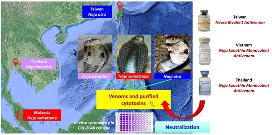
Cytotoxicity of Venoms and Cytotoxins from Asiatic Cobras (Naja kaouthia, Naja sumatrana, Naja atra) and Neutralization by Antivenoms from Thailand, Vietnam, and Taiwan

1
Venom Research and Toxicology Laboratory, Department of Pharmacology, Faculty of Medicine, University of Malaya, Kuala Lumpur 50603, Malaysia
2
Protein and Interactomics Laboratory, Department of Molecular Medicine, Faculty of Medicine, University of Malaya, Kuala Lumpur 50603, Malaysia
3
National Institute of Infectious Diseases and Vaccinology, National Health Research Institutes, Miaoli 35053, Taiwan
*
Authors to whom correspondence should be addressed.
Submission received: 20 March 2022 / Revised: 3 May 2022 / Accepted: 4 May 2022 / Published: 10 May 2022
(This article belongs to the Section Animal Venoms)

Abstract

:
Envenoming by cobras (Naja spp.) often results in extensive local tissue necrosis when optimal treatment with antivenom is not available. This study investigated the cytotoxicity of venoms and purified cytotoxins from the Monocled Cobra (Naja kaouthia), Taiwan Cobra (Naja atra), and Equatorial Spitting Cobra (Naja sumatrana) in a mouse fibroblast cell line, followed by neutralization of the cytotoxicity by three regional antivenoms: the Thai Naja kaouthia monovalent antivenom (NkMAV), Vietnamese snake antivenom (SAV) and Taiwanese Neuro bivalent antivenom (NBAV). The cytotoxins of N. atra (NA-CTX) and N. sumatrana (NS-CTX) were identified as P-type cytotoxins, whereas that of N. kaouthia (NK-CTX) is S-type. All venoms and purified cytotoxins demonstrated varying concentration-dependent cytotoxicity in the following trend: highest for N. atra, followed by N. sumatrana and N. kaouthia. The antivenoms moderately neutralized the cytotoxicity of N. kaouthia venom but were weak against N. atra and N. sumatrana venom cytotoxicity. The neutralization potencies of the antivenoms against the cytotoxins were varied and generally low across NA-CTX, NS-CTX, and NK-CTX, possibly attributed to limited antigenicity of CTXs and/or different formulation of antivenom products. The study underscores the need for antivenom improvement and/or new therapies in treating local tissue toxicity caused by cobra envenomings.
Key Contribution: The study investigates the in vitro cytotoxicity of venom and purified cytotoxins from the Monocled Cobra (Naja kaouthia), Taiwan Cobra (Naja atra), and Equatorial Spitting Cobra (Naja sumatrana) and cytotoxicity neutralization by three regional antivenoms from Thailand, Vietnam, and Taiwan were also studied. The study highlights considerations in antivenom manufacturing in treating cobra venom cytotoxicity.

Graphical Abstract

1. Introduction

Snakebite envenoming is a priority neglected tropical disease according to the World Health Organization [1,2]. Each year, approximately 83,000–138,000 deaths and 3 times as many limb amputations or other permanent disabilities are caused by snakebite envenoming [1]. In Asia and Africa, cobras (Naja spp.) are commonly listed under Category 1 of medically important venomous snakes by the WHO as they are frequently implicated in snakebite envenoming and associated with high mortality and morbidity [3,4]. Envenoming caused by cobras (Naja spp.) can result in systemic neurotoxicity and local tissue necrosis at the bite site and beyond [3,5,6]. In addition, venom ophthalmia can occur if venoms are sprayed by “spitting” cobras into the victim’s eyes, resulting in ocular injuries and blindness [7,8,9]. The immense tissue-damaging effect of cobra venoms is attributed to the cytotoxins (CTX) present in virtually all cobra venoms, constituting 20–80% of total venom proteins [5,10,11,12]
Cytotoxins are non-enzymatic, highly basic (pI > 10) β-sheet single-chained polypeptides consisting of 60–70 amino acid residues and have a molecular weight of ~6.5–7.0 kDa. Structurally, they have a three-fingered loop-folding topology reinforced by four disulfide bonds between eight conserved cysteine residues [13,14]. CTXs exhibit strong amphiphilic properties on their molecular surface and the hydrophobic tips of the three-finger structure flanked by polar residues are shown to be the principal membrane-binding motif [15], in particular toward the anionic phosphatidylserine lipid [16]. Previous studies proposed 2 distinct types of CTX, i.e., the P-type and S-type CTXs, where the former is characterized by the presence of Pro-31 within a putative phospholipid binding site near the tip of loop 2, while S-type CTXs are characterized by the presence of Ser-29 within the same but more hydrophilic region [15,17]. CTXs have been previously shown to be highly cytotoxic in vitro toward various mammalian cell lines (both cancer and non-cancer) [18,19,20] and are able to induce dermonecrosis in mice [21,22]. This is consistent with the local tissue damage reported in most cobra bite envenoming and is a cause of limb deformity, amputation, and permanent disabilities when treatment is inadequate [23,24].
To date, antivenoms remain the definitive treatment for snakebite envenoming. The gold standard advocated by the WHO for antivenom efficacy assessment is the neutralization of lethality in rodents, an assay that essentially tests against the systemic toxicity of venom under acute conditions [25]. The efficacy of antivenom in neutralizing cobra CTX-induced cytotoxicity, however, has not been commonly addressed, presumably because of the general perception that antivenom is effective systemically but has limitations in the treatment of local envenoming [2,26]. Nevertheless, antivenom treatment is still recommended by most management guidelines to alleviate the effect of local envenoming, indicated when there is rapid swelling that extends beyond a joint or involves half the limb [3,5]. It is therefore important to assess the efficacy of antivenom in neutralizing the cytotoxic activity of different cobra venoms and, importantly, the causative cytotoxins contained therein. Preclinically, a mouse model is used to demonstrate dermonecrosis induced by snake venoms and to serve as a model for neutralization studies [27,28,29]. The development of dermonecrosis takes up to a few days and has been deemed rather controversial nowadays from an animal ethics point of view. Many authorities, including the WHO, encourage implementing the 3Rs Principle (3R: replacement, reduction, and refinement) in antivenom production and assessment procedures before the use of in vivo models [25,30]. Therefore, in vitro approaches such as cell-based assays may be opted as a surrogate model for venom-induced cytotoxicity [31,32,33]. With apt modifications, the in vitro assays can be applied to examine the neutralization efficacy of antivenom against CTX-induced necrosis while minimizing animal use. Therefore, the present study aimed to investigate the cytotoxic activities of the venoms and purified CTX from three Asiatic cobra species known for causing significant local tissue damage, Monocled Cobra (Naja kaouthia), Equatorial Spitting Cobra (Naja sumatrana), and Taiwanese or Chinese Cobra (Naja atra), through a cell-based assay using rodent fibroblast cell line (CRL-2648) derived from the subcutaneous tissue of the skin.
The present study further investigated the neutralization efficacy of the Naja kaouthia Monovalent Antivenom (NkMAV, Thailand and SAV, Vietnam) and the Neuro Bivalent Antivenom raised against Naja kaouthia and Bungarus multicintus (NBAV, Taiwan) against the cytotoxicity of the cobra venoms and CTX. The three antivenoms were studied as they are the regional cobra antivenoms available and clinically used for cobra envenoming. Also, they are different in terms of formulation (monovalent vs. bivalent) and the venom immunogen used in raising the antivenoms. Thai N. kaouthia monovalent antivenom (NkMAV) and Vietnamese Snake antivenom (as per manufacturer’s label, SAV) are monovalent antivenoms (raised against N. kaouthia), and Taiwanese neurobivalent antivenom (NBAV) is a bivalent antivenom raised against N. atra and Bungarus multicinctus). The difference in the formulation might affect the specific antibody concentration and, thus, the efficacy against specific CTX of different species. In terms of CTX, N. kaouthia venom contains mainly S-type, while N. atra and N. sumatrana have mainly P-type CTX [32,34,35]. Therefore, the antivenoms produced from different venoms could have varied efficacy against the two CTX subtypes from different species. Hence, the comparison of the efficacy of the three antivenoms against the different CTX subtypes may provide insights into the optimization of antivenom use for the treatment of local necrosis caused by cobra envenoming.

2. Results

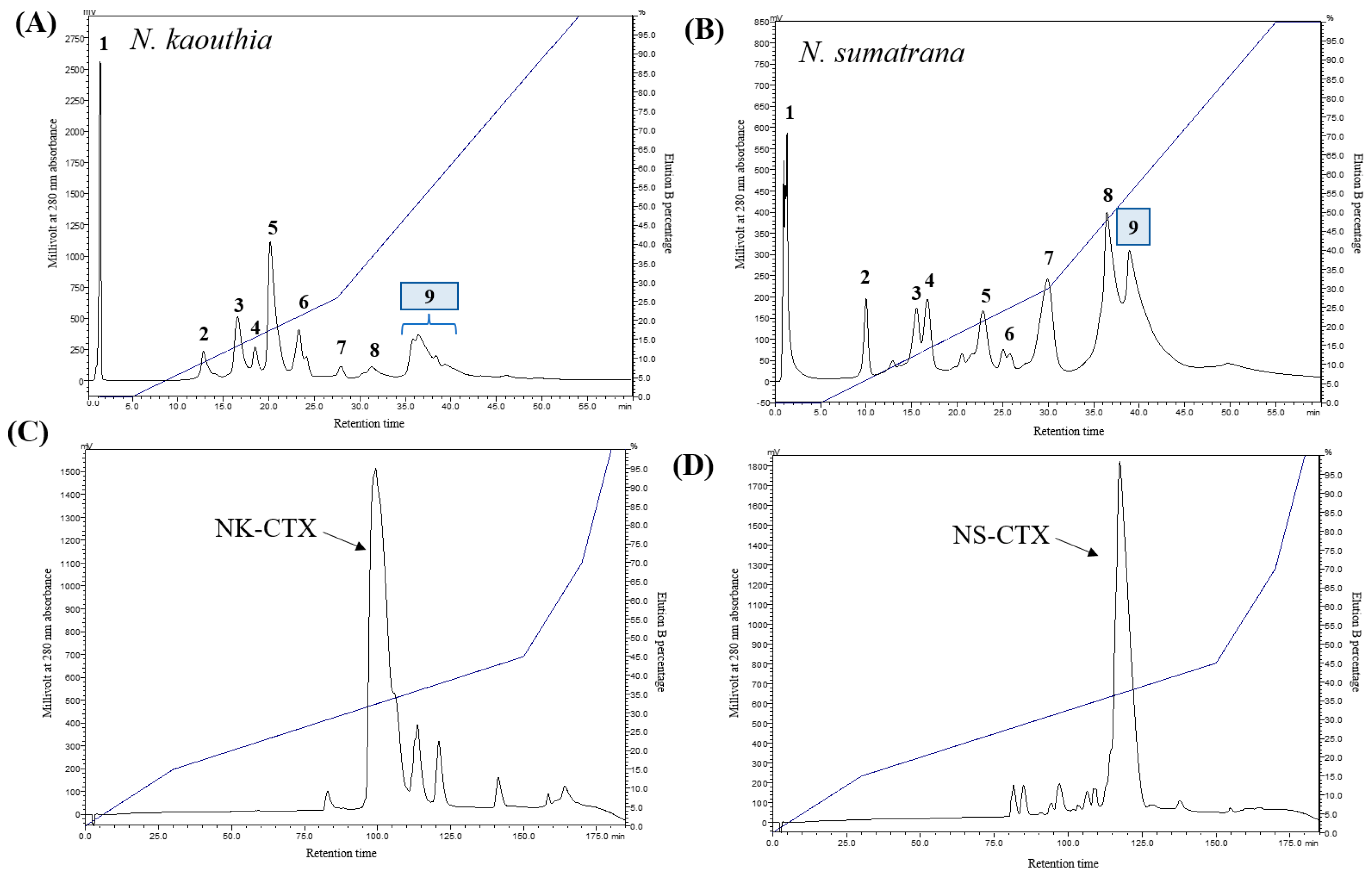
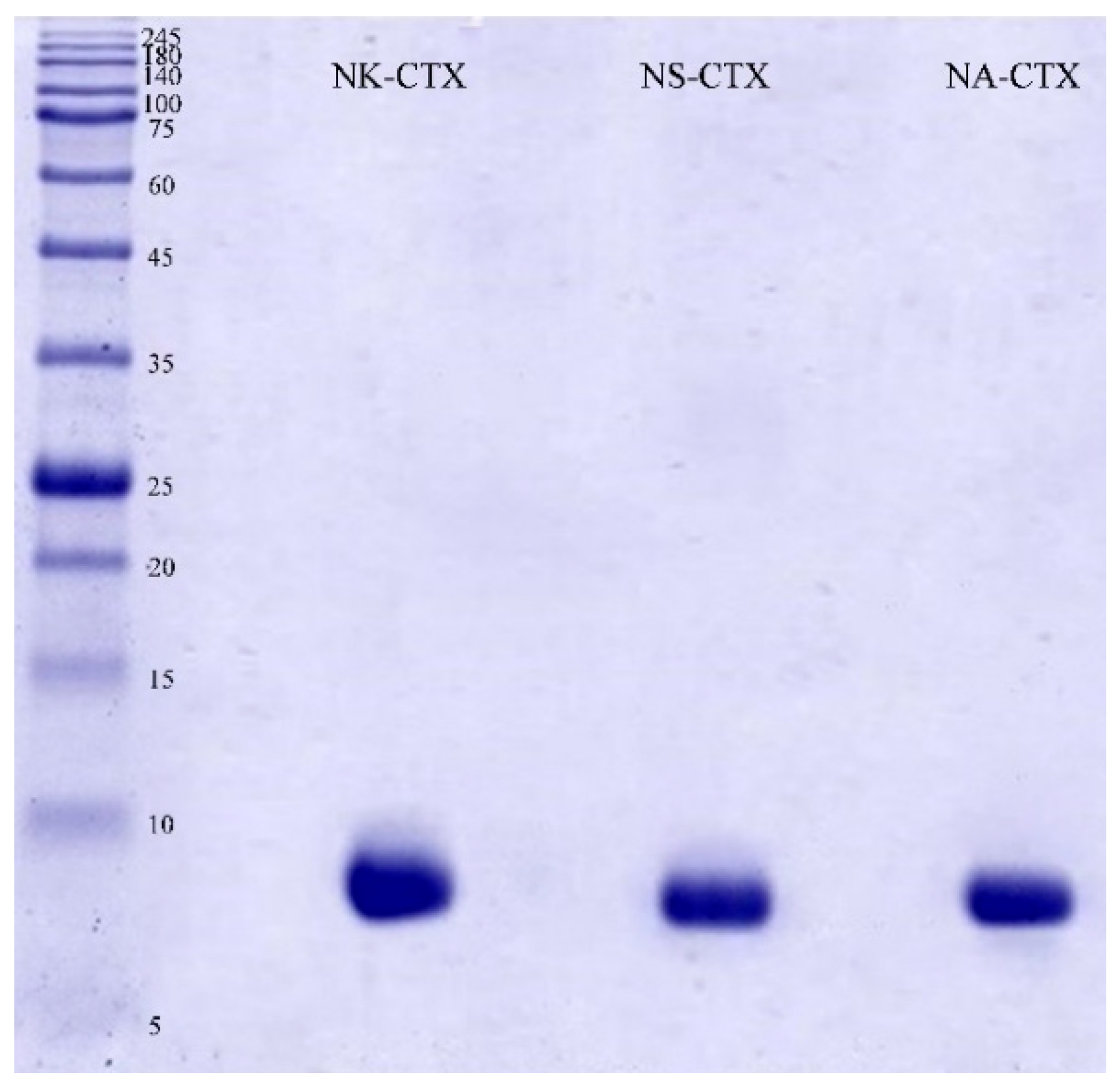
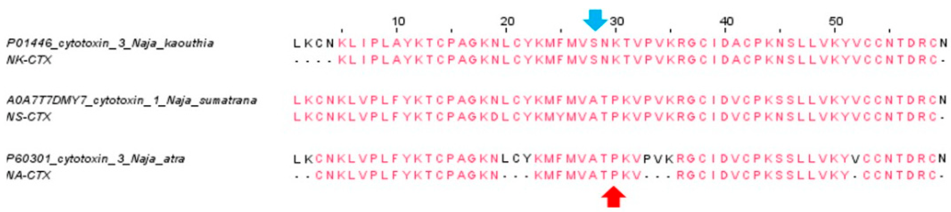
2.1. Isolation and Validation of Cobra Cytotoxins

Cation-exchange HPLC resolved Naja kaouthia and Naja sumatrana venoms into nine protein fractions (Figure 1A,B). Fraction 9 of both venoms, which contained basic proteins, were further separated by C18 reverse-phase HPLC. The major protein fractions containing CTXs were eluted at similar retention times as previously reported [18], from 90 to 105 min for N. kaouthia (NK-CTX; Figure 1C) and from 110 to 125 min for N. sumatrana (NS-CTX; Figure 1D).
The purity of the cobra cytotoxins was validated on SDS-PAGE under reducing conditions. The cytotoxins (NK-CTX, NS-CTX, and NA-CTX) each showed a homogeneous band with an estimated molecular weight of approximately 7.0–7.5 kDa (Figure 2).
With nano-LCMS/MS, these cytotoxins were identified and annotated as cytotoxin 3 (P01446, N. kaouthia) for NK-CTX, cytotoxin 1 (A0A7T7DMY7, N. sumatrana) for NS-CTX, and cytotoxin 3 (Cardiotoxin A3; P60301, N. atra) for NA-CTX. All identifications were validated with a high protein score (>200) and peptide sequence coverage (85–93%) (Figure 3, Table 1). Mass spectrometric analysis of the tryptic peptides (protein score, scored peak intensity, peptide sequence, mass-to-charge ratio, and spectral intensity) of NK-CTX, NS-CTX, and NA-CTX is provided in Supplementary File S1.

2.2. Cytotoxicity of Cobra Venoms and Purified Cytotoxins

Figure 4 shows the cytotoxic activities induced by N. kaouthia, N. sumatrana, and N. atra venoms in the mouse subcutaneous fibroblast cells (CRL-2648) and the neutralization effects of the regional antivenoms from Thailand, Vietnam, and Taiwan. Cell viability was impaired by all cobra venoms in a dose-dependent manner. Among the 3 cobra venoms, N. atra and N. sumatrana venoms were comparably the most cytotoxic (IC50 = 26.87 ± 8.86 μg/mL and 28.83 ± 2.86 μg/mL, respectively), while the cytotoxic potency of N. kaouthia venom was significantly lower (IC50 = 47.40 ± 6.50 μg/mL; p < 0.01) (Table 2). The venom-induced cytotoxicity corroborated with microscopic examination, where N. atra and N. sumatrana venoms caused more prominent alterations in cell morphology than N. kaouthia venom after 8 h incubation. Cells treated with N. atra and N. sumatrana venoms showed a more prominent cell shriveling effect, cell vesicles, and cell debris compared to those treated with N. kaouthia venom (Figure 5).
Under the same experimental conditions, the cells were treated with the purified cobra cytotoxins (NK-CTX, NS-CTX, and NA-CTX), and the findings revealed a dose-dependent reduction in cell viability (Figure 6). The cytotoxic activities exerted by the purified cytotoxins were significantly higher than their corresponding crude venoms at all doses by 1.5–2.0 folds (p < 0.05). NA-CTX was found to be most cytotoxic (IC50 = 13.17 ± 2.72 μg/mL), followed by NS-CTX (IC50 = 20.90 ± 3.73 μg/mL) and NK-CTX (IC50 = 32.90 ± 3.11 μg/mL) (p < 0.01). The microscopic morphology of cells was altered to varying degrees by the respective cytotoxins, as shown in Figure 7. Consistent with the varying IC50 values, while NK-CTX and NS-CTX treated cells showed increased cell vesicles and cell aggregation, cells treated with the more cytotoxic NA-CTX had complete loss of cell morphology, leaving substantial cell debris (Figure 7).

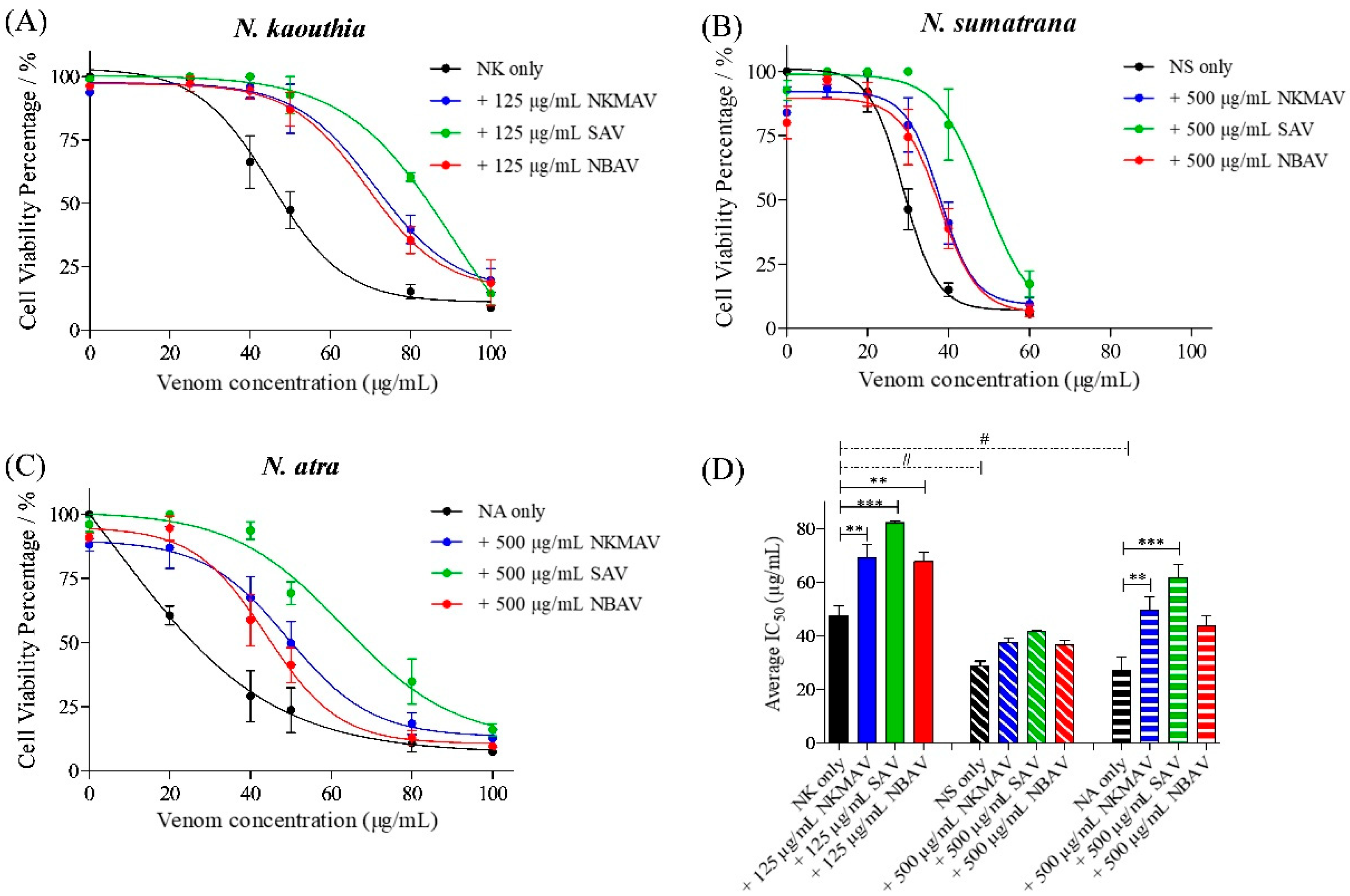
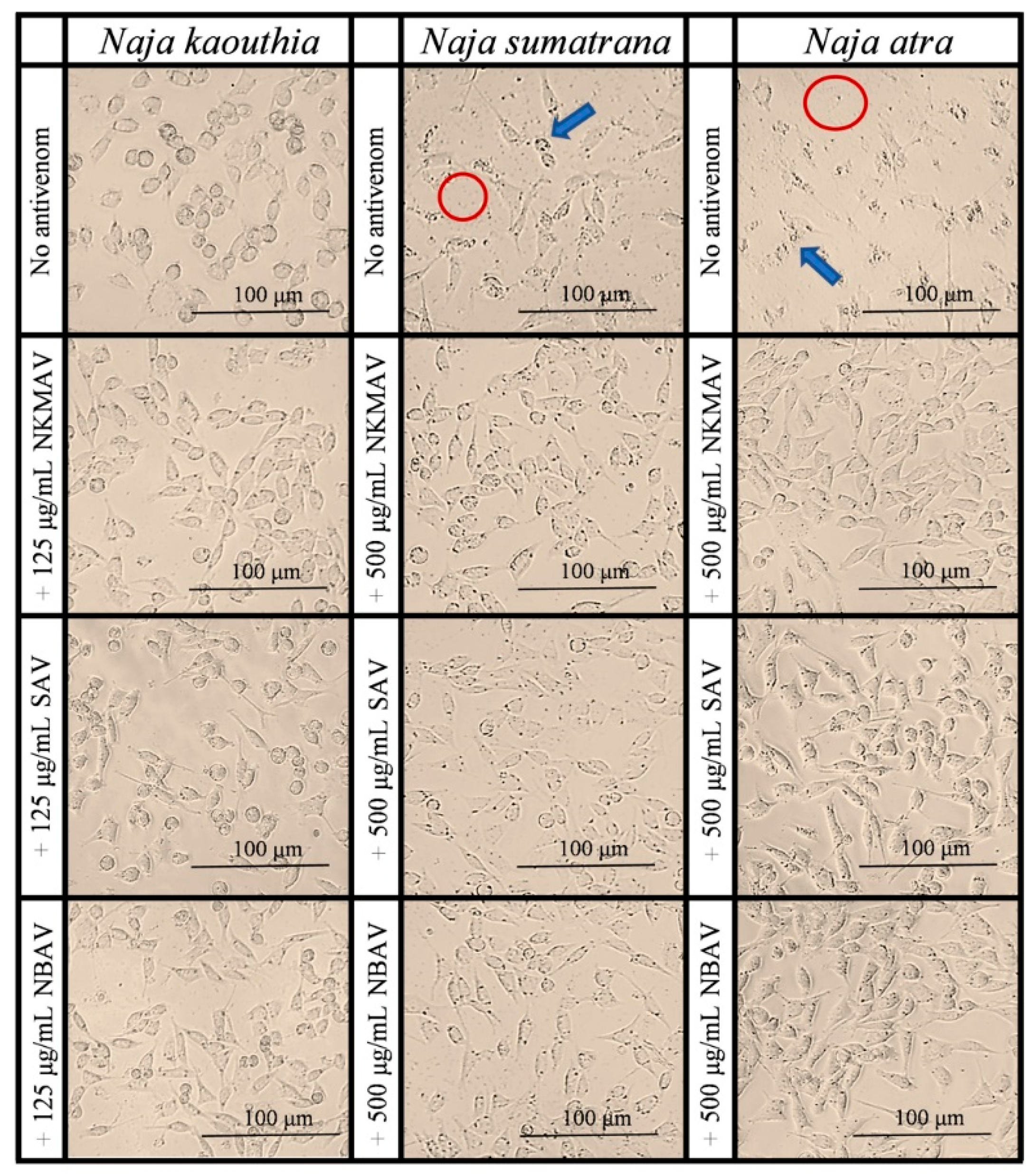
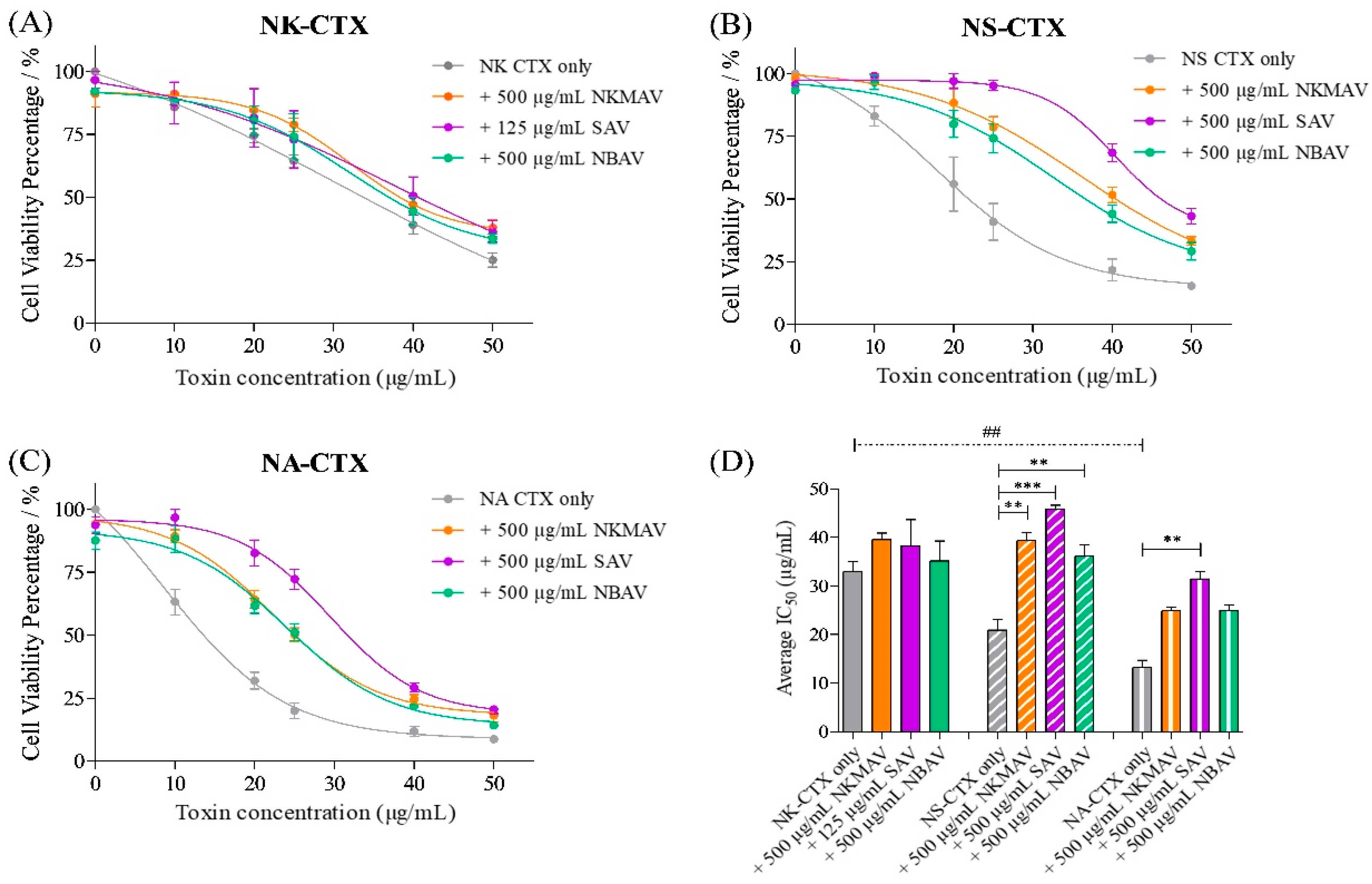
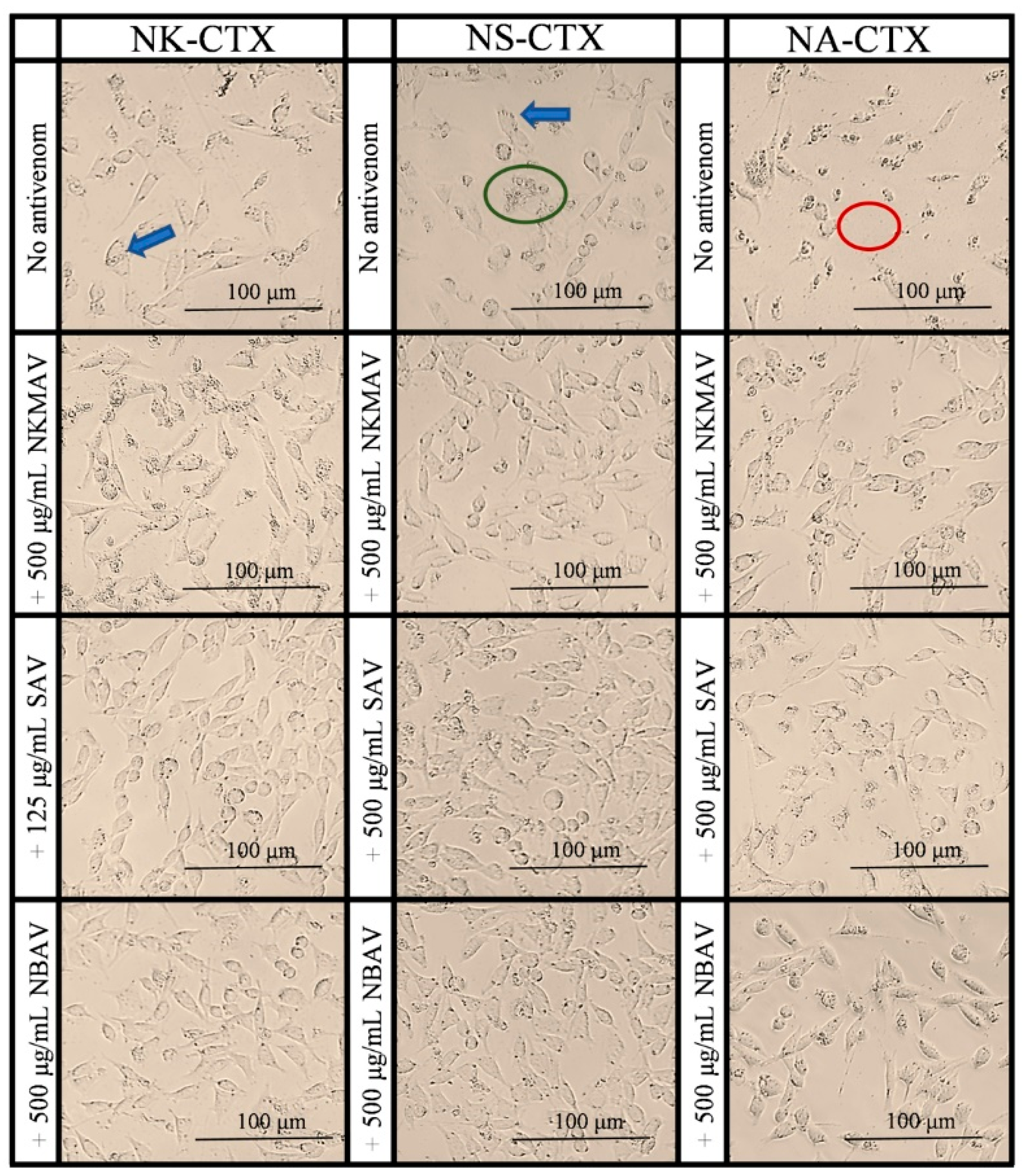
2.3. Neutralization of Cytotoxicity by Antivenoms

The neutralization efficacy of antivenom was determined with an experimental design adapted from Liu et al. [32], quantifying the difference in IC50 between the neutralizing group and control (no antivenom) group. Treatments with antivenom effectively reduced the cytotoxicity of cobra venoms and their purified CTXs, as evidenced by the right-shifted cell viability curves and increased IC50 values (Figure 4 and Table 2). All 3 antivenoms tested, i.e., NkMAV, SAV, and NBAV were able to reduce the cytotoxicity of N. kaouthia venom at varying degrees with 125 µg/mL antivenom concentration (Figure 4A). However, 500 μg/mL antivenom treatments were required to produce noticeable changes in IC50 against N. sumatrana and N. atra venoms (Figure 4B,C). The efficacy levels of different antivenoms against the three venoms were compared in terms of Potency (Pc), which an amount of venom or cytotoxin (in mg) is completely neutralized by a unit amount (g) of antivenom protein, showing at least 1:1000 ratio of venom/CTX to antivenom required for stoichiometric neutralization. All antivenoms exhibited high potencies against N. kaouthia venom-induced cytotoxicity, with SAV being the most efficacious (Pc = 278.16 mg/g), followed by NkMAV (Pc = 174.16 mg/g) and NBAV (Pc = 134.40 mg/g) (Table 2). The antivenoms were moderately efficacious against the cytotoxicity of N. atra venom (Pc ~33.32–69.86 mg/g, with SAV > NkMAV > NBAV) but weak against the N. sumatrana venom (Pc ~15.34–25.34 mg/g, with SAV > NkMAV > NBAV).
In the neutralization study of CTX-induced cytotoxicity, a high dose of antivenom (500 μg/mL) was required to reduce the cytotoxic effects of cytotoxins purified from N. kaouthia, N. sumatrana, and N. atra venoms, except for SAV in which a dose of 125 µg/mL of antivenom protein was adequate to effect neutralization (Table 2). SAV also showed an overall higher potency against the CTXs of individual venoms (Pc of 42.64 mg/g against NK-CTX, 49.74 mg/g against NS-CTX, and 36.66 mg/g against NA-CTX), outperforming the other antivenoms. In comparison between NkMAV and NBAV, both were similarly efficacious against NS-CTX (Pc ~30–37 mg/g) and NA-CTX (Pc ~23 mg/g), while against NK-CTX, NkMAV was much more potent (Pc = 13.20 mg/g) than NBAV (Pc = 4.54 mg/g).

3. Discussion

Envenoming caused by cobras (Naja spp.) is characterized by two hallmark pathologies: systemic neurotoxicity, which is typically the cause of death, and local cytotoxicity, which often leads to tissue necrosis and permanent disabilities [3,5,36,37]. The severity of neurotoxicity is known to correlate with the amount or abundance of alpha-neurotoxins (post-synaptic blocking neurotoxins) in the cobra venoms [38], but the local tissue effect may not necessarily conform to the amount of cytotoxins/cardiotoxins (CTX), suggesting varying cytotoxic activity of different CTX forms, and/or the presence of other modulatory toxins variably evolved in the different lineages of cobras [39]. The present study showed that the venoms of N. sumatrana, N. kaouthia, and N. atra, three medically important cobra species in Asia known for causing severe local necrosis, exert potent cytotoxicity (with low IC50 < 50 μg/mL) when investigated in vitro in the mouse subcutaneous fibroblast cell line (CRL-2648). The degree of cytotoxicity of the three venoms appears to reflect the abundance of CTX in the venoms. The high cytotoxicity of N. atra and N. sumatrana venoms (IC50 = 26–28.83 μg/mL) is consistent with the higher CTX abundance (~50% of total venom proteins) reported in the venom proteomes [12,40] venoms, whereas the N. kaouthia venom (IC50 ~ 47 μg/mL) had a lower abundance of CTX (~28% of total venom proteins) [41]. Comparatively, the venoms of African spitting cobras, such as Naja nigricollis, Naja mossambica, and Naja pallida, have relatively higher CTX abundances (64–72%) [42] and thus higher cytotoxicity when examined in the rat myogenic cell line (C2C12) (IC50 of ~15 μg/mL) [33]. It has been proposed that the enhanced cobra venom cytotoxicity facilitates the evolution of the defensive spitting behavior of cobras, especially the African spitters from the subgenus of Afronaja [39]. The phenotype of venom-spitting behavior and highly cytotoxic venom is in sharp contrast to the African non-spitting cobras (subgenus: Uraeus), which cause minimal or negligible local envenoming effect and have lower cytotoxicity [39,43,44], presumably because the CTX of the non-spitting cobras of Uraeus subgenus are structurally atypical, containing a substitution of histidine at the 4th amino acid residue [45,46,47], a CTX “variant” reported to exhibit low cytotoxicity [48,49]. The association between venom-spitting behavior and high venom cytotoxicity is, however, weak when examined in the Asiatic cobras (subgenus: Naja), as also shown in this study. Notably, the spitting Philippine Cobra, Naja philippinensis causes minimal local envenoming effect and has a venom with low cytotoxicity [38,50], while N. atra and N. kaouthia, conventionally known to be non-spitting, have highly cytotoxic venoms and cause severe tissue necrosis in envenomings much as the typical spitter N. sumatrana does [5,51,52]. Nevertheless, it is noteworthy that both N. atra and N. kaouthia are increasingly perceived as “partial” spitting cobras, where a few specimens have been found to be able to eject venom through the fangs, facilitated with a physical maneuver to enable forward spitting [53,54]. The spitting behavior in N. atra and N. kaouthia is rare, however, and it lacks the precision of typical spitting cobras.
Our findings of the high cytotoxic activity of N. kaouthia, N. sumatrana, and N. atra venoms are congruent with the clinical phenotype of local envenoming caused by these species (manifested as severe local necrosis), irrespective of their inclination of venom spitting. Further characterization revealed that the purified cytotoxins, i.e., NK-CTX, NS-CTX, and NA-CTX have IC50 that is lower than their corresponding venoms, indicating their role as the major cytotoxic components in the venoms. The CTX cytotoxicity is variable among the three cobra species, with the most potent being NA-CTX, followed by NS-CTX and NK-CTX, suggesting structural variation that influences the intrinsic activity of these homologous proteins. The cytotoxicity of CTX also varies depending on the target cells, as NA-CTX exhibited low IC50 (IC50 = 6.82 μg/mL) in human leukemic myeoloblast cell line (HL60), showing high CTX-specificity toward human cell line compared to murine cell line (IC50 = 26.87 μg/mL). The more cytotoxic NA-CTX and NS-CTX belong to the P-type CTX, whereas the relatively less cytotoxic NK-CTX is an S-type CTX. P-type CTXs are generally more cytotoxic than S-type CTXs due to higher lipid-binding activity and deeper penetration into phospholipid bilayers, which readily destabilizes the membrane architecture, causing pore formation and cytolysis [15,17]. This is also supported by the differences in cytotoxicity between NS-CTX (P-type) and NK-CTX (S-type) in a variety of human cell lines of breast (MCF-7), lung (A549), and prostate (PC-3) cancers reported recently [18]. The cytotoxin subtypes (P- and S-type), however, seem independent from the systemic lethality as shown in vivo, where NS-CTX was the most lethal (P-type, LD50 = 1.13 μg/g), followed by NK-CTX (S-type, LD50 = 1.41 μg/g), and NA-CTX (P-type, LD50 = 2.12 μg/g) [32,55,56]. Therefore, the role of CTX subtypes mainly reflects the degree of local tissue cytotoxicity and necrosis evident from the extensive tissue necrosis from N. sumatrana and N. atra envenomation [32,51].
The findings of distinct CTX with high cytotoxicity in the three cobra venoms underscores the need to evaluate antivenom efficacy in neutralizing not only the whole venoms but also the CTXs specific to the respective venoms. In the neutralization study, the antivenoms NkMAV, SAV, and NBAV demonstrated variable efficacy against the venom- and CTX-induced cytotoxicity, with SAV being the most potent, followed by NkMAV and NBAV. The cross-neutralization capability of the different antivenoms suggests conserved epitopes among the CTXs studied, while their varying efficacy could be due to factors associated with the CTXs and/or the antivenom formulation. The subtypes and abundances of CTX in different cobra venoms are variable, and this can result in diverse immunogenicity and antigenicity of the venom proteins and their neutralization by antivenom [12,57]. The findings showed that between both monovalent antivenoms (NkMAV and SAV), SAV raised against the Vietnamese N. kaouthia was more potent (by 1.6 folds) in neutralizing the cytotoxicity induced by N. kaouthia venom. This is possibly attributed to the higher abundance of CTX (44.9% of total venom proteins) in the Vietnamese N. kaouthia venom compared to the CTX content in the Thai N. kaouthia venom (27.6%) [41], which therefore elicited a higher antibody titer in SAV against the CTXs. Furthermore, the content or amount of neutralizing antibodies that specifically target cobra CTX may vary between different antivenom formulations. Naturally, the specific CTX-neutralizing antibody content is anticipated to be higher in a monovalent (mono-specific) antivenom compared with a poly-specific product per mass unit of protein. In comparison, NBAV showed generally lower neutralization potency against the venoms and CTXs tested compared with the monovalent SAV and NkMAV. This is perhaps because NBAV has a proportionally lower antibody titer in neutralizing the cytotoxic components as it is a bivalent product raised against the Taiwanese N. atra and B. multicintus, the latter of which is an elapid whose venom lacks cytotoxic properties and contains no CTX [32,58]. This is also supported by an earlier study that showed a lower immunocapturing activity of NBAV against the CTX of Taiwanese N. atra (<20%) in contrast to SAV, which showed higher immunocapturing activity of >50% [32]. The suggestion of non-monovalent antivenoms having a lower content of specific neutralizing antibodies is also consistent with the observation from another recent study, which showed that the bivalent NBAV is less potent in neutralizing B. multicinctus venom compared to a regional B. multicinctus monovalent antivenom product [58]. Additionally, the cytotoxin subtypes seem to play a vital role in neutralization as NBAV (raised against the Taiwanese N. atra, possessing P-type cytotoxin) exhibited low neutralization potency (Pc = 4.54 mg/g) against the S-type NK-CTX, highlighting the need for cytotoxins subtype considerations in antivenom development.
When compared across the three venoms tested, the three cobra antivenoms consistently showed limited neutralization activity against the cytotoxicity induced by the heterologous N. sumatrana venom (Pc = 15.34–25.34 mg/g) despite having modest neutralization efficacy against the NS-CTX (Pc = 30.40–49.74 mg/g). The discrepancy could be due to the presence of toxic neutral PLA2 in the N. sumatrana venom, which can act synergistically with CTX to potentiate the venom cytotoxicity [59]. The antivenoms tested (raised against N. kaouthia and N. atra) were probably less effective against this form of toxic PLA2 found uniquely in N. sumatrana venom, as the PLA2s present in N. kaouthia and N. atra venoms are primarily the non-lethal acidic forms [32,55,56,60]. Moreover, the N. sumatrana venom exhibited a much higher PLA2 enzymatic activity (enzymatic rate = 82.11 nmol/min/mg) than N. kaouthia and N. atra venoms (enzymatic rates < 40 nmol/min/mg) [61], a characteristic which may have a synergistic effect on the CTX activity of N. sumatrana venom, rendering the reduced efficacy of the antivenoms.
Despite the present findings showing that the three regional antivenoms generally had low efficacy against the in vitro cytotoxicity induced by the cobra venoms and cytotoxins, WHO and Ministry of Health guidelines recommend the use of antivenom on patients exhibiting prominent local cytotoxicity involving more than half of the bitten extremity, rapid swelling extension beyond ankles or wrists within hours, and development of enlarged and tender lymph node draining of the bitten limb [3,62]. Clinical reports of the use of these antivenoms in treating cytotoxicity have been inconsistent, with Vietnamese patients reporting neither mortality nor morbidity with SAV therapy [29], while Taiwanese patients still develop necrosis and require surgical interventions despite early (<6 h) NBAV administration [51]. Nevertheless, antivenom effectiveness is limited in treating tissue necrosis probably due to limited immunogenicity and antigenicity of CTX in view of their small molecular sizes (60–70 amino acid residues) and variable amounts (abundances) in the venoms that reduce the efficiency of eliciting specific antibody production against the toxins during hyperimmunization. In addition, the formulation of an antivenom product as being monovalent or polyvalent probably also has an impact on its neutralization potency against the venom cytotoxicity.

4. Conclusions

The present study elucidated the differential cytotoxicity of the three medically important Asiatic cobras (N. kaouthia, N. atra, and N. sumatrana) venoms and their purified cytotoxins applying an in vitro model of mouse fibroblast cell line (CRL-2648) which can be considered prior to the use of the in vivo tissue necrosis assay. The findings underscore the need to improve antivenom efficacy for the neutralization of cytotoxicity induced by cobra CTXs which are small proteins with variable abundances and subtypes across different species. It is suggested that innovative approaches such as the use of toxin-enriched immunogens [63], synthetic or recombinant species-specific antivenoms [64], or small molecule inhibitors [65,66] should be further explored for better treatment of local tissue-damaging effects. Further research should also aim to establish the correlation of cytotoxicity neutralization study between in vitro and in vivo models and how this can be clinically translated for improved treatment outcome of local tissue toxicity in cobra bite envenomings.

5. Materials and Methods

5.1. Venoms and Antivenoms

Naja kaouthia venom was supplied by the Queen Saovabha Memorial Institute (QSMI), Bangkok, Thailand. Naja sumatrana venom was a sample collected from adult snakes in Seremban, Peninsular Malaysia. Naja atra venom was obtained from a local snake farm in Tainan, Taiwan. All venoms were pooled samples of more than 10 adult snakes, lyophilized, and stored at −20 ℃ until use. N. kaouthia and N. sumatrana venoms were subjected to the purification of cytotoxins as described below. The cytotoxin of N. atra was provided by the National Health Research Institutes, Taiwan, obtained through a purification process as reported earlier by Liu et al. [32].
Thai Naja kaouthia Monovalent Antivenom (NkMAV), Vietnamese Naja kaouthia Monovalent Antivenom (SAV, as Snake Antivenom per manufacturer’s labeling), and Taiwanese Neuro Bivalent Antivenom (NBAV) were used in the present study. NkMAV (batch no.: NK00116) is a monospecific antivenom raised against Thai Naja kaouthia venom, produced by the Queen Saovabha Memorial Institute (QSMI), Bangkok, Thailand. SAV (batch no.: 039-00-18) is a monospecific antivenom raised against Vietnamese Naja kaouthia venom, produced by the Venom Research Unit, Ho Chi Minh City, Vietnam. NBAV (batch no.: 61-06-0002) is a bivalent antivenom raised against Taiwanese Bungarus multicintus and Naja atra venoms, manufactured by Taiwan Central for Disease Control, Taipei, Taiwan. All antivenoms were lyophilized F(ab’)2 derived from horse antisera. NkMAV and NBAV are lyophilized types of antivenoms that were reconstituted in 10 mL normal saline per vial prior to use.

5.2. Resource S Cation-Exchange and C18 Reverse-Phase High-Performance Liquid Chromatography

Cytotoxins of N. kaouthia (NK-CTX) and N. sumatrana (NS-CTX) venoms were purified with sequential fractionation using Resource S cation-exchange and C18 reverse-phase high-performance liquid chromatography using Shimadzu LC-20AD High-Performance Liquid Chromatography (HPLC) system (Kyoto, Japan), as previously described [18,56]. Briefly, the venom samples were reconstituted in 20 mM 2-(N-morpholino)ethanesulfonic acid (MES; eluent A; pH 6) and subjected to Resource S cation-exchange (GE Healthcare, Sweden) chromatographic fractionation. The HPLC system was pre-equilibrated with eluent A, and elution was carried out with 0.8 M sodium chloride in 20 mM MES (eluent B; pH 6) at a linear gradient of 0 to 30% eluent B from 5 to 40 min, followed by 30 to 100% from 40 to 65 min. The flow rate was kept at 1 mL/min, and the elution was monitored at 280 nm absorbance. The protein fractions were manually collected and subjected to buffer exchange using Vivaspin 20 (5000 MWCO) polyethersulfone membrane (Sartorius, Göttingen, Germany).
The retentates from cation-exchange chromatography, which contains NK-CTX and NS-CTX, were subjected to reverse-phase fractionation via LiChrospher WP 300 C18 (5 µm pore size) reverse-phase column (Kyoto, Japan). In brief, the retentates were reconstituted in 0.1% trifluoroacetic acid (TFA) in ultrapure water (solvent A). Column pre-equilibration was performed with solvent A, and retentates were subsequently separated with 0.1% TFA in acetonitrile (solvent B), applying linear gradient elution as follows: 5% for 10 min, 5 to 15% for 20 min, 15 to 45% for 120 min, and 45 to 70% for 20 min. The flow rate was maintained at 1 mL/min, and the elution was monitored at 215 nm absorbance. Protein fractions were collected manually, freeze-dried, and stored at −20 ℃ until further use.

5.3. Sodium Dodecyl Sulfate-Polyacrylamide Gel Electrophoresis (SDS-PAGE)

The cytotoxins of N. kaouthia (NK-CTX), N. sumatrana (NS-CTX), and N. atra (NA-CTX) were subjected to 15% SDS-PAGE under reducing conditions at 90 V for 2.5 h. ExcelBand™ 3-colour Broad Range Protein Marker (Smobio, Hsinchu, Taiwan) was used for molecular weight estimation. Proteins were visualized by staining the gel with Coomassie Brilliant Blue R-250 (Sigma-Aldrich, St. Louis, MO, USA). The molecular weights of the cytotoxins were estimated using GelAnalyzer (Version 19.1) [67].

5.4. Nano-Electrospray Ionization Liquid Chromatography-Tandem Mass Spectrometry (Nano-ESI LC-MS/MS)

A total of 5 micrograms of the purified NK-CTX, NA-CTX, and NS-CTX were reduced by dithiothreitol (DTT), alkylated with iodoacetamide (IA), and subjected to in-solution digestion with 0.1 μg/μL of MS grade Pierce trypsin protease (trypsin to sample ratio 1:10; Thermo Scientific Pierce, Rockford, IL, USA). Peptide desalting and extraction were carried out with Millipore ZipTip®® C18 pipette tip (Merck, Branchburg, NJ, USA). The extracted peptides then underwent nano-electrospray ionization-liquid chromatography-tandem mass spectrometry (nano-ESI-LC-MS/MS) with Agilent 1200 HPLC-Chip/MS Interface, paired with Agilent 6520 Accurate-Mass Q-TOF LC/MS system (Santa Clara, CA, USA). A total of 0.1% formic acid in water and 90% acetonitrile in water with 0.1% formic acid were utilized, respectively, as eluting solvents A and B. Samples were then analyzed with Agilent HPLC-Chip II: G42040-62010, comprised of 160 nL enrichment column and a 75 μm × 150 mm analytical column packed with Agilent ZORBAX 300SB-C18, 5 µm, 300Å. Sample elution was carried out with the same linear gradient as previously described [68]: 3–50% solvent B for 30 min, 50–95% solvent B for 2 min, and 95% solvent B for 5 min. The drying gas flow rate was set to 5 l/min at 325 ℃, and the ion polarity was set to positive. Fragmentor voltage was adjusted to 175 V while capillary voltage was 1995 V. Spectra acquisition was assigned at MS 110–3000 m/z and 50–3000 m/z scan range. The precursor was adjusted to include a doubly or more charged state, apart from reference precursor ions (922.0098 m/z (z = 1) and 121.0509 (z = 1). MH+ range between 600 and 4000 Da was acquired and processed with Agilent Spectrum Mill MS Proteomics Workbench software packages (B.04.00). Peptide sequences obtained were searched against the non-redundant NCBI database (taxonomy: Serpentes, taxid: 8570) (https://0-www-ncbi-nlm-nih-gov.brum.beds.ac.uk/Taxonomy/Browser/wwwtax.cgi?mode=Info&id=8570, accessed on 22 July 2020). Carbamidomethylation was set as a fixed modification. The identified proteins were validated with the following parameters: peptide score filter > 6, protein score filter > 10, and scored peak intensity (SPI) > 60%.

5.5. Multiple Sequence Alignment

Tryptic peptides were aligned with annotated toxins with the highest homology. The annotated toxin sequences were obtained from the UniProtKB depository. Multiple sequence alignment was conducted with Jalview [69] and MUSCLE [70] software (version 2.10.5).

5.6. Cell Culture

The murine subcutaneous fibroblast cell line (L cells; ATCC®® CRL-2648) was supplied by American Type Culture Collection (ATCC; Manassas, VA, USA). The cells were cultured in Dulbecco’s Modified Eagle’s Medium (DMEM; Nacalai tesque, Kyoto, Japan) with 10% fetal bovine serum (TICO Europe, Amstelveen, The Netherlands) and 1% penicillin-streptomycin (Nacalai tesque; Kyoto, Japan) in a humidified atmosphere of 5% CO2 and 37 ℃ in T25 flasks. The cells were passaged in accordance with ATCC guidelines using trypsin-EDTA (0.05% trypsin and 0.02% EDTA; Nacalai tesque, Kyoto, Japan).

5.7. In Vitro Cytotoxicity Induced by Cobra Venoms and CTX

In vitro assay protocols were modified protocols adapted from Liu et al. [32] and Chong et al. [18]. The cytotoxic activities of cobra venoms and their purified cytotoxins were tested with 3-(4,5-dimethylthiazol-2-yl)-2,5-diphenyltetrazolium bromide (MTT; Sigma-Aldrich, MO, USA). A hundred microliters of the aliquot of cells (15,000 cells/well) were seeded into 96-well microtiter plates 24 h prior to venom or cytotoxin treatment. The cells were treated with 200 μL of serially diluted (10 to 100 μg/mL) venom or purified cytotoxin (NK-CTX, NS-CTX, NA-CTX) of the respective cobra species in a complete medium. Cells without venom served as the negative control. The cells were then treated for 8 h, in conjunction with a previous report by Omran et al. [71] demonstrating the highest cytotoxicity in N. haje venom at 8 h treatment. After incubation, cytotoxicity was determined by introducing 10% MTT solution and subsequently incubated in the dark and at 37 ℃ for 3 h. Purple formazans created were then dissolved with 200 μL DMSO, and their absorbance was quantified at 570 nm using Chameleon™ vs. Multilabel microplate reader (Hidex, Turku, Finland). All assays were performed with biological and technical triplicates. The cell viability percentage was calculated according to the formula below:
Cell   viability   percentage = ( Absorbance   of   experimental   sample )     ( Absorbance   of   blank   sample ) Absorbance   of   the   untreated   sample     ( Absorbance   of   blank   sample ) × 100 %
Half maximal inhibitory concentrations (IC50) were then determined for the respective venoms and cytotoxins with GraphPad Prism (Version 5.03) with nonlinear regression analysis, applying the 4-parameter logistic equation for dose-response inhibition study, and the data (encompassing 3 biological and 3 technical triplicates) were expressed as means ± standard error of triplicates.

5.8. Neutralization Assay of Cytotoxicity

The cobra venoms or toxins were prepared at a serial dose of 10 to 100 μg/mL as described above for the determination of cytotoxicity. The venom or toxins were pre-incubated with a fixed dose of antivenom treatment (125 μg/mL of SAV, or 500 μg/mL of NBAV and NKMAV) in complete medium at 37 ℃ for 30 min. Subsequently, the mixture was added to the cell-seeded wells. Cytotoxicity and IC50 values were determined after 8 h with the MTT assay as described above, also performed in biological and technical triplicates.
The in vitro efficacy of cytotoxicity neutralization by antivenom, termed Potency (Pc), was interpreted in terms of the absolute change in IC50 of the venom or toxin affected by the antivenom treatment given at a normalized dose. This was calculated with the following formula modified from a previous study [32]:
Potency   Pc mg / g = IC 50 Treated IC 50 Untreated   in   μ g / mL   Antivenom   concentration   in   mg / mL
The IC50 values of the venom/cytotoxin-induced cytotoxicity tests with and without antivenom treatment. The calculated difference in IC50 was then normalized by the dose of antivenom (in mg/mL) used in the corresponding treatment group. The resultant Pc value represents the amount of venom or cytotoxin (in mg) neutralized per gram of antivenom protein under the experimental conditions to effect the change in IC50 between treated and untreated groups.

5.9. Statistical Analysis

One-way ANOVA with Tukey’s multiple post hoc comparison test on GraphPad Prism (Version 5.03) was used in comparing cell viability. Differences in means were statistically significant when p-value < 0.05.

Supplementary Materials

The following supporting information can be downloaded at: https://0-www-mdpi-com.brum.beds.ac.uk/article/10.3390/toxins14050334/s1, Supplementary File S1: nano-LC-MS/MS of purified cytotoxins from Naja kaouthia (NK-CTX), Naja sumatrana (NS-CTX), and Naja atra (NA-CTX)

Author Contributions

Conceptualization, K.Y.T., C.H.T. and W.-C.S.; Methodology, H.P.C. and B.-S.L.; Validation, H.P.C., K.Y.T. and C.H.T.; Formal analysis, H.P.C. and K.Y.T.; Investigation, H.P.C., K.Y.T. and C.H.T.; Resources, C.H.T. and W.-C.S.; Data curation, H.P.C., K.Y.T. and C.H.T.; Writing—original draft preparation, H.P.C. and K.Y.T.; Writing—review and editing, H.P.C., K.Y.T., C.H.T., B.-S.L. and W.-C.S.; Visualization, H.P.C. and K.Y.T.; Supervision, K.Y.T. and C.H.T.; Project administration, K.Y.T. and C.H.T.; Funding acquisition, K.Y.T., C.H.T. and W.-C.S. All authors have read and agreed to the published version of the manuscript.

Funding

The study was supported by research grants from the University of Malaya (ST011–2020) and National Health and Research Institutes (IV-111-PP-25).

Institutional Review Board Statement

Not applicable.

Informed Consent Statement

Not applicable.

Data Availability Statement

The data presented in this study are available in this article and Supplementary Materials.

Acknowledgments

The authors would like to thank the University of Malaya and National Health Research Institutes (Taiwan) for research funding and facility support.

Conflicts of Interest

The authors declare no conflict of interest.

References

  1. Gutierrez, J.M.; Calvete, J.J.; Habib, A.G.; Harrison, R.A.; Williams, D.J.; Warrell, D.A. Snakebite envenoming. Nat. Rev. Dis. Primers 2017, 3, 17079. [Google Scholar] [CrossRef] [PubMed]
  2. WHO. Global snakebite burden. In Proceedings of the Seventy-First World Health Assembly, Geneva, Switzerland, 21–26 May 2018. [Google Scholar]
  3. WHO. Guidelines for the Management of Snake Bites; Regional office for South-East Asia: New Delhi, India, 2016. [Google Scholar]
  4. WHO. Snakebite Envenoming: A Strategy for Prevention and Control; WHO: Geneva, Switzerland, 2019. [Google Scholar]
  5. Tan, N.H.; Tan, K.Y.; Tan, C.H. Snakebite in Southeast Asia: Envenomation and Clinical Management. In Handbook of Venoms and Toxins of Reptiles, 2nd ed.; CRC Press: Boca Raton, FL, USA, 2021. [Google Scholar]
  6. Wong, O.F.; Lam, T.S.; Fung, H.T.; Choy, C.H. Five-year experience with Chinese cobra (Naja atra)—Related injuries in two acute hospitals in Hong Kong. Hong Kong Med. J. 2010, 16, 36–43. [Google Scholar] [PubMed]
  7. Ang, L.J.; Sanjay, S.; Sangtam, T. Ophthalmia due to spitting cobra venom in an urban setting--a report of three cases. Middle East Afr. J. Ophthalmol. 2014, 21, 259–261. [Google Scholar] [CrossRef] [PubMed]
  8. Chu, E.R.; Weinstein, S.A.; White, J.; Warrell, D.A. Venom ophthalmia caused by venoms of spitting elapid and other snakes: Report of ten cases with review of epidemiology, clinical features, pathophysiology and management. Toxicon 2010, 56, 259–272. [Google Scholar] [CrossRef]
  9. Tsai, T.H.; Lin, C.C.; Mao, Y.C.; Hung, C.L.; Yang, Y.C.; Yang, C.C.; Jeng, M.J. Naja atra venom-spit ophthalmia in Taiwan: An epidemiological survey from 1990 to 2016. J. Chin. Med. Assoc. 2020, 83, 77–83. [Google Scholar] [CrossRef]
  10. Tan, K.Y.; Wong, K.Y.; Tan, N.H.; Tan, C.H. Quantitative proteomics of Naja annulifera (sub-Saharan snouted cobra) venom and neutralization activities of two antivenoms in Africa. Int. J. Biol. Macromol. 2020, 158, 605–616. [Google Scholar] [CrossRef]
  11. Wong, K.Y.; Tan, K.Y.; Tan, N.H.; Tan, C.H. A Neurotoxic Snake Venom without Phospholipase A2: Proteomics and Cross-Neutralization of the Venom from Senegalese Cobra, Naja senegalensis (Subgenus: Uraeus). Toxins 2021, 13, 60. [Google Scholar] [CrossRef]
  12. Huang, H.W.; Liu, B.S.; Chien, K.Y.; Chiang, L.C.; Huang, S.Y.; Sung, W.C.; Wu, W.G. Cobra venom proteome and glycome determined from individual snakes of Naja atra reveal medically important dynamic range and systematic geographic variation. J. Proteomics 2015, 128, 92–104. [Google Scholar] [CrossRef]
  13. Antil-Delbeke, S.; Gaillard, C.; Tamiya, T.; Corringer, P.J.; Changeux, J.P.; Servent, D.; Menez, A. Molecular determinants by which a long chain toxin from snake venom interacts with the neuronal alpha 7-nicotinic acetylcholine receptor. J. Biol. Chem. 2000, 275, 29594–29601. [Google Scholar] [CrossRef] [Green Version]
  14. Kini, R.M. Molecular moulds with multiple missions: Functional sites in three-finger toxins. Clin. Exp. Pharmacol. Physiol. 2002, 29, 815–822. [Google Scholar] [CrossRef]
  15. Dubovskii, P.V.; Lesovoy, D.M.; Dubinnyi, M.A.; Konshina, A.G.; Utkin, Y.N.; Efremov, R.G.; Arseniev, A.S. Interaction of three-finger toxins with phospholipid membranes: Comparison of S- and P-type cytotoxins. Biochem. J. 2005, 387, 807–815. [Google Scholar] [CrossRef] [PubMed]
  16. Konshina, A.G.; Boldyrev, I.A.; Utkin, Y.N.; Omel’kov, A.V.; Efremov, R.G. Snake Cytotoxins Bind to Membranes via Interactions with Phosphatidylserine Head Groups of Lipids. PLoS ONE 2011, 6, e19064. [Google Scholar] [CrossRef] [PubMed] [Green Version]
  17. Chien, K.Y.; Chiang, C.M.; Hseu, Y.C.; Vyas, A.A.; Rule, G.S.; Wu, W. Two distinct types of cardiotoxin as revealed by the structure and activity relationship of their interaction with zwitterionic phospholipid dispersions. J. Biol. Chem. 1994, 269, 14473–14483. [Google Scholar] [CrossRef]
  18. Chong, H.P.; Tan, K.Y.; Tan, C.H. Cytotoxicity of Snake Venoms and Cytotoxins From Two Southeast Asian Cobras (Naja sumatrana, Naja kaouthia): Exploration of Anticancer Potential, Selectivity, and Cell Death Mechanism. Front. Mol. Biosci. 2020, 7, 583587. [Google Scholar] [CrossRef] [PubMed]
  19. Attarde, S.S.; Pandit, S.V. Cytotoxic activity of NN-32 toxin from Indian spectacled cobra venom on human breast cancer cell lines. BMC Complement. Altern. Med. 2017, 17, 503. [Google Scholar] [CrossRef] [Green Version]
  20. Liu, Y.; Ming, W.; Wang, Y.; Liu, S.; Qiu, Y.; Xiang, Y.; Hu, L.; Fan, L.; Peng, X.; Wang, H.; et al. Cytotoxin 1 from Naja atra Cantor venom induced necroptosis of leukemia cells. Toxicon 2019, 165, 110–115. [Google Scholar] [CrossRef]
  21. Liu, C.C.; Chou, Y.S.; Chen, C.Y.; Liu, K.L.; Huang, G.J.; Yu, J.S.; Wu, C.J.; Liaw, G.W.; Hsieh, C.H.; Chen, C.K. Pathogenesis of local necrosis induced by Naja atra venom: Assessment of the neutralization ability of Taiwanese freeze-dried neurotoxic antivenom in animal models. PLoS Negl. Trop. Dis. 2020, 14, e0008054. [Google Scholar] [CrossRef] [Green Version]
  22. Rivel, M.; Solano, D.; Herrera, M.; Vargas, M.; Villalta, M.; Segura, A.; Arias, A.S.; Leon, G.; Gutierrez, J.M. Pathogenesis of dermonecrosis induced by venom of the spitting cobra, Naja nigricollis: An experimental study in mice. Toxicon 2016, 119, 171–179. [Google Scholar] [CrossRef]
  23. Hsieh, Y.H.; Hsueh, J.H.; Liu, W.C.; Yang, K.C.; Hsu, K.C.; Lin, C.T.; Ho, Y.Y.; Chen, L.W. Contributing Factors for Complications and Outcomes in Patients With Snakebite: Experience in a Medical Center in Southern Taiwan. Ann. Plast. Surg. 2017, 78, S32–S36. [Google Scholar] [CrossRef]
  24. Huang, L.-W.; Wang, J.-D.; Huang, J.-A.; Hu, S.-Y.; Wang, L.-M.; Tsan, Y.-T. Wound infections secondary to snakebite in central Taiwan. J. Venom. Anim. Toxins Incl. Trop. Dis. 2012, 18, 272–276. [Google Scholar] [CrossRef] [Green Version]
  25. WHO. Guidelines for the Management of Snake-Bites; World Health Organization: New Delhi, India, 2010. [Google Scholar]
  26. Gutiérrez, J.; Solano, G.; Pla, D.; Herrera, M.; Segura, Á.; Vargas, M.; Villalta, M.; Sánchez, A.; Sanz, L.; Lomonte, B.; et al. Preclinical Evaluation of the Efficacy of Antivenoms for Snakebite Envenoming: State-of-the-Art and Challenges Ahead. Toxins 2017, 9, 163. [Google Scholar] [CrossRef] [PubMed] [Green Version]
  27. Jimenez, N.; Escalante, T.; Gutierrez, J.M.; Rucavado, A. Skin pathology induced by snake venom metalloproteinase: Acute damage, revascularization, and re-epithelization in a mouse ear model. J. Investig. Dermatol. 2008, 128, 2421–2428. [Google Scholar] [CrossRef] [PubMed]
  28. Rudresha, G.V.; Urs, A.P.; Manjuprasanna, V.N.; Milan Gowda, M.D.; Jayachandra, K.; Rajaiah, R.; Vishwanath, B.S. Echis carinatus snake venom metalloprotease-induced toxicities in mice: Therapeutic intervention by a repurposed drug, Tetraethyl thiuram disulfide (Disulfiram). PLoS Negl. Trop. Dis. 2021, 15, e0008596. [Google Scholar] [CrossRef] [PubMed]
  29. Silva, A.; Gunawardena, P.; Weilgama, D.; Maduwage, K.; Gawarammana, I. Comparative in-vivo toxicity of venoms from South Asian hump-nosed pit vipers (Viperidae: Crotalinae: Hypnale). BMC Res. Notes 2012, 5, 471. [Google Scholar] [CrossRef] [Green Version]
  30. Gutierrez, J.M.; Vargas, M.; Segura, A.; Herrera, M.; Villalta, M.; Solano, G.; Sanchez, A.; Herrera, C.; Leon, G. In vitro Tests for Assessing the Neutralizing Ability of Snake Antivenoms: Toward the 3Rs Principles. Front. Immunol. 2020, 11, 617429. [Google Scholar] [CrossRef]
  31. Kalam, Y.; Isbister, G.K.; Mirtschin, P.; Hodgson, W.C.; Konstantakopoulos, N. Validation of a cell-based assay to differentiate between the cytotoxic effects of elapid snake venoms. J. Pharmacol. Toxicol. Methods 2011, 63, 137–142. [Google Scholar] [CrossRef]
  32. Liu, B.S.; Wu, W.G.; Lin, M.H.; Li, C.H.; Jiang, B.R.; Wu, S.C.; Leng, C.H.; Sung, W.C. Identification of Immunoreactive Peptides of Toxins to Simultaneously Assess the Neutralization Potency of Antivenoms against Neurotoxicity and Cytotoxicity of Naja atra Venom. Toxins 2017, 10, 10. [Google Scholar] [CrossRef] [Green Version]
  33. Mendez, I.; Gutierrez, J.M.; Angulo, Y.; Calvete, J.J.; Lomonte, B. Comparative study of the cytolytic activity of snake venoms from African spitting cobras (Naja spp., Elapidae) and its neutralization by a polyspecific antivenom. Toxicon 2011, 58, 558–564. [Google Scholar] [CrossRef]
  34. Tan, K.Y.; Tan, C.H.; Chanhome, L.; Tan, N.H. Comparative venom gland transcriptomics of Naja kaouthia (monocled cobra) from Malaysia and Thailand: Elucidating geographical venom variation and insights into sequence novelty. PeerJ 2017, 5, e3142. [Google Scholar] [CrossRef] [Green Version]
  35. Chong, H.P.; Tan, K.Y.; Tan, N.H.; Tan, C.H. Exploring the Diversity and Novelty of Toxin Genes in Naja sumatrana, the Equatorial Spitting Cobra from Malaysia through De Novo Venom-Gland Transcriptomics. Toxins 2019, 11, 104. [Google Scholar] [CrossRef] [Green Version]
  36. Jayawardana, S.; Arambepola, C.; Chang, T.; Gnanathasan, A. Long-term health complications following snake envenoming. J. Multidiscip. Healthc. 2018, 11, 279–285. [Google Scholar] [CrossRef] [PubMed] [Green Version]
  37. Ranawaka, U.K.; Lalloo, D.G.; de Silva, H.J. Neurotoxicity in snakebite—The limits of our knowledge. PLoS Negl. Trop. Dis. 2013, 7, e2302. [Google Scholar] [CrossRef] [PubMed] [Green Version]
  38. Tan, C.H.; Wong, K.Y.; Chong, H.P.; Tan, N.H.; Tan, K.Y. Proteomic insights into short neurotoxin-driven, highly neurotoxic venom of Philippine cobra (Naja philippinensis) and toxicity correlation of cobra envenomation in Asia. J. Proteomics 2019, 206, 103418. [Google Scholar] [CrossRef] [PubMed]
  39. Panagides, N.; Jackson, T.N.; Ikonomopoulou, M.P.; Arbuckle, K.; Pretzler, R.; Yang, D.C.; Ali, S.A.; Koludarov, I.; Dobson, J.; Sanker, B.; et al. How the Cobra Got Its Flesh-Eating Venom: Cytotoxicity as a Defensive Innovation and Its Co-Evolution with Hooding, Aposematic Marking, and Spitting. Toxins 2017, 9, 103. [Google Scholar] [CrossRef] [Green Version]
  40. Yap, M.K.; Fung, S.Y.; Tan, K.Y.; Tan, N.H. Proteomic characterization of venom of the medically important Southeast Asian Naja sumatrana (Equatorial spitting cobra). Acta Trop. 2014, 133, 15–25. [Google Scholar] [CrossRef]
  41. Tan, K.Y.; Tan, C.H.; Fung, S.Y.; Tan, N.H. Venomics, lethality and neutralization of Naja kaouthia (monocled cobra) venoms from three different geographical regions of Southeast Asia. J. Proteomics 2015, 120, 105–125. [Google Scholar] [CrossRef]
  42. Petras, D.; Sanz, L.; Segura, A.; Herrera, M.; Villalta, M.; Solano, D.; Vargas, M.; Leon, G.; Warrell, D.A.; Theakston, R.D.; et al. Snake venomics of African spitting cobras: Toxin composition and assessment of congeneric cross-reactivity of the pan-African EchiTAb-Plus-ICP antivenom by antivenomics and neutralization approaches. J. Proteome Res. 2011, 10, 1266–1280. [Google Scholar] [CrossRef]
  43. WHO. Guidelines for the Prevention and Clinical Management of Snakebite in Africa; World Health Organization: Brazzaville, Congo, 2010. [Google Scholar]
  44. Blaylock, R.S.; Lichtman, A.R.; Potgieter, P.D. Clinical manifestations of Cape cobra (Naja nivea) bites. A report of 2 cases. S. Afr. Med. J. 1985, 68, 342–344. [Google Scholar]
  45. Botes, D.P.; Viljoen, C.C. The amino acid sequence of three non-curarimimetic toxins from Naja nivea venom. Biochim. Biophys. Acta (BBA) Protein Struct. 1976, 446, 1–9. [Google Scholar] [CrossRef]
  46. Joubert, F.J.; Taljaard, N. Naja haje haje (Egyptian cobra) venom. Some properties and the complete primary structure of three toxins (CM-2, CM-11 and CM-12). Eur. J. Biochem. 1978, 90, 359–367. [Google Scholar] [CrossRef]
  47. Weise, K.H.; Carlsson, F.H.; Joubert, F.J.; Strydom, D.J. Snake venom toxins. The purification of toxins VII1 and VII2, two cytotoxin homologues from banded Egyptian cobra (Naja haje annulifera) venom, and the complete amino acid sequence of toxin VII1. Hoppe Seylers Z. Physiol Chem 1973, 354, 1317–1326. [Google Scholar] [CrossRef]
  48. Tan, C.H.; Fung, S.Y.; Yap, M.K.; Leong, P.K.; Liew, J.L.; Tan, N.H. Unveiling the elusive and exotic: Venomics of the Malayan blue coral snake (Calliophis bivirgata flaviceps). J. Proteomics 2016, 132, 1–12. [Google Scholar] [CrossRef] [PubMed]
  49. Chiang, C.M.; Chien, K.Y.; Lin, H.J.; Lin, J.F.; Yeh, H.C.; Ho, P.L.; Wu, W.G. Conformational change and inactivation of membrane phospholipid-related activity of cardiotoxin V from Taiwan cobra venom at acidic pH. Biochemistry 1996, 35, 9167–9176. [Google Scholar] [CrossRef]
  50. Watt, G.; Padre, L.; Tuazon, L.; Theakston, R.D.; Laughlin, L. Bites by the Philippine cobra (Naja naja philippinensis): Prominent neurotoxicity with minimal local signs. Am. J. Trop. Med. Hyg. 1988, 39, 306–311. [Google Scholar] [CrossRef] [PubMed]
  51. Mao, Y.C.; Liu, P.Y.; Chiang, L.C.; Lai, C.S.; Lai, K.L.; Ho, C.H.; Wang, T.H.; Yang, C.C. Naja atra snakebite in Taiwan. Clin. Toxicol. 2018, 56, 273–280. [Google Scholar] [CrossRef] [PubMed]
  52. Wongtongkam, N.; Wilde, H.; Sitthi-Amorn, C.; Ratanabanangkoon, K. A study of Thai cobra (Naja kaouthia) bites in Thailand. Mil. Med. 2005, 170, 336–341. [Google Scholar] [CrossRef] [Green Version]
  53. Paterna, A. Spitting behaviour in the Chinese cobra Naja atra. Herpetol. Bull. 2019, 22–25. [Google Scholar] [CrossRef]
  54. Wuster, W.; Thorpe, R.S. Dentitional Phenomena in Cobras Revisited: Spitting and Fang Structure in the Asiatic Species of Naja (Serpentes: Elapidae). Herpetologica 1992, 48, 424–434. [Google Scholar]
  55. Leong, P.K.; Fung, S.Y.; Tan, C.H.; Sim, S.M.; Tan, N.H. Immunological cross-reactivity and neutralization of the principal toxins of Naja sumatrana and related cobra venoms by a Thai polyvalent antivenom (Neuro Polyvalent Snake Antivenom). Acta Trop. 2015, 149, 86–93. [Google Scholar] [CrossRef]
  56. Tan, K.Y.; Tan, C.H.; Fung, S.Y.; Tan, N.H. Neutralization of the Principal Toxins from the Venoms of Thai Naja kaouthia and Malaysian Hydrophis schistosus: Insights into Toxin-Specific Neutralization by Two Different Antivenoms. Toxins 2016, 8, 86. [Google Scholar] [CrossRef] [Green Version]
  57. Casewell, N.R.; Jackson, T.N.W.; Laustsen, A.H.; Sunagar, K. Causes and Consequences of Snake Venom Variation. Trends Pharmacol. Sci. 2020, 41, 570–581. [Google Scholar] [CrossRef] [PubMed]
  58. Oh, A.M.F.; Tan, K.Y.; Tan, N.H.; Tan, C.H. Proteomics and neutralization of Bungarus multicinctus (Many-banded Krait) venom: Intra-specific comparisons between specimens from China and Taiwan. Comp. Biochem. Physiol. C Toxicol. Pharmacol. 2021, 247, 109063. [Google Scholar] [CrossRef] [PubMed]
  59. Gasanov, S.E.; Dagda, R.K.; Rael, E.D. Snake Venom Cytotoxins, Phospholipase A2s, and Zn(2+)-dependent Metalloproteinases: Mechanisms of Action and Pharmacological Relevance. J. Clin. Toxicol. 2014, 4, 1000181. [Google Scholar] [CrossRef] [PubMed] [Green Version]
  60. Wong, K.Y.; Tan, C.H.; Tan, N.H. Venom and Purified Toxins of the Spectacled Cobra (Naja naja) from Pakistan: Insights into Toxicity and Antivenom Neutralization. Am. J. Trop. Med. Hyg. 2016, 94, 1392–1399. [Google Scholar] [CrossRef] [PubMed] [Green Version]
  61. Tan, C.H.; Wong, K.Y.; Tan, N.H.; Ng, T.S.; Tan, K.Y. Distinctive Distribution of Secretory Phospholipases A(2) in the Venoms of Afro-Asian Cobras (Subgenus: Naja, Afronaja, Boulengerina and Uraeus). Toxins 2019, 11, 116. [Google Scholar] [CrossRef] [Green Version]
  62. Ministry of Health Malaysia. Guideline: Management of Snakebite; Ministry of Health Malaysia: Putrajaya, Malaysia, 2017.
  63. Ratanabanangkoon, K.; Tan, K.Y.; Pruksaphon, K.; Klinpayom, C.; Gutierrez, J.M.; Quraishi, N.H.; Tan, C.H. A pan-specific antiserum produced by a novel immunization strategy shows a high spectrum of neutralization against neurotoxic snake venoms. Sci. Rep. 2020, 10, 11261. [Google Scholar] [CrossRef]
  64. Laustsen, A.H.; Johansen, K.H.; Engmark, M.; Andersen, M.R. Recombinant snakebite antivenoms: A cost-competitive solution to a neglected tropical disease? PLoS Negl. Trop. Dis. 2017, 11, e0005361. [Google Scholar] [CrossRef] [Green Version]
  65. Albulescu, L.O.; Xie, C.; Ainsworth, S.; Alsolaiss, J.; Crittenden, E.; Dawson, C.A.; Softley, R.; Bartlett, K.E.; Harrison, R.A.; Kool, J.; et al. A therapeutic combination of two small molecule toxin inhibitors provides broad preclinical efficacy against viper snakebite. Nat. Commun. 2020, 11, 6094. [Google Scholar] [CrossRef]
  66. Tan, C.H.; Lingam, T.M.C.; Tan, K.Y. Varespladib (LY315920) rescued mice from fatal neurotoxicity caused by venoms of five major Asiatic kraits (Bungarus spp.) in an experimental envenoming and rescue model. Acta Trop. 2021, 227, 106289. [Google Scholar] [CrossRef]
  67. Lazar, I.J.; Lazar, I.S. Gel Analyzer 2010a: Freeware 1D gel Electrophoresis Image Analysis Software. Available online: http://www.gelanalyzer.com (accessed on 11 May 2020).
  68. Tan, C.H.; Wong, K.Y.; Tan, K.Y.; Tan, N.H. Venom proteome of the yellow-lipped sea krait, Laticauda colubrina from Bali: Insights into subvenomic diversity, venom antigenicity and cross-neutralization by antivenom. J. Proteomics 2017, 166, 48–58. [Google Scholar] [CrossRef]
  69. Waterhouse, A.M.; Procter, J.B.; Martin, D.M.; Clamp, M.; Barton, G.J. Jalview Version 2—A multiple sequence alignment editor and analysis workbench. Bioinformatics 2009, 25, 1189–1191. [Google Scholar] [CrossRef] [PubMed] [Green Version]
  70. Edgar, R.C. MUSCLE: A multiple sequence alignment method with reduced time and space complexity. BMC Bioinform. 2004, 5, 113. [Google Scholar] [CrossRef] [PubMed] [Green Version]
  71. Omran, M.A.A.; Fabb, S.A.; Dickson, G. Biochemical and morphological analysis of cell death induced by Egyptian cobra (Naja haje) venom on cultured cells. J. Venom. Anim. Toxins Incl. Trop. Dis. 2004, 10, 219–241. [Google Scholar] [CrossRef]
Figure 1. Sequential purification of cytotoxins from Naja kaouthia and Naja sumatrana venoms via high-performance liquid chromatography (HPLC). Fractionation of N. kaouthia (A) and N. sumatrana (B) venoms with Resource S cation-exchange chromatography. Purification of cytotoxin-containing fraction 9 of N. kaouthia (C) and N. sumatrana (D) venoms with reverse-phase chromatography. Pointers depict major cytotoxins purified from the cobra venoms.
Toxins 14 00334 g001
Figure 2. Electrophoretic profile of purified cobra cytotoxins on 15% sodium dodecyl sulfate-polyacrylamide gel electrophoresis (SDS-PAGE) under reducing conditions. Abbreviations: NK-CTX: cytotoxin purified from N. kaouthia venom; NS-CTX: cytotoxin purified from N. sumatrana venom; NA-CTX: cytotoxin purified from N. atra venom.
Toxins 14 00334 g002
Figure 3. Multiple sequence alignments of cytotoxins identified (NK-CTX, NS-CTX, and NA-CTX), and the sequences of cytotoxin matched in database. Matched amino acid sequences are in red. Dashed lines represent amino acids that were not covered by nano-ESI-LCMS/MS. Arrow in blue depicts Serine28 of S-type cytotoxin, and arrow in red depicts Proline30 of P-type cytotoxin. UniProt ID: P01446 is cytotoxin 3 from N. kaouthia, A0A7T7DMY7 is cytotoxin 1 from N. sumatrana, P60301 is cytotoxin 3 from N. atra.
Toxins 14 00334 g003
Figure 4. Cytotoxicity of cobra venoms and their neutralization by antivenoms on the CRL-2648 cell lines. Cell viability plot of mouse fibroblast cell line treated with N. kaouthia (NK; A), N. sumatrana (NS; B), and N. atra (NA; C) venoms and their neutralization by cobra antivenoms (NkMAV, SAV, and NBAV). Average IC50 of venoms of NK, NS, and NA and neutralization with cobra antivenom (D). The assay represents biological and technical triplicates. One-way ANOVA with Tukey’s post hoc test was used in determining statistical significance. ** and *** represents statistical significance between venom IC50 and antivenom neutralized IC50 within the respective cobra group, p < 0.001 and p < 0.0001 respectively. # Represents statistical significance between venom IC50 of cobra species, p < 0.01.
Toxins 14 00334 g004
Figure 5. Microscopic observations of CRL-2648 cell lines treated with 20 μg/mL of N. kaouthia, N. sumatrana, and N. atra venoms and its neutralization effect by antivenoms (NkMAV, SAV, or NBAV). Arrow in blue indicates cell vesicles, red ring highlights cell debris.
Toxins 14 00334 g005
Figure 6. Cytotoxicity of purified cytotoxins from N. kaouthia (NK-CTX), N. sumatrana (NS-CTX), and N. atra (NA-CTX) venoms and neutralization with NkMAV, SAV, and NBAV on CRL-2648 cell line. Cell viability plot of NK-CTX (A), NS-CTX (B), and NA-CTX (C) in mouse fibroblast cell lines and neutralization with NkMAV, SAV, and NBAV. Average IC50 of cobra cytotoxins, NK-CTX, NS-CTX, and NA-CTX and neutralization with antivenom (D). The assay represents biological and technical triplicates. One-way ANOVA with Tukey’s post hoc test was used in determining statistical significance. ** and *** represents statistical significance between venom IC50 and antivenom neutralized IC50 within the respective cobra group, p < 0.001 and p < 0.0001 respectively. ## represents statistical significance between venom IC50 of cobra species, p < 0.001.
Toxins 14 00334 g006
Figure 7. Microscopic observations of CRL-2648 cell lines treated with 20 μg/mL of purified cytotoxins from N. kaouthia, N. sumatrana, and N. atra and its neutralization effect by antivenoms (NkMAV, SAV, or NBAV). Arrow in blue shows cell vesicles, green ring shows cell aggregation, red ring shows cell debris.
Toxins 14 00334 g007
Table 1. Protein identification of cobra cytotoxins from Naja kaouthia (NK-CTX), Naja sumatrana (NS-CTX), and Naja atra (NA-CTX) by nano-ESI-LCMS/MS.
Protein Designated IdentityProtein ScoreProtein Name MatchedAccession Code (Species)Matched Distinct Peptide SequencesCoverage (%)
NK-CTX201.05Cytotoxin 3P01446
(N. kaouthia)
LIPLAYK
LIPLAYKTCPAGK
LVPLFYK
MFMVAAPK
MFMVATPK
MFMVSNK
MFMVSNKTVPVK
NSLLVKYVCCNTDR
SSLLVK
TCPAGKNLCYK
YVCCNTDR
GCIDACPK
GCIDACPKNSLLVK
92.7
NS-CTX252.30Cytotoxin 1A0A7T7DMY7
(N. sumatrana)
LKCNKLVPLFYK
CNKLVPLFYK
DLCYK
LIPLAYK
LVPLFYK
LVPLFYKTCPAGK
MYMVATPK
MYMVATPKVPVK
NSLLVK
RGCIDVCPK
SSLLVK
SSLLVKYVCCNTDR
YVCCNTDR
GCIDVCPK
GCIDVCPKSSLLVK
93.3
NA-CTX208.83Cytotoxin 3P60301
(N. atra)
CNKLVPLFYK
LVPLFYK
LVPLFYKTCPAGK
MFMVAAPK
MFMVATPK
MFMVSNK
MYMVATPK
NSLLVK
RGCIDVCPK
SSLLVK
SSLLVKYVCCNTDR
YVCCNTDR
GCIDVCPK
85.0
Table 2. Cytotoxicity of N. kaouthia, N. sumatrana, and N. atra venoms and NK-CTX, NS-CTX, and NA-CTX in CRL-2648 cells and neutralization by NBAV, NkMAV, and SAV.
Venom/ToxinIC50 (Untreated) a (μg/mL)NkMAVSAVNBAV
IC50 (Treated) b (μg/mL)Antivenom Concentration (μg/mL)Pc c
(mg/g)
IC50 (Treated) b (μg/mL)Antivenom Concentration (μg/mL)Pc c
(mg/g)
IC50 (Treated) b (μg/mL)Antivenom Concentration (μg/mL)Pc c
(mg/g)
N. kaouthia (Thailand)47.40 ± 6.5069.17 ± 8.39125174.1682.17 ± 0.85125278.1664.20 ± 7.00125134.40
N. sumatrana (Malaysia)28.83 ± 2.8637.47 ± 2.7750017.2841.50 ± 1.2550025.3436.50 ± 2.9550015.34
N. atra (Taiwan)26.87 ± 8.8649.60 ± 8.4750045.4661.80 ± 6.7350069.8643.53 ± 6.4850033.32
NK-CTX32.90 ± 3.1139.50 ± 2.4050013.2038.23 ± 1.4812542.6435.17 ± 7.105004.54
NS-CTX20.90 ± 3.7339.33 ± 2.8250036.8645.77 ± 9.4050049.7436.10 ± 4.0850030.40
NA-CTX13.17 ± 2.7224.87 ± 1.3650023.4031.50 ± 2.7150036.6624.97 ± 1.9550023.60
NkMAV: Thai Naja kaouthia monovalent antivenom; SAV: Vietnamese Naja kaouthia monovalent antivenom; NBAV: Taiwanese neurobivalent antivenom; IC50: Half maximal inhibitory concentration; Pc: Potency. a IC50(Untreated), defined as half maximal inhibitory concentration of venom/cytotoxin without antivenom. b IC50(Treated), defined as half maximal inhibitory concentration of venom/cytotoxin with antivenom treatment. c Pc, Potency, defined as amount of venom/cytotoxin, in mg, neutralized by 1 g of antivenom.
Publisher’s Note: MDPI stays neutral with regard to jurisdictional claims in published maps and institutional affiliations.

