Article

Vitamin B12 Uptake by the Gut Commensal Bacteria Bacteroides thetaiotaomicron Limits the Production of Shiga Toxin by Enterohemorrhagic Escherichia coli

by
Charlotte Cordonnier
1,†,
Guillaume Le Bihan
2,†,
Jean-Guillaume Emond-Rheault
2,
Annie Garrivier
1,
Josée Harel
2 and
Grégory Jubelin
1,*
1
INRA, UR454 Microbiologie, F-63122 Saint-Genès-Champanelle, France
2
Groupe de Recherche sur les Maladies Infectieuses du Porc (GREMIP), Centre de Recherche en Infectiologie Porcine et Avicole (CRIPA), Université de Montréal, Faculté de Médecine Vétérinaire, C.P. 5000, Saint-Hyacinthe, QC J2S 7C6, Canada
*
Author to whom correspondence should be addressed.
These authors contributed equally to this work.
Submission received: 14 October 2015 / Revised: 17 December 2015 / Accepted: 29 December 2015 / Published: 5 January 2016
(This article belongs to the Collection Shiga Toxins)

Abstract

:
Enterohemorrhagic Escherichia coli (EHEC) are foodborne pathogens responsible for the development of bloody diarrhea and renal failure in humans. Many environmental factors have been shown to regulate the production of Shiga toxin 2 (Stx2), the main virulence factor of EHEC. Among them, soluble factors produced by human gut microbiota and in particular, by the predominant species Bacteroides thetaiotaomicron (B. thetaiotaomicron), inhibit Stx2 gene expression. In this study, we investigated the molecular mechanisms underlying the B. thetaiotaomicron-dependent inhibition of Stx2 production by EHEC. We determined that Stx2-regulating molecules are resistant to heat treatment but do not correspond to propionate and acetate, two short-chain fatty acids produced by B. thetaiotaomicron. Moreover, screening of a B. thetaiotaomicron mutant library identified seven mutants that do not inhibit Stx2 synthesis by EHEC. One mutant has impaired production of BtuB, an outer membrane receptor for vitamin B12. Together with restoration of Stx2 level after vitamin B12 supplementation, these data highlight vitamin B12 as a molecule produced by gut microbiota that modulates production of a key virulence factor of EHEC and consequently may affect the outcome of an infection.

1. Introduction

The human gut is colonized by microbiota that is a dense population of microorganisms, primarily bacteria belonging to two major phyla: Bacteroidetes and Firmicutes. Gut microbiota provides many benefits for humans, including the digestion of food and production of end-products available for the host; production of key vitamins and hormones; and development of the immune system [1]. Gut microbiota efficiently limits infection by intestinal pathogenic bacteria, a phenomenon termed colonization resistance [2]. This concept is supported by numerous studies showing disruption of the normal microbiota. Antibiotic treatment, for example, leads to animals with a higher susceptibility to enteric pathogens [3,4,5]. To date, the molecular basis for colonization resistance remains elusive and relies on four possible mechanisms [6]. First, the microbiota can directly inhibit pathogen growth through the production of inhibitory metabolites and bacteriocins. Second, its ability to consume a very high variety of nutrients gives rise to gut resource depletion that is incompatible with the development of incoming bacteria. Third, mucosal surface–microbiota interactions stimulate the immune system. This may induce the release of antimicrobial peptides or secretory IgA providing host defense features against enteric pathogens. Fourth, activities of the gut microbiota have been shown to decrease the virulence properties of pathogens through the modification of expression levels for virulence genes.
EHEC, particularly those belonging to the O157:H7 serotype, are intestinal pathogens responsible for foodborne infections in humans. EHEC infection leads to the development of various disorders ranging from watery or bloody diarrhea to life-threatening diseases such as hemolytic and uremic syndrome or thrombotic thrombocytopenic purpura. Two cardinal virulence factors of EHEC are the type 3 secretion system (T3SS) and Stx. T3SS, encoded by the Locus of Enterocyte Effacement (LEE), is involved in the formation of attaching and effacing (A/E) lesions on the colonic epithelium [7] through the injection of specific effectors into epithelial cells. Interactions of bacterial effectors with eukaryotic signal transduction pathways lead to host cytoskeleton reorganization that is characterized by two key markers of A/E lesions: an effacement of microvilli and formation of pedestals beneath adherent EHEC [7]. Stx, the other critical virulence factor of EHEC, is encoded by genes located on lysogenic phages. Stx is an AB5 toxin that binds specifically to a receptor at the surface of intestinal and glomerular endothelial cells. Stx blocks translation in intoxicated cells resulting in cell death by apoptosis and renal dysfunction [8].
During the course of infection, EHEC encounter the intestinal environment and its microbiota. Several studies have clearly illustrated that EHEC create an adaptive response at the transcriptional level to successfully colonize the host, including activation of pathways such as neoglucogenesis, ethanolamine and mucus-derived sugar catabolism [9,10,11,12,13,14]. In response to intestinal signals, EHEC also modulate expression and/or activity of its virulence factors. We recently determined that LEE gene expression is significantly reduced by microbial activities in human gut microbiota-associated rats [13]. The two host hormones, epinephrine and norepinephrine, were also identified as signals sensed by EHEC to adjust the expression level of LEE genes [15]. Production of Stx and its translocation across the intestinal barrier are also subject to regulation by digestive molecules [16,17]. Transcriptional inhibition of Stx2-encoding genes is mediated by unknown molecule(s) produced by commensal bacteria of the gut including a predominant species of the phylum Bacteroidetes, Bacteroides thetaiotaomicron (B. thetaiotaomicron) [16].
The goal of our study was to identify molecules involved in the repressive effect of B. thetaiotaomicron on Stx2 production. Screening of a B. thetaiotaomicron mutant library uncovered several mutants that are unable to repress the synthesis of Stx2. Interestingly, one of these mutants has the transposon inserted into a gene encoding a specific transporter of vitamin B12 (also known as cobalamin). Together with restoration of Stx synthesis by cobalamin supplementation, our results highlight vitamin B12 as an additional digestive molecule that modulates production of virulence factors in EHEC and consequently may affect the outcome of an infection.

2. Results

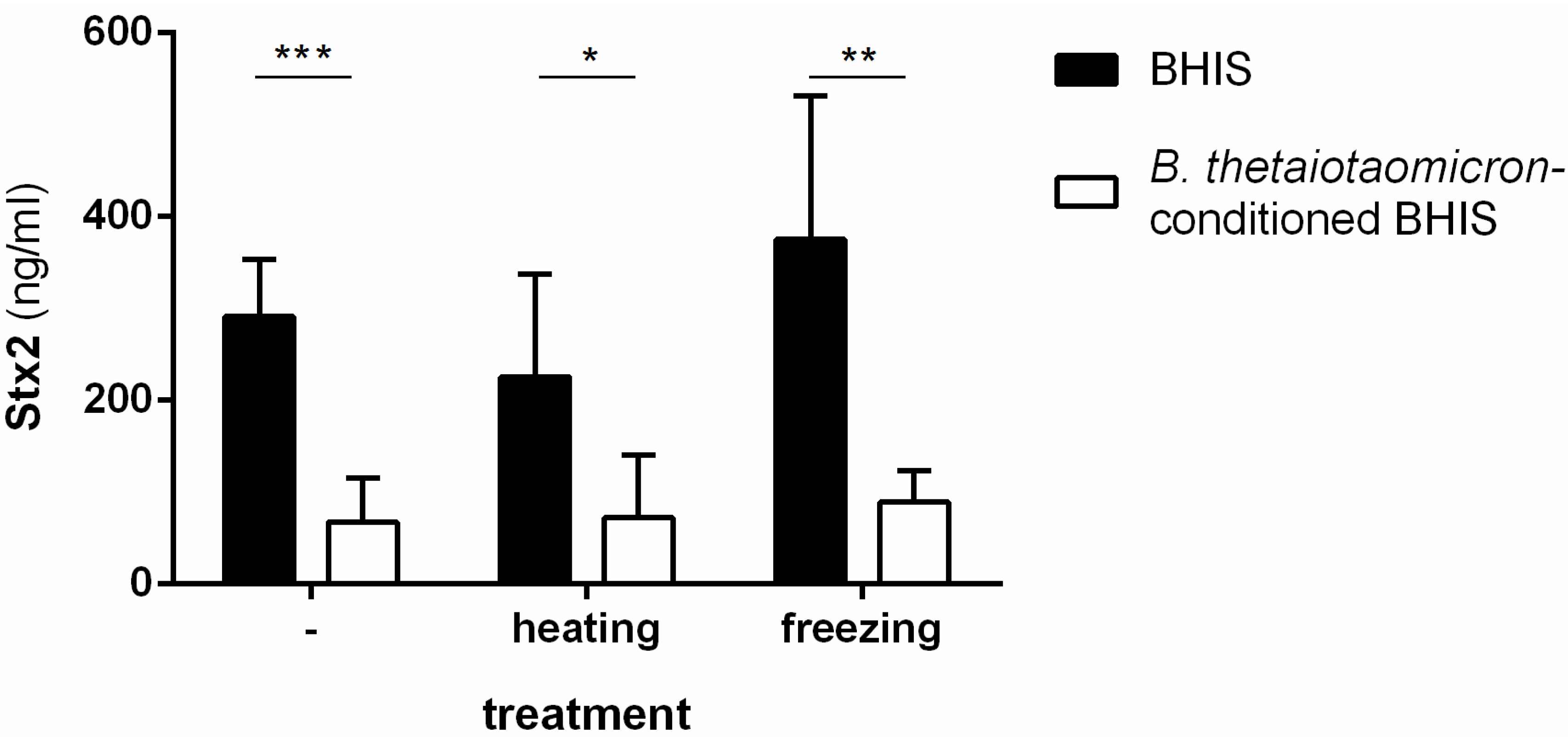
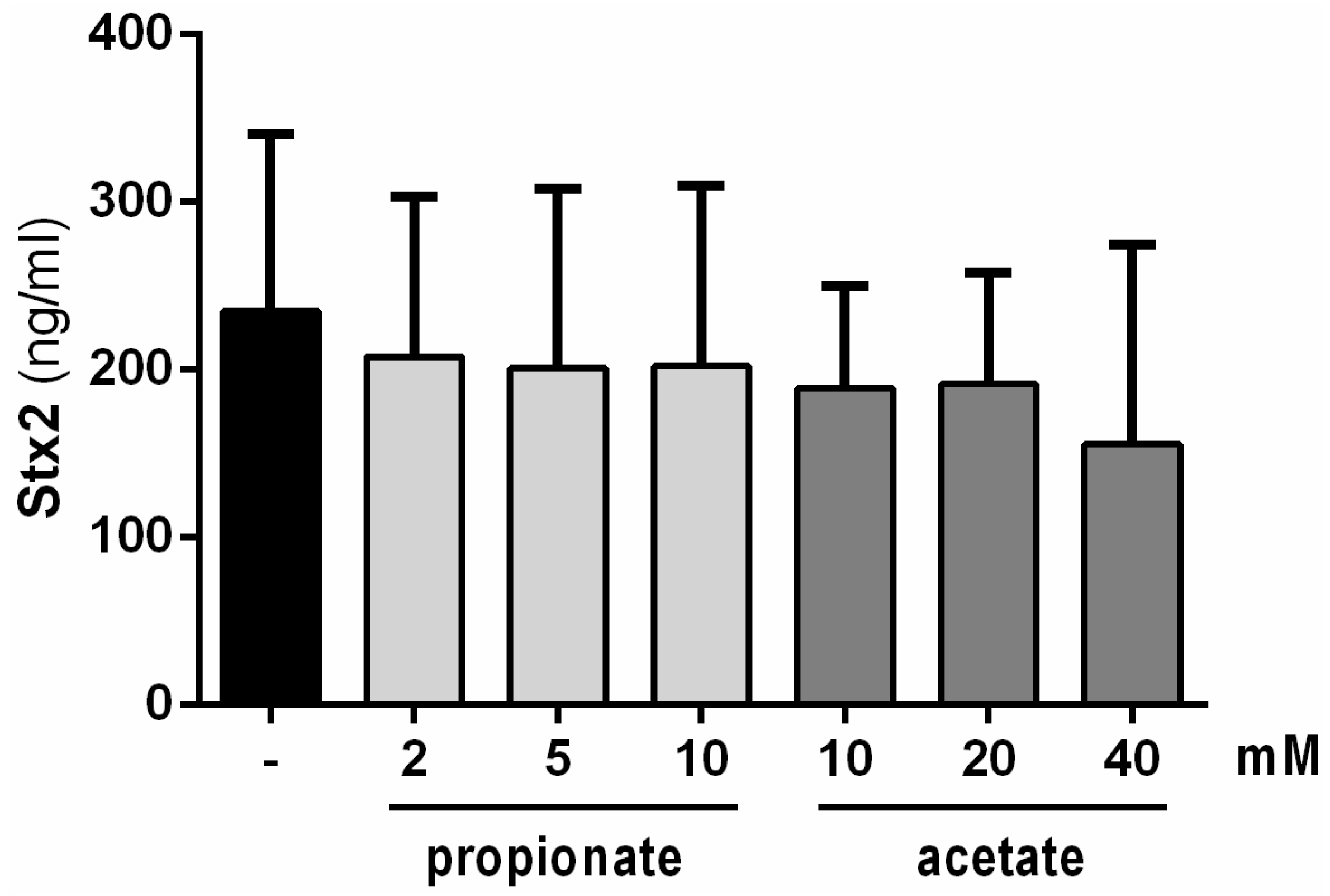
2.1. Stx2-Repressing Molecules Are Heat-Resistant but Do Not Correspond to the Short-Chain Fatty Acids Produced by B. thetaiotaomicron

To characterize biochemical properties of the Stx2 inhibitory molecule(s) produced by B. thetaiotaomicron, we subjected supernatants from culture of the commensal species in supplemented brain-heart infusion medium (BHIS) to either a 95 °C heat treatment or to storage at −80 °C before use as the growth medium for the EHEC O157:H7 strain EDL933. Production of Stx2 remained inhibited after growth of EHEC in all B. thetaiotaomicron-conditioned media (Figure 1), indicating that the inhibitory compounds are unaltered in these conditions.
Figure 1. Biochemical characterization of Stx2 inhibitory molecules produced by B. thetaiotaomicron. Stx2 concentrations were measured after 6 h of growth of EDL933 in BHIS or in B. thetaiotaomicron-conditioned BHIS treated 15 min at 95 °C (heating) or 24 h at −80 °C (freezing) or left untreated as indicated. Student’s t-test: * p < 0.05, ** p < 0.01, *** p < 0.001.
Toxins 08 00014 g001
Our previous study demonstrated that the Stx2 inhibitory molecules have a molecular size less than 3 kDa [16]. Short-chain fatty acids (SCFAs), the end-products of fermentation of dietary fibers by gut microbiota, are small heat-resistant molecules and one of them, butyrate, regulates LEE gene expression in EHEC [18]. We investigated the potential effect of SCFAs produced by B. thetaiotaomicron (specifically acetate and propionate) on Stx2 production level. The range of tested concentrations for both SCFAs were selected based on experimental measurements after in vitro growth of B. thetaiotaomicron or from the digestive tract of B. thetaiotaomicron mono-associated rodents [19,20]. Figure 2 shows no differences in Stx2 production levels in the absence or presence of propionate or acetate under any concentration used. This result indicates that propionate and acetate have no impact on toxin synthesis under these conditions.
Figure 2. Impact of propionate and acetate on Stx2 production levels. Stx2 concentrations were measured after 6 h of growth of EDL933 in BHIS supplemented or not with indicated concentrations of SCFAs. No significant differences were observed. ANOVA: p > 0.05.
Toxins 08 00014 g002

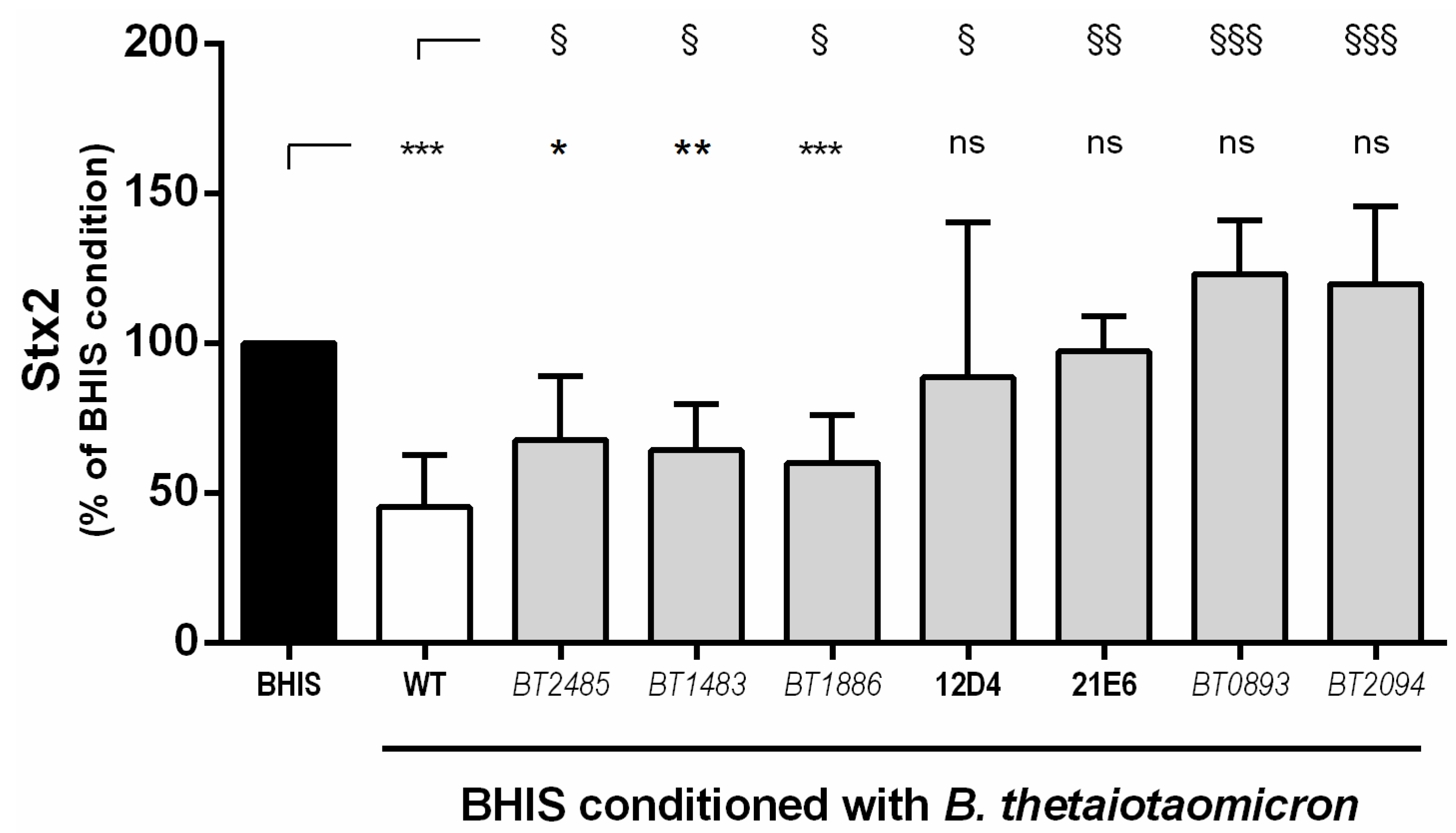
2.2. Screening of A B. thetaiotaomicron Mutant Library Identifies Seven Mutants that Lost Their Ability to Inhibit Stx2 Production by EHEC

To find genes involved in the production of Stx2 inhibitory molecules, a library of 4000 transposon mutants was generated using the B. thetaiotaomicron strain VPI-5482 (see Experimental Section for details). Mutants were cultivated individually to prepare conditioned media and Stx2 production levels were quantified after growth of EHEC strain EDL933. Media conditioned by eleven mutants led to higher level of Stx2 synthesis than in medium conditioned by wild-type B. thetaiotaomicron. To verify that these eleven clones were B. thetaiotaomicron isolates with integrated Tn4351 in their genome, we performed PCR controls using primers specific to a B. thetaiotaomicron gene or primers targeting ermF gene from transposon Tn4351. Of the eleven mutants, four were discarded after the PCR controls. We determined that the remaining seven B. thetaiotaomicron mutants lost their capacity to limit the production of Stx2 by EHEC (Figure 3) either partially or totally. Among them, three mutants demonstrated a repressive effect that was less strong than one with wild-type B. thetaiotaomicron. For these mutants, Tn4351 was inserted into gene BT_2485 encoding the major outer membrane porin OmpA, gene BT_1483 involved in the synthesis of sensor protein PhoR from the two-component system PhoBR or gene BT_1886, encoding a protein of unknown function with a domain similar to 2-nitropropane dioxygenase. Another four B. thetaiotaomicron mutants fully lost their capacity to inhibit the synthesis of Stx2 by EDL933. Indeed, amounts of toxin detected in BHIS conditioned with these mutants were not significantly different from those obtained following the growth of EHEC in unconditioned BHIS (Figure 3). Despite our efforts, transposon insertion sites were not identified for clones 12D4 and 21E6. For two other clones, genes BT_0893 or BT_2094, were inactivated by the integration of Tn4351. BT_0893 corresponds to gene trmD that directs the synthesis of a tRNA-methyltransferase and BT_2094 encodes a functional outer membrane transporter of vitamin B12 [21].
Figure 3. Seven B. thetaiotaomicron mutants have lost their ability to repress Stx2 synthesis. Stx2 concentrations were measured after 6 h of growth of EDL933 in BHIS or in BHIS conditioned with wild-type B. thetaiotaomicron or seven transposon-inserted mutants. ANOVA: ns p > 0.05, * p < 0.05, ** p < 0.01, *** p < 0.001 for BHIS versus conditioned media; § p < 0.05, §§ p < 0.01, §§§ p < 0.001 for wild-type-conditioned versus mutant-conditioned media.
Toxins 08 00014 g003

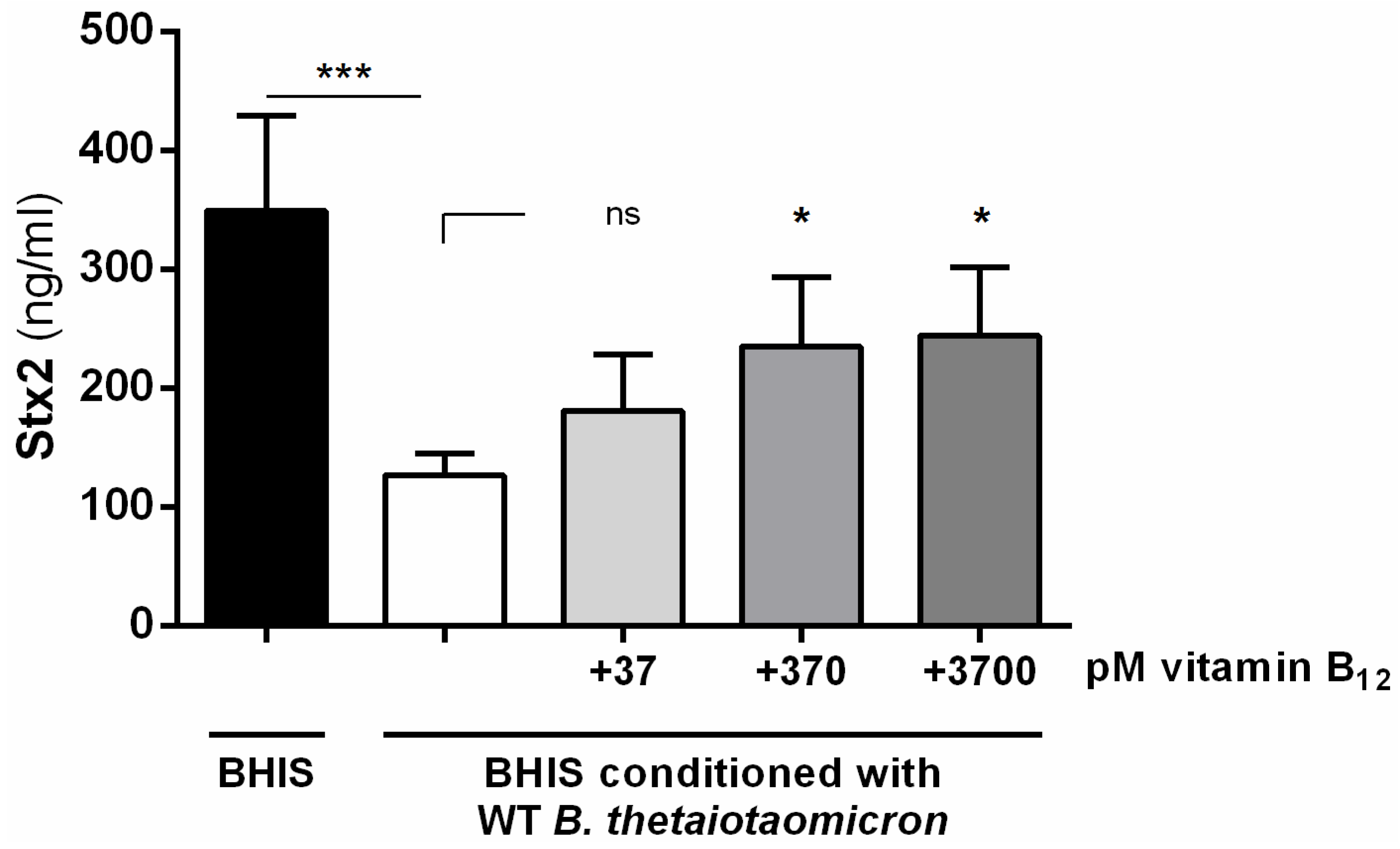
2.3. Vitamin B12 Activates the Production of Stx2 by EHEC

By inactivating gene BT_2094 in B. thetaiotaomicron, the commensal bacterium is unable to repress the production of Stx2 by EHEC. We focused our attention on this mutant because: (i) gene BT_2094 has been functionally described (renamed btuB3, a term used hereafter) as a gene encoding one of the three transporters of vitamin B12 produced by B. thetaiotaomicron [21]; (ii) vitamin B12 is a key vitamin for the host and is exclusively produced by prokaryotes [22]; and (iii) it has been well described that vitamin B12 affects gene expression in bacteria via riboswitches [23]. To explore the potential role of vitamin B12 in the B. thetaiotaomicron-dependent inhibition of Stx2, we first quantified vitamin B12 in BHIS before and after the growth of wild-type or btuB3 strains of B. thetaiotaomicron. As expected, the wild-type strain consumed vitamin B12 during its growth in BHIS (551 ± 73 pM vs. 92 ± 8 pM in BHIS before and after the growth, respectively; p < 0.0001 Student’s t-test). While the btuB3 mutant consumed a large amount of vitamin B12, concentration of the remaining vitamin B12 into the medium after growth was slightly but significantly higher with the btuB3 mutant than with the wild-type strain (105 ± 4 pM vs. 92 ± 8 pM in BHIS, respectively; p < 0.05 Student’s t-test). This indicates that inactivation of btuB3 partially affects the capacity of B. thetaiotaomicron to internalize vitamin B12. Next, we performed Stx2 quantification after the growth of EDL933 in BHIS conditioned with wild-type B. thetaiotaomicron and supplemented with different concentrations of cobalamin. Figure 4 shows that the addition of exogenous vitamin B12 led to a significant dose-dependent increase in Stx2 production. This indicates that vitamin B12 is an inducer of Stx2 synthesis and suggests that its consumption by B. thetaiotaomicron affects the level of toxins produced by EHEC. It is noteworthy that restoration remained partial even with the highest concentration of vitamin B12 since the amount of produced Stx2 was not as high as in unconditioned medium. Because vitamin B12 can modulate gene expression through binding to riboswitches, we searched the genome of EDL933 for a B12 riboswitch using the RFAM search tool. The only detected B12 riboswitch was the well characterized riboswitch located in the 5’-UTR of the btuB gene. This suggests that the influence of vitamin B12 on Stx2 production by EHEC may not be mediated by a B12 riboswitch-dependent regulation mechanism.
Figure 4. Vitamin B12 induces the production of Stx2. Stx2 concentrations were measured after 6 h of growth of EDL933 in BHIS or in wild-type B. thetaiotaomicron-conditioned BHIS supplemented or not with indicated concentrations of vitamin B12. ANOVA: ns p > 0.05, * p < 0.05, *** p < 0.001.
Toxins 08 00014 g004

3. Discussion

In this study, we investigated the repressive effect of B. thetaiotaomicron on the ability of EHEC to produce Stx2. Screening of a B. thetaiotaomicron mutant library revealed that a mutant affected in the production of the vitamin B12 transporter BtuB3 lost its ability to inhibit the synthesis of Stx2. Properties of vitamin B12 are compatible with previous data showing that Stx2-affecting molecules recovered from the supernatant of B. thetaiotaomicron culture were resistant to heat treatment and were less than 3 kDa in size (this study and [16]). In addition, supplementation of wild-type B. thetaiotaomicron-conditioned medium with cobalamin partially restores the production of Stx by EHEC. This indicates that vitamin B12 stimulates EHEC to produce Stx2 and that growth of B. thetaiotaomicron inhibits toxin synthesis probably through medium depletion of vitamin B12. Quantification of vitamin B12 in BHIS after the growth of B. thetaiotaomicron clearly demonstrated that the commensal bacterium internalizes the vitamin, which is required for the function of many key enzymes including the methionine synthase [24]. The btuB3 mutant was also able to uptake cobalamin but with slightly reduced efficiency compared to the wild-type strain. This difference is probably too small to explain by itself the inability of the btuB3 mutant to repress Stx2 synthesis by EHEC. Recently, Degnan and coworkers showed that B. thetaiotaomicron uses three functional vitamin B12 transporters (named BtuB1–3) that exhibit specific preferences to distinct B12 analogs [21]. BtuB3 has a greater affinity to B12 analogs containing 2-methyladenine, 5-methoxybenzimidazole, 5-methylbenzimidazole, or 5,6-dimethylbenzimidazole as the lower ligand linked to cobinamide [21]. Our vitamin B12 quantification method does not provide distinction between the different forms of cobalamin. We speculate that the btuB3 mutant is unable to uptake some particular B12 analog(s) that might specifically affect Stx2 synthesis by EDL933.
Few studies have directly investigated the role of vitamin B12 in the virulence of pathogens. Whereas inactivation of the vitamin B12 uptake or synthesis pathway does not affect the virulence of Salmonella enterica (S. enterica) serovar Typhimurium in mice [25], a defective mutant in vitamin B12 production of S. enterica serovar Gallinarum, a fowl pathogen, is avirulent in chickens [26]. Vitamin B12 also affects the virulence of Listeria monocytogenes in mice through the binding to a riboswitch element controlling the expression of a small non-coding RNA, Rli55 [27]. Rli55 sequesters and inhibits the action of EutV, a transcriptional activator of the eut operon required for the use of ethanolamine, an abundant substrate in the gut of mammalians. If the eut operon exists in EHEC, it differs from the one in Listeria, especially for the regulatory network controlling its expression. In addition, no B12 riboswitch element was found in the genome of EHEC EDL933 except the well described riboswitch regulating the expression of the btuB gene. Ethanolamine catabolic pathways are widely conserved in gut pathogenic bacteria [28] and have been involved in the virulence of several [27,29,30,31,32]. Interestingly, vitamin B12 is essential for ethanolamine degradation since it is a co-factor of the enzyme ethanolamine ammonia-lyase [33]. Moreover, the main activator of eut genes in Escherichia and Salmonella species, EutR, also regulates the expression of other genes related to virulence. Indeed, Luzader and coworkers demonstrated that EutR activates LEE gene expression in EHEC O157:H7 in an ethanolamine-dependent manner [34]. Although there is no direct evidence, vitamin B12 is probably a co-factor of EutR since vitamin B12 with ethanolamine is required by EutR for full activation of eut and LEE operons [34,35,36]. Production of Stx2 has been determined to be also affected by the presence of ethanolamine in the environment [29]. This effect occurs, at least partially, in a EutR-dependent way. Such might explain why the consumption of vitamin B12 by B. thetaiotaomicron leads to a decrease in Stx2 production levels by EHEC.
With vitamin B12 supplementation even in high concentration, full restoration of Stx2 synthesis is not achieved. These data suggest that Stx2 modulation by B. thetaiotaomicron is certainly multifactorial. We have determined that acetate and propionate, the SCFAs produced by B. thetaiotaomicron, have no impact on Stx2 production. However, acetate has been proposed to affect the translocation of Stx2 across intestinal mucosa [17], suggesting that B. thetaiotaomicron may provide protection from an EHEC infection at different levels. In contrast, Crane and coworkers have shown that EHEC infection triggers the production of xantine oxidase by intestinal epithelial cells [37]. Xantine oxidase activity increases the ability of Stx2 to translocate across intestinal monolayers and leads to the production of hydrogen peroxide that negatively impacts commensal bacteria, including B. thetaiotaomicron. Taken together, these data illustrate that EHEC and B. thetaiotaomicron are mutually antagonistic and that the outcome of interactions between the two species may contribute important consequences to disease progression.
The phoR mutant (gene BT_1483), is one of the B. thetaiotaomicron mutants that is unable to repress the production of Stx2. PhoR is the sensor protein of the PhoBR two-component system involved in the regulation of many genes in response to the concentration of extracellular inorganic phosphate [38]. Disruption of the PhoBR system disallows the bacterium to properly regulate phosphate internalization. Interestingly, a recent report has shown that production of Stx2 by EHEC is modulated by the concentration of extracellular inorganic phosphate [39]. This suggests that both an effective regulation of phosphate uptake and assimilation by B. thetaiotaomicron might also have important roles in protection against Stx2 production by EHEC.
In this study, we have investigated how B. thetaiotaomicron affects the production of Stx2 by EHEC. We have demonstrated that the two SCFAs, acetate and propionate, produced by this gut commensal bacterial species, have no effect on toxin synthesis. In contrast, a mutant of B. thetaiotaomicron with impaired production of a specific transporter of vitamin B12 is no longer able to inhibit the production of Stx2. This work suggests that concentration of vitamin B12 in the gut and by extension, activities of commensal bacterial species producing and/or consuming vitamin B12, may modulate the production of the main virulence factor of EHEC. Our research provides an additional element to the growing list of environmental cues EHEC can sense in the gut during the infectious process in order to successfully manage its virulence program.

4. Experimental Section

4.1. Bacterial Strains, Media and Growth Conditions

The human gut commensal bacteria B. thetaiotaomicron VPI-5482 was grown in BHIS medium [40] under anaerobic conditions at 37 °C. The E. coli strain WM3064 used for mating experiments was grown in Lysogeny broth (LB) supplemented with 100 µM of 2,6-diamino-pimelic acid. Wild-type EHEC O157:H7 strain EDL933 was grown in LB medium, in BHIS or in B. thetaiotaomicron-conditioned BHIS as described previously [16]. Briefly, B. thetaiotaomicron-conditioned BHIS was prepared after a 24-h growth of the commensal strain in BHIS. After culture centrifugation, the supernatant, filtered through 0.2 µm filters was supplemented with concentrated LB to reach a final concentration of 0.5 X. pH was adjusted to 7.0 and the medium was filtered with a 0.2-µm filter. When required, acetate or propionate (pH 7.0) were added to BHIS medium with indicated concentrations; antibiotics were used at the following concentrations: 10 µg·mL−1 erythromycin; 50 or 200 µg·mL−1 gentamycin; 25 µg·mL−1 chloramphenicol; and 10 µg·mL−1 tetracycline.

4.2. Construction and Screening of the B. thetaiotaomicron Mutant Library

The transposon Tn4351 was used to construct an insertion mutant library in B. thetaiotaomicron strain VPI-5482 as described previously [41]. The suicide vector pEP4351 carrying the transposon Tn4351 was transferred to B. thetaiotaomicron by mating experiments using WM3064 as the donor strain. After independent growth, donor and recipient strains were mixed at ratio 1:1 or 1:5, respectively, and spotted on a nitrocellulose filter placed on a BHIS + 2,6-diamino-pimelic acid agar plate. After an overnight incubation at 37 °C under aerobic condition, bacteria were recovered from filters and washed twice with BHIS. After dilution, bacteria were plated on BHIS + erythromycin and incubated under anaerobic conditions for 48 h at 37 °C in order to select B. thetaiotaomicron transconjugants.
To identify B. thetaiotaomicron mutants that no longer repress the production of Stx2 by EHEC, the mutant library was screened as follows: first, each mutant was grown in 1 ml of BHIS in Deepwell 96 microplates for 48 h. The plates were centrifuged and supernatants were transferred to new Deepwell microplates. After supplementation with LB (0.5 X final) and a determined volume of NaOH to yield a neutral pH, the B. thetaiotaomicron-conditioned media were inoculated with EDL933 strain and incubated for 6 h with orbital agitation. Controls were included in each plate; EDL933 was cultivated in BHIS and BHIS conditioned with wild-type B. thetaiotaomicron. The plates were centrifuged and supernatants were collected to perform Stx2 quantification. Selected mutants were checked by PCR using B. thetaiotaomicron specific primers BT-F (5’-AACAGGTGGAAGCTGCGGA-3’) and BT-R (5’-AGCCTCCAACCGCATCAA-3’) ([42]) or primers 341 (5’-TTGGAAATTTTCTGGGAGG-3’) and Tn4351-Rev (5’-TAACCATTGACTTGGAGAC-3’), targeting ermF gene from Tn4351. Identification of transposon insertion regions was performed using arbitrarily primed PCR as described previously [43].

4.3. Biochemical Methods

Amounts of Stx2 in culture supernatants were quantified using an enzyme-linked immunoassay as previously described with slight modifications [44]. Sandwich ELISAs were performed in 96-well plates using a monoclonal antibody against Stx2 (Stx2-BB12, Toxin Technology, Sarasota, FL, USA) diluted 1:1600 to coat the plates. Two-fold serial dilutions of samples were incubated in coated plates. When indicated, samples were beforehand treated 15 min at 95 °C or 24 h at −80 °C. Rabbit polyclonal antiserum against Stx2 diluted 1:1000 was used to detect the toxin. A standard curve was obtained with several dilutions of purified Stx2 (Toxin Technology, Sarasota, FL, USA). Detection was performed with a 1: 5 000 dilution of horseradish peroxidase-goat IgG anti-rabbit (ThermoFisher Pierce, Waltham, MA, USA) and o-phenylenediamine stable peroxidase substrate (OPD, Sigma-Aldrich, St. Louis, MO, USA).
The concentration of vitamin B12 in BHIS was analyzed using the Dimension VISTA immunoassay system and the analytical kit supplied by the manufacturer (Siemens Healthcare, Erlangen, Germany).

4.4. Statistical Analysis

All data represent the mean ± the standard deviation of at least three independent experiments. Student’s t-test or ANOVA with Fisher’s LSD test were used to determine significant differences between two or several test groups, respectively as shown in the figures.

Acknowledgments

We thank C. Barrachina for her technical assistance; R. Minet-Quinard for vitamin B12 quantification; and A.P. Gobert for his critical reading of the manuscript. This work was supported by grants from INRA, PHC Sakura and, in part, by the 61st Session de la Commission permanente de coopération franco-québécoise (Project 61.116) and by the Natural Sciences and Engineering Research Council of Canada (NSERC) (Discovery Grant SD-25120). GLB was supported by a scholarship from the Institut de recherche en santé publique de l’Université de Montréal (FRSQ 40148) and CRIPA (FQRNT RS-17946).

Author Contributions

G.J. and J.H. conceived and designed the experiments; C.C., G.L.B., J.G.E.R. and A.G. performed the experiments; C.C., G.L.B. and G.J. analyzed the data; and G.J. wrote the paper.

Conflicts of Interest

The authors declare no conflict of interest.

References

  1. Sekirov, I.; Russell, S.L.; Antunes, L.C.; Finlay, B.B. Gut microbiota in health and disease. Physiol. Rev. 2010, 90, 859–904. [Google Scholar] [CrossRef] [PubMed]
  2. Stecher, B.; Hardt, W.D. The role of microbiota in infectious disease. Trends Microbiol. 2008, 16, 107–114. [Google Scholar] [CrossRef] [PubMed]
  3. Bailey, M.T.; Dowd, S.E.; Parry, N.M.; Galley, J.D.; Schauer, D.B.; Lyte, M. Stressor exposure disrupts commensal microbial populations in the intestines and leads to increased colonization by Citrobacter rodentium. Infect. Immun. 2010, 78, 1509–1519. [Google Scholar] [CrossRef] [PubMed]
  4. Ferreira, R.B.; Gill, N.; Willing, B.P.; Antunes, L.C.; Russell, S.L.; Croxen, M.A.; Finlay, B.B. The intestinal microbiota plays a role in Salmonella-induced colitis independent of pathogen colonization. PLoS ONE 2011, 6, e20338. [Google Scholar] [CrossRef] [PubMed]
  5. Sekirov, I.; Tam, N.M.; Jogova, M.; Robertson, M.L.; Li, Y.; Lupp, C.; Finlay, B.B. Antibiotic-induced perturbations of the intestinal microbiota alter host susceptibility to enteric infection. Infect. Immun. 2008, 76, 4726–4736. [Google Scholar] [CrossRef] [PubMed]
  6. Stecher, B.; Hardt, W.D. Mechanisms controlling pathogen colonization of the gut. Curr. Opin. Microbiol. 2011, 14, 82–91. [Google Scholar] [CrossRef] [PubMed]
  7. Schmidt, M.A. LEEways: Tales of EPEC, ATEC and EHEC. Cell Microbiol. 2010, 12, 1544–1552. [Google Scholar] [CrossRef] [PubMed]
  8. Zoja, C.; Buelli, S.; Morigi, M. Shiga toxin-associated hemolytic uremic syndrome: Pathophysiology of endothelial dysfunction. Pediatr. Nephrol. 2010, 25, 2231–2240. [Google Scholar] [CrossRef] [PubMed]
  9. Bertin, Y.; Chaucheyras-Durand, F.; Robbe-Masselot, C.; Durand, A.; de la Foye, A.; Harel, J.; Cohen, P.S.; Conway, T.; Forano, E.; Martin, C. Carbohydrate utilization by enterohaemorrhagic Escherichia coli O157:H7 in bovine intestinal content. Environ. Microbiol. 2013, 15, 610–622. [Google Scholar]
  10. Bertin, Y.; Deval, C.; de la Foye, A.; Masson, L.; Gannon, V.; Harel, J.; Martin, C.; Desvaux, M.; Forano, E. The gluconeogenesis pathway is involved in maintenance of enterohaemorrhagic Escherichia coli O157:H7 in bovine intestinal content. PLoS ONE 2014, 9, e98367. [Google Scholar] [CrossRef] [PubMed] [Green Version]
  11. Bertin, Y.; Girardeau, J.P.; Chaucheyras-Durand, F.; Lyan, B.; Pujos-Guillot, E.; Harel, J.; Martin, C. Enterohaemorrhagic Escherichia coli gains a competitive advantage by using ethanolamine as a nitrogen source in the bovine intestinal content. Environ. Microbiol. 2011, 13, 365–377. [Google Scholar] [CrossRef] [PubMed]
  12. Fabich, A.J.; Jones, S.A.; Chowdhury, F.Z.; Cernosek, A.; Anderson, A.; Smalley, D.; McHargue, J.W.; Hightower, G.A.; Smith, J.T.; Autieri, S.M.; et al. Comparison of carbon nutrition for pathogenic and commensal Escherichia coli strains in the mouse intestine. Infect. Immun. 2008, 76, 1143–1152. [Google Scholar] [CrossRef] [PubMed]
  13. Le Bihan, G.; Jubelin, G.; Garneau, P.; Bernalier-Donadille, A.; Martin, C.; Beaudry, F.; Harel, J. Transcriptome analysis of Escherichia coli O157:H7 grown in vitro in the sterile-filtrated cecal content of human gut microbiota associated rats reveals an adaptive expression of metabolic and virulence genes. Microbes Infect. 2015, 17, 23–33. [Google Scholar] [CrossRef] [PubMed]
  14. Miranda, R.L.; Conway, T.; Leatham, M.P.; Chang, D.E.; Norris, W.E.; Allen, J.H.; Stevenson, S.J.; Laux, D.C.; Cohen, P.S. Glycolytic and gluconeogenic growth of Escherichia coli O157:H7 (EDL933) and E. coli K-12 (MG1655) in the mouse intestine. Infect. Immun. 2004, 72, 1666–1676. [Google Scholar] [CrossRef] [PubMed]
  15. Reading, N.C.; Rasko, D.A.; Torres, A.G.; Sperandio, V. The two-component system QseEF and the membrane protein QseG link adrenergic and stress sensing to bacterial pathogenesis. Proc. Natl. Acad. Sci. USA 2009, 106, 5889–5894. [Google Scholar] [CrossRef] [PubMed]
  16. De Sablet, T.; Chassard, C.; Bernalier-Donadille, A.; Vareille, M.; Gobert, A.P.; Martin, C. Human microbiota-secreted factors inhibit shiga toxin synthesis by enterohemorrhagic Escherichia coli O157:H7. Infect. Immun. 2009, 77, 783–790. [Google Scholar] [CrossRef] [PubMed]
  17. Fukuda, S.; Toh, H.; Hase, K.; Oshima, K.; Nakanishi, Y.; Yoshimura, K.; Tobe, T.; Clarke, J.M.; Topping, D.L.; Suzuki, T.; et al. Bifidobacteria can protect from enteropathogenic infection through production of acetate. Nature 2011, 469, 543–547. [Google Scholar] [CrossRef] [PubMed]
  18. Nakanishi, N.; Tashiro, K.; Kuhara, S.; Hayashi, T.; Sugimoto, N.; Tobe, T. Regulation of virulence by butyrate sensing in enterohaemorrhagic Escherichia coli. Microbiology 2009, 155, 521–530. [Google Scholar] [CrossRef] [PubMed]
  19. Rios-Covian, D.; Arboleya, S.; Hernandez-Barranco, A.M.; Alvarez-Buylla, J.R.; Ruas-Madiedo, P.; Gueimonde, M.; de los Reyes-Gavilan, C.G. Interactions between Bifidobacterium and Bacteroides species in cofermentations are affected by carbon sources, including exopolysaccharides produced by bifidobacteria. Appl. Environ. Microbiol. 2013, 79, 7518–7524. [Google Scholar] [CrossRef] [PubMed]
  20. Wrzosek, L.; Miquel, S.; Noordine, M.L.; Bouet, S.; Joncquel Chevalier-Curt, M.; Robert, V.; Philippe, C.; Bridonneau, C.; Cherbuy, C.; Robbe-Masselot, C.; et al. Bacteroides thetaiotaomicron and Faecalibacterium prausnitzii influence the production of mucus glycans and the development of goblet cells in the colonic epithelium of a gnotobiotic model rodent. BMC Biol. 2013. [Google Scholar] [CrossRef] [PubMed]
  21. Degnan, P.H.; Barry, N.A.; Mok, K.C.; Taga, M.E.; Goodman, A.L. Human gut microbes use multiple transporters to distinguish vitamin B12 analogs and compete in the gut. Cell Host Microbe 2014, 15, 47–57. [Google Scholar] [CrossRef] [PubMed]
  22. Martens, J.H.; Barg, H.; Warren, M.J.; Jahn, D. Microbial production of vitamin B12. Appl. Microbiol. Biotechnol. 2002, 58, 275–285. [Google Scholar] [CrossRef] [PubMed]
  23. Nahvi, A.; Sudarsan, N.; Ebert, M.S.; Zou, X.; Brown, K.L.; Breaker, R.R. Genetic control by a metabolite binding mRNA. Chem. Biol. 2002, 9, 1043–1049. [Google Scholar] [CrossRef]
  24. Varel, V.H.; Bryant, M.P. Nutritional features of Bacteroides fragilis subsp. fragilis. Appl. Microbiol. 1974, 28, 251–257. [Google Scholar] [PubMed]
  25. Sampson, B.A.; Gotschlich, E.C. Elimination of the vitamin B12 uptake or synthesis pathway does not diminish the virulence of Escherichia coli K1 or Salmonella typhimurium in three model systems. Infect. Immun. 1992, 60, 3518–3522. [Google Scholar] [PubMed]
  26. De Paiva, J.B.; Penha Filho, R.A.; Arguello, Y.M.; Berchieri Junior, A.; Lemos, M.V.; Barrow, P.A. A defective mutant of Salmonella enterica Serovar Gallinarum in cobalamin biosynthesis is avirulent in chickens. Braz. J. Microbiol. 2009, 40, 495–504. [Google Scholar] [CrossRef] [PubMed]
  27. Mellin, J.R.; Koutero, M.; Dar, D.; Nahori, M.A.; Sorek, R.; Cossart, P. Riboswitches. Sequestration of a two-component response regulator by a riboswitch-regulated noncoding RNA. Science 2014, 345, 940–943. [Google Scholar] [CrossRef] [PubMed]
  28. Korbel, J.O.; Doerks, T.; Jensen, L.J.; Perez-Iratxeta, C.; Kaczanowski, S.; Hooper, S.D.; Andrade, M.A.; Bork, P. Systematic association of genes to phenotypes by genome and literature mining. PLoS Biol. 2005, 3, e134. [Google Scholar] [CrossRef] [PubMed] [Green Version]
  29. Kendall, M.M.; Gruber, C.C.; Parker, C.T.; Sperandio, V. Ethanolamine controls expression of genes encoding components involved in interkingdom signaling and virulence in enterohemorrhagic Escherichia coli O157:H7. MBio 2012, 3, e00050-12. [Google Scholar] [CrossRef] [PubMed]
  30. Maadani, A.; Fox, K.A.; Mylonakis, E.; Garsin, D.A. Enterococcus faecalis mutations affecting virulence in the Caenorhabditis elegans model host. Infect. Immun. 2007, 75, 2634–2637. [Google Scholar] [CrossRef] [PubMed]
  31. Stojiljkovic, I.; Baumler, A.J.; Heffron, F. Ethanolamine utilization in Salmonella typhimurium: Nucleotide sequence, protein expression, and mutational analysis of the cchA cchB eutE eutJ eutG eutH gene cluster. J. Bacteriol. 1995, 177, 1357–1366. [Google Scholar] [PubMed]
  32. Thiennimitr, P.; Winter, S.E.; Winter, M.G.; Xavier, M.N.; Tolstikov, V.; Huseby, D.L.; Sterzenbach, T.; Tsolis, R.M.; Roth, J.R.; Baumler, A.J. Intestinal inflammation allows Salmonella to use ethanolamine to compete with the microbiota. Proc. Natl. Acad. Sci. USA 2011, 108, 17480–17485. [Google Scholar] [CrossRef] [PubMed]
  33. Scarlett, F.A.; Turner, J.M. Microbial metabolism of amino alcohols. Ethanolamine catabolism mediated by coenzyme B12-dependent ethanolamine ammonia-lyase in Escherichia coli and Klebsiella aerogenes. J. Gen. Microbiol. 1976, 95, 173–176. [Google Scholar] [CrossRef] [PubMed]
  34. Luzader, D.H.; Clark, D.E.; Gonyar, L.A.; Kendall, M.M. EutR is a direct regulator of genes that contribute to metabolism and virulence in enterohemorrhagic Escherichia coli O157:H7. J. Bacteriol. 2013, 195, 4947–4953. [Google Scholar] [CrossRef] [PubMed]
  35. Roof, D.M.; Roth, J.R. Autogenous regulation of ethanolamine utilization by a transcriptional activator of the eut operon in Salmonella typhimurium. J. Bacteriol. 1992, 174, 6634–6643. [Google Scholar] [PubMed]
  36. Sheppard, D.E.; Roth, J.R. A rationale for autoinduction of a transcriptional activator: Ethanolamine ammonia-lyase (EutBC) and the operon activator (EutR) compete for adenosyl-cobalamin in Salmonella typhimurium. J. Bacteriol. 1994, 176, 1287–1296. [Google Scholar] [PubMed]
  37. Crane, J.K.; Naeher, T.M.; Broome, J.E.; Boedeker, E.C. Role of host xanthine oxidase in infection due to enteropathogenic and Shiga-toxigenic Escherichia coli. Infect. Immun. 2013, 81, 1129–1139. [Google Scholar] [CrossRef] [PubMed]
  38. Santos-Beneit, F. The Pho regulon: A huge regulatory network in bacteria. Front. Microbiol. 2015. [Google Scholar] [CrossRef] [PubMed]
  39. Chekabab, S.M.; Jubelin, G.; Dozois, C.M.; Harel, J. PhoB activates Escherichia coli O157:H7 virulence factors in response to inorganic phosphate limitation. PLoS ONE 2014, 9, e94285. [Google Scholar] [CrossRef] [PubMed]
  40. Bacic, M.K.; Smith, C.J. Laboratory maintenance and cultivation of Bacteroides species. Curr. Protoc. Microbiol. 2008. [Google Scholar] [CrossRef]
  41. Shoemaker, N.B.; Getty, C.; Gardner, J.F.; Salyers, A.A. Tn4351 transposes in Bacteroides spp. and mediates the integration of plasmid R751 into the Bacteroides chromosome. J. Bacteriol. 1986, 165, 929–936. [Google Scholar] [PubMed]
  42. Teng, L.J.; Hsueh, P.R.; Huang, Y.H.; Tsai, J.C. Identification of Bacteroides thetaiotaomicron on the basis of an unexpected specific amplicon of universal 16S ribosomal DNA PCR. J. Clin. Microbiol. 2004, 42, 1727–1730. [Google Scholar] [CrossRef] [PubMed]
  43. Peterson, D.A.; McNulty, N.P.; Guruge, J.L.; Gordon, J.I. IgA response to symbiotic bacteria as a mediator of gut homeostasis. Cell Host Microbe 2007, 2, 328–339. [Google Scholar] [CrossRef] [PubMed]
  44. De Sablet, T.; Bertin, Y.; Vareille, M.; Girardeau, J.P.; Garrivier, A.; Gobert, A.P.; Martin, C. Differential expression of Stx2 variants in Shiga toxin-producing Escherichia coli belonging to seropathotypes A and C. Microbiology 2008, 154, 176–186. [Google Scholar] [CrossRef] [PubMed]

