Article

Potential of Tyrosine Kinase Receptor TIE-1 as Novel Therapeutic Target in High-PI3K-Expressing Ovarian Cancer

1
Department of Obstetrics and Gynecology, Tohoku University Graduate School of Medicine, Tohoku University, Sendai 980-8577, Japan
2
Cancer Science Institute of Singapore, National University of Singapore, Singapore 119077, Singapore
3
Laboratory of Immunopharmacology, Faculty of Pharmaceutical Sciences, Setsunan University, Hirakata, Osaka 572-8508, Japan
4
Department of Obstetrics and Gynecology, Japanese Red Cross Ishinomaki Hospital, Ishinomaki 986-8522, Japan
*
Author to whom correspondence should be addressed.
Submission received: 12 May 2020 / Revised: 17 June 2020 / Accepted: 23 June 2020 / Published: 26 June 2020
(This article belongs to the Section Molecular Cancer Biology)

Abstract

:
Tyrosine kinase receptor TIE-1 plays a critical role in angiogenesis and blood-vessel stability. In recent years, increased TIE-1 expression has been observed in many types of cancers; however, the biological significance and underlying mechanisms remain unknown. Thus, in the present study, we investigated the tumor biological functions of TIE-1 in ovarian cancer. The treatment of SKOV3 ovarian-cancer cells with siRNA against TIE-1 decreased the expression of key molecules in the PI3K/Akt signaling pathway, such as p110α and phospho-Akt, suggesting that TIE-1 is related to the PI3K/Akt pathway. Furthermore, the knockdown of TIE-1 significantly decreased cell proliferation in high-PI3K-expressing cell lines (SKOV3, CAOV3) but not low-PI3K-expressing cell lines (TOV112D, A2780). These results suggested that inhibition of TIE-1 decreases cell growth in high-PI3K-expressing cells. Moreover, in low-PI3K-expressing TOV112D ovarian-cancer cells, TIE-1 overexpression induced PI3K upregulation and promoted a PI3K-mediated cell proliferative phenotype. Mechanistically, TIE-1 participates in cell growth and proliferation by regulating the PI3K/Akt signaling pathway. Taken together, our findings strongly implicate TIE-1 as a novel therapeutic target in high-PI3K-expressing ovarian-cancer cells.

1. Introduction

Ovarian cancer is a highly lethal global gynecologic malignancy [1,2]. Most patients are diagnosed at an advanced stage, and the five-year survival rate is in the range of 30–40% [3]. Although ovarian-cancer cells are sensitive to platinum-based chemotherapy, recurrence and the development of drug resistance make ovarian cancer difficult to treat [4]. Thus, the establishment of novel therapeutic strategies for refractory ovarian cancer is an urgent need.
Tyrosine kinase with immunoglobulin-like and EGF-like domains (TIE)-1 is a cell-membrane protein expressed in endothelial cells [5,6]. Although TIE-1 is considered an orphan receptor thus far [7,8], recently LECT2, a functional ligand of TIE-1, has been reported [9]. Xu and colleagues showed that LECT2/TIE-1 signaling pathway promotes liver fibrogenesis; however, the significance of LECT2/TIE-1 in cancer is still not clear. The known functions of TIE-1 are in angiogenesis and blood-vessel stability [10,11]. Recently, it was reported that TIE-1 upregulation predicts shorter survival in patients with early chronic-phase chronic myeloid leukemia [12,13]. Moreover, TIE-1 overexpression was observed in many types of cancer, including gastric- [14,15], colon- [16], and breast-cancer cells [17], but the significance of TIE-1 overexpression in cancer remains unknown. We previously reported that TIE-1 promotes DNA damage repair, thereby rendering ovarian-cancer cells resistant to cisplatin [18]. Therefore, TIE-1 might be a potential target for anticancer treatment.
Phosphoinositide 3-kinases (PI3Ks) are members of the lipid-kinase family originally discovered in the 1980s [19,20]. On the basis of their structure and substrate specificity, PI3Ks are categorized into three distinct classes: I, II, and III [21,22]. Class I is divided into Class IA and IB on the basis of differences in activating receptors [23,24]. In Class IA, PI3Ks are heterodimers consisting of a p85 regulatory subunit and a p110 catalytic subunit that has three isomers (α, β, and γ) encoded by three distinct genes, PIK3CA, PIK3CB, and PIK3CD, respectively. Of these, PIK3CA is the most frequently mutated in human cancer [25,26].
The PI3K/Akt pathway is one of the most essential intracellular pathways that mediates cellular metabolism, survival, differentiation, and cell growth [27,28]. Recent evidence suggests that different kinds of receptor tyrosine kinases (RTKs) are associated with the activation of the PI3K/Akt signaling pathway [29,30] that promotes cell proliferation, differentiation, migration, and survival. However, the involvement of TIE-1, one of the RTKs, in PI3K/Akt pathway remains unknown and requires further research.
Therefore, in the present study, we explore the biological significance of TIE-1 in the PI3K/Akt signaling pathway and demonstrate a novel molecular mechanism of action for TIE-1 in ovarian cancer, identifying TIE-1 as a target for the treatment of high-PI3K-expressing ovarian cancer.

2. Results

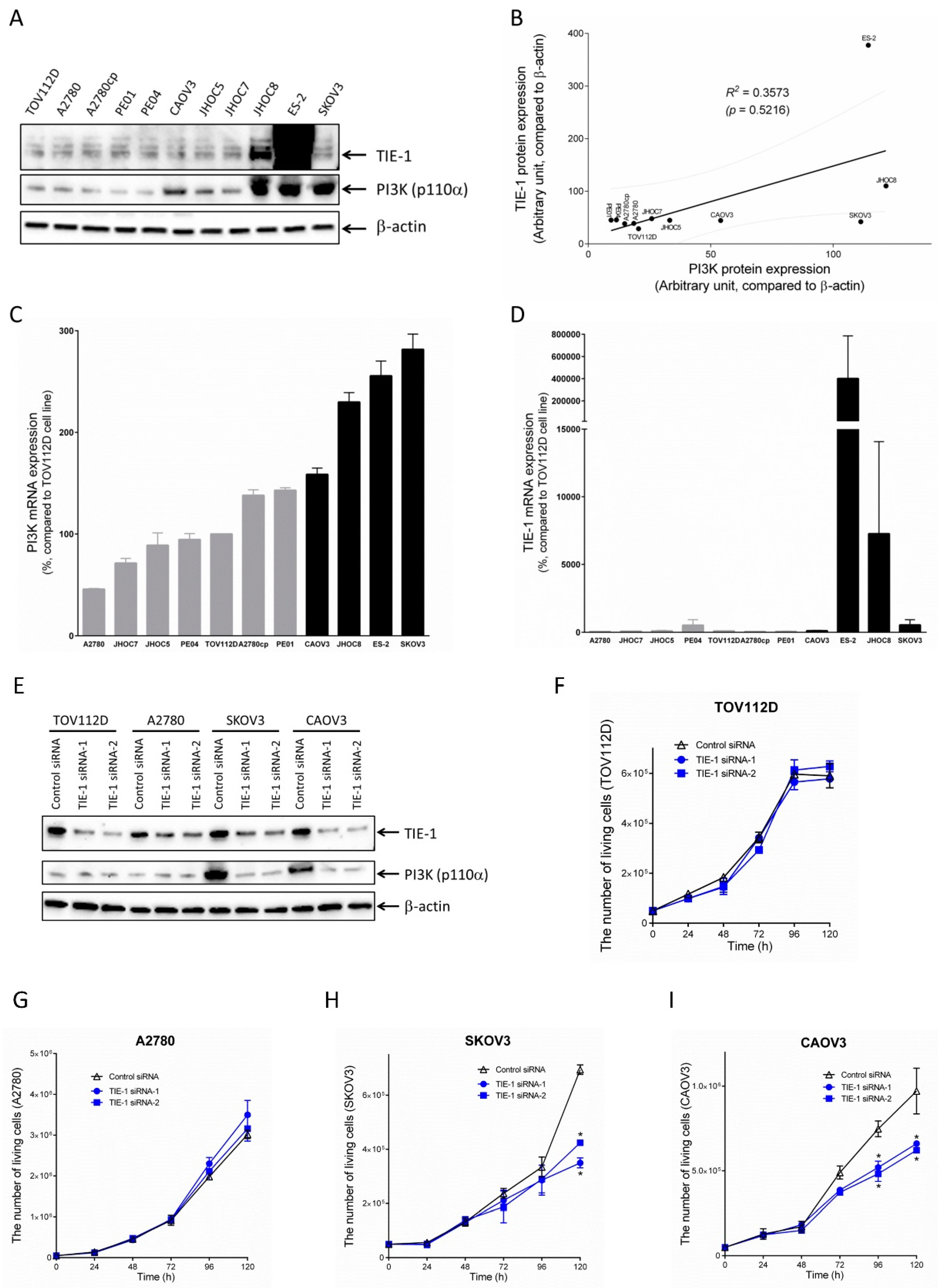
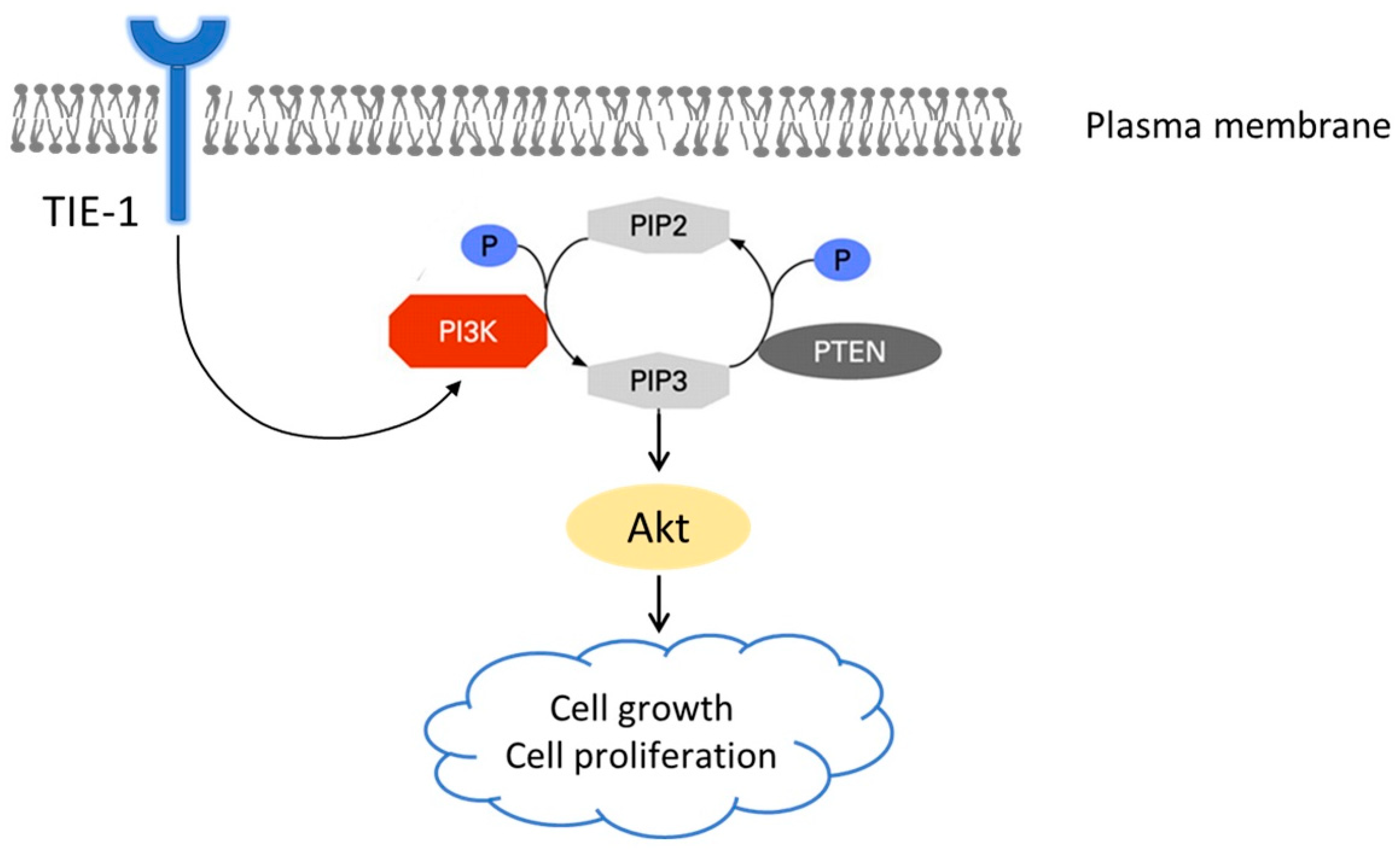
2.1. TIE-1 Might Signal through PI3K/Akt Pathway

To determine the biological functions of tyrosine kinase receptor TIE-1, we introduced TIE-1 knockdown using siRNA and measured the expression of key proteins in the PI3K/Akt signaling pathway. Immunoblotting analysis showed that TIE-1 knockdown significantly suppressed the protein expression of PI3K p110α and phospho-Akt, with no change in total Akt (Figure S1), in SKOV3 ovarian cancer cells (Figure 1A–C). Immunoblotting analysis showed that overexpression of TIE-1 with a V5-tagged TIE-1 vector significantly enhanced the protein expression of PI3K p110α and phospho-Akt, with no change in total Akt (Figure S1), in SKOV3 cells (Figure 1D–F). These results suggested that TIE-1 might signal through the PI3K/Akt signaling pathway.

2.2. TIE-1 Inhibition Decreases Cell Growth in High-PI3K-Expressing Cell Line.

TIE-1 and PI3K expression levels in 11 ovarian-cancer cell lines were confirmed. TIE-1 and PI3K p110α expression varied in the tested ovarian-cancer cell lines (Figure 2A,C,D). There was positive-correlation tendency between the protein-expression levels of TIE-1 and PI3K p110α (Figure 2B). However, there was no significant difference (p > 0.05). We next investigated the effect of TIE-1 knockdown on cell growth using multiple ovarian-cancer cell lines. On the basis of the endogenous expression of PI3K in cells and/or whether TIE-1 knockdown induced cell-growth inhibition, we divided 11 ovarian-cancer cell lines into two groups: low-PI3K-expressing and high-PI3K-expressing cell lines (Figure 2C, Figure S2). Low-PI3K-expressing cell lines TOV112D, or A2780 and high-PI3K-expressing cell lines SKOV3 or CAOV3, were used. Treatment with TIE-1 siRNA for 120 h significantly reduced cell number by approximately 45% in high-PI3K-expressing SKOV3 cells (Figure 2H), and by approximately 33% in CAOV3 cells (Figure 2I) relative to control siRNA group. Interestingly, the knockdown of TIE-1 significantly decreased cell proliferation in high-PI3K-expressing cell lines SKOV3 and CAOV3 (Figure 2E,H,I), but not in low-PI3K-expressing cell lines TOV112D and A2780 (Figure 2E–G). These results suggested that inhibition of TIE-1 selectively suppresses PI3K/Akt-mediated cell growth in high-PI3K-expressing cells.

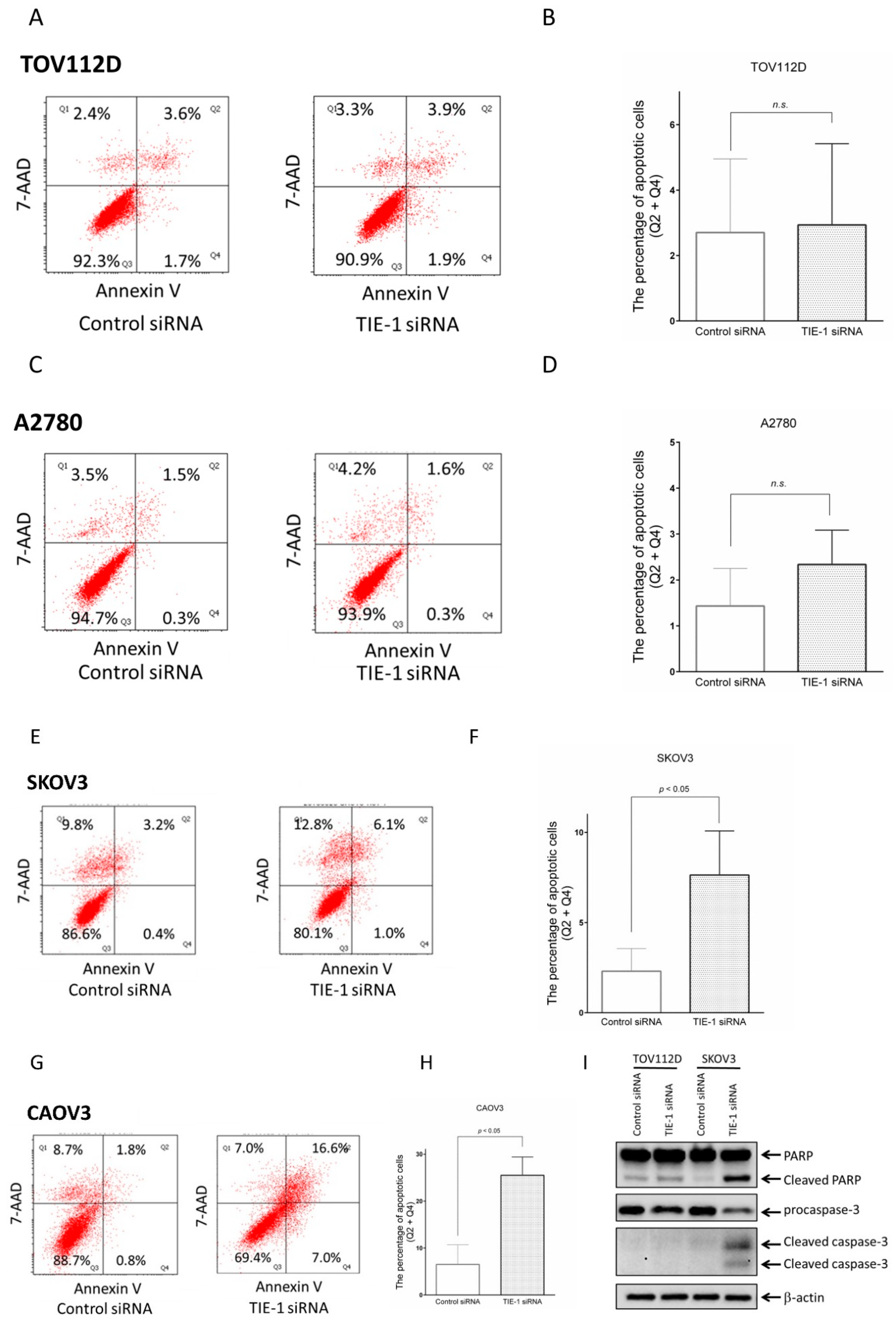
2.3. TIE-1 Inhibition Induces Apoptosis in High-PI3K-Expressing Cell Lines

Given that PI3K inhibitor GDC-0941 is a potent inducer of apoptosis (Figure S3), we determined whether TIE-1 inhibition-induced PI3K downregulation triggers apoptosis. To elucidate the underlying molecular mechanism of TIE-1 inhibition-induced cell death, we performed an annexin V/7-annexin-apoptosis-detection (AAD) assay. TIE-1 inhibition had no effect on the percentage of apoptotic cells in low-PI3K-expressing TOV112D (Figure 3A,B) or A2780 (Figure 3C,D) cells. In contrast, TIE-1 inhibition increased the percentage of apoptotic high-PI3K-expressing SKOV3 (Figure 3E,F) and CAOV3 (Figure 3G,H) cells.
Furthermore, cleaved poly ADP-ribose polymerase (PARP) and cleaved caspase-3 expression levels were significantly increased in TIE-1-deficient high-PI3K-expressing SKOV3 cells (Figure 3I). In addition, procaspase-3 expression was decreased in TIE-1 siRNA-treated SKOV3 cells (Figure 3I). Similar results were observed in CAOV3 ovarian-cancer cells (Figure S4). These findings suggest that inhibition of TIE-1 induces apoptosis in high- but not low-PI3K-expressing cells. Therefore, TIE-1 may function as an antiapoptotic regulator in high-PI3K-expressing ovarian-cancer cells.

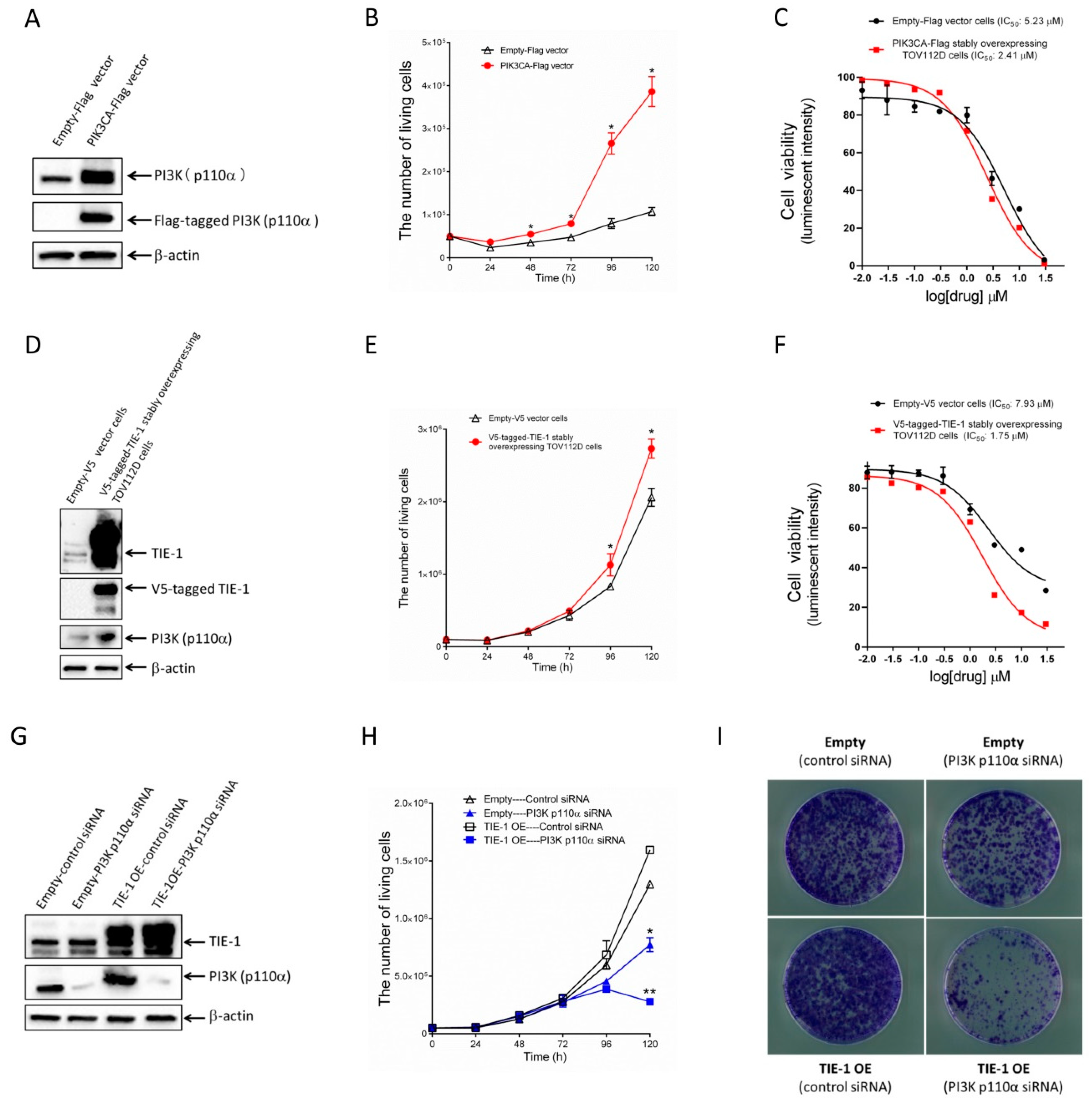
2.4. Increased TIE-1 Expression Induces TIE-1/PI3K-Mediated Cell Growth in Low-PI3K-Expressing Ovarian-Cancer Cells

To determine the mechanism by which TIE-1 selectively exhibits an antitumor effect in high-PI3K-expressing cells, we investigated whether high-PI3K-expressing cells showed PI3K-mediated cell growth. We transfected low-PI3K-expressing TOV112D cells with a PIK3CA–Flag vector, and measured cell growth.
As shown in Figure 4A, transfection with PIK3CA–Flag vectors was confirmed to increase PI3K protein expression. Importantly, PIK3CA–Flag-overexpressing significantly promoted cell growth compared to empty vectors (Figure 4B). We treated PIK3CA–Flag stably overexpressing TOV112D cells with a PI3K inhibitor (GDC-0941), and determined its IC50 value. IC50 in control cells or PI3K p110α-overexpressed cell number was 5.23 or 2.41 µM (Figure 4C), respectively. These findings suggested that PI3K upregulation promotes PI3K-mediated cell growth in low-PI3K-expressing cells.
Given our data demonstrating that PI3K upregulation induces PI3K-mediated cell growth, V5-tagged TIE-1 vector-induced PI3K upregulation may also promote PI3K-mediated cell growth. To confirm this hypothesis, we established a V5-tagged TIE-1 stably overexpressing TOV112D cell line and measured TIE-1 and PI3K expression, and cell growth. In V5-tagged TIE-1 stably overexpressing TOV112D cells, high TIE-1 levels promoted PI3K expression and cell proliferation compared with empty V5 vector cells (Figure 4D,E). The IC50 of the PI3K inhibitor (GDC-0941) in empty V5 vector cells was 7.93 µM (Figure 4F). In contrast, the IC50 in TIE-1-overexpressed cells was 1.75 µM (Figure 4F). These results suggested that TIE-1 overexpressed cells were more sensitive to the PI3K inhibitor compared with empty-vector cells.
Then, we treated empty V5 vector cells or V5-tagged TIE-1 stably overexpressing TOV112D cells with PI3K p110α siRNA and measured cell viability. The knockdown of PI3K in both V5-tagged TIE-1 stably overexpressing TOV112D cells and empty V5 vector cells significantly inhibited cell growth (Figure 4G,H). Moreover, in V5-tagged TIE-1 stably overexpressing TOV112D cells, inhibition of PI3K significantly decreased cell proliferation (Figure 4H). Consistent with Figure 4H, clonogenic-assay results suggested that PI3K inhibition significantly induced cell death in TIE-1-overexpressed cells compared with empty-vector cells (Figure 4I). These findings suggested that PI3K inhibition abolished high-TIE-1-expression-induced cell growth. Furthermore, TIE-1 overexpression increased PI3K expression and promoted a PI3K-mediated cell-proliferative phenotype (Figure 5).

3. Discussion

To the best of our knowledge, we demonstrated for the first time that tyrosine kinase receptor TIE-1 participates in cell growth and proliferation by targeting the PI3K/Akt signaling pathway in high-PI3K-expressing ovarian cancer cells. As shown in Table 1, various histological subtypes of ovarian-cancer cells were used in this study (Cancer Cell Line Encyclopedia: https://portals.broadinstitute.org/ccle). The effect of TIE-1 inhibition depended on the high level of PI3K expression in ovarian-cancer cells, not histological types. These findings provide evidence that TIE-1 may be a novel therapeutic target in high-PI3K-expressing ovarian cancer.
Endothelial cell-surface protein TIE-1 was considered as a nonfunctional orphan receptor except for the maintenance of vascular integrity [31,32]. Recently, Kontos CD et al. showed that TIE-1 activates the PI3K/Akt signaling pathway to inhibit UV-irradiation-induced apoptosis [33]. Our results suggested that TIE-1 regulates the PI3K/Akt pathway by increasing/decreasing PI3K expression in ovarian cancer. However, there is no significant difference between the protein-expression levels of TIE-1 and PI3K p110α. Considering that PIK3CA mutations were reported not only in SKOV3 but also in A2780 ovarian-cancer cells [34], it was inferred that TIE-1 and PI3K p110α expression levels might not always correlate because PIK3CA is independently mutated/amplified in some ovarian-cancer cells. Although the PI3K/Akt signaling pathway is proposed to be a downstream target of tyrosine kinase receptor TIE-1, whether PI3K is directly targeted by TIE-1 remains to be investigated. Our ongoing studies aim to identify the transcription factors responsible for the TIE-1-mediated regulation of PI3K.
The PI3K/Akt/mTOR pathway is altered in approximately 70% of ovarian-cancer cases, leading to enhanced cellular growth, proliferation, and survival, through an intricate series of hyperactive signaling cascades [35,36]. This pathway activation is associated with higher invasive and migratory capacities in human ovarian cancer; thus, the PI3K/Akt/mTOR pathway is a potential predictor of invasiveness in ovarian-tumor cells [37]. Therefore, the inhibition of this pathway represents a potential therapeutic strategy for cancer treatment [38,39]. However, due to the nonspecific growth inhibition of normal cells and other adverse effects, the use of oral selective PI3K inhibitors remains challenging [40,41]. Our findings demonstrated that TIE-1 inhibition selectively exhibited an antitumor effect by decreasing PI3K expression in high-PI3K-expressing ovarian-cancer cells. Furthermore, TIE-1 inhibition-induced PI3K downregulation did not affect low-PI3K-expressing ovarian-cancer or normal cells. These data suggested that TIE-1 inhibition-based therapy may provide an alternative approach to PI3K/Akt pathway inhibitors for the treatment of cancers with high PI3K expression.
In summary, TIE-1 functions as a potential carcinogenic molecule by activating the PI3K/Akt signaling pathway in high-PI3K-expressing ovarian cancer. Therefore, the future development of specific TIE-1 inhibitors could be an effective strategy to improve the prognosis of ovarian-cancer patients. Furthermore, PI3K could serve as a possible biomarker to predict the therapeutic efficiency of TIE-1-inhibition-based therapy in the future.

4. Materials and Methods

4.1. Antibodies and Reagents

Peroxidase AffiniPure Donkey Anti-Rabbit IgG (#711-035-152) and Peroxidase AffiniPure Donkey Anti-Mouse IgG (#715-035-150) were from Jackson ImmunoResearch (West Grove, PA, USA). Antibodies specific to TIE-1 (ab111547) and PTEN (ab32199) were obtained from Abcam (Cambridge, MA, USA). Other antibodies for phospho-Akt (S473, #9271) and PI3K p110α (#4249) were from Cell Signaling Technology (Danvers, MA, USA). Antibodies specific to V5 (#R960-25) were obtained from Thermo Fisher Scientific (Waltham, MA, USA). The β-actin antibody (A5441) was obtained from Sigma (St Louis, MO, USA). RNAiMax; Lipofectamine 2000; and siRNAs for control (4390846), TIE-1 (s14140 and s14141), and PI3K (s10521) were from Life Technologies (Carlsbad, CA, USA). The CellTiter-Glo luminescent cell-viability-assay kit was obtained from Promega (Fitchburg, WI, USA). The annexin V/7-AAD kit was from BD Biosciences (San Jose, CA, USA).

4.2. Cell Culture

Ovarian-cancer cells were cultured in Dulbecco’s modified Eagle’s medium, supplemented with 10% fetal bovine serum at 37 °C in a humidified incubator containing 5% CO2. Human ovarian cancer cell lines (TOV112D, CAOV3, SKOV3, A2780, A2780CP, PE01, PE04, and ES2) were obtained from the American Type Culture Collection. JHOC5, JHOC7, and JHOC8 human ovarian cancer cell lines were kindly provided by Dr. Katsutoshi Oda (University of Tokyo, Tokyo, Japan).

4.3. Preparation of TIE-1–V5 Vector

The preparation of TIE-1–V5-vector was previously described by Ishibashi et al. [18].

4.4. siRNA transfections

Cells were transfected with 5 nM of siRNAs using Lipofectamine RNAiMAX transfection reagent (Life Technologies) according to the manufacturer’s instructions.

4.5. Vector transfections

Cells were transfected with a TIE-1–V5 or empty vector (2 μg/dish) using Lipofectamine 2000 transfection reagent (Life Technologies) according to the manufacturer’s instructions.

4.6. Immunoblotting

Cells (2 × 10⁵ cells/well) were seeded in 60 mm dishes and treated with siRNAs or vectors for 72 h. Cells were harvested, washed three times with ice-cold phosphate-buffered saline (PBS), and lysed in RIPA lysis buffer. The protein content of the samples was measured using the BCA protein assay reagent. Equal amounts of proteins were subjected to SDS-PAGE (4–20% gradient gels). Proteins were electrophoretically transferred to nitrocellulose membranes and blocked with PBS/0.1% Tween 20 (PBS-T) containing 5% nonfat dried milk for 30 min at room temperature. Membranes were incubated overnight at 4 °C with the following primary antibodies: TIE-1 (1:500), PI3K p110α (1:1000), phospho-Akt (S473) (1: 1000), PTEN (1:500), and V5 (1:5000). β-actin (1:200,000) was used as a loading control. After washing three times with PBS-T, membranes were incubated with a secondary antibody conjugated with horseradish peroxidase in PBS-T containing 5% nonfat dried milk for 1 h at 4 °C. Proteins were detected using SuperSignal West Dura Extended Duration Substrate and ChemiDocTM MP (Bio-Rad, Hercules, CA, USA).

4.7. Cell-Viability Assay

Cells (1 × 10³ cells/well) were plated into a 96-well plate, followed by growth for 24 h in a humidified 37 °C cell-culture incubator. Next, cells were treated with 0.01, 0.03, 0.1, 0.3, 1, 3, 10, or 30 µM of PI3K inhibitor GDC-0941 (Pictilisib). Cell viability was determined using a CellTiter-Glo luminescent cell-viability assay (Promega) according to the manufacturer’s protocol.

4.8. Quantitative Real-Time PCR

Total RNA was extracted after the homogenization of cell samples using the QIAshredder and RNeasy mini kits (Qiagen, Hilden, Germany). Extracted RNAs were reverse-transcribed with SuperScript III (Thermo Fisher Scientific) for cDNA synthesis. GAPDH was used as the input reference. Real-time PCR for determining TIE-1 and PI3K mRNA expression was performed using the StepOne Plus Real-Time PCR System with Taqman Universal Master Mix II and a TaqMan probe specific for each gene (Thermo Fisher Scientific).

4.9. Apoptosis Assay (Annexin V/7-AAD)

To determine the proportion of apoptotic cells, the annexin V apoptosis-detection kit was used according to the manufacturer’s protocol. Each cell line was treated with control siRNA, TIE-1 siRNA, DMSO (negative control), or PI3K inhibitor GDC-0941. Cells were trypsinized and collected by centrifugation. After removing the supernatant, cells were washed with PBS and resuspended in annexin V binding buffer. Cells were labeled with 2.5 μL of annexin V-FITC and 1 µL of 7-AAD in 50 μL annexin V binding buffer, and incubated in the dark at room temperature for 30 min. Apoptotic cells were then detected by flow cytometer. The percentage of cells undergoing apoptosis was quantified. Cells are considered necrotic if they allow the penetration of 7-AAD without annexin V staining.

4.10. Clonogenic Assay

Cells were trypsinized, and 2 × 10⁴ cells/dish were seeded in 6 cm culture dishes. The following day, cells were treated with control siRNA or PI3K p110α siRNA for 9 days. Colonies were fixed in 4% formaldehyde for 30 min at room temperature. Fixed cells were stained with 0.5% crystal violet in ethanol for 10 min, and all images were captured using ApeosPort-VI C4471 (Fuji Xerox, Tokyo, Japan).

4.11. Trypan-Blue Exclusion Assay

Cells were plated into six-well plates (5 × 10⁴ cells/well) and preincubated with growth medium overnight. The following day, cells were treated with control siRNA or TIE-1 siRNA for up to 120 h. Trypan-blue-negative living cells were counted.

4.12. Statistical Analysis

Results are presented as the mean ± SD of data from at least three independent experiments. Statistical analyses were performed using GraphPad Prism 6.0 (GraphPad Software Inc., CA, USA). Statistical significance was assessed by performing individual t-tests. P values lower than 0.05 were considered significant.

5. Conclusions

In this work, we demonstrated that tyrosine kinase receptor TIE-1 plays a critical role in cell proliferation and growth by modulating the PI3K/Akt signaling pathway in high-PI3K-expressing ovarian-cancer cells. Although further studies are still needed, our findings shed light on a possible mechanism to improve the poor prognosis of patients with high-PI3K-expressing ovarian cancer.

Supplementary Materials

The following are available online at https://0-www-mdpi-com.brum.beds.ac.uk/2072-6694/12/6/1705/s1. Figure S1: TIE-1 knockdown/overexpression effect on the expression of key proteins in PI3K/Akt pathway; Figure S2: The effects of TIE-1 knockdown on cell proliferation in multiple ovarian-cancer cell lines; Figure S3: PI3K inhibitor GDC-0941 induces apoptosis in multiple ovarian-cancer cell lines; Figure S4: Inhibition of TIE-1 induces apoptosis in PI3K high expressing CAOV3 cell lines; Figure S5: Whole blots of Western blot analysis.

Author Contributions

Conceptualization: X.Z., M.I., K.K., S.S., H.T., M.T., M.S., and N.Y.; methodology: X.Z., M.I., K.K., and S.S.; validation and investigation: X.Z. and M.I.; data curation: X.Z.; writing—original draft preparation: X.Z.; writing—review and editing: M.I. and K.K.; supervision: M.I., K.K., S.S., H.T., M.T., M.S., and N.Y.; project administration: M.I., K.K., S.S., and N.Y. All authors have read and agreed to the published version of the manuscript.

Funding

This study was financially supported by JSPS KAKENHI grants (18H06172 and 19K21280 to M. Ishibashi).

Acknowledgments

We express our gratitude to the laboratory members of the Biomedical Research Unit of Tohoku University Hospital and Department of Obstetrics and Gynecology (Tohoku University, Sendai, Japan) for the critical discussion. We thank the staff at Enago (https://www.enago.jp/).

Conflicts of Interest

The authors declare no conflict of interest.

References

  1. Ferlay, J.; Soerjomataram, I.; Dikshit, R.; Eser, S.; Mathers, C.; Rebelo, M.; Parkin, D.M.; Forman, D.; Bray, F. Cancer incidence and mortality worldwide: Sources, methods and major patterns in GLOBOCAN 2012. Int. J. Cancer 2015, 136, E359–E386. [Google Scholar] [CrossRef] [PubMed]
  2. Banerjee, S.; Kaye, S.B. New strategies in the treatment of ovarian cancer: Current clinical perspectives and future potential. Clinical. Cancer Res. 2013, 19, 961–968. [Google Scholar] [CrossRef] [PubMed] [Green Version]
  3. Lengyel, E. Ovarian cancer development and metastasis. Am. J. Pathol. 2010, 177, 1053–1064. [Google Scholar] [CrossRef]
  4. Agarwal, R.; Kaye, S.B. Ovarian cancer: Strategies for overcoming resistance to chemotherapy. Nat. Rev. Cancer 2003, 3, 502–516. [Google Scholar] [CrossRef]
  5. Puri, M.C.; Partanen, J.; Rossant, J.; Bernstein, A. Interaction of the TEK and TIE receptor tyrosine kinases during cardiovascular development. Development 1999, 126, 4569–4580. [Google Scholar]
  6. Eklund, L.; Kangas, J.; Saharinen, P. Angiopoietin-Tie signalling in the cardiovascular and lymphatic systems. Clin. Sci. 2017, 131, 87–103. [Google Scholar] [CrossRef] [PubMed] [Green Version]
  7. Sato, T.N.; Tozawa, Y.; Deutsch, U.; Wolburg-Buchholz, K.; Fujiwara, Y.; Gendron-Maguire, M.; Gridley, T.; Wolburg, H.; Risau, W.; Qin, Y. Distinct roles of the receptor tyrosine kinases Tie-1 and Tie-2 in blood vessel formation. Nature 1995, 376, 70–74. [Google Scholar] [CrossRef]
  8. Fagiani, E.; Christofori, G. Angiopoietins in angiogenesis. Cancer Lett. 2013, 328, 18–26. [Google Scholar] [CrossRef]
  9. Xu, M.; Xu, H.H.; Lin, Y.; Sun, X.; Wang, L.J.; Fang, Z.P.; Su, X.H.; Liang, X.J.; Hu, Y.; Liu, Z.M.; et al. LECT2, a ligand for Tie1, plays a crucial role in liver fibrogenesis. Cell 2019, 178, 1478–1492. [Google Scholar] [CrossRef]
  10. Yang, P.; Chen, N.; Jia, J.H.; Gao, X.J.; Li, S.H.; Cai, J.; Wang, Z. Tie-1: A potential target for anti-angiogenesis therapy. J. Huazhong Univ. Sci. Technol. [Med. Sci.] 2015, 35, 615–622. [Google Scholar] [CrossRef]
  11. Puri, M.C.; Rossant, J.; Alitalo, K.; Bernstein, A.; Partanen, J. The receptor tyrosine kinase TIE is required for integrity and survival of vascular endothelial cells. EMBO J. 1995, 14, 5884–5891. [Google Scholar] [CrossRef] [PubMed]
  12. Verstovsek, S.; Kantarjian, H.; Manshouri, T.; O’Brien, S.; Faderl, S.; Talpaz, M.; Cortes, J.; Albitar, M. Prognostic significance of Tie-1 protein expression in patients with early chronic phase chronic myeloid leukemia. Cancer 2002, 94, 1517–1521. [Google Scholar] [CrossRef] [PubMed] [Green Version]
  13. Verstovsek, S.; Estey, E.; Manshouri, T.; Keating, M.; Kantarjian, H.; Giles, F.J.; Albitar, M. High expression of the receptor tyrosine kinase Tie-1 in acute myeloid leukemia and myelodysplastic syndrome. Leuk. Lymphom. 2001, 42, 511–516. [Google Scholar] [CrossRef] [PubMed]
  14. Rees, K.A.; Singh, H.; Brindle, N.P. The receptor tyrosine kinase Tie1 is expressed and activated in epithelial tumour cell lines. Int. J. Oncol. 2007, 31, 893–897. [Google Scholar] [CrossRef] [PubMed] [Green Version]
  15. Zeisberg, E.M.; Potenta, S.; Xie, L.; Zeisberg, M.; Kalluri, R. Discovery of endothelial to mesenchymal transition as a source for carcinoma-associated fibroblasts. Cancer Res. 2007, 67, 10123–10128. [Google Scholar] [CrossRef] [Green Version]
  16. Ito, Y.; Yoshida, H.; Uruno, T.; Nakano, K.; Takamura, Y.; Miya, A.; Kobayashi, K.; Yokozawa, T.; Matsuzuka, F.; Kuma, K.; et al. Tie-1 tyrosine kinase expression in human thyroid neoplasms. Histopathology 2004, 44, 318–322. [Google Scholar] [CrossRef]
  17. Salven, P.; Joensuu, H.; Heikkila, P.; Matikainen, M.T.; Wasenius, V.M.; Alanko, A.; Alitalo, K. Endothelial Tie growth factor receptor provides antigenic marker for assessment of breast cancer angiogenesis. Br. J. Cancer 1996, 74, 69–72. [Google Scholar] [CrossRef] [Green Version]
  18. Ishibashi, M.; Toyoshima, M.; Zhang, X.; Hasegawa-Minato, J.; Shigeta, S.; Usui, T.; Kemp, C.J.; Grandori, C.; Kitatani, K.; Yaegashi, N. Tyrosine kinase receptor TIE-1 mediates platinum resistance by promoting nucleotide excision repair in ovarian cancer. Sci. Rep. 2018, 8, 13207. [Google Scholar] [CrossRef] [Green Version]
  19. Sugimoto, Y.; Whitman, M.; Cantley, L.C.; Erikson, R.L. Evidence that the Rous sarcoma virus transforming gene product phosphorylates phosphatidylinositol and diacylglycerol. Proc. Natl. Acad. Sci. USA 1984, 81, 2117–2121. [Google Scholar] [CrossRef] [Green Version]
  20. Vivanco, I.; Sawyers, C.L. The phosphatidylinositol 3-Kinase AKT pathway in human cancer. Nat. Rev. Cancer 2002, 2, 489–501. [Google Scholar] [CrossRef]
  21. Engelman, J.A.; Luo, J.; Cantley, L.C. The evolution of phosphatidylinositol 3-kinases as regulators of growth and metabolism. Nat. Rev. Genet. 2006, 7, 606–619. [Google Scholar] [CrossRef]
  22. Fruman, D.A.; Meyers, R.E.; Cantley, L.C. Phosphoinositide kinases. Annu. Rev. Biochem. 1998, 67, 481–507. [Google Scholar] [CrossRef] [PubMed]
  23. Vanhaesebroeck, B.; Guillermet-Guibert, J.; Graupera, M.; Bilanges, B. The emerging mechanisms of isoform-specific PI3K signalling. Nat. Rev. Mol. Cell Biol. 2010, 11, 329–341. [Google Scholar] [CrossRef] [PubMed]
  24. Jean, S.; Kiger, A.A. Classes of phosphoinositide 3-kinases at a glance. J. Cell Sci. 2014, 127 Pt 5, 923–928. [Google Scholar] [CrossRef] [Green Version]
  25. Samuels, Y.; Waldman, T. Oncogenic mutations of PIK3CA in human cancers. Curr. Top. Microbiol. Immunol. 2010, 347, 21–41. [Google Scholar] [PubMed]
  26. Karakas, B.; Bachman, K.E.; Park, B.H. Mutation of the PIK3CA oncogene in human cancers. Br. J. Cancer 2006, 94, 455–459. [Google Scholar] [CrossRef] [PubMed] [Green Version]
  27. Cantley, L.C. The phosphoinositide 3-kinase pathway. Science 2002, 296, 1655–1657. [Google Scholar] [CrossRef]
  28. Fruman, D.A.; Chiu, H.; Hopkins, B.D.; Bagrodia, S.; Cantley, L.C.; Abraham, R.T. The PI3K Pathway in Human Disease. Cell 2017, 170, 605–635. [Google Scholar] [CrossRef] [Green Version]
  29. Vanhaesebroeck, B.; Ali, K.; Bilancio, A.; Geering, B.; Foukas, L.C. Signalling by PI3K isoforms: Insights from gene-targeted mice. Trends Biochem. Sci. 2005, 30, 194–204. [Google Scholar] [CrossRef]
  30. Regad, T. Targeting RTK Signaling Pathways in Cancer. Cancers 2015, 7, 1758–1784. [Google Scholar] [CrossRef]
  31. Korhonen, E.A.; Lampinen, A.; Giri, H.; Anisimov, A.; Kim, M.; Allen, B.; Fang, S.; D’Amico, G.; Sipila, T.J.; Lohela, M.; et al. Tie1 controls angiopoietin function in vascular remodeling and inflammation. J. Clin. Investig. 2016, 126, 3495–3510. [Google Scholar] [CrossRef] [PubMed] [Green Version]
  32. Savant, S.; La Porta, S.; Budnik, A.; Busch, K.; Hu, J.; Tisch, N.; Korn, C.; Valls, A.F.; Benest, A.V.; Terhardt, D.; et al. The orphan receptor Tie1 controls angiogenesis and vascular remodeling by differentially regulating Tie2 in Tip and stalk cells. Cell Rep. 2015, 12, 1761–1773. [Google Scholar] [CrossRef] [PubMed] [Green Version]
  33. Kontos, C.D.; Cha, E.H.; York, J.D.; Peters, K.G. The endothelial receptor tyrosine kinase Tie1 activates phosphatidylinositol 3-kinase and Akt to inhibit apoptosis. Mol. Cell. Biol. 2002, 22, 1704–1713. [Google Scholar] [CrossRef] [PubMed] [Green Version]
  34. Domcke, S.; Sinha, R.; Levine, D.A.; Sander, C.; Schultz, N. Evaluating cell lines as tumour models by comparison of genomic profiles. Nat. Commun. 2013, 4, 2126. [Google Scholar] [CrossRef] [PubMed]
  35. Gasparri, M.L.; Bardhi, E.; Ruscito, I.; Papadia, A.; Farooqi, A.A.; Marchetti, C.; Bogani, G.; Ceccacci, I.; Mueller, M.D.; Benedetti Panici, P. PI3K/AKT/mTOR pathway in ovarian cancer treatment: Are we on the right track? Geburtshilfe und Frauenheilkd. 2017, 77, 1095–1103. [Google Scholar] [CrossRef] [Green Version]
  36. Li, H.; Zeng, J.; Shen, K. PI3K/AKT/mTOR signaling pathway as a therapeutic target for ovarian cancer. Arch. Gynecol. Obstet. 2014, 290, 1067–1078. [Google Scholar] [CrossRef] [PubMed]
  37. Bai, H.; Li, H.; Li, W.; Gui, T.; Yang, J.; Cao, D.; Shen, K. The PI3K/AKT/mTOR pathway is a potential predictor of distinct invasive and migratory capacities in human ovarian cancer cell lines. Oncotarget 2015, 6, 25520–25532. [Google Scholar] [CrossRef]
  38. Wu, P.; Liu, T.; Hu, Y. PI3K inhibitors for cancer therapy: What has been achieved so far? Curr. Med. Chem. 2009, 16, 916–930. [Google Scholar] [CrossRef]
  39. Deng, J.; Bai, X.; Feng, X.; Ni, J.; Beretov, J.; Graham, P.; Li, Y. Inhibition of PI3K/Akt/mTOR signaling pathway alleviates ovarian cancer chemoresistance through reversing epithelial–mesenchymal transition and decreasing cancer stem cell marker expression. BMC Cancer 2019, 19, 618. [Google Scholar] [CrossRef]
  40. Cheaib, B.; Auguste, A.; Leary, A. The PI3K/Akt/mTOR pathway in ovarian cancer: Therapeutic opportunities and challenges. Chin. J. Cancer 2015, 34, 4–16. [Google Scholar] [CrossRef]
  41. Yang, J.; Nie, J.; Ma, X.; Wei, Y.; Peng, Y.; Wei, X. Targeting PI3K in cancer: Mechanisms and advances in clinical trials. Mol. Cancer 2019, 18, 26. [Google Scholar] [CrossRef] [PubMed] [Green Version]
Figure 1. TIE-1 might signal through the PI3K/Akt pathway. (A) SKOV3 ovarian-cancer cells were transfected with 5 nM TIE-1 siRNA-1, TIE-1 siRNA-2, or control siRNA, for 72 h. Extracted cellular proteins were subjected to immunoblot analysis with antibodies against TIE-1, PI3K p110α, phospho-Akt, PTEN, and β-actin. Equal amounts of proteins were loaded in each lane. Three independent experiments were performed, and representative images are shown. Intensity of (B) PI3K p110α and (C) phospho-Akt protein expression quantified using Image Lab and shown as mean ± SD of three independent experiments; *, p < 0.05 compared with control siRNA group. (D) SKOV3 ovarian-cancer cells transfected with empty vector or V5-tagged TIE-1 vector for 72 h. Extracted cellular proteins subjected to immunoblot analysis with antibodies against TIE-1, V5, PI3K p110α, phospho-Akt, PTEN, and β-actin. Equal amounts of proteins were loaded in each lane. Three independent experiments were performed, and representative images are shown. Intensity of (E) PI3K p110α and (F) phospho-Akt protein expression quantified using Image Lab and shown as mean ± SD of three independent experiments; *, p < 0.05 compared with empty-vector group.
Cancers 12 01705 g001
Figure 2. Inhibition of TIE-1 decreases cell growth in high-PI3K-expressing cell lines. (A) Extracted cellular proteins from indicated ovarian-cancer cell lines were subjected to immunoblot analysis with antibodies against TIE-1, PI3K p110α, and β-actin. Equal amounts of protein were loaded in each lane. Three independent experiments were performed, and representative images are shown. Intensity of TIE-1 and PI3K p110α protein expression quantified using Image Lab. (B) TIE-1 protein expression plotted with PI3K p110α protein expression, and R2 values determined by GraphPad prism. mRNA expression levels of (C) PI3K p110α and (D) TIE-1 in eleven ovarian-cancer cell lines confirmed by quantitative real-time PCR. (E) TOV112D, A2780, SKOV3, and CAOV3 ovarian-cancer cells transfected with 5 nM TIE-1 siRNA-1, TIE-1 siRNA-2, or control siRNA for 72 h. Extracted cellular proteins subjected to immunoblot analysis with antibodies against TIE-1, PI3K p110α, and β-actin. Equal amounts of proteins loaded in each lane. Three independent experiments were performed, and representative images are shown. (F) TOV112D, (G) A2780, (H) SKOV3, and (I) CAOV3 ovarian-cancer cells transfected with 5 nM TIE-1 siRNA-1, TIE-1 siRNA-2, or control siRNA, for up to 120 h. Number of living cells was counted. Values shown as mean ± SD (n = 3); *, p < 0.05 compared with control siRNA group.
Cancers 12 01705 g002
Figure 3. Inhibition of TIE-1 induces apoptosis in high-PI3K-expressing cell lines. (A) TOV112D cells were transfected with 5 nM TIE-1 siRNA or control siRNA for 72 h and assessed as described in Materials and Methods. Annexin V/7-annexin-apoptosis-detection (AAD) assays performed to analyze apoptotic cell death. (B) Percentage of apoptotic cells determined and presented as mean ± SD of three independent experiments. (C) A2780 cells transfected with 5 nM TIE-1 siRNA or control siRNA for 72 h and assessed as described in Materials and Methods. Annexin V/7-AAD assays performed to measure apoptotic cell death. (D) Percentage of apoptotic cells determined and presented as mean ± SD of three independent experiments. (E) SKOV3 cells transfected with 5 nM TIE-1 siRNA or control siRNA for 72 h and assessed as described in Materials and Methods. Annexin V/7-AAD assays performed to measure apoptotic cell death. (F) Percentage of apoptotic cells determined and presented as mean ± SD of three independent experiments; *, p < 0.05 compared with control siRNA group. (G) CAOV3 cells transfected with 5 nM TIE-1 siRNA or control siRNA for 72 h and assessed as described in Materials and Methods. Annexin V/7-AAD assays performed to analyze apoptotic cell death. (H) Percentage of apoptotic cells determined and presented as mean ± SD of three independent experiments; *, p < 0.05 compared with control siRNA group. (I) TOV112D and SKOV3 cells transfected with 5 nM TIE-1 siRNA or control siRNA for 72 h, and extracted cellular proteins subjected to immunoblot analysis with antibodies for poly ADP-ribose polymerase (PARP), procaspase-3, cleaved caspase-3, and β-actin. Equal amounts of proteins loaded in each lane. Three independent experiments were performed, and representative images are shown.
Cancers 12 01705 g003
Figure 4. Increased TIE-1 expression induces TIE-1/PI3K-mediated cell growth in low-PI3K-expressing TOV112D ovarian-cancer cells. (A) TOV112D ovarian-cancer cells transfected with empty vector or PI3K–Flag vector for 72 h. Extracted cellular proteins subjected to immunoblot analysis with antibodies against PI3K p110α, Flag, and β-actin. Equal amounts of proteins loaded in each lane. Three independent experiments were performed, and representative images are shown. (B) TOV112D ovarian-cancer cells transfected with empty vector or PI3K–Flag vector for up to 120 h. Living cells were counted. Values shown as mean ± SD (n = 3); *, p < 0.05 compared with empty-vector group. (C) Empty Flag vector cells and TOV112D cells stably overexpressing Flag-tagged PI3K were treated with 0.01, 0.03, 0.1, 0.3, 1, 3, 10, or 30 µM of PI3K inhibitor GDC-0941 (Pictilisib). Cell viability determined using CellTiter-Glo luminescent cell-viability assay according to manufacturer’s protocol. Data shown (mean ± SD, n = 3) are percentages relative to cells treated with negative control. (D) Extracted cellular proteins from empty V5 vector cells and V5-tagged TIE-1 stably overexpressing TOV112D cells subjected to immunoblot analysis with antibodies against TIE-1, V5, PI3K p110α, and β-actin. Equal amounts of protein loaded in each lane. Three independent experiments were performed, and representative images shown. (E) V5-tagged TIE-1 stably overexpressing TOV112D cells or empty-vector cells seeded in six-well plates and incubated for up to 120 h. Living cells were counted. Values shown as mean ± SD (n = 3); *, p < 0.05 compared with empty-vector group. (F) Empty V5 vector cells and V5-tagged TIE-1 stably overexpressing TOV112D cells treated with 0.01, 0.03, 0.1, 0.3, 1, 3, 10, or 30 µM of PI3K inhibitor GDC-0941 (Pictilisib). Cell viability determined using CellTiter-Glo luminescent cell-viability assay according to manufacturer’s protocol. Data shown (mean ± SD, n = 3) are percentages relative to cells treated with negative control. (G) V5-tagged TIE-1 stably overexpressing TOV112D cells or empty V5 vector cells transfected with 5 nM PI3K p110α siRNA or control siRNA for 72 h. Extracted cellular proteins subjected to immunoblot analysis with antibodies against TIE-1, PI3K p110α, and β-actin. Equal amounts of proteins loaded in each lane. Three independent experiments were performed, and representative images are shown. (H) V5-tagged TIE-1 stably overexpressing TOV112D cells or empty vector cells transfected with 5 nM PI3K p110α siRNA or control siRNA for up to 120 h. Living cells were counted. Values shown as mean ± SD (n = 3); *, p < 0.05 compared with empty control siRNA group; **, p < 0.05 compared with TIE-1 control siRNA group. (I) V5-tagged TIE-1 stably overexpressing TOV112D cells or empty-vector cells transfected with 5 nM PI3K p110α siRNA or control siRNA for 9 days. Three independent experiments were performed. Imaging performed using ApeosPort-VI C4471 (Fuji Xerox, Japan), and representative images are shown.
Cancers 12 01705 g004
Figure 5. TIE-1 regulates PI3K expression and promotes PI3K-mediated cell proliferative phenotype. Proposed action mechanisms of TIE-1: high TIE-1 expression induces PI3K upregulation and PI3K-mediated cell proliferative phenotype.
Cancers 12 01705 g005
Table 1. Histological type of 11 ovarian cancer cell lines.
Cell NameHistological Type
TOV112DEndometrioid
A2780Non-serous
A2780cpNon-serous
PE01High grade serous carcinoma
PE04High grade serous carcinoma
CAOV3High grade serous carcinoma
JHOC5Clear cell carcinoma
JHOC7Clear cell carcinoma
JHOC8Clear cell carcinoma
ES-2Clear cell carcinoma
SKOV3Non-serous

