
The Role of miRNAs in the Regulation of Endometrial Cancer Invasiveness and Metastasis—A Systematic Review

 ,
,  ,
, 
Abstract
:Simple Summary
Abstract
1. Introduction
2. Materials and Methods
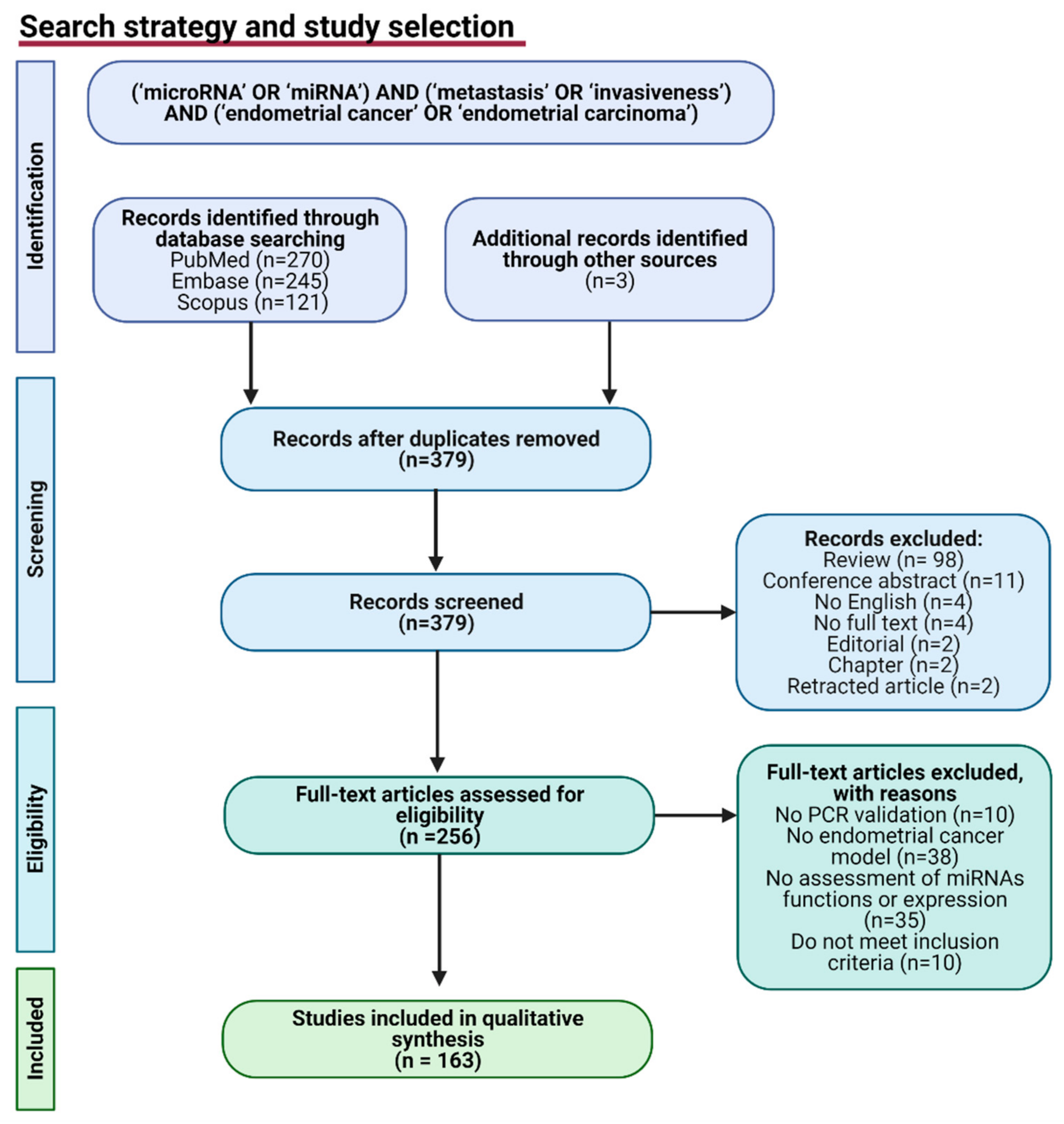
2.1. Search Strategy
2.2. Inclusion and Exclusion Criteria
2.3. Data Extraction
3. Results
3.1. MiRNAs Dysregulation in Endometrial Cancer
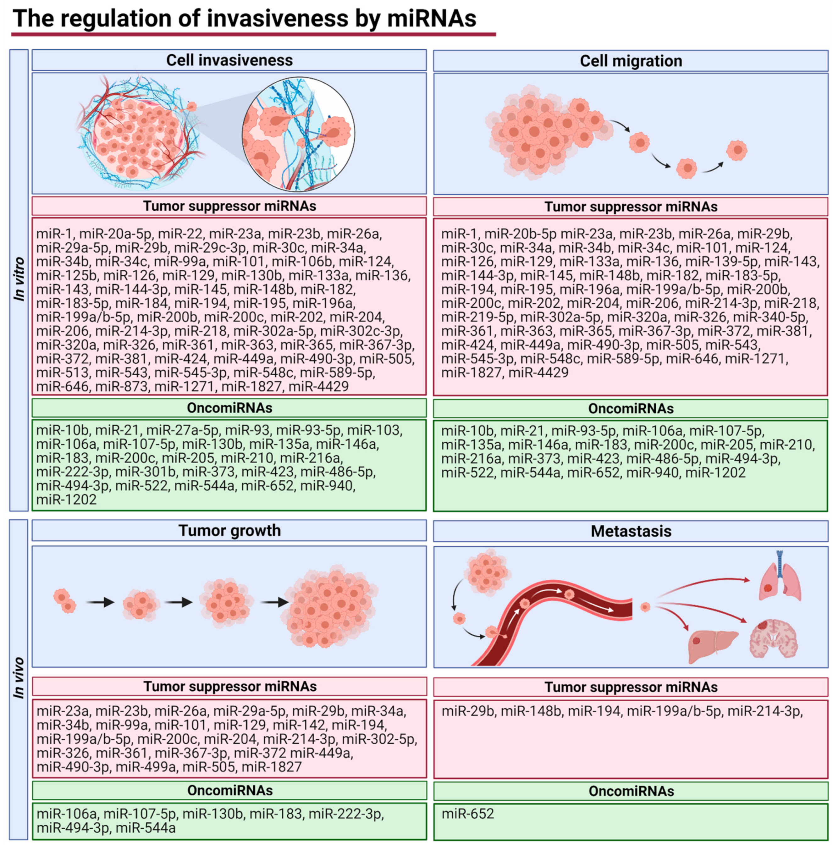
3.2. MiRNAs Regulating Endometrial Cancer (EC) Invasiveness and Metastasis
3.3. Relationship between miRNA Expression and Clinical Parameters
4. Discussion
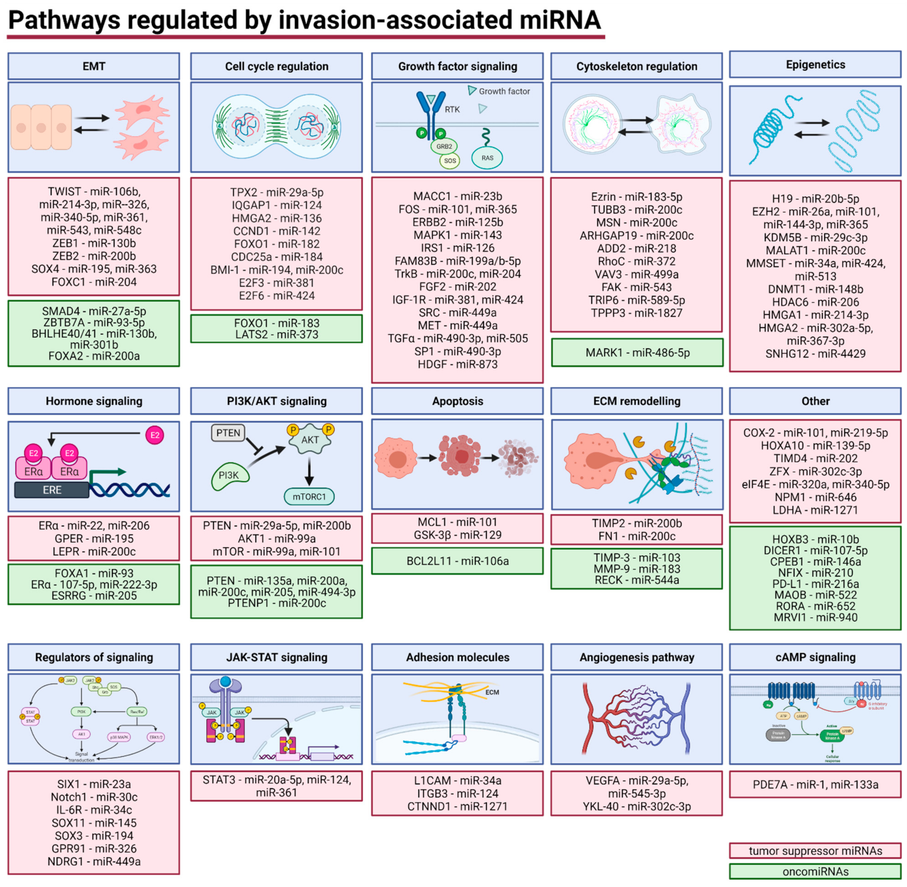
4.1. Regulatory Network of Invasion-Associated miRNAs
4.1.1. Epithelial–Mesenchymal Transition (EMT)
4.1.2. Cell Cycle
4.1.3. Growth Factors and Regulators of Signaling
4.1.4. Cytoskeleton Regulation
4.1.5. Epigenetics
4.1.6. Hormone Signaling
4.1.7. Phosphatidylinositol 3-Kinase/Protein Kinase B (PI3K/AKT) Pathways
4.1.8. Apoptosis
4.1.9. Extracellular Matrix (ECM) Remodeling
4.1.10. Janus Kinase/Signal Transducers and Activators of Transcription (JAK-STAT) Signaling
4.1.11. Adhesion Molecules
4.1.12. Angiogenesis
4.1.13. Cyclic Adenosine Monophosphate (cAMP) Signaling
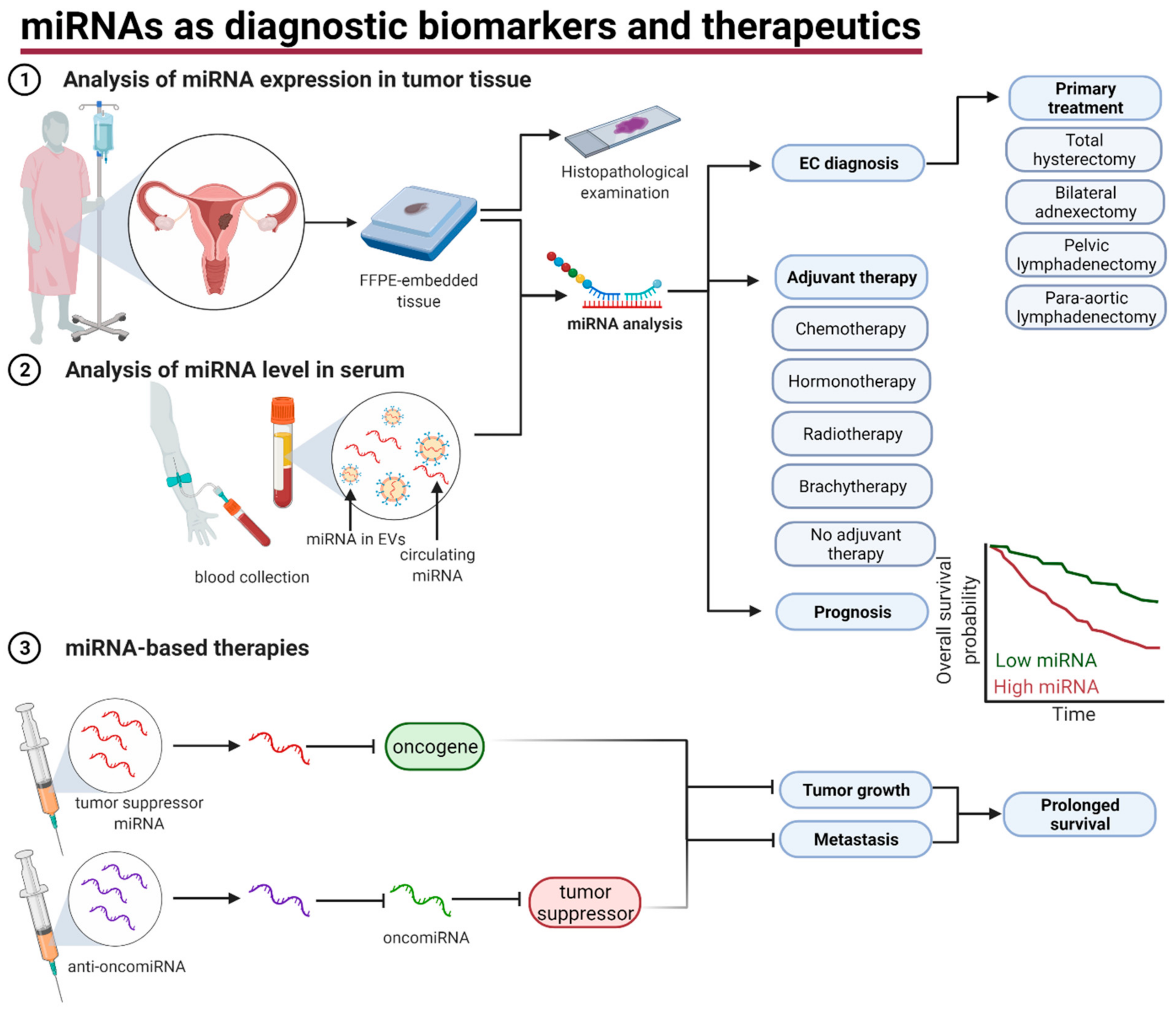
4.2. The Role of miRNAs in EC Diagnosis and Management
5. Conclusions
Author Contributions
Funding
Conflicts of Interest
References
- Siegel, R.L.; Miller, K.D.; Jemal, A. Cancer statistics, 2020. CA Cancer J. Clin. 2020, 70, 7–30. [Google Scholar] [CrossRef] [PubMed]
- Lu, K.H.; Broaddus, R.R. Endometrial Cancer. N. Engl. J. Med. 2020, 383, 2053–2064. [Google Scholar] [CrossRef] [PubMed]
- Rahatli, S.; Dizdar, O.; Kucukoztas, N.; Oguz, A.; Yalcin, S.; Ozen, O.; Reyhan, N.H.; Tarhan, C.; Yildiz, F.; Dursun, P.; et al. Good outcomes of patients with stage IB endometrial cancer with surgery alone. Asian Pac. J. Cancer Prev. 2014, 15, 3891–3893. [Google Scholar] [CrossRef] [PubMed] [Green Version]
- Chen, K.S.; Berhane, H.; Gill, B.S.; Olawaiye, A.; Sukumvanich, P.; Kelley, J.L.; Boisen, M.M.; Courtney-Brooks, M.; Comerci, J.T.; Edwards, R.; et al. Outcomes of stage II endometrial cancer: The UPMC Hillman Cancer Center experience. Gynecol. Oncol. 2017, 147, 315–319. [Google Scholar] [CrossRef] [PubMed]
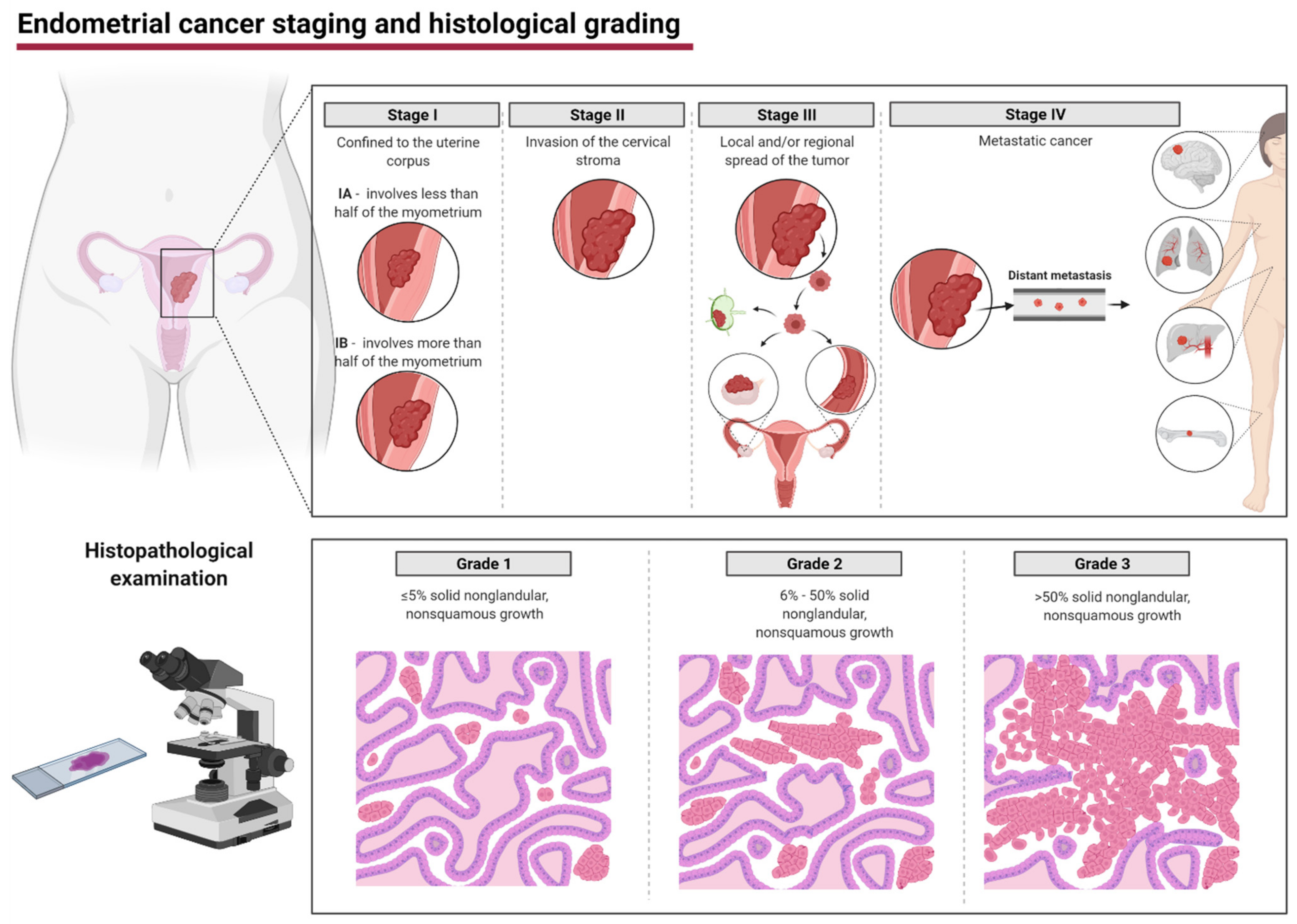
- Saleh, M.; Virarkar, M.; Bhosale, P.; El Sherif, S.; Javadi, S.; Faria, S.C. Endometrial Cancer, the Current International Federation of Gynecology and Obstetrics Staging System, and the Role of Imaging. J. Comput. Assist. Tomogr. 2020, 44, 714–729. [Google Scholar] [CrossRef] [PubMed]
- Soslow, R.A.; Tornos, C.; Park, K.J.; Malpica, A.; Matias-Guiu, X.; Oliva, E.; Parkash, V.; Carlson, J.; McCluggage, W.G.; Gilks, C.B. Endometrial Carcinoma Diagnosis: Use of FIGO Grading and Genomic Subcategories in Clinical Practice: Recommendations of the International Society of Gynecological Pathologists. Int. J. Gynecol. Pathol. Off. J. Int. Soc. Gynecol. Pathol. 2019, 38, S64–S74. [Google Scholar] [CrossRef] [PubMed] [Green Version]
- Mehlich, D.; Garbicz, F.; Włodarski, P.K. The emerging roles of the polycistronic miR-106b∼25 cluster in cancer—A comprehensive review. Biomed. Pharmacother. 2018, 107, 1183–1195. [Google Scholar] [CrossRef]
- Williams, M.; Cheng, Y.Y.; Blenkiron, C.; Reid, G. Exploring Mechanisms of MicroRNA Downregulation in Cancer. MicroRNA 2017, 6, 2–16. [Google Scholar] [CrossRef]
- Peng, Y.; Croce, C.M. The role of MicroRNAs in human cancer. Signal. Transduct. Target. Ther. 2016, 1, 15004. [Google Scholar] [CrossRef] [Green Version]
- Pełka, K.; Klicka, K.; Grzywa, T.M.; Gondek, A.; Marczewska, J.M.; Garbicz, F.; Szczepaniak, K.; Paskal, W.; Włodarski, P.K. miR-96-5p, miR-134-5p, miR-181b-5p and miR-200b-3p heterogenous expression in sites of prostate cancer versus benign prostate hyperplasia-archival samples study. Histochem. Cell Biol. 2021, 155, 423–433. [Google Scholar] [CrossRef]
- Vedanayagam, J.; Chatila, W.K.; Aksoy, B.A.; Majumdar, S.; Skanderup, A.J.; Demir, E.; Schultz, N.; Sander, C.; Lai, E.C. Cancer-associated mutations in DICER1 RNase IIIa and IIIb domains exert similar effects on miRNA biogenesis. Nat. Commun. 2019, 10, 3682. [Google Scholar] [CrossRef]
- Grzywa, T.M.; Klicka, K.; Włodarski, P.K. Regulators at Every Step-How microRNAs Drive Tumor Cell Invasiveness and Metastasis. Cancers 2020, 12, 3709. [Google Scholar] [CrossRef]
- Grzywa, T.M.; Klicka, K.; Rak, B.; Mehlich, D.; Garbicz, F.; Zieliński, G.; Maksymowicz, M.; Sajjad, E.; Włodarski, P.K. Lineage-dependent role of miR-410-3p as oncomiR in gonadotroph and corticotroph pituitary adenomas or tumor suppressor miR in somatotroph adenomas via MAPK, PTEN/AKT, and STAT3 signaling pathways. Endocrine 2019, 65, 646–655. [Google Scholar] [CrossRef] [Green Version]
- Hanahan, D.; Weinberg, R.A. Hallmarks of Cancer: The Next Generation. Cell 2011, 144, 646–674. [Google Scholar] [CrossRef] [Green Version]
- Ghafouri-Fard, S.; Shoorei, H.; Anamag, F.T.; Taheri, M. The Role of Non-Coding RNAs in Controlling Cell Cycle Related Proteins in Cancer Cells. Front. Oncol. 2020, 10. [Google Scholar] [CrossRef]
- Leone, P.; Buonavoglia, A.; Fasano, R.; Solimando, A.G.; De Re, V.; Cicco, S.; Vacca, A.; Racanelli, V. Insights into the Regulation of Tumor Angiogenesis by Micro-RNAs. J. Clin. Med. 2019, 8, 2030. [Google Scholar] [CrossRef] [Green Version]
- Si, W.; Shen, J.; Zheng, H.; Fan, W. The role and mechanisms of action of microRNAs in cancer drug resistance. Clin. Epigenetics 2019, 11, 25. [Google Scholar] [CrossRef]
- Bao, W.; Wang, H.H.; Tian, F.J.; He, X.Y.; Qiu, M.T.; Wang, J.Y.; Zhang, H.J.; Wang, L.H.; Wan, X.P. A TrkB-STAT3-miR-204-5p regulatory circuitry controls proliferation and invasion of endometrial carcinoma cells. Mol. Cancer 2013, 12, 155. [Google Scholar] [CrossRef] [Green Version]
- Bao, W.; Zhang, Y.; Li, S.; Fan, Q.; Qiu, M.; Wang, Y.; Li, Y.; Ji, X.; Yang, Y.; Sang, Z.; et al. miR-107-5p promotes tumor proliferation and invasion by targeting estrogen receptor-α in endometrial carcinoma. Oncol. Rep. 2019, 41, 1575–1585. [Google Scholar] [CrossRef]
- Bing, L.; Hong, C.; Li-Xin, S.; Wei, G. MicroRNA-543 suppresses endometrial cancer oncogenicity via targeting FAK and TWIST1 expression. Arch. Gynecol. Obstet. 2014, 290, 533–541. [Google Scholar] [CrossRef]
- Chang, L.; Yuan, Z.; Shi, H.; Bian, Y.; Guo, R. miR-145 targets the SOX11 3’UTR to suppress endometrial cancer growth. Am. J. Cancer Res. 2017, 7, 2305–2317. [Google Scholar] [PubMed]
- Chang, L.; Zhang, D.; Shi, H.; Bian, Y.; Guo, R. MiR-143 inhibits endometrial cancer cell proliferation and metastasis by targeting MAPK1. Oncotarget 2017, 8, 84384–84395. [Google Scholar] [CrossRef] [PubMed] [Green Version]
- Chen, H.; Fan, Y.; Xu, W.; Chen, J.; Meng, Y.; Fang, D.; Wang, J. Exploration of miR-1202 and miR-196a in human endometrial cancer based on high throughout gene screening analysis. Oncol. Rep. 2017, 37, 3493–3501. [Google Scholar] [CrossRef] [PubMed] [Green Version]
- Chen, H.X.; Xu, X.X.; Tan, B.Z.; Zhang, Z.; Zhou, X.D. MicroRNA-29b Inhibits Angiogenesis by Targeting VEGFA through the MAPK/ERK and PI3K/Akt Signaling Pathways in Endometrial Carcinoma. Cell. Physiol. Biochem. 2017, 41, 933–946. [Google Scholar] [CrossRef]
- Chen, P.; Xing, T.; Wang, Q.; Liu, A.; Liu, H.; Hu, Y.; Ji, Y.; Song, Y.; Wang, D. MicroRNA-202 inhibits cell migration and invasion through targeting FGF2 and inactivating Wnt/β-catenin signaling in endometrial carcinoma. Biosci. Rep. 2019, 39. [Google Scholar] [CrossRef] [Green Version]
- Chen, R.; Zhang, M.; Liu, W.; Chen, H.; Cai, T.; Xiong, H.; Sheng, X.; Liu, S.; Peng, J.; Wang, F.; et al. Estrogen affects the negative feedback loop of PTENP1-miR200c to inhibit PTEN expression in the development of endometrioid endometrial carcinoma. Cell Death Dis. 2018, 10, 4. [Google Scholar] [CrossRef]
- Chen, S.; Chen, X.; Sun, K.X.; Xiu, Y.L.; Liu, B.L.; Feng, M.X.; Sang, X.B.; Zhao, Y. MicroRNA-93 Promotes Epithelial-Mesenchymal Transition of Endometrial Carcinoma Cells. PLoS ONE 2016, 11, e0165776. [Google Scholar] [CrossRef] [Green Version]
- Chen, Z.; Zhu, Y.; Fan, X.; Liu, Y.; Feng, Q. Decreased expression of miR-184 restrains the growth and invasion of endometrial carcinoma cells through CDC25A-dependent Notch signaling pathway. Am. J. Transl. Res. 2019, 11, 755–764. [Google Scholar]
- Chung, T.K.; Cheung, T.H.; Huen, N.Y.; Wong, K.W.; Lo, K.W.; Yim, S.F.; Siu, N.S.; Wong, Y.M.; Tsang, P.T.; Pang, M.W.; et al. Dysregulated microRNAs and their predicted targets associated with endometrioid endometrial adenocarcinoma in Hong Kong women. Int. J. Cancer 2009, 124, 1358–1365. [Google Scholar] [CrossRef]
- Chung, T.K.; Lau, T.S.; Cheung, T.H.; Yim, S.F.; Lo, K.W.; Siu, N.S.; Chan, L.K.; Yu, M.Y.; Kwong, J.; Doran, G.; et al. Dysregulation of microRNA-204 mediates migration and invasion of endometrial cancer by regulating FOXC1. Int. J. Cancer 2012, 130, 1036–1045. [Google Scholar] [CrossRef]
- Dai, Y.; Xia, W.; Song, T.; Su, X.; Li, J.; Li, S.; Chen, Y.; Wang, W.; Ding, H.; Liu, X.; et al. MicroRNA-200b is overexpressed in endometrial adenocarcinomas and enhances MMP2 activity by downregulating TIMP2 in human endometrial cancer cell line HEC-1A cells. Nucleic Acid Ther. 2013, 23, 29–34. [Google Scholar] [CrossRef] [Green Version]
- Dong, P. Mutant p53 gain-of-function induces epithelial-mesenchymal transition through modulation of the miR-130b-ZEB1 axis. J. Cancer Sci. Ther. 2012, 4, 131. [Google Scholar] [CrossRef] [Green Version]
- Dong, P.; Ihira, K.; Xiong, Y.; Watari, H.; Hanley, S.J.; Yamada, T.; Hosaka, M.; Kudo, M.; Yue, J.; Sakuragi, N. Reactivation of epigenetically silenced miR-124 reverses the epithelial-to-mesenchymal transition and inhibits invasion in endometrial cancer cells via the direct repression of IQGAP1 expression. Oncotarget 2016, 7, 20260–20270. [Google Scholar] [CrossRef]
- Dong, P.; Xiong, Y.; Yue, J.; Hanley, S.J.B.; Watari, H. miR-34a, miR-424 and miR-513 inhibit MMSET expression to repress endometrial cancer cell invasion and sphere formation. Oncotarget 2018, 9, 23253–23263. [Google Scholar] [CrossRef]
- Dong, Y.; Si, J.W.; Li, W.T.; Liang, L.; Zhao, J.; Zhou, M.; Li, D.; Li, T. miR-200a/miR-141 and miR-205 upregulation might be associated with hormone receptor status and prognosis in endometrial carcinomas. Int. J. Clin. Exp. Pathol. 2015, 8, 2864–2875. [Google Scholar]
- Fang, Y.Y.; Tan, M.R.; Zhou, J.; Liang, L.; Liu, X.Y.; Zhao, K.; Bao, E.C. MiR-214-3p inhibits epithelial-to-mesenchymal transition and metastasis of endometrial cancer cells by targeting TWISTI. OncoTargets Ther. 2019, 12, 9449–9458. [Google Scholar] [CrossRef] [Green Version]
- He, S.; Zeng, S.; Zhou, Z.W.; He, Z.X.; Zhou, S.F. Hsa-microRNA-181a is a regulator of a number of cancer genes and a biomarker for endometrial carcinoma in patients: A bioinformatic and clinical study and the therapeutic implication. Drug Des. Dev. Ther. 2015, 9, 1103–1175. [Google Scholar] [CrossRef] [Green Version]
- Hiroki, E.; Akahira, J.; Suzuki, F.; Nagase, S.; Ito, K.; Suzuki, T.; Sasano, H.; Yaegashi, N. Changes in microRNA expression levels correlate with clinicopathological features and prognoses in endometrial serous adenocarcinomas. Cancer Sci. 2010, 101, 241–249. [Google Scholar] [CrossRef]
- Hiroki, E.; Suzuki, F.; Akahira, J.I.; Nagase, S.; Ito, K.; Sugawara, J.I.; Miki, Y.; Suzuki, T.; Sasano, H.; Yaegashi, N. MicroRNA-34b functions as a potential tumor suppressor in endometrial serous adenocarcinoma. Int. J. Cancer 2012, 131, E395–E404. [Google Scholar] [CrossRef]
- Huang, Y.; Yang, N. MicroRNA-20a-5p inhibits epithelial to mesenchymal transition and invasion of endometrial cancer cells by targeting STAT3. Int. J. Clin. Exp. Pathol. 2018, 11, 5715–5724. [Google Scholar]
- Ihira, K.; Dong, P.; Xiong, Y.; Watari, H.; Konno, Y.; Hanley, S.J.; Noguchi, M.; Hirata, N.; Suizu, F.; Yamada, T.; et al. EZH2 inhibition suppresses endometrial cancer progression via miR-361/Twist axis. Oncotarget 2017, 8, 13509–13520. [Google Scholar] [CrossRef]
- Jiang, F.Z.; He, Y.Y.; Wang, H.H.; Zhang, H.L.; Zhang, J.; Yan, X.F.; Wang, X.J.; Che, Q.; Ke, J.Q.; Chen, Z.; et al. Mutant p53 induces EZH2 expression and promotes epithelial-mesenchymal transition by disrupting p68-Drosha complex assembly and attenuating miR-26a processing. Oncotarget 2015, 6, 44660–44674. [Google Scholar] [CrossRef] [Green Version]
- Jiang, L.; Meng, W.; Zeng, J.; Hu, H.; Lu, L. MiR-34c oligonucleotide enhances chemosensitivity of Ishikawa cell to cisplatin by inducing apoptosis. Cell Biol. Int. 2013, 37, 577–583. [Google Scholar] [CrossRef]
- Jiang, T.; Sui, D.; You, D.; Yao, S.; Zhang, L.; Wang, Y.; Zhao, J.; Zhang, Y. MiR-29a-5p inhibits proliferation and invasion and induces apoptosis in endometrial carcinoma via targeting TPX2. Cell Cycle 2018, 17, 1268–1278. [Google Scholar] [CrossRef] [Green Version]
- Jing, L.; Hua, X.; Yuanna, D.; Rukun, Z.; Junjun, M. Exosomal miR-499a-5p Inhibits Endometrial Cancer Growth and Metastasis via Targeting VAV3. Cancer Manag. Res. 2020, 12, 13541–13552. [Google Scholar] [CrossRef]
- Konno, Y.; Dong, P.; Xiong, Y.; Suzuki, F.; Lu, J.; Cai, M.; Watari, H.; Mitamura, T.; Hosaka, M.; Hanley, S.J.; et al. MicroRNA-101 targets EZH2, MCL-1 and FOS to suppress proliferation, invasion and stem cell-like phenotype of aggressive endometrial cancer cells. Oncotarget 2014, 5, 6049–6062. [Google Scholar] [CrossRef] [Green Version]
- Kottaridi, C.; Spathis, A.; Margari, N.; Koureas, N.; Terzakis, E.; Chrelias, C.; Pappas, A.; Bilirakis, E.; Pouliakis, A.; Panayiotides, I.J.; et al. Evaluation Analysis of miRNAs Overexpression in Liquid-Based Cytology Endometrial Samples. J. Cancer 2017, 8, 2699–2703. [Google Scholar] [CrossRef] [Green Version]
- Lee, H.; Choi, H.J.; Kang, C.S.; Lee, H.J.; Lee, W.S.; Park, C.S. Expression of miRNAs and PTEN in endometrial specimens ranging from histologically normal to hyperplasia and endometrial adenocarcinoma. Mod. Pathol. Off. J. USA Acad. Pathol. Inc. 2012, 25, 1508–1515. [Google Scholar] [CrossRef] [PubMed] [Green Version]
- Li, Y.; Sun, D.; Gao, J.; Shi, Z.; Chi, P.; Meng, Y.; Zou, C.; Wang, Y. MicroRNA-373 promotes the development of endometrial cancer by targeting LATS2 and activating the Wnt/β-Catenin pathway. J. Cell. Biochem. 2018. [Google Scholar] [CrossRef] [PubMed]
- Li, Y.; Zhang, Z.; Liu, X.; Huang, T.; He, W.; Shen, Y.; Liu, X.; Hong, K.; Cao, Q. miR-124 functions as a tumor suppressor in the endometrial carcinoma cell line HEC-1B partly by suppressing STAT3. Mol. Cell. Biochem. 2014, 388, 219–231. [Google Scholar] [CrossRef] [PubMed]
- Li, Y.; Zhang, Z.; Zhang, X.; Lin, Y.; Luo, T.; Xiao, Z.; Zhou, Q. A dual PI3K/AKT/mTOR signaling inhibitor miR-99a suppresses endometrial carcinoma. Am. J. Transl. Res. 2016, 8, 719–731. [Google Scholar]
- Liu, B.; Che, Q.; Qiu, H.; Bao, W.; Chen, X.; Lu, W.; Li, B.; Wan, X. Elevated MiR-222-3p promotes proliferation and invasion of endometrial carcinoma via targeting ERα. PLoS ONE 2014, 9, e87563. [Google Scholar] [CrossRef] [Green Version]
- Li, Q.; Zhang, C.; Chen, R.; Xiong, H.; Qiu, F.; Liu, S.; Zhang, M.; Wang, F.; Wang, Y.; Zhou, X.; et al. Disrupting MALAT1/miR-200c sponge decreases invasion and migration in endometrioid endometrial carcinoma. Cancer Lett. 2016, 383, 28–40. [Google Scholar] [CrossRef]
- Liu, J.H.; Li, C.Y.; Jiang, Y.; Wan, Y.C.; Zhou, S.L.; Cheng, W.J. Tumor-Suppressor role of miR-139-5p in endometrial cancer. Cancer Cell Int. 2018, 18. [Google Scholar] [CrossRef]
- Liu, W.; Zhang, B.; Xu, N.; Wang, M.J.; Liu, Q. miR-326 regulates EMT and metastasis of endometrial cancer through targeting TWIST1. Eur. Rev. Med. Pharmacol. Sci. 2017, 21, 3787–3793. [Google Scholar]
- Liu, Y.; Li, H.; Zhao, C.; Jia, H. MicroRNA-101 inhibits angiogenesis via COX-2 in endometrial carcinoma. Mol. Cell. Biochem. 2018, 448, 61–69. [Google Scholar] [CrossRef]
- Lu, L.; Shen, Y.; Tseng, K.; Jiang, L.; Liu, W.; Duan, H.; Meng, W. Oncogenic function of miR-301 to predicts poor prognosis of endometrial cancer. Int. J. Clin. Exp. Med. 2016, 9, 12937–12942. [Google Scholar]
- Lu, Z.; Nian, Z.; Jingjing, Z.; Tao, L.; Quan, L. MicroRNA-424/E2F6 feedback loop modulates cell invasion, migration and EMT in endometrial carcinoma. Oncotarget 2017, 8, 114281–114291. [Google Scholar] [CrossRef] [Green Version]
- Ma, Y.J.; Ha, C.F.; Bai, Z.M.; Li, H.N.; Xiong, Y.; Jiang, J. Overexpression of microRNA-205 predicts lymph node metastasis and indicates an unfavorable prognosis in endometrial cancer. Oncol. Lett. 2016, 12, 4403–4410. [Google Scholar] [CrossRef] [Green Version]
- Mozos, A.; Catasús, L.; D’Angelo, E.; Serrano, E.; Espinosa, I.; Ferrer, I.; Pons, C.; Prat, J. The FOXO1-miR27 tandem regulates myometrial invasion in endometrioid endometrial adenocarcinoma. Hum. Pathol. 2014, 45, 942–951. [Google Scholar] [CrossRef]
- Notaro, S.; Reimer, D.; Duggan-Peer, M.; Fiegl, H.; Wiedermair, A.; Rössler, J.; Altevogt, P.; Marth, C.; Zeimet, A.G. Evaluating L1CAM expression in human endometrial cancer using qRT-PCR. Oncotarget 2016, 7, 40221–40232. [Google Scholar] [CrossRef] [Green Version]
- Ruan, H.; Liang, X.; Zhao, W.; Ma, L.; Zhao, Y. The effects of microRNA-183 promots cell proliferation and invasion by targeting MMP-9 in endometrial cancer. Biomed. Pharmacother. Biomed. Pharmacother. 2017, 89, 812–818. [Google Scholar] [CrossRef]
- Shang, C.; Lu, Y.M.; Meng, L.R. MicroRNA-125b down-regulation mediates endometrial cancer invasion by targeting ERBB2. Med. Sci. Monit. Int. Med. J. Exp. Clin. Res. 2012, 18, BR149–BR155. [Google Scholar] [CrossRef]
- Shao, W.; Li, Y.; Chen, F.; Jia, H.; Jia, J.; Fu, Y. Long non-coding RNA DLEU1 contributes to the development of endometrial cancer by sponging miR-490 to regulate SP1 expression. Pharmazie 2018, 73, 379–385. [Google Scholar] [CrossRef]
- Shi, Y.; Jia, L.; Wen, H. Circ_0109046 Promotes the Progression of Endometrial Cancer via Regulating miR-136/HMGA2 Axis. Cancer Manag. Res. 2020, 12, 10993–11003. [Google Scholar] [CrossRef]
- Shu, S.; Liu, X.; Xu, M.; Gao, X.; Fan, J.; Liu, H.; Li, R. MicroRNA-424 regulates epithelial-mesenchymal transition of endometrial carcinoma by directly targeting insulin-like growth factor 1 receptor. J. Cell. Biochem. 2019, 120, 2171–2179. [Google Scholar] [CrossRef] [PubMed]
- Song, N.; Zhang, Y.; Kong, F.; Yang, H.; Ma, X. HOXA-AS2 promotes type I endometrial carcinoma via miRNA-302c-3p-mediated regulation of ZFX. Cancer Cell Int. 2020, 20, 359. [Google Scholar] [CrossRef] [PubMed]
- Su, N.; Qiu, H.; Chen, Y.; Yang, T.; Yan, Q.; Wan, X. miR-205 promotes tumor proliferation and invasion through targeting ESRRG in endometrial carcinoma. Oncol. Rep. 2013, 29, 2297–2302. [Google Scholar] [CrossRef] [PubMed]
- Su, Y.; Wang, J.; Ma, Z.; Gong, W.; Yu, L. miR-142 Suppresses Endometrial Cancer Proliferation In Vitro and In Vivo by Targeting Cyclin D1. DNA Cell Biol. 2019, 38, 144–150. [Google Scholar] [CrossRef] [PubMed]
- Sun, K.X.; Chen, Y.; Chen, S.; Liu, B.L.; Feng, M.X.; Zong, Z.H.; Zhao, Y. The correlation between microRNA490-3p and TGFα in endometrial carcinoma tumorigenesis and progression. Oncotarget 2016, 7, 9236–9249. [Google Scholar] [CrossRef]
- Sun, X.; Cui, M.; Zhang, A.; Tong, L.; Wang, K.; Li, K.; Wang, X.; Sun, Z.; Zhang, H. MiR-548c impairs migration and invasion of endometrial and ovarian cancer cells via downregulation of Twist. J. Exp. Clin. Cancer Res. 2016, 35, 10. [Google Scholar] [CrossRef] [Green Version]
- Sun, X.; Dongol, S.; Qiu, C.; Xu, Y.; Sun, C.; Zhang, Z.; Yang, X.; Zhang, Q.; Kong, B. miR-652 Promotes Tumor Proliferation and Metastasis by Targeting RORA in Endometrial Cancer. Mol. Cancer Res. 2018, 16, 1927–1939. [Google Scholar] [CrossRef] [Green Version]
- Tang, W.; Li, J.; Liu, H.; Zhou, F.; Liu, M. MiR-106a promotes tumor growth, migration, and invasion by targeting BCL2L11 in human endometrial adenocarcinoma. Am. J. Transl. Res. 2017, 9, 4984–4993. [Google Scholar]
- Tian, Y.; Chen, Y.Y.; Han, A.L. MiR-1271 inhibits cell proliferation and metastasis by targeting LDHA in endometrial cancer. Eur. Rev. Med. Pharmacol. Sci. 2019, 23, 5648–5656. [Google Scholar] [CrossRef]
- Torres, A.; Torres, K.; Paszkowski, T.; Radej, S.; Staśkiewicz, G.J.; Ceccaroni, M.; Pesci, A.; Maciejewski, R. Highly increased maspin expression corresponds with up-regulation of miR-21 in endometrial cancer: A preliminary report. Int. J. Gynecol. Cancer Off. J. Int. Gynecol. Cancer Soc. 2011, 21, 8–14. [Google Scholar] [CrossRef]
- Torres, A.; Torres, K.; Pesci, A.; Ceccaroni, M.; Paszkowski, T.; Cassandrini, P.; Zamboni, G.; Maciejewski, R. Deregulation of miR-100, miR-99a and miR-199b in tissues and plasma coexists with increased expression of mTOR kinase in endometrioid endometrial carcinoma. BMC Cancer 2012, 12. [Google Scholar] [CrossRef] [Green Version]
- Torres, A.; Torres, K.; Pesci, A.; Ceccaroni, M.; Paszkowski, T.; Cassandrini, P.; Zamboni, G.; Maciejewski, R. Diagnostic and prognostic significance of miRNA signatures in tissues and plasma of endometrioid endometrial carcinoma patients. Int. J. Cancer 2013, 132, 1633–1645. [Google Scholar] [CrossRef]
- Tsukamoto, O.; Miura, K.; Mishima, H.; Abe, S.; Kaneuchi, M.; Higashijima, A.; Miura, S.; Kinoshita, A.; Yoshiura, K.; Masuzaki, H. Identification of endometrioid endometrial carcinoma-associated microRNAs in tissue and plasma. Gynecol. Oncol. 2014, 132, 715–721. [Google Scholar] [CrossRef]
- Tu, C.; Wang, F.; Wan, J. MicroRNA-381 inhibits cell proliferation and invasion in endometrial carcinoma by targeting the IGF-1R. Mol. Med. Rep. 2018, 17, 4090–4098. [Google Scholar] [CrossRef]
- Wang, C.; Su, K.; Zhang, Y.; Zhang, W.; Chu, D.; Zhao, Q.; Guo, R. MicroRNA-365 targets multiple oncogenes to inhibit proliferation, invasion, and self-renewal of aggressive endometrial cancer cells. Cancer Manag. Res. 2018, 10, 5171–5185. [Google Scholar] [CrossRef] [Green Version]
- Wang, H.; Wang, T.T.; Lv, X.P. Expression and prognostic value of miRNA-29b in peripheral blood for endometrial cancer. Future Oncol. 2018, 14, 1365–1376. [Google Scholar] [CrossRef]
- Wang, J.; Zhao, X.; Guo, Z.; Ma, X.; Song, Y.; Guo, Y. Regulation of NEAT1/miR-214-3p on the growth, migration and invasion of endometrial carcinoma cells. Arch. Gynecol. Obstet. 2017, 295, 1469–1475. [Google Scholar] [CrossRef]
- Wang, L.; Zhao, S.; Mingxin, Y.U. LncRNA NR2F1-AS1 is involved in the progression of endometrial cancer by sponging miR-363 to target SOX4. Pharmazie 2019, 74, 295–300. [Google Scholar] [CrossRef]
- Wang, Q.; Zhu, W. MicroRNA-873 inhibits the proliferation and invasion of endometrial cancer cells by directly targeting hepatoma-derived growth factor. Exp. Ther. Med. 2019, 18, 1291–1298. [Google Scholar] [CrossRef] [Green Version]
- Wang, W.; Ge, L.; Xu, X.J.; Yang, T.; Yuan, Y.; Ma, X.L.; Zhang, X.H. LncRNA NEAT1 promotes endometrial cancer cell proliferation, migration and invasion by regulating the miR-144-3p/EZH2 axis. Radiol. Oncol. 2019, 53, 434–442. [Google Scholar] [CrossRef] [Green Version]
- Wang, Y.; Dong, L.; Liu, Y. Targeting Thyroid Receptor Interacting Protein 6 by MicroRNA-589-5p Inhibits Cell Proliferation, Migration, and Invasion in Endometrial Carcinoma. Cancer Biother. Radiopharm. 2019, 34, 529–536. [Google Scholar] [CrossRef]
- Wang, Y.; Yin, L. LINC00461 Promoted Endometrial Carcinoma Growth and Migration by Targeting MicroRNA-219-5p/Cyclooxygenase-2 Signaling Axis. Cell Transplant. 2021, 30. [Google Scholar] [CrossRef]
- Wei, D.; Tian, M.; Fan, W.; Zhong, X.; Wang, S.; Chen, Y.; Zhang, S. Circular RNA circ_0000043 promotes endometrial carcinoma progression by regulating miR-1271-5p/CTNND1 axis. Arch. Gynecol. Obstet. 2020. [Google Scholar] [CrossRef]
- Wilczynski, M.; Danielska, J.; Dzieniecka, M.; Szymanska, B.; Wojciechowski, M.; Malinowski, A. Prognostic and Clinical Significance of miRNA-205 in Endometrioid Endometrial Cancer. PLoS ONE 2016, 11, e0164687. [Google Scholar] [CrossRef] [Green Version]
- Wu, D.; Huang, H.J.; He, C.N.; Wang, K.Y. MicroRNA-199a-3p regulates endometrial cancer cell proliferation by targeting mammalian target of rapamycin (mTOR). Int. J. Gynecol. Cancer Off. J. Int. Gynecol. Cancer Soc. 2013, 23, 1191–1197. [Google Scholar] [CrossRef]
- Xin, W.; Gao, X.; Zhao, S.; Zhao, P.; Yu, H.; Wu, Q.; Hua, K. LncRNA RP11-395G23.3 suppresses the endometrial cancer progression via regulating microRNA-205-5p/PTEN axis. Am. J. Transl. Res. 2020, 12, 4422–4433. [Google Scholar] [PubMed]
- Xiong, H.; Chen, R.; Liu, S.; Lin, Q.; Chen, H.; Jiang, Q. MicroRNA-183 induces epithelial-mesenchymal transition and promotes endometrial cancer cell migration and invasion in by targeting CPEB1. J. Cell. Biochem. 2018, 119, 8123–8137. [Google Scholar] [CrossRef] [PubMed]
- Xiong, H.; Wang, N.; Chen, H.; Zhang, M.; Lin, Q. MicroRNA-199a/b-5p inhibits endometrial cancer cell metastasis and invasion by targeting FAM83B in the epithelial-to-mesenchymal transition signaling pathway. Mol. Med. Rep. 2021, 23. [Google Scholar] [CrossRef] [PubMed]
- Xu, C.; Zhai, J.; Fu, Y. Overexpression of Nuclear Enriched Autosomal Transcript 1 Facilitates Cell Proliferation, Migration Invasion, and Suppresses Apoptosis in Endometrial Cancer by Targeting MicroRNA-202-3p/T Cell Immunoglobulin and Mucin Domain 4 Axis. Cancer Biother. Radiopharm. 2020. [Google Scholar] [CrossRef]
- Xu, J.B. MicroRNA-93-5p/IFNAR1 axis accelerates metastasis of endometrial carcinoma by activating the STAT3 pathway. Eur. Rev. Med. Pharmacol. Sci. 2019, 23, 5657–5666. [Google Scholar] [CrossRef]
- Xu, X.; Kong, X.; Liu, T.; Zhou, L.; Wu, J.; Fu, J.; Wang, Y.; Zhu, M.; Yao, S.; Ding, Y.; et al. Metastasis-associated protein 1, modulated by miR-30c, promotes endometrial cancer progression through AKT/mTOR/4E-BP1 pathway. Gynecol. Oncol. 2019, 154, 207–217. [Google Scholar] [CrossRef]
- Yamamoto, N.; Nishikawa, R.; Chiyomaru, T.; Goto, Y.; Fukumoto, I.; Usui, H.; Mitsuhashi, A.; Enokida, H.; Nakagawa, M.; Shozu, M.; et al. The tumor-suppressive microRNA-1/133a cluster targets PDE7A and inhibits cancer cell migration and invasion in endometrial cancer. Int. J. Oncol. 2015, 47, 325–334. [Google Scholar] [CrossRef]
- Yan, H.; Sun, B.M.; Zhang, Y.Y.; Li, Y.J.; Huang, C.X.; Feng, F.Z.; Li, C. Upregulation of miR-183-5p is responsible for the promotion of apoptosis and inhibition of the epithelial-mesenchymal transition, proliferation, invasion and migration of human endometrial cancer cells by downregulating Ezrin. Int. J. Mol. Med. 2018, 42, 2469–2480. [Google Scholar] [CrossRef] [Green Version]
- Yang, B.; Li, W.; Ma, X.; Guo, Y.; Shen, G. The correlation between tumor tissue mir-373 and mir-124 expressions and prognosis in patients with endometrial cancer. Int. J. Clin. Exp. Med. 2020, 13, 4470–4477. [Google Scholar]
- Yang, L.; Yang, Z.; Yao, R.; Li, Y.; Liu, Z.; Chen, X.; Zhang, G. miR-210 promotes progression of endometrial carcinoma by regulating the expression of NFIX. Int. J. Clin. Exp. Pathol. 2018, 11, 5213–5222. [Google Scholar]
- Yanokura, M.; Banno, K.; Aoki, D. MicroRNA-34b expression enhances chemosensitivity of endometrial cancer cells to paclitaxel. Int. J. Oncol. 2020, 57, 1145–1156. [Google Scholar] [CrossRef]
- Yoneyama, K.; Ishibashi, O.; Kawase, R.; Kurose, K.; Takeshita, T. miR-200a, miR-200b and miR-429 are onco-miRs that target the PTEN gene in endometrioid endometrial carcinoma. Anticancer Res. 2015, 35, 1401–1410. [Google Scholar]
- Zhang, H.H.; Li, R.; Li, Y.J.; Yu, X.X.; Sun, Q.N.; Li, A.Y.; Kong, Y. eIF4E-related miR-320a and miR-340-5p inhibit endometrial carcinoma cell metastatic capability by preventing TGF-β1-induced epithelial-mesenchymal transition. Oncol. Rep. 2020, 43, 447–460. [Google Scholar] [CrossRef] [Green Version]
- Zhang, H.M.; Fan, T.T.; Li, W.; Li, X.X. Expressions and significances of TTF-1 and PTEN in early endometrial cancer. Eur. Rev. Med. Pharmacol. Sci. 2017, 21, 20–26. [Google Scholar]
- Zhang, K.; Cai, Y.; Zhou, Q.; Sun, H.; Wei, J. Long non-coding RNA SNHG14 impedes viability, migration and invasion of endometrial carcinoma cells through modulating miR-93-5p/ ZBTB7A axis. Cancer Manag. Res. 2020, 12, 9515–9525. [Google Scholar] [CrossRef]
- Zhang, S.; Wang, M.; Li, Q.; Zhu, P. MiR-101 reduces cell proliferation and invasion and enhances apoptosis in endometrial cancer via regulating PI3K/Akt/mTOR. Cancer Biomark. Sect. A Dis. Markers 2017, 21, 179–186. [Google Scholar] [CrossRef]
- Zhao, W.; Geng, P.; Li, Y.; Wei, X.; Cheng, J. MicroRNA-21 promotes endometrial carcinoma proliferation and invasion by targeting PTEN. Int. J. Clin. Exp. Pathol. 2017, 10, 11489–11495. [Google Scholar]
- Zhao, X.; Dai, L.; Yue, Q.; Wang, H.; Wang, X.U.; Li, Y.; Chen, R. MiR-195 inhibits migration, invasion and epithelial-mesenchymal transition (EMT) of endometrial carcinoma cells by targeting SOX4. J. Biosci. 2019, 44, 146. [Google Scholar] [CrossRef]
- Zhao, X.; Zhu, D.; Lu, C.; Yan, D.; Li, L.; Chen, Z. MicroRNA-126 inhibits the migration and invasion of endometrial cancer cells by targeting insulin receptor substrate 1. Oncol. Lett. 2016, 11, 1207–1212. [Google Scholar] [CrossRef] [Green Version]
- Zheng, W.; Yang, J.; Wang, Y.; Liu, X. Exosomal miRNA-93 and miRNA-205 expression in endometrial cancer. J. King Saud Univ. Sci. 2020, 32, 1111–1115. [Google Scholar] [CrossRef]
- Zheng, W.P.; Meng, F.L.; Wang, L.Y. miR-544a Stimulates endometrial carcinoma growth via targeted inhibition of reversion-inducing cysteine-rich protein with Kazal motifs. Mol. Cell. Probes 2020, 53, 101572. [Google Scholar] [CrossRef] [PubMed]
- Zheng, X.; Xu, K.; Zhu, L.; Mao, M.; Zhang, F.; Cui, L. MiR-486-5p Act as a Biomarker in Endometrial Carcinoma: Promotes Cell Proliferation, Migration, Invasion by Targeting MARK1. OncoTargets Ther. 2020, 13, 4843–4853. [Google Scholar] [CrossRef] [PubMed]
- Zhou, Y.X.; Wang, C.; Mao, L.W.; Wang, Y.L.; Xia, L.Q.; Zhao, W.; Shen, J.; Chen, J. Long noncoding RNA HOTAIR mediates the estrogen-induced metastasis of endometrial cancer cells via the miR-646/NPM1 axis. Am. J. Physiol. Cell Physiol. 2018, 314, C690–C701. [Google Scholar] [CrossRef] [PubMed]
- Zhou, Y.X.; Zhao, W.; Mao, L.W.; Wang, Y.L.; Xia, L.Q.; Cao, M.; Shen, J.; Chen, J. Long non-coding RNA NIFK-AS1 inhibits M2 polarization of macrophages in endometrial cancer through targeting miR-146a. Int. J. Biochem. Cell Biol. 2018, 104, 25–33. [Google Scholar] [CrossRef]
- Zhou, Z.; Xu, Y.P.; Wang, L.J.; Kong, Y. miR-940 potentially promotes proliferation and metastasis of endometrial carcinoma through regulation of MRVI1. Biosci. Rep. 2019, 39. [Google Scholar] [CrossRef] [Green Version]
- Zhu, H.; Jin, Y.M.; Lyu, X.M.; Fan, L.M.; Wu, F. Long noncoding RNA H19 regulates HIF-1α/AXL signaling through inhibiting miR-20b-5p in endometrial cancer. Cell Cycle 2019, 18, 2454–2464. [Google Scholar] [CrossRef]
- Zhu, L.; Wang, X.; Wang, T.; Zhu, W.; Zhou, X. MiR-494-3p promotes the progression of endometrial cancer by regulating the PTEN/PI3K/AKT pathway. Mol. Med. Rep. 2019, 19, 581–588. [Google Scholar] [CrossRef] [Green Version]
- Ritter, A.; Hirschfeld, M.; Berner, K.; Jaeger, M.; Grundner-Culemann, F.; Schlosser, P.; Asberger, J.; Weiss, D.; Noethling, C.; Mayer, S.; et al. Discovery of potential serum and urine-based microRNA as minimally-invasive biomarkers for breast and gynecological cancer. Cancer Biomark. Sect. A Dis. Markers 2020, 27, 225–242. [Google Scholar] [CrossRef]
- Wang, Y.; Zhang, S. Berberine suppresses growth and metastasis of endometrial cancer cells via miR-101/COX-2. Biomed. Pharmacother. Biomed. Pharmacother. 2018, 103, 1287–1293. [Google Scholar] [CrossRef]
- Zheng, X.; Liu, M.; Song, Y.; Feng, C. Long Noncoding RNA-ATB Impairs the Function of Tumor Suppressor miR-126-Mediated Signals in Endometrial Cancer for Tumor Growth and Metastasis. Cancer Biother. Radiopharm. 2019, 34, 47–55. [Google Scholar] [CrossRef]
- Li, S.; Hu, R.; Wang, C.; Guo, F.; Li, X.; Wang, S. miR-22 inhibits proliferation and invasion in estrogen receptor α-positive endometrial endometrioid carcinomas cells. Mol. Med. Rep. 2014, 9, 2393–2399. [Google Scholar] [CrossRef]
- Li, B.L.; Lu, C.; Lu, W.; Yang, T.T.; Qu, J.; Hong, X.; Wan, X.P. MiR-130b is an EMT-related microRNA that targets DICER1 for aggression in endometrial cancer. Med. Oncol. 2013, 30. [Google Scholar] [CrossRef]
- Li, F.; Chen, H.; Huang, Y.; Zhang, Q.; Xue, J.; Liu, Z.; Zheng, F. MiR-34c plays a role of tumor suppressor in HEC-1-B cells by targeting E2F3 protein. Oncol. Rep. 2015, 33, 3069–3074. [Google Scholar] [CrossRef]
- Zheng, Y.; Yang, X.; Wang, C.; Zhang, S.; Wang, Z.; Li, M.; Wang, Y.; Wang, X.; Yang, X. HDAC6, modulated by miR-206, promotes endometrial cancer progression through the PTEN/AKT/mTOR pathway. Sci. Rep. 2020, 10, 3576. [Google Scholar] [CrossRef]
- Li, X.C.; Hai, J.J.; Tan, Y.J.; Yue, Q.F.; Liu, L.J. MiR-218 suppresses metastasis and invasion of endometrial cancer via negatively regulating ADD2. Eur. Rev. Med. Pharmacol. Sci. 2019, 23, 1408–1417. [Google Scholar] [CrossRef]
- Ma, J.; Li, D.; Kong, F.F.; Yang, D.; Yang, H.; Ma, X.X. miR-302a-5p/367-3p-HMGA2 axis regulates malignant processes during endometrial cancer development. J. Exp. Clin. Cancer Res. 2018, 37, 19. [Google Scholar] [CrossRef] [Green Version]
- Ye, W.; Xue, J.; Zhang, Q.; Li, F.; Zhang, W.; Chen, H.; Huang, Y.; Zheng, F. MiR-449a functions as a tumor suppressor in endometrial cancer by targeting CDC25A. Oncol. Rep. 2014, 32, 1193–1199. [Google Scholar] [CrossRef] [Green Version]
- Li, H.L.; Sun, J.J.; Ma, H.; Liu, S.J.; Li, N.; Guo, S.J.; Shi, Y.; Xu, Y.Y.; Qi, Z.Y.; Wang, Y.Q.; et al. MicroRNA-23a inhibits endometrial cancer cell development by targeting SIX1. Oncol. Lett. 2019, 18, 3792–3802. [Google Scholar] [CrossRef] [Green Version]
- Chen, H.; Fan, Y.; Xu, W.; Chen, J.; Xu, C.; Wei, X.; Fang, D.; Feng, Y. miR-10b Inhibits Apoptosis and Promotes Proliferation and Invasion of Endometrial Cancer Cells via Targeting HOXB3. Cancer Biother. Radiopharm. 2016, 31, 225–231. [Google Scholar] [CrossRef]
- Liu, B.L.; Sun, K.X.; Zong, Z.H.; Chen, S.; Zhao, Y. MicroRNA-372 inhibits endometrial carcinoma development by targeting the expression of the Ras homolog gene family member C (RhoC). Oncotarget 2016, 7, 6649–6664. [Google Scholar] [CrossRef] [Green Version]
- Chen, S.; Sun, K.X.; Liu, B.L.; Zong, Z.H.; Zhao, Y. MicroRNA-505 functions as a tumor suppressor in endometrial cancer by targeting TGF-α. Mol. Cancer 2016, 15, 11. [Google Scholar] [CrossRef] [Green Version]
- Zhang, H.C.; Han, Y.Y.; Zhang, X.M.; Xiao, N.; Jiang, T.; Zhu, S.; Wang, E.P.; Chen, C.B. miR-522 facilitates the prosperities of endometrial carcinoma cells by directly binding to monoamine oxidase B. Kaohsiung J. Med. Sci. 2019, 35, 598–606. [Google Scholar] [CrossRef]
- Bai, J.X.; Yan, B.; Zhao, Z.N.; Xiao, X.; Qin, W.W.; Zhang, R.; Jia, L.T.; Meng, Y.L.; Jin, B.Q.; Fan, D.M.; et al. Tamoxifen represses miR-200 microRNAs and promotes epithelial-to-mesenchymal transition by up-regulating c-Myc in endometrial carcinoma cell lines. Endocrinology 2013, 154, 635–645. [Google Scholar] [CrossRef] [Green Version]
- Cai, P.; Wu, M.; Zhang, B.; Wu, S.; Wei, H.; Wei, L. Long non-coding RNA SNHG12 regulates cell proliferation, invasion and migration in endometrial cancer by targeting miR-4429. Mol. Med. Rep. 2020, 22, 2842–2850. [Google Scholar] [CrossRef]
- Che, X.; Jian, F.; Chen, C.; Liu, C.; Liu, G.; Feng, W. PCOS serum-derived exosomal miR-27a-5p stimulates endometrial cancer cells migration and invasion. J. Mol. Endocrinol. 2020, 64, 1–12. [Google Scholar] [CrossRef]
- Chen, S.; Sun, K.X.; Liu, B.L.; Zong, Z.H.; Zhao, Y. The role of glycogen synthase kinase-3β (GSK-3β) in endometrial carcinoma: A carcinogenesis, progression, prognosis, and target therapy marker. Oncotarget 2016, 7, 27538–27551. [Google Scholar] [CrossRef] [Green Version]
- Chen, S.; Zong, Z.H.; Wu, D.D.; Sun, K.X.; Liu, B.L.; Zhao, Y. The role of metastasis-associated in colon cancer 1 (MACC1) in endometrial carcinoma tumorigenesis and progression. Mol. Carcinog. 2017, 56, 1361–1371. [Google Scholar] [CrossRef] [PubMed]
- Cochrane, D.R.; Spoelstra, N.S.; Howe, E.N.; Nordeen, S.K.; Richer, J.K. MicroRNA-200c mitigates invasiveness and restores sensitivity to microtubule-targeting chemotherapeutic agents. Mol. Cancer Ther. 2009, 8, 1055–1066. [Google Scholar] [CrossRef] [PubMed] [Green Version]
- Deng, J.; Wang, W.; Yu, G.; Ma, X. MicroRNA-195 inhibits epithelial-mesenchymal transition by targeting G protein-coupled estrogen receptor 1 in endometrial carcinoma. Mol. Med. Rep. 2019, 20, 4023–4032. [Google Scholar] [CrossRef] [PubMed] [Green Version]
- Dong, M.K.N.S.P. Micro rna-106b modulates epithelial-mesenchymal transition by targeting twist1 in invasive endometrial cancer cell lines. Int. J. Gynecol. Cancer 2013, 23, 1076. [Google Scholar]
- Dong, P.; Kaneuchi, M.; Watari, H.; Hamada, J.; Sudo, S.; Ju, J.; Sakuragi, N. MicroRNA-194 inhibits epithelial to mesenchymal transition of endometrial cancer cells by targeting oncogene BMI-1. Mol. Cancer 2011, 10, 99. [Google Scholar] [CrossRef] [Green Version]
- Fang, Q.; Sang, L.; Du, S. Long noncoding RNA LINC00261 regulates endometrial carcinoma progression by modulating miRNA/FOXO1 expression. Cell Biochem. Funct. 2018, 36, 323–330. [Google Scholar] [CrossRef]
- Gao, X.; Yu, L.; Zhang, J.; Xue, P. Silencing of long non-coding RNA LINC01106 suppresses the proliferation, migration and invasion of endometrial cancer cells through regulating the miR-449a/MET axis. OncoTargets Ther. 2020, 13, 9643–9655. [Google Scholar] [CrossRef]
- Gao, Y.; Qian, H.; Tang, X.; Du, X.; Wang, G.; Zhang, H.; Ye, F.; Liu, T. Superparamagnetic iron oxide nanoparticle-mediated expression of miR-326 inhibits human endometrial carcinoma stem cell growth. Int. J. Nanomed. 2019, 14, 2719–2731. [Google Scholar] [CrossRef] [Green Version]
- Gong, B.; Yue, Y.; Wang, R.; Zhang, Y.; Jin, Q.; Zhou, X. Overexpression of microRNA-194 suppresses the epithelial-mesenchymal transition in targeting stem cell transcription factor Sox3 in endometrial carcinoma stem cells. Tumour Biol. J. Int. Soc. Oncodev. Biol. Med. 2017, 39. [Google Scholar] [CrossRef] [Green Version]
- Howe, E.N.; Cochrane, D.R.; Richer, J.K. Targets of miR-200c mediate suppression of cell motility and anoikis resistance. Breast Cancer Res. 2011, 13, R45. [Google Scholar] [CrossRef] [Green Version]
- Hu, Y.; Wu, A.Y.; Xu, C.; Song, K.Q.; Wang, W.J.; Yin, X.; Di, W.; Hong, Z.B.; Qiu, L.H. MicroRNA-449a Inhibits Tumor Metastasis through AKT/ERK1/2 Inactivation by Targeting Steroid Receptor Coactivator (SRC) in Endometrial Cancer. J. Cancer 2019, 10, 547–555. [Google Scholar] [CrossRef]
- Jia, J.; Guo, S.; Zhang, D.; Tian, X.; Xie, X. Exosomal-lncRNA DLEU1 Accelerates the Proliferation, Migration, and Invasion of Endometrial Carcinoma Cells by Regulating microRNA-E2F3. OncoTargets Ther. 2020, 13, 8651–8663. [Google Scholar] [CrossRef]
- Jin, C.; Liang, R. miR-205 promotes epithelial-mesenchymal transition by targeting AKT signaling in endometrial cancer cells. J. Obstet. Gynaecol. Res. 2015, 41, 1653–1660. [Google Scholar] [CrossRef]
- Kong, J.; He, X.; Wang, Y.; Li, J. Effect of microRNA-29b on proliferation, migration, and invasion of endometrial cancer cells. J. Int. Med. Res. 2019, 47, 3803–3817. [Google Scholar] [CrossRef]
- Li, B.L.; Lu, W.; Qu, J.J.; Ye, L.; Du, G.Q.; Wan, X.P. Loss of exosomal miR-148b from cancer-associated fibroblasts promotes endometrial cancer cell invasion and cancer metastasis. J. Cell. Physiol. 2019, 234, 2943–2953. [Google Scholar] [CrossRef]
- Li, F.; Liang, A.; Lv, Y.; Liu, G.; Jiang, A.; Liu, P. MicroRNA-200c Inhibits Epithelial-Mesenchymal Transition by Targeting the BMI-1 Gene Through the Phospho-AKT Pathway in Endometrial Carcinoma Cells In Vitro. Med. Sci. Monit. Int. Med. J. Exp. Clin. Res. 2017, 23, 5139–5149. [Google Scholar] [CrossRef] [Green Version]
- Li, J.; Sun, H.; Liu, T.; Kong, J. MicroRNA-423 promotes proliferation, migration and invasion and induces chemoresistance of endometrial cancer cells. Exp. Ther. Med. 2018, 16, 4213–4224. [Google Scholar] [CrossRef]
- Li, L.; Shou, H.; Wang, Q.; Liu, S. Investigation of the potential theranostic role of KDM5B/miR-29c signaling axis in paclitaxel resistant endometrial carcinoma. Gene 2019, 694, 76–82. [Google Scholar] [CrossRef]
- Liu, Y.; Chen, S.; Zong, Z.H.; Guan, X.; Zhao, Y. CircRNA WHSC1 targets the miR-646/NPM1 pathway to promote the development of endometrial cancer. J. Cell. Mol. Med. 2020, 24, 6898–6907. [Google Scholar] [CrossRef]
- Pandey, D.P.; Picard, D. miR-22 inhibits estrogen signaling by directly targeting the estrogen receptor alpha mRNA. Mol. Cell Biol. 2009, 29, 3783–3790. [Google Scholar] [CrossRef] [Green Version]
- Shen, A.; Tong, X.; Li, H.; Chu, L.; Jin, X.; Ma, H.; Ouyang, Y. TPPP3 inhibits the proliferation, invasion and migration of endometrial carcinoma targeted with miR-1827. Clin. Exp. Pharmacol. Physiol. 2021. [Google Scholar] [CrossRef]
- Shi, W.; Wang, X.L.; Ruan, L.; Fu, J.; Liu, F.; Qu, J. MiR-200a promotes epithelial-mesenchymal transition of endometrial cancer cells by negatively regulating FOXA2 expression. Pharmazie 2017, 72, 694–699. [Google Scholar] [CrossRef] [PubMed]
- Wang, J.; Zhang, L.; Jiang, W.; Zhang, R.; Zhang, B.; Silayiding, A.; Duan, X. MicroRNA-135a promotes proliferation, migration, invasion and induces chemoresistance of endometrial cancer cells. Eur. J. Obstet. Gynecol. Reprod. Biol. X 2020, 5, 100103. [Google Scholar] [CrossRef] [PubMed]
- Wu, A.Y.; Hu, Y.; Cang, W.; Li, D.; Wang, W.J.; Tian, Q.; Gu, L.Y.; Zhang, N.; Ji, F.; Qiu, L.H. Suppressive effect of microRNA-449a on the NDRG1/PTEN/AKT axis regulates endometrial cancer growth and metastasis. Exp. Cell Res. 2019, 382, 111468. [Google Scholar] [CrossRef] [PubMed]
- Xia, X.; Wang, J.; Liu, Y.; Yue, M. Lower cystic fibrosis transmembrane conductance regulator (CFTR) promotes the proliferation and migration of endometrial carcinoma. Med. Sci. Monit. 2017, 23, 966–974. [Google Scholar] [CrossRef] [Green Version]
- Xu, D.; Dong, P.; Xiong, Y.; Chen, R.; Konno, Y.; Ihira, K.; Yue, J.; Watari, H. PD-L1 Is a Tumor Suppressor in Aggressive Endometrial Cancer Cells and Its Expression Is Regulated by miR-216a and lncRNA MEG3. Front. Cell Dev. Biol. 2020, 8, 598205. [Google Scholar] [CrossRef]
- Yao, H.; Kong, F.; Zhou, Y. MiR-182 promotes cell proliferation, migration and invasion by targeting FoxF2 in endometrial carcinoma cells. Int. J. Clin. Exp. Pathol. 2019, 12, 1248–1259. [Google Scholar]
- Yu, D.; Zhou, H.; Xun, Q.; Xu, X.; Ling, J.; Hu, Y. microRNA-103 regulates the growth and invasion of endometrial cancer cells through the downregulation of tissue inhibitor of metalloproteinase 3. Oncol. Lett. 2012, 3, 1221–1226. [Google Scholar] [CrossRef] [Green Version]
- Zhong, Y.; Wang, Y.; Dang, H.; Wu, X. LncRNA AFAP1-AS1 contributes to the progression of endometrial carcinoma by regulating miR-545-3p/VEGFA pathway. Mol. Cell. Probes 2020, 53. [Google Scholar] [CrossRef]
- Wang, Z.; Wang, W.; Huang, K.; Wang, Y.; Li, J.; Yang, X. MicroRNA-34a inhibits cells proliferation and invasion by downregulating Notch1 in endometrial cancer. Oncotarget 2017, 8, 111258–111270. [Google Scholar] [CrossRef]
- Liu, S.; Qiu, J.; Tang, X.; Cui, H.; Zhang, Q.; Yang, Q. LncRNA-H19 regulates cell proliferation and invasion of ectopic endometrium by targeting ITGB3 via modulating miR-124-3p. Exp. Cell Res. 2019, 381, 215–222. [Google Scholar] [CrossRef]
- Chen, X.; Yan, Q.; Li, S.; Zhou, L.; Yang, H.; Yang, Y.; Liu, X.; Wan, X. Expression of the tumor suppressor miR-206 is associated with cellular proliferative inhibition and impairs invasion in ERα-positive endometrioid adenocarcinoma. Cancer Lett. 2012, 314, 41–53. [Google Scholar] [CrossRef]
- Dong, P.; Xiong, Y.; Yue, J.; Xu, D.; Ihira, K.; Konno, Y.; Kobayashi, N.; Todo, Y.; Watari, H. Long noncoding RNA NEAT1 drives aggressive endometrial cancer progression via miR-361-regulated networks involving STAT3 and tumor microenvironment-related genes. J. Exp. Clin. Cancer Res. 2019, 38, 295. [Google Scholar] [CrossRef] [Green Version]
- Schirmer, U.; Doberstein, K.; Rupp, A.K.; Bretz, N.P.; Wuttig, D.; Kiefel, H.; Breunig, C.; Fiegl, H.; Müller-Holzner, E.; Zeillinger, R.; et al. Role of miR-34a as a suppressor of L1CAM in endometrial carcinoma. Oncotarget 2014, 5, 462–472. [Google Scholar] [CrossRef] [Green Version]
- Asanoma, K.; Hori, E.; Yoshida, S.; Yagi, H.; Onoyama, I.; Kodama, K.; Yasunaga, M.; Ohgami, T.; Kaneki, E.; Okugawa, K.; et al. Mutual suppression between BHLHE40/BHLHE41 and the MIR301B-MIR130B cluster is involved in epithelial-to-mesenchymal transition of endometrial cancer cells. Oncotarget 2019, 10, 4640–4654. [Google Scholar] [CrossRef] [Green Version]
- Qiu, L. MiR-449a suppresses endometrial cancer invasion and metastasis by targeting NDRG1. Ann. Oncol. 2018, 29, viii11. [Google Scholar] [CrossRef]
- Zhou, H.; Xu, X.; Xun, Q.; Yu, D.; Ling, J.; Guo, F.; Yan, Y.; Shi, J.; Hu, Y. microRNA-30c negatively regulates endometrial cancer cells by targeting metastasis-associated gene-1. Oncol. Rep. 2012, 27, 807–812. [Google Scholar] [CrossRef]
- Van Nyen, T.; Moiola, C.P.; Colas, E.; Annibali, D.; Amant, F. Modeling Endometrial Cancer: Past, Present, and Future. Int. J. Mol. Sci. 2018, 19, 2348. [Google Scholar] [CrossRef] [Green Version]
- Buchynska, L.G.; Borykun, T.V.; Iurchenko, N.P.; Nespryadko, S.V.; Nesina, I.P. Expression of microRNA in tumor cells of endmetrioid carcinoma of endometrium. Exp. Oncol. 2020, 42, 289–294. [Google Scholar] [CrossRef]
- Canlorbe, G.; Wang, Z.; Laas, E.; Bendifallah, S.; Castela, M.; Lefevre, M.; Chabbert-Buffet, N.; Daraï, E.; Aractingi, S.; Méhats, C.; et al. Identification of microRNA expression profile related to lymph node status in women with early-stage grade 1-2 endometrial cancer. Mod. Pathol. Off. J. US Can. Acad. Pathol. Inc. 2016, 29, 391–401. [Google Scholar] [CrossRef] [Green Version]
- Cohn, D.E.; Fabbri, M.; Valeri, N.; Alder, H.; Ivanov, I.; Liu, C.G.; Croce, C.M.; Resnick, K.E. Comprehensive miRNA profiling of surgically staged endometrial cancer. Am. J. Obstet. Gynecol. 2010, 202, 656.e1–656.e8. [Google Scholar] [CrossRef] [Green Version]
- Lee, L.J.; Ratner, E.; Uduman, M.; Winter, K.; Boeke, M.; Greven, K.M.; King, S.; Burke, T.W.; Underhill, K.; Kim, H.; et al. The KRAS-variant and miRNA expression in RTOG endometrial cancer clinical trials 9708 and 9905. PLoS ONE 2014, 9, e94167. [Google Scholar] [CrossRef]
- Xu, X.; Liu, T.; Wang, Y.; Fu, J.; Yang, Q.; Wu, J.; Zhou, H. MiRNA–mRNA associated with survival in endometrial cancer. Front. Genet. 2019, 10. [Google Scholar] [CrossRef] [Green Version]
- Zhai, H.; Karaayvaz, M.; Dong, P.; Sakuragi, N.; Ju, J. Prognostic significance of miR-194 in endometrial cancer. Biomark. Res. 2013, 1, 12. [Google Scholar] [CrossRef] [PubMed] [Green Version]
- Romero-Pérez, L.; López-García, M.Á.; Díaz-Martín, J.; Biscuola, M.; Castilla, M.Á.; Tafe, L.J.; Garg, K.; Oliva, E.; Matias-Guiu, X.; Soslow, R.A.; et al. ZEB1 overexpression associated with E-cadherin and microRNA-200 downregulation is characteristic of undifferentiated endometrial carcinoma. Mod. Pathol. 2013, 26, 1514–1524. [Google Scholar] [CrossRef] [PubMed] [Green Version]
- Ravo, M.; Cordella, A.; Rinaldi, A.; Bruno, G.; Alexandrova, E.; Saggese, P.; Nassa, G.; Giurato, G.; Tarallo, R.; Marchese, G.; et al. Small non-coding RNA deregulation in endometrial carcinogenesis. Oncotarget 2015, 6, 4677–4691. [Google Scholar] [CrossRef] [PubMed]
- Donkers, H.; Bekkers, R.; Galaal, K. Diagnostic value of microRNA panel in endometrial cancer: A systematic review. Oncotarget 2020, 11, 2010–2023. [Google Scholar] [CrossRef] [PubMed]
- Delangle, R.; De Foucher, T.; Larsen, A.K.; Sabbah, M.; Azaïs, H.; Bendifallah, S.; Daraï, E.; Ballester, M.; Mehats, C.; Uzan, C.; et al. The Use of microRNAs in the Management of Endometrial Cancer: A Meta-Analysis. Cancers 2019, 11, 832. [Google Scholar] [CrossRef] [Green Version]
- Favier, A.; Rocher, G.; Larsen, A.K.; Delangle, R.; Uzan, C.; Sabbah, M.; Castela, M.; Duval, A.; Mehats, C.; Canlorbe, G. MicroRNA as Epigenetic Modifiers in Endometrial Cancer: A Systematic Review. Cancers 2021, 13, 1137. [Google Scholar] [CrossRef]
- Ouyang, T.; Liu, Z.; Han, Z.; Ge, Q. MicroRNA Detection Specificity: Recent Advances and Future Perspective. Anal. Chem. 2019, 91, 3179–3186. [Google Scholar] [CrossRef] [Green Version]
- Kent, C.N.; Guttilla Reed, I.K. Regulation of epithelial–mesenchymal transition in endometrial cancer: Connecting PI3K, estrogen signaling, and microRNAs. Clin. Transl. Oncol. 2016, 18, 1056–1061. [Google Scholar] [CrossRef]
- Chiu, H.C.; Li, C.J.; Yiang, G.T.; Tsai, A.P.; Wu, M.Y. Epithelial to Mesenchymal Transition and Cell Biology of Molecular Regulation in Endometrial Carcinogenesis. J. Clin. Med. 2019, 8, 439. [Google Scholar] [CrossRef] [Green Version]
- Ribatti, D.; Tamma, R.; Annese, T. Epithelial-Mesenchymal Transition in Cancer: A Historical Overview. Transl. Oncol. 2020, 13, 100773. [Google Scholar] [CrossRef]
- Tang, H.; Massi, D.; Hemmings, B.A.; Mandalà, M.; Hu, Z.; Wicki, A.; Xue, G. AKT-ions with a TWIST between EMT and MET. Oncotarget 2016, 7, 62767–62777. [Google Scholar] [CrossRef] [Green Version]
- Colas, E.; Pedrola, N.; Devis, L.; Ertekin, T.; Campoy, I.; Martínez, E.; Llauradó, M.; Rigau, M.; Olivan, M.; Garcia, M.; et al. The EMT signaling pathways in endometrial carcinoma. Clin. Transl. Oncol. Off. Publ. Fed. Span. Oncol. Soc. Natl. Cancer Inst. Mex. 2012, 14, 715–720. [Google Scholar] [CrossRef]
- Leal-Esteban, L.C.; Fajas, L. Cell cycle regulators in cancer cell metabolism. Biochim. Biophys. Acta. Mol. Basis Dis. 2020, 1866, 165715. [Google Scholar] [CrossRef]
- Ueno, D.; Dancik, G.M.; Shuch, B. The Cell Cycle Progression Score: Unclear Role in Renal Cell Carcinoma. Eur. Urol. 2018, 74, 128–129. [Google Scholar] [CrossRef]
- Giannone, G.; Tuninetti, V.; Ghisoni, E.; Genta, S.; Scotto, G.; Mittica, G.; Valabrega, G. Role of Cyclin-Dependent Kinase Inhibitors in Endometrial Cancer. Int. J. Mol. Sci. 2019, 20, 2353. [Google Scholar] [CrossRef] [Green Version]
- Witek, Ł.; Janikowski, T.; Gabriel, I.; Bodzek, P.; Olejek, A. Analysis of microRNA regulating cell cycle-related tumor suppressor genes in endometrial cancer patients. Hum. Cell 2021, 34, 564–569. [Google Scholar] [CrossRef]
- Xu, J.; Lin, D.I. Oncogenic c-terminal cyclin D1 (CCND1) mutations are enriched in endometrioid endometrial adenocarcinomas. PLoS ONE 2018, 13, e0199688. [Google Scholar] [CrossRef] [Green Version]
- Brooks, R.A.; Fleming, G.F.; Lastra, R.R.; Lee, N.K.; Moroney, J.W.; Son, C.H.; Tatebe, K.; Veneris, J.L. Current recommendations and recent progress in endometrial cancer. CA Cancer J. Clin. 2019, 69, 258–279. [Google Scholar] [CrossRef]
- Mitsuhashi, Y.; Horiuchi, A.; Miyamoto, T.; Kashima, H.; Suzuki, A.; Shiozawa, T. Prognostic significance of Notch signalling molecules and their involvement in the invasiveness of endometrial carcinoma cells. Histopathology 2012, 60, 826–837. [Google Scholar] [CrossRef] [Green Version]
- Wang, Z.; Li, Y.; Kong, D.; Banerjee, S.; Ahmad, A.; Azmi, A.S.; Ali, S.; Abbruzzese, J.L.; Gallick, G.E.; Sarkar, F.H. Acquisition of epithelial-mesenchymal transition phenotype of gemcitabine-resistant pancreatic cancer cells is linked with activation of the notch signaling pathway. Cancer Res. 2009, 69, 2400–2407. [Google Scholar] [CrossRef] [Green Version]
- Mayor, R.; Etienne-Manneville, S. The front and rear of collective cell migration. Nat. Rev. Mol. Cell Biol. 2016, 17, 97–109. [Google Scholar] [CrossRef] [Green Version]
- Ong, M.S.; Deng, S.; Halim, C.E.; Cai, W.; Tan, T.Z.; Huang, R.Y.-J.; Sethi, G.; Hooi, S.C.; Kumar, A.P.; Yap, C.T. Cytoskeletal Proteins in Cancer and Intracellular Stress: A Therapeutic Perspective. Cancers 2020, 12, 238. [Google Scholar] [CrossRef] [Green Version]
- Köbel, M.; Langhammer, T.; Hüttelmaier, S.; Schmitt, W.D.; Kriese, K.; Dittmer, J.; Strauss, H.-G.; Thomssen, C.; Hauptmann, S. Ezrin expression is related to poor prognosis in FIGO stage I endometrioid carcinomas. Mod. Pathol. 2006, 19, 581–587. [Google Scholar] [CrossRef] [Green Version]
- Sulaiman, S.A.; Ab Mutalib, N.-S.; Jamal, R. miR-200c Regulation of Metastases in Ovarian Cancer: Potential Role in Epithelial and Mesenchymal Transition. Front. Pharmacol. 2016, 7. [Google Scholar] [CrossRef] [Green Version]
- Natalia, M.A.; Alejandro, G.T.; Virginia, T.J.; Alvarez-Salas, L.M. MARK1 is a Novel Target for miR-125a-5p: Implications for Cell Migration in Cervical Tumor Cells. MicroRNA 2018, 7, 54–61. [Google Scholar] [CrossRef]
- Tang, X.; Yang, M.; Wang, Z.; Wu, X.; Wang, D. MicroRNA-23a promotes colorectal cancer cell migration and proliferation by targeting at MARK1. Acta Biochim. Biophys. Sin. 2019, 51, 661–668. [Google Scholar] [CrossRef]
- Cheng, Y.; He, C.; Wang, M.; Ma, X.; Mo, F.; Yang, S.; Han, J.; Wei, X. Targeting epigenetic regulators for cancer therapy: Mechanisms and advances in clinical trials. Signal Transduct. Target. Ther. 2019, 4, 62. [Google Scholar] [CrossRef] [Green Version]
- Eich, M.-L.; Athar, M.; Ferguson, J.E.; Varambally, S. EZH2-Targeted Therapies in Cancer: Hype or a Reality. Cancer Res. 2020, 80, 5449–5458. [Google Scholar] [CrossRef]
- Krill, L.; Deng, W.; Eskander, R.; Mutch, D.; Zweizig, S.; Hoang, B.; Ioffe, O.; Randall, L.; Lankes, H.; Miller, D.S.; et al. Overexpression of enhance of Zeste homolog 2 (EZH2) in endometrial carcinoma: An NRG Oncology/Gynecologic Oncology Group Study. Gynecol. Oncol. 2020, 156, 423–429. [Google Scholar] [CrossRef]
- Roh, J.-W.; Choi, J.E.; Han, H.D.; Hu, W.; Matsuo, K.; Nishimura, M.; Lee, J.-S.; Kwon, S.Y.; Cho, C.H.; Kim, J.; et al. Clinical and biological significance of EZH2 expression in endometrial cancer. Cancer Biol. Ther. 2020, 21, 147–156. [Google Scholar] [CrossRef]
- Hou, X.; Zhao, M.; Wang, T.; Zhang, G. Upregulation of estrogen receptor mediates migration, invasion and proliferation of endometrial carcinoma cells by regulating the PI3K/AKT/mTOR pathway. Oncol. Rep. 2014, 31, 1175–1182. [Google Scholar] [CrossRef] [PubMed] [Green Version]
- Guo, R.X.; Wei, L.H.; Tu, Z.; Sun, P.M.; Wang, J.L.; Zhao, D.; Li, X.P.; Tang, J.M. 17 beta-estradiol activates PI3K/Akt signaling pathway by estrogen receptor (ER)-dependent and ER-independent mechanisms in endometrial cancer cells. J. Steroid Biochem. Mol. Biol. 2006, 99, 9–18. [Google Scholar] [CrossRef] [PubMed]
- Petrie, W.K.; Dennis, M.K.; Hu, C.; Dai, D.; Arterburn, J.B.; Smith, H.O.; Hathaway, H.J.; Prossnitz, E.R. G Protein-Coupled Estrogen Receptor-Selective Ligands Modulate Endometrial Tumor Growth. Obstet. Gynecol. Int. 2013, 2013, 472720. [Google Scholar] [CrossRef] [PubMed] [Green Version]
- Zhou, J.; Song, T.; Gong, S.; Zhong, M.; Su, G. microRNA regulation of the expression of the estrogen receptor in endometrial cancer. Mol. Med. Rep. 2010, 3, 387–392. [Google Scholar] [CrossRef] [PubMed] [Green Version]
- Gao, Q.; Ye, F.; Xia, X.; Xing, H.; Lu, Y.; Zhou, J.; Ma, D. Correlation between PTEN expression and PI3K/Akt signal pathway in endometrial carcinoma. J. Huazhong Univ. Sci. Technol. Med. Sci. Hua Zhong Ke Ji Da Xue Xue Bao Yi Xue Ying De Wen Ban Huazhong Keji Daxue Xuebao. Yixue Yingdewen Ban 2009, 29, 59–63. [Google Scholar] [CrossRef]
- Xu, X.; Lai, Y.; Hua, Z.C. Apoptosis and apoptotic body: Disease message and therapeutic target potentials. Biosci. Rep. 2019, 39. [Google Scholar] [CrossRef] [Green Version]
- Carneiro, B.A.; El-Deiry, W.S. Targeting apoptosis in cancer therapy. Nat. Rev. Clin. Oncol. 2020, 17, 395–417. [Google Scholar] [CrossRef]
- Lin, J.; Song, T.; Li, C.; Mao, W. GSK-3β in DNA repair, apoptosis, and resistance of chemotherapy, radiotherapy of cancer. Biochim. Biophys. Acta. Mol. Cell Res. 2020, 1867, 118659. [Google Scholar] [CrossRef]
- Luo, S.; Rubinsztein, D.C. BCL2L11/BIM: A novel molecular link between autophagy and apoptosis. Autophagy 2013, 9, 104–105. [Google Scholar] [CrossRef] [Green Version]
- Mohr, A.M.; Mott, J.L. Overview of microRNA biology. Semin. Liver Dis. 2015, 35, 3–11. [Google Scholar] [CrossRef] [Green Version]
- Owen, K.L.; Brockwell, N.K.; Parker, B.S. JAK-STAT Signaling: A Double-Edged Sword of Immune Regulation and Cancer Progression. Cancers 2019, 11, 2002. [Google Scholar] [CrossRef] [Green Version]
- Subramaniam, K.S.; Omar, I.S.; Kwong, S.C.; Mohamed, Z.; Woo, Y.L.; Mat Adenan, N.A.; Chung, I. Cancer-associated fibroblasts promote endometrial cancer growth via activation of interleukin-6/STAT-3/c-Myc pathway. Am. J. Cancer Res. 2016, 6, 200–213. [Google Scholar]
- Janiszewska, M.; Primi, M.C.; Izard, T. Cell adhesion in cancer: Beyond the migration of single cells. J. Biol. Chem. 2020, 295, 2495–2505. [Google Scholar] [CrossRef] [Green Version]
- Rutherford, E.J.; Hill, A.D.K.; Hopkins, A.M. Adhesion in Physiological, Benign and Malignant Proliferative States of the Endometrium: Microenvironment and the Clinical Big Picture. Cells 2018, 7, 43. [Google Scholar] [CrossRef] [Green Version]
- Schäfer, M.K.E.; Altevogt, P. L1CAM malfunction in the nervous system and human carcinomas. Cell. Mol. Life Sci. 2010, 67, 2425–2437. [Google Scholar] [CrossRef]
- Giordano, M.; Cavallaro, U. Different Shades of L1CAM in the Pathophysiology of Cancer Stem Cells. J. Clin. Med. 2020, 9, 1502. [Google Scholar] [CrossRef]
- Zeimet, A.G.; Reimer, D.; Huszar, M.; Winterhoff, B.; Puistola, U.; Azim, S.A.; Müller-Holzner, E.; Ben-Arie, A.; van Kempen, L.C.; Petru, E.; et al. L1CAM in early-stage type I endometrial cancer: Results of a large multicenter evaluation. J. Natl. Cancer Inst. 2013, 105, 1142–1150. [Google Scholar] [CrossRef]
- Kommoss, F.K.F.; Karnezis, A.N.; Kommoss, F.; Talhouk, A.; Taran, F.-A.; Staebler, A.; Gilks, C.B.; Huntsman, D.G.; Krämer, B.; Brucker, S.Y.; et al. L1CAM further stratifies endometrial carcinoma patients with no specific molecular risk profile. Br. J. Cancer 2018, 119, 480–486. [Google Scholar] [CrossRef]
- Dellinger, T.H.; Smith, D.D.; Ouyang, C.; Warden, C.D.; Williams, J.C.; Han, E.S. L1CAM is an independent predictor of poor survival in endometrial cancer—An analysis of The Cancer Genome Atlas (TCGA). Gynecol. Oncol. 2016, 141, 336–340. [Google Scholar] [CrossRef] [Green Version]
- Berger, A.A.; Dao, F.; Levine, D.A. Angiogenesis in endometrial carcinoma: Therapies and biomarkers, current options, and future perspectives. Gynecol. Oncol. 2021, 160, 844–850. [Google Scholar] [CrossRef]
- Rubinstein, M.M.; Dickinson, S.; Narayan, P.; Zhou, Q.; Iasonos, A.; Ma, W.; Lakhman, Y.; Makker, V. Bevacizumab in advanced endometrial cancer. Gynecol. Oncol. 2021. [Google Scholar] [CrossRef]
- Cheng, Y.; Gao, X.H.; Li, X.J.; Cao, Q.H.; Zhao, D.D.; Zhou, J.R.; Wu, H.X.; Wang, Y.; You, L.J.; Yang, H.B.; et al. Depression promotes prostate cancer invasion and metastasis via a sympathetic-cAMP-FAK signaling pathway. Oncogene 2018, 37, 2953–2966. [Google Scholar] [CrossRef] [Green Version]
- Jiang, K.; Yao, G.; Hu, L.; Yan, Y.; Liu, J.; Shi, J.; Chang, Y.; Zhang, Y.; Liang, D.; Shen, D.; et al. MOB2 suppresses GBM cell migration and invasion via regulation of FAK/Akt and cAMP/PKA signaling. Cell Death Dis. 2020, 11, 230. [Google Scholar] [CrossRef]
- Zhang, H.; Kong, Q.; Wang, J.; Jiang, Y.; Hua, H. Complex roles of cAMP-PKA-CREB signaling in cancer. Exp. Hematol. Oncol. 2020, 9, 32. [Google Scholar] [CrossRef]
- Levine, D.A.; Getz, G.; Gabriel, S.B.; Cibulskis, K.; Lander, E.; Sivachenko, A.; Sougnez, C.; Lawrence, M.; Kandoth, C.; Dooling, D.; et al. Integrated genomic characterization of endometrial carcinoma. Nature 2013, 497, 67–73. [Google Scholar] [CrossRef] [Green Version]
- Coll-de la Rubia, E.; Martinez-Garcia, E.; Dittmar, G.; Gil-Moreno, A.; Cabrera, S.; Colas, E. Prognostic Biomarkers in Endometrial Cancer: A Systematic Review and Meta-Analysis. J. Clin. Med. 2020, 9, 1900. [Google Scholar] [CrossRef]
- Hutt, S.; Tailor, A.; Ellis, P.; Michael, A.; Butler-Manuel, S.; Chatterjee, J. The role of biomarkers in endometrial cancer and hyperplasia: A literature review. Acta Oncol. 2019, 58, 342–352. [Google Scholar] [CrossRef] [Green Version]
- Xie, Y.; Ma, X.; Chen, L.; Li, H.; Gu, L.; Gao, Y.; Zhang, Y.; Li, X.; Fan, Y.; Chen, J.; et al. MicroRNAs with prognostic significance in bladder cancer: A systematic review and meta-analysis. Sci. Rep. 2017, 7, 5619. [Google Scholar] [CrossRef]
- Parol, M.; Gzil, A.; Bodnar, M.; Grzanka, D. Systematic review and meta-analysis of the prognostic significance of microRNAs related to metastatic and EMT process among prostate cancer patients. J. Transl. Med. 2021, 19, 28. [Google Scholar] [CrossRef]
- Ferreira, P.; Roela, R.A.; Lopez, R.V.M.; Del Pilar Estevez-Diz, M. The prognostic role of microRNA in epithelial ovarian cancer: A systematic review of literature with an overall survival meta-analysis. Oncotarget 2020, 11, 1085. [Google Scholar] [CrossRef] [PubMed]
- Zografos, E.; Zagouri, F.; Kalapanida, D.; Zakopoulou, R.; Kyriazoglou, A.; Apostolidou, K.; Gazouli, M.; Dimopoulos, M.-A. Prognostic role of microRNAs in breast cancer: A systematic review. Oncotarget 2019, 10, 7156–7178. [Google Scholar] [CrossRef] [PubMed] [Green Version]
- Hong, D.S.; Kang, Y.K.; Borad, M.; Sachdev, J.; Ejadi, S.; Lim, H.Y.; Brenner, A.J.; Park, K.; Lee, J.L.; Kim, T.Y.; et al. Phase 1 study of MRX34, a liposomal miR-34a mimic, in patients with advanced solid tumours. Br. J. Cancer 2020, 122, 1630–1637. [Google Scholar] [CrossRef] [PubMed]
- Beg, M.S.; Brenner, A.J.; Sachdev, J.; Borad, M.; Kang, Y.K.; Stoudemire, J.; Smith, S.; Bader, A.G.; Kim, S.; Hong, D.S. Phase I study of MRX34, a liposomal miR-34a mimic, administered twice weekly in patients with advanced solid tumors. Investig. New Drugs 2017, 35, 180–188. [Google Scholar] [CrossRef] [PubMed]
- Forterre, A.; Komuro, H.; Aminova, S.; Harada, M. A Comprehensive Review of Cancer MicroRNA Therapeutic Delivery Strategies. Cancers 2020, 12, 1852. [Google Scholar] [CrossRef]
- Van Zandwijk, N.; Pavlakis, N.; Kao, S.C.; Linton, A.; Boyer, M.J.; Clarke, S.; Huynh, Y.; Chrzanowska, A.; Fulham, M.J.; Bailey, D.L.; et al. Safety and activity of microRNA-loaded minicells in patients with recurrent malignant pleural mesothelioma: A first-in-man, phase 1, open-label, dose-escalation study. Lancet Oncol. 2017, 18, 1386–1396. [Google Scholar] [CrossRef]
- Callegari, E.; Elamin, B.K.; Giannone, F.; Milazzo, M.; Altavilla, G.; Fornari, F.; Giacomelli, L.; D’Abundo, L.; Ferracin, M.; Bassi, C.; et al. Liver tumorigenicity promoted by microRNA-221 in a mouse transgenic model. Hepatology 2012, 56, 1025–1033. [Google Scholar] [CrossRef]
- Kalinkova, L.; Kajo, K.; Karhanek, M.; Wachsmannova, L.; Suran, P.; Zmetakova, I.; Fridrichova, I. Discriminating miRNA Profiles between Endometrioid Well- and Poorly-Differentiated Tumours and Endometrioid and Serous Subtypes of Endometrial Cancers. Int. J. Mol. Sci. 2020, 21, 6071. [Google Scholar] [CrossRef]
- Fridrichova, I.; Kalinkova, L.; Karhanek, M.; Smolkova, B.; Machalekova, K.; Wachsmannova, L.; Nikolaieva, N.; Kajo, K. miR-497-5p Decreased Expression Associated with High-Risk Endometrial Cancer. Int. J. Mol. Sci. 2020, 22, 127. [Google Scholar] [CrossRef]


| Domain | Inclusion Criteria |
|---|---|
| Patients (P) | Patients with endometrial cancer or endometrial cancer cells |
| Interventions (I) | Differentially expressed miRNAs |
| Comparators (C) | Non-neoplastic endometrium or cells |
| Outcomes (O) | Tumor invasiveness or metastasis |
| MiRNA | Human Tissue | Cell Line | Circulating miRNA | Ref. | MiRNA | Human Tissue | Cell Line | Circulating miRNA | Ref. |
|---|---|---|---|---|---|---|---|---|---|
| let-7b | ↓ | ↓ | n/d | [41] | miR-10b * | ↑ | n/d | ↑ | [118,129] |
| miR-1 | ↓ | ↓ | n/d | [97] | miR-21 | ↑ | n/d | n/d | [57,75,107] |
| miR-10b * | ↓ | n/d | n/d | [38,78] | miR-27 | ↑ | n/d | ↑ | [60] |
| miR-20a-5p | ↓ | ↓ | n/d | [40] | miR-34a * | ↑ | n/d | n/d | [61] |
| miR-20b-5p | ↓ | ↓ | n/d | [116] | miR-93 | ↑ | n/d | n/d | [27] |
| miR-22 | ↓ | ↓ | n/d | [77,121] | miR-93-5p | ↑ | ↑ | n/d | [95,105] |
| miR-23a | ↓ | n/d | ↓ | [118,128] | miR-95 | ↑ | n/d | ↑ | [110] |
| miR-26a | n/d | n/d | ↓ | [42] | miR-99a | n/d | n/d | ↑ | [76] |
| miR-29a-5p | ↓ | ↓ | n/d | [44] | miR-100 | n/d | n/d | ↑ | [76] |
| miR-29b | ↓ | n/d | ↓ | [24,38,81] | miR-106a | ↑ | ↑ | n/d | [73] |
| miR-30c | ↓ | n/d | n/d | [96] | miR-107-5p | ↑ | ↑ | n/d | [19] |
| miR-34 | ↓ | n/d | n/d | [57] | miR-130b * | ↑ | n/d | n/d | [122] |
| miR-34a * | ↓ | n/d | n/d | [34] | miR-141 | ↑ | n/d | ↑ | [35,47,77] |
| miR-34b | ↓ | n/d | n/d | [38,39,101] | miR-145 | ↑ | n/d | n/d | [21] |
| miR-34c | ↓ | n/d | n/d | [43,123] | miR-146a | ↑ | n/d | n/d | [114] |
| miR-99a | ↓ | n/d | n/d | [51,76] | miR-148b | ↑ | n/d | n/d | [57] |
| miR-100 | ↓ | n/d | n/d | [76] | miR-181a | ↑ | n/d | n/d | [37] |
| miR-101 | ↓ | ↓ | n/d | [38,46,56,106,119] | miR-182 | ↑ | n/d | n/d | [30,47,48,77] |
| miR-124 | ↓ | n/d | n/d | [33,50,99] | miR-183 | ↑ | n/d | n/d | [30,48,62,77,92] |
| miR-125 | ↓ | n/d | n/d | [57] | miR-192 | ↑ | n/d | n/d | [30] |
| miR-125b | ↓ | n/d | n/d | [63,104] | miR-194 | ↑ | n/d | n/d | [30] |
| miR-126 | ↓ | n/d | n/d | [109,120] | miR-200a | ↑ | n/d | ↑ | [35,48,77,102] |
| miR-130b * | ↓ | n/d | n/d | [32] | miR-200b | ↑ | n/d | n/d | [31,77,102] |
| miR-133a | ↓ | ↓ | n/d | [38,97] | miR-200c | ↑ | ↑ | n/d | [26,47,48,53,77] |
| miR-133b | ↓ | n/d | n/d | [38] | miR-203 | ↑ | n/d | ↑ | [77] |
| miR-136 | ↓ | n/d | n/d | [65] | miR-205* | ↑ | n/d | n/d | [29,30,35,38,47,48,59,68,77,78,89,91] |
| miR-139-5p | ↓ | n/d | n/d | [54] | miR-210 | ↑ | n/d | n/d | [100] |
| miR-142 | ↓ | n/d | n/d | [69] | miR-218 * | ↑ | n/d | n/d | [30] |
| miR-143 | ↓ | ↓ | ↓ | [22] | miR-222-3p | ↑ | ↑ | n/d | [47,52] |
| miR-144-3p | ↓ | ↓ | n/d | [85] | miR-301 | ↑ | n/d | n/d | [57] |
| miR-152 | ↓ | n/d | n/d | [38] | miR-373 | ↑ | ↑ | n/d | [49,99] |
| miR-183-5p | ↓ | n/d | n/d | [98] | miR-429 | ↑ | n/d | n/d | [77,102] |
| miR-184 | ↓ | n/d | n/d | [28] | miR-449 | ↑ | n/d | n/d | [30] |
| miR-195 | ↓ | ↓ | n/d | [78,108] | miR-449a | n/d | n/d | ↑ | [77] |
| miR-196a | ↓ | n/d | n/d | [23] | miR-486-5p | ↑ | n/d | ↑ | [112] |
| miR-199a-3p | ↓ | n/d | n/d | [90] | miR-494-3p | ↑ | n/d | n/d | [117] |
| miR-199a/b-5p | ↓ | ↓ | n/d | [93] | miR-499 | ↑ | n/d | n/d | [78] |
| miR-202 | ↓ | ↓ | n/d | [25,94] | miR-522 | ↑ | ↑ | n/d | [132] |
| miR-204 | ↓ | n/d | n/d | [18,30] | miR-544a | ↑ | ↑ | n/d | [111] |
| miR-205 * | n/d | n/d | ↓ | [110,118] | miR-652 | ↑ | n/d | n/d | [72] |
| miR-206 | ↓ | ↓ | n/d | [124] | miR-940 | ↑ | ↑ | n/d | [115] |
| miR-214-3p | ↓ | ↓ | n/d | [36,82] | miR-1202 | ↑ | n/d | n/d | [23] |
| miR-218 * | ↓ | ↓ | n/d | [125] | miR-1228 | n/d | n/d | ↑ | [77] |
| miR-219-5p | ↓ | n/d | n/d | [87] | |||||
| miR-301b | n/d | n/d | ↓ | [77] | |||||
| miR-302a-5p | ↓ | n/d | n/d | [126] | |||||
| miR-302c-3p | ↓ | n/d | n/d | [67] | |||||
| miR-320a | ↓ | n/d | n/d | [103] | |||||
| miR-326 | ↓ | ↓ | n/d | [55] | |||||
| miR-340-5p | ↓ | n/d | n/d | [103] | |||||
| miR-361 | ↓ | ↓ | n/d | [41] | |||||
| miR-363 | ↓ | ↓ | n/d | [83] | |||||
| miR-365 | ↓ | ↓ | n/d | [80] | |||||
| miR-367-3p | ↓ | n/d | n/d | [126] | |||||
| miR-372 | ↓ | n/d | n/d | [130] | |||||
| miR-381 | ↓ | ↓ | n/d | [79] | |||||
| miR-424 | ↓ | ↓ | n/d | [34,58,66] | |||||
| miR-449a | ↓ | n/d | n/d | [127] | |||||
| miR-490-3p | ↓ | n/d | n/d | [64,70] | |||||
| miR-499a | ↓ | ↓ | n/d | [45] | |||||
| miR-505 | ↓ | n/d | n/d | [131] | |||||
| miR-513 | ↓ | n/d | n/d | [34] | |||||
| miR-543 | ↓ | ↓ | n/d | [20] | |||||
| miR-548c | ↓ | n/d | n/d | [71] | |||||
| miR-589-5p | ↓ | n/d | n/d | [86] | |||||
| miR-646 | ↓ | ↓ | n/d | [113] | |||||
| miR-873 | ↓ | ↓ | n/d | [84] | |||||
| miR-1271-5p | ↓ | ↓ | n/d | [74,88] |
| MiRNA | Target 1 | Migration | Invasion | EMT | Tumor Growth In Vivo | Metastasis In Vivo | Ref. |
|---|---|---|---|---|---|---|---|
| miR-1 | PDE7A | ↓ | ↓ | n/d | n/d | n/d | [97] |
| miR-20a-5p | STAT3 | n/d | ↓ | ↓ | n/d | n/d | [40] |
| miR-20b-5p | H19 | ↓ | n/d | n/d | n/d | n/d | [116] |
| miR-22 | ERα | n/d | ↓ | n/d | n/d | n/d | [121,156] |
| miR-23a | SIX1 | ↓ | ↓ | n/d | ↓ | n/d | [128] |
| miR-23b | MACC1 | ↓ | ↓ | n/d | ↓ | n/d | [137] |
| miR-26a | EZH2 | ↓ | ↓ | ↓ | ↓ | n/d | [42] |
| miR-29a-5p | TPX2 | n/d | ↓ | n/d | ↓ | n/d | [44] |
| miR-29b | VEGFA, PTEN | ↓ | ↓ | n/d | ↓ | ↓ | [24,150] |
| miR-29c-3p | KDM5B | n/d | ↓ | n/d | n/d | n/d | [154] |
| miR-30c | MTA-1 | ↓ | ↓ | n/d | n/d | n/d | [96,173] |
| miR-34a | Notch1, L1CAM, MMSET | ↓ | ↓ | ↓ | ↓ | n/d | [34,166,170] |
| miR-34b | n/d | ↓ | ↓ | n/d | ↓ | n/d | [39,101] |
| miR-34c | IL-6R | ↓ | ↓ | n/d | n/d | n/d | [43,123] |
| miR-99a | AKT1, mTOR | n/d | ↓ | n/d | ↓ | n/d | [51] |
| miR-101 | COX-2, EZH2, MCL-1, FOS, mTOR | ↓ | ↓ | ↓ | ↓ | n/d | [46,56,106,119] |
| miR-106b | TWIST1 | n/d | ↓ | ↓ | n/d | n/d | [140] |
| miR-124 | STAT3, IQGAP1, ITGB3 | ↓ | ↓ | ↓ | n/d | n/d | [33,50,167] |
| miR-125b | ERBB2 | n/d | ↓ | n/d | n/d | n/d | [63,161] |
| miR-126 | IRS1 | ↓ | ↓ | n/d | n/d | n/d | [109] |
| miR-129 | GSK-3β | ↓ | ↓ | n/d | ↓ | n/d | [136] |
| miR-130b* | ZEB1 | n/d | ↓ | ↓ | n/d | n/d | [32] |
| miR-133a | PDE7A | ↓ | ↓ | n/d | n/d | n/d | [97] |
| miR-136 | HMGA2 | ↓ | ↓ | n/d | n/d | n/d | [65] |
| miR-139-5p | HOXA10 | ↓ | n/d | n/d | n/d | n/d | [54] |
| miR-142 | CCND1 | n/d | n/d | n/d | ↓ | n/d | [69] |
| miR-143 | MAPK1 | ↓ | ↓ | n/d | n/d | n/d | [22] |
| miR-144-3p | EZH2 | ↓ | ↓ | n/d | n/d | n/d | [85] |
| miR-145 | SOX11 | ↓ | ↓ | n/d | n/d | n/d | [21] |
| miR-148b | DNMT1 | ↓ | ↓ | n/d | n/d | ↓ | [151] |
| miR-182 | FOXO1 | ↓ | ↓ | n/d | n/d | n/d | [142,163] |
| miR-183-5p | Ezrin | ↓ | ↓ | ↓ | n/d | n/d | [98] |
| miR-184 | CDC25A | n/d | ↓ | n/d | n/d | n/d | [28] |
| miR-194 | BMI-1, Sox3 | ↓ | ↓ | ↓ | ↓ | ↓ | [141,145] |
| miR-195 | SOX4, GPER | ↓ | ↓ | ↓ | n/d | n/d | [108,139] |
| miR-196a | n/d | ↓ | ↓ | n/d | n/d | n/d | [23] |
| miR-199a/b-5p | FAM83B | ↓ | ↓ | ↓ | ↓ | ↓ | [93] |
| miR-200b | TIMP2, PTEN, ZEB2 | ↓ | ↓ | n/d | n/d | n/d | [31,102,133] |
| miR-200c * | TUBB3, BMI-1, MSN, FN1, TrkB, ARHGAP19, LEPR | ↓ | ↓ | ↓ | ↓ | n/d | [138,146,152] |
| miR-202 | FGF2, TIMD4 | ↓ | ↓ | ↓ | n/d | n/d | [25,94] |
| miR-204 | TrkB, FOXC1 | ↓ | ↓ | n/d | ↓ | n/d | [18,30] |
| miR-206 | HDAC6, ERα | ↓ | ↓ | n/d | n/d | n/d | [124,168] |
| miR-214-3p | TWIST1, HMGA1 | ↓ | ↓ | ↓ | ↓ | ↓ | [36,82] |
| miR-218 | ADD2 | ↓ | ↓ | n/d | n/d | n/d | [125] |
| miR-219-5p | COX-2 | ↓ | n/d | n/d | n/d | n/d | [87] |
| miR-302a-5p | HMGA2 | ↓ | ↓ | n/d | ↓ | n/d | [126] |
| miR-302c-3p | ZFX, YKL-40 | n/d | ↓ | n/d | n/d | n/d | [67] |
| miR-320a | eIF4E | ↓ | ↓ | ↓ | n/d | n/d | [103] |
| miR-326 | TWIST1, GPR91 | ↓ | ↓ | ↓ | ↓ | n/d | [55,144] |
| miR-340-5p | eIF4E | ↓ | n/d | ↓ | n/d | n/d | [103] |
| miR-361 | TWIST, STAT3 | ↓ | ↓ | n/d | ↓ | n/d | [41,169] |
| miR-363 | SOX4 | ↓ | ↓ | n/d | n/d | n/d | [83] |
| miR-365 | EZH2, FOS | ↓ | ↓ | ↓ | n/d | n/d | [80] |
| miR-367-3p | HMGA2 | ↓ | ↓ | n/d | ↓ | n/d | [126] |
| miR-372 | RhoC | ↓ | ↓ | n/d | ↓ | n/d | [130] |
| miR-381 | IGF-1R, E2F3 | ↓ | ↓ | n/d | n/d | n/d | [79,148] |
| miR-424 | IGF-1R, E2F6, MMSET | ↓ | ↓ | ↓ | n/d | n/d | [34,58,66] |
| miR-449a | NDRG1, SRC, MET | ↓ | ↓ | n/d | ↓ | n/d | [127,143,147,160,172] |
| miR-490-3p | TGFα, SP1 | ↓ | ↓ | n/d | ↓ | n/d | [64,70] |
| miR-499a | VAV3 | n/d | n/d | n/d | ↓ | n/d | [45] |
| miR-505 | TGFα | ↓ | ↓ | n/d | ↓ | n/d | [131] |
| miR-513 | MMSET | n/d | ↓ | ↓ | n/d | n/d | [34] |
| miR-543 | FAK, TWIST1 | ↓ | ↓ | n/d | n/d | n/d | [20] |
| miR-545-3p | VEGF | ↓ | ↓ | n/d | n/d | n/d | [165] |
| miR-548c | TWIST | ↓ | ↓ | n/d | n/d | n/d | [71] |
| miR-589-5p | TRIP6 | ↓ | ↓ | n/d | n/d | n/d | [86] |
| miR-646 | NPM1 | ↓ | ↓ | n/d | n/d | n/d | [113,155] |
| miR-873 | HDGF | n/d | ↓ | n/d | n/d | n/d | [84] |
| miR-1271 | LDHA, CTNND1 | ↓ | ↓ | n/d | n/d | n/d | [74,88] |
| miR-1827 | TPPP3 | ↓ | ↓ | n/d | ↓ | n/d | [157] |
| miR-4429 | SNHG12 | ↓ | ↓ | n/d | n/d | n/d | [134] |
| miR-10b | HOXB3 | ↑ | ↑ | n/d | n/d | n/d | [129] |
| miR-21 | n/d | ↑ | ↑ | n/d | n/d | n/d | [107] |
| miR-27a-5p | SMAD4 | n/d | ↑ | n/d | n/d | n/d | [135] |
| miR-93 | FOXA1 | n/d | ↑ | ↑ | n/d | n/d | [27] |
| miR-93-5p | ZBTB7A | ↑ | ↑ | n/d | n/d | n/d | [95,105] |
| miR-103 | TIMP3 | n/d | ↑ | n/d | n/d | n/d | [164] |
| miR-106a | BCL2L11 | ↑ | ↑ | n/d | ↑ | n/d | [73] |
| miR-107-5p | ERα | ↑ | ↑ | n/d | ↑ | n/d | [19] |
| miR-130b * | DICER1, BHLHE40/41 | n/d | ↑ | ↑ | ↑ | n/d | [122,171] |
| miR-135a | PTEN | ↑ | ↑ | n/d | n/d | n/d | [159] |
| miR-146a | n/d | ↑ | ↑ | n/d | n/d | n/d | [114] |
| miR-183 | CPEB1, MMP-9, FOXO1 | ↑ | ↑ | ↑ | ↑ | n/d | [62,92,142] |
| miR-200a | FOXA2, PTEN | n/d | n/d | ↑ | n/d | n/d | [102,158] |
| miR-200c * | PTENP1, PTEN, MALAT1 | ↑ | ↑ | ↑ | n/d | n/d | [26,53] |
| miR-205 | ESRRG, PTEN | ↑ | ↑ | ↑ | n/d | n/d | [68,91,149] |
| miR-210 | NFIX | ↑ | ↑ | n/d | n/d | n/d | [100] |
| miR-216a | PD-L1 | ↑ | ↑ | n/d | n/d | n/d | [162] |
| miR-222-3p | ERa | n/d | ↑ | n/d | ↑ | n/d | [52] |
| miR-301b | BHLHE40/41 | n/d | ↑ | ↑ | n/d | n/d | [171] |
| miR-373 | LATS2 | ↑ | ↑ | n/d | n/d | n/d | [49] |
| miR-423 | n/d | ↑ | ↑ | n/d | n/d | n/d | [153] |
| miR-486-5p | MARK1 | ↑ | ↑ | n/d | n/d | n/d | [112] |
| miR-494-3p | PTEN | ↑ | ↑ | n/d | ↑ | n/d | [117] |
| miR-522 | MAOB | ↑ | ↑ | n/d | n/d | n/d | [132] |
| miR-544a | RECK | ↑ | ↑ | n/d | ↑ | n/d | [111] |
| miR-652 | RORA | ↑ | ↑ | n/d | n/d | ↑ | [72] |
| miR-940 | MRVI1 | ↑ | ↑ | n/d | n/d | n/d | [115] |
| miR-1202 | n/d | ↑ | ↑ | n/d | n/d | n/d | [23] |
| MiRNA | Expression | OS | DFS or PFS | FIGO Stage | Histological Grade | Myometrial Invasion | Lymph Node Metastases | Ref. |
|---|---|---|---|---|---|---|---|---|
| miR-10a | Upregulation | n/d | n/d | n/d | n/d | n/d | ↑ | [29] |
| miR-27a | Upregulation | n/d | n/d | ↑ | n/d | n/d | n/d | [60] |
| miR-30c-3p | Upregulation | n/d | n/d | n/d | n/d | n/d | ↑a | [178] |
| miR-34a * | Upregulation | n/d | n/d | n/d | n/d | n/d | ↑ | [29] |
| miR-93-5p | Upregulation | ↓ | n/d | ↑ | n/d | n/d | ↑ | [95] |
| miR-95 | Upregulation | ↓ | n/d | ↑ | ↑ | n/d | ↑ | [29,110] |
| miR-107-5p | Upregulation | n/d | n/d | ↑ | n/d | ↑ | ↑ | [19] |
| miR-130b | Upregulation | ↑ | n/d | ↑ | n/d | ↑ | n/d | [32,122] |
| miR-181a | Upregulation | n/d | n/d | ↑ | n/d | n/d | n/d | [37] |
| miR-192 | Upregulation | n/d | n/d | n/d | n/d | n/d | ↑a | [178] |
| miR-194 | Upregulation | n/d | n/d | n/d | n/d | n/d | ↑a | [178] |
| miR-199a | Upregulation | ↑ | ↑ | n/d | n/d | n/d | n/d | [177] |
| miR-200a | Upregulation | n/d | n/d | ↑ | n/d | n/d | n/d | [29] |
| miR-203 | Upregulation | n/d | n/d | n/d | n/d | n/d | ↑a | [178] |
| miR-205 | Upregulation | ↓ | n/d | ↑ | n/d | n/d | n/d | [29] |
| miR-210 | Upregulation | n/d | n/d | ↑ | ↑ | n/d | ↑ | [100,178] |
| miR-222-3p | Upregulation | n/d | n/d | ↑ | ↑ | n/d | ↑ | [52] |
| miR-301 | Upregulation | ↓ | n/d | ↑ | ↑ | ↑ | ↑ | [57,178] |
| miR-345 | Upregulation | n/d | n/d | n/d | n/d | n/d | ↑a | [178] |
| miR-373 | Upregulation | ↓ | n/d | ↑ | ↑ | ↑ | ↑ | [49,99] |
| miR-494-3p | Upregulation | ↓ | n/d | n/d | n/d | n/d | n/d | [117] |
| miR-499 | Upregulation | n/d | n/d | ↑ | ↑ | n/d | n/d | [78] |
| miR-522 | Upregulation | ↓ | n/d | n/d | ↑ | n/d | n/d | [132] |
| miR-544a | Upregulation | ↓ | n/d | n/d | n/d | n/d | n/d | [111] |
| miR-940 | Upregulation | ↓ | n/d | n/d | ↑ | n/d | n/d | [115] |
| miR-10b | Downregulation | ↓ | n/d | n/d | n/d | n/d | n/d | [38] |
| miR-29b | Downregulation | ↓ | ↓ | ↑ | n/d | n/d | ↑ | [24,38,81] |
| miR-29a-5p | Downregulation | n/d | n/d | ↑ | ↑ | ↑ | ↑ | [44] |
| miR-29c | Downregulation | ↓ | n/d | n/d | n/d | n/d | n/d | [154] |
| miR-34a * | Downregulation | ↓ | n/d | ↑ | ↑ | ↑ | ↑ | [34,175] |
| miR-34b-5p | Downregulation | n/d | n/d | n/d | n/d | n/d | ↑ | [176] |
| miR-34c-3p | Downregulation | n/d | n/d | n/d | n/d | n/d | ↑ | [176] |
| miR-34c-5p | Downregulation | n/d | n/d | n/d | n/d | n/d | ↑ | [176] |
| miR-100 | Downregulation | ↓ | n/d | n/d | n/d | n/d | n/d | [76] |
| miR-101 | Downregulation | ↓ | n/d | n/d | ↑ | ↑ | n/d | [38,56,175] |
| miR-124 | Downregulation | ↓ | n/d | ↑ | ↑ | ↑ | ↑ | [99] |
| miR-125b | Downregulation | n/d | n/d | n/d | ↑ | ↑ | n/d | [175] |
| miR-126 | Downregulation | n/d | n/d | ↑ | ↑ | n/d | n/d | [120] |
| miR-139-5p | Downregulation | ↓ | n/d | n/d | n/d | n/d | n/d | [38] |
| miR-141 | Downregulation | ↓ | ↓ | n/d | n/d | n/d | n/d | [77] |
| miR-142 | Downregulation | ↓ | n/d | n/d | ↑ | ↑ | n/d | [69,175] |
| miR-152 | Downregulation | ↓ | ↓ | n/d | n/d | n/d | n/d | [38] |
| miR-184 | Downregulation | ↓ | n/d | n/d | n/d | n/d | ↑ | [28,176] |
| miR-194 | Downregulation | ↓ | n/d | ↑ | n/d | n/d | n/d | [180] |
| miR-195 | Downregulation | n/d | n/d | ↑ | n/d | n/d | ↑a | [108] |
| miR-202 | Downregulation | ↓ | n/d | ↑ | n/d | n/d | ↑ | [25] |
| miR-203 | Downregulation | ↓ | ↓ | n/d | n/d | n/d | n/d | [77] |
| miR-204 | Downregulation | n/d | n/d | ↑ | n/d | n/d | ↑ | [18] |
| miR-301b | Downregulation | ↓ | ↓ | n/d | n/d | n/d | n/d | [77] |
| miR-361 | Downregulation | n/d | n/d | n/d | ↑ | n/d | n/d | [41] |
| miR-372 | Downregulation | n/d | n/d | n/d | n/d | n/d | ↑ | [130] |
| miR-375 | Downregulation | n/d | n/d | n/d | n/d | n/d | ↑ | [176] |
| miR-381 | Downregulation | n/d | n/d | ↑ | n/d | ↑ | ↑ | [79] |
| miR-424 | Downregulation | ↓ | n/d | ↑ | ↑ | n/d | ↑ | [34,58] |
| miR-429 | Downregulation | n/d | ↓ | n/d | n/d | n/d | n/d | [77] |
| miR-449a | Downregulation | ↓ | n/d | ↑ | ↑ | n/d | ↑ | [147,160] |
| miR-455-5p | Downregulation | ↓ | ↓ | n/d | n/d | n/d | n/d | [38] |
| miR-490-3p | Downregulation | n/d | n/d | n/d | n/d | ↑ | ↑ | [70] |
| miR-497 | Downregulation | ↓ | n/d | ↑ | ↑ | n/d | n/d | [179] |
| miR-505 | Downregulation | n/d | n/d | ↑ | n/d | n/d | ↑ | [131] |
| miR-513 | Downregulation | ↓ | n/d | n/d | n/d | n/d | n/d | [34] |
| miR-548c | Downregulation | ↓ | n/d | n/d | n/d | n/d | n/d | [71] |
| miR-873 | Downregulation | n/d | n/d | ↑ | ↑ | n/d | n/d | [84] |
| miR-99a | Downregulation | n/d | n/d | ↑ | ↑ | n/d | n/d | [51] |
| miR-200 family | Downregulation | n/d | n/d | n/d | ↑ | n/d | n/d | [181] |
| miR-1228 | Downregulation | n/d | ↓ | n/d | n/d | n/d | n/d | [77] |
Publisher’s Note: MDPI stays neutral with regard to jurisdictional claims in published maps and institutional affiliations. |
© 2021 by the authors. Licensee MDPI, Basel, Switzerland. This article is an open access article distributed under the terms and conditions of the Creative Commons Attribution (CC BY) license (https://creativecommons.org/licenses/by/4.0/).
Share and Cite
Klicka, K.; Grzywa, T.M.; Klinke, A.; Mielniczuk, A.; Włodarski, P.K. The Role of miRNAs in the Regulation of Endometrial Cancer Invasiveness and Metastasis—A Systematic Review. Cancers 2021, 13, 3393. https://0-doi-org.brum.beds.ac.uk/10.3390/cancers13143393
Klicka K, Grzywa TM, Klinke A, Mielniczuk A, Włodarski PK. The Role of miRNAs in the Regulation of Endometrial Cancer Invasiveness and Metastasis—A Systematic Review. Cancers. 2021; 13(14):3393. https://0-doi-org.brum.beds.ac.uk/10.3390/cancers13143393
Chicago/Turabian StyleKlicka, Klaudia, Tomasz M. Grzywa, Alicja Klinke, Aleksandra Mielniczuk, and Paweł K. Włodarski. 2021. "The Role of miRNAs in the Regulation of Endometrial Cancer Invasiveness and Metastasis—A Systematic Review" Cancers 13, no. 14: 3393. https://0-doi-org.brum.beds.ac.uk/10.3390/cancers13143393