
Molecular Oncology of Bladder Cancer from Inception to Modern Perspective

 , and
, and
Abstract
:Simple Summary
Abstract
1. Introduction
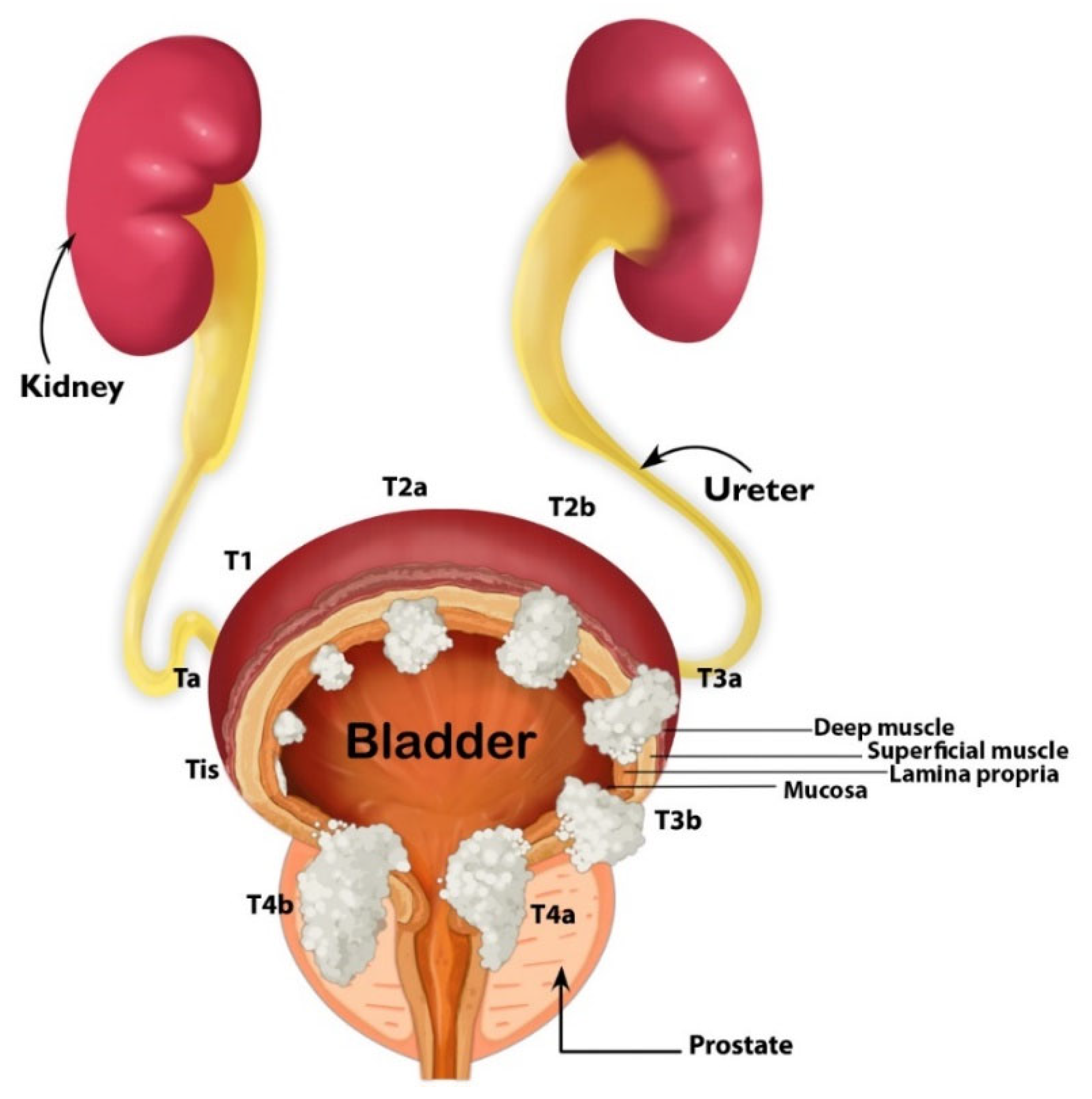
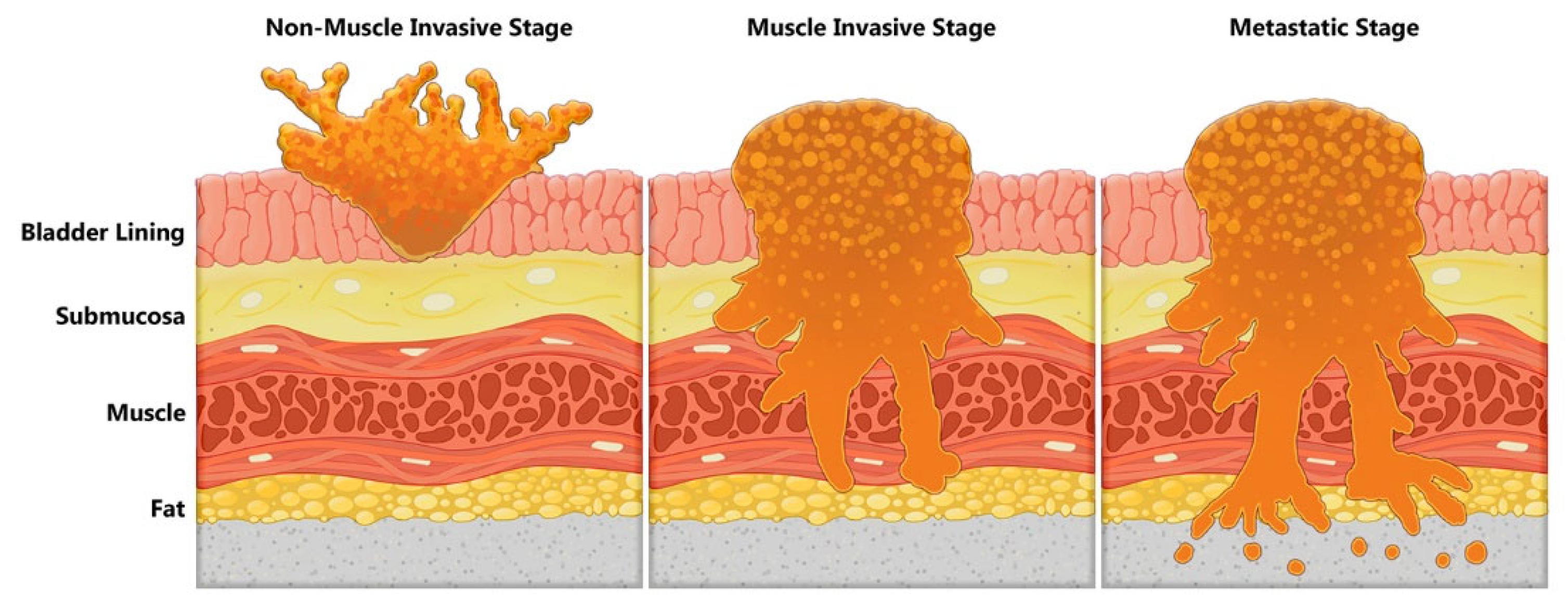
2. Overview on BC
3. Clinical Management of BC
4. Molecular Basis of BC and Clinical Translation
5. Stem Cell Origin of BC
6. Genetic Hallmarks of Papillary and Non-Papillary Invasive Pathways of BC Development
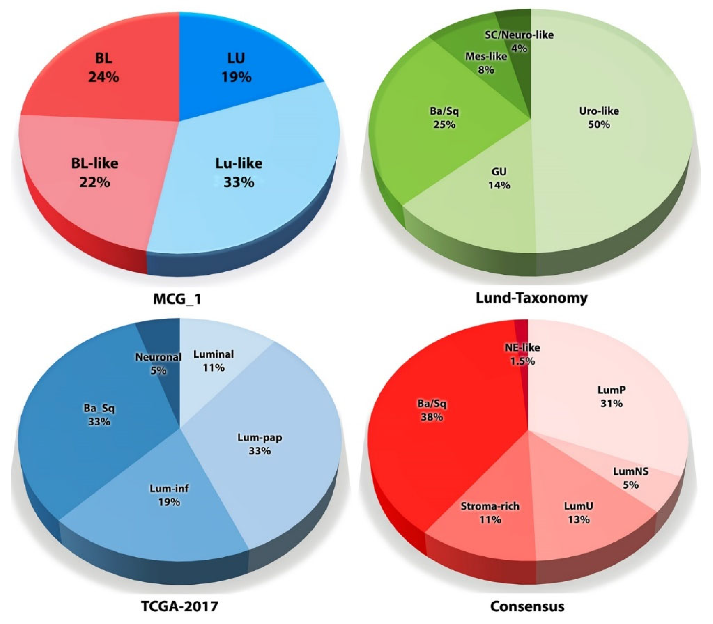
7. Transcriptome Profiling and the Quest for Personalized Medicine
8. Genomic Alterations/Tumor Mutation Burden in BC
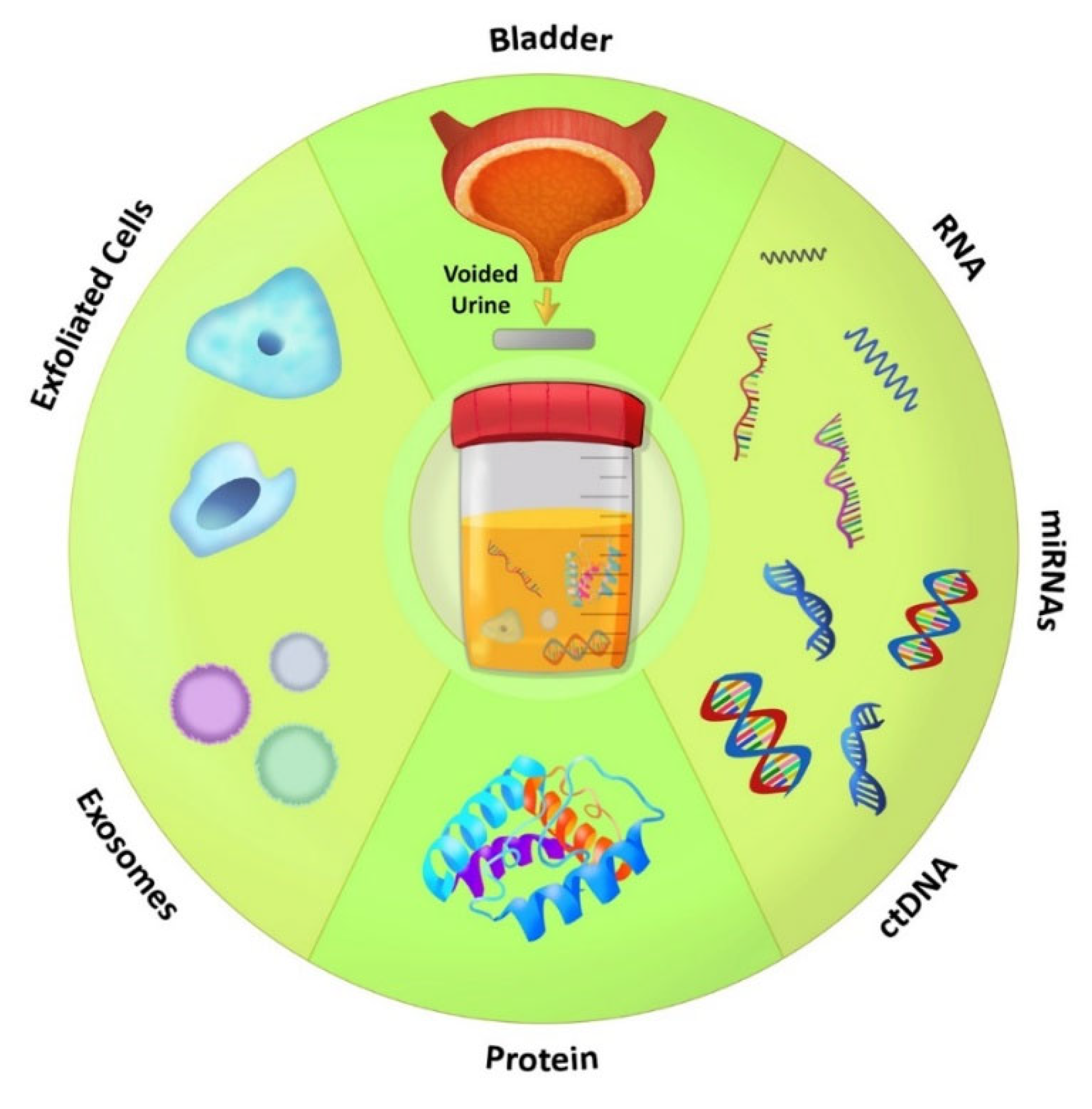
9. Urine Biomarkers for BC: Clinical Need and Reality
10. Ease of Developing Urine Biomarkers
11. Clinical Translation of Urine Biomarkers: Where Are We?
12. Conclusions
Author Contributions
Funding
Conflicts of Interest
Abbreviations
References
- Cathomas, R.; Lorch, A.; Bruins, H.M.; Compérat, E.M.; Cowan, N.C.; Efstathiou, J.A.; Fietkau, R.; Gakis, G.; Hernández, V.; Espinós, E.L.; et al. The 2021 Updated European Association of Urology Guidelines on Metastatic Urothelial Carcinoma. Eur. Urol. 2021, 81, 95–103. [Google Scholar] [CrossRef] [PubMed]
- Miller, K.D.; Ortiz, A.P.; Pinheiro, P.S.; Bandi, P.; Minihan, A.; Fuchs, H.E.; Martinez Tyson, D.; Tortolero-Luna, G.; Fedewa, S.A.; Jemal, A.M.; et al. Cancer statistics for the US Hispanic/Latino population, 2021. CA A Cancer J. Clin. 2021, 71, 466–487. [Google Scholar] [CrossRef] [PubMed]
- Rubio-Briones, J.; Algaba, F.; Gallardo, E.; Marcos-Rodriguez, J.A.; Climent, M.A.; SOGUG Multidisciplinary Working Group. Recent Advances in the Management of Patients with Non-Muscle-Invasive Bladder Cancer Using a Multidisciplinary Approach: Practical Recommendations from the Spanish Oncology Genitourinary (SOGUG) Working Group. Cancers 2021, 13, 4762. [Google Scholar] [CrossRef] [PubMed]
- Mainwaring, A.; Bullock, N.; Ellul, T.; Hughes, O.; Featherstone, J. The top 100 most cited manuscripts in bladder cancer: A bibliometric analysis (review article). Int. J. Surg. 2020, 75, 130–138. [Google Scholar] [CrossRef]
- Droller, M.J. Bladder cancer. Curr. Probl. Surg. 1981, 18, 205–279. [Google Scholar] [CrossRef]
- Jones, P.A.; Droller, M.J. Pathways of development and progression in bladder cancer: New correlations between clinical observations and molecular mechanisms. Semin. Urol. 1993, 11, 177–192. [Google Scholar]
- Audenet, F.; Attalla, K.; Sfakianos, J.P. The evolution of bladder cancer genomics: What have we learned and how can we use it? Urol. Oncol. Semin. Orig. Investig. 2018, 36, 313–320. [Google Scholar] [CrossRef]
- Lee, R.; Droller, M.J. The natural history of bladder cancer. Implications for therapy. Urol. Clin. N. Am. 2000, 27, 1–13. [Google Scholar] [CrossRef]
- van Doeveren, T.; van de Werken, H.J.G.; van Riet, J.; Aben, K.K.H.; van Leeuwen, P.J.; Zwarthoff, E.C.; Boormans, J.L. Synchronous and metachronous urothelial carcinoma of the upper urinary tract and the bladder: Are they clonally related? A systematic review. Urol. Oncol. Semin. Orig. Investig. 2020, 38, 590–598. [Google Scholar] [CrossRef]
- Dakubo, G.D.; Jakupciak, J.P.; Birch-Machin, M.A.; Parr, R.L. Clinical implications and utility of field cancerization. Cancer Cell Int. 2007, 7, 2. [Google Scholar] [CrossRef] [Green Version]
- Hafner, C.; Knuechel, R.; Stoehr, R.; Hartmann, A. Clonality of multifocal urothelial carcinomas: 10 years of molecular genetic studies. Int. J. Cancer 2002, 101, 1–6. [Google Scholar] [CrossRef] [PubMed]
- Dhaini, H.R.; El Hafi, B.; Khamis, A.M. NAT1 genotypic and phenotypic contribution to urinary bladder cancer risk: A systematic review and meta-analysis. Drug Metab. Rev. 2018, 50, 208–219. [Google Scholar] [CrossRef] [PubMed]
- Yu, C.; Hequn, C.; Longfei, L.; Long, W.; Zhi, C.; Feng, Z.; Jinbo, C.; Chao, L.; Xiongbing, Z. GSTM1 and GSTT1 polymorphisms are associated with increased bladder cancer risk: Evidence from updated meta-analysis. Oncotarget 2017, 8, 3246–3258. [Google Scholar] [CrossRef] [PubMed] [Green Version]
- Hein, D.W. Molecular genetics and function of NAT1 and NAT2: Role in aromatic amine metabolism and carcinogenesis. Mutat. Res. 2002, 506, 65–77. [Google Scholar] [CrossRef] [Green Version]
- Vineis, P.; Martone, T.; Randone, D. Molecular epidemiology of bladder cancer: Known chemical causes of bladder cancer: Occupation and smoking. Urol. Oncol. Semin. Orig. Investig. 1995, 1, 137–143. [Google Scholar] [CrossRef]
- Rhea, L.P.; Mendez-Marti, S.; Kim, D.; Aragon-Ching, J.B. Role of immunotherapy in bladder cancer. Cancer Treat. Res. Commun. 2020, 26, 100296. [Google Scholar] [CrossRef]
- Mukherjee, N.; Wheeler, K.M.; Svatek, R.S. Bacillus Calmette-Guerin treatment of bladder cancer: A systematic review and commentary on recent publications. Curr. Opin. Urol. 2019, 29, 181–188. [Google Scholar] [CrossRef]
- Epand, R.F.; Epand, R.M. 25-Hydroxycholesterol promotes myelin basic protein-induced leakage of phospholipid vesicles. Biochim. Biophys. Acta 1988, 938, 131–134. [Google Scholar] [CrossRef]
- Lee, S.; Jeong, J.; Majewski, T.; Scherer, S.E.; Kim, M.S.; Tuziak, T.; Tang, K.S.; Baggerly, K.; Grossman, H.B.; Zhou, J.H.; et al. Forerunner genes contiguous to RB1 contribute to the development of in situ neoplasia. Proc. Natl. Acad. Sci. USA 2007, 104, 13732–13737. [Google Scholar] [CrossRef] [Green Version]
- Majewski, T.; Lee, S.; Jeong, J.; Yoon, D.S.; Kram, A.; Kim, M.S.; Tuziak, T.; Bondaruk, J.; Lee, S.; Park, W.S.; et al. Understanding the development of human bladder cancer by using a whole-organ genomic mapping strategy. Lab. Investig. 2008, 88, 694–721. [Google Scholar] [CrossRef] [Green Version]
- Kryger, J.V.; Messing, E. Bladder cancer screening. Semin. Oncol. 1996, 23, 585–597. [Google Scholar] [PubMed]
- Laukhtina, E.; Pradere, B.; Mori, K.; Schuettfort, V.M.; Quhal, F.; Mostafaei, H.; Sari Motlangh, R.; Katayama, S.; Grossmann, N.C.; Moschini, M.; et al. Catalog of prognostic tissue-based biomarkers in patients treated with neoadjuvant systemic therapy for urothelial carcinoma of the bladder: A systematic review. Urol. Oncol. Semin. Orig. Investig. 2021, 39, 180–190. [Google Scholar] [CrossRef]
- Messing, E.M.; Vaillancourt, A. Hematuria screening for bladder cancer. J. Occup. Med. 1990, 32, 838–845. [Google Scholar] [CrossRef] [PubMed]
- Ng, K.; Stenzl, A.; Sharma, A.; Vasdev, N. Urinary Biomarkers in Bladder Cancer: A Review of the Current Landscape and Future Directions. Urol. Oncol. Semin. Orig. Investig. 2020, 39, 41–51. [Google Scholar] [CrossRef] [PubMed]
- Satyal, U.; Srivastava, A.; Abbosh, P.H. Urine Biopsy-Liquid Gold for Molecular Detection and Surveillance of Bladder Cancer. Front. Oncol. 2019, 9, 1266. [Google Scholar] [CrossRef] [PubMed] [Green Version]
- Sjodahl, G.; Jackson, C.L.; Bartlett, J.M.; Siemens, D.R.; Berman, D.M. Molecular profiling in muscle-invasive bladder cancer: More than the sum of its parts. J. Pathol. 2019, 247, 563–573. [Google Scholar] [CrossRef] [PubMed] [Green Version]
- Da Costa, J.B.; Gibb, E.A.; Nykopp, T.K.; Mannas, M.; Wyatt, A.W.; Black, P.C. Molecular Tumor Heterogeneity in Muscle Invasive Bladder Cancer: Biomarkers, Subtypes, and Implications for Therapy. Urol. Oncol. Semin. Orig. Investig. 2018. online ahead of print. [Google Scholar] [CrossRef]
- Kripnerova, M.; Parmar, H.S.; Pesta, M.; Kohoutova, M.; Kuncova, J.; Drbal, K.; Rajtmajerova, M.; Hatina, J. Urothelial Cancer Stem Cell Heterogeneity. Adv. Exp. Med. Biol. 2019, 1139, 127–151. [Google Scholar] [CrossRef]
- Siegel, R.L.; Miller, K.D.; Fuchs, H.E.; Jemal, A. Cancer Statistics, 2021. CA Cancer J. Clin. 2021, 71, 7–33. [Google Scholar] [CrossRef]
- Kaufman, D.S.; Shipley, W.U.; Feldman, A.S. Bladder cancer. Lancet 2009, 374, 239–249. [Google Scholar] [CrossRef]
- Shokeir, A. Squamous cell carcinoma of the bladder: Pathology, diagnosis and treatment. BJU Int. 2004, 93, 216–220. [Google Scholar] [CrossRef] [PubMed]
- Freedman, N.D.; Silverman, D.T.; Hollenbeck, A.R.; Schatzkin, A.; Abnet, C.C. Association between smoking and risk of bladder cancer among men and women. JAMA 2011, 306, 737–745. [Google Scholar] [CrossRef] [PubMed]
- Lokeshwar, S.; Klaassen, Z.; Terris, M. A Contemporary Review of Risk Factors for Bladder. Clin. Oncol. 2016, 1, 1–3. [Google Scholar]
- Aben, K.K.; Witjes, J.A.; Schoenberg, M.P.; Hulsbergen-van de Kaa, C.; Verbeek, A.L.; Kiemeney, L.A. Familial aggregation of urothelial cell carcinoma. Int. J. Cancer 2002, 98, 274–278. [Google Scholar] [CrossRef]
- Siegel, R.L.; Miller, K.D.; Jemal, A. Cancer statistics, 2020. CA A Cancer J. Clin. 2020, 70, 7–30. [Google Scholar] [CrossRef]
- Shephard, E.A.; Stapley, S.; Neal, R.D.; Rose, P.; Walter, F.M.; Hamilton, W.T. Clinical features of bladder cancer in primary care. Br. J. Gen. Pract. 2012, 62, e598–e604. [Google Scholar] [CrossRef] [Green Version]
- Davis, R.; Jones, J.S.; Barocas, D.A.; Castle, E.P.; Lang, E.K.; Leveillee, R.J.; Messing, E.M.; Miller, S.D.; Peterson, A.C.; Turk, T.M. Diagnosis, evaluation and follow-up of asymptomatic microhematuria (AMH) in adults: AUA guideline. J. Urol. 2012, 188, 2473–2481. [Google Scholar] [CrossRef]
- Babjuk, M.; Bohle, A.; Burger, M.; Capoun, O.; Cohen, D.; Comperat, E.M.; Hernandez, V.; Kaasinen, E.; Palou, J.; Roupret, M.; et al. EAU Guidelines on Non-Muscle-invasive Urothelial Carcinoma of the Bladder: Update 2016. Eur. Urol. 2017, 71, 447–461. [Google Scholar] [CrossRef]
- Majewski, T.; Yao, H.; Bondaruk, J.; Chung, W.; Lee, S.; Lee, J.G.; Zhang, S.; Cogdell, D.; Yang, G.; Choi, W.; et al. Whole-Organ Genomic Characterization of Mucosal Field Effects Initiating Bladder Carcinogenesis. Cell Rep. 2019, 26, 2241–2256.e4. [Google Scholar] [CrossRef] [Green Version]
- Wolff, E.M.; Chihara, Y.; Pan, F.; Weisenberger, D.J.; Siegmund, K.D.; Sugano, K.; Kawashima, K.; Laird, P.W.; Jones, P.A.; Liang, G. Unique DNA methylation patterns distinguish noninvasive and invasive urothelial cancers and establish an epigenetic field defect in premalignant tissue. Cancer Res. 2010, 70, 8169–8178. [Google Scholar] [CrossRef] [Green Version]
- Heney, N.M.; Ahmed, S.; Flanagan, M.J.; Frable, W.; Corder, M.P.; Hafermann, M.D.; Hawkins, I.R. Superficial bladder cancer: Progression and recurrence. J. Urol. 1983, 130, 1083–1086. [Google Scholar] [CrossRef]
- Jakse, G.; Loidl, W.; Seeber, G.; Hofstadter, F. Stage T1, grade 3 transitional cell carcinoma of the bladder: An unfavorable tumor? J. Urol. 1987, 137, 39–43. [Google Scholar] [CrossRef]
- Kobayashi, H.; Kikuchi, E.; Mikami, S.; Maeda, T.; Tanaka, N.; Miyajima, A.; Nakagawa, K.; Oya, M. Long term follow-up in patients with initially diagnosed low grade Ta non-muscle invasive bladder tumors: Tumor recurrence and worsening progression. BMC Urol. 2014, 14, 5. [Google Scholar] [CrossRef] [PubMed] [Green Version]
- Simon, M.; Bosset, P.O.; Rouanne, M.; Benhamou, S.; Radulescu, C.; Molinie, V.; Neuzillet, Y.; Paoletti, X.; Lebret, T. Multiple recurrences and risk of disease progression in patients with primary low-grade (TaG1) non-muscle-invasive bladder cancer and with low and intermediate EORTC-risk score. PLoS ONE 2019, 14, e0211721. [Google Scholar] [CrossRef]
- Ritch, C.R.; Velasquez, M.C.; Kwon, D.; Becerra, M.F.; Soodana-Prakash, N.; Atluri, V.S.; Almengo, K.; Alameddine, M.; Kineish, O.; Kava, B.R.; et al. Use and Validation of the AUA/SUO Risk Grouping for Nonmuscle Invasive Bladder Cancer in a Contemporary Cohort. J. Urol. 2020, 203, 505–511. [Google Scholar] [CrossRef]
- Chamie, K.; Litwin, M.S.; Bassett, J.C.; Daskivich, T.J.; Lai, J.; Hanley, J.M.; Konety, B.R.; Saigal, C.S.; Urologic Diseases in America, P. Recurrence of high-risk bladder cancer: A population-based analysis. Cancer 2013, 119, 3219–3227. [Google Scholar] [CrossRef] [Green Version]
- Hamid, A.; Ridwan, F.R.; Parikesit, D.; Widia, F.; Mochtar, C.A.; Umbas, R. Meta-analysis of neoadjuvant chemotherapy compared to radical cystectomy alone in improving overall survival of muscle-invasive bladder cancer patients. BMC Urol. 2020, 20, 158. [Google Scholar] [CrossRef]
- Chang, S.S.; Bochner, B.H.; Chou, R.; Dreicer, R.; Kamat, A.M.; Lerner, S.P.; Lotan, Y.; Meeks, J.J.; Michalski, J.M.; Morgan, T.M.; et al. Treatment of Non-Metastatic Muscle-Invasive Bladder Cancer: AUA/ASCO/ASTRO/SUO Guideline. J. Urol. 2017, 198, 552–559. [Google Scholar] [CrossRef]
- Abufaraj, M.; Dalbagni, G.; Daneshmand, S.; Horenblas, S.; Kamat, A.M.; Kanzaki, R.; Zlotta, A.R.; Shariat, S.F. The Role of Surgery in Metastatic Bladder Cancer: A Systematic Review. Eur. Urol. 2018, 73, 543–557. [Google Scholar] [CrossRef]
- Nadal, R.; Bellmunt, J. Management of metastatic bladder cancer. Cancer Treat. Rev. 2019, 76, 10–21. [Google Scholar] [CrossRef]
- Pardo, J.C.; Ruiz de Porras, V.; Plaja, A.; Carrato, C.; Etxaniz, O.; Buisan, O.; Font, A. Moving towards Personalized Medicine in Muscle-Invasive Bladder Cancer: Where Are We Now and Where Are We Going? Int. J. Mol. Sci. 2020, 21, 6271. [Google Scholar] [CrossRef] [PubMed]
- Rouanne, M.; Bajorin, D.F.; Hannan, R.; Galsky, M.D.; Williams, S.B.; Necchi, A.; Sharma, P.; Powles, T. Rationale and Outcomes for Neoadjuvant Immunotherapy in Urothelial Carcinoma of the Bladder. Eur. Urol. Oncol. 2020, 3, 728–738. [Google Scholar] [CrossRef] [PubMed]
- Leow, J.J.; Bedke, J.; Chamie, K.; Collins, J.W.; Daneshmand, S.; Grivas, P.; Heidenreich, A.; Messing, E.M.; Royce, T.J.; Sankin, A.I.; et al. SIU-ICUD consultation on bladder cancer: Treatment of muscle-invasive bladder cancer. World J. Urol. 2019, 37, 61–83. [Google Scholar] [CrossRef]
- Patel, V.G.; Oh, W.K.; Galsky, M.D. Treatment of muscle-invasive and advanced bladder cancer in 2020. CA A Cancer J. Clin. 2020, 70, 404–423. [Google Scholar] [CrossRef]
- von der Maase, H.; Hansen, S.W.; Roberts, J.T.; Dogliotti, L.; Oliver, T.; Moore, M.J.; Bodrogi, I.; Albers, P.; Knuth, A.; Lippert, C.M.; et al. Gemcitabine and cisplatin versus methotrexate, vinblastine, doxorubicin, and cisplatin in advanced or metastatic bladder cancer: Results of a large, randomized, multinational, multicenter, phase III study. J. Clin. Oncol. 2000, 18, 3068–3077. [Google Scholar] [CrossRef] [PubMed]
- von der Maase, H.; Sengelov, L.; Roberts, J.T.; Ricci, S.; Dogliotti, L.; Oliver, T.; Moore, M.J.; Zimmermann, A.; Arning, M. Long-term survival results of a randomized trial comparing gemcitabine plus cisplatin, with methotrexate, vinblastine, doxorubicin, plus cisplatin in patients with bladder cancer. J. Clin. Oncol. 2005, 23, 4602–4608. [Google Scholar] [CrossRef] [PubMed]
- Lee, Y.; Kim, Y.S.; Hong, B.; Cho, Y.M.; Lee, J.L. Comparison of clinical outcomes in patients with localized or locally advanced urothelial carcinoma treated with neoadjuvant chemotherapy involving gemcitabine-cisplatin and high dose-intensity MVAC. J. Cancer Res. Clin. Oncol. 2021, 147, 3421–3429. [Google Scholar] [CrossRef]
- Qu, H.C.; Huang, Y.; Mu, Z.Y.; Lv, H.; Xie, Q.P.; Wang, K.; Hu, B. Efficacy and Safety of Chemotherapy Regimens in Advanced or Metastatic Bladder and Urothelial Carcinomas: An Updated Network Meta-Analysis. Front. Pharmacol. 2019, 10, 1507. [Google Scholar] [CrossRef]
- Cooley, L.F.; Glaser, A.P.; Meeks, J.J. Mutation signatures to Pan-Cancer Atlas: Investigation of the genomic landscape of muscle-invasive bladder cancer. Urol. Oncol. Semin. Orig. Investig. 2020. online ahead of print. [Google Scholar] [CrossRef]
- Czerniak, B.; Dinney, C.; McConkey, D. Origins of Bladder Cancer. Annu. Rev. Pathol. Mech. Dis. 2016, 11, 149–174. [Google Scholar] [CrossRef]
- Kamoun, A.; de Reynies, A.; Allory, Y.; Sjodahl, G.; Robertson, A.G.; Seiler, R.; Hoadley, K.A.; Groeneveld, C.S.; Al-Ahmadie, H.; Choi, W.; et al. A Consensus Molecular Classification of Muscle-invasive Bladder Cancer. Eur. Urol. 2020, 77, 420–433. [Google Scholar] [CrossRef]
- Kang, H.W.; Kim, W.J.; Choi, W.; Yun, S.J. Tumor heterogeneity in muscle-invasive bladder cancer. Transl. Androl. Urol. 2020, 9, 2866–2880. [Google Scholar] [CrossRef] [PubMed]
- Li, Y.; Lin, K.; Yang, Z.; Han, N.; Quan, X.; Guo, X.; Li, C. Bladder cancer stem cells: Clonal origin and therapeutic perspectives. Oncotarget 2017, 8, 66668–66679. [Google Scholar] [CrossRef] [PubMed] [Green Version]
- Meeks, J.J.; Al-Ahmadie, H.; Faltas, B.M.; Taylor, J.A., 3rd; Flaig, T.W.; DeGraff, D.J.; Christensen, E.; Woolbright, B.L.; McConkey, D.J.; Dyrskjot, L. Genomic heterogeneity in bladder cancer: Challenges and possible solutions to improve outcomes. Nat. Rev. Urol. 2020, 17, 259–270. [Google Scholar] [CrossRef] [PubMed]
- Minoli, M.; Kiener, M.; Thalmann, G.N.; Kruithof-de Julio, M.; Seiler, R. Evolution of Urothelial Bladder Cancer in the Context of Molecular Classifications. Int. J. Mol. Sci. 2020, 21, 5670. [Google Scholar] [CrossRef] [PubMed]
- Necchi, A.; Raggi, D.; Gallina, A.; Ross, J.S.; Fare, E.; Giannatempo, P.; Marandino, L.; Colecchia, M.; Luciano, R.; Bianchi, M.; et al. Impact of Molecular Subtyping and Immune Infiltration on Pathological Response and Outcome Following Neoadjuvant Pembrolizumab in Muscle-invasive Bladder Cancer. Eur. Urol. 2020, 77, 701–710. [Google Scholar] [CrossRef] [PubMed]
- Sfakianos, J.P.; Daza, J.; Hu, Y.; Anastos, H.; Bryant, G.; Bareja, R.; Badani, K.K.; Galsky, M.D.; Elemento, O.; Faltas, B.M.; et al. Epithelial plasticity can generate multi-lineage phenotypes in human and murine bladder cancers. Nat. Commun. 2020, 11, 2540. [Google Scholar] [CrossRef] [PubMed]
- Dinney, C.P.; Hansel, D.; McConkey, D.; Shipley, W.; Hagan, M.; Dreicer, R.; Lerner, S.; Czerniak, B.; Waldman, F.; Groshen, S.; et al. Novel neoadjuvant therapy paradigms for bladder cancer: Results from the National Cancer Center Institute Forum. Urol. Oncol. 2014, 32, 1108–1115. [Google Scholar] [CrossRef] [Green Version]
- Seiler, R.; Ashab, H.A.D.; Erho, N.; van Rhijn, B.W.G.; Winters, B.; Douglas, J.; Van Kessel, K.E.; Fransen van de Putte, E.E.; Sommerlad, M.; Wang, N.Q.; et al. Impact of Molecular Subtypes in Muscle-invasive Bladder Cancer on Predicting Response and Survival after Neoadjuvant Chemotherapy. Eur. Urol. 2017, 72, 544–554. [Google Scholar] [CrossRef]
- Abugomaa, A.; Elbadawy, M.; Yamawaki, H.; Usui, T.; Sasaki, K. Emerging Roles of Cancer Stem Cells in Bladder Cancer Progression, Tumorigenesis, and Resistance to Chemotherapy: A Potential Therapeutic Target for Bladder Cancer. Cells 2020, 9, 235. [Google Scholar] [CrossRef] [Green Version]
- Najfeld, V.; Zucker-Franklin, D.; Adamson, J.; Singer, J.; Troy, K.; Fialkow, P.J. Evidence for clonal development and stem cell origin of M7 megakaryocytic leukemia. Leukemia 1988, 2, 351–357. [Google Scholar] [PubMed]
- Aghaalikhani, N.; Rashtchizadeh, N.; Shadpour, P.; Allameh, A.; Mahmoodi, M. Cancer stem cells as a therapeutic target in bladder cancer. J. Cell Physiol. 2019, 234, 3197–3206. [Google Scholar] [CrossRef] [PubMed]
- Hicks, R.M. The mammalian urinary bladder: An accommodating organ. Biol. Rev. 1975, 50, 215–246. [Google Scholar] [CrossRef]
- Jost, S.P. Cell cycle of normal bladder urothelium in developing and adult mice. Virchows Arch. B Cell Pathol. Incl. Mol. Pathol. 1989, 57, 27–36. [Google Scholar] [CrossRef] [PubMed]
- Khandelwal, P.; Abraham, S.N.; Apodaca, G. Cell biology and physiology of the uroepithelium. Am. J. Physiol. Physiol. 2009, 297, F1477–F1501. [Google Scholar] [CrossRef] [PubMed] [Green Version]
- Balsara, Z.R.; Li, X. Sleeping beauty: Awakening urothelium from its slumber. Am. J. Physiol. Physiol. 2017, 312, F732–F743. [Google Scholar] [CrossRef] [Green Version]
- Fuchs, E.; Horsley, V. More than one way to skin. Genes Dev. 2008, 22, 976–985. [Google Scholar] [CrossRef] [Green Version]
- Gandhi, D.; Molotkov, A.; Batourina, E.; Schneider, K.; Dan, H.; Reiley, M.; Laufer, E.; Metzger, D.; Liang, F.; Liao, Y.; et al. Retinoid signaling in progenitors controls specification and regeneration of the urothelium. Dev. Cell 2013, 26, 469–482. [Google Scholar] [CrossRef] [Green Version]
- Shin, K.; Lim, A.; Odegaard, J.I.; Honeycutt, J.D.; Kawano, S.; Hsieh, M.H.; Beachy, P.A. Cellular origin of bladder neoplasia and tissue dynamics of its progression to invasive carcinoma. Nat. Cell Biol. 2014, 16, 469–478. [Google Scholar] [CrossRef] [Green Version]
- Shin, K.; Lim, A.; Zhao, C.; Sahoo, D.; Pan, Y.; Spiekerkoetter, E.; Liao, J.C.; Beachy, P.A. Hedgehog signaling restrains bladder cancer progression by eliciting stromal production of urothelial differentiation factors. Cancer Cell 2014, 26, 521–533. [Google Scholar] [CrossRef] [Green Version]
- Van Batavia, J.; Yamany, T.; Molotkov, A.; Dan, H.; Mansukhani, M.; Batourina, E.; Schneider, K.; Oyon, D.; Dunlop, M.; Wu, X.R.; et al. Bladder cancers arise from distinct urothelial sub-populations. Nat. Cell Biol. 2014, 16, 982–991. [Google Scholar] [CrossRef] [PubMed]
- Solomon, J.P.; Hansel, D.E. The Emerging Molecular Landscape of Urothelial Carcinoma. Surg. Pathol. Clin. 2016, 9, 391–404. [Google Scholar] [CrossRef] [PubMed]
- McConkey, D.J.; Lee, S.; Choi, W.; Tran, M.; Majewski, T.; Lee, S.; Siefker-Radtke, A.; Dinney, C.; Czerniak, B. Molecular genetics of bladder cancer: Emerging mechanisms of tumor initiation and progression. Urol. Oncol. 2010, 28, 429–440. [Google Scholar] [CrossRef] [PubMed] [Green Version]
- Nassar, A.H.; Umeton, R.; Kim, J.; Lundgren, K.; Harshman, L.; Van Allen, E.M.; Preston, M.; Dong, F.; Bellmunt, J.; Mouw, K.W.; et al. Mutational Analysis of 472 Urothelial Carcinoma Across Grades and Anatomic Sites. Clin. Cancer Res. 2019, 25, 2458–2470. [Google Scholar] [CrossRef]
- Rogan, P.K. Multigene signatures of responses to chemotherapy derived by biochemically-inspired machine learning. Mol. Genet. Metab. 2019, 128, 45–52. [Google Scholar] [CrossRef]
- Matulay, J.T.; Narayan, V.M.; Kamat, A.M. Clinical and Genomic Considerations for Variant Histology in Bladder Cancer. Curr. Oncol. Rep. 2019, 21, 23. [Google Scholar] [CrossRef]
- Tsai, Y.C.; Nichols, P.W.; Hiti, A.L.; Williams, Z.; Skinner, D.G.; Jones, P.A. Allelic losses of chromosomes 9, 11, and 17 in human bladder cancer. Cancer Res. 1990, 50, 44–47. [Google Scholar]
- Knowles, M.A. The genetics of transitional cell carcinoma: Progress and potential clinical application. BJU Int. 1999, 84, 412–427. [Google Scholar] [CrossRef]
- Knowles, M.A.; Elder, P.A.; Williamson, M.; Cairns, J.P.; Shaw, M.E.; Law, M.G. Allelotype of human bladder cancer. Cancer Res. 1994, 54, 531–538. [Google Scholar]
- Olumi, A.F.; Tsai, Y.C.; Nichols, P.W.; Skinner, D.G.; Cain, D.R.; Bender, L.I.; Jones, P.A. Allelic loss of chromosome 17p distinguishes high grade from low grade transitional cell carcinomas of the bladder. Cancer Res. 1990, 50, 7081–7083. [Google Scholar]
- Pollard, C.; Smith, S.C.; Theodorescu, D. Molecular genesis of non-muscle-invasive urothelial carcinoma (NMIUC). Expert Rev. Mol. Med. 2010, 12, e10. [Google Scholar] [CrossRef] [PubMed] [Green Version]
- Simoneau, A.R.; Spruck, C.H., 3rd; Gonzalez-Zulueta, M.; Gonzalgo, M.L.; Chan, M.F.; Tsai, Y.C.; Dean, M.; Steven, K.; Horn, T.; Jones, P.A. Evidence for two tumor suppressor loci associated with proximal chromosome 9p to q and distal chromosome 9q in bladder cancer and the initial screening for GAS1 and PTC mutations. Cancer Res. 1996, 56, 5039–5043. [Google Scholar] [PubMed]
- Ploussard, G.; Dubosq, F.; Soliman, H.; Verine, J.; Desgrandchamps, F.; De The, H.; Mongiat-Artus, P. Prognostic value of loss of heterozygosity at chromosome 9p in non-muscle-invasive bladder cancer. Urology 2010, 76, 513.e13–513.e18. [Google Scholar] [CrossRef] [PubMed]
- Dalbagni, G.; Presti, J.C., Jr.; Reuter, V.E.; Zhang, Z.F.; Sarkis, A.S.; Fair, W.R.; Cordon-Cardo, C. Molecular genetic alterations of chromosome 17 and p53 nuclear overexpression in human bladder cancer. Diagn. Mol. Pathol. 1993, 2, 4–13. [Google Scholar] [CrossRef] [PubMed]
- Li, B.; Kanamaru, H.; Noriki, S.; Fukuda, M.; Okada, K. Numeric aberration of chromosome 17 is strongly correlated with p53 overexpression, tumor proliferation and histopathology in human bladder cancer. Int. J. Urol. 1998, 5, 317–323. [Google Scholar] [CrossRef] [Green Version]
- Hoglund, M.; Sall, T.; Heim, S.; Mitelman, F.; Mandahl, N.; Fadl-Elmula, I. Identification of cytogenetic subgroups and karyotypic pathways in transitional cell carcinoma. Cancer Res. 2001, 61, 8241–8246. [Google Scholar]
- Neuzillet, Y.; Paoletti, X.; Ouerhani, S.; Mongiat-Artus, P.; Soliman, H.; de The, H.; Sibony, M.; Denoux, Y.; Molinie, V.; Herault, A.; et al. A meta-analysis of the relationship between FGFR3 and TP53 mutations in bladder cancer. PLoS ONE 2012, 7, e48993. [Google Scholar] [CrossRef]
- Ouerhani, S.; Elgaaied, A.B. The mutational spectrum of HRAS, KRAS, NRAS and FGFR3 genes in bladder cancer. Cancer Biomark. 2011, 10, 259–266. [Google Scholar] [CrossRef]
- Jebar, A.H.; Hurst, C.D.; Tomlinson, D.C.; Johnston, C.; Taylor, C.F.; Knowles, M.A. FGFR3 and Ras gene mutations are mutually exclusive genetic events in urothelial cell carcinoma. Oncogene 2005, 24, 5218–5225. [Google Scholar] [CrossRef] [Green Version]
- Taylor, C.F.; Platt, F.M.; Hurst, C.D.; Thygesen, H.H.; Knowles, M.A. Frequent inactivating mutations of STAG2 in bladder cancer are associated with low tumour grade and stage and inversely related to chromosomal copy number changes. Hum. Mol. Genet. 2014, 23, 1964–1974. [Google Scholar] [CrossRef] [Green Version]
- Gordon, N.S.; Humayun-Zakaria, N.; Goel, A.; Abbotts, B.; Zeegers, M.P.; Cheng, K.K.; James, N.D.; Arnold, R.; Bryan, R.T.; Ward, D.G. STAG2 Protein Expression in Non-muscle-invasive Bladder Cancer: Associations with Sex, Genomic and Transcriptomic Changes, and Clinical Outcomes. Eur. Urol. Open Sci. 2022, 38, 88–95. [Google Scholar] [CrossRef] [PubMed]
- Tuziak, T.; Jeong, J.; Majewski, T.; Kim, M.S.; Steinberg, J.; Wang, Z.; Yoon, D.S.; Kuang, T.C.; Baggerly, K.; Johnston, D.; et al. High-resolution whole-organ mapping with SNPs and its significance to early events of carcinogenesis. Lab. Investig. 2005, 85, 689–701. [Google Scholar] [CrossRef] [PubMed] [Green Version]
- Czerniak, B. Molecular pathology and biomarkers of bladder cancer. Cancer Biomark. 2010, 9, 159–176. [Google Scholar] [CrossRef] [PubMed]
- Alhalabi, O.; Zhu, Y.; Hamza, A.; Qiao, W.; Lin, Y.; Wang, R.M.; Shah, A.Y.; Campbell, M.T.; Holla, V.; Kamat, A.; et al. Integrative Clinical and Genomic Characterization of MTAP-deficient Metastatic Urothelial Cancer. Eur. Urol. Oncol. 2021. online ahead of print. [Google Scholar] [CrossRef]
- Williamson, M.P.; Elder, P.A.; Shaw, M.E.; Devlin, J.; Knowles, M.A. p16 (CDKN2) is a major deletion target at 9p21 in bladder cancer. Hum. Mol. Genet. 1995, 4, 1569–1577. [Google Scholar] [CrossRef]
- Cordon-Cardo, C.; Sheinfeld, J. Molecular and immunopathology studies of oncogenes and tumor-suppressor genes in bladder cancer. World J. Urol. 1997, 15, 112–119. [Google Scholar] [CrossRef] [PubMed]
- Dalbagni, G.; Ren, Z.P.; Herr, H.; Cordon-Cardo, C.; Reuter, V. Genetic alterations in tp53 in recurrent urothelial cancer: A longitudinal study. Clin. Cancer Res. 2001, 7, 2797–2801. [Google Scholar]
- Shariat, S.F.; Bolenz, C.; Karakiewicz, P.I.; Fradet, Y.; Ashfaq, R.; Bastian, P.J.; Nielsen, M.E.; Capitanio, U.; Jeldres, C.; Rigaud, J.; et al. p53 expression in patients with advanced urothelial cancer of the urinary bladder. BJU Int. 2010, 105, 489–495. [Google Scholar] [CrossRef]
- Brunelli, M.; Tafuri, A.; Cima, L.; Cerruto, M.A.; Milella, M.; Zivi, A.; Buti, S.; Bersanelli, M.; Fornarini, G.; Vellone, V.G.; et al. MDM2 gene amplification as selection tool for innovative targeted approaches in PD-L1 positive or negative muscle-invasive urothelial bladder carcinoma. J. Clin. Pathol. 2022, 75, 39–44. [Google Scholar] [CrossRef]
- Vandekerkhove, G.; Todenhofer, T.; Annala, M.; Struss, W.J.; Wong, A.; Beja, K.; Ritch, E.; Brahmbhatt, S.; Volik, S.V.; Hennenlotter, J.; et al. Circulating Tumor DNA Reveals Clinically Actionable Somatic Genome of Metastatic Bladder Cancer. Clin. Cancer Res. 2017, 23, 6487–6497. [Google Scholar] [CrossRef] [Green Version]
- Lopez-Knowles, E.; Hernandez, S.; Kogevinas, M.; Lloreta, J.; Amoros, A.; Tardon, A.; Carrato, A.; Kishore, S.; Serra, C.; Malats, N.; et al. The p53 pathway and outcome among patients with T1G3 bladder tumors. Clin. Cancer Res. 2006, 12, 6029–6036. [Google Scholar] [CrossRef] [PubMed] [Green Version]
- Sanchez-Carbayo, M.; Socci, N.D.; Kirchoff, T.; Erill, N.; Offit, K.; Bochner, B.H.; Cordon-Cardo, C. A polymorphism in HDM2 (SNP309) associates with early onset in superficial tumors, TP53 mutations, and poor outcome in invasive bladder cancer. Clin. Cancer Res. 2007, 13, 3215–3220. [Google Scholar] [CrossRef] [Green Version]
- Cancer Genome Atlas Research, N. Comprehensive molecular characterization of urothelial bladder carcinoma. Nature 2014, 507, 315–322. [Google Scholar] [CrossRef] [PubMed] [Green Version]
- Grossman, H.B.; Liebert, M.; Antelo, M.; Dinney, C.P.; Hu, S.X.; Palmer, J.L.; Benedict, W.F. p53 and RB expression predict progression in T1 bladder cancer. Clin. Cancer Res. 1998, 4, 829–834. [Google Scholar] [PubMed]
- Miyamoto, H.; Shuin, T.; Ikeda, I.; Hosaka, M.; Kubota, Y. Loss of heterozygosity at the p53, RB, DCC and APC tumor suppressor gene loci in human bladder cancer. J. Urol. 1996, 155, 1444–1447. [Google Scholar] [CrossRef]
- Pollack, A.; Czerniak, B.; Zagars, G.K.; Hu, S.X.; Wu, C.S.; Dinney, C.P.; Chyle, V.; Benedict, W.F. Retinoblastoma protein expression and radiation response in muscle-invasive bladder cancer. Int. J. Radiat. Oncol. Biol. Phys. 1997, 39, 687–695. [Google Scholar] [CrossRef]
- He, F.; Mo, L.; Zheng, X.Y.; Hu, C.; Lepor, H.; Lee, E.Y.; Sun, T.T.; Wu, X.R. Deficiency of pRb family proteins and p53 in invasive urothelial tumorigenesis. Cancer Res. 2009, 69, 9413–9421. [Google Scholar] [CrossRef] [PubMed] [Green Version]
- Stadler, W.M.; Lerner, S.P.; Groshen, S.; Stein, J.P.; Shi, S.R.; Raghavan, D.; Esrig, D.; Steinberg, G.; Wood, D.; Klotz, L.; et al. Phase III study of molecularly targeted adjuvant therapy in locally advanced urothelial cancer of the bladder based on p53 status. J. Clin. Oncol. 2011, 29, 3443–3449. [Google Scholar] [CrossRef] [PubMed]
- Chan, K.S.; Espinosa, I.; Chao, M.; Wong, D.; Ailles, L.; Diehn, M.; Gill, H.; Presti, J., Jr.; Chang, H.Y.; van de Rijn, M.; et al. Identification, molecular characterization, clinical prognosis, and therapeutic targeting of human bladder tumor-initiating cells. Proc. Natl. Acad. Sci. USA 2009, 106, 14016–14021. [Google Scholar] [CrossRef] [Green Version]
- He, X.; Marchionni, L.; Hansel, D.E.; Yu, W.; Sood, A.; Yang, J.; Parmigiani, G.; Matsui, W.; Berman, D.M. Differentiation of a highly tumorigenic basal cell compartment in urothelial carcinoma. Stem. Cells 2009, 27, 1487–1495. [Google Scholar] [CrossRef] [Green Version]
- Volkmer, J.P.; Sahoo, D.; Chin, R.K.; Ho, P.L.; Tang, C.; Kurtova, A.V.; Willingham, S.B.; Pazhanisamy, S.K.; Contreras-Trujillo, H.; Storm, T.A.; et al. Three differentiation states risk-stratify bladder cancer into distinct subtypes. Proc. Natl. Acad. Sci. USA 2012, 109, 2078–2083. [Google Scholar] [CrossRef] [PubMed] [Green Version]
- Perou, C.M.; Jeffrey, S.S.; van de Rijn, M.; Rees, C.A.; Eisen, M.B.; Ross, D.T.; Pergamenschikov, A.; Williams, C.F.; Zhu, S.X.; Lee, J.C.; et al. Distinctive gene expression patterns in human mammary epithelial cells and breast cancers. Proc. Natl. Acad. Sci. USA 1999, 96, 9212–9217. [Google Scholar] [CrossRef] [PubMed] [Green Version]
- Szymiczek, A.; Lone, A.; Akbari, M.R. Molecular intrinsic versus clinical subtyping in breast cancer: A comprehensive review. Clin. Genet. 2020, 9, 613–637. [Google Scholar] [CrossRef] [PubMed]
- Sorlie, T.; Perou, C.M.; Tibshirani, R.; Aas, T.; Geisler, S.; Johnsen, H.; Hastie, T.; Eisen, M.B.; van de Rijn, M.; Jeffrey, S.S.; et al. Gene expression patterns of breast carcinomas distinguish tumor subclasses with clinical implications. Proc. Natl. Acad. Sci. USA 2001, 98, 10869–10874. [Google Scholar] [CrossRef] [PubMed] [Green Version]
- Chan, K.S.; Volkmer, J.P.; Weissman, I. Cancer stem cells in bladder cancer: A revisited and evolving concept. Curr. Opin. Urol. 2010, 20, 393–397. [Google Scholar] [CrossRef] [PubMed] [Green Version]
- Choi, W.; Porten, S.; Kim, S.; Willis, D.; Plimack, E.R.; Hoffman-Censits, J.; Roth, B.; Cheng, T.; Tran, M.; Lee, I.L.; et al. Identification of distinct basal and luminal subtypes of muscle-invasive bladder cancer with different sensitivities to frontline chemotherapy. Cancer Cell 2014, 25, 152–165. [Google Scholar] [CrossRef] [PubMed] [Green Version]
- Dadhania, V.; Zhang, M.; Zhang, L.; Bondaruk, J.; Majewski, T.; Siefker-Radtke, A.; Guo, C.C.; Dinney, C.; Cogdell, D.E.; Zhang, S.; et al. Meta-Analysis of the Luminal and Basal Subtypes of Bladder Cancer and the Identification of Signature Immunohistochemical Markers for Clinical Use. EBioMedicine 2016, 12, 105–117. [Google Scholar] [CrossRef] [Green Version]
- Lindgren, D.; Frigyesi, A.; Gudjonsson, S.; Sjodahl, G.; Hallden, C.; Chebil, G.; Veerla, S.; Ryden, T.; Mansson, W.; Liedberg, F.; et al. Combined gene expression and genomic profiling define two intrinsic molecular subtypes of urothelial carcinoma and gene signatures for molecular grading and outcome. Cancer Res. 2010, 70, 3463–3472. [Google Scholar] [CrossRef] [Green Version]
- Sjodahl, G.; Lauss, M.; Lovgren, K.; Chebil, G.; Gudjonsson, S.; Veerla, S.; Patschan, O.; Aine, M.; Ferno, M.; Ringner, M.; et al. A molecular taxonomy for urothelial carcinoma. Clin. Cancer Res. 2012, 18, 3377–3386. [Google Scholar] [CrossRef] [Green Version]
- Robertson, A.G.; Kim, J.; Al-Ahmadie, H.; Bellmunt, J.; Guo, G.; Cherniack, A.D.; Hinoue, T.; Laird, P.W.; Hoadley, K.A.; Akbani, R.; et al. Comprehensive Molecular Characterization of Muscle-Invasive Bladder Cancer. Cell 2018, 174, 1033. [Google Scholar] [CrossRef] [Green Version]
- Marzouka, N.A.; Eriksson, P.; Rovira, C.; Liedberg, F.; Sjodahl, G.; Hoglund, M. A validation and extended description of the Lund taxonomy for urothelial carcinoma using the TCGA cohort. Sci. Rep. 2018, 8, 3737. [Google Scholar] [CrossRef] [PubMed] [Green Version]
- Tan, T.Z.; Rouanne, M.; Tan, K.T.; Huang, R.Y.; Thiery, J.P. Molecular Subtypes of Urothelial Bladder Cancer: Results from a Meta-cohort Analysis of 2411 Tumors. Eur. Urol. 2019, 75, 423–432. [Google Scholar] [CrossRef] [PubMed]
- Seiler, R.; Gibb, E.A.; Wang, N.Q.; Black, P.C.; Lam, H.M.; van Kessel, K.E.; Voskuilen, C.S.; Winters, B.; Erho, N.; Takhar, M.M.; et al. Divergent Biological Response to Neoadjuvant Chemotherapy in Muscle-invasive Bladder Cancer. Clin. Cancer Res. 2019, 25, 5082–5093. [Google Scholar] [CrossRef] [PubMed] [Green Version]
- Song, B.N.; Kim, S.K.; Mun, J.Y.; Choi, Y.D.; Leem, S.H.; Chu, I.S. Identification of an immunotherapy-responsive molecular subtype of bladder cancer. EBioMedicine 2019, 50, 238–245. [Google Scholar] [CrossRef] [Green Version]
- Dieci, M.V.; Miglietta, F.; Griguolo, G.; Guarneri, V. Biomarkers for HER2-positive metastatic breast cancer: Beyond hormone receptors. Cancer Treat. Rev. 2020, 88, 102064. [Google Scholar] [CrossRef]
- Guido, L.P.; Gomez-Fernandez, C. Advances in the Molecular Taxonomy of Breast Cancer. Arch. Med. Res. 2020, 51, 777–783. [Google Scholar] [CrossRef]
- Damrauer, J.S.; Hoadley, K.A.; Chism, D.D.; Fan, C.; Tiganelli, C.J.; Wobker, S.E.; Yeh, J.J.; Milowsky, M.I.; Iyer, G.; Parker, J.S.; et al. Intrinsic subtypes of high-grade bladder cancer reflect the hallmarks of breast cancer biology. Proc. Natl. Acad. Sci. USA 2014, 111, 3110–3115. [Google Scholar] [CrossRef] [Green Version]
- Rinaldetti, S.; Rempel, E.; Worst, T.S.; Eckstein, M.; Steidler, A.; Weiss, C.A.; Bolenz, C.; Hartmann, A.; Erben, P. Subclassification, survival prediction and drug target analyses of chemotherapy-naive muscle-invasive bladder cancer with a molecular screening. Oncotarget 2018, 9, 25935–25945. [Google Scholar] [CrossRef] [Green Version]
- Hedegaard, J.; Lamy, P.; Nordentoft, I.; Algaba, F.; Hoyer, S.; Ulhoi, B.P.; Vang, S.; Reinert, T.; Hermann, G.G.; Mogensen, K.; et al. Comprehensive Transcriptional Analysis of Early-Stage Urothelial Carcinoma. Cancer Cell 2016, 30, 27–42. [Google Scholar] [CrossRef] [Green Version]
- Hurst, C.D.; Alder, O.; Platt, F.M.; Droop, A.; Stead, L.F.; Burns, J.E.; Burghel, G.J.; Jain, S.; Klimczak, L.J.; Lindsay, H.; et al. Genomic Subtypes of Non-invasive Bladder Cancer with Distinct Metabolic Profile and Female Gender Bias in KDM6A Mutation Frequency. Cancer Cell 2017, 32, 701–715.e7. [Google Scholar] [CrossRef]
- Dyrskjot, L. Molecular Subtypes of Bladder Cancer: Academic Exercise or Clinical Relevance? Eur. Urol. 2019, 75, 433–434. [Google Scholar] [CrossRef] [PubMed]
- Morera, D.S.; Hasanali, S.L.; Belew, D.; Ghosh, S.; Klaassen, Z.; Jordan, A.R.; Wang, J.; Terris, M.K.; Bollag, R.J.; Merseburger, A.S.; et al. Clinical Parameters Outperform Molecular Subtypes for Predicting Outcome in Bladder Cancer: Results from Multiple Cohorts, Including TCGA. J. Urol. 2020, 203, 62–72. [Google Scholar] [CrossRef] [PubMed]
- Thomsen, M.B.H.; Nordentoft, I.; Lamy, P.; Vang, S.; Reinert, L.; Mapendano, C.K.; Hoyer, S.; Orntoft, T.F.; Jensen, J.B.; Dyrskjot, L. Comprehensive multiregional analysis of molecular heterogeneity in bladder cancer. Sci. Rep. 2017, 7, 11702. [Google Scholar] [CrossRef] [PubMed] [Green Version]
- Sirab, N.; Drubay, D.; Maille, P.; Popova, T.; Ngo, C.; Gentien, D.; Moktefi, A.; Soyeux-Porte, P.; Pelletier, R.; Reyes, C.; et al. Multilayer spectrum of intratumoral heterogeneity in basal bladder cancer. J. Pathol. 2021, 256, 108–118. [Google Scholar] [CrossRef] [PubMed]
- Ochoa, A.E.; Choi, W.; Su, X.; Siefker-Radtke, A.; Czerniak, B.; Dinney, C.; McConkey, D.J. Specific micro-RNA expression patterns distinguish the basal and luminal subtypes of muscle-invasive bladder cancer. Oncotarget 2016, 7, 80164–80174. [Google Scholar] [CrossRef] [Green Version]
- Damrauer, J.S.; Roell, K.R.; Smith, M.A.; Sun, X.; Kirk, E.L.; Hoadley, K.A.; Benefield, H.C.; Iyer, G.; Solit, D.B.; Milowsky, M.I.; et al. Identification of a Novel Inflamed Tumor Microenvironment Signature as a Predictive Biomarker of Bacillus Calmette-Guerin Immunotherapy in Non-Muscle-Invasive Bladder Cancer. Clin. Cancer Res. 2021, 27, 4599–4609. [Google Scholar] [CrossRef]
- Teo, M.Y.; Mota, J.M.; Whiting, K.A.; Li, H.A.; Funt, S.A.; Lee, C.H.; Solit, D.B.; Al-Ahmadie, H.; Milowsky, M.I.; Balar, A.V.; et al. Fibroblast Growth Factor Receptor 3 Alteration Status is Associated with Differential Sensitivity to Platinum-based Chemotherapy in Locally Advanced and Metastatic Urothelial Carcinoma. Eur. Urol. 2020, 78, 907–915. [Google Scholar] [CrossRef]
- Owyong, M.; Lotan, Y.; Kapur, P.; Panwar, V.; McKenzie, T.; Lee, T.K.; Zi, X.; Martin, J.W.; Mosbah, A.; Abol-Enein, H.; et al. Expression and prognostic utility of PD-L1 in patients with squamous cell carcinoma of the bladder. Urol. Oncol. 2019, 37, 478–484. [Google Scholar] [CrossRef]
- Veskimae, E.; Espinos, E.L.; Bruins, H.M.; Yuan, Y.; Sylvester, R.; Kamat, A.M.; Shariat, S.F.; Witjes, J.A.; Comperat, E.M. What Is the Prognostic and Clinical Importance of Urothelial and Nonurothelial Histological Variants of Bladder Cancer in Predicting Oncological Outcomes in Patients with Muscle-invasive and Metastatic Bladder Cancer? A European Association of Urology Muscle Invasive and Metastatic Bladder Cancer Guidelines Panel Systematic Review. Eur. Urol. Oncol. 2019, 2, 625–642. [Google Scholar] [CrossRef]
- Park, S.; Reuter, V.E.; Hansel, D.E. Non-urothelial carcinomas of the bladder. Histopathology 2019, 74, 97–111. [Google Scholar] [CrossRef] [Green Version]
- Kollberg, P.; Chebil, G.; Eriksson, P.; Sjodahl, G.; Liedberg, F. Molecular subtypes applied to a population-based modern cystectomy series do not predict cancer-specific survival. Urol. Oncol. 2019, 37, 791–799. [Google Scholar] [CrossRef] [PubMed]
- Kim, S.K.; Park, S.H.; Kim, Y.U.; Byun, Y.J.; Piao, X.M.; Jeong, P.; Kim, K.; Lee, H.Y.; Seo, S.P.; Kang, H.W.; et al. A Molecular Signature Determines the Prognostic and Therapeutic Subtype of Non-Muscle-Invasive Bladder Cancer Responsive to Intravesical Bacillus Calmette-Guerin Therapy. Int. J. Mol. Sci. 2021, 22, 1450. [Google Scholar] [CrossRef]
- Lokeshwar, S.D.; Ruiz-Cordero, R.; Hupe, M.C.; Jorda, M.; Soloway, M.S. Impact of 2004 ISUP/WHO classification on bladder cancer grading. World J. Urol. 2015, 33, 1929–1936. [Google Scholar] [CrossRef] [PubMed]
- Miyamoto, H.; Brimo, F.; Schultz, L.; Ye, H.; Miller, J.S.; Fajardo, D.A.; Lee, T.K.; Epstein, J.I.; Netto, G.J. Low-grade papillary urothelial carcinoma of the urinary bladder: A clinicopathologic analysis of a post-World Health Organization/International Society of Urological Pathology classification cohort from a single academic center. Arch. Pathol. Lab. Med. 2010, 134, 1160–1163. [Google Scholar] [CrossRef] [PubMed]
- Sharma, P.; Kini, H.; Pai, R.R.; Sahu, K.K.; Kini, J. Study of the reproducibility of the 2004 World Health Organization classification of urothelial neoplasms. Indian J. Pathol. Microbiol. 2015, 58, 59–61. [Google Scholar] [CrossRef]
- Burguin, A.; Diorio, C.; Durocher, F. Breast Cancer Treatments: Updates and New Challenges. J. Pers. Med. 2021, 11, 808. [Google Scholar] [CrossRef]
- Ross, D.S.; Pareja, F. Molecular Pathology of Breast Tumors: Diagnostic and Actionable Genetic Alterations. Surg. Pathol. Clin. 2021, 14, 455–471. [Google Scholar] [CrossRef]
- Lokeshwar, V.B.; Morera, D.S.; Hasanali, S.L.; Yates, T.J.; Hupe, M.C.; Knapp, J.; Lokeshwar, S.D.; Wang, J.; Hennig, M.J.P.; Baskar, R.; et al. A Novel Splice Variant of HYAL-4 Drives Malignant Transformation and Predicts Outcome in Patients with Bladder Cancer. Clin. Cancer Res. 2020, 26, 3455–3467. [Google Scholar] [CrossRef] [Green Version]
- Hasanali, S.L.; Morera, D.S.; Racine, R.R.; Hennig, M.; Ghosh, S.; Lopez, L.E.; Hupe, M.C.; Escudero, D.O.; Wang, J.; Zhu, H.; et al. HYAL4-V1/Chondroitinase (Chase) Drives Gemcitabine Resistance and Predicts Chemotherapy Failure in Patients with Bladder Cancer. Clin. Cancer Res. 2021, 27, 4410–4421. [Google Scholar] [CrossRef]
- Futreal, P.A.; Coin, L.; Marshall, M.; Down, T.; Hubbard, T.; Wooster, R.; Rahman, N.; Stratton, M.R. A census of human cancer genes. Nat. Rev. Cancer 2004, 4, 177–183. [Google Scholar] [CrossRef]
- Alexandrov, L.B.; Nik-Zainal, S.; Wedge, D.C.; Aparicio, S.A.; Behjati, S.; Biankin, A.V.; Bignell, G.R.; Bolli, N.; Borg, A.; Borresen-Dale, A.L.; et al. Signatures of mutational processes in human cancer. Nature 2013, 500, 415–421. [Google Scholar] [CrossRef] [PubMed] [Green Version]
- Tate, J.G.; Bamford, S.; Jubb, H.C.; Sondka, Z.; Beare, D.M.; Bindal, N.; Boutselakis, H.; Cole, C.G.; Creatore, C.; Dawson, E.; et al. COSMIC: The Catalogue of Somatic Mutations In Cancer. Nucleic Acids Res. 2019, 47, D941–D947. [Google Scholar] [CrossRef] [PubMed] [Green Version]
- Laderian, B.; Zhou, M.; Fojo, T. Distribution of cancer genes in human chromosomes. Semin. Oncol. 2020, 47, 409–413. [Google Scholar] [CrossRef]
- Kim, J.; Akbani, R.; Creighton, C.J.; Lerner, S.P.; Weinstein, J.N.; Getz, G.; Kwiatkowski, D.J. Invasive Bladder Cancer: Genomic Insights and Therapeutic Promise. Clin. Cancer Res. 2015, 21, 4514–4524. [Google Scholar] [CrossRef] [PubMed] [Green Version]
- Roberts, S.A.; Lawrence, M.S.; Klimczak, L.J.; Grimm, S.A.; Fargo, D.; Stojanov, P.; Kiezun, A.; Kryukov, G.V.; Carter, S.L.; Saksena, G.; et al. An APOBEC cytidine deaminase mutagenesis pattern is widespread in human cancers. Nat. Genet. 2013, 45, 970–976. [Google Scholar] [CrossRef]
- Nakanishi, R.; Shimizu, T.; Kumagai, K.; Takai, A.; Marusawa, H. Genetic Pathogenesis of Inflammation-Associated Cancers in Digestive Organs. Pathogens 2021, 10, 453. [Google Scholar] [CrossRef]
- Harris, R.S. Molecular mechanism and clinical impact of APOBEC3B-catalyzed mutagenesis in breast cancer. Breast Cancer Res. 2015, 17, 8. [Google Scholar] [CrossRef] [Green Version]
- Nik-Zainal, S.; Alexandrov, L.B.; Wedge, D.C.; Van Loo, P.; Greenman, C.D.; Raine, K.; Jones, D.; Hinton, J.; Marshall, J.; Stebbings, L.A.; et al. Mutational processes molding the genomes of 21 breast cancers. Cell 2012, 149, 979–993. [Google Scholar] [CrossRef] [Green Version]
- Roberts, S.A.; Sterling, J.; Thompson, C.; Harris, S.; Mav, D.; Shah, R.; Klimczak, L.J.; Kryukov, G.V.; Malc, E.; Mieczkowski, P.A.; et al. Clustered mutations in yeast and in human cancers can arise from damaged long single-strand DNA regions. Mol. Cell 2012, 46, 424–435. [Google Scholar] [CrossRef] [Green Version]
- Glaser, A.P.; Fantini, D.; Wang, Y.; Yu, Y.; Rimar, K.J.; Podojil, J.R.; Miller, S.D.; Meeks, J.J. APOBEC-mediated mutagenesis in urothelial carcinoma is associated with improved survival, mutations in DNA damage response genes, and immune response. Oncotarget 2018, 9, 4537–4548. [Google Scholar] [CrossRef] [Green Version]
- Jaguva Vasudevan, A.A.; Kreimer, U.; Schulz, W.A.; Krikoni, A.; Schumann, G.G.; Haussinger, D.; Munk, C.; Goering, W. APOBEC3B Activity Is Prevalent in Urothelial Carcinoma Cells and Only Slightly Affected by LINE-1 Expression. Front. Microbiol. 2018, 9, 2088. [Google Scholar] [CrossRef] [PubMed] [Green Version]
- Alexandrov, L.B.; Ju, Y.S.; Haase, K.; Van Loo, P.; Martincorena, I.; Nik-Zainal, S.; Totoki, Y.; Fujimoto, A.; Nakagawa, H.; Shibata, T.; et al. Mutational signatures associated with tobacco smoking in human cancer. Science 2016, 354, 618–622. [Google Scholar] [CrossRef] [PubMed] [Green Version]
- Fantini, D.; Seiler, R.; Meeks, J.J. Molecular footprints of muscle-invasive bladder cancer in smoking and nonsmoking patients. Urol. Oncol. 2019, 37, 818–825. [Google Scholar] [CrossRef] [PubMed]
- Borcsok, J.; Sztupinszki, Z.; Bekele, R.; Gao, S.P.; Diossy, M.; Samant, A.S.; Dillon, K.M.; Tisza, V.; Spisak, S.; Rusz, O.; et al. Identification of a Synthetic Lethal Relationship between Nucleotide Excision Repair Deficiency and Irofulven Sensitivity in Urothelial Cancer. Clin. Cancer Res. 2021, 27, 2011–2022. [Google Scholar] [CrossRef] [PubMed]
- Pietzak, E.J.; Zabor, E.C.; Bagrodia, A.; Armenia, J.; Hu, W.; Zehir, A.; Funt, S.; Audenet, F.; Barron, D.; Maamouri, N.; et al. Genomic Differences Between “Primary” and “Secondary” Muscle-invasive Bladder Cancer as a Basis for Disparate Outcomes to Cisplatin-based Neoadjuvant Chemotherapy. Eur. Urol. 2019, 75, 231–239. [Google Scholar] [CrossRef]
- Pietzak, E.J.; Al-Ahmadie, H.; Walasek, A.; Bajorin, D.F.; Rosenberg, J.E.; Bochner, B.H.; Solit, D.B.; Iyer, G. “Primary” and “secondary” muscle-invasive bladder cancer is more than just a surrogate for molecular subtypes. Transl. Cancer Res. 2019, 8, S642–S644. [Google Scholar] [CrossRef]
- Taber, A.; Christensen, E.; Lamy, P.; Nordentoft, I.; Prip, F.; Lindskrog, S.V.; Birkenkamp-Demtroder, K.; Okholm, T.L.H.; Knudsen, M.; Pedersen, J.S.; et al. Molecular correlates of cisplatin-based chemotherapy response in muscle invasive bladder cancer by integrated multi-omics analysis. Nat. Commun. 2020, 11, 4858. [Google Scholar] [CrossRef]
- di Meo, N.A.; Loizzo, D.; Pandolfo, S.D.; Autorino, R.; Ferro, M.; Porta, C.; Stella, A.; Bizzoca, C.; Vincenti, L.; Crocetto, F.; et al. Metabolomic Approaches for Detection and Identification of Biomarkers and Altered Pathways in Bladder Cancer. Int. J. Mol. Sci. 2022, 23, 4173. [Google Scholar] [CrossRef]
- Loras, A.; Segovia, C.; Ruiz-Cerda, J.L. Epigenomic and Metabolomic Integration Reveals Dynamic Metabolic Regulation in Bladder Cancer. Cancers 2021, 13, 2719. [Google Scholar] [CrossRef]
- Shahid, M.; Yeon, A.; Kim, J. Metabolomic and lipidomic approaches to identify biomarkers for bladder cancer and interstitial cystitis (Review). Mol. Med. Rep. 2020, 22, 5003–5011. [Google Scholar] [CrossRef]
- Amara, C.S.; Vantaku, V.; Lotan, Y.; Putluri, N. Recent advances in the metabolomic study of bladder cancer. Expert Rev. Proteom. 2019, 16, 315–324. [Google Scholar] [CrossRef] [PubMed]
- Piao, X.M.; Byun, Y.J.; Kim, W.J.; Kim, J. Unmasking molecular profiles of bladder cancer. Investig. Clin. Urol. 2018, 59, 72–82. [Google Scholar] [CrossRef] [PubMed]
- Soloway, M.S. Do we have a prostate specific antigen for bladder cancer? J. Urol. 1999, 161, 447–448. [Google Scholar] [CrossRef]
- Messing, E.M.; Young, T.B.; Hunt, V.B.; Emoto, S.E.; Wehbie, J.M. The significance of asymptomatic microhematuria in men 50 or more years old: Findings of a home screening study using urinary dipsticks. J. Urol. 1987, 137, 919–922. [Google Scholar] [CrossRef]
- Messing, E.M.; Young, T.B.; Hunt, V.B.; Roecker, E.B.; Vaillancourt, A.M.; Hisgen, W.J.; Greenberg, E.B.; Kuglitsch, M.E.; Wegenke, J.D. Home screening for hematuria: Results of a multiclinic study. J. Urol. 1992, 148, 289–292. [Google Scholar] [CrossRef]
- Messing, E.M.; Young, T.B.; Hunt, V.B.; Wehbie, J.M.; Rust, P. Urinary tract cancers found by homescreening with hematuria dipsticks in healthy men over 50 years of age. Cancer 1989, 64, 2361–2367. [Google Scholar] [CrossRef]
- Dahmcke, C.M.; Steven, K.E.; Larsen, L.K.; Poulsen, A.L.; Abdul-Al, A.; Dahl, C.; Guldberg, P. A Prospective Blinded Evaluation of Urine-DNA Testing for Detection of Urothelial Bladder Carcinoma in Patients with Gross Hematuria. Eur. Urol. 2016, 70, 916–919. [Google Scholar] [CrossRef]
- Elmussareh, M.; Young, M.; Ordell Sundelin, M.; Bak-Ipsen, C.B.; Graumann, O.; Jensen, J.B. Outcomes of haematuria referrals: Two-year data from a single large university hospital in Denmark. Scand. J. Urol. 2017, 51, 282–289. [Google Scholar] [CrossRef]
- Johnson, E.K.; Daignault, S.; Zhang, Y.; Lee, C.T. Patterns of hematuria referral to urologists: Does a gender disparity exist? Urology 2008, 72, 498–502, discussion 502–493. [Google Scholar] [CrossRef]
- Kiriluk, K.J.; Prasad, S.M.; Patel, A.R.; Steinberg, G.D.; Smith, N.D. Bladder cancer risk from occupational and environmental exposures. Urol. Oncol. 2012, 30, 199–211. [Google Scholar] [CrossRef]
- Loo, R.K.; Lieberman, S.F.; Slezak, J.M.; Landa, H.M.; Mariani, A.J.; Nicolaisen, G.; Aspera, A.M.; Jacobsen, S.J. Stratifying risk of urinary tract malignant tumors in patients with asymptomatic microscopic hematuria. Mayo Clin. Proc. 2013, 88, 129–138. [Google Scholar] [CrossRef] [PubMed]
- Lotan, Y.; Black, P.C.; Caba, L.; Chang, S.S.; Cookson, M.S.; Daneshmand, S.; Kamat, A.M.; McKiernan, J.M.; Pruthi, R.S.; Ritch, C.R.; et al. Optimal Trial Design for Studying Urinary Markers in Bladder Cancer: A Collaborative Review. Eur. Urol. Oncol. 2018, 1, 223–230. [Google Scholar] [CrossRef]
- Mariani, A.J.; Mariani, M.C.; Macchioni, C.; Stams, U.K.; Hariharan, A.; Moriera, A. The significance of adult hematuria: 1,000 hematuria evaluations including a risk-benefit and cost-effectiveness analysis. J. Urol. 1989, 141, 350–355. [Google Scholar] [CrossRef]
- Mbeutcha, A.; Lucca, I.; Mathieu, R.; Lotan, Y.; Shariat, S.F. Current Status of Urinary Biomarkers for Detection and Surveillance of Bladder Cancer. Urol. Clin. N. Am. 2016, 43, 47–62. [Google Scholar] [CrossRef] [PubMed]
- Mishriki, S.F.; Grimsley, S.J.; Nabi, G. Incidence of recurrent frank hematuria and urological cancers: Prospective 6.9 years of followup. J. Urol. 2009, 182, 1294–1298. [Google Scholar] [CrossRef]
- Shinagare, A.B.; Silverman, S.G.; Gershanik, E.F.; Chang, S.L.; Khorasani, R. Evaluating hematuria: Impact of guideline adherence on urologic cancer diagnosis. Am. J. Med. 2014, 127, 625–632. [Google Scholar] [CrossRef]
- Yafi, F.A.; Aprikian, A.G.; Tanguay, S.; Kassouf, W. Patients with microscopic and gross hematuria: Practice and referral patterns among primary care physicians in a universal health care system. Can. Urol. Assoc. J. 2011, 5, 97–101. [Google Scholar] [CrossRef] [Green Version]
- Yaxley, J.P. Urinary tract cancers: An overview for general practice. J. Fam. Med. Prim. Care 2016, 5, 533–538. [Google Scholar] [CrossRef] [Green Version]
- Saginala, K.; Barsouk, A.; Aluru, J.S.; Rawla, P.; Padala, S.A.; Barsouk, A. Epidemiology of Bladder Cancer. Med. Sci. 2020, 8, 15. [Google Scholar] [CrossRef] [Green Version]
- Lokeshwar, V.B.; Habuchi, T.; Grossman, H.B.; Murphy, W.M.; Hautmann, S.H.; Hemstreet, G.P., 3rd; Bono, A.V.; Getzenberg, R.H.; Goebell, P.; Schmitz-Drager, B.J.; et al. Bladder tumor markers beyond cytology: International Consensus Panel on bladder tumor markers. Urology 2005, 66, 35–63. [Google Scholar] [CrossRef]
- Al-Husseini, M.J.; Kunbaz, A.; Saad, A.M.; Santos, J.V.; Salahia, S.; Iqbal, M.; Alahdab, F. Trends in the incidence and mortality of transitional cell carcinoma of the bladder for the last four decades in the USA: A SEER-based analysis. BMC Cancer 2019, 19, 46. [Google Scholar] [CrossRef] [PubMed] [Green Version]
- Linder, B.J.; Bass, E.J.; Mostafid, H.; Boorjian, S.A. Guideline of guidelines: Asymptomatic microscopic haematuria. BJU Int. 2018, 121, 176–183. [Google Scholar] [CrossRef] [PubMed] [Green Version]
- Barocas, D.A.; Boorjian, S.A.; Alvarez, R.D.; Downs, T.M.; Gross, C.P.; Hamilton, B.D.; Kobashi, K.C.; Lipman, R.R.; Lotan, Y.; Ng, C.K.; et al. Microhematuria: AUA/SUFU Guideline. J. Urol. 2020, 204, 778–786. [Google Scholar] [CrossRef] [PubMed]
- Babjuk, M.; Burger, M.; Comperat, E.M.; Gontero, P.; Mostafid, A.H.; Palou, J.; van Rhijn, B.W.G.; Roupret, M.; Shariat, S.F.; Sylvester, R.; et al. European Association of Urology Guidelines on Non-muscle-invasive Bladder Cancer (TaT1 and Carcinoma In Situ)-2019 Update. Eur. Urol. 2019, 76, 639–657. [Google Scholar] [CrossRef] [PubMed]
- Gold, S.A.; Kenigsberg, A.P.; Lotan, Y. Diagnostic and Cost Implications of the 2020 AUA Microhematuria Guidelines: Modeling Impact in a Large Public Health Care System. J. Urol. 2022, 207, 52–60. [Google Scholar] [CrossRef]
- Laukhtina, E.; Shim, S.R.; Mori, K.; D’Andrea, D.; Soria, F.; Rajwa, P.; Mostafaei, H.; Comperat, E.; Cimadamore, A.; Moschini, M.; et al. Diagnostic Accuracy of Novel Urinary Biomarker Tests in Non-muscle-invasive Bladder Cancer: A Systematic Review and Network Meta-analysis. Eur. Urol. Oncol. 2021, 4, 927–942. [Google Scholar] [CrossRef]
- Chou, R.; Gore, J.L.; Buckley, D.; Fu, R.; Gustafson, K.; Griffin, J.C.; Grusing, S.; Selph, S. Urinary Biomarkers for Diagnosis of Bladder Cancer: A Systematic Review and Meta-analysis. Ann. Intern. Med. 2015, 163, 922–931. [Google Scholar] [CrossRef] [Green Version]
- Maas, M.; Bedke, J.; Stenzl, A.; Todenhofer, T. Can urinary biomarkers replace cystoscopy? World J. Urol. 2019, 37, 1741–1749. [Google Scholar] [CrossRef]
- Murakami, K.; Pagano, I.; Furuya, H.; Daskivich, T.; Mori, D.; Rosser, C.J. Clinical Utility of Oncuria, a Multiplexed Liquid Biopsy for the Non-Invasive Detection of Bladder Cancer-A Pilot Study. Diagnostics 2022, 12, 131. [Google Scholar] [CrossRef]
- van Kessel, K.E.; Beukers, W.; Lurkin, I.; Ziel-van der Made, A.; van der Keur, K.A.; Boormans, J.L.; Dyrskjot, L.; Marquez, M.; Orntoft, T.F.; Real, F.X.; et al. Validation of a DNA Methylation-Mutation Urine Assay to Select Patients with Hematuria for Cystoscopy. J. Urol. 2017, 197, 590–595. [Google Scholar] [CrossRef]
- Soloway, M.S. Active Surveillance or Office Fulguration for Low Grade Ta Bladder Tumors: A Win-Win for Patients and Urologists. J. Urol. 2018, 199, 1120–1122. [Google Scholar] [CrossRef] [PubMed]
- Matulay, J.T.; Soloway, M.; Witjes, J.A.; Buckley, R.; Persad, R.; Lamm, D.L.; Boehle, A.; Palou, J.; Colombel, M.; Brausi, M.; et al. Risk-adapted management of low-grade bladder tumours: Recommendations from the International Bladder Cancer Group (IBCG). BJU Int. 2020, 125, 497–505. [Google Scholar] [CrossRef] [PubMed]
- Soloway, M.S. Bladder cancer: Active surveillance for low-grade Ta bladder tumours. Nat. Rev. Urol. 2016, 13, 303–304. [Google Scholar] [CrossRef] [PubMed]
- Schroeck, F.R.; Lynch, K.E.; Li, Z.; MacKenzie, T.A.; Han, D.S.; Seigne, J.D.; Robertson, D.J.; Sirovich, B.; Goodney, P.P. The impact of frequent cystoscopy on surgical care and cancer outcomes among patients with low-risk, non-muscle-invasive bladder cancer. Cancer 2019, 125, 3147–3154. [Google Scholar] [CrossRef]
- Feldman, A.S.; Lokeshwar, V.; Lin, D.W. A 25-year perspective on evaluation and understanding of biomarkers in urologic cancers. Urol. Oncol. 2021, 39, 602–617. [Google Scholar] [CrossRef] [PubMed]
- Duquesne, I.; Weisbach, L.; Aziz, A.; Kluth, L.A.; Xylinas, E. The contemporary role and impact of urine-based biomarkers in bladder cancer. Transl. Androl. Urol. 2017, 6, 1031–1042. [Google Scholar] [CrossRef]
- Xu, J.; Zeng, S.; Li, J.; Gao, L.; Le, W.; Huang, X.; Wang, G.; Chen, B.; Zhang, Z.; Xu, C. Novel Non-Invasive Diagnosis of Bladder Cancer in Urine Based on Multifunctional Nanoparticles. Front. Cell Dev. Biol. 2021, 9, 813420. [Google Scholar] [CrossRef]
- Hentschel, A.E.; Nieuwenhuijzen, J.A.; Bosschieter, J.; Splunter, A.P.V.; Lissenberg-Witte, B.I.; Voorn, J.P.V.; Segerink, L.I.; Moorselaar, R.; Steenbergen, R.D.M. Comparative Analysis of Urine Fractions for Optimal Bladder Cancer Detection Using DNA Methylation Markers. Cancers 2020, 12, 859. [Google Scholar] [CrossRef] [Green Version]
- Hentschel, A.E.; van der Toom, E.E.; Vis, A.N.; Ket, J.C.F.; Bosschieter, J.; Heymans, M.W.; van Moorselaar, R.J.A.; Steenbergen, R.D.M.; Nieuwenhuijzen, J.A. A systematic review on mutation markers for bladder cancer diagnosis in urine. BJU Int. 2021, 127, 12–27. [Google Scholar] [CrossRef]
- Lopez-Beltran, A.; Cheng, L.; Gevaert, T.; Blanca, A.; Cimadamore, A.; Santoni, M.; Massari, F.; Scarpelli, M.; Raspollini, M.R.; Montironi, R. Current and emerging bladder cancer biomarkers with an emphasis on urine biomarkers. Expert Rev. Mol. Diagn. 2020, 20, 231–243. [Google Scholar] [CrossRef]
- Oeyen, E.; Hoekx, L.; De Wachter, S.; Baldewijns, M.; Ameye, F.; Mertens, I. Bladder Cancer Diagnosis and Follow-Up: The Current Status and Possible Role of Extracellular Vesicles. Int. J. Mol. Sci. 2019, 20, 821. [Google Scholar] [CrossRef] [PubMed] [Green Version]
- Hirasawa, Y.; Pagano, I.; Chen, R.; Sun, Y.; Dai, Y.; Gupta, A.; Tikhonenkov, S.; Goodison, S.; Rosser, C.J.; Furuya, H. Diagnostic performance of Oncuria, a urinalysis test for bladder cancer. J. Transl. Med. 2021, 19, 141. [Google Scholar] [CrossRef] [PubMed]
- Murakami, K.; Pagano, I.; Chen, R.; Sun, Y.; Goodison, S.; Rosser, C.J.; Furuya, H. Influencing Factors on the Oncuria Urinalysis Assay: An Experimental Model. Diagnostics 2021, 11, 1023. [Google Scholar] [CrossRef] [PubMed]
- Furuya, H.; Tabula, L.; Lee, R.; Kralovec, P.; Ramsden, M.; Wong, R.; Rosser, C.J. Analytical validation of ONCURIA a multiplex bead-based immunoassay for the non-invasive bladder cancer detection. Pract. Lab. Med. 2020, 22, e00189. [Google Scholar] [CrossRef] [PubMed]
- Chai, C.A.; Yeoh, W.S.; Rajandram, R.; Aung, K.P.; Ong, T.A.; Kuppusamy, S.; Nazran, A.; Kumaran, K.; Razack, A.H.A.; Teoh, J.Y. Comparing CxBladder to Urine Cytology as Adjunct to Cystoscopy in Surveillance of Non-muscle Invasive Bladder Cancer-A Pilot Study. Front. Surg. 2021, 8, 659292. [Google Scholar] [CrossRef]
- Konety, B.; Shore, N.; Kader, A.K.; Porten, S.; Daneshmand, S.; Lough, T.; Lotan, Y. Evaluation of Cxbladder and Adjudication of Atypical Cytology and Equivocal Cystoscopy. Eur. Urol. 2019, 76, 238–243. [Google Scholar] [CrossRef] [Green Version]
- Koya, M.; Osborne, S.; Chemasle, C.; Porten, S.; Schuckman, A.; Kennedy-Smith, A. An evaluation of the real world use and clinical utility of the Cxbladder Monitor assay in the follow-up of patients previously treated for bladder cancer. BMC Urol. 2020, 20, 12. [Google Scholar] [CrossRef]
- Raman, J.D.; Kavalieris, L.; Konety, B.; Porten, S.; Daneshmand, S.; Lotan, Y.; Loo, R. The Diagnostic Performance of Cxbladder Resolve, Alone and in Combination with Other Cxbladder Tests, in the Identification and Priority Evaluation of Patients at Risk for Urothelial Carcinoma. J. Urol. 2021, 206, 1380–1389. [Google Scholar] [CrossRef]
- Fasulo, V.; Paciotti, M.; Lazzeri, M.; Contieri, R.; Casale, P.; Saita, A.; Lughezzani, G.; Diana, P.; Frego, N.; Avolio, P.P.; et al. Xpert Bladder Cancer Monitor May Avoid Cystoscopies in Patients Under "Active Surveillance" for Recurrent Bladder Cancer (BIAS Project): Longitudinal Cohort Study. Front. Oncol. 2022, 12, 832835. [Google Scholar] [CrossRef]
- Singer, G.; Ramakrishnan, V.M.; Rogel, U.; Schotzau, A.; Disteldorf, D.; Maletzki, P.; Adank, J.P.; Hofmann, M.; Niemann, T.; Stadlmann, S.; et al. The Role of New Technologies in the Diagnosis and Surveillance of Non-Muscle Invasive Bladder Carcinoma: A Prospective, Double-Blinded, Monocentric Study of the XPERT(c) Bladder Cancer Monitor and Narrow Band Imaging(c) Cystoscopy. Cancers 2022, 14, 618. [Google Scholar] [CrossRef]
- Cowan, B.; Klein, E.; Jansz, K.; Westenfelder, K.; Bradford, T.; Peterson, C.; Scherr, D.; Karsh, L.I.; Egerdie, B.; Witjes, A.; et al. Longitudinal follow-up and performance validation of an mRNA-based urine test (Xpert((R)) Bladder Cancer Monitor) for surveillance in patients with non-muscle-invasive bladder cancer. BJU Int. 2021, 128, 713–721. [Google Scholar] [CrossRef] [PubMed]
- Elsawy, A.A.; Awadalla, A.; Abdullateef, M.; Ahmed, A.E.; Abol-Enein, H. Can repeat biopsy be skipped after initial complete resection of T1 bladder cancer? The role of a novel urinary mRNA biomarker. Urol. Oncol. 2021, 39, e411–e419. [Google Scholar] [CrossRef] [PubMed]
- Cancel-Tassin, G.; Roupret, M.; Pinar, U.; Gaffory, C.; Vanie, F.; Ondet, V.; Comperat, E.; Cussenot, O. Assessment of Xpert Bladder Cancer Monitor test performance for the detection of recurrence during non-muscle invasive bladder cancer follow-up. World J. Urol. 2021, 39, 3329–3335. [Google Scholar] [CrossRef] [PubMed]
- Valenberg, F.; Hiar, A.M.; Wallace, E.; Bridge, J.A.; Mayne, D.J.; Beqaj, S.; Sexton, W.J.; Lotan, Y.; Weizer, A.Z.; Jansz, G.K.; et al. Validation of an mRNA-based Urine Test for the Detection of Bladder Cancer in Patients with Haematuria. Eur. Urol. Oncol. 2021, 4, 93–101. [Google Scholar] [CrossRef]
- Roupret, M.; Gontero, P.; McCracken, S.R.C.; Dudderidge, T.; Stockley, J.; Kennedy, A.; Rodriguez, O.; Sieverink, C.; Vanie, F.; Allasia, M.; et al. Diagnostic Accuracy of MCM5 for the Detection of Recurrence in Nonmuscle Invasive Bladder Cancer Followup: A Blinded, Prospective Cohort, Multicenter European Study. J. Urol. 2020, 204, 685–690. [Google Scholar] [CrossRef] [PubMed]
- Dudderidge, T.; Stockley, J.; Nabi, G.; Mom, J.; Umez-Eronini, N.; Hrouda, D.; Cresswell, J.; McCracken, S.R.C. A Novel, non-invasive Test Enabling Bladder Cancer Detection in Urine Sediment of Patients Presenting with Haematuria—A Prospective Multicentre Performance Evaluation of ADXBLADDER. Eur. Urol. Oncol. 2020, 3, 42–46. [Google Scholar] [CrossRef]
- Anastasi, E.; Maggi, M.; Tartaglione, S.; Angeloni, A.; Gennarini, G.; Leoncini, P.P.; Sperduti, I.; Di Lascio, G.; De Stefano, V.; Di Pierro, G.B.; et al. Predictive value of MCM5 (ADXBLADDER) analysis in urine of men evaluated for the initial diagnosis of bladder cancer: A comparative prospective study. Diagn. Cytopathol. 2020, 48, 1034–1040. [Google Scholar] [CrossRef]
- van Kessel, K.E.; Van Neste, L.; Lurkin, I.; Zwarthoff, E.C.; Van Criekinge, W. Evaluation of an Epigenetic Profile for the Detection of Bladder Cancer in Patients with Hematuria. J. Urol. 2016, 195, 601–607. [Google Scholar] [CrossRef]
- Sathianathen, N.J.; Butaney, M.; Weight, C.J.; Kumar, R.; Konety, B.R. Urinary Biomarkers in the Evaluation of Primary Hematuria: A Systematic Review and Meta-Analysis. Bladder Cancer 2018, 4, 353–363. [Google Scholar] [CrossRef] [Green Version]
- Eich, M.L.; Rodriguez Pena, M.D.C.; Springer, S.U.; Taheri, D.; Tregnago, A.C.; Salles, D.C.; Bezerra, S.M.; Cunha, I.W.; Fujita, K.; Ertoy, D.; et al. Incidence and distribution of UroSEEK gene panel in a multi-institutional cohort of bladder urothelial carcinoma. Mod. Pathol. 2019, 32, 1544–1550. [Google Scholar] [CrossRef]
- Springer, S.U.; Chen, C.H.; Rodriguez Pena, M.D.C.; Li, L.; Douville, C.; Wang, Y.; Cohen, J.D.; Taheri, D.; Silliman, N.; Schaefer, J.; et al. Non-invasive detection of urothelial cancer through the analysis of driver gene mutations and aneuploidy. eLife 2018, 7, e32143. [Google Scholar] [CrossRef] [PubMed]
- Rodriguez Pena, M.D.C.; Springer, S.U.; Taheri, D.; Li, L.; Tregnago, A.C.; Eich, M.L.; Eltoum, I.A.; VandenBussche, C.J.; Papadopoulos, N.; Kinzler, K.W.; et al. Correction to: Performance of novel non-invasive urine assay UroSEEK in cohorts of equivocal urine cytology. Virchows Arch. 2022, 480, 719. [Google Scholar] [CrossRef] [PubMed]
- Wong, R.; Rosser, C.J. UroSEEK gene panel for bladder cancer surveillance. Transl. Androl. Urol. 2019, 8, S546–S549. [Google Scholar] [CrossRef] [PubMed]
- Sieverink, C.A.; Batista, R.P.M.; Prazeres, H.J.M.; Vinagre, J.; Sampaio, C.; Leao, R.R.; Maximo, V.; Witjes, J.A.; Soares, P. Clinical Validation of a Urine Test (Uromonitor-V2((R))) for the Surveillance of Non-Muscle-Invasive Bladder Cancer Patients. Diagnostics 2020, 10, 745. [Google Scholar] [CrossRef] [PubMed]
- Batista, R.; Vinagre, J.; Prazeres, H.; Sampaio, C.; Peralta, P.; Conceicao, P.; Sismeiro, A.; Leao, R.; Gomes, A.; Furriel, F.; et al. Validation of a Novel, Sensitive, and Specific Urine-Based Test for Recurrence Surveillance of Patients with Non-Muscle-Invasive Bladder Cancer in a Comprehensive Multicenter Study. Front. Genet. 2019, 10, 1237. [Google Scholar] [CrossRef] [Green Version]
- Mancini, M.; Righetto, M.; Zumerle, S.; Montopoli, M.; Zattoni, F. The Bladder EpiCheck Test as a Non-Invasive Tool Based on the Identification of DNA Methylation in Bladder Cancer Cells in the Urine: A Review of Published Evidence. Int. J. Mol. Sci. 2020, 21, 6542. [Google Scholar] [CrossRef]
- Witjes, J.A.; Morote, J.; Cornel, E.B.; Gakis, G.; van Valenberg, F.J.P.; Lozano, F.; Sternberg, I.A.; Willemsen, E.; Hegemann, M.L.; Paitan, Y.; et al. Performance of the Bladder EpiCheck Methylation Test for Patients Under Surveillance for Non-muscle-invasive Bladder Cancer: Results of a Multicenter, Prospective, Blinded Clinical Trial. Eur. Urol. Oncol. 2018, 1, 307–313. [Google Scholar] [CrossRef]
- Trenti, E.; Pycha, S.; Mian, C.; Schwienbacher, C.; Hanspeter, E.; Kafka, M.; Spedicato, G.A.; Vjaters, E.; Degener, S.; Pycha, A.; et al. Comparison of 2 new real-time polymerase chain reaction-based urinary markers in the follow-up of patients with non-muscle-invasive bladder cancer. Cancer Cytopathol. 2020, 128, 341–347. [Google Scholar] [CrossRef]
- D’Andrea, D.; Soria, F.; Zehetmayer, S.; Gust, K.M.; Korn, S.; Witjes, J.A.; Shariat, S.F. Diagnostic accuracy, clinical utility and influence on decision-making of a methylation urine biomarker test in the surveillance of non-muscle-invasive bladder cancer. BJU Int. 2019, 123, 959–967. [Google Scholar] [CrossRef] [Green Version]
- Cochetti, G.; Rossi de Vermandois, J.A.; Maula, V.; Cari, L.; Cagnani, R.; Suvieri, C.; Balducci, P.M.; Paladini, A.; Del Zingaro, M.; Nocentini, G.; et al. Diagnostic performance of the Bladder EpiCheck methylation test and photodynamic diagnosis-guided cystoscopy in the surveillance of high-risk non-muscle invasive bladder cancer: A single centre, prospective, blinded clinical trial. Urol. Oncol. 2022, 40, 105.e11–105.e18. [Google Scholar] [CrossRef]
- Schmitz-Drager, B.J.; Droller, M.; Lokeshwar, V.B.; Lotan, Y.; Hudson, M.A.; van Rhijn, B.W.; Marberger, M.J.; Fradet, Y.; Hemstreet, G.P.; Malmstrom, P.U.; et al. Molecular markers for bladder cancer screening, early diagnosis, and surveillance: The WHO/ICUD consensus. Urol. Int. 2015, 94, 1–24. [Google Scholar] [CrossRef] [PubMed]
- Mowatt, G.; Zhu, S.; Kilonzo, M.; Boachie, C.; Fraser, C.; Griffiths, T.R.; N’Dow, J.; Nabi, G.; Cook, J.; Vale, L. Systematic review of the clinical effectiveness and cost-effectiveness of photodynamic diagnosis and urine biomarkers (FISH, ImmunoCyt, NMP22) and cytology for the detection and follow-up of bladder cancer. Health Technol. Assess. 2010, 14, 1–331. [Google Scholar] [CrossRef] [PubMed] [Green Version]
- Gleichenhagen, J.; Arndt, C.; Casjens, S.; Meinig, C.; Gerullis, H.; Raiko, I.; Bruning, T.; Ecke, T.; Johnen, G. Evaluation of a New Survivin ELISA and UBC((R)) Rapid for the Detection of Bladder Cancer in Urine. Int. J. Mol. Sci. 2018, 19, 226. [Google Scholar] [CrossRef] [PubMed] [Green Version]
- Shariat, S.F.; Casella, R.; Khoddami, S.M.; Hernandez, G.; Sulser, T.; Gasser, T.C.; Lerner, S.P. Urine detection of survivin is a sensitive marker for the noninvasive diagnosis of bladder cancer. J. Urol. 2004, 171, 626–630. [Google Scholar] [CrossRef] [PubMed]
- Denice Smith, G.; Sangle, N.A.; Wilson, A.; Chadwick, B.E.; Bentz, J.S.; Gopez, E.V.; Pyne, M.T.; Tripp, S.R.; Layton, M.; Pasi, A.; et al. A retrospective review of UroVysion fish interpretations over 8.6 years: A major shift in the patient test population. Diagn. Cytopathol. 2013, 41, 437–447. [Google Scholar] [CrossRef]
- Matuszczak, M.; Salagierski, M. Diagnostic and Prognostic Potential of Biomarkers CYFRA 21.1, ERCC1, p53, FGFR3 and TATI in Bladder Cancers. Int. J. Mol. Sci. 2020, 21, 3360. [Google Scholar] [CrossRef]
- Gong, Y.W.; Wang, Y.R.; Fan, G.R.; Niu, Q.; Zhao, Y.L.; Wang, H.; Svatek, R.; Rodriguez, R.; Wang, Z.P. Diagnostic and prognostic role of BTA, NMP22, survivin and cytology in urothelial carcinoma. Transl. Cancer Res. 2021, 10, 3192–3205. [Google Scholar] [CrossRef]
- Muhammad, A.S.; Mungadi, I.A.; Darlington, N.N.; Kalayi, G.D. Effectiveness of bladder tumor antigen quantitative test in the diagnosis of bladder carcinoma in a schistosoma endemic area. Urol. Ann. 2019, 11, 143–148. [Google Scholar] [CrossRef]
- Lotan, Y.; O’Sullivan, P.; Raman, J.D.; Shariat, S.F.; Kavalieris, L.; Frampton, C.; Guilford, P.; Luxmanan, C.; Suttie, J.; Crist, H.; et al. Clinical comparison of noninvasive urine tests for ruling out recurrent urothelial carcinoma. Urol. Oncol. 2017, 35, 531.e15–531.e22. [Google Scholar] [CrossRef]
- Furuya, H.; Pagano, I.; Chee, K.; Kobayashi, T.; Wong, R.S.; Lee, R.; Rosser, C.J. Comparison of Commercial ELISA Kits, a Prototype Multiplex Electrochemoluminescent Assay, and a Multiplex Bead-Based Immunoassay for Detecting a Urine-Based Bladder-Cancer-Associated Diagnostic Signature. Diagnostics 2019, 9, 166. [Google Scholar] [CrossRef] [Green Version]
- D´elia, C.; Pycha, A.; Folchini, D.M.; Mian, C.; Hanspeter, E.; Schwienbacher, C.; Vjaters, E.; Pycha, A.; Trenti, E. Diagnostic predictive value of Xpert Bladder Cancer Monitor in the follow-up of patients affected by non-muscle invasive bladder cancer. J. Clin. Pathol. 2019, 72, 140–144. [Google Scholar] [CrossRef] [PubMed]
- Pichler, R.; Fritz, J.; Tulchiner, G.; Klinglmair, G.; Soleiman, A.; Horninger, W.; Klocker, H.; Heidegger, I. Increased accuracy of a novel mRNA-based urine test for bladder cancer surveillance. BJU Int. 2018, 121, 29–37. [Google Scholar] [CrossRef] [PubMed] [Green Version]
- Matulewicz, R.S.; DeLancey, J.O.; Pavey, E.; Schaeffer, E.M.; Popescu, O.; Meeks, J.J. Dipstick Urinalysis as a Test for Microhematuria and Occult Bladder Cancer. Bladder Cancer 2017, 3, 45–49. [Google Scholar] [CrossRef] [PubMed] [Green Version]
- Jain, M.; Kamalov, D.; Tivtikyan, A.; Balatsky, A.; Samokhodskaya, L.; Okhobotov, D.; Kozlova, P.; Pisarev, E.; Zvereva, M.; Kamalov, A. Urine TERT promoter mutations-based tumor DNA detection in patients with bladder cancer: A pilot study. Mol. Clin. Oncol. 2021, 15, 253. [Google Scholar] [CrossRef] [PubMed]
- Cohen, J.F.; Korevaar, D.A.; Altman, D.G.; Bruns, D.E.; Gatsonis, C.A.; Hooft, L.; Irwig, L.; Levine, D.; Reitsma, J.B.; de Vet, H.C.; et al. STARD 2015 guidelines for reporting diagnostic accuracy studies: Explanation and elaboration. BMJ Open 2016, 6, e012799. [Google Scholar] [CrossRef]
- Moher, D.; Liberati, A.; Tetzlaff, J.; Altman, D.G.; Group, P. Preferred reporting items for systematic reviews and meta-analyses: The PRISMA statement. J. Clin. Epidemiol. 2009, 62, 1006–1012. [Google Scholar] [CrossRef]
- Salameh, J.P.; Bossuyt, P.M.; McGrath, T.A.; Thombs, B.D.; Hyde, C.J.; Macaskill, P.; Deeks, J.J.; Leeflang, M.; Korevaar, D.A.; Whiting, P.; et al. Preferred reporting items for systematic review and meta-analysis of diagnostic test accuracy studies (PRISMA-DTA): Explanation, elaboration, and checklist. BMJ 2020, 370, m2632. [Google Scholar] [CrossRef]
- McInnes, M.D.F.; Moher, D.; Thombs, B.D.; McGrath, T.A.; Bossuyt, P.M.; Clifford, T.; Cohen, J.F.; Deeks, J.J.; Gatsonis, C.; Hooft, L.; et al. Preferred Reporting Items for a Systematic Review and Meta-analysis of Diagnostic Test Accuracy Studies: The PRISMA-DTA Statement. JAMA 2018, 319, 388–396. [Google Scholar] [CrossRef]


| Test/Manufacturer | Molecule Detected and Assay Type | Reported Sensitivity% | Reported Overall Specificity | Reference |
|---|---|---|---|---|
| BTA Stat® Polymedco™ |
| 64 (58–69); 58 (29–74) Modest: low-grade and low- volume tumors | 77 (73–81) 73 (56–86) Low: benign urologic diseases; hematuria | [155,162,163,206] |
| BTA TRAK® Polymedco™ |
| 65 (54–75) | 74 (64–82) Could be ~50% for benign conditions | [155,162,163,206] |
| ALERE NMP22® TEST Abbott |
| 69 (62–75) 65 (50–80) | 77 (70–83) 81 (40–87) Modest for some benign urologic diseases | [155,206,207] |
| ALERE NMP22® BladderChek® Test Abbott |
| 58 (39–75) | 88 (78–94) Modest for some benign urologic diseases | [155,206,207] |
| Vysis® UroVysion diagnostic test Abbott, Vysis |
| 63 (50–75) 72 (69–75) 76 (65–84) | 87 (79–93) 83 (82–85) 85 (78–92) | [155,162,163,206,207,210] |
| ImmunoCyt/ uCyt+™ Scimedex |
| 81 (42–100) 84 (77–91) 78 (68–85) | 75 (62–95) 75 (68–83) 78 (72–82) | [155,162,163,206,207] |
| UBC® Rapid IDL Biotech |
| 36–79 50–59 | 88–92 82–86 | [155,206] |
| Survivin Fujirebio Diagnostics Inc. |
| 36–64 | 93–98 | [163,206,208,209] |
| CYFRA 21-1 Bio International; Roche Diagnostics |
| 70–90 Low for low-grade tumors (~55) | 73–86 67–71 Low in benign conditions, intravesical instillations | [155,163,206,211] |
| ONCURIA™ |
| 79–92.2% | ~80 | [164,177,178,179] |
| Assure MDx MDx Heath USA |
| 93–97 Claimed as highest diagnostic odds ratio; limited testing | 83–86 | [165,193,194] Not extensively evaluated |
| ADXBLADDER™ |
| 45–73 May rule out aggressive tumors with 99% negative predictive value | 80–88 | [190,191,192] |
| CxBladder™ Cxb CxbD CxbR Pacific Edge, Dunedin New Zealand |
| 91.1–100 Possible 35% to 39% reduction in cystoscopy Hematuria: 85 | 75–93%; surveillance 73.3% (hematuria) | [161,180,181,182,183] |
| XPERT© Bladder Cancer Monitor, XBCM Cephaid Sunnyvale, USA |
| 58–86 Low-grade 40–78 High-grade: 86–92 Possible ~75% reduction in cystoscopies but misses ~25% of recurrence | 73–91 | [184,185,186,187,188,189] |
| Uromonitor® Uromonitor-V2®, U-monitor, Porto, Portugal |
| Uromonitor®: 73 Uromonitor-V2®: 93 | 93 85 | [199,200] |
| Bladder EpiCheck Nucleix, Rehovot, Israel |
| 64–90 | 82–88 | [201,202,203,204,205] |
Publisher’s Note: MDPI stays neutral with regard to jurisdictional claims in published maps and institutional affiliations. |
© 2022 by the authors. Licensee MDPI, Basel, Switzerland. This article is an open access article distributed under the terms and conditions of the Creative Commons Attribution (CC BY) license (https://creativecommons.org/licenses/by/4.0/).
Share and Cite
Lokeshwar, S.D.; Lopez, M.; Sarcan, S.; Aguilar, K.; Morera, D.S.; Shaheen, D.M.; Lokeshwar, B.L.; Lokeshwar, V.B. Molecular Oncology of Bladder Cancer from Inception to Modern Perspective. Cancers 2022, 14, 2578. https://0-doi-org.brum.beds.ac.uk/10.3390/cancers14112578
Lokeshwar SD, Lopez M, Sarcan S, Aguilar K, Morera DS, Shaheen DM, Lokeshwar BL, Lokeshwar VB. Molecular Oncology of Bladder Cancer from Inception to Modern Perspective. Cancers. 2022; 14(11):2578. https://0-doi-org.brum.beds.ac.uk/10.3390/cancers14112578
Chicago/Turabian StyleLokeshwar, Soum D., Maite Lopez, Semih Sarcan, Karina Aguilar, Daley S. Morera, Devin M. Shaheen, Bal L. Lokeshwar, and Vinata B. Lokeshwar. 2022. "Molecular Oncology of Bladder Cancer from Inception to Modern Perspective" Cancers 14, no. 11: 2578. https://0-doi-org.brum.beds.ac.uk/10.3390/cancers14112578