Review

Racial Disparity in Quadruple Negative Breast Cancer: Aggressive Biology and Potential Therapeutic Targeting and Prevention

by
Nikita Jinna
1,*,
Tijana Jovanovic-Talisman
1,2,
Mark LaBarge
1,2,
Rama Natarajan
1,2,
Rick Kittles
1,2,
Christopher Sistrunk
1,
Padmashree Rida
3 and
Victoria L. Seewaldt
1,*
1
City of Hope Comprehensive Cancer Center, Duarte, CA 91010, USA
2
Beckman Research Institute of City of Hope, Duarte, CA 91010, USA
3
Novazoi Theranostics, Salt Lake City, UT 84105, USA
*
Authors to whom correspondence should be addressed.
Submission received: 30 June 2022 / Revised: 8 September 2022 / Accepted: 13 September 2022 / Published: 16 September 2022
(This article belongs to the Collection Cancer Biomarkers)

Abstract

:

Simple Summary

Quadruple negative breast cancer (QNBC), a subgroup of triple negative BC, has emerged as a highly aggressive BC subtype that disproportionately afflicts and impacts Black/African-American (AA) women. In this article, we review molecular distinctions in Black/AA and White/European-American (EA) QNBC biology as well as address potential non-genetic risk factors that could be underlying this racially disparate burden. We aim to provide deeper insight and provide a framework for novel discovery of actionable therapeutic targets and identify lifestyle changes to improve outcomes for Black/AA QNBC patients.

Abstract

Black/African-American (AA) women, relative to their White/European-American (EA) counterparts, experience disproportionately high breast cancer mortality. Central to this survival disparity, Black/AA women have an unequal burden of aggressive breast cancer subtypes, such as triple-negative breast cancer (ER/PR-, HER2-wild type; TNBC). While TNBC has been well characterized, recent studies have identified a highly aggressive androgen receptor (AR)-negative subtype of TNBC, quadruple-negative breast cancer (ER/PR-, HER2-wildtype, AR-; QNBC). Similar to TNBC, QNBC disproportionately impacts Black/AA women and likely plays an important role in the breast cancer survival disparities experienced by Black/AA women. Here, we discuss the racial disparities of QNBC and molecular signaling pathways that may contribute to the aggressive biology of QNBC in Black/AA women. Our immediate goal is to spotlight potential prevention and therapeutic targets for Black/AA QNBC; ultimately our goal is to provide greater insight into reducing the breast cancer survival burden experienced by Black/AA women.

1. Introduction

In the United States (US), breast cancer is the leading cancer diagnosis and the second cause of cancer-related death in women [1]. Breast cancer is a heterogeneous disease that can be subdivided into 4 major intrinsic molecular subtypes—either by immunohistochemical (IHC) staining or PAM50 gene expression profiling [2]. The four breast cancer subtypes are: (1) luminal A (ER+/PR+/HER), (2) luminal B (ER+/PR+/−/HER2+), (3) HER2-enriched (ER/PR/HER2+), and triple-negative breast cancer (ER/PR/HER2, TNBC); these breast cancer subtypes are used to identify targeted therapeutic treatment and potential prevention options.
Breast cancer incidence and mortality rates differ significantly by race/ethnicity in the US. Black/African-American (AA) and White/European-American (EA) experience notably higher incidence and mortality rates of female breast cancer across all ages compared to American Indian/Alaskan Native (AI/AN), Hispanic, and Asian Pacific Islander (API) subpopulations, with rates being the lowest among APIs [3,4]. These racial/ethnic differences have been suggested to be primarily driven by higher rates of luminal A and B molecular subtypes observed among AI/AN, Hispanic, and API women but higher rates of TNBC observed among Black/AA and White/EA women [3]. Furthermore, White/EA and API women display the highest rates of localized breast cancers (64–66%) but lowest rates of regional stage breast cancer (27–30%). Whereas Black/AA and Hispanic women display the lowest rates of localized disease (56–60%) but highest rates of regional disease (35%) [4]. Distant-stage (metastatic) breast cancer contributes to 8% of diagnoses in Black/AA women compared to only 5–6% of diagnoses reported among other racial/ethnicities [4].
Among Black/AA and White/EA women, breast cancer disproportionately impacts Black/AA patients. Although the incidence rates between Black/AA and White/EA women are similar (126.7 vs. 130.8 per 100,000, respectively), Black/AA women experience a 40% higher death rate than White/EA women (28.4 vs. 20.3 per 100,000, respectively). Black/AA women are significantly more likely to present clinically with aggressive breast cancer subtypes such as TNBC, that lack an effective targeted therapy [3,5,6]. Black/AA women are twice as likely to be diagnosed with TNBC than White/EA women (38 vs. 19 per 100,000, respectively). In contrast, White/EA women are more likely to present with the least aggressive breast cancer subtypes, particularly luminal A breast cancer, that is effectively targeted with current therapies [3,4]. Thus, Black/AA women have fewer targeted treatment options compared to White/EA women, which has been suggested to underlie the racially disparate burden in breast cancer. Additionally, among all breast cancer subtypes, Black/AA women have the highest rate of recurrence and the lowest survival [5,6,7]. Within TNBC, even after adjusting for age, stage, grade, and poverty index, Black/AA patients experience significantly shorter survival (HR = 2.1, 95% CI: 1.1–4.0) compared to White/EA patients [8].
Recently a new molecular TNBC subtype has been identified —quadruple-negative breast cancer (QNBC) [9,10,11,12]. Similar to TNBC, QNBC lacks expression of ER/PR and does not overexpress HER2. In addition, QNBC lacks expression of the androgen receptor (AR) [13]. The absence of AR expression in TNBC is linked to more aggressive clinical features upon presentation, younger age at diagnosis, and shorter disease-free survival. QNBC more frequently occurs in Black/AA women (relative to White/EA women) and is emerging as a highly aggressive breast cancer subtype [10,13,14,15,16].
In this review, we examine and survey the current landscape of the racial health disparity in QNBC and review unique molecular features that distinguish racially distinct QNBC tumors. We aim to encourage the discovery of novel targets for therapeutic intervention in QNBC. Since QNBC frequently impacts Black/AA women and is highly aggressive, improving the therapeutic targeting and prevention of QNBC has the potential to substantially reduce Black/AA breast cancer disparities.

2. TNBC—The Triple Threat

TNBC is frequently referred to as a “triple threat” due to the absence of all three major therapeutic breast cancer targets—ER, PR, and HER2. [17,18]. In addition, TNBC is inherently more clinically aggressive than the other breast cancer subtypes as evidenced by the higher frequency of metastasis and recurrence within 5 years of diagnosis [17,19,20]. Linked with these poor survival statistics, TNBC is characterized by the highest inter-patient and intra-tumoral heterogeneity among the breast cancer subtypes [21,22]. Multiple groups have dissected the heterogeneity of TNBC starting with Lehmann and colleagues, who subdivided TNBC into six distinct molecular subtypes [23,24]. These subtypes include two basal-like subtypes (BL1 and BL2), immunomodulatory (IM), mesenchymal (M), mesenchymal stem–like (MSL) and luminal androgen receptor (LAR). Liu et al., recently integrated both long coding RNAs and mRNAs to classify TNBCs into 4 distinct subtypes, (1) immunomodulatory (IM)—enriched with immune cell and cytokine signaling; (2) luminal androgen receptor (LAR)—enriched with AR signaling; (3) mesenchymal-like subtype (MES)—enriched with growth factor signaling, and (4) basal-like immunosuppressed (BLIS)—enriched with cell cycle and DNA repair processes and downregulated immune response [25].
TNBC subtypes differ in their aggressive biological potential. The two basal-like subtypes (BL1 and BL2 or BLIS) and immunomodulatory (IM) subtypes carry a worse prognosis, while the AR+ LAR subtype carries a more favorable prognosis. Black/AA women more frequently have aggressive TNBC subtypes (BL1, BL2, and IM) and White/EA women more frequently present with the less aggressive LAR subtype [7,10,26].

3. AR Signaling/Pathway

AR is a type I nuclear receptor that is expressed in multiple tissue types in both sexes, including in the breast [27]. Although widely known to be instrumental in male biology as ER is in female biology, AR also plays a critical role in female biology [28]. AR is indispensable for both female fertility and breast growth [29]. The androgen, testosterone, is synthesized in the ovaries and adrenal glands in women and is converted to dihydrotestosterone (DHT) or 17β-estradiol (E2) in breast tissue. DHT or E2 binds to the AR or ERα to stimulate or inhibit cell proliferation [30,31,32]. When AR is not bound to its ligand, it is located in the cytoplasm, bound to heat shock proteins. Upon ligand binding, AR undergoes a conformational change, disassociates from heat shock proteins, becomes activated, and dimerizes with another activated AR [27]. These AR dimers translocate to the nucleus to bind to androgen-responsive elements (AREs) within target genes to modulate transcription. This AR-mediated gene transcription can result in differentiation, proliferation, apoptosis, or angiogenesis [33]. AR can also be activated independent of its ligand via crosstalk with key signaling pathways such as PI3K/Akt, ERK, mammalian target of rapamycin (mTOR), Wnt/β-catenin or via interaction with FOXA1 [34].

4. AR Pathway as a Therapeutic Target for TNBC

Nuclear AR is expressed in approximately 12–35% of TNBCs and has emerged as a promising therapeutic target [35]. AR inhibitors and antagonists, such as enzalutamide and bicalutamide, have elicited a promising response in vitro and in clinical testing. LAR TNBC cell lines had a higher sensitivity to bicalutamide [23]. In AR-positive TNBC models, both in vitro and in vivo, enzalutamide reduced proliferation, blocked invasion, and increased apoptosis [36,37,38]. In women with metastatic AR-positive TNBC treated with enzalutamide, in a nonrandomized phase II clinical trial, Traina and colleagues observed a clinical response rate of 25% at 24 weeks and a median progression-free survival (PFS) of 14.7 weeks to [39]. Similarly, in AR-positive metastatic TNBC, bicalutamide elicited a clinical benefit rate of 19% at 24 weeks and a median PFS of 12 weeks [40].
Apalutamide is structurally similar to enzalutamide but does not induce AR nuclear translocation or DNA binding [41]. Darolutamide antagonizes AR mutants such as F876 L, W741L, and T877A [42]. Apalutamide and darolutamide are currently under evaluation as promising new-generation AR inhibitors in phase III clinical trials for non-metastatic castration-resistant prostate cancer (NCT01946204 and NCT02200614, respectively) [43]. Thus, these new AR inhibitors may be tested in AR-positive TNBC patients in the future. Agents that target intracrine and adrenal androgen biosynthesis, such as the CYP17 inhibitors, abiraterone acetate and seviteronel, are also promising alternative treatments for AR-positive TNBC. In a phase II multicenter trial with metastatic or inoperable locally advanced AR-positive TNBC patients, abiraterone acetate (in combination with prednisone to offset aldosterone production) elicited a clinical benefit rate (CBR) of 20.0% at 6 months and median PFS at 2.8 months. Preliminary pharmacokinetic data from a phase I/II trial with seviteronel showed a significant reduction in testosterone in AR-positive TNBC patients [44]. Preclinical studies also demonstrate that seviteronel may sensitize AR-positive TNBC patients to radiotherapy [45]. Furthermore, since compensatory pathways often crosstalk with the AR pathway, the future of AR inhibition will likely require the inclusion of targeted therapies that impair these alternative pathways. Cyclin D1 and Rb protein expression is often upregulated in AR-positive TNBCs [46]. Thus, clinical trials are already underway that combine AR inhibitors with CDK4/6 inhibitors, such as palbociclib and ribociclib [44]. AR-positive TNBC biology is also characterized by PIK3CA mutations and p-AKT [23,47,48]. A multi-institutional phase I/II study (TBCRC032) has commenced to determine the safety and efficacy of combining AR inhibitors such as enzalutamide with the PI3K inhibitor, taselisib, in metastatic AR-positive BC patients [49]. The combination resulted in a significant increase in the CBR among AR-positive TNBC patients.

5. A Double-Edged Sword: Controversial Role of ARs in ER+ Breast Cancer and TNBC

Similar to ERs and PRs, the AR is a member of the nuclear steroid hormone receptor family and transcriptionally regulates target genes. Testosterone and dihydrotestosterone are androgens that directly or indirectly (as prohormones) stimulate AR-signaling [50,51]. Upon binding of androgens to an AR, the receptor translocates into the nucleus and binds to the promoter of target genes to enhance transcriptional activity [51]. AR-signaling plays an important role in both the development of normal breast tissue and in breast tumorigenesis and progression [52,53]. Several studies have defined androgens as potential tumor suppressors in ER-positive breast cancer with anti-proliferative activities. The anti-proliferative activity of ARs in ER-positive breast cancer is thought to be the result of crosstalk between AR and ER signaling pathways [54]. ERs promote proliferation by binding to estrogen response elements (EREs) in cis-regulatory elements of estrogen-regulated genes [51,55]. ARs can competitively bind to EREs to suppress estrogen-mediated tumor proliferation [52,56].
In contrast to ER-positive breast cancer, ARs promote proliferation of ER-/PR-negative breast cancer cell lines [57]. This finding is supported by studies by Garay et al. and Doanne et al. who raised the possibility of therapeutic targeting of the androgen pathway in TNBC [47,58]. Mechanistic studies in TNBC cell lines provide evidence that the AR interacts with AREs and stimulates tumor cell proliferation in an androgen-dependent manner [51].
Despite mechanistic studies in ER-/PR-negative cell lines, the role of AR signaling in TNBC is controversial [59,60,61]. AR expression in TNBC has been reported to range from as low as 7% to as high as 75% [61,62,63,64,65]. Studies investigating the prognostic role of the AR in TNBCs have similarly reported diverse results. Multiple groups have reported that negative AR status confers an aggressive disease course in TNBCs [12,63,64,66,67]. Loss of AR expression has been associated with younger age at presentation, lower stage, grade, mitotic scores, Ki-67, and lymphovascular invasion [60,61,63,68]. Additionally, several groups have reported that lack of AR expression in TNBC is associated with an increased risk of recurrence, distant metastasis, shorter DFS and shorter OS [63,69,70,71,72,73,74]. Paradoxically, some other studies have shown the opposite trend where AR positivity has been associated with younger age at diagnosis, higher nuclear grade, higher tumor stage, greater lymph node metastases and increased mortality [38,61,63,64,68,69,75,76,77].
The controversy over the AR is attributed to multiple factors, including variation in sample preservation (e.g., cold hypoxia time), use of different AR antibodies, staining methods, scoring methods, cut off values, lack of external validation, confounding effects of patient selection, and the existence of 15 different AR splice variants [78]. But perhaps one of the most significant contributors to this controversy is biogeographic ancestry. In a multi-institutional study, AR in TNBC was found to be a positive prognostic biomarker in US and Nigerian (West African) cohorts but a negative prognostic biomarker in women from Norway and Ireland [9]. This finding suggests that AR expression may confer a poor prognosis in TNBC that occurs in women of European ancestry but a favorable prognosis in women of West African ancestry. Differences in AR signaling networks in women of different genetic ancestry, however, is poorly understood.

6. QNBC—The Quadruple Threat

While some TNBC express AR, approximately 65–88% of TNBC lack AR expression. AR-negative TNBC is called quadruple-negative breast cancer (QNBC: ER/PR/HER2/AR) and is considered a “quadruple threat”. Accumulating evidence suggests that QNBCs are significantly more aggressive than AR-positive TNBCs. QNBC has also been linked to the clinically aggressive basal-like molecular phenotype; in contrast, AR-positive TNBCs are linked with a less aggressive luminal phenotype [10,13]. Thus, QNBC is increasingly recognized as an aggressive, hard-to-treat breast cancer subtype.

7. A New Racial Disparity in Breast Cancer: Characterization of QNBC Disparity in Women of African versus European Ancestry

Recent studies provide evidence that the AR is differentially expressed in TNBC from women of African- versus European-ancestry. Gasparini et al. showed that the frequency of AR-positive TNBC was greater in White/EA versus Black/AA women (25.5% versus 16.7%, respectively) [12]. In a US cohort, it was revealed that among Black/AA compared to White/EA TNBC patients, the percentage of patients negative for AR expression was significantly higher (80.1% vs. 70.3%, respectively) [9]. Davis et al. corroborated these findings by showing that in multiple publicly available cohorts, AR mRNA expression was lower in TNBCs from Black/AA versus White/EA women, irrespective of ER and PR status [10]. In the same study, 100% of Black/AA women with TNBC were shown to be AR-negative. Several groups have also reported that QNBC is even more prevalent among native West African than Black/AA women. In TNBC from East African and White/EA women, AR expression levels were similar [9,11,79]. These findings suggest that low AR expression in TNBC is strongly associated with West African-ancestry as opposed to East African- or European-ancestry.
Emerging evidence suggests that QNBC is clinically more aggressive in Black/AA versus White/EA women. PAM50 subtyping of AR-negative TNBC in TCGA, showed that Black/AAs have a higher percentage of basal-like tumors than White/EAs (77% versus 70%, respectively) [10]. In addition, subtyping of the same AR-negative tumors, showed that Black/AA women (compared to White/EA) have a higher percentage of aggressive TNBC subtypes such as BL1 (24% versus 16%), BL2 (16% versus 12%), and IM (24% versus 19%) subtypes but a lower percentage of the LAR (0% versus 2%) subtype [10,26]. Furthermore, among women with AR-negative basal-like TNBC, Black/AA women exhibited a significantly shorter time of progression than White/EA women [10].

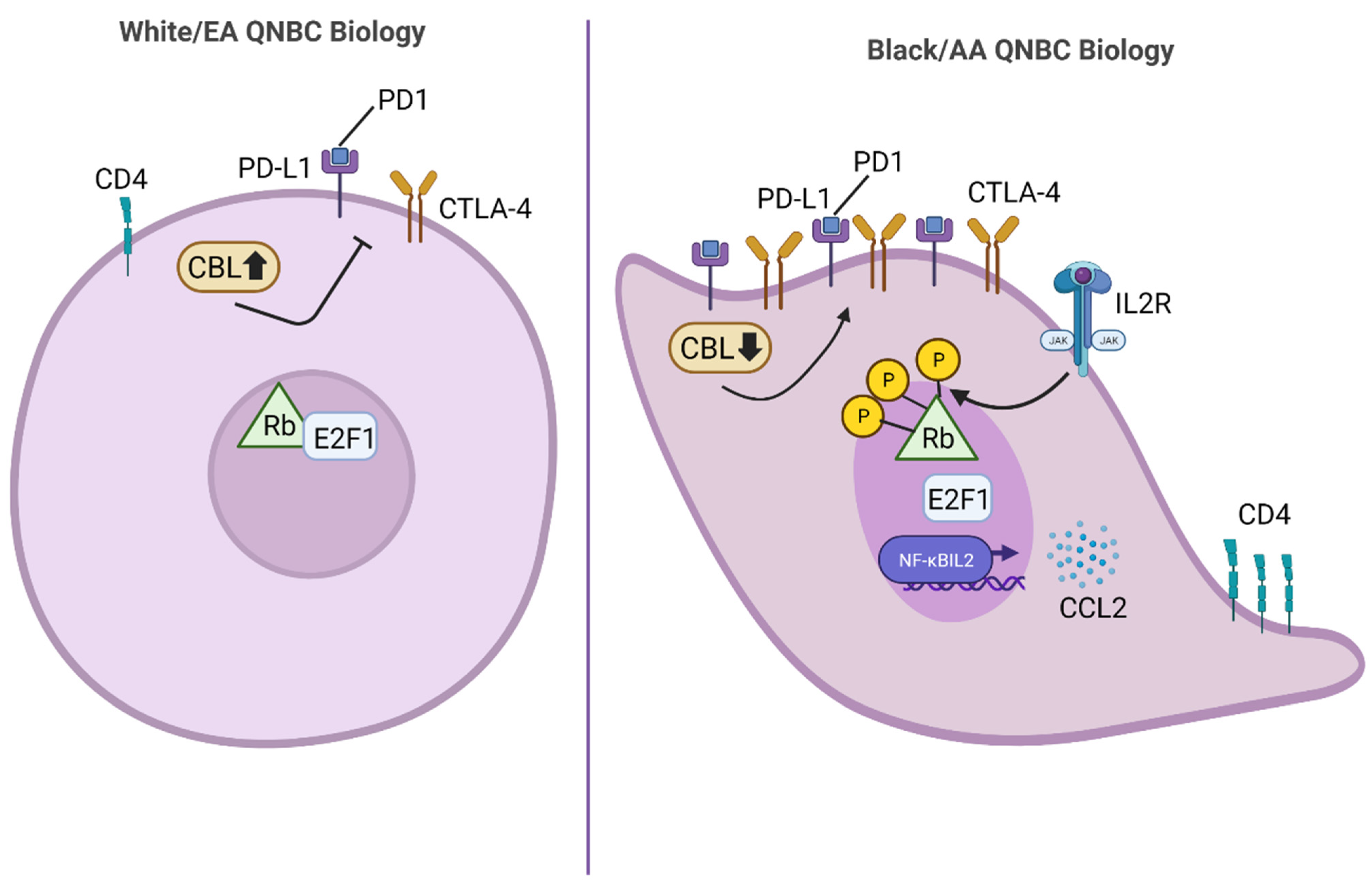
8. Black/AA versus White/EA QNBC Biology: Novel Therapeutic Strategies to Reduce the Racially Disparate Burden in QNBC

Accumulating evidence suggests that QNBC is a highly aggressive breast cancer subtype and disproportionately impacts Black/AA women. Many groups have identified unique genes and pathways differentially regulated in QNBCs versus AR-positive TNBCs for targeted therapeutic intervention. Consistent with the basal-like phenotype associated with QNBCs, some studies have discovered that QNBCs are highly upregulated in proliferative markers such as EGFR, Ki-67, CDK6, and topoisomerase 2a (TOPO2A) compared to AR-positive TNBCs [12]. This evidence suggests QNBCs would be highly susceptible to inhibitors of proliferation such as EGFR, CDK6, and TOPO2A inhibitors. The cellular metabolic marker, long chain acyl-CoA synthetase 4 (ASCL4), was also found to be significantly upregulated in TNBCs absent of AR expression, suggesting ASCL4 inhibitors may be another viable therapeutic candidate [80]. Furthermore, as aligned with the highly immunogenic biology of QNBCs versus AR-positive TNBCs, QNBCs have also been found to be highly upregulated in T cell markers (CD4 and CD8), immune checkpoints (PD1, PD-L1, and CTLA4), and immune signaling pathways (ILR2B, CCR5, NFKBII2) compared to AR-enriched tumors [10]. These findings suggest that QNBCs may be highly sensitive to immunotherapeutic strategies, such as PD-L1 inhibitors.
However, with reports suggesting that QNBC is more aggressive in Black/AA than White/EA women, there is an urgent need for therapeutic targets specifically associated with QNBC disease in Black/AA women. Although limited, evidence suggests that differentially expressed genes and pathways in Black/AA versus White/EA QNBC exist as illustrated in Figure 1. Specifically, genes involved in immune cell signaling were found to be differentially expressed between Black/AA and White/EA women with QNBC, such as E2F1, NFKBIL2, CCL2, TGFB2, CEBPB, PDK1, IL12RB2, IL2RA, and SOS1 [10]. Furthermore, the CD4 T-cell marker and immune checkpoint genes PD-1, PD-L1, and CTLA-4, were found to be significantly upregulated in Black/AA compared to White/EA QNBC. Thus, QNBCs in Black/AA women may be highly susceptible to immunotherapeutic intervention, which could aid in reducing the racially disparate burden in QNBC.

9. Spotlight on KIFC1: A Promising Target for QNBC in Women of West African Descent

Kinesin family member C1 (KIFC1) is a microtubule binding protein that confers the survival of cancer cells with centrosome amplification, which is when a cell consists of more than 2 centrosomes per cell or has abnormally voluminous centrosomes [81,82]. Centrosome amplification is a hallmark of aggressive cancer as centrosome numbers are highly conserved in normal cells [83]. When cancer cells are burdened with extra centrosomes, they undergo erroneous multipolar spindle mitoses. These mitotic cell division errors can lead to improper segregation of chromosomes into each daughter cell, resulting in apoptotic cell death. KIFC1 blocks the apoptotic cell death program from occurring by clustering the extra centrosomes at opposite poles of the cell via crosslinking microtubules. This crosslinking allows cancer cells to undergo “pseudobipolar” spindle mitoses [82,84]. Thus, KIFC1 facilitates the persistence of genomically unstable cells and the expansion of aggressive cellular clones. Therefore, KIFC1 has emerged as a biomarker of aggressive breast cancer. Primary breast tumors overexpress KIFC1 relative to matched normal breast tissue, while KIFC1 is expressed higher in TNBCs compared to non-TNBCs [85,86]. Nuclear KIFC1 expression is associated with an advanced tumor grade as well as poorer OS and progression-free survival in breast cancer [86]. In TNBC, KIFC1 is among the top 1% of genes that are upregulated in TNBCs versus non-TNBCs and its overexpression is associated with a survival rate of <5 years [87]. Overexpression of KIFC1 in MDA-MB-231 and MDA-MB-468 TNBC cell lines conferred enhanced cell survival versus vector controls [88].
It was shown that nuclear KIFC1 expression is an independent biomarker of poor prognosis for Black/AA but not White/EA women with TNBC [89]. Furthermore, KIFC1 inhibition hindered proliferation and migration of Black/AA-derived TNBC cell lines to a greater extent compared to White/EA-derived TNBC cell lines [89]. These findings suggest that KIFC1 may be specifically critical for the progression of Black/AA TNBC biology and that targeting KIFC1 in Black/AA women may help reduce the racially disparate burden in TNBC. Since emerging evidence suggest that QNBCs are more aggressive in Black/AAs versus White/EAs, we hypothesize that KIFC1 may also be a promising target for Black/AA women with QNBC.
Furthermore, KIFC1 has been identified as the top hit among centrosome clustering genes in a genome-wide Drosophila screen, suggesting it may be the best centrosome clustering protein to target [82]. In TNBC, KIFC1 has also been identified as a malignant cell-specific dependency factor, which supports KIFC1 inhibition as a highly effective pharmacological strategy for aggressive TNBC [87]. Additionally, KIFC1 is non-essential for healthy cells with 2 centrosomes but indispensable for proper cell division of cancer cells with supernumerary centrosomes. Thus, KIFC1 inhibitors can selectively kill cancer cells burdened with extra centrosomes unlike standard chemotherapies [90]. Hence, KIFC1 inhibition may be a promising anti-cancer strategy for QNBC women of West African ancestry.

10. Black/AA versus White/EA QNBC Epigenomics: The Potential Role of Epigenetic Modifications in the Racially Disparate Burden in QNBC

Epigenetic modifications such as DNA methylation, microRNA (miRNA)-mediated gene silencing, histone posttranslational modifications and associated alterations in chromatin structure have long been shown to be key players in aggressive breast pathogenesis. An increasing body of evidence suggests that differential epigenetic patterns exhibited between Black/AA and White/EA women may be underlying the racially disparate burden in breast cancer [91,92,93,94,95]. Specifically, certain epigenetic markers have been linked to the predisposal of more aggressive breast cancer subtypes, such as TNBC and QNBC [96]. Recent evidence has identified distinct epigenomic profiles between TNBC and QNBC tumors as both DNA and RNA epigenetic modifications have been known to modulate androgen receptor expression. In particular, a molecular switch has been suggested to be responsible for the transition of TNBC to QNBC, or inactivation of the AR in TNBC, via hypermethylation of the AR promoter and/or miRNA-mediated transcriptional repression of the AR gene [97]. This transition has been suggested to promote more aggressive TNBC. These epigenetic changes can also alter genes involved in critical processes such as DNA repair, metabolism, and invasion/metastasis. Thus, differential epigenetic profiles could be conferring increased prevalence of QNBC and more aggressive QNBC disease in Black/AA compared to White/EA women.
Although limited, emerging evidence has begun to dissect the role of DNA methylation and miRNA-mediated gene regulation in conferring increased QNBC incidence and aggressiveness. Differential DNA methylation patterns in oncogenes and tumor suppressor genes among Black/AA and White/EA women have been observed to occur more frequently among ER-/PR-negative as opposed to ER-/PR-positive breast cancers, which could be underlying the increased incidence of TNBC and QNBC in Black/AA women [91,94]. miRNA regulatory networks have been suggested to alter the expression of genes in signaling pathways associated with QNBC and Black/AA QNBC, such as PD-1, PD-L1, CTLA-4, epidermal growth factor receptor (EGFR), phosphatase and tensin homolog (PTEN), ACSL4, S-phase kinase associated protein 2, and engrailed homobox1 [98]. Specifically, overexpression of miR-17-5p has been shown to downregulate PTEN, also suppressed in QNBC, via targeting the 3′-untranslated mRNA region [99]. Overexpression of ACSL4, observed in QNBC, has been discovered to occur via differential transcriptional mechanisms in the ACSL4 gene and downregulation of miR-211-5p [100,101]. Overexpression of the EGFR, observed in QNBC, was associated with downregulated miR-133a [102]. Increased miR-135b expression was associated with AR-negativity in TNBC as well as a high proliferation index, which occurs in QNBC, and suggested to promote QNBC pathogenesis via targeting the TGF-β, WNT, and ERBB signaling pathways [103]. In an unpublished study, Angajala and colleagues discovered miRNAs differentially expressed between Black/AA and White/EA QNBC patients in The Cancer Genome Atlas (TCGA), which also correlated with AR expression, such as hsa-mir-135b, has-mir-18a, and has-mir-577, hsa-mir-500a, has-mir-181a-2, has-let-7d, has-mir-92a-2, has-mir-150, has-mir-17, hsa-mir-92a-1, has-mir-30a, has-mir-210, has-mir-455, hsa-mir-455, hsa-mir-130a, and has-mir-20a. The Women’s Circle of Health Study identified single nucleotide polymorphisms (SNPs) in miRNAs in Black/AA women that were associated with an increased risk for ER-negative and/or TNBC, specifically in hsa-mir-219, hsa-mir-595, hsa-mir-204, and hsa-mir-513a-2 [104]. Hence, epigenetic reprogramming via DNA methylation inhibitors or miRNA mimics/inhibitors in Black/AA women may help reduce QNBC incidence and aggressiveness in this patient subpopulation.

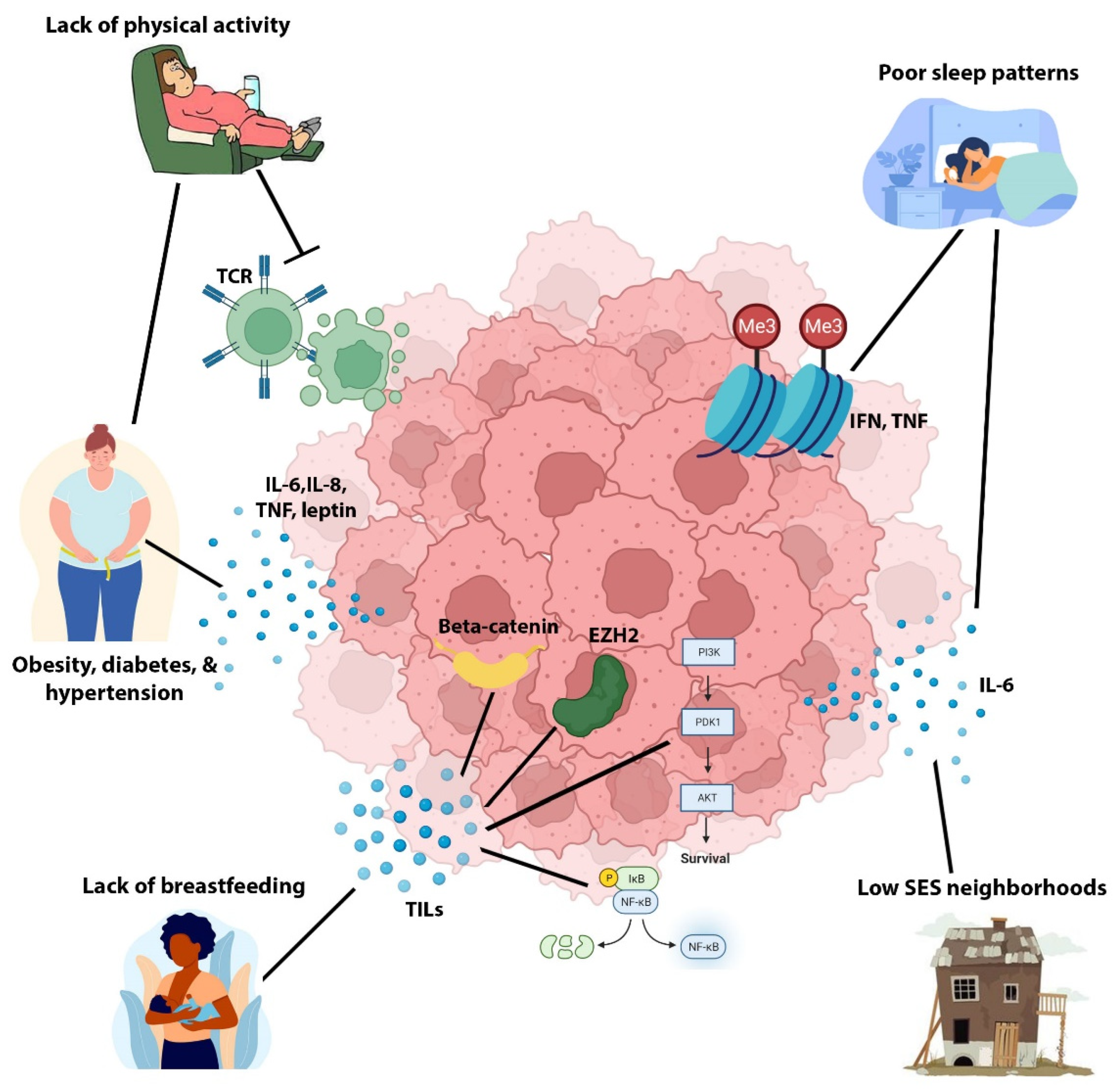
11. Black/AA versus White/EA QNBC Non-Biology: The Potential Role of Non-Genetic Risk Factors in the Racially Disparate Burden in QNBC

Disparities in non-genetic risk factors have long been reported to contribute to the gap in survival rates between Black/AA and White/EA women with breast cancer. These non-genetic risk factors include lifestyle (i.e., diet, physical activity, sleep), socioeconomic status (i.e., income level, education), access to quality oncological care (i.e., screening, health insurance, transportation), reproductive factors (i.e., parity, age at menarche, breastfeeding), anthropometrics (i.e., body mass index, waist/hip ratio), and comorbidities (i.e., obesity, diabetes, hypertension) [7,105,106,107]. Black/AA women experience disproportionately poorer access to adequate foods, physical activity, sleep patterns, breastfeeding, income levels, education, healthcare/screening, and health insurance compared to their White/EA counterparts [105,108]. Additionally, Black/AA women experience a greater burden of co-morbid diseases than White/EA women [109]. These unequal living standards and increased prevalence of co-morbid illnesses have also been suggested to underlie the acquisition of more aggressive subtypes, such as TNBC, in Black/AA versus White/EA women [7,105,108].
The Carolina Breast Cancer Study (CBCS) identified an association between increased waist/hip ratio and an increased risk of developing the basal-like subset of TNBC [110]. Other non-genetic risk factors such as co-morbid conditions, poor sleep patterns, lack of physical activity, stress, and lack of breast feeding have been linked to increased development of TNBC versus other subtypes via increased tumor-promoting inflammation. Specifically, poor sleep can suppress the immune system, cause alterations in the methylation of inflammatory genes such as IFN and tumor necrosis factor (TNF), as well as upregulate cancer-stimulatory cytokines [111,112]. Physical activity can boost the anti-tumoral immune response as well as prevent a high body mass index and waist/hip ratio [113]. A lack of breastfeeding postpartum can lead to the mammary gland undergoing involution, which can result in increased tissue inflammation [114,115]. Higher rates of residence in low SES neighborhoods among Black/AA communities may lead to increased psychological distress, which has been reported to elevate levels of inflammatory markers such as interleukin 6 (IL-6) [105,116]. Lastly, co-morbid diseases such as obesity, diabetes mellitus, and hypertension have all been reported to increase tissue inflammation. Obesity can upregulate circulating levels of insulin, inflammatory cytokines such as IL-6, IL-8, TNF, leptin, chemokines, and transforming growth factor beta (TGF-β) [105,117,118]. These inflammatory markers can activate signaling networks that promote cell proliferation and genomic instability, such as PI3K-AKT, signal transducer and activator of transcription 3 (STAT3), nuclear factor-ΚB (NFΚB), EZH2 and Wnt signaling. Diabetes and hypertension can also promote the upregulation of inflammatory cytokines [7,105]. Since the basal-like and immunogenic subsets of TNBC are reflective of QNBC and Black/AA QNBC biology, we postulate that these non-genetic risk factors may also play a role in the greater incidence of QNBC and more aggressive QNBC disease among Black/AA women compared to White/EA women as shown in Figure 2. Thus, further investigation into this link is warranted to adequately address the racially disparate burden in QNBC.

12. Conclusions and Future Perspectives

Addressing the racial disparity in the highly aggressive breast cancer subtype, QNBC, could significantly contribute to reducing the racially disparate burden in breast cancer. We assert that successfully addressing this subtype disparity would require a multifaceted approach. Particularly, the US and worldwide racial disparity in QNBC is still poorly defined and documented. We suggest large cancer statistics programs such as the Surveillance, Epidemiology, and End Results (SEER), Center for Disease Control and Prevention (CDC) as well as predominately Black/AA cohorts such as the CBCS and Women’s Circle of Health Study (WCHS) investigate current statistics on the prevalence and impact of QNBC. Within these studies, we recommend controlling for non-genetic confounding factors such as SES and access to care. We also suggest increasing the pool of Black/AA women in publicly available datasets to better define the impact of AR expression in TNBC tumors of West African descent, and thorough investigating potential molecular targets for racially distinct QNBC tumors. Furthermore, we propose increased investigation of AR IHC-based expression for identification of optimal staining methodologies and cut-off values to successfully incorporate AR evaluation into routine clinical practice for QNBC women of West African descent. We strongly advocate for high-granularity genetic ancestry typing in these studies to refine risk-stratification of subpopulations most susceptible to specific therapeutic strategies for QNBC.
In addition, we recommend increased research into the molecular and biological mechanisms underlying increased QNBC incidence and mortality among individuals of West African descent, as well as how AR signaling differs between TNBC from Black/AA and White/EA women for optimal therapeutic intervention. We propose investigations into the role of KIFC1 as a potential therapeutic target for QNBCs of West African ancestry. Our overview of preliminary evidence suggesting that epigenetic modifications may be predisposing Black/AA women to QNBC pathogenesis suggest increased investigation into the role of other epigenetic modifications in the racially disparate burden in QNBC as well as into novel treatment strategies targeting these modifications. Furthermore, we encourage addressing the previously mentioned non-genetic risk factors, such as lifestyle and SES, in order to adequately address this new clinical challenge.

Author Contributions

Conceptualization, N.J. and V.L.S.; Writing-Original Draft Preparation, N.J.; Writing-Review and Editing, N.J., T.J.-T., M.L., R.N., R.K., C.S., P.R. and V.L.S.; Visualization, N.J.; Supervision, V.L.S. All authors have read and agreed to the published version of the manuscript.

Funding

National Institutes of Health/National Cancer Institute (NIH/NCI) grants T32CA221709 (VLS, NW) U01CA189283 (VLS), and R01CA220693 (VLS, TJ-T), P20CA24619 (VLS), and P20CA252717 (VLS). The funders had no role in the manuscript design, data analysis, decision to publish, or preparation of the manuscript.

Conflicts of Interest

The authors declare no conflict of interest.

References

  1. DeSantis, C.; Ma, J.; Bryan, L.; Jemal, A. Breast cancer statistics, 2013. CA Cancer J. Clin. 2014, 64, 52–62. [Google Scholar] [CrossRef] [PubMed]
  2. Provenzano, E.; Ulaner, G.A.; Chin, S.F. Molecular Classification of Breast Cancer. PET Clin. 2018, 13, 325–338. [Google Scholar] [CrossRef]
  3. DeSantis, C.E.; Ma, J.; Goding Sauer, A.; Newman, L.A.; Jemal, A. Breast cancer statistics, 2017, racial disparity in mortality by state. CA Cancer J. Clin. 2017, 67, 439–448. [Google Scholar] [CrossRef]
  4. DeSantis, C.E.; Ma, J.; Gaudet, M.M.; Newman, L.A.; Miller, K.D.; Goding Sauer, A.; Jemal, A.; Siegel, R.L. Breast cancer statistics, 2019. CA Cancer J. Clin. 2019, 69, 438–451. [Google Scholar] [CrossRef] [PubMed]
  5. Zhao, F.; Copley, B.; Niu, Q.; Liu, F.; Johnson, J.A.; Sutton, T.; Khramtsova, G.; Sveen, E.; Yoshimatsu, T.F.; Zheng, Y.; et al. Racial disparities in survival outcomes among breast cancer patients by molecular subtypes. Breast Cancer Res. Treat. 2021, 185, 841–849. [Google Scholar] [CrossRef] [PubMed]
  6. Troester, M.A.; Sun, X.; Allott, E.H.; Geradts, J.; Cohen, S.M.; Tse, C.K.; Kirk, E.L.; Thorne, L.B.; Mathews, M.; Li, Y.; et al. Racial Differences in PAM50 Subtypes in the Carolina Breast Cancer Study. J. Natl. Cancer Inst. 2018, 110, 176–182. [Google Scholar] [CrossRef] [PubMed]
  7. Dietze, E.C.; Sistrunk, C.; Miranda-Carboni, G.; O’Regan, R.; Seewaldt, V.L. Triple-negative breast cancer in African-American women: Disparities versus biology. Nat. Rev. Cancer 2015, 15, 248–254. [Google Scholar] [CrossRef]
  8. Lund, M.J.; Trivers, K.F.; Porter, P.L.; Coates, R.J.; Leyland-Jones, B.; Brawley, O.W.; Flagg, E.W.; O’Regan, R.M.; Gabram, S.G.; Eley, J.W. Race and triple negative threats to breast cancer survival: A population-based study in Atlanta, GA. Breast Cancer Res. Treat. 2009, 113, 357–370. [Google Scholar] [CrossRef]
  9. Bhattarai, S.; Klimov, S.; Mittal, K.; Krishnamurti, U.; Li, X.B.; Oprea-Ilies, G.; Wetherilt, C.S.; Riaz, A.; Aleskandarany, M.A.; Green, A.R.; et al. Prognostic Role of Androgen Receptor in Triple Negative Breast Cancer: A Multi-Institutional Study. Cancers 2019, 11, 995. [Google Scholar] [CrossRef]
  10. Davis, M.; Tripathi, S.; Hughley, R.; He, Q.; Bae, S.; Karanam, B.; Martini, R.; Newman, L.; Colomb, W.; Grizzle, W.; et al. AR negative triple negative or “quadruple negative” breast cancers in African American women have an enriched basal and immune signature. PLoS ONE 2018, 13, e0196909. [Google Scholar] [CrossRef] [Green Version]
  11. Newman, L.A.; Jenkins, B.; Chen, Y.; Oppong, J.K.; Adjei, E.; Jibril, A.S.; Hoda, S.; Cheng, E.; Chitale, D.; Bensenhaver, J.M.; et al. Hereditary Susceptibility for Triple Negative Breast Cancer Associated With Western Sub-Saharan African Ancestry: Results From an International Surgical Breast Cancer Collaborative. Ann. Surg. 2019, 270, 484–492. [Google Scholar] [CrossRef] [PubMed]
  12. Gasparini, P.; Fassan, M.; Cascione, L.; Guler, G.; Balci, S.; Irkkan, C.; Paisie, C.; Lovat, F.; Morrison, C.; Zhang, J.; et al. Androgen receptor status is a prognostic marker in non-basal triple negative breast cancers and determines novel therapeutic options. PLoS ONE 2014, 9, e88525. [Google Scholar] [CrossRef] [PubMed]
  13. Hon, J.D.; Singh, B.; Sahin, A.; Du, G.; Wang, J.; Wang, V.Y.; Deng, F.M.; Zhang, D.Y.; Monaco, M.E.; Lee, P. Breast cancer molecular subtypes: From TNBC to QNBC. Am. J. Cancer Res. 2016, 6, 1864–1872. [Google Scholar]
  14. Huang, M.; Wu, J.; Ling, R.; Li, N. Quadruple negative breast cancer. Breast Cancer 2020, 27, 527–533. [Google Scholar] [CrossRef]
  15. Angajala, A.; Mothershed, E.; Davis, M.B.; Tripathi, S.; He, Q.; Bedi, D.; Dean-Colomb, W.; Yates, C. Quadruple Negative Breast Cancers (QNBC) Demonstrate Subtype Consistency among Primary and Recurrent or Metastatic Breast Cancer. Transl. Oncol. 2019, 12, 493–501. [Google Scholar] [CrossRef] [PubMed]
  16. Jovanovic, B.; Mayer, I.A.; Mayer, E.L.; Abramson, V.G.; Bardia, A.; Sanders, M.E.; Kuba, M.G.; Estrada, M.V.; Beeler, J.S.; Shaver, T.M.; et al. A Randomized Phase II Neoadjuvant Study of Cisplatin, Paclitaxel With or Without Everolimus in Patients with Stage II/III Triple-Negative Breast Cancer (TNBC): Responses and Long-term Outcome Correlated with Increased Frequency of DNA Damage Response Gene Mutations, TNBC Subtype, AR Status, and Ki67. Clin. Cancer Res. 2017, 23, 4035–4045. [Google Scholar] [CrossRef] [PubMed]
  17. Dent, R.; Trudeau, M.; Pritchard, K.I.; Hanna, W.M.; Kahn, H.K.; Sawka, C.A.; Lickley, L.A.; Rawlinson, E.; Sun, P.; Narod, S.A. Triple-negative breast cancer: Clinical features and patterns of recurrence. Clin. Cancer Res. 2007, 13, 4429–4434. [Google Scholar] [CrossRef]
  18. Perou, C.M.; Sorlie, T.; Eisen, M.B.; van de Rijn, M.; Jeffrey, S.S.; Rees, C.A.; Pollack, J.R.; Ross, D.T.; Johnsen, H.; Akslen, L.A.; et al. Molecular portraits of human breast tumours. Nature 2000, 406, 747–752. [Google Scholar] [CrossRef]
  19. Bonotto, M.; Gerratana, L.; Poletto, E.; Driol, P.; Giangreco, M.; Russo, S.; Minisini, A.M.; Andreetta, C.; Mansutti, M.; Pisa, F.E.; et al. Measures of outcome in metastatic breast cancer: Insights from a real-world scenario. Oncologist 2014, 19, 608–615. [Google Scholar] [CrossRef]
  20. Liedtke, C.; Mazouni, C.; Hess, K.R.; Andre, F.; Tordai, A.; Mejia, J.A.; Symmans, W.F.; Gonzalez-Angulo, A.M.; Hennessy, B.; Green, M.; et al. Response to neoadjuvant therapy and long-term survival in patients with triple-negative breast cancer. J. Clin. Oncol. 2008, 26, 1275–1281. [Google Scholar] [CrossRef]
  21. Bianchini, G.; Balko, J.M.; Mayer, I.A.; Sanders, M.E.; Gianni, L. Triple-negative breast cancer: Challenges and opportunities of a heterogeneous disease. Nat. Rev. Clin. Oncol. 2016, 13, 674–690. [Google Scholar] [CrossRef] [PubMed]
  22. Wright, N.; Rida, P.C.G.; Aneja, R. Tackling intra- and inter-tumor heterogeneity to combat triple negative breast cancer. Front. Biosci. (Landmark Ed.) 2017, 22, 1549–1580. [Google Scholar] [CrossRef] [PubMed]
  23. Lehmann, B.D.; Bauer, J.A.; Chen, X.; Sanders, M.E.; Chakravarthy, A.B.; Shyr, Y.; Pietenpol, J.A. Identification of human triple-negative breast cancer subtypes and preclinical models for selection of targeted therapies. J. Clin. Investig. 2011, 121, 2750–2767. [Google Scholar] [CrossRef]
  24. Burstein, M.D.; Tsimelzon, A.; Poage, G.M.; Covington, K.R.; Contreras, A.; Fuqua, S.A.; Savage, M.I.; Osborne, C.K.; Hilsenbeck, S.G.; Chang, J.C.; et al. Comprehensive genomic analysis identifies novel subtypes and targets of triple-negative breast cancer. Clin. Cancer Res. 2015, 21, 1688–1698. [Google Scholar] [CrossRef] [PubMed]
  25. Liu, Y.R.; Jiang, Y.Z.; Xu, X.E.; Yu, K.D.; Jin, X.; Hu, X.; Zuo, W.J.; Hao, S.; Wu, J.; Liu, G.Y.; et al. Comprehensive transcriptome analysis identifies novel molecular subtypes and subtype-specific RNAs of triple-negative breast cancer. Breast Cancer Res. 2016, 18, 33. [Google Scholar] [CrossRef]
  26. Keenan, T.; Moy, B.; Mroz, E.A.; Ross, K.; Niemierko, A.; Rocco, J.W.; Isakoff, S.; Ellisen, L.W.; Bardia, A. Comparison of the Genomic Landscape Between Primary Breast Cancer in African American Versus White Women and the Association of Racial Differences With Tumor Recurrence. J. Clin. Oncol. 2015, 33, 3621–3627. [Google Scholar] [CrossRef]
  27. Anestis, A.; Zoi, I.; Papavassiliou, A.G.; Karamouzis, M.V. Androgen Receptor in Breast Cancer-Clinical and Preclinical Research Insights. Molecules 2020, 25, 358. [Google Scholar] [CrossRef]
  28. Zhou, X. Roles of androgen receptor in male and female reproduction: Lessons from global and cell-specific androgen receptor knockout (ARKO) mice. J. Androl. 2010, 31, 235–243. [Google Scholar] [CrossRef]
  29. Walters, K.A.; Simanainen, U.; Handelsman, D.J. Molecular insights into androgen actions in male and female reproductive function from androgen receptor knockout models. Hum. Reprod. Update 2010, 16, 543–558. [Google Scholar] [CrossRef]
  30. Zhou, J.; Ng, S.; Adesanya-Famuiya, O.; Anderson, K.; Bondy, C.A. Testosterone inhibits estrogen-induced mammary epithelial proliferation and suppresses estrogen receptor expression. FASEB J. 2000, 14, 1725–1730. [Google Scholar] [CrossRef]
  31. Dimitrakakis, C.; Zhou, J.; Wang, J.; Belanger, A.; LaBrie, F.; Cheng, C.; Powell, D.; Bondy, C. A physiologic role for testosterone in limiting estrogenic stimulation of the breast. Menopause 2003, 10, 292–298. [Google Scholar] [CrossRef] [PubMed]
  32. Tiefenbacher, K.; Daxenbichler, G. The Role of Androgens in Normal and Malignant Breast Tissue. Breast Care 2008, 3, 325–331. [Google Scholar] [CrossRef] [PubMed]
  33. Venema, C.M.; Bense, R.D.; Steenbruggen, T.G.; Nienhuis, H.H.; Qiu, S.Q.; van Kruchten, M.; Brown, M.; Tamimi, R.M.; Hospers, G.A.P.; Schroder, C.P.; et al. Consideration of breast cancer subtype in targeting the androgen receptor. Pharmacol. Ther. 2019, 200, 135–147. [Google Scholar] [CrossRef]
  34. Sarker, D.; Reid, A.H.; Yap, T.A.; de Bono, J.S. Targeting the PI3K/AKT pathway for the treatment of prostate cancer. Clin. Cancer Res. 2009, 15, 4799–4805. [Google Scholar] [CrossRef] [PubMed]
  35. Dong, S.; Alahari, S.K. Combination treatment of bicalutamide and curcumin has a strong therapeutic effect on androgen receptor-positive triple-negative breast cancers. Anticancer Drugs 2020, 31, 359–367. [Google Scholar] [CrossRef]
  36. Barton, V.N.; D’Amato, N.C.; Gordon, M.A.; Lind, H.T.; Spoelstra, N.S.; Babbs, B.L.; Heinz, R.E.; Elias, A.; Jedlicka, P.; Jacobsen, B.M.; et al. Multiple molecular subtypes of triple-negative breast cancer critically rely on androgen receptor and respond to enzalutamide in vivo. Mol. Cancer Ther. 2015, 14, 769–778. [Google Scholar] [CrossRef]
  37. Caiazza, F.; Murray, A.; Madden, S.F.; Synnott, N.C.; Ryan, E.J.; O’Donovan, N.; Crown, J.; Duffy, M.J. Preclinical evaluation of the AR inhibitor enzalutamide in triple-negative breast cancer cells. Endocr. Relat. Cancer 2016, 23, 323–334. [Google Scholar] [CrossRef]
  38. Cochrane, D.R.; Bernales, S.; Jacobsen, B.M.; Cittelly, D.M.; Howe, E.N.; D’Amato, N.C.; Spoelstra, N.S.; Edgerton, S.M.; Jean, A.; Guerrero, J.; et al. Role of the androgen receptor in breast cancer and preclinical analysis of enzalutamide. Breast Cancer Res. 2014, 16, R7. [Google Scholar] [CrossRef]
  39. Traina, T.A.; Miller, K.; Yardley, D.A.; Eakle, J.; Schwartzberg, L.S.; O’Shaughnessy, J.; Gradishar, W.; Schmid, P.; Winer, E.; Kelly, C.; et al. Enzalutamide for the Treatment of Androgen Receptor-Expressing Triple-Negative Breast Cancer. J. Clin. Oncol. 2018, 36, 884–890. [Google Scholar] [CrossRef]
  40. Gucalp, A.; Tolaney, S.; Isakoff, S.J.; Ingle, J.N.; Liu, M.C.; Carey, L.A.; Blackwell, K.; Rugo, H.; Nabell, L.; Forero, A.; et al. Phase II trial of bicalutamide in patients with androgen receptor-positive, estrogen receptor-negative metastatic Breast Cancer. Clin. Cancer Res. 2013, 19, 5505–5512. [Google Scholar] [CrossRef]
  41. Clegg, N.J.; Wongvipat, J.; Joseph, J.D.; Tran, C.; Ouk, S.; Dilhas, A.; Chen, Y.; Grillot, K.; Bischoff, E.D.; Cai, L.; et al. ARN-509: A novel antiandrogen for prostate cancer treatment. Cancer Res. 2012, 72, 1494–1503. [Google Scholar] [CrossRef] [PubMed] [Green Version]
  42. Fizazi, K.; Albiges, L.; Loriot, Y.; Massard, C. ODM-201: A new-generation androgen receptor inhibitor in castration-resistant prostate cancer. Expert Rev. Anticancer. Ther. 2015, 15, 1007–1017. [Google Scholar] [CrossRef]
  43. Jinna, N.; Rida, P.; Smart, M.; LaBarge, M.; Jovanovic-Talisman, T.; Natarajan, R.; Seewaldt, V. Adaptation to Hypoxia May Promote Therapeutic Resistance to Androgen Receptor Inhibition in Triple-Negative Breast Cancer. Int. J. Mol. Sci. 2022, 23, 8844. [Google Scholar] [CrossRef] [PubMed]
  44. Gucalp, A.; Traina, T.A. Targeting the androgen receptor in triple-negative breast cancer. Curr. Probl. Cancer 2016, 40, 141–150. [Google Scholar] [CrossRef] [PubMed]
  45. Michmerhuizen, A.R.; Chandler, B.; Olsen, E.; Wilder-Romans, K.; Moubadder, L.; Liu, M.; Pesch, A.M.; Zhang, A.; Ritter, C.; Ward, S.T.; et al. Seviteronel, a Novel CYP17 Lyase Inhibitor and Androgen Receptor Antagonist, Radiosensitizes AR-Positive Triple Negative Breast Cancer Cells. Front. Endocrinol. 2020, 11, 35. [Google Scholar] [CrossRef]
  46. Finn, R.S.; Dering, J.; Conklin, D.; Kalous, O.; Cohen, D.J.; Desai, A.J.; Ginther, C.; Atefi, M.; Chen, I.; Fowst, C.; et al. PD 0332991, a selective cyclin D kinase 4/6 inhibitor, preferentially inhibits proliferation of luminal estrogen receptor-positive human breast cancer cell lines in vitro. Breast Cancer Res. 2009, 11, R77. [Google Scholar] [CrossRef]
  47. Doane, A.S.; Danso, M.; Lal, P.; Donaton, M.; Zhang, L.; Hudis, C.; Gerald, W.L. An estrogen receptor-negative breast cancer subset characterized by a hormonally regulated transcriptional program and response to androgen. Oncogene 2006, 25, 3994–4008. [Google Scholar] [CrossRef]
  48. Aleskandarany, M.A.; Rakha, E.A.; Ahmed, M.A.; Powe, D.G.; Ellis, I.O.; Green, A.R. Clinicopathologic and molecular significance of phospho-Akt expression in early invasive breast cancer. Breast Cancer Res. Treat. 2011, 127, 407–416. [Google Scholar] [CrossRef]
  49. Lehmann, B.D.; Abramson, V.G.; Sanders, M.E.; Mayer, E.L.; Haddad, T.C.; Nanda, R.; Van Poznak, C.; Storniolo, A.M.; Nangia, J.R.; Gonzalez-Ericsson, P.I.; et al. TBCRC 032 IB/II Multicenter Study: Molecular Insights to AR Antagonist and PI3K Inhibitor Efficacy in Patients with AR(+) Metastatic Triple-Negative Breast Cancer. Clin. Cancer Res. 2020, 26, 2111–2123. [Google Scholar] [CrossRef]
  50. Birrell, S.N.; Butler, L.M.; Harris, J.M.; Buchanan, G.; Tilley, W.D. Disruption of androgen receptor signaling by synthetic progestins may increase risk of developing breast cancer. FASEB J. 2007, 21, 2285–2293. [Google Scholar] [CrossRef]
  51. Zhu, X.; Li, H.; Liu, J.P.; Funder, J.W. Androgen stimulates mitogen-activated protein kinase in human breast cancer cells. Mol. Cell. Endocrinol. 1999, 152, 199–206. [Google Scholar] [CrossRef]
  52. Peters, A.A.; Buchanan, G.; Ricciardelli, C.; Bianco-Miotto, T.; Centenera, M.M.; Harris, J.M.; Jindal, S.; Segara, D.; Jia, L.; Moore, N.L.; et al. Androgen receptor inhibits estrogen receptor-alpha activity and is prognostic in breast cancer. Cancer Res. 2009, 69, 6131–6140. [Google Scholar] [CrossRef] [PubMed]
  53. Wong, Y.C.; Xie, B. The role of androgens in mammary carcinogenesis. Ital. J. Anat. Embryol. 2001, 106, 111–125. [Google Scholar] [PubMed]
  54. Hickey, T.; Robinson, J.; Carroll, J.; Tilley, W. Minireview: The androgen receptor in breast tissues: Growth inhibitor, tumor suppressor, oncogene? Mol. Endocrinol. 2012, 26, 1252–1267. [Google Scholar] [CrossRef]
  55. Wilson, J.D.; Griffin, J.E.; Leshin, M.; George, F.W. Role of gonadal hormones in development of the sexual phenotypes. Hum. Genet. 1981, 58, 78–84. [Google Scholar] [CrossRef]
  56. Yu, K.-D.; Zhu, R.; Zhan, M.; Rodriguez, A.A.; Yang, W.; Wong, S.; Makris, A.; Lehmann, B.D.; Chen, X.; Mayer, I. Identification of prognosis-relevant subgroups in patients with chemoresistant triple-negative breast cancer. Clin. Cancer Res. 2013, 19, 2723–2733. [Google Scholar] [CrossRef]
  57. Birrell, S.; Hall, R.; Tilley, W. Role of the androgen receptor in human breast cancer. J. Mammary Gland. Biol. Neoplasia 1998, 3, 95–103. [Google Scholar] [CrossRef]
  58. Garay, J.P.; Karakas, B.; Abukhdeir, A.M.; Cosgrove, D.P.; Gustin, J.P.; Higgins, M.J.; Konishi, H.; Konishi, Y.; Lauring, J.; Mohseni, M. The growth response to androgen receptor signaling in ER α-negative human breast cells is dependent on p21 and mediated by MAPK activation. Breast Cancer Res. 2012, 14, R27. [Google Scholar] [CrossRef]
  59. Kuenen-Boumeester, V.; Van der Kwast, T.H.; Claassen, C.C.; Look, M.P.; Liem, G.S.; Klijn, J.G.; Henzen-Logmans, S.C. The clinical significance of androgen receptors in breast cancer and their relation to histological and cell biological parameters. Eur. J. Cancer 1996, 32, 1560–1565. [Google Scholar] [CrossRef]
  60. Ogawa, Y.; Hai, E.; Matsumoto, K.; Ikeda, K.; Tokunaga, S.; Nagahara, H.; Sakurai, K.; Inoue, T.; Nishiguchi, Y. Androgen receptor expression in breast cancer: Relationship with clinicopathological factors and biomarkers. Int. J. Clin. Oncol. 2008, 13, 431–435. [Google Scholar] [CrossRef]
  61. Park, S.; Koo, J.; Park, H.S.; Kim, J.H.; Choi, S.Y.; Lee, J.H.; Park, B.W.; Lee, K.S. Expression of androgen receptors in primary breast cancer. Ann. Oncol. 2010, 21, 488–492. [Google Scholar] [CrossRef] [PubMed]
  62. Mirzania, M. Approach to the Triple Negative Breast Cancer in new drugs area. Int. J. Hematol.-Oncol. Stem Cell Res. 2016, 10, 115. [Google Scholar] [PubMed]
  63. Sutton, L.M.; Cao, D.; Sarode, V.; Molberg, K.H.; Torgbe, K.; Haley, B.; Peng, Y. Decreased androgen receptor expression is associated with distant metastases in patients with androgen receptor–expressing triple-negative breast carcinoma. Am. J. Clin. Pathol. 2012, 138, 511–516. [Google Scholar] [CrossRef]
  64. Tang, D.; Xu, S.; Zhang, Q.; Zhao, W. The expression and clinical significance of the androgen receptor and E-cadherin in triple-negative breast cancer. Med. Oncol. 2012, 29, 526–533. [Google Scholar] [CrossRef]
  65. Yue, Y.; Astvatsaturyan, K.; Cui, X.; Zhang, X.; Fraass, B.; Bose, S. Stratification of prognosis of triple-negative breast cancer patients using combinatorial biomarkers. PLoS ONE 2016, 11, e0149661. [Google Scholar] [CrossRef] [PubMed]
  66. He, J.; Peng, R.; Yuan, Z.; Wang, S.; Peng, J.; Lin, G.; Jiang, X.; Qin, T. Prognostic value of androgen receptor expression in operable triple-negative breast cancer: A retrospective analysis based on a tissue microarray. Med. Oncol. 2012, 29, 406–410. [Google Scholar] [CrossRef]
  67. Robinson, J.L.; MacArthur, S.; Ross-Innes, C.S.; Tilley, W.D.; Neal, D.E.; Mills, I.G.; Carroll, J.S. Androgen receptor driven transcription in molecular apocrine breast cancer is mediated by FoxA1. EMBO J. 2012, 31, 1617. [Google Scholar] [CrossRef]
  68. Mrklić, I.; Pogorelić, Z.; Ćapkun, V.; Tomić, S. Expression of androgen receptors in triple negative breast carcinomas. Acta Histochem. 2013, 115, 344–348. [Google Scholar] [CrossRef]
  69. Rakha, E.A.; El-Sayed, M.E.; Green, A.R.; Lee, A.H.; Robertson, J.F.; Ellis, I.O. Prognostic markers in triple-negative breast cancer. Cancer 2007, 109, 25–32. [Google Scholar] [CrossRef]
  70. Qu, Q.; Mao, Y.; Fei, X.-C.; Shen, K.-W. The impact of androgen receptor expression on breast cancer survival: A retrospective study and meta-analysis. PLoS ONE 2013, 8, e82650. [Google Scholar] [CrossRef]
  71. Wang, C.; Pan, B.; Zhu, H.; Zhou, Y.; Mao, F.; Lin, Y.; Xu, Q.; Sun, Q. Prognostic value of androgen receptor in triple negative breast cancer: A meta-analysis. Oncotarget 2016, 7, 46482. [Google Scholar] [CrossRef] [PubMed] [Green Version]
  72. Gonzalez-Angulo, A.M.; Stemke-Hale, K.; Palla, S.L.; Carey, M.; Agarwal, R.; Meric-Berstam, F.; Traina, T.A.; Hudis, C.; Hortobagyi, G.N.; Gerald, W.L. Androgen receptor levels and association with PIK3CA mutations and prognosis in breast cancer. Clin. Cancer Res. 2009, 15, 2472–2478. [Google Scholar] [CrossRef] [PubMed]
  73. Luo, X.; Shi, Y.; Li, Z.; Jiang, W. Expression and clinical significance of androgen receptor in triple negative breast cancer. Chin. J. Cancer 2010, 29, 585–590. [Google Scholar] [CrossRef] [PubMed]
  74. Asano, Y.; Kashiwagi, S.; Goto, W.; Tanaka, S.; Morisaki, T.; Takashima, T.; Noda, S.; Onoda, N.; Ohsawa, M.; Hirakawa, K.; et al. Expression and Clinical Significance of Androgen Receptor in Triple-Negative Breast Cancer. Cancers 2017, 9, 4. [Google Scholar] [CrossRef]
  75. McGhan, L.J.; McCullough, A.E.; Protheroe, C.A.; Dueck, A.C.; Lee, J.J.; Nunez-Nateras, R.; Castle, E.P.; Gray, R.J.; Wasif, N.; Goetz, M.P. Androgen receptor-positive triple negative breast cancer: A unique breast cancer subtype. Ann. Surg. Oncol. 2014, 21, 361–367. [Google Scholar] [CrossRef]
  76. Choi, J.E.; Kang, S.H.; Lee, S.J.; Bae, Y.K. Androgen receptor expression predicts decreased survival in early stage triple-negative breast cancer. Ann. Surg. Oncol. 2015, 22, 82–89. [Google Scholar] [CrossRef]
  77. Hu, R.; Dawood, S.; Holmes, M.D.; Collins, L.C.; Schnitt, S.J.; Cole, K.; Marotti, J.D.; Hankinson, S.E.; Colditz, G.A.; Tamimi, R.M. Androgen receptor expression and breast cancer survival in postmenopausal women. Clin. Cancer Res. 2011, 17, 1867–1874. [Google Scholar] [CrossRef]
  78. Vera-Badillo, F.E.; Templeton, A.J.; de Gouveia, P.; Diaz-Padilla, I.; Bedard, P.L.; Al-Mubarak, M.; Seruga, B.; Tannock, I.F.; Ocana, A.; Amir, E. Androgen receptor expression and outcomes in early breast cancer: A systematic review and meta-analysis. JNCI J. Natl. Cancer Inst. 2014, 106, djt319. [Google Scholar] [CrossRef]
  79. Jiagge, E.; Jibril, A.S.; Davis, M.; Murga-Zamalloa, C.; Kleer, C.G.; Gyan, K.; Divine, G.; Hoenerhoff, M.; Bensenhave, J.; Awuah, B.; et al. Androgen Receptor and ALDH1 Expression Among Internationally Diverse Patient Populations. J. Glob. Oncol. 2018, 4, 1–8. [Google Scholar] [CrossRef]
  80. Wu, X.; Li, Y.; Wang, J.; Wen, X.; Marcus, M.T.; Daniels, G.; Zhang, D.Y.; Ye, F.; Wang, L.H.; Du, X.; et al. Long chain fatty Acyl-CoA synthetase 4 is a biomarker for and mediator of hormone resistance in human breast cancer. PLoS ONE 2013, 8, e77060. [Google Scholar] [CrossRef]
  81. Kleylein-Sohn, J.; Pollinger, B.; Ohmer, M.; Hofmann, F.; Nigg, E.A.; Hemmings, B.A.; Wartmann, M. Acentrosomal spindle organization renders cancer cells dependent on the kinesin HSET. J. Cell Sci. 2012, 125, 5391–5402. [Google Scholar] [CrossRef] [Green Version]
  82. Kwon, M.; Godinho, S.A.; Chandhok, N.S.; Ganem, N.J.; Azioune, A.; Thery, M.; Pellman, D. Mechanisms to suppress multipolar divisions in cancer cells with extra centrosomes. Genes Dev. 2008, 22, 2189–2203. [Google Scholar] [CrossRef]
  83. Zhao, J.Z.; Ye, Q.; Wang, L.; Lee, S.C. Centrosome amplification in cancer and cancer-associated human diseases. Biochim. Biophys. Acta Rev. Cancer 2021, 1876, 188566. [Google Scholar] [CrossRef] [PubMed]
  84. Yang, W.X.; Sperry, A.O. C-terminal kinesin motor KIFC1 participates in acrosome biogenesis and vesicle transport. Biol. Reprod. 2003, 69, 1719–1729. [Google Scholar] [CrossRef] [PubMed]
  85. Wu, J.; Mikule, K.; Wang, W.; Su, N.; Petteruti, P.; Gharahdaghi, F.; Code, E.; Zhu, X.; Jacques, K.; Lai, Z.; et al. Discovery and mechanistic study of a small molecule inhibitor for motor protein KIFC1. ACS Chem. Biol. 2013, 8, 2201–2208. [Google Scholar] [CrossRef] [PubMed]
  86. Pannu, V.; Rida, P.C.; Ogden, A.; Turaga, R.C.; Donthamsetty, S.; Bowen, N.J.; Rudd, K.; Gupta, M.V.; Reid, M.D.; Cantuaria, G.; et al. HSET overexpression fuels tumor progression via centrosome clustering-independent mechanisms in breast cancer patients. Oncotarget 2015, 6, 6076–6091. [Google Scholar] [CrossRef]
  87. Patel, N.; Weekes, D.; Drosopoulos, K.; Gazinska, P.; Noel, E.; Rashid, M.; Mirza, H.; Quist, J.; Braso-Maristany, F.; Mathew, S.; et al. Integrated genomics and functional validation identifies malignant cell specific dependencies in triple negative breast cancer. Nat. Commun. 2018, 9, 1044. [Google Scholar] [CrossRef]
  88. De, S.; Cipriano, R.; Jackson, M.W.; Stark, G.R. Overexpression of kinesins mediates docetaxel resistance in breast cancer cells. Cancer Res. 2009, 69, 8035–8042. [Google Scholar] [CrossRef]
  89. Ogden, A.; Garlapati, C.; Li, X.B.; Turaga, R.C.; Oprea-Ilies, G.; Wright, N.; Bhattarai, S.; Mittal, K.; Wetherilt, C.S.; Krishnamurti, U.; et al. Multi-institutional study of nuclear KIFC1 as a biomarker of poor prognosis in African American women with triple-negative breast cancer. Sci. Rep. 2017, 7, 42289. [Google Scholar] [CrossRef] [PubMed]
  90. Li, Y.; Lu, W.; Chen, D.; Boohaker, R.J.; Zhai, L.; Padmalayam, I.; Wennerberg, K.; Xu, B.; Zhang, W. KIFC1 is a novel potential therapeutic target for breast cancer. Cancer Biol. Ther. 2015, 16, 1316–1322. [Google Scholar] [CrossRef]
  91. Mehrotra, J.; Ganpat, M.M.; Kanaan, Y.; Fackler, M.J.; McVeigh, M.; Lahti-Domenici, J.; Polyak, K.; Argani, P.; Naab, T.; Garrett, E.; et al. Estrogen receptor/progesterone receptor-negative breast cancers of young African-American women have a higher frequency of methylation of multiple genes than those of Caucasian women. Clin. Cancer Res. 2004, 10, 2052–2057. [Google Scholar] [CrossRef] [Green Version]
  92. Wang, S.; Dorsey, T.H.; Terunuma, A.; Kittles, R.A.; Ambs, S.; Kwabi-Addo, B. Relationship between tumor DNA methylation status and patient characteristics in African-American and European-American women with breast cancer. PLoS ONE 2012, 7, e37928. [Google Scholar] [CrossRef] [PubMed]
  93. Conway, K.; Edmiston, S.N.; Tse, C.K.; Bryant, C.; Kuan, P.F.; Hair, B.Y.; Parrish, E.A.; May, R.; Swift-Scanlan, T. Racial variation in breast tumor promoter methylation in the Carolina Breast Cancer Study. Cancer Epidemiol. Biomark. Prev. 2015, 24, 921–930. [Google Scholar] [CrossRef] [PubMed]
  94. Ambrosone, C.B.; Young, A.C.; Sucheston, L.E.; Wang, D.; Yan, L.; Liu, S.; Tang, L.; Hu, Q.; Freudenheim, J.L.; Shields, P.G.; et al. Genome-wide methylation patterns provide insight into differences in breast tumor biology between American women of African and European ancestry. Oncotarget 2014, 5, 237–248. [Google Scholar] [CrossRef] [PubMed]
  95. Joshi, S.; Garlapati, C.; Aneja, R. Epigenetic Determinants of Racial Disparity in Breast Cancer: Looking beyond Genetic Alterations. Cancers 2022, 14, 1903. [Google Scholar] [CrossRef] [PubMed]
  96. Karsli-Ceppioglu, S.; Dagdemir, A.; Judes, G.; Lebert, A.; Penault-Llorca, F.; Bignon, Y.J.; Bernard-Gallon, D. The Epigenetic Landscape of Promoter Genome-wide Analysis in Breast Cancer. Sci. Rep. 2017, 7, 6597. [Google Scholar] [CrossRef]
  97. Muhammad, A.; Forcados, G.E.; Katsayal, B.S.; Bako, R.S.; Aminu, S.; Sadiq, I.Z.; Abubakar, M.B.; Yusuf, A.P.; Malami, I.; Faruk, M.; et al. Potential epigenetic modifications implicated in triple- to quadruple-negative breast cancer transition: A review. Epigenomics 2022, 14, 711–726. [Google Scholar] [CrossRef]
  98. Qattan, A.; Al-Tweigeri, T.; Suleman, K. Translational Implications of Dysregulated Pathways and microRNA Regulation in Quadruple-Negative Breast Cancer. Biomedicines 2022, 10, 366. [Google Scholar] [CrossRef]
  99. Wu, J.; Zhou, Z. MicroRNA-432 Acts as a Prognostic Biomarker and an Inhibitor of Cell Proliferation, Migration, and Invasion in Breast Cancer. Clin. Breast Cancer 2021, 21, e462–e470. [Google Scholar] [CrossRef]
  100. Dattilo, M.A.; Benzo, Y.; Herrera, L.M.; Prada, J.G.; Castillo, A.F.; Orlando, U.D.; Podesta, E.J.; Maloberti, P.M. Regulatory mechanisms leading to differential Acyl-CoA synthetase 4 expression in breast cancer cells. Sci. Rep. 2019, 9, 10324. [Google Scholar] [CrossRef]
  101. Qin, X.; Zhang, J.; Lin, Y.; Sun, X.M.; Zhang, J.N.; Cheng, Z.Q. Identification of MiR-211-5p as a tumor suppressor by targeting ACSL4 in Hepatocellular Carcinoma. J. Transl. Med. 2020, 18, 326. [Google Scholar] [CrossRef] [PubMed]
  102. Cui, W.; Zhang, S.; Shan, C.; Zhou, L.; Zhou, Z. microRNA-133a regulates the cell cycle and proliferation of breast cancer cells by targeting epidermal growth factor receptor through the EGFR/Akt signaling pathway. FEBS J. 2013, 280, 3962–3974. [Google Scholar] [CrossRef] [PubMed]
  103. Uva, P.; Cossu-Rocca, P.; Loi, F.; Pira, G.; Murgia, L.; Orru, S.; Floris, M.; Muroni, M.R.; Sanges, F.; Carru, C.; et al. miRNA-135b Contributes to Triple Negative Breast Cancer Molecular Heterogeneity: Different Expression Profile in Basal-like Versus non-Basal-like Phenotypes. Int. J. Med. Sci. 2018, 15, 536–548. [Google Scholar] [CrossRef]
  104. Yao, S.; Graham, K.; Shen, J.; Campbell, L.E.; Singh, P.; Zirpoli, G.; Roberts, M.; Ciupak, G.; Davis, W.; Hwang, H.; et al. Genetic variants in microRNAs and breast cancer risk in African American and European American women. Breast Cancer Res. Treat. 2013, 141, 447–459. [Google Scholar] [CrossRef]
  105. Wright, N.; Akinyemiju, T.; Subhedar, P.; Rida, P.; Aneja, R. Targeting risk factors for reducing the racially disparate burden in breast cancer. Front. Biosci. (Schol. Ed.) 2019, 11, 136–160. [Google Scholar] [CrossRef] [PubMed]
  106. Kantor, O.; Wang, M.L.; Bertrand, K.; Pierce, L.; Freedman, R.A.; Chavez-MacGregor, M.; King, T.A.; Mittendorf, E.A. Racial and Socioeconomic Disparities in Breast Cancer Outcomes within the AJCC Pathologic Prognostic Staging System. Ann. Surg. Oncol. 2022, 29, 686–696. [Google Scholar] [CrossRef]
  107. Siddharth, S.; Sharma, D. Racial Disparity and Triple-Negative Breast Cancer in African-American Women: A Multifaceted Affair between Obesity, Biology, and Socioeconomic Determinants. Cancers 2018, 10, 514. [Google Scholar] [CrossRef]
  108. Martini, R.; Newman, L.; Davis, M. Breast cancer disparities in outcomes; unmasking biological determinants associated with racial and genetic diversity. Clin. Exp. Metastasis 2022, 39, 7–14. [Google Scholar] [CrossRef]
  109. Black, S.A. Diabetes, diversity, and disparity: What do we do with the evidence? Am. J. Public Health 2002, 92, 543–548. [Google Scholar] [CrossRef]
  110. Carey, L.A.; Perou, C.M.; Livasy, C.A.; Dressler, L.G.; Cowan, D.; Conway, K.; Karaca, G.; Troester, M.A.; Tse, C.K.; Edmiston, S.; et al. Race, breast cancer subtypes, and survival in the Carolina Breast Cancer Study. JAMA 2006, 295, 2492–2502. [Google Scholar] [CrossRef]
  111. Kelsey, J.L.; Gammon, M.D.; John, E.M. Reproductive factors and breast cancer. Epidemiol. Rev. 1993, 15, 36–47. [Google Scholar] [CrossRef] [PubMed]
  112. Forshee, R.A.; Storey, M.L.; Ritenbaugh, C. Breast cancer risk and lifestyle differences among premenopausal and postmenopausal African-American women and white women. Cancer 2003, 97, 280–288. [Google Scholar] [CrossRef] [PubMed]
  113. Schmidt, S.; Monk, J.M.; Robinson, L.E.; Mourtzakis, M. The integrative role of leptin, oestrogen and the insulin family in obesity-associated breast cancer: Potential effects of exercise. Obes. Rev. 2015, 16, 473–487. [Google Scholar] [CrossRef] [PubMed]
  114. Lyons, T.R.; O’Brien, J.; Borges, V.F.; Conklin, M.W.; Keely, P.J.; Eliceiri, K.W.; Marusyk, A.; Tan, A.C.; Schedin, P. Postpartum mammary gland involution drives progression of ductal carcinoma in situ through collagen and COX-2. Nat. Med. 2011, 17, 1109–1115. [Google Scholar] [CrossRef] [PubMed]
  115. Basree, M.M.; Shinde, N.; Koivisto, C.; Cuitino, M.; Kladney, R.; Zhang, J.; Stephens, J.; Palettas, M.; Zhang, A.; Kim, H.K.; et al. Abrupt involution induces inflammation, estrogenic signaling, and hyperplasia linking lack of breastfeeding with increased risk of breast cancer. Breast Cancer Res. 2019, 21, 80. [Google Scholar] [CrossRef] [PubMed]
  116. Crosswell, A.D.; Bower, J.E.; Ganz, P.A. Childhood adversity and inflammation in breast cancer survivors. Psychosom. Med. 2014, 76, 208–214. [Google Scholar] [CrossRef]
  117. Creighton, C.J.; Li, X.; Landis, M.; Dixon, J.M.; Neumeister, V.M.; Sjolund, A.; Rimm, D.L.; Wong, H.; Rodriguez, A.; Herschkowitz, J.I.; et al. Residual breast cancers after conventional therapy display mesenchymal as well as tumor-initiating features. Proc. Natl. Acad. Sci. USA 2009, 106, 13820–13825. [Google Scholar] [CrossRef]
  118. Hartman, Z.C.; Poage, G.M.; den Hollander, P.; Tsimelzon, A.; Hill, J.; Panupinthu, N.; Zhang, Y.; Mazumdar, A.; Hilsenbeck, S.G.; Mills, G.B.; et al. Growth of triple-negative breast cancer cells relies upon coordinate autocrine expression of the proinflammatory cytokines IL-6 and IL-8. Cancer Res. 2013, 73, 3470–3480. [Google Scholar] [CrossRef] [Green Version]
Figure 1. Distinctions in QNBC molecular cell biology among Black/AA and White/EA patients. Black/AA QNBCs have been shown to exhibit significantly upregulated levels of the T cell marker antigen, CD4, and immune checkpoint genes, PDL-1, PD1, CTLA-4 compared to White/EA QNBCs. In addition, Black/AA QNBCs have been reported to display significantly lower levels of CBL and significantly higher levels of E2F1, NFKBIL2, and CCL2 gene expression. CBL negatively regulates immune checkpoints; thus, downregulation of CBL in Black/AA QNBC cells can result in increased immune checkpoint activity. Observed upregulation of E2F1 in Black/AA QNBCs may be a result of increased IL2R signaling which can lead to hyperphosphorylation of Rb and thus, release of the E2F1 transcription factor. Upregulation of NFKBIL2 in Black/AA QNBCs can lead to increased transcription of cytokines and chemokines, such as CCL2. Abbreviations: PD-L1, programmed death-ligand; PD-1, programmed cell death protein 1; CTLA-4, cytotoxic T-lymphocyte-associated antigen 4; CBL, casitas B-lineage lymphoma; E2F1, E2 promoter binding factor 1; NFKBIL2, nuclear factor kappa B subunit 2; CCL2, chemokine (C-C motif) ligand 2; CD4, cluster of differentiation 4; IL2R, interleukin-2 receptor; JAK, Janus kinase; Rb, retinoblastoma.
Cancers 14 04484 g001
Figure 2. Potential influence of non-genetic risk factors on aggressive QNBC biology. Non-genetic risk factors such as co-morbid conditions (obesity, diabetes mellitus, and hypertension) poor sleep patterns, lack of physical activity, stress, low socioeconomic status (SES), and lack of breast feeding has been linked to aggressive basal-like TNBC biology, which reflects QNBC, via upregulating tumor-promoting inflammation and downregulating antitumor immunity. Abbreviations: IFN, interferon; TNF, tumor necrosis factor; IL-6, interleukin-6; IL-8, interleukin-8; TILs, tumor infiltrating lymphocytes; TCR, T cell receptor.
Cancers 14 04484 g002
Publisher’s Note: MDPI stays neutral with regard to jurisdictional claims in published maps and institutional affiliations.

