

Mapping Lymph Node during Indocyanine Green Fluorescence-Imaging Guided Gastric Oncologic Surgery: Current Applications and Future Directions
Abstract
:Simple Summary
Abstract
1. Introduction
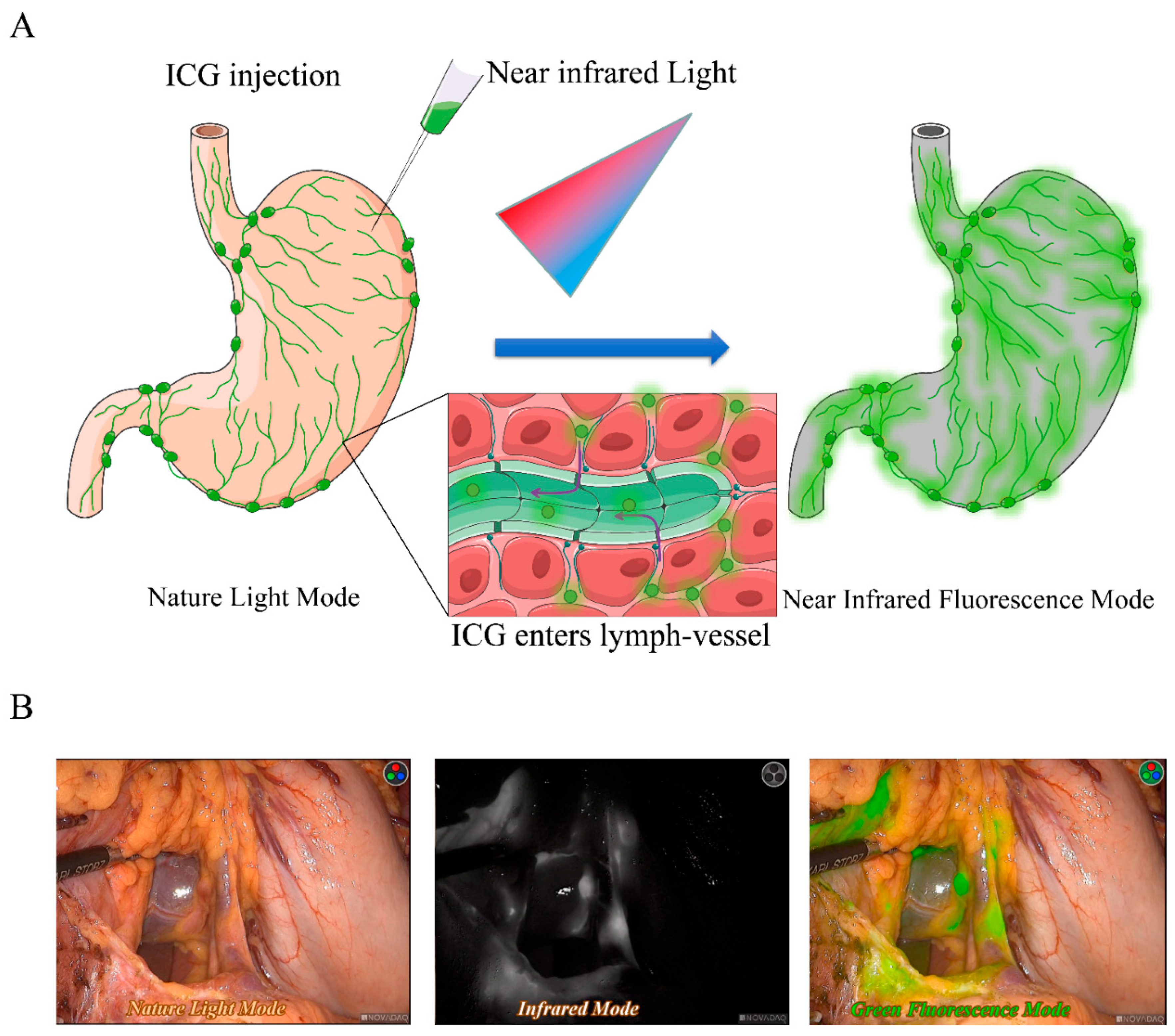
2. Current Applications of ICG-FI Guided Surgery
2.1. Assessment of Sentinel Lymph Nodes to Achieve Tailored Partial Resection with Selective Lymphadenectomy
2.2. ICG-FI Allows for Precise Lymphadenectomy in Gastric Oncologic Surgery
3. Limitations of ICG-FI in Current Gastric Cancer Practice
3.1. Absence of Specificity Contributes Additional Lymph Nodes Harvest but Loss in Precision Control of Gastric Cancer
3.2. Unpredictable False Negative Lymph Nodes Restrict Application of ICG-FI
4. Future Directions for ICG-FI Guided Surgery
4.1. Applications of Multi-Tracers Integrated with ICG-FI
4.2. Intraoperative Biopsy Technologies Improve Diagnostic Yield of ICG-FI
4.3. Artificial Intelligence, Augmented Reality and Machine Learning Algorithms for Enhancement of ICG-FI
5. Discussion
6. Conclusions
Supplementary Materials
Author Contributions
Funding
Institutional Review Board Statement
Informed Consent Statement
Data Availability Statement
Acknowledgments
Conflicts of Interest
Abbreviations
References
- Sung, H.; Ferlay, J.; Siegel, R.L.; Laversanne, M.; Soerjomataram, I.; Jemal, A.; Bray, F. Global cancer statistics 2020: GLOBOCAN estimates of incidence and mortality worldwide for 36 cancers in 185 countries. CA Cancer J. Clin. 2021, 71, 209–249. [Google Scholar] [CrossRef]
- Kano, K.; Yamada, T.; Yamamoto, K.; Komori, K.; Watanabe, H.; Hara, K.; Shimoda, Y.; Maezawa, Y.; Fujikawa, H.; Aoyama, T.; et al. Association Between Lymph Node Ratio and Survival in Patients with Pathological Stage II/III Gastric Cancer. Ann. Surg. Oncol. 2020, 27, 4235–4247. [Google Scholar] [CrossRef]
- Wang, H.; Chen, M.M.; Zhu, G.S.; Ma, M.G.; Du, H.S.; Long, Y.P. Lymph node mapping with carbon nanoparticles and the risk factors of lymph node metastasis in gastric cancer. J. Huazhong Univ. Sci. Technol. 2016, 36, 865–870. [Google Scholar] [CrossRef]
- Minarich, M.J.; Schwarz, R.E. A Simplified Two-Step Technique for Extended Lymphadenectomy During Resection of Gastroesophageal Malignancy: Early Results Compared to En Bloc Dissection. J. Gastrointest. Surg. 2019, 23, 393–401. [Google Scholar] [CrossRef]
- Borowski, D.W.; Banky, B.; Banerjee, A.K.; Agarwal, A.K.; Tabaqchali, M.A.; Garg, D.K.; Hobday, C.; Hegab, M.; Gill, T.S. Intra-arterial methylene blue injection into ex vivo colorectal cancer specimens improves lymph node staging accuracy: A randomized controlled trial. Colorectal. Dis. 2014, 16, 681–689. [Google Scholar] [CrossRef]
- Chen, D.; Chen, G.; Jiang, W.; Fu, M.; Liu, W.; Sui, J.; Xu, S.; Liu, Z.; Zheng, X.; Chi, L.; et al. Association of the Collagen Signature in the Tumor Microenvironment With Lymph Node Metastasis in Early Gastric Cancer. JAMA Surg. 2019, 154, e185249. [Google Scholar] [CrossRef]
- Benjamin, A.; Merkow, R.P. Importance of Adequate Lymphadenectomy in Gastrointestinal Cancer. Cancer Treat. Res. 2016, 168, 331–343. [Google Scholar] [CrossRef]
- Tajima, Y.; Murakami, M.; Yamazaki, K.; Masuda, Y.; Kato, M.; Sato, A.; Goto, S.; Otsuka, K.; Kato, T.; Kusano, M. Sentinel node mapping guided by indocyanine green fluorescence imaging during laparoscopic surgery in gastric cancer. Ann. Surg. Oncol. 2010, 17, 1787–1793. [Google Scholar] [CrossRef]
- Qian, Y.; Cai, S. A safe and effective surgical navigation technique in laparoscopic radical gastrectomy: Indocyanine green-mediated near-infrared fluorescent imaging. Cancer Commun. 2020, 40, 270–272. [Google Scholar] [CrossRef]
- Nishino, H.; Hatano, E.; Seo, S.; Nitta, T.; Saito, T.; Nakamura, M.; Hattori, K.; Takatani, M.; Fuji, H.; Taura, K.; et al. Real-time Navigation for Liver Surgery Using Projection Mapping With Indocyanine Green Fluorescence: Development of the Novel Medical Imaging Projection System. Ann. Surg. 2018, 267, 1134–1140. [Google Scholar] [CrossRef]
- Egloff-Juras, C.; Bezdetnaya, L.; Dolivet, G.; Lassalle, H.P. NIR fluorescence-guided tumor surgery: New strategies for the use of indocyanine green. Int. J. Nanomed. 2019, 14, 7823–7838. [Google Scholar] [CrossRef] [PubMed] [Green Version]
- Schaafsma, B.E.; Mieog, J.S.; Hutteman, M.; van der Vorst, J.R.; Kuppen, P.J.; Lowik, C.W.; Frangioni, J.V.; van de Velde, C.J.; Vahrmeijer, A.L. The clinical use of indocyanine green as a near-infrared fluorescent contrast agent for image-guided oncologic surgery. J. Surg. Oncol. 2011, 104, 323–332. [Google Scholar] [CrossRef] [PubMed] [Green Version]
- Leung, K. IRDye 800CW-Human serum albumin. In Molecular Imaging and Contrast Agent Database (MICAD); National Center for Biotechnology Information: Bethesda, MD, USA, 2004. [Google Scholar]
- Miyashiro, I.; Miyoshi, N.; Hiratsuka, M.; Kishi, K.; Yamada, T.; Ohue, M.; Ohigashi, H.; Yano, M.; Ishikawa, O.; Imaoka, S. Detection of sentinel node in gastric cancer surgery by indocyanine green fluorescence imaging: Comparison with infrared imaging. Ann. Surg. Oncol. 2008, 15, 1640–1643. [Google Scholar] [CrossRef] [PubMed]
- Chen, Q.Y.; Zhong, Q.; Li, P.; Xie, J.W.; Liu, Z.Y.; Huang, X.B.; Lin, G.T.; Wang, J.B.; Lin, J.X.; Lu, J.; et al. Comparison of submucosal and subserosal approaches toward optimized indocyanine green tracer-guided laparoscopic lymphadenectomy for patients with gastric cancer (FUGES-019): A randomized controlled trial. BMC Med. 2021, 19, 276. [Google Scholar] [CrossRef] [PubMed]
- Ishizaki, M.; Kurita, A.; Kubo, Y.; Takashima, S.; Nishina, T.; Nishimura, E. Evaluation of sentinel node identification with isosulfan blue in gastric cancer. Eur. J. Surg. Oncol. 2006, 32, 191–196. [Google Scholar] [CrossRef]
- Miwa, K. Sentinel node concept and its application for cancer surgery. Nihon Geka Gakkai Zasshi 2000, 101, 307–310. [Google Scholar]
- Kinami, S.; Nakamura, N.; Miyashita, T.; Kitakata, H.; Fushida, S.; Fujimura, T.; Iida, Y.; Inaki, N.; Ito, T.; Takamura, H. Life prognosis of sentinel node navigation surgery for early-stage gastric cancer: Outcome of lymphatic basin dissection. World J. Gastroenterol. 2021, 27, 8010–8030. [Google Scholar] [CrossRef]
- Wong, J.H.; Cagle, L.A.; Morton, D.L. Lymphatic drainage of skin to a sentinel lymph node in a feline model. Ann. Surg. 1991, 214, 637–641. [Google Scholar] [CrossRef]
- Cabanas, R.M. An approach for the treatment of penile carcinoma. Cancer 1977, 39, 456–466. [Google Scholar] [CrossRef]
- Wang, X.; Zhang, G.; Zuo, Z.; Zhu, Q.; Wu, S.; Zhou, Y.; Mao, F.; Lin, Y.; Shen, S.; Zhang, X.; et al. Sentinel Lymph Node Positive Rate Predicts Non-Sentinel Lymph Node Metastasis in Breast Cancer. J. Surg. Res. 2021, 271, 59–66. [Google Scholar] [CrossRef]
- El-Achi, V.; Burling, M.; Al-Aker, M. Sentinel lymph node biopsy at robotic-assisted hysterectomy for atypical hyperplasia and endometrial cancer. J. Robot. Surg. 2021, 16, 1111–1115. [Google Scholar] [CrossRef] [PubMed]
- Hora, M.; Travnicek, I.; Nykodymova, S.; Ferda, J.; Kacerovska, D.; Michalova, K.; Hes, O.; Minhas, S. VEILND (Video Endoscopic Inguinal Lymph Node Dissection) with Florescence Indocyanine Green (ICG): A Novel Technique to Identify the Sentinel Lymph Node in Men with >/=pT1G2 and cN0 Penile Cancer. Contrast Media Mol. Imaging 2021, 2021, 5575730. [Google Scholar] [CrossRef]
- Natsugoe, S.; Arigami, T.; Uenosono, Y.; Yanagita, S. Novel surgical approach based on the sentinel node concept in patients with early gastric cancer. Ann. Gastroenterol. Surg. 2017, 1, 180–185. [Google Scholar] [CrossRef] [PubMed]
- Park, D.J.; Lee, H.J.; Lee, H.S.; Kim, W.H.; Kim, H.H.; Lee, K.U.; Choe, K.J.; Yang, H.K. Sentinel node biopsy for cT1 and cT2a gastric cancer. Eur. J. Surg. Oncol. 2006, 32, 48–54. [Google Scholar] [CrossRef] [PubMed]
- Hiratsuka, M.; Miyashiro, I.; Ishikawa, O.; Furukawa, H.; Motomura, K.; Ohigashi, H.; Kameyama, M.; Sasaki, Y.; Kabuto, T.; Ishiguro, S.; et al. Application of sentinel node biopsy to gastric cancer surgery. Surgery 2001, 129, 335–340. [Google Scholar] [CrossRef] [PubMed]
- Kusano, M.; Tajima, Y.; Yamazaki, K.; Kato, M.; Watanabe, M.; Miwa, M. Sentinel node mapping guided by indocyanine green fluorescence imaging: A new method for sentinel node navigation surgery in gastrointestinal cancer. Dig. Surg. 2008, 25, 103–108. [Google Scholar] [CrossRef]
- Kelder, W.; Nimura, H.; Takahashi, N.; Mitsumori, N.; van Dam, G.M.; Yanaga, K. Sentinel node mapping with indocyanine green (ICG) and infrared ray detection in early gastric cancer: An accurate method that enables a limited lymphadenectomy. Eur. J. Surg. Oncol. 2010, 36, 552–558. [Google Scholar] [CrossRef]
- Park, D.J.; Kim, H.H.; Park, Y.S.; Lee, H.S.; Lee, W.W.; Lee, H.J.; Yang, H.K. Simultaneous indocyanine green and (99m)Tc-antimony sulfur colloid-guided laparoscopic sentinel basin dissection for gastric cancer. Ann. Surg. Oncol. 2011, 18, 160–165. [Google Scholar] [CrossRef]
- Miyashiro, I.; Hiratsuka, M.; Kishi, K.; Takachi, K.; Yano, M.; Takenaka, A.; Tomita, Y.; Ishiguro, S. Intraoperative diagnosis using sentinel node biopsy with indocyanine green dye in gastric cancer surgery: An institutional trial by experienced surgeons. Ann. Surg. Oncol. 2013, 20, 542–546. [Google Scholar] [CrossRef]
- Kim, D.W.; Jeong, B.; Shin, I.H.; Kang, U.; Lee, Y.; Park, Y.S.; Ahn, S.H.; Park, D.J.; Kim, H.H. Sentinel node navigation surgery using near-infrared indocyanine green fluorescence in early gastric cancer. Surg. Endosc. 2019, 33, 1235–1243. [Google Scholar] [CrossRef]
- Kitagawa, Y.; Takeuchi, H.; Takagi, Y.; Natsugoe, S.; Terashima, M.; Murakami, N.; Fujimura, T.; Tsujimoto, H.; Hayashi, H.; Yoshimizu, N.; et al. Sentinel node mapping for gastric cancer: A prospective multicenter trial in Japan. J. Clin. Oncol. 2013, 31, 3704–3710. [Google Scholar] [CrossRef] [PubMed]
- Nozaki, I.; Kubo, Y.; Kurita, A.; Tanada, M.; Yokoyama, N.; Takiyama, W.; Takashima, S. Long-term outcome after laparoscopic wedge resection for early gastric cancer. Surg. Endosc. 2008, 22, 2665–2669. [Google Scholar] [CrossRef] [PubMed]
- Shinohara, T.; Ohyama, S.; Muto, T.; Kato, Y.; Yanaga, K.; Yamaguchi, T. Clinical outcome of high segmental gastrectomy for early gastric cancer in the upper third of the stomach. Br. J. Surg. 2006, 93, 975–980. [Google Scholar] [CrossRef] [PubMed]
- Takiguchi, N.; Takahashi, M.; Ikeda, M.; Inagawa, S.; Ueda, S.; Nobuoka, T.; Ota, M.; Iwasaki, Y.; Uchida, N.; Kodera, Y.; et al. Long-term quality-of-life comparison of total gastrectomy and proximal gastrectomy by postgastrectomy syndrome assessment scale (PGSAS-45): A nationwide multi-institutional study. Gastric Cancer 2015, 18, 407–416. [Google Scholar] [CrossRef]
- Nimura, H.; Narimiya, N.; Mitsumori, N.; Yamazaki, Y.; Yanaga, K.; Urashima, M. Infrared ray electronic endoscopy combined with indocyanine green injection for detection of sentinel nodes of patients with gastric cancer. Br. J. Surg. 2004, 91, 575–579. [Google Scholar] [CrossRef]
- Koyama, T.; Tsubota, A.; Nariai, K.; Mitsunaga, M.; Yanaga, K.; Takahashi, H. Novel biomedical imaging approach for detection of sentinel nodes in an experimental model of gastric cancer. Br. J. Surg. 2007, 94, 996–1001. [Google Scholar] [CrossRef]
- Miyashiro, I.; Hiratsuka, M.; Sasako, M.; Sano, T.; Mizusawa, J.; Nakamura, K.; Nashimoto, A.; Tsuburaya, A.; Fukushima, N.; Gastric Cancer Surgical Study Group (GCSSG) in the Japan Clinical Oncology Group (JCOG). High false-negative proportion of intraoperative histological examination as a serious problem for clinical application of sentinel node biopsy for early gastric cancer: Final results of the Japan Clinical Oncology Group multicenter trial JCOG0302. Gastric Cancer 2014, 17, 316–323. [Google Scholar] [CrossRef] [Green Version]
- Quaresma, L.; Fradique, A.C.; Pupo, A.; Silva, G.; Da Silva, G.; Da Costa, L.B.; Oliveira, M.; Esteves, J.; Marques, M.; Fernandez, G. Sentinel lymph node in gastric cancer. J. Clin. Oncol. 2015, 33, e15014. [Google Scholar] [CrossRef] [Green Version]
- Tummers, Q.R.; Boogerd, L.S.; de Steur, W.O.; Verbeek, F.P.; Boonstra, M.C.; Handgraaf, H.J.; Frangioni, J.V.; van de Velde, C.J.; Hartgrink, H.H.; Vahrmeijer, A.L. Near-infrared fluorescence sentinel lymph node detection in gastric cancer: A pilot study. World J. Gastroenterol. 2016, 22, 3644–3651. [Google Scholar] [CrossRef]
- Takahashi, N.; Nimura, H.; Fujita, T.; Mitsumori, N.; Shiraishi, N.; Kitano, S.; Satodate, H.; Yanaga, K. Laparoscopic sentinel node navigation surgery for early gastric cancer: A prospective multicenter trial. Langenbeck’s Arch. Surg. 2017, 402, 27–32. [Google Scholar] [CrossRef]
- Mayanagi, S.; Takahashi, N.; Mitsumori, N.; Arigami, T.; Natsugoe, S.; Yaguchi, Y.; Suda, T.; Kinami, S.; Ohi, M.; Kawakubo, H.; et al. Sentinel node mapping for post-endoscopic resection gastric cancer: Multicenter retrospective cohort study in Japan. Gastric Cancer 2020, 23, 716–724. [Google Scholar] [CrossRef] [PubMed]
- Miyashiro, I. What is the problem in clinical application of sentinel node concept to gastric cancer surgery? J. Gastric Cancer 2012, 12, 7–12. [Google Scholar] [CrossRef] [Green Version]
- Kim, S.G.; Eom, B.W.; Yoon, H.M.; Kim, C.G.; Kook, M.C.; Kim, Y.W.; Ryu, K.W. Recent updates and current issues of sentinel node navigation surgery for early gastric cancer. Chin. J. Cancer Res. 2021, 33, 142–149. [Google Scholar] [CrossRef] [PubMed]
- An, J.Y.; Min, J.S.; Hur, H.; Lee, Y.J.; Cho, G.S.; Park, Y.K.; Jung, M.R.; Park, J.H.; Hyung, W.J.; Jeong, S.H.; et al. Laparoscopic sentinel node navigation surgery versus laparoscopic gastrectomy with lymph node dissection for early gastric cancer: Short-term outcomes of a multicentre randomized controlled trial (SENORITA). Br. J. Surg. 2020, 107, 1429–1439. [Google Scholar] [CrossRef]
- Park, D.J.; Park, Y.S.; Son, S.Y.; Lee, J.H.; Lee, H.S.; Park, Y.S.; Lee, K.H.; Kim, Y.H.; Park, K.U.; Lee, W.W.; et al. Long-Term Oncologic Outcomes of Laparoscopic Sentinel Node Navigation Surgery in Early Gastric Cancer: A Single-Center, Single-Arm, Phase II Trial. Ann. Surg. Oncol. 2018, 25, 2357–2365. [Google Scholar] [CrossRef] [PubMed]
- Lee, J.H.; Ryu, K.W.; Kim, C.G.; Kim, S.K.; Choi, I.J.; Kim, Y.W.; Chang, H.J.; Bae, J.M.; Hong, E.K. Comparative study of the subserosal versus submucosal dye injection method for sentinel node biopsy in gastric cancer. Eur. J. Surg. Oncol. 2005, 31, 965–968. [Google Scholar] [CrossRef]
- Yaguchi, Y.; Ichikura, T.; Ono, S.; Tsujimoto, H.; Sugasawa, H.; Sakamoto, N.; Matsumoto, Y.; Yoshida, K.; Kosuda, S.; Hase, K. How should tracers be injected to detect for sentinel nodes in gastric cancer--submucosally from inside or subserosally from outside of the stomach? J. Exp. Clin. Cancer Res. 2008, 27, 79. [Google Scholar] [CrossRef] [Green Version]
- Lan, Y.T.; Huang, K.H.; Chen, P.H.; Liu, C.A.; Lo, S.S.; Wu, C.W.; Shyr, Y.M.; Fang, W.L. A pilot study of lymph node mapping with indocyanine green in robotic gastrectomy for gastric cancer. SAGE Open Med. 2017, 5, 2050312117727444. [Google Scholar] [CrossRef]
- Huang, L.; Li, T.J. Laparoscopic surgery for gastric cancer: Where are we now and where are we going? Expert Rev. Anticancer Ther. 2018, 18, 1145–1157. [Google Scholar] [CrossRef]
- Cianchi, F.; Indennitate, G.; Paoli, B.; Ortolani, M.; Lami, G.; Manetti, N.; Tarantino, O.; Messeri, S.; Foppa, C.; Badii, B.; et al. The Clinical Value of Fluorescent Lymphography with Indocyanine Green During Robotic Surgery for Gastric Cancer: A Matched Cohort Study. J. Gastrointest. Surg. 2020, 24, 2197–2203. [Google Scholar] [CrossRef]
- Liu, M.; Xing, J.; Xu, K.; Yuan, P.; Cui, M.; Zhang, C.; Yang, H.; Yao, Z.; Zhang, N.; Tan, F.; et al. Application of Near-Infrared Fluorescence Imaging with Indocyanine Green in Totally Laparoscopic Distal Gastrectomy. J. Gastric Cancer 2020, 20, 290–299. [Google Scholar] [CrossRef]
- Huang, Z.N.; Su, Y.; Qiu, W.W.; Liu, C.H.; Chen, Q.Y.; Zheng, C.H.; Li, P.; Wang, J.B.; Lin, J.X.; Lu, J.; et al. Assessment of indocyanine green tracer-guided lymphadenectomy in laparoscopic gastrectomy after neoadjuvant chemotherapy for locally advanced gastric cancer: Results from a multicenter analysis based on propensity matching. Gastric Cancer 2021, 24, 1355–1364. [Google Scholar] [CrossRef]
- Lu, X.; Liu, S.; Xia, X.; Sun, F.; Liu, Z.; Wang, J.; Li, X.; Yang, Z.; Kang, X.; Ai, S.; et al. The short-term and long-term outcomes of indocyanine green tracer-guided laparoscopic radical gastrectomy in patients with gastric cancer. World J. Surg. Oncol. 2021, 19, 271. [Google Scholar] [CrossRef] [PubMed]
- Ma, S.; Zhang, Y.M.; Dou, L.Z.; Liu, H.; Ma, F.H.; Wang, G.Q.; Tian, Y.T. Efficacy and Feasibility of Indocyanine Green for Mapping Lymph Nodes in Advanced Gastric Cancer Patients Undergoing Laparoscopic Distal Gastrectomy. J. Gastrointest. Surg. 2020, 24, 2306–2309. [Google Scholar] [CrossRef]
- Kwon, I.G.; Son, T.; Kim, H.I.; Hyung, W.J. Fluorescent Lymphography-Guided Lymphadenectomy During Robotic Radical Gastrectomy for Gastric Cancer. JAMA Surg. 2019, 154, 150–158. [Google Scholar] [CrossRef] [PubMed] [Green Version]
- Chen, Q.Y.; Xie, J.W.; Zhong, Q.; Wang, J.B.; Lin, J.X.; Lu, J.; Cao, L.L.; Lin, M.; Tu, R.H.; Huang, Z.N.; et al. Safety and Efficacy of Indocyanine Green Tracer-Guided Lymph Node Dissection During Laparoscopic Radical Gastrectomy in Patients with Gastric Cancer: A Randomized Clinical Trial. JAMA Surg. 2020, 155, 300–311. [Google Scholar] [CrossRef]
- Deng, C.; Zhang, Z.; Qi, H.; Guo, Z.; Liu, Y.; Xiao, H.; Li, X. Safety and efficacy of indocyanine green near-infrared fluorescent imaging-guided lymph nodes dissection during radical gastrectomy for gastric cancer: A systematic review and meta-analysis. Front. Oncol. 2022, 12, 917541. [Google Scholar] [CrossRef]
- Ushimaru, Y.; Omori, T.; Fujiwara, Y.; Yanagimoto, Y.; Sugimura, K.; Yamamoto, K.; Moon, J.H.; Miyata, H.; Ohue, M.; Yano, M. The Feasibility and Safety of Preoperative Fluorescence Marking with Indocyanine Green (ICG) in Laparoscopic Gastrectomy for Gastric Cancer. J. Gastrointest. Surg. 2019, 23, 468–476. [Google Scholar] [CrossRef]
- Kim, T.H.; Kong, S.H.; Park, J.H.; Son, Y.G.; Huh, Y.J.; Suh, Y.S.; Lee, H.J.; Yang, H.K. Assessment of the Completeness of Lymph Node Dissection Using Near-infrared Imaging with Indocyanine Green in Laparoscopic Gastrectomy for Gastric Cancer. J. Gastric Cancer 2018, 18, 161–171. [Google Scholar] [CrossRef]
- Liu, C.G.; Lu, P.; Lu, Y.; Jin, F.; Xu, H.M.; Wang, S.B.; Chen, J.Q. Distribution of solitary lymph nodes in primary gastric cancer: A retrospective study and clinical implications. World J. Gastroenterol. 2007, 13, 4776–4780. [Google Scholar] [CrossRef] [PubMed]
- Zuo, C.H.; Xie, H.; Liu, J.; Qiu, X.X.; Lin, J.G.; Hua, X.; Qin, A. Characterization of lymph node metastasis and its clinical significance in the surgical treatment of gastric cancer. Mol. Clin. Oncol. 2014, 2, 821–826. [Google Scholar] [CrossRef] [PubMed]
- Park, S.H.; Berlth, F.; Choi, J.H.; Park, J.H.; Suh, Y.S.; Kong, S.H.; Park, D.J.; Lee, H.J.; Yang, H.K. Near-infrared fluorescence-guided surgery using indocyanine green facilitates secure infrapyloric lymph node dissection during laparoscopic distal gastrectomy. Surg. Today 2020, 50, 1187–1196. [Google Scholar] [CrossRef] [PubMed]
- Lee, S.; Song, J.H.; Choi, S.; Cho, M.; Kim, Y.M.; Kim, H.I.; Hyung, W.J. Fluorescent lymphography during minimally invasive total gastrectomy for gastric cancer: An effective technique for splenic hilar lymph node dissection. Surg. Endosc. 2022, 36, 2914–2924. [Google Scholar] [CrossRef] [PubMed]
- Romanzi, A.; Mancini, R.; Ioni, L.; Picconi, T.; Pernazza, G. ICG-NIR-guided lymph node dissection during robotic subtotal gastrectomy for gastric cancer. A single-centre experience. Int. J. Med Robot. Comput. Assist. Surg. 2021, 17, e2213. [Google Scholar] [CrossRef]
- Zhong, Q.; Chen, Q.Y.; Huang, X.B.; Lin, G.T.; Liu, Z.Y.; Chen, J.Y.; Wang, H.G.; Weng, K.; Li, P.; Xie, J.W.; et al. Clinical implications of Indocyanine Green Fluorescence Imaging-Guided laparoscopic lymphadenectomy for patients with gastric cancer: A cohort study from two randomized, controlled trials using individual patient data. Int. J. Surg. 2021, 94, 106120. [Google Scholar] [CrossRef]
- Deng, J.Y.; Liang, H.; Sun, D.; Pan, Y.; Zhang, R.P.; Wang, B.G.; Zhan, H.J. Outcome in relation to numbers of nodes harvested in lymph node-positive gastric cancer. Eur. J. Surg. Oncol. 2009, 35, 814–819. [Google Scholar] [CrossRef]
- Saito, H.; Fukumoto, Y.; Osaki, T.; Fukuda, K.; Tatebe, S.; Tsujitani, S.; Ikeguchi, M. Prognostic significance of level and number of lymph node metastases in patients with gastric cancer. Ann. Surg. Oncol. 2007, 14, 1688–1693. [Google Scholar] [CrossRef]
- Alatengbaolide; Lin, D.; Li, Y.; Xu, H.; Chen, J.; Wang, B.; Liu, C.; Lu, P. Lymph node ratio is an independent prognostic factor in gastric cancer after curative resection (R0) regardless of the examined number of lymph nodes. Am. J. Clin. Oncol. 2013, 36, 325–330. [Google Scholar] [CrossRef]
- Topcu, R.; Sahiner, I.T.; Kendirci, M.; Erkent, M.; Sezikli, I.; Tutan, M.B. Does lymph node ratio (metastasis/total lymph node count) affect survival and prognosis in gastric cancer? Saudi Med. J. 2022, 43, 139–145. [Google Scholar] [CrossRef]
- Jiang, J.; Chen, J.; Zhang, H.; Rao, X.; Hao, T.; Li, M.; Zhang, C.; Wu, W.; He, Y. Combination of the ratio between metastatic and harvested lymph nodes and negative lymph node count as a prognostic indicator in advanced gastric cancer: A retrospective cohort study. J. Gastrointest. Oncol. 2021, 12, 2022–2034. [Google Scholar] [CrossRef]
- Nelen, S.D.; van Steenbergen, L.N.; Dassen, A.E.; van der Wurff, A.A.; Lemmens, V.E.; Bosscha, K. The lymph node ratio as a prognostic factor for gastric cancer. Acta Oncol. 2013, 52, 1751–1759. [Google Scholar] [CrossRef] [PubMed]
- Sevieri, M.; Sitia, L.; Bonizzi, A.; Truffi, M.; Mazzucchelli, S.; Corsi, F. Tumor Accumulation and Off-Target Biodistribution of an Indocyanine-Green Fluorescent Nanotracer: An Ex Vivo Study on an Orthotopic Murine Model of Breast Cancer. Int. J. Mol. Sci. 2021, 22, 1601. [Google Scholar] [CrossRef] [PubMed]
- Tajima, Y.; Yamazaki, K.; Masuda, Y.; Kato, M.; Yasuda, D.; Aoki, T.; Kato, T.; Murakami, M.; Miwa, M.; Kusano, M. Sentinel node mapping guided by indocyanine green fluorescence imaging in gastric cancer. Ann. Surg. 2009, 249, 58–62. [Google Scholar] [CrossRef] [PubMed]
- Tonouchi, H.; Mohri, Y.; Tanaka, K.; Kobayashi, M.; Ohmori, Y.; Kusunoki, M. Laparoscopic lymphatic mapping and sentinel node biopsies for early-stage gastric cancer: The cause of false negativity. World J. Surg. 2005, 29, 418–421. [Google Scholar] [CrossRef]
- Su, Z.; Shu, K.; Zheng, M.; Sun, X.; Fang, Z.; Wang, G. Sentinel lymph node and skip metastases in gastric cancer: A prospective study. Hepatogastroenterology 2013, 60, 1513–1518. [Google Scholar] [CrossRef]
- Wang, Z.; Cui, Y.; Zheng, M.; Ge, H.; Huang, Y.; Peng, J.; Xie, H.; Wang, S. Comparison of indocyanine green fluorescence and methylene blue dye in the detection of sentinel lymph nodes in breast cancer. Gland Surg. 2020, 9, 1495–1501. [Google Scholar] [CrossRef]
- Guo, J.; Yang, H.; Wang, S.; Cao, Y.; Liu, M.; Xie, F.; Liu, P.; Zhou, B.; Tong, F.; Cheng, L.; et al. Comparison of sentinel lymph node biopsy guided by indocyanine green, blue dye, and their combination in breast cancer patients: A prospective cohort study. World J. Surg. Oncol. 2017, 15, 196. [Google Scholar] [CrossRef] [Green Version]
- Huang, L.; Wei, T.; Chen, J.; Zhou, D. Feasibility and diagnostic performance of dual-tracer-guided sentinel lymph node biopsy in cT1-2N0M0 gastric cancer: A systematic review and meta-analysis of diagnostic studies. World J. Surg. Oncol. 2017, 15, 103. [Google Scholar] [CrossRef] [Green Version]
- Inoue, K.; Fukuhara, H.; Shimamoto, T.; Kamada, M.; Iiyama, T.; Miyamura, M.; Kurabayashi, A.; Furihata, M.; Tanimura, M.; Watanabe, H.; et al. Comparison between intravesical and oral administration of 5-aminolevulinic acid in the clinical benefit of photodynamic diagnosis for nonmuscle invasive bladder cancer. Cancer 2012, 118, 1062–1074. [Google Scholar] [CrossRef]
- Namikawa, T.; Iwabu, J.; Munekage, M.; Uemura, S.; Maeda, H.; Kitagawa, H.; Nakayama, T.; Inoue, K.; Sato, T.; Kobayashi, M.; et al. Evolution of photodynamic medicine based on fluorescence image-guided diagnosis using indocyanine green and 5-aminolevulinic acid. Surg. Today 2020, 50, 821–831. [Google Scholar] [CrossRef]
- Osterkamp, J.; Strandby, R.B.; Nerup, N.; Svendsen, M.B.S.; Svendsen, L.B.; Achiam, M.P. Time to maximum indocyanine green fluorescence of gastric sentinel lymph nodes and feasibility of combined indocyanine green/sodium fluorescein gastric lymphography. Langenbecks Arch. Surg. 2021, 406, 2717–2724. [Google Scholar] [CrossRef] [PubMed]
- Kuwahata, A.; Ahmed, M.; Saeki, K.; Chikaki, S.; Kaneko, M.; Qiu, W.; Xin, Z.; Yamaguchi, S.; Kaneko, A.; Douek, M.; et al. Combined use of fluorescence with a magnetic tracer and dilution effect upon sentinel node localization in a murine model. Int. J. Nanomed. 2018, 13, 2427–2433. [Google Scholar] [CrossRef] [PubMed] [Green Version]
- Azargoshasb, S.; Molenaar, L.; Rosiello, G.; Buckle, T.; van Willigen, D.M.; van de Loosdrecht, M.M.; Welling, M.M.; Alic, L.; van Leeuwen, F.W.B.; Winter, A.; et al. Advancing intraoperative magnetic tracing using 3D freehand magnetic particle imaging. Int. J. Comput. Assist. Radiol. Surg. 2022, 17, 211–218. [Google Scholar] [CrossRef] [PubMed]
- Kamada, T.; Yoshida, M.; Takeuchi, H.; Narihiro, S.; Ohdaira, H.; Suzuki, Y. A new method of sentinel node mapping for early gastric cancer using a fluorescent laparoscope that can adjust the intensity of excitation light and quantify the intensity of indocyanine green fluorescence: Report of a case. Int. J. Surg. Case Rep. 2020, 73, 248–252. [Google Scholar] [CrossRef]
- Tian, Y.; Lin, Y.; Guo, H.; Hu, Y.; Li, Y.; Fan, L.; Zhao, X.; Wang, D.; Tan, B.; Zhao, Q. Safety and efficacy of carbon nanoparticle suspension injection and indocyanine green tracer-guided lymph node dissection during robotic distal gastrectomy in patients with gastric cancer. Surg. Endosc. 2021, 36, 3209–3216. [Google Scholar] [CrossRef]
- Ankersmit, M.; Hoekstra, O.S.; van Lingen, A.; Bloemena, E.; Jacobs, M.; Vugts, D.J.; Bonjer, H.J.; van Dongen, G.; Meijerink, W. Perioperative PET/CT lymphoscintigraphy and fluorescent real-time imaging for sentinel lymph node mapping in early staged colon cancer. Eur. J. Pediatr. 2019, 46, 1495–1505. [Google Scholar] [CrossRef] [Green Version]
- Park, J.Y.; Kook, M.C.; Eom, B.W.; Yoon, H.M.; Kim, S.J.; Rho, J.Y.; Kim, S.K.; Kim, Y.I.; Cho, S.J.; Lee, J.Y.; et al. Practical intraoperative pathologic evaluation of sentinel lymph nodes during sentinel node navigation surgery in gastric cancer patients—Proposal of the pathologic protocol for the upcoming SENORITA trial. Surg. Oncol. 2016, 25, 139–146. [Google Scholar] [CrossRef]
- Esposito, F.; Noviello, A.; Moles, N.; Coppola Bottazzi, E.; Baiamonte, M.; Macaione, I.; Ferbo, U.; Lepore, M.; Miro, A.; Crafa, F. Sentinel Lymph Node Analysis in Colorectal Cancer Patients Using One-Step Nucleic Acid Amplification in Combination With Fluorescence and Indocyanine Green. Ann. Coloproctol. 2019, 35, 174–180. [Google Scholar] [CrossRef] [Green Version]
- Shimizu, Y.; Takeuchi, H.; Sakakura, Y.; Saikawa, Y.; Nakahara, T.; Mukai, M.; Kitajima, M.; Kitagawa, Y. Molecular detection of sentinel node micrometastases in patients with clinical N0 gastric carcinoma with real-time multiplex reverse transcription-polymerase chain reaction assay. Ann. Surg. Oncol. 2012, 19, 469–477. [Google Scholar] [CrossRef]
- Diana, M.; Robinet, E.; Liu, Y.Y.; Legner, A.; Kong, S.H.; Schiraldi, L.; Marchegiani, F.; Halvax, P.; Swanstrom, L.; Dallemagne, B.; et al. Confocal Imaging and Tissue-Specific Fluorescent Probes for Real-Time In Vivo Immunohistochemistry. Proof of the Concept in a Gastric Lymph Node Metastasis Model. Ann. Surg. Oncol. 2016, 23, 567–573. [Google Scholar] [CrossRef]
- Yuan, S.; Roney, C.A.; Wierwille, J.; Chen, C.W.; Xu, B.; Griffiths, G.; Jiang, J.; Ma, H.; Cable, A.; Summers, R.M.; et al. Co-registered optical coherence tomography and fluorescence molecular imaging for simultaneous morphological and molecular imaging. Phys. Med. Biol. 2010, 55, 191–206. [Google Scholar] [CrossRef] [PubMed]
- Nolan, R.M.; Adie, S.G.; Marjanovic, M.; Chaney, E.J.; South, F.A.; Monroy, G.L.; Shemonski, N.D.; Erickson-Bhatt, S.J.; Shelton, R.L.; Bower, A.J.; et al. Intraoperative optical coherence tomography for assessing human lymph nodes for metastatic cancer. BMC Cancer 2016, 16, 144. [Google Scholar] [CrossRef] [PubMed] [Green Version]
- Lee, B.; Chen, S.; Moult, E.M.; Yu, Y.; Alibhai, A.Y.; Mehta, N.; Baumal, C.R.; Waheed, N.K.; Fujimoto, J.G. High-Speed, Ultrahigh-Resolution Spectral-Domain OCT with Extended Imaging Range Using Reference Arm Length Matching. Transl. Vis. Sci. Technol. 2020, 9, 12. [Google Scholar] [CrossRef]
- Lee, S.; Lee, M.W.; Cho, H.S.; Song, J.W.; Nam, H.S.; Oh, D.J.; Park, K.; Oh, W.Y.; Yoo, H.; Kim, J.W. Fully integrated high-speed intravascular optical coherence tomography/near-infrared fluorescence structural/molecular imaging in vivo using a clinically available near-infrared fluorescence-emitting indocyanine green to detect inflamed lipid-rich atheromata in coronary-sized vessels. Circ. Cardiovasc. Interv. 2014, 7, 560–569. [Google Scholar] [CrossRef] [PubMed] [Green Version]
- Ughi, G.J.; Verjans, J.; Fard, A.M.; Wang, H.; Osborn, E.; Hara, T.; Mauskapf, A.; Jaffer, F.A.; Tearney, G.J. Dual modality intravascular optical coherence tomography (OCT) and near-infrared fluorescence (NIRF) imaging: A fully automated algorithm for the distance-calibration of NIRF signal intensity for quantitative molecular imaging. Int. J. Cardiovasc. Imaging 2015, 31, 259–268. [Google Scholar] [CrossRef] [Green Version]
- Kim, S.; Lee, M.W.; Kim, T.S.; Song, J.W.; Nam, H.S.; Cho, H.S.; Jang, S.J.; Ryu, J.; Oh, D.J.; Gweon, D.G.; et al. Intracoronary dual-modal optical coherence tomography-near-infrared fluorescence structural-molecular imaging with a clinical dose of indocyanine green for the assessment of high-risk plaques and stent-associated inflammation in a beating coronary artery. Eur. Heart J. 2016, 37, 2833–2844. [Google Scholar] [CrossRef] [Green Version]
- Yeung, T.M.; Wang, L.M.; Colling, R.; Kraus, R.; Cahill, R.; Hompes, R.; Mortensen, N.J. Intraoperative identification and analysis of lymph nodes at laparoscopic colorectal cancer surgery using fluorescence imaging combined with rapid OSNA pathological assessment. Surg. Endosc. 2018, 32, 1073–1076. [Google Scholar] [CrossRef] [Green Version]
- Yanagita, S.; Uenosono, Y.; Arigami, T.; Daisuke, M.; Okubo, K.; Kijima, T.; Arima, H.; Hirata, M.; Haraguchi, N.; Hagihara, T.; et al. The clinical usefulness of the intraoperative detection of sentinel lymph node metastases by a rapid RT-PCR system in patients with gastric cancer. Cancer 2016, 122, 386–392. [Google Scholar] [CrossRef]
- Park, S.H.; Park, H.M.; Baek, K.R.; Ahn, H.M.; Lee, I.Y.; Son, G.M. Artificial intelligence based real-time microcirculation analysis system for laparoscopic colorectal surgery. World J. Gastroenterol. 2020, 26, 6945–6962. [Google Scholar] [CrossRef]
- Ma, Z.; Wang, F.; Wang, W.; Zhong, Y.; Dai, H. Deep learning for in vivo near-infrared imaging. Proc. Natl. Acad. Sci. USA 2021, 118, e2021446118. [Google Scholar] [CrossRef]
- Knospe, L.; Gockel, I.; Jansen-Winkeln, B.; Thieme, R.; Niebisch, S.; Moulla, Y.; Stelzner, S.; Lyros, O.; Diana, M.; Marescaux, J.; et al. New Intraoperative Imaging Tools and Image-Guided Surgery in Gastric Cancer Surgery. Diagnostics 2022, 12, 507. [Google Scholar] [CrossRef] [PubMed]
- Lee, S.H.; Quan, Y.H.; Kim, M.S.; Kwon, K.H.; Choi, B.H.; Kim, H.K.; Kim, B.M. Design and Testing of Augmented Reality-Based Fluorescence Imaging Goggle for Intraoperative Imaging-Guided Surgery. Diagnostics 2021, 11, 927. [Google Scholar] [CrossRef] [PubMed]
- Spaide, R.F.; Fujimoto, J.G.; Waheed, N.K. Optical Coherence Tomography Angiography. Retina 2015, 35, 2161–2162. [Google Scholar] [CrossRef] [Green Version]
- Lee, S.E.; Lee, J.H.; Ryu, K.W.; Cho, S.J.; Lee, J.Y.; Kim, C.G.; Choi, I.J.; Kook, M.C.; Nam, B.H.; Park, S.R.; et al. Sentinel node mapping and skip metastases in patients with early gastric cancer. Ann. Surg. Oncol. 2009, 16, 603–608. [Google Scholar] [CrossRef] [PubMed]


| Ref. | NO. of Patients | Detection Rate | Sensitivity | Specificity | Considerations |
|---|---|---|---|---|---|
| Hiratsuka, M. 2001 [26] | Total: 74 T1 (n = 44) T2 (n = 30) | 99% | 90% | 100% | T1 (n = 44) Sensitivity: 100% T2 (n = 30) Sensitivity: 88% |
| Nimura, H. 2004 [36] | 84 | 99% | 100% | 67% | ICG and infrared ray electronic endoscopy (IREE) |
| Park, D.J. 2006 [25] | 100 | 94% | 78.6% | 100% | FNR occurred in cases with tumor size > 4 cm |
| Koyama, T. 2007 [37] | 50 | 95% | 81% | 50% | Low specificity |
| Kusano, M. 2008 [27] | 22 | 90.9% | T1: 66.7% Overall: 40% | T1: 100% Overall: 100% | T1 stage FNR:33.3%% Overall FNR:60% (T1,T2,T3) |
| Kelder, W. 2010 [28] | 212 | 99.5% | 85.8% | 100% | ICG and infrared ray electronic endoscopy (IREE) |
| Tajima, Y. 2010 [8] | 77 | LAG: 94.7% OG: 94.9% | LAG: 75% OG: 76.9% | LAG: 100% OG: 100% | LAG: FNR:25% OG: FNR:23.1% |
| Park, D.J. 2011 [29] | 68 | 91.2% | 72.2% | 100% | Dual technique (ICG and isotope) |
| Miyashiro, I. 2013 [30] | 241 | 99.6% | 89.7% | 98.6% | Intraoperative FNR: 10.3% |
| Miyashiro, I. 2014 [38] | 440 | 97.8% | 85.7% | 99.3% | High FNR |
| Quaresma, L. 2015 [39] | 20 | 92.85% | 83.3% | 100% | size of the tumor is less than 4 cm |
| Tummers, Q.R. 2016 [40] | 22 | 95% | 75% | 100% | FNR occurred in T3 and T4 cases |
| Takahashi, N. 2017 [41] | 44 | 100% | 100% | 100% | cT1 gastric adenocarcinomas less than 4 cm |
| Kim, D.W. 2019 [31] | 28 | 55% | 98.9% | 76% | Dual tracer method as gold standard |
| Mayanagi, S. 2020 [42] | 132 | 100% | 95.7% | 100% | multicenter retrospective cohort study |
| Ref. | Patients | ICG Group | Non-ICG Group | ICG-Mean (SD) Nodules | Non-ICG-Mean (SD) Nodules | p Value |
|---|---|---|---|---|---|---|
| Ushimaru, Y. 2019 [59] | 168 | 84 | 84 | 47.5 ± 1.7 | 42.6±1.7 | 0.046 * |
| Kwon, I.G. 2019 [56] | 80 | 40 | 40 | 48.9 (14.6) | 35.2 (11.2) | <0.001 *** |
| Chen, Q.Y. 2020 [57] | 258 | 129 | 129 | 50.5 (15.9) | 42 (10.3) | <0.01 ** |
| Cianchi, F. 2020 [51] | 74 | 37 | 37 | 50.8 (17.1) | 40.1 (23) | 0.03 * |
| Liu, M. 2020 [52] | 136 | 61 | 75 | 33.72 (9.06) | 29.36 (8.76) | 0.005 ** |
| Ma, S. 2020 [55] | 65 | 31 | 34 | 49.6 (13.3) | 36.4 (13.3) | 0 *** |
| Huang, Z.N. 2021 [53] | 313 | 102 | 211 | 41.5 (12.9) | 28.4 (12.4) | <0.001 *** |
| Lu, X. 2021 [54] | 56 | 28 | 28 | 27.50 (10.60) | 21.79 (6.73) | 0.0196 * |
| Romanzi, A. 2021 [65] | 20 | 10 | 10 | 40 | 24 | <0.05 * |
| Zhong, Q. 2021 [66] | 514 | 385 | 129 | 49.9 | 42.0 | <0.001 *** |
| Integrate Modalities | Rationale | Superiority | Drawbacks |
|---|---|---|---|
| Traditional biological Blue dye | |||
| Patent blue (PB) | Biological dye, visible in white light | Relies solely on a visual identification of the SLNs and no requirement of additional equipment | Restricted dying time and fades quickly when exposed to light |
| Methylene blue (MB) | Toxicity | ||
| Isosulfide blue (ISB) | Allergic reactions and long-term skin discoloration | ||
| Traditional radiocolloid tracer | |||
| Technetium-99 (99mTc) [80] | Identification of decays emitting X-rays by gamma probe | Auxiliary SPECT–CT enhance SLN mapping in terms of overall and bilateral detection rates and identify SLNs located in unusual anatomic sites | The dose is time-dependent, the decay of TC-99m may compromise the effectiveness and Radioactive drug (99mTc) toxicity |
| Fluorescent agent | |||
| 5-aminolevulinic acid (5-ALA) [82] | Tumor-specific accumulation of photoactive PpIX | Accumulates specifically in cancer cells and provide fluorescence signal | Diffuse-type tumors shown only nominal red fluorescent signal |
| Sodium fluorescein (SF) [83] | Excited by 400–550 nm wavelengths light and provide fluorescence signal | The excitation and emission wavelengths of SF do not overlap with ICG, SF can be a remedial measures for contaminated surgical field caused by spillage or incorrect injection of ICG | Potential leak from lymphatic vessels |
| Carbon nano-tracers | |||
| Carbon nanoparticles [86] | Carbon nanoparticles pass through the lymphatic vessels and accumulate in the lymph nodes, thus staining them black | With average diameter of 150 nm, Carbon nanoparticles only enter lymphatic vessels but not blood capillaries | Carbon nanoparticles remain in the body for a long time, thus extravasation will affect the surgical field |
| magnetic tracer | |||
| Sienna+® [84] | Magnetic resonance imaging (MRI) | High resolution, ideal tissue penetration depth | Potential adverse including patient discomfort, artifacts on magnetic resonance imaging (MRI) and brown skin staining |
| Superparamagnetic iron-oxide Nanoparticles (SPIONs) [85] | The integration of ICG avoids higher echelon node involvement during SLN procedure | Skin staining issues; potential unnecessary nodal retrieval if there is no co-localization of sentinel nodes by the two techniques | |
| other | |||
| PET/CT [87] | - | Detect nodes near the primary tumor | High cost and Radioactive drug toxicity |
| Biopsy Techniques | Rationale | Superiority | Drawback |
|---|---|---|---|
| One-step nucleic acid (OSNA) [98] | mRNA assay that can detect micro metastases in lymph nodes based on cytokeratin 19 (CK19) levels | Highly concordant with standard histology | requires fresh nodal tissue |
| Transcription-polymerase chain reaction (RT-PCR) [99] | RT-PCR assay, detecting metastases in lymph nodes based on cytokeratin 19 (CK19) or carcinoembryonic antigen (CEA) levels | Detecting micro metastases of lymph nodes | Time cost, requires fresh nodal tissue |
| Confocal laser endomicroscopy (CLE) [91] | Recognize Tumor-specific fluorescent antibodies at a cellular or tissue level | Provide tumor-specific real-time in vivo immunohistochemistry | Fluorescence signal dependence, unable to detect tissue without fluorescent antibody |
| Optical coherence tomography (OCT) [92] | Optical imaging at a cellular or tissue level | Real-time, in vivo, no-invasive | The scanning of OCT is insufficient to scan the tissue in depth |
Publisher’s Note: MDPI stays neutral with regard to jurisdictional claims in published maps and institutional affiliations. |
© 2022 by the authors. Licensee MDPI, Basel, Switzerland. This article is an open access article distributed under the terms and conditions of the Creative Commons Attribution (CC BY) license (https://creativecommons.org/licenses/by/4.0/).
Share and Cite
Liao, Y.; Zhao, J.; Chen, Y.; Zhao, B.; Fang, Y.; Wang, F.; Wei, C.; Ma, Y.; Ji, H.; Wang, D.; et al. Mapping Lymph Node during Indocyanine Green Fluorescence-Imaging Guided Gastric Oncologic Surgery: Current Applications and Future Directions. Cancers 2022, 14, 5143. https://0-doi-org.brum.beds.ac.uk/10.3390/cancers14205143
Liao Y, Zhao J, Chen Y, Zhao B, Fang Y, Wang F, Wei C, Ma Y, Ji H, Wang D, et al. Mapping Lymph Node during Indocyanine Green Fluorescence-Imaging Guided Gastric Oncologic Surgery: Current Applications and Future Directions. Cancers. 2022; 14(20):5143. https://0-doi-org.brum.beds.ac.uk/10.3390/cancers14205143
Chicago/Turabian StyleLiao, Yiqun, Jiahao Zhao, Yuji Chen, Bin Zhao, Yongkun Fang, Fei Wang, Chen Wei, Yichao Ma, Hao Ji, Daorong Wang, and et al. 2022. "Mapping Lymph Node during Indocyanine Green Fluorescence-Imaging Guided Gastric Oncologic Surgery: Current Applications and Future Directions" Cancers 14, no. 20: 5143. https://0-doi-org.brum.beds.ac.uk/10.3390/cancers14205143