
Predictors for the Recurrence of Clinically Uterine-Confined Endometrial Cancer and the Role of Cytokeratin Immunohistochemistry Stain in the Era of Sentinel Lymph Node Mapping

 , ,
, ,
Abstract
:Simple Summary
Abstract
1. Introduction
2. Materials and Methods
3. Results
4. Discussion
5. Conclusions
Author Contributions
Funding
Institutional Review Board Statement
Informed Consent Statement
Data Availability Statement
Conflicts of Interest
References
- Abu-Rustum, N.R.; Iasonos, A.; Zhou, Q.; Oke, E.; Soslow, R.A.; Alektiar, K.M.; Chi, D.S.; Barakat, R.R. Is there a therapeutic impact to regional lymphadenectomy in the surgical treatment of endometrial carcinoma? Am. J. Obstet. Gynecol. 2008, 198, 457.e1–457.e6. [Google Scholar] [CrossRef] [PubMed]
- Bogani, G.; Murgia, F.; Ditto, A.; Raspagliesi, F. Sentinel node mapping vs. lymphadenectomy in endometrial cancer: A systematic review and meta-analysis. Gynecol. Oncol. 2019, 153, 676–683. [Google Scholar] [CrossRef] [PubMed]
- Bodurtha Smith, A.J.; Fader, A.N.; Tanner, E.J. Sentinel lymph node assessment in endometrial cancer: A systematic review and meta-analysis. Am. J. Obstet. Gynecol. 2017, 216, 459–476.e10. [Google Scholar] [CrossRef] [PubMed] [Green Version]
- Holloway, R.W.; Gupta, S.; Stavitzski, N.M.; Zhu, X.; Takimoto, E.L.; Gubbi, A.; Bigsby, G.E.; Brudie, L.A.; Kendrick, J.E.; Ahmad, S. Sentinel lymph node mapping with staging lymphadenectomy for patients with endometrial cancer increases the detection of metastasis. Gynecol. Oncol. 2016, 141, 206–210. [Google Scholar] [CrossRef]
- How, J.; Gauthier, C.; Abitbol, J.; Lau, S.; Salvador, S.; Gotlieb, R.; Pelmus, M.; Ferenczy, A.; Probst, S.; Brin, S.; et al. Impact of sentinel lymph node mapping on recurrence patterns in endometrial cancer. Gynecol. Oncol. 2017, 144, 503–509. [Google Scholar] [CrossRef]
- Daraï, E.; Dubernard, G.; Bats, A.S.; Heitz, D.; Mathevet, P.; Marret, H.; Querleu, D.; Golfier, F.; Leblanc, E.; Rouzier, R.; et al. Sentinel node biopsy for the management of early stage endometrial cancer: Long-term results of the SENTI-ENDO study. Gynecol. Oncol. 2015, 136, 54–59. [Google Scholar] [CrossRef]
- Bogani, G.; Papadia, A.; Buda, A.; Casarin, J.; Di Donato, V.; Gasparri, M.L.; Plotti, F.; Pinelli, C.; Paderno, M.C.; Lopez, S.; et al. Sentinel node mapping vs. sentinel node mapping plus back-up lymphadenectomy in high-risk endometrial cancer patients: Results from a multi-institutional study. Gynecol. Oncol. 2021, 161, 122–129. [Google Scholar] [CrossRef]
- National Comprehensive Cancer Network. Uterine Neoplasms (Version 1.2021). Available online: http://www.nccn.org/professionals/physician_gls/pdf/uterine.pdf (accessed on 20 October 2020).
- Kim, C.H.; Soslow, R.A.; Park, K.J.; Barber, E.L.; Khoury-Collado, F.; Barlin, J.N.; Sonoda, Y.; Hensley, M.L.; Barakat, R.R.; Abu-Rustum, N.R. Pathologic ultrastaging improves micrometastasis detection in sentinel lymph nodes during endometrial cancer staging. Int. J. Gynecol. Cancer 2013, 23, 964–970. [Google Scholar] [CrossRef]
- Burg, L.C.; Hengeveld, E.M.; in ’t Hout, J.; Bulten, J.; Bult, P.; Zusterzeel, P.L.M. Ultrastaging methods of sentinel lymph nodes in endometrial cancer—A systematic review. Int. J. Gynecol. Cancer 2021, 31, 744–753. [Google Scholar] [CrossRef]
- Ordóñez, N.G. Broad-spectrum immunohistochemical epithelial markers: A review. Hum. Pathol. 2013, 44, 1195–1215. [Google Scholar] [CrossRef]
- Abu-Rustum, N.R. Sentinel lymph node mapping for endometrial cancer: A modern approach to surgical staging. J. Natl. Compr Cancer Netw. 2014, 12, 288–297. [Google Scholar] [CrossRef] [Green Version]
- Edge, S.B.; Compton, C.C. AJCC Cancer Staging Manual, 7th ed.; Springer: New York, NY, USA, 2010; pp. 347–376. [Google Scholar]
- Chen, H.H.; Ting, W.H.; Sun, H.D.; Wei, M.C.; Lin, H.H.; Hsiao, S.M. Predictors of Survival in Women with High-Risk Endometrial Cancer and Comparisons of Sandwich versus Concurrent Adjuvant Chemotherapy and Radiotherapy. Int. J. Environ. Res. Public Health 2020, 17, 5941. [Google Scholar] [CrossRef]
- Pijnenborg, J.M.A.; Reijnen, C.; Vergeldt, T.F.M.; Zusterzeel, P.L.M. Optimizing the treatment algorithm for sentinel lymph node mapping in endometrial cancer. Semin. Oncol. 2020, 47, 138–143. [Google Scholar] [CrossRef]
- Bogani, G.; Cappuccio, S.; Casarin, J.; Narasimhulu, D.M.M.; Cilby, W.A.; Glaser, G.E.; Weaver, A.L.; McGree, M.E.; Keeney, G.L.; Weroha, J.; et al. Role of adjuvant therapy in stage IIIC2 endometrial cancer. Int. J. Gynecol. Cancer 2020, 30, 1169–1176. [Google Scholar] [CrossRef]
- Gould, V.E.; Bloom, K.J.; Franke, W.W.; Warren, W.H.; Moll, R. Increased number of cytokeratin-positive interstitial reticulum cells (CIRC) in reactive, inflammatory and neoplastic lymphadenopathies: Hyperplastic or induced expression? Virchows Arch. 1995, 425, 617–629. [Google Scholar] [CrossRef]
- Yared, M.A.; Middleton, L.P.; Smith, T.L.; Kim, H.W.; Ross, M.I.; Hunt, K.K.; Sahin, A.A. Recommendations for sentinel lymph node processing in breast cancer. Am. J. Surg. Path 2002, 26, 377–382. [Google Scholar] [CrossRef]
- Yabushita, H.; Shimazu, M.; Yamada, H.; Sawaguchi, K.; Noguchi, M.; Nakanishi, M.; Kawai, M. Occult lymph node metastases detected by cytokeratin immunohistochemistry predict recurrence in node-negative endometrial cancer. Gynecol. Oncol. 2001, 80, 139–144. [Google Scholar] [CrossRef]
- Ducie, J.A.; Eriksson, A.G.Z.; Ali, N.; McGree, M.E.; Weaver, A.L.; Bogani, G.; Cliby, W.A.; Dowdy, S.C.; Bakkum-Gamez, J.N.; Soslow, R.A.; et al. Comparison of a sentinel lymph node mapping algorithm and comprehensive lymphadenectomy in the detection of stage IIIC endometrial carcinoma at higher risk for nodal disease. Gynecol. Oncol. 2017, 147, 541–548. [Google Scholar] [CrossRef]
- Rossi, E.C.; Kowalski, L.D.; Scalici, J.; Cantrell, L.; Schuler, K.; Hanna, R.K.; Method, M.; Ade, M.; Ivanova, A.; Boggess, J.F. A comparison of sentinel lymph node biopsy to lymphadenectomy for endometrial cancer staging (FIRES trial): A multicentre, prospective, cohort study. Lancet Oncol. 2017, 18, 384–392. [Google Scholar] [CrossRef]
- Cusimano, M.C.; Vicus, D.; Pulman, K.; Maganti, M.; Bernardini, M.Q.; Bouchard-Fortier, G.; Laframboise, S.; May, T.; Hogen, L.F.; Covens, A.L.; et al. Assessment of sentinel lymph node biopsy vs lymphadenectomy for intermediate- and high-grade endometrial cancer staging. JAMA Surg. 2021, 156, 157–164. [Google Scholar] [CrossRef]
- Orr, J.W.; Holloway, R.W.; Orr, P.F.; Holimon, J.L. Surgical staging of uterine cancer: An analysis of perioperative morbidity. Gynecol. Oncol. 1991, 42, 209–216. [Google Scholar] [CrossRef]
- Yost, K.J.; Cheville, A.L.; Al-Hilli, M.M.; Mariani, A.; Barrette, B.A.; McGree, M.E.; Weaver, A.L.; Dowdy, S.C. Lymphedema after surgery for endometrial cancer: Prevalence, risk factors, and quality of life. Obstet. Gynecol. 2014, 124, 307–315. [Google Scholar] [CrossRef] [Green Version]
- Abu-Rustum, N.R.; Alektiar, K.; Iasonos, A.; Lev, G.; Sonoda, Y.; Aghajanian, C.; Chi, D.S.; Barakat, R.R. The incidence of symptomatic lower-extremity lymphedema following treatment of uterine corpus malignancies: A 12-year experience at Memorial Sloan-Kettering Cancer Center. Gynecol. Oncol. 2006, 103, 714–718. [Google Scholar] [CrossRef]
- Kakubari, R.; Kobayashi, E.; Kakuda, M.; Iwamiya, T.; Takiuchi, T.; Kodama, M.; Hashimoto, K.; Ueda, Y.; Sawada, K.; Tomimatsu, T.; et al. Postoperative lymphocyst formation after pelvic lymphadenectomy for gynecologic cancers: Comparison between laparoscopy and laparotomy. Int. J. Clin. Oncol. 2022, 27, 602–608. [Google Scholar] [CrossRef]
- Mueller, J.J.; Pedra Nobre, S.; Braxton, K.; Alektiar, K.M.; Leitao, M.M., Jr.; Aghajanian, C.; Ellenson, L.H.; Abu-Rustum, N.R. Incidence of pelvic lymph node metastasis using modern FIGO staging and sentinel lymph node mapping with ultrastaging in surgically staged patients with endometrioid and serous endometrial carcinoma. Gynecol. Oncol. 2020, 157, 619–623. [Google Scholar] [CrossRef]
- Walker, J.L.; Piedmonte, M.R.; Spirtos, N.M.; Eisenkop, S.M.; Schlaerth, J.B.; Mannel, R.S.; Barakat, R.; Pearl, M.L.; Sharma, S.K. Recurrence and survival after random assignment to laparoscopy versus laparotomy for comprehensive surgical staging of uterine cancer: Gynecologic Oncology Group LAP2 Study. J. Clin. Oncol. 2012, 30, 695–700. [Google Scholar] [CrossRef] [Green Version]
- Janda, M.; Gebski, V.; Davies, L.C.; Forder, P.; Brand, A.; Hogg, R.; Jobling, T.W.; Land, R.; Manolitsas, T.; Nascimento, M.; et al. Effect of Total Laparoscopic Hysterectomy vs Total Abdominal Hysterectomy on Disease-Free Survival Among Women With Stage I Endometrial Cancer: A Randomized Clinical Trial. JAMA 2017, 317, 1224–1233. [Google Scholar] [CrossRef] [PubMed] [Green Version]
- Gallotta, V.; Conte, C.; D’Indinosante, M.; Federico, A.; Biscione, A.; Vizzielli, G.; Bottoni, C.; Carbone, M.V.; Legge, F.; Uccella, S.; et al. Robotic Surgery in Elderly and Very Elderly Gynecologic Cancer Patients. J. Minim. Invasive Gynecol. 2018, 25, 872–877. [Google Scholar] [CrossRef]
- Gallotta, V.; Giudice, M.T.; Conte, C.; Sarandeses, A.V.; D’Indinosante, M.; Federico, A.; Tortorella, L.; Carbone, M.V.; Gueli Alletti, S.; Vizzielli, G.; et al. Minimally invasive salvage lymphadenectomy in gynecological cancer patients: A single institution series. Eur. J. Surg. Oncol. 2018, 44, 1568–1572. [Google Scholar] [CrossRef] [PubMed]
- How, J.; Boldeanu, I.; Lau, S.; Salvador, S.; How, E.; Gotlieb, R.; Abitbol, J.; Halder, A.; Amajoud, Z.; Probst, S.; et al. Unexpected locations of sentinel lymph nodes in endometrial cancer. Gynecol. Oncol. 2017, 147, 18–23. [Google Scholar] [CrossRef] [PubMed]
- Ballester, M.; Dubernard, G.; Lécuru, F.; Heitz, D.; Mathevet, P.; Marret, H.; Querleu, D.; Golfier, F.; Leblanc, E.; Rouzier, R.; et al. Detection rate and diagnostic accuracy of sentinel-node biopsy in early stage endometrial cancer: A prospective multicentre study (SENTI-ENDO). Lancet Oncol. 2011, 12, 469–476. [Google Scholar] [CrossRef]
- Barlin, J.N.; Khoury-Collado, F.; Kim, C.H.; Leitao, M.M., Jr.; Chi, D.S.; Sonoda, Y.; Alektiar, K.; DeLair, D.F.; Barakat, R.R.; Abu-Rustum, N.R. The importance of applying a sentinel lymph node mapping algorithm in endometrial cancer staging: Beyond removal of blue nodes. Gynecol. Oncol. 2012, 125, 531–535. [Google Scholar] [CrossRef]
- Abu-Rustum, N.R.; Gomez, J.D.; Alektiar, K.M.; Soslow, R.A.; Hensley, M.L.; Leitao Jr, M.M.; Gardner, G.J.; Sonoda, Y.; Chi, D.S.; Barakat, R.R. The incidence of isolated paraaortic nodal metastasis in surgically staged endometrial cancer patients with negative pelvic lymph nodes. Gynecol. Oncol. 2009, 115, 236–238. [Google Scholar] [CrossRef]
- Holloway, R.W.; Abu-Rustum, N.R.; Backes, F.J.; Boggess, J.F.; Gotlieb, W.H.; Jeffrey Lowery, W.; Rossi, E.C.; Tanner, E.J.; Wolsky, R.J. Sentinel lymph node mapping and staging in endometrial cancer: A Society of Gynecologic Oncology literature review with consensus recommendations. Gynecol. Oncol. 2017, 146, 405–415. [Google Scholar] [CrossRef]
- Todo, Y.; Okamoto, K.; Hayashi, M.; Minobe, S.; Nomura, E.; Hareyama, H.; Takeda, M.; Ebina, Y.; Watari, H.; Sakuragi, N. A validation study of a scoring system to estimate the risk of lymph node metastasis for patients with endometrial cancer for tailoring the indication of lymphadenectomy. Gynecol. Oncol. 2007, 104, 623–628. [Google Scholar] [CrossRef]
- AlHilli, M.M.; Mariani, A. The role of para-aortic lymphadenectomy in endometrial cancer. Int. J. Clin. Oncol. 2013, 18, 193–199. [Google Scholar] [CrossRef]
- Shimada, C.; Todo, Y.; Yamazaki, H.; Takeshita, S.; Okamoto, K.; Minobe, S.; Yamashiro, K.; Kato, H. A feasibility study of sentinel lymph node mapping by cervical injection of a tracer in Japanese women with early stage endometrial cancer. Taiwan J. Obstet. Gynecol. 2018, 57, 541–545. [Google Scholar] [CrossRef]
- Khoury-Collado, F.; Glaser, G.E.; Zivanovic, O.; Sonoda, Y.; Levine, D.A.; Chi, D.S.; Gemignani, M.L.; Barakat, R.R.; Abu-Rustum, N.R. Improving sentinel lymph node detection rates in endometrial cancer: How many cases are needed? Gynecol. Oncol. 2009, 115, 453–455. [Google Scholar] [CrossRef]
- Tucker, K.; Staley, S.A.; Gehrig, P.A.; Soper, J.T.; Boggess, J.F.; Ivanova, A.; Rossi, E. Defining the learning curve for successful staging with sentinel lymph node biopsy for endometrial cancer among surgeons at an academic institution. Int. J. Gynecol. Cancer 2020, 30, 346–351. [Google Scholar] [CrossRef]


| Variables | Traditional (n = 272) | SLN (n = 62) | † p |
|---|---|---|---|
| Age (years) | 55.5 ± 10.0 | 57.3 ± 10.6 | 0.08 |
| Body mass index (kg/m2) | 27.0 ± 6.1 | 28.1 ± 4.4 | 0.04 |
| Operative method | |||
| Laparotomic staging | 178 (65) | 8 (13) | <0.001 |
| Laparoscopic staging | 61 (22) | 44 (71) | |
| Robotic staging | 33 (12) | 9 (15) | |
| ECOG score | |||
| 0 | 122 (45) | 22 (35) | 0.41 |
| 1 | 141 (52) | 39 (63) | |
| 2 | 5 (2) | 1 (0) | |
| 3 | 3 (1) | 0 (0) | |
| Parity | 2.1 ± 1.3 | 1.8 ± 1.3 | 0.23 |
| Endometrioid cell type | 229 (84) | 52 (84) | 0.70 |
| Cell grade | |||
| 1 | 125 (46) | 31 (50) | 0.21 |
| 2 | 91 (33) | 14 (23) | |
| 3 | 42 (15) | 15 (14) | |
| Deep (>1/2) myometrial invasion | 64 (24) | 15 (24) | 0.99 |
| Lymphovascular space invasion | 97 (36) | 21 (34) | 0.26 |
| Lymph node pelvic metastasis | 18 (7) | 5 (8) | 0.69 |
| Tumor size (cm) | 3.2 ± 2.6 | 2.4 ± 1.6 | 0.04 |
| Washing cytology | |||
| Malignant cell | 10 (4) | 2 (3) | 0.20 |
| Atypical cell | 53 (19) | 19 (31) | - |
| CA-125 (U/mL) | 61.6 ± 142.4 | 47.3 ± 142.4 | 0.59 |
| SLN mapping | |||
| Left hemipelvis | - | 51 (82) | - |
| Right hemipelvis | - | 49 (79) | - |
| Bilateral hemipelvis | - | 42 (68) | - |
| Unilateral hemipelvis | - | 16 (26) | - |
| Mapping failure | - | 4 (6) | - |
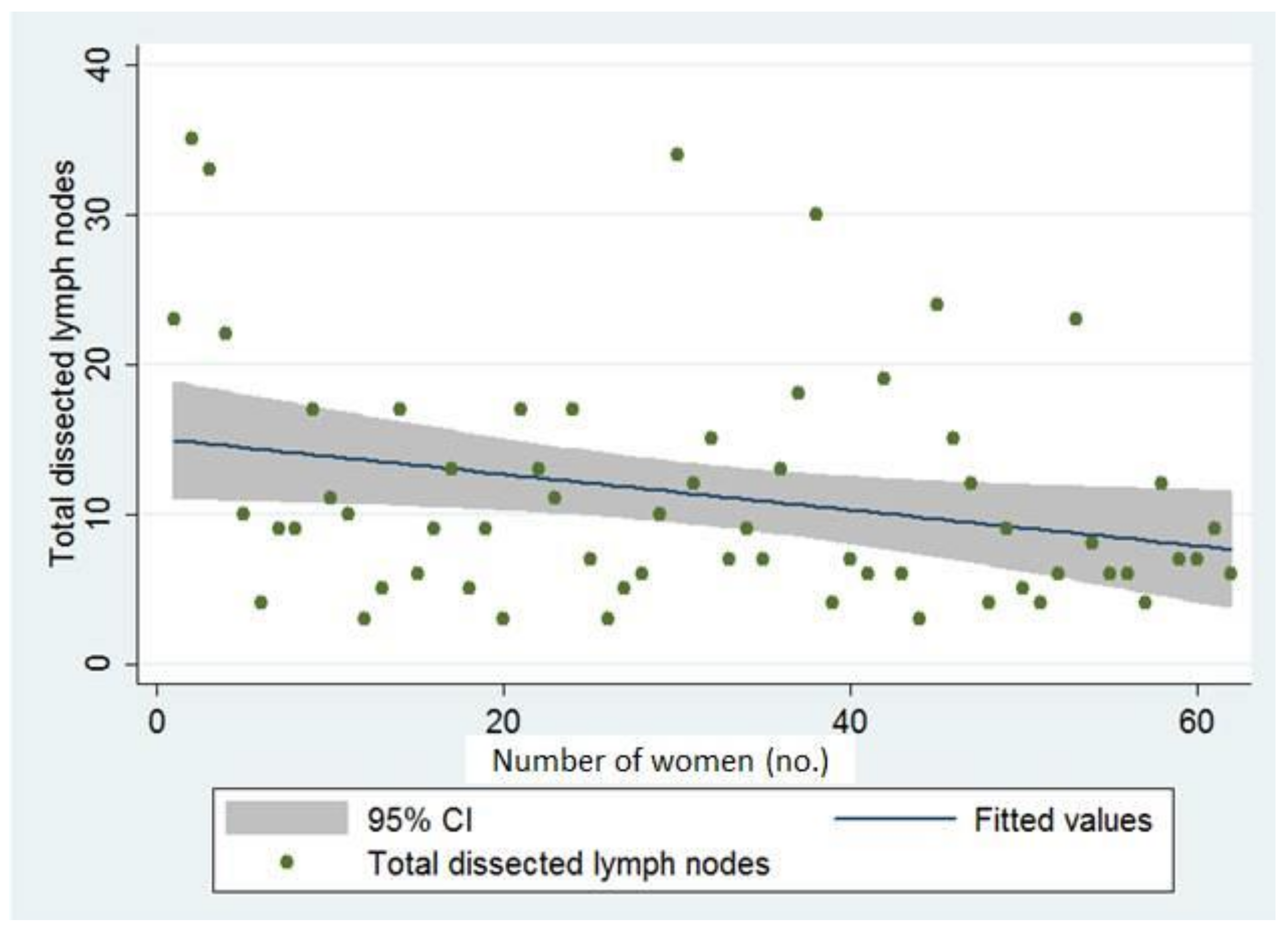
| Total number of dissected lymph nodes | 18.0 ± 9.8 | 11.3 ± 7.9 | <0.0001 |
| Total number of positive pelvic lymph nodes | 0.19 ± 0.97 | 0.18 ± 1.15 | 0.48 |
| Para-aortic lymph nodes dissection | 54 (20) | 8 (13) | <0.001 |
| Stage | 0.71 | ||
| IA | 187 (69) | 39 (63) | |
| IB | 46 (17) | 13 (21) | |
| II | 13 (5) | 2 (3) | |
| IIIA | 6 (2) | 2 (3) | |
| IIIC1 | 15 (6) | 4 (6) | |
| IIIC2 | 4 (1) | 1 (2) | |
| IVB | 1 (0) | 1 (2) | |
| Adjuvant radiotherapy | 135 (48) | 27 (44) | 0.47 |
| Adjuvant chemotherapy | 48 (11) | 12 (19) | 0.41 |
| Operation time (min) | 185 ± 72 | 209 ± 70 | 0.008 |
| Blood loss (mL) | 349 ± 300 | 196 ± 197 | <0.0001 |
| Complications | 74 (27) | 8 (13) | 0.02 |
| Lymphocyst/lymphedema | 33 (7) | 3 (5) | 0.11 |
| Average size of lymphocyst (cm) | 4.9 ± 2.2 | 2.5 ± 0.8 | 0.03 |
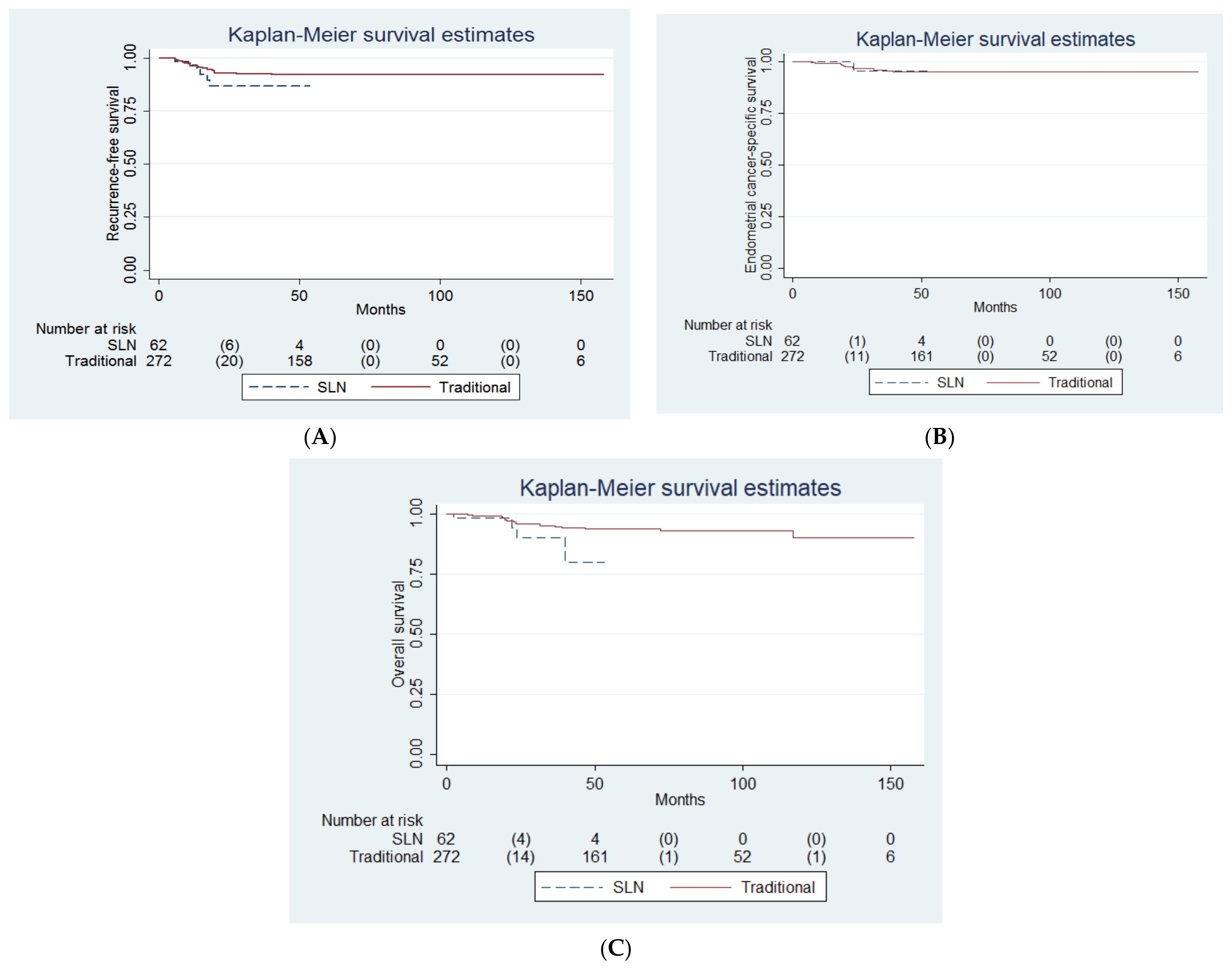
| Median follow-up (months) | 65.0 ± 39.9 | 22.6 ± 12.8 | <0.0001 |
| Recurrence | 20 (11) | 6 (10) | ‡ 0.24 |
| Death | 16 (6) | 4 (7) | ‡ 0.09 |
| Endometrial cancer related | 11 (4) | 1 (2) | ‡ 0.79 |
| Other causes (i.e., sepsis, n = 4; second malignancy, n = 2; pneumonia, n = 2) | 5 (2) | 3 (5) | - |
| Location | Left Hemipelvis (n = 51) | Right Hemipelvis (n = 49) |
|---|---|---|
| External iliac | 38 (75) | 34 (69) |
| Obturator | 24 (47) | 26 (53) |
| Common iliac | 2 (4) | 3 (6) |
| Presacral | 0 (0) | 1 (2) |
| Para-aortic | 0 (0) | 2 (4) |
| Not specified | 5 (10) | 6 (12) |
| Variables | Univariate | Multivariable | ||
|---|---|---|---|---|
| Hazard Ratio (95% CI) | † p | Hazard Ratio (95% CI) | ‡ p | |
| SLN mapping | 1.73 (0.69, 4.34) | 0.24 | 3.54 (0.52, 23.86) | 0.19 |
| Age (years) | 1.05 (1.01, 1.08) | 0.02 | 1.00 (0.94, 1.07) | 0.97 |
| BMI (kg/m2) | 0.93 (0.87, 0.99) | 0.02 | 0.88 (0.74, 1.05) | 0.15 |
| ECOG scale | ||||
| 0 (reference) | 1.00 | - | - | - |
| 1 | 0.71 (0.33, 1.57) | 0.40 | - | - |
| 2 | 1.85 (0.24, 14.1) | 0.55 | - | - |
| 3 | 3.80 × 10⁻¹⁵ (0, infinity) | 1.00 | - | - |
| Parity | 1.20 (0.84, 1.73) | 0.31 | - | - |
| CA-125 (U/mL) | 1.00 (1.00, 1.00) | 0.91 | - | - |
| Surgical method | ||||
| Laparotomic (reference) | 1.00 | - | - | - |
| Laparoscopic | 1.15 (0.48, 2.75) | 0.75 | - | - |
| Robotic | 1.37 (0.45, 4.15) | 0.58 | - | - |
| Total number of dissected pelvic lymph node | 1.00 (0.96, 1.04) | 0.89 | - | - |
| Endometrioid cell type | 0.17 (0.08, 0.36) | <0.001 | 0.84 (0.17, 4.10) | 0.83 |
| Cell grade | ||||
| 1 (reference) | 1.00 | - | 1.00 | - |
| 2 | 7.90 (1.73, 36.1) | 0.008 | 3.57 (0.37, 34.85) | 0.27 |
| 3 | 19.9 (4.46, 89.2) | <0.001 | 7.13 (0.63, 80.85) | 0.11 |
| Deep (>1/2) myometrial invasion | 2.59 (1.18, 5.71) | 0.02 | 0.47 (0.10, 2.33) | 0.36 |
| LVSI | 4.75 (1.89, 11.98) | 0.001 | 0.75 (0.16, 3.55) | 0.72 |
| Tumor size (cm) | 1.01 (1.01,1.02) | <0.001 | 1.01 (0.99, 1.04) | 0.28 |
| Pelvic lymph node metastasis | 6.90 (3.00, 15.88) | <0.001 | 1.52 (0.29, 7.95) | 0.62 |
| Para-aortic lymph node metastasis | 12.74 (3.90, 41.59) | <0.001 | 7.60 (1.28, 45.16) | 0.03 |
| Ascites cytology. | ||||
| Normal cell | 1.00 | - | - | - |
| Atypical cell | 1.79 (0.76,4.21) | 0.19 | - | - |
| Malignant cell | 2.51 (0.57, 11.00) | 0.22 | - | - |
| Stage | ||||
| IA | 1.00 (reference) | - | ||
| IB | 2.56 (0.99, 6.59) | 0.052 | ||
| II | 1.70 × 10⁻¹⁵ (0, infinity) | 1.00 | ||
| IIIA | 1.71 × 10⁻¹⁵ (0, infinity) | 1.00 | ||
| IIIC1 | 4.77 (1.52, 14.97) | 0.008 | ||
| IIIC2 | 14.6 (4.05, 52.79) | <0.001 | ||
| IVB | 22.76 (2.79, 185.42) | 0.003 |
| Variables | Univariate | Multivariate | ||
|---|---|---|---|---|
| Odds Ratio (95% CI) | † p | Odds Ratio (95% CI) | ‡ p | |
| SLN mapping | 1.11 (0.40, 3.07) | 0.85 | 0.73 (0.24, 2.24) | 0.58 |
| Cytokeratin IHC staining | 2.69 (1.06, 6.86) | 0.04 | 3.04 (1.09, 8.44) | 0.03 |
Publisher’s Note: MDPI stays neutral with regard to jurisdictional claims in published maps and institutional affiliations. |
© 2022 by the authors. Licensee MDPI, Basel, Switzerland. This article is an open access article distributed under the terms and conditions of the Creative Commons Attribution (CC BY) license (https://creativecommons.org/licenses/by/4.0/).
Share and Cite
Ting, W.-H.; Hsieh, S.-W.; Chen, H.-H.; Wei, M.-C.; Lin, H.-H.; Hsiao, S.-M. Predictors for the Recurrence of Clinically Uterine-Confined Endometrial Cancer and the Role of Cytokeratin Immunohistochemistry Stain in the Era of Sentinel Lymph Node Mapping. Cancers 2022, 14, 1973. https://0-doi-org.brum.beds.ac.uk/10.3390/cancers14081973
Ting W-H, Hsieh S-W, Chen H-H, Wei M-C, Lin H-H, Hsiao S-M. Predictors for the Recurrence of Clinically Uterine-Confined Endometrial Cancer and the Role of Cytokeratin Immunohistochemistry Stain in the Era of Sentinel Lymph Node Mapping. Cancers. 2022; 14(8):1973. https://0-doi-org.brum.beds.ac.uk/10.3390/cancers14081973
Chicago/Turabian StyleTing, Wan-Hua, Shu-Wei Hsieh, Hui-Hua Chen, Ming-Chow Wei, Ho-Hsiung Lin, and Sheng-Mou Hsiao. 2022. "Predictors for the Recurrence of Clinically Uterine-Confined Endometrial Cancer and the Role of Cytokeratin Immunohistochemistry Stain in the Era of Sentinel Lymph Node Mapping" Cancers 14, no. 8: 1973. https://0-doi-org.brum.beds.ac.uk/10.3390/cancers14081973