Article

A Bioinformatics Evaluation of the Role of Dual-Specificity Tyrosine-Regulated Kinases in Colorectal Cancer

by
Amina Jamal Laham
1,2,3,
Raafat El-Awady
1,3,*,
Jean-Jacques Lebrun
4 and
Maha Saber Ayad
1,2,*
1
Sharjah Institute for Medical Research, University of Sharjah, Sharjah 27272, United Arab Emirates
2
College of Medicine, University of Sharjah, Sharjah 27272, United Arab Emirates
3
College of Pharmacy, University of Sharjah, Sharjah 27272, United Arab Emirates
4
Cancer Research Program, Department of Medicine, McGill University Health Center, Montreal, QC H4A 3J1, Canada
*
Authors to whom correspondence should be addressed.
Submission received: 23 March 2022 / Revised: 9 April 2022 / Accepted: 12 April 2022 / Published: 18 April 2022
(This article belongs to the Special Issue Bioinformatics, Big Data and Cancer)

Abstract

:

Simple Summary

The dual-specificity tyrosine-regulated kinase (DYRK) family has been implicated in various diseases, including cancer. However, its role in colorectal cancer has not been elucidated. In this research, we used publicly available web-based tools to investigate DYRKs status in colorectal cancer. Our results showed that among DYRKs, only DYRK1A was upregulated significantly in late tumor stages, and it is associated with poor prognosis for colorectal cancer patients. These finding comprehensively characterized DYRK1A as a potential new therapeutic approach in CRC, especially in late tumor stages.

Abstract

Colorectal cancer (CRC) is the third most common cancer worldwide and has an increasing incidence in younger populations. The dual-specificity tyrosine-regulated kinase (DYRK) family has been implicated in various diseases, including cancer. However, the role and contribution of the distinct family members in regulating CRC tumorigenesis has not been addressed yet. Herein, we used publicly available CRC patient datasets (TCGA RNA sequence) and several bioinformatics webtools to perform in silico analysis (GTEx, GENT2, GEPIA2, cBioPortal, GSCALite, TIMER2, and UALCAN). We aimed to investigate the DYRK family member expression pattern, prognostic value, and oncological roles in CRC. This study shed light on the role of distinct DYRK family members in CRC and their potential outcome predictive value. Based on mRNA level, DYRK1A is upregulated in late tumor stages, with lymph node and distant metastasis. All DYRKs were found to be implicated in cancer-associated pathways, indicating their key role in CRC pathogenesis. No significant DYRK mutations were identified, suggesting that DYRK expression variation in normal vs. tumor samples is likely linked to epigenetic regulation. The expression of DYRK1A and DYRK3 expression correlated with immune-infiltrating cells in the tumor microenvironment and was upregulated in MSI subtypes, pointing to their potential role as biomarkers for immunotherapy. This comprehensive bioinformatics analysis will set directions for future biological studies to further exploit the molecular basis of these findings and explore the potential of DYRK1A modulation as a novel targeted therapy for CRC.

1. Introduction

Colorectal cancer (CRC) is the third most common diagnosed type of cancer and represents the third leading cause of cancer-related deaths worldwide [1]. The vast majority of colorectal cancer patients are diagnosed at late stages and often exhibit poor prognosis with an estimated 5-year survival rate of less than 10% for stage 4 metastatic CRC patients [2]. The standard of care for stage 4 metastatic CRC includes surgery and chemotherapy; the latter improves the 5-year survival rate by 30–50% [3,4]. However, patients will eventually develop resistance to chemotherapy and often develop tumor relapse following surgery [5]. Extensive research over the past 20 years defined important prognostic biomarkers and therapeutic targets in CRC, further improving the survival of CRC patients.
One of the most mutated genes in cancer are protein kinases. The dysregulation of kinases is found to be an early event in carcinogenesis [6]. Kinases are classified into different group, and one of the major kinases groups is the proline-directed kinases “(CMGC)” group, which includes cyclin-dependent kinase (CDK), mitogen-activated protein kinase (MAPK), glycogen synthase kinase (GSK3), CDC-like kinase (CLK)) and dual-specificity tyrosine-regulated kinase (DYRK) [7].
As potential targets for colorectal cancer treatment, protein kinases have attracted remarkable attention in recent years and many such kinases have been found to be implicated in CRC initiation, progression, and/or metastasis [6]. During the last decade, a lot of attention has been focused on MAPK/ERK and PI3 Kinase pathways due to their broad involvement in tumorigenic processes in various tissues [8]. These studies led to the development of several targeted therapies, such as cetuximab and panitumumab, which successfully improved the survival of metastatic CRC patients [9]. Besides MAPK and PI3K, the dual-specificity tyrosine-regulated kinase (DYRK) family has also been found to be implicated in regulating tumorigenesis [10]. In humans, there are five DYRK family members, which are classified into class 1 (DYRK1A and DYRK1B) and class 2 (DYRK2, 3, and 4) based on sequence homology of their kinase domain [11]. DYRKs have the ability to auto-phosphorylate their own activation loop on tyrosine residue while still attached to the ribosome during translation. This phosphorylation event has been reported to be essential for DYRK kinase activity such as serine and threonine kinases [12]. DYRK family members play essential cellular functions, and the dysregulation of their kinase activity was found to be implicated in various diseases, such as neurological disorders, metabolic disorders and cancer [11,13,14]. Class 1 DYRKs (1A and 1B) were found to be essential for cell cycle regulation in neurological, mesenchymal and epithelial tissues [15,16,17,18]. Studies have reported class 1 DYRKs to be involved in tumorigenesis, but their precise role and functions remain unclear (10,19). DYRK1A can exert tumor-suppressive activities in cancer and was found to regulate cancer-related key pathways, such as apoptosis, DNA damage, activation of receptor tyrosine kinase and angiogenesis [19]. However, DYRK1A expression levels vary among different types of cancer. For example, it was found to be upregulated in some solid cancers, such as glioblastoma, lung, head and neck, and pancreatic cancers [20,21,22]. Recently, it was also reported to be upregulated in B cell acute lymphoblastic leukemia [23]. In contrast, it was found to be downregulated in acute myeloid leukemia [24]. Unlike DYRK1A, DYRK1B was reported to exert pro-oncogenic functions in ovarian, pancreatic, breast tumors and liposarcoma [25,26,27]. DYRK1B expression is also increased upon the inhibition of MAPK and PI3K/AKT/MTORC pathways in ovarian and pancreatic tumors, suggesting its involvement in the resistance to targeted therapies in these tumors [28,29]. From the class 2 DYRK subgroup, only DYRK2 was extensively studied in terms of tumorigenesis and found to have a controversial role in cancer [30]. DYRK2 is overexpressed in various cancer types [10] and is involved in the proteo-stasis pathway that can enhance cancer cells survival under stressful conditions [31]. However, other studies reported that it could exert a tumor suppressor role through inhibiting epithelial-to-mesenchymal transition (EMT) in breast and ovarian cancers [32,33]. Furthermore, DYRK2 was also reported to suppress liver metastasis and to act as a good prognostic marker in CRC [34,35]. A few studies have been conducted to investigate the role of DYRK3 and 4 in cancer. The cellular role of DYRK3 was in fact recently discovered [36,37]. It has been reported to activate mTORC1 in stress granules and to allow cytoplasmic compartmentalization via liquid phase transitions [36]. Additionally, it was reported to be essential for mitosis; acting as a regulator for dissolution or condensation of membrane organelles during mitosis [37]. In cancer, DYRK3 was proposed to act in parallel with DYRK1A to promote cell survival under stressful conditions by phosphorylating nicotinamide adenosine dinucleotide (NAD)-dependent deacetylase SIRT1 [38].
Despite the important roles played by DYRK family members in various types of cancers with the exception of DYRK2, no study investigated their role and broad contribution to colorectal cancer. Recently, bioinformatics-based analysis of publicly available databases has gained popularity in biomedical research due to their robustness in identifying key cancer vulnerabilities and potential biomarkers. The available pathological-sample database provides a large RNA seq and proteomic datasets that can be instrumental in investigating the specific role and function of key regulatory genes in multiple types of cancer [39]. Moreover, they can provide invaluable information when the analysis is conducted in view of the clinical data including disease staging.
In this study, we analyzed DYRKs expression levels in tumor versus normal samples and in the different CRC molecular subtypes, in correlation with clinical features of CRC and patient survival outcomes. For this, we used various bioinformatics tools that are publicly available, including Genotype-Tissue Expression (GTEx), Gene Expression database of Normal and Tumor tissues 2 (GENT2), Gene Expression Profiling Interactive Analysis2 (GEPIA2), cBioPortal, Gene Set Cancer Analysis (GSCALite), TIMER2, UALCAN, Kaplan–Meier and Cox regression analyses. The overall aim of this study is to address the role of DYRKs in CRC carcinogenesis and to evaluate their capacity as potential prognostic markers and/or therapeutic targets in CRC.

2. Materials and Methods

2.1. Genotype-Tissue Expression (GTEx)

To measure the expression level of DYRKs in normal colon tissues, we used the Genotype-Tissue Expression (GTEx) web tool (https://gtexportal.org, accessed on 6 April 2022), which provides RNA seq data to study the expression of specific genes in different non-diseased tissue. We chose the expression tab with multi-gene query. We queried DYRKs in tumor tissue of the sigmoid and transverse colon. The website provides TPM (transcript per million) for each gene.

2.2. Gene Expression Database of Normal and Tumor Tissues 2 (GENT2)

To measure the expression level of DYRKs in different CRC subtypes, we used the GENT2 database (http://gent2.appex.kr/gent2/, accessed on 5 April 2022) [40]. This tool uses NCBI GEO database that is derived from microarray platforms (Affymetrix U133A or U133Plus2) [40]. We queried each DYRK alone by searching its gene across the subtypes tab; we used this tool to investigate the expression of DYRKs in CRC molecular subtypes.

2.3. Gene Expression Profiling Interactive Analysis2 (GEPIA2)

To measure the DYRKs expression level in different CRC stages, we used the GEPIA2 web-based server. The data are derived from the TCGA RNA sequence dataset (http://gepia2.cancer-pku.cn/#index, accessed on 6 April 2022) [41]. We queried each DYRK alone by searching its gene across stages tab. Moreover, we used this tool to generate the overall survival Kaplan–Meier plots for each DYRK in CRC patient. Hazard ratios were calculated based on Cox regression analysis.

2.4. cBioPortal

To further analyze the DYRKs status in tumor samples, we used cBioPortal [42,43]. We selected the Colorectal Adenocarcinoma TCGA PanCancer dataset (592 samples). We chose the gene specific query and selected mRNA expression z-score relative to normal samples (Log RNAseq V2 RSEM). For each DYRK, we filtered the samples based on z-score. Samples with z-score < 1 were considered to have low expression, and samples with z-score >1 were considered to have high expression. For DYRK2, all z-scores were below −1 in this dataset, so we were unable to compare it with other group, so we excluded it from the comparison.

2.5. GeneSet Cancer Analysis (GSCALite)

Gene Set Cancer Analysis (GSCALite) is a web-based server that uses to investigate the status of genes in cancer patient’s datasets (http://bioinfo.life.hust.edu.cn/web/GSCALite/, accessed on 5 April 2022) [44]. Interestingly, it can predict the gene activity in a cancer-associated pathway; we used this tool to investigate the role of DYRKs in cancer-related pathways in a colon adenocarcinoma (COAD) data set from TCGA.

2.6. STRING

We used STRING web tool to predict functional protein interaction network in Homo sapiens for DYRKs (https://string-db.org, accessed on 6 April 2022). We customized the active interaction sources and selected only experimental, database and co-expression proteins.

2.7. TIMER2

TIMER 2 is a web tool that uses multiple algorithms to analyze and measure the tumor-infiltrating immune cells from TCGA dataset (http://timer.cistrome.org, accessed on 5 April 2022). This tool measures the correlation between the expression of the gene of interest and immune-infiltrating cells abundancy. We used this tool to investigate the correlation between DYRKs and tumor-infiltrating immune cells in CRC [45]. We selected immune association to each gene and each type of immune cells, and then we chose TIMER algorithm and Spearman correlation.

2.8. UALCAN

UALCAN is a web-based tool that uses OMICS data from cancer data sets such as TCGA, MET500 and CPTAC (http://ualcan.path.uab.edu, accessed on 6 April 2022). It is used to study the gene status in different cancers such as expression and methylation in correlation to survival and calculate significance by Student’s t-test [46]. We used this tool to investigate the expression of DYRKs in normal vs. tumor samples and to investigate DYRKs promoter methylation status in CRC TCGA data sets.

3. Results

3.1. DYRKs Expression in Colon Tissues

DYRKs are expressed ubiquitously in all human tissue, and they have low tissue specificity [11]. We used the GTEx portal web tool to investigate the expression level of DYRKs in normal colon tissues in two anatomical parts: the sigmoid and the transverse colon. As represented in Figure 1, DYRK1A was found to have a higher expression level in colon tissues compared to other DYRKs in both anatomical parts of the colon. DYRK1B and DYRK2 showed intermediate expression levels, whereas DYRK3 and DYRK4 were found to have low expression in colon tissues.

3.2. DYRKs Genes Expression in Normal vs. Colon and Rectum Adenocarcinoma

Next, we measured the expression level of DYRKs in colon and rectum adenocarcinoma [COAD and rectal adenocarcinoma (READ)] vs. normal tissues. Using the RNA seq dataset from TCGA data in UALCAN web tool, we found no significant change in DYRK1A and 1B (Figure 2a,b), whereas DYRK2 (Figure 2c) had a significantly lower expression in COAD samples compared to normal tissues. On the other hand, both DYRK3 (Figure 2d) and DYRK4 (Figure 2e) were found to be significantly upregulated in COAD samples vs. normal tissues. As shown in Figure 3, both DYRK1A (Figure 3a) and DYRK2 (Figure 3c) showed a significantly lower expression in READ tumor vs. normal tissues, whereas DYRK1B, DYRK3 and DYRK4 had no significant change in expression.

3.3. DYRKs Expression Level in CRC Molecular Subtypes

CRC are classified into major pathological molecular subtypes. The first group is microsatellite instable group (MSI) due to defects in mismatch repair (MMR) genes (16%) or DNA polymerase epsilon proof reading (3%). The second group is microsatellite stable (MSS) and represents 84% of CRC cases. MSS CRC are characterized by high rate of somatic mutations in several genes such as APC, TP53, PIK3CA, KRAS and SMAD4 [47]. To study DYRK members’ gene expression in the different tumor subtypes, we used the web-based tool GENT2 [40]. Comparing the MSI and MSS subtypes, DYRK1A, DYRK3 and DYRK4 (Figure 4a,d,e) are all significantly higher in MSI, whereas DYRK2 is significantly higher in MSS (Figure 4c). For DYRK1B, there were no significant differences in its expression in MSI and MSS (Figure 4b).

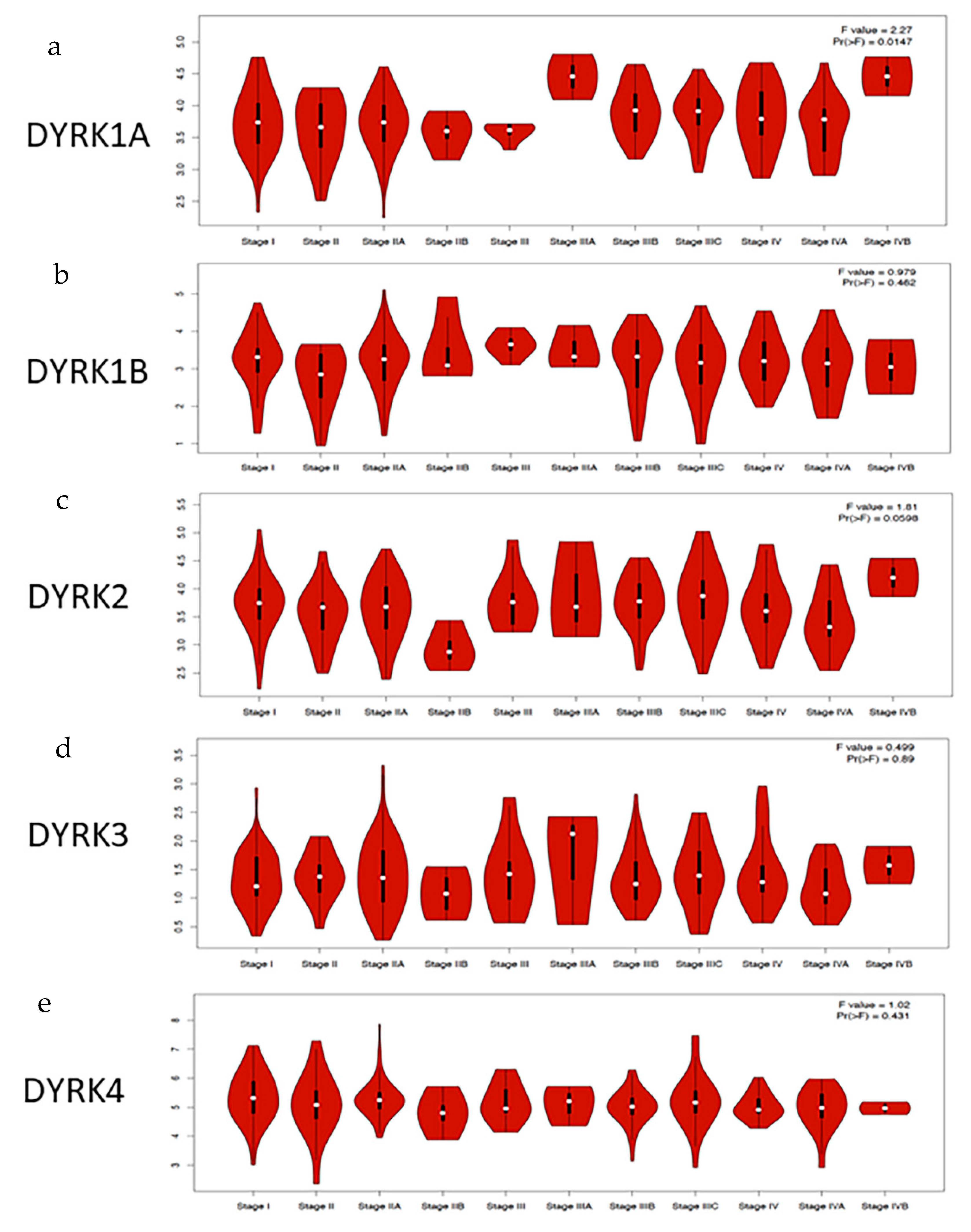
3.4. DYRKs Expression Level in CRC Stages

Using GEPIA2, we investigated the level of DYRKs member in different CRC stages. As shown in Figure 5, DYRK1A is significantly upregulated in late tumor stages from IIIA to IVB (Figure 5a). DYRK1B expression did not show any significant differences among CRC stages (Figure 5b). Similarly, class 2 DYRKs expression did not have any significant differences in CRC stages. This indicates that among DYRKs members, only DYRK1A is overexpressed in later tumor stages of CRC patients and suggests that DYRK1A may play a role in the advanced stages of the disease.

3.5. Clinical Feature of DYRKs in CRC

To further explore the expression status of DYRK members in relation to the CRC clinical features, we used cBioPortal and filtered the patient data set based on z-score to low or high expression to each DYRK member.

3.5.1. Histological Cancer Subtypes

Consistent with what was observed with our RNA seq from UALCAN web tool analysis (F2-F3), the expression level of DYRK3 and 4 (z-score = or >1) were high in the majority of the tumor samples. Tumor samples with high DYRK1A were found to be significantly associated with mucinous colorectal adenocarcinoma cancer (Figure 6a). A similar pattern was obtained for DYRK4, as shown in Figure 6d. On the other hand, DYRK1B expression was higher in non-mucinous COAD than in mucinous subtype (Figure 6b). For DYRK3, there was no significant change in its expression in all three histological subtypes (Figure 6c).

3.5.2. Metastasis, Lymph Node Stages and New Neoplasm Event Post Therapy

As shown in Figure 7, although DYRK1A was found to have a low expression in CRC compared to normal tissues, cancer samples with high DYRK1A expression were found to be enriched in late metastasis and lymph node stage (Figure 7a,b). For other DYRKs members, there were no statistical differences between their expression and metastasis and lymph node stages (Figure 7c–h). Moreover, tumor samples with higher DYRK1A level exhibited higher tumor recurrence following initial therapy (Figure 8a), whereas none of the other DYRKs members showed any significant change (Figure 8b–d). These results suggest that DYRK1A could serve as a potential negative tumor biomarker in the late stages of the CRC disease.

3.6. Mutations and Promotor Methylation of DYRKs in CRC

DYRKs have differential expression in normal vs. tumor samples in CRC. To identify if this variation in expression is related to specific mutations, we used the Pan TCGA data from cBioPortal and analyze the DYRKs mutations in this dataset. As shown in Figure 9, DYRKs in general are not mutated in CRC; some samples have amplification mutations in DYRK1B, DYRK3 and DYRK4. For DYRK1A, there is a missense mutation with unknown significance. In general, the differential expression of DYRKs found to be related to mRNA level. Then, we used the UALCAN web tool to investigate the methylation of DYRKs genes promotor in different tumor stage. We found that DYRK1A methylation in COAD was not significantly changed (Figure 10a); however, in READ, the methylation level decreased significantly in stage 3 compared to normal samples (Figure 10b). The methylation of DYRK1B was found to be significantly increased in stage 1, 2, and 3 compared to normal in COAD (Figure 10c); in READ, the methylation increased significantly in stage 1 and decreased significantly in stage 4 compared to normal samples (Figure 10d). DYRK2 was reported to have lower methylation level in stage 4 in COAD only (Figure 10e), and no significant change was observed in READ (Figure 10f). Investigating the mRNA expression in normal vs. tumor samples, DYRK3 (Figure 10g) and DYRK4 (Figure 10i) were found to have significantly lower methylation level compared to the control in COAD. In READ, only DYRK4 was found to have lower methylation level in stage 1 and 4 (Figure 10i). These results suggest that variation in DYRKs expression in normal vs. tumors might be related to epigenetic regulation rather than gene mutations.

3.7. DYRKs Activity in Cancer Associated Pathways in CRC

After exploring the status of DYRKs in clinical features of CRC, we used GSCALite to predict the molecular function of DYRKs in COAD Figure 11a and in all cancers combined (Figure 11b). Class 1 DYRKs (1A and 1B) and DYRK4 were found to correlate with cell cycle inhibition. However, DYRK1B also correlated with the inhibition of apoptosis, DNA damage response, and PI3K/AKT pathway activation. DYRK2 was found to correlate with the inhibition of DNA damage response, whereas DYRK3 correlated positively with epithelial mesenchymal transition and estrogen hormone pathway. These data are predicted data based on gene correlation with major regulators of cancer-associated pathway and indicate that DYRKs may play a significant role in several cancer-related pathways. To further predict DYRKs functions in cancer, we used the protein functional association network tool (STRING) to predict the interacting partners of DYRKs. As shown in Figure S1a, DYRK1A interacts with the DREAM complex that is involved in cell cycle inhibition. Moreover, it interacts with the ubiquitin ligase RNF169 that is involved in DNA damage response. These interactions were further demonstrated experimentally [48,49]. Moreover, DYRK1B was found to associate with Signal-induced proliferation-associated 1-like protein 1,2 and 3 (Figure S1b), which are involved in morphogenesis and cytoskeletal organization. In correlation with the predicted cancer pathways, DYRK1B was also found to have a functional association with RNF169, which predicts its role in DNA damage. Another DYRK1B interactor is LZTS2, a negative regulator of beta catenin, suggesting its involvement in colorectal cancer [50]. In correlation with predicted cancer pathways, DYRK2 associated proteins were mainly DNA damage response factors such as P53 and ATM Figure S1c. Finally, all DYRK members were found to have functional association with DCAF7, which was previously reported as an adaptor protein for DYRK1A [51].

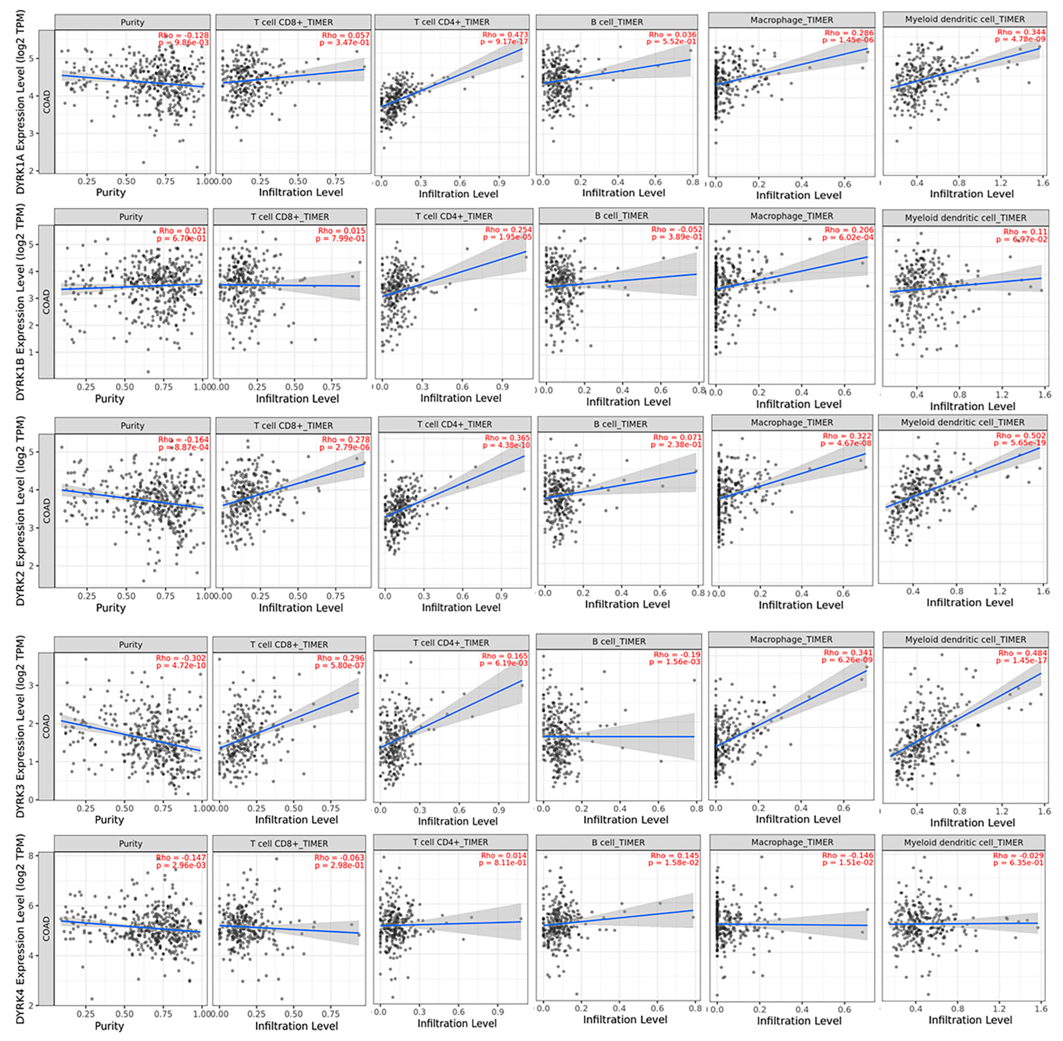
3.8. Correlation between DYRKs and Tumor Infiltrating Immune Cells in CRC Patient

Because we found differential DYRK members expression in MSI and MSS subtypes and the fact that these two CRC subtypes exhibit different immune cells abundance in the tumor microenvironment [47], we used TIMER 2 web tool to explore potential correlation between DYRKs expression and tumor infiltrating immune cells in CRC.
As shown in Figure 12, DYRK1A significantly positively correlated with CD4+ T cells, macrophages and myeloid dendritic cells, with no significant correlation with CD8+ T cells and B cells. For DYRK1B, we only found a significant positive correlation with CD4+ T cells and macrophages. Although DYRK2 has higher expression in the MSS subtype, its expression positively correlated with CD8+ T cells, CD4+ T cells, macrophages, and myeloid dendritic cells. This can be explained by the fact that the TIMIR web tool calculates the correlation in all CRC patient data sets not in a specific subtype. In addition, DYRK3 was also found to be positively correlate with CD8+ T cells, CD4+ T cells, macrophages, and myeloid dendritic cells. DYRK4 did not show significant correlation with any of the immune infiltrating cells. In conclusion, only DYRK1A and DYRK3 are upregulated in the MSI subtype with their expression correlating positively with immune infiltrating cells.

3.9. Progression Free Survival

Based on our analysis of z-score using the Pan TCGA data from cBioPortal, we found that CRC samples with high DYRK1A expression levels have significantly lower progression free survival rate (Figure 13a); however, for other DYRKs, no significant change in PFS was observed (Figure 13b–d). These results are consistent with what described above and further support that the notion that DYRK1A expression in late-stage CRC tumors may reflect poor patient prognosis.

3.10. Overall Survival

To further confirm our finding from cBioPortal, we performed Kaplan–Meier analysis for overall survival (obtained from CRC samples using GEPIA2 web tool). Interestingly, CRC patients with higher DYRK1A expression showed significantly lower overall survival (Figure 14a), whereas other DYRK members did not show any significant correlation with overall survival (Figure 14b–f). This indicates that expression of DYRK1A, but not other DYRK members, can predict cancer survival in CRC.

4. Discussion

DYRKs were shown to be implicated in various human diseases, including cancer [10]. However, no study investigated the role of DYRKs specifically in colorectal cancer. In this study, using various web tools and large RNA sequence datasets derived from cancer patients, we analyzed specific expression levels of all DYRK family members in relation to CRC clinical features and patient survival outcome.
According to our analysis, DYRK1A is the predominantly expressed one in CRC; however, its physiological role in colonic epithelium is not yet fully elucidated. DYRK1A is well-known to be involved in various essential cellular pathways such as cell cycle regulation, specifically as a regulator for RNA polymerase II at specific genes loci that are involved in cell cycle, translation, and RNA processing [15,52]. This might give an insight into the predominance of DYRK1A in colon tissues.
It is noteworthy that, one of the main cellular functions of DYRK1A in neuronal cells is regulating cell cycle. Studies have shown that, in Down syndrome, when DYRK1A is overexpressed, it modulates the differentiation of neuronal cells, resulting in immature neurons via inhibiting the cell cycle [15]. Moreover, it was also found to inhibit cell cycle in cardiomyocytes [16], and it could be used as a therapeutic target to initiate pancreatic beta cells proliferation [53]. Together with DYRK1B, DYRK1A was found to have an inhibitory effect on cell cycle in COAD. One of the molecular mechanisms of class 1 DYRKs in cell cycle is that both were reported to phosphorylate cyclin D and induce its degradation and activate P27 [16,54,55].
One of the interesting findings of our analysis is that DYRK1A is the only member of the DYRK family to be associated with worse prognosis in CRC. It is significantly upregulated in later tumor stages, associated with lymph node and distant metastasis. Intriguingly, high expression of DYRK1A was associated with mucinous colorectal adenocarcinoma. It was reported that mucinous colorectal adenocarcinoma has a worse prognosis than non-mucinous colorectal adenocarcinoma [56]. Recently, it was reported that DYRK1A inhibition decreases metastases in tumors other than CRC, e.g., triple-negative breast cancer and glioblastoma cells [57,58]. Moreover, DYRK1A in endothelial cells was found to be involved in angiogenesis [59], which is an essential process for metastasis [60]. However, further studies should be conducted to investigate the role of DYRK1A in CRC metastasis.
Additionally, tumors with high DYRK1A level have been shown to have more potential of recurrence, which is associated with cancer stemness [61]. Interestingly, DYRK1A was recently reported to play a role in cancer stemness in oral squamous cell carcinoma. Therefore, inhibition of DYRK1A has been reported to reduce cancer stem cell phenotype [62]. These findings correlate with the clinical outcome; patients with high DYRK1A expression were found to have significantly lower PFS and lower OS.
The CRC is classified into major pathological molecular subtypes, (MSI) and (MSS). Usually, patients with MSI have a better prognosis than patients with MSS [63]. Here, we found that DYRK1A, DYRK3 and 4 expression is higher in MSI, whereas DYRK2 is higher in MSS. Both DYRK1A and DYRK2 have been reported to be involved in DNA-damage response, with depleted DYRK2 cells found to have lower repair efficiency which correlates with our finding that DYRK2 is higher in MSS subtype, which has a proficient DNA repair machinery [64].
Furthermore, DYRK1A was not reported to be involved in MMR pathway, it was only reported to act as a binding partner with ubiquitin ligase RNF169 that promote homologous recombination pathways after DNA double strand break [65]. Both DYRK1A and DYRK3 were found to activate the nicotinamide adenosine dinucleotide (NAD)-dependent deacetylase that inactivates TP53 under stressful conditions [38], because MSI has a higher mutational load and cellular stress, DYRK1A and 3 might be required to overcome cellular stress by activating SIRT1 and enhance the survival of cancer cells [38]. In metastatic dMMR–MSI-H CRC, immune checkpoint inhibitors (ICIs), specifically monoclonal antibodies targeting programmed cell death 1 (PD1) and cytotoxic T lymphocyte antigen 4 (CTLA4), increase survival, but pMMR–MSI-L CRC is mainly insensitive to existing ICIs [66].
Besides the variable genomic instability, MSI and MSS subtypes have differential immunogenic microenvironment. The MSI tumors were found to have a higher number of immune infiltrating cells than MSS [67]. In correlation with DYRKs expressions in CRC subtypes, DYRK1A and 3 significantly correlated with tumor infiltrating immune cells; this suggests that those two kinases can act as a prognostic marker for MSI and immunotherapy response. In the era of personalized medicine, finding the appropriate biomarkers that denote MSI-H is expected to predict the potential success of immunotherapy and to advance the management of CRC.
DYRK1B was found to have an anti-apoptotic effect in colon adenocarcinoma (COAD) and in global cancer predicted pathways (Figure 10). Additionally, it was found to inhibit the DNA damage response. In support of this role inhibiting DYRK1B in ovarian cancer cells was found to initiate apoptosis and to sensitize cancer cells to cisplatin (DNA damaging agent) [68], supporting the assumption that DYRK1B could act in a similar way in COAD. However, further studies are needed to support this hypothesis. Additionally, DYRK1B was found to activate PI3K/AKT pathway in COAD, its role in activating this pathway was reported in pancreatic and ovarian cancer cells [69].
DYRK2 is the only kinase that was found to be significantly downregulated in CRC, and was reported to have a tumor suppressor role in CRC [35]. DYRK2 could also be implicated in inhibiting both cell cycle and DNA damage response. It inhibits the cell cycle by inducing c-myc and c-Jun degradation [70]. However, DYRK2 role in inhibiting DNA damage response was not previously reported. In contrast it was reported to act as partner with ubiquitin ligase RNF8 to regulate DNA repair [64]. In our analysis, DYRK2 was found to be upregulated in MSS subtype that have a stable DNA repair machinery. Downregulation of DYRK2 impairs DNA repair efficiency [64]. Moreover, DYRK2 was reported to interact with P53, MDM2 and ATM [71]. DYRK2 is activated by ATM under DNA damage, this activation protects DYRK2 from MDM2-induced degradation and recruits DYRK2 to phosphorylate P53 and induce apoptosis. Such findings suggest that DYRK2 is a favorable kinase that suppresses the progression of CRC [71,72].
COAD-DYRK3 associated pathways included EMT and estrogen hormone pathway. DYRK3 was recently reported to be involved in glioblastoma multiform invasion and migration. Knocking down DYRK3 increased E-cadherin (epithelial marker) expression and decreased N-cadherin and Vimentin (Mesenchymal markers) expression in glioblastoma cancer cell [73]. Although in most cancers, DYRK4 contributes to cell cycle activation, its expression was not predicted in association with cancer pathways in COAD. Further studies should be conducted to understand the cellular role of DYRK4.
In general, DYRK genes were not mutated in studied CRC patient datasets, but variation in mRNA level was observed. We suggest that the variation in DYRKs expression in normal vs. tumor samples and in different tumor stages as in case of DYRK1A might not be linked to mutations, instead it might be due to epigenetic alterations. In correlation with DYRK3 and DYRK4 overexpression in tumors vs. normal, promotor methylation was significantly decreased in different tumor stages compared to normal. In COAD, there was upregulation in DYRK1B promoter methylation. DYRK1B had lower expression in COAD, however, it did not reach statistical significance. In READ DYRK1B was significantly less methylated compared to normal in READ stage 4. This correlates with its expression in READ, which is found to be non-significantly higher. For DYRK1A and DYRK2, there was no significant change in promotor methylation.
The current study provides an integrated comprehensive analysis of expression level, mutations, and immunological aspects of DYRKs in CRC carcinogenesis and progression. The study integrated pathological and clinical data to investigate the role of this family of dual kinases. Due to the limited availability of the molecular classification in most datasets and the very limited number of control cases in some analyses, our study has intrinsic limitations similar to other bioinformatics approaches. Furthermore, functional data such as enzyme activity status and ligand–receptor interactions have not been revealed in the TCGA and GTEx databases until recently. Therefore, we tried to overcome this challenge by studying protein network functional analysis, gene–gene, and protein–protein interaction analysis. To apply our results, more specific and extensive experimental work is required. Further studies should be conducted to understand the role of DYRKs in CRC, especially DYRK1A, hoping for promising new therapeutics to be developed targeting this kinase.

5. Conclusions

In the current study, we used publicly available data and web-based tools to investigate the status of DYRKs in colorectal cancer. We analyzed the expression in normal vs. tumor samples, then we investigated the expression of various DYRKs in different tumor stages and in CRC molecular and histological subtypes. Based on mRNA level, DYRK1A is the only member of DYRKs that was found to be upregulated in late tumor stages, with lymph node and distant metastatic stages. Additionally, it is the only kinase that was found to be associated with a worse progression free survival and overall survival when upregulated in patients‘ tumors. All DYRKs were found to be implicated in cancer-associated pathways which further indicate their involvement in cancer. No significant DYRK mutations were identified, and we suggested that DYRK expression variation in normal vs. tumor samples in different tumor stages might be because of epigenetics regulation. The majority of DYRKs showed a positive correlation with immune-infiltrating cells, which might suggest them as biomarkers for immunotherapy, namely, DYRK1A and DYRK3, which were upregulated in MSI subtypes. Our results reveal the role of DYRKs in CRC and provide deep insight into CRC biomarkers and potential novel therapies.

Supplementary Materials

The following supporting information can be downloaded at: https://0-www-mdpi-com.brum.beds.ac.uk/article/10.3390/cancers14082034/s1. Figure S1: Functional protein association network of different DYRK family members, as predicted from STRING.

Author Contributions

Conceptualization, A.J.L. and M.S.A.; methodology, A.J.L.; software, A.J.L., writing—original draft preparation, A.J.L. and M.S.A.; writing—review and editing, R.E.-A. and J.-J.L.; visualization, A.J.L. All authors have read and agreed to the published version of the manuscript.

Funding

A.J.L. and R.E.-A. are funded by the University of Sharjah, targeted Research Project No.: 2001110343. M.S.-A. is funded by the MBRU-AlMahmeed Research Award, (ALM#1914) and Al-Jalila Foundation Seed Grant (#AJF2017-68).

Institutional Review Board Statement

Not applicable.

Informed Consent Statement

Not applicable.

Data Availability Statement

Not applicable.

Acknowledgments

The authors acknowledge the support of the University of Sharjah and McGill University.

Conflicts of Interest

The authors declare no conflict of interest.

References

  1. Sung, H.; Ferlay, J.; Siegel, R.L.; Laversanne, M.; Soerjomataram, I.; Jemal, A.; Bray, F. Global Cancer Statistics 2020: GLOBOCAN Estimates of Incidence and Mortality Worldwide for 36 Cancers in 185 Countries. CA Cancer J. Clin. 2021, 71, 209–249. [Google Scholar] [CrossRef] [PubMed]
  2. Zacharakis, M.; Xynos, I.D.; Lazaris, A.; Smaro, T.; Kosmas, C.; Dokou, A.; Felekouras, E.; Antoniou, E.; Polyzos, A.; Sarantonis, J.; et al. Predictors of survival in stage IV metastatic colorectal cancer. Anticancer Res. 2010, 30, 653–660. [Google Scholar] [PubMed]
  3. Gallagher, D.; Kemeny, N. Metastatic Colorectal Cancer: From Improved Survival to Potential Cure. Oncology 2010, 78, 237–248. [Google Scholar] [CrossRef] [PubMed]
  4. Kow, A.W.C. Hepatic metastasis from colorectal cancer. J. Gastrointest. Oncol. 2019, 10, 1274–1298. [Google Scholar] [CrossRef] [PubMed]
  5. Al Bandar, M.H.; Kim, N.K. Current status and future perspectives on treatment of liver metastasis in colorectal cancer (Review). Oncol. Rep. 2017, 37, 2553–2564. [Google Scholar] [CrossRef] [Green Version]
  6. Cicenas, J.; Zalyte, E.; Bairoch, A.; Gaudet, P. Kinases and Cancer. Cancers 2018, 10, 63. [Google Scholar] [CrossRef] [Green Version]
  7. Varjosalo, M.; Keskitalo, S.; Van Drogen, A.; Nurkkala, H.; Vichalkovski, A.; Aebersold, R.; Gstaiger, M. The protein interaction landscape of the human CMGC kinase group. Cell Rep. 2013, 3, 1306–1320. [Google Scholar] [CrossRef] [Green Version]
  8. Bhullar, K.S.; Lagarón, N.O.; McGowan, E.M.; Parmar, I.; Jha, A.; Hubbard, B.P.; Rupasinghe, H.P.V. Kinase-targeted cancer therapies: Progress, challenges and future directions. Mol. Cancer 2018, 17, 48. [Google Scholar] [CrossRef]
  9. García-Foncillas, J.; Sunakawa, Y.; Aderka, D.; Wainberg, Z.; Ronga, P.; Witzler, P.; Stintzing, S. Distinguishing Features of Cetuximab and Panitumumab in Colorectal Cancer and Other Solid Tumors. Front. Oncol. 2019, 9, 849. [Google Scholar] [CrossRef]
  10. Boni, J.; Rubio-Perez, C.; López-Bigas, N.; Fillat, C.; De La Luna, S. The DYRK Family of Kinases in Cancer: Molecular Functions and Therapeutic Opportunities. Cancers 2020, 12, 2106. [Google Scholar] [CrossRef]
  11. Aranda, S.; Laguna, A.; de la Luna, S. DYRK family of protein kinases: Evolutionary relationships, biochemical properties, and functional roles. FASEB J. 2011, 25, 449–462. [Google Scholar] [CrossRef] [PubMed]
  12. Lochhead, P.A.; Sibbet, G.; Morrice, N.; Cleghon, V. Activation-loop autophosphorylation is mediated by a novel transitional intermediate form of DYRKs. Cell 2005, 121, 925–936. [Google Scholar] [CrossRef] [PubMed] [Green Version]
  13. Abu Jhaisha, S.; Widowati, E.W.; Kii, I.; Sonamoto, R.; Knapp, S.; Papadopoulos, C.; Becker, W. DYRK1B mutations associated with metabolic syndrome impair the chaperone-dependent maturation of the kinase domain. Sci. Rep. 2017, 7, 6420. [Google Scholar] [CrossRef] [Green Version]
  14. Abbassi, R.; Johns, T.G.; Kassiou, M.; Munoz, L. DYRK1A in neurodegeneration and cancer: Molecular basis and clinical implications. Pharmacol. Ther. 2015, 151, 87–98. [Google Scholar] [CrossRef] [PubMed]
  15. Hämmerle, B.; Ulin, E.; Guimera, J.; Becker, W.; Guillemot, F.; Tejedor, F.J. Transient expression of Mnb/Dyrk1a couples cell cycle exit and differentiation of neuronal precursors by inducing p27KIP1 expression and suppressing NOTCH signaling. Development 2011, 138, 2543–2554. [Google Scholar] [CrossRef] [Green Version]
  16. Hille, S.; Dierck, F.; Kühl, C.; Sosna, J.; Adam-Klages, S.; Adam, D.; Lüllmann-Rauch, R.; Frey, N.; Kuhn, C. Dyrk1a regulates the cardiomyocyte cell cycle via D-cyclin-dependent Rb/E2f-signalling. Cardiovasc. Res. 2016, 110, 381–394. [Google Scholar] [CrossRef] [Green Version]
  17. Kuhn, C.; Frank, D.; Will, R.; Jaschinski, C.; Frauen, R.; Katus, H.A.; Frey, N. DYRK1A is a novel negative regulator of cardiomyocyte hypertrophy. J. Biol. Chem. 2009, 284, 17320–17327. [Google Scholar] [CrossRef] [Green Version]
  18. Becker, W. Emerging role of DYRK family protein kinases as regulators of protein stability in cell cycle control. Cell Cycle 2012, 11, 3389–3394. [Google Scholar] [CrossRef] [Green Version]
  19. Laham, A.J.; Saber-Ayad, M.; El-Awady, R. DYRK1A: A down syndrome-related dual protein kinase with a versatile role in tumorigenesis. Cell. Mol. Life Sci. 2020, 78, 603–619. [Google Scholar] [CrossRef]
  20. Luna, J.; Boni, J.; Cuatrecasas, M.; Bofill-De Ros, X.; Núñez-Manchón, E.; Gironella, M.; Vaquero, E.C.; Arbones, M.; de la Luna, S.; Fillat, C. DYRK1A modulates c-MET in pancreatic ductal adenocarcinoma to drive tumour growth. Gut 2019, 68, 1465–1476. [Google Scholar] [CrossRef]
  21. Pozo, N.; Zahonero, C.; Fernández, P.; Liñares, J.M.; Ayuso-Sacido, A.; Hagiwara, M.; Pérez, A.; Ricoy, J.R.; Hernández-Laín, A.; Sepúlveda, J.M.; et al. Inhibition of DYRK1A destabilizes EGFR and reduces EGFR-dependent glioblastoma growth. J. Clin. Investig. 2013, 123, 2475–2487. [Google Scholar] [CrossRef] [PubMed] [Green Version]
  22. Li, Y.L.; Ding, K.; Hu, X.; Wu, L.W.; Zhou, D.M.; Rao, M.J.; Lin, N.-M.; Zhang, C. DYRK1A inhibition suppresses STAT3/EGFR/Met signalling and sensitizes EGFR wild-type NSCLC cells to AZD9291. J. Cell. Mol. Med. 2019, 23, 7427–7437. [Google Scholar] [CrossRef] [PubMed] [Green Version]
  23. Bhansali, R.S.; Rammohan, M.; Lee, P.; Laurent, A.P.; Wen, Q.; Suraneni, P.; Yip, B.H.; Tsai, Y.; Jenni, S.; Bornhauser, B.; et al. DYRK1A regulates B cell acute lymphoblastic leukemia through phosphorylation of FOXO1 and STAT3. J. Clin. Investig. 2021, 131, e135937. [Google Scholar] [CrossRef] [PubMed]
  24. Liu, Q.; Liu, N.; Zang, S.; Liu, H.; Wang, P.; Ji, C.; Sun, X. Tumor suppressor DYRK1A effects on proliferation and chemoresistance of AML cells by downregulating c-Myc. PLoS ONE 2014, 9, e98853. [Google Scholar] [CrossRef] [PubMed]
  25. Becker, W. A wake-up call to quiescent cancer cells—Potential use of DYRK1B inhibitors in cancer therapy. FEBS J. 2018, 285, 1203–1211. [Google Scholar] [CrossRef] [PubMed] [Green Version]
  26. Chen, H.; Shen, J.; Choy, E.; Hornicek, F.J.; Shan, A.; Duan, Z. Targeting DYRK1B suppresses the proliferation and migration of liposarcoma cells. Oncotarget 2018, 9, 13154–13166. [Google Scholar] [CrossRef] [Green Version]
  27. Chen, Y.; Wang, S.; He, Z.; Sun, F.; Huang, Y.; Ni, Q.; Wang, H.; Wang, Y.; Cheng, C. Dyrk1B overexpression is associated with breast cancer growth and a poor prognosis. Hum. Pathol. 2017, 66, 48–58. [Google Scholar] [CrossRef]
  28. Gao, J.; Zhao, Y.; Lv, Y.; Chen, Y.; Wei, B.; Tian, J.; Yang, Z.; Kong, F.; Pang, J.; Liu, J.; et al. Mirk/Dyrk1B mediates G0/G1 to S phase cell cycle progression and cell survival involving MAPK/ERK signaling in human cancer cells. Cancer Cell Int. 2013, 13, 2. [Google Scholar] [CrossRef] [Green Version]
  29. Deng, X.; Hu, J.; Ewton, D.Z.; Friedman, E. Mirk/dyrk1B kinase is upregulated following inhibition of mTOR. Carcinogenesis 2014, 35, 1968–1976. [Google Scholar] [CrossRef] [Green Version]
  30. Tandon, V.; de la Vega, L.; Banerjee, S. Emerging roles of DYRK2 in cancer. J. Biol. Chem. 2021, 296, 100233. [Google Scholar] [CrossRef]
  31. Guo, X.; Wang, X.; Wang, Z.; Banerjee, S.; Yang, J.; Huang, L.; Dixon, J.E. Site-specific proteasome phosphorylation controls cell proliferation and tumorigenesis. Nat. Cell Biol. 2016, 18, 202–212. [Google Scholar] [CrossRef] [PubMed]
  32. Yamaguchi, N.; Mimoto, R.; Yanaihara, N.; Imawari, Y.; Hirooka, S.; Okamoto, A.; Yoshida, K. DYRK2 regulates epithelial-mesenchymal-transition and chemosensitivity through Snail degradation in ovarian serous adenocarcinoma. Tumour Biol. 2015, 36, 5913–5923. [Google Scholar] [CrossRef] [PubMed]
  33. Mimoto, R.; Taira, N.; Takahashi, H.; Yamaguchi, T.; Okabe, M.; Uchida, K.; Miki, Y.; Yoshida, K. DYRK2 controls the epithelial-mesenchymal transition in breast cancer by degrading Snail. Cancer Lett. 2013, 339, 214–225. [Google Scholar] [CrossRef] [PubMed]
  34. Ito, D.; Yogosawa, S.; Mimoto, R.; Hirooka, S.; Horiuchi, T.; Eto, K.; Yanaga, K.; Yoshida, K. Dual-specificity tyrosine-regulated kinase 2 is a suppressor and potential prognostic marker for liver metastasis of colorectal cancer. Cancer Sci. 2017, 108, 1565–1573. [Google Scholar] [CrossRef] [PubMed] [Green Version]
  35. Yan, H.; Hu, K.; Wu, W.; Li, Y.; Tian, H.; Chu, Z.; Koeffler, H.P.; Yin, D. Low Expression of DYRK2 (Dual Specificity Tyrosine Phosphorylation Regulated Kinase 2) Correlates with Poor Prognosis in Colorectal Cancer. PLoS ONE 2016, 11, e0159954. [Google Scholar] [CrossRef] [PubMed]
  36. Wippich, F.; Bodenmiller, B.; Trajkovska, M.G.; Wanka, S.; Aebersold, R.; Pelkmans, L. Dual specificity kinase DYRK3 couples stress granule condensation/dissolution to mTORC1 signaling. Cell 2013, 152, 791–805. [Google Scholar] [CrossRef] [PubMed] [Green Version]
  37. Rai, A.K.; Chen, J.X.; Selbach, M.; Pelkmans, L. Kinase-controlled phase transition of membraneless organelles in mitosis. Nature 2018, 559, 211–216. [Google Scholar] [CrossRef] [Green Version]
  38. Guo, X.; Williams, J.G.; Schug, T.T.; Li, X. DYRK1A and DYRK3 promote cell survival through phosphorylation and activation of SIRT1. J. Biol. Chem. 2010, 285, 13223–13232. [Google Scholar] [CrossRef] [Green Version]
  39. Zheng, H.; Zhang, G.; Zhang, L.; Wang, Q.; Li, H.; Han, Y.; Xie, L.; Yan, Z.; Li, Y.; An, Y.; et al. Comprehensive Review of Web Servers and Bioinformatics Tools for Cancer Prognosis Analysis. Front. Oncol. 2020, 10, 68. [Google Scholar] [CrossRef]
  40. Park, S.J.; Yoon, B.H.; Kim, S.K.; Kim, S.Y. GENT2: An updated gene expression database for normal and tumor tissues. BMC Med. Genom. 2019, 12, 101. [Google Scholar] [CrossRef]
  41. Tang, Z.; Kang, B.; Li, C.; Chen, T.; Zhang, Z. GEPIA2: An enhanced web server for large-scale expression profiling and interactive analysis. Nucleic Acids Res. 2019, 47, W556–W560. [Google Scholar] [CrossRef] [PubMed] [Green Version]
  42. Cerami, E.; Gao, J.; Dogrusoz, U.; Gross, B.E.; Sumer, S.O.; Aksoy, B.A.; Jacobsen, A.; Byrne, C.J.; Heuer, M.L.; Larsson, E.; et al. The cBio cancer genomics portal: An open platform for exploring multidimensional cancer genomics data. Cancer Discov. 2012, 2, 401–404. [Google Scholar] [CrossRef] [PubMed] [Green Version]
  43. Gao, J.; Aksoy, B.A.; Dogrusoz, U.; Dresdner, G.; Gross, B.E.; Sumer, S.O.; Sun, Y.; Jacobsen, A.; Sinha, R.; Larsson, E.; et al. Integrative analysis of complex cancer genomics and clinical profiles using the cBioPortal. Sci. Signal 2013, 6, pl1. [Google Scholar] [CrossRef] [PubMed] [Green Version]
  44. Liu, C.J.; Hu, F.F.; Xia, M.X.; Han, L.; Zhang, Q.; Guo, A.Y. GSCALite: A web server for gene set cancer analysis. Bioinformatics 2018, 34, 3771–3772. [Google Scholar] [CrossRef]
  45. Li, T.; Fu, J.; Zeng, Z.; Cohen, D.; Li, J.; Chen, Q.; Li, B.; Liu, X.S. TIMER2.0 for analysis of tumor-infiltrating immune cells. Nucleic Acids Res. 2020, 48, W509–W514. [Google Scholar] [CrossRef]
  46. Chandrashekar, D.S.; Bashel, B.; Balasubramanya, S.A.H.; Creighton, C.J.; Ponce-Rodriguez, I.; Chakravarthi, B.V.S.K.; Varambally, S. UALCAN: A Portal for Facilitating Tumor Subgroup Gene Expression and Survival Analyses. Neoplasia 2017, 19, 649–658. [Google Scholar] [CrossRef]
  47. Müller, M.F.; Ibrahim, A.E.; Arends, M.J. Molecular pathological classification of colorectal cancer. Virchows Arch. 2016, 469, 125–134. [Google Scholar] [CrossRef] [Green Version]
  48. Guard, S.E.; Poss, Z.C.; Ebmeier, C.C.; Pagratis, M.; Simpson, H.; Taatjes, D.J.; Old, W.M. The nuclear interactome of DYRK1A reveals a functional role in DNA damage repair. Sci. Rep. 2019, 9, 6539. [Google Scholar] [CrossRef] [Green Version]
  49. Litovchick, L.; Florens, L.A.; Swanson, S.K.; Washburn, M.P.; DeCaprio, J.A. DYRK1A protein kinase promotes quiescence and senescence through DREAM complex assembly. Genes Dev. 2011, 25, 801–813. [Google Scholar] [CrossRef] [Green Version]
  50. Thyssen, G.; Li, T.H.; Lehmann, L.; Zhuo, M.; Sharma, M.; Sun, Z. LZTS2 is a novel beta-catenin-interacting protein and regulates the nuclear export of beta-catenin. Mol. Cell. Biol. 2006, 26, 8857–8867. [Google Scholar] [CrossRef] [Green Version]
  51. Glenewinkel, F.; Cohen, M.J.; King, C.; Kaspar, S.; Bamberg-Lemper, S.; Mymryk, J.S.; Becker, W. The adaptor protein DCAF7 mediates the interaction of the adenovirus E1A oncoprotein with the protein kinases DYRK1A and HIPK2. Sci. Rep. 2016, 6, 28241. [Google Scholar] [CrossRef] [PubMed] [Green Version]
  52. Di Vona, C.; Bezdan, D.; Islam, A.B.; Salichs, E.; López-Bigas, N.; Ossowski, S.; de la Luna, S. Chromatin-wide profiling of DYRK1A reveals a role as a gene-specific RNA polymerase II CTD kinase. Mol. Cell 2015, 57, 506–520. [Google Scholar] [CrossRef] [PubMed] [Green Version]
  53. Dirice, E.; Walpita, D.; Vetere, A.; Meier, B.C.; Kahraman, S.; Hu, J.; Dančík, V.; Burns, S.M.; Gilbert, T.J.; Olson, D.E.; et al. Inhibition of DYRK1A Stimulates Human β-Cell Proliferation. Diabetes 2016, 65, 1660–1671. [Google Scholar] [CrossRef] [Green Version]
  54. Najas, S.; Arranz, J.; Lochhead, P.A.; Ashford, A.L.; Oxley, D.; Delabar, J.M.; Cook, S.; Barallobre, M.J.; Arbonés, M.L. DYRK1A-mediated Cyclin D1 Degradation in Neural Stem Cells Contributes to the Neurogenic Cortical Defects in Down Syndrome. EBioMedicine 2015, 2, 120–134. [Google Scholar] [CrossRef] [Green Version]
  55. Zou, Y.; Ewton, D.Z.; Deng, X.; Mercer, S.E.; Friedman, E. Mirk/dyrk1B kinase destabilizes cyclin D1 by phosphorylation at threonine 288. J. Biol. Chem. 2004, 279, 27790–27798. [Google Scholar] [CrossRef] [PubMed] [Green Version]
  56. Luo, C.; Cen, S.; Ding, G.; Wu, W. Mucinous colorectal adenocarcinoma: Clinical pathology and treatment options. Cancer Commun. 2019, 39, 13. [Google Scholar] [CrossRef] [PubMed] [Green Version]
  57. Liu, H.; Sun, Q.; Chen, S.; Chen, L.; Jia, W.; Zhao, J.; Sun, X. DYRK1A activates NFATC1 to increase glioblastoma migration. Cancer Med. 2021, 10, 6416–6427. [Google Scholar] [CrossRef]
  58. Sato, K.; Padgaonkar, A.A.; Baker, S.J.; Cosenza, S.C.; Rechkoblit, O.; Subbaiah, D.R.C.V.; Domingo-Domenech, J.; Bartkowski, A.; Port, E.R.; Aggarwal, A.K.; et al. Simultaneous CK2/TNIK/DYRK1 inhibition by 108600 suppresses triple negative breast cancer stem cells and chemotherapy-resistant disease. Nat. Commun. 2021, 12, 4671. [Google Scholar] [CrossRef]
  59. Rozen, E.J.; Roewenstrunk, J.; Barallobre, M.J.; Di Vona, C.; Jung, C.; Figueiredo, A.F.; Luna, J.; Fillat, C.; Arbonés, M.L.; Graupera, M.; et al. DYRK1A Kinase Positively Regulates Angiogenic Responses in Endothelial Cells. Cell Rep. 2018, 23, 1867–1878. [Google Scholar] [CrossRef] [Green Version]
  60. Kazerounian, S.; Lawler, J. Integration of pro- and anti-angiogenic signals by endothelial cells. J. Cell Commun. Signal 2018, 12, 171–179. [Google Scholar] [CrossRef] [Green Version]
  61. Chang, J.C. Cancer stem cells: Role in tumor growth, recurrence, metastasis, and treatment resistance. Medicine 2016, 95, S20–S25. [Google Scholar] [CrossRef]
  62. Martin, C.E.; Nguyen, A.; Kang, M.K.; Kim, R.H.; Park, N.H.; Shin, K.H. DYRK1A is required for maintenance of cancer stemness, contributing to tumorigenic potential in oral/oropharyngeal squamous cell carcinoma. Exp. Cell Res. 2021, 405, 112656. [Google Scholar] [CrossRef] [PubMed]
  63. Kawakami, H.; Zaanan, A.; Sinicrope, F.A. Microsatellite instability testing and its role in the management of colorectal cancer. Curr. Treat Options Oncol. 2015, 16, 30. [Google Scholar] [CrossRef] [PubMed]
  64. Yamamoto, T.; Nihira, N.T.; Yogosawa, S.; Aoki, K.; Takeda, H.; Sawasaki, T.; Yoshida, K. Interaction between RNF8 and DYRK2 is required for the recruitment of DNA repair molecules to DNA double-strand breaks. FEBS Lett. 2017, 591, 842–853. [Google Scholar] [CrossRef] [PubMed] [Green Version]
  65. An, L.; Dong, C.; Li, J.; Chen, J.; Yuan, J.; Huang, J.; Chan, K.M.; Yu, C.-H.; Huen, M.S.Y. RNF169 limits 53BP1 deposition at DSBs to stimulate single-strand annealing repair. Proc. Natl. Acad. Sci. USA 2018, 115, E8286–E8295. [Google Scholar] [CrossRef] [Green Version]
  66. Ganesh, K.; Stadler, Z.K.; Cercek, A.; Mendelsohn, R.B.; Shia, J.; Segal, N.H.; Diaz, L.A., Jr. Immunotherapy in colorectal cancer: Rationale, challenges and potential. Nat. Rev. Gastroenterol. Hepatol. 2019, 16, 361–375. [Google Scholar] [CrossRef]
  67. Xiao, Y.; Freeman, G.J. The microsatellite instable subset of colorectal cancer is a particularly good candidate for checkpoint blockade immunotherapy. Cancer Discov. 2015, 5, 16–18. [Google Scholar] [CrossRef] [Green Version]
  68. Gao, J.; Yang, X.; Yin, P.; Hu, W.; Liao, H.; Miao, Z.; Pan, C.; Li, N. The involvement of FoxO in cell survival and chemosensitivity mediated by Mirk/Dyrk1B in ovarian cancer. Int. J. Oncol. 2012, 40, 1203–1209. [Google Scholar] [CrossRef]
  69. Singh, R.; Dhanyamraju, P.K.; Lauth, M. DYRK1B blocks canonical and promotes non-canonical Hedgehog signaling through activation of the mTOR/AKT pathway. Oncotarget 2017, 8, 833–845. [Google Scholar] [CrossRef] [Green Version]
  70. Nihira, N.T.; Yoshida, K. Engagement of DYRK2 in proper control for cell division. Cell Cycle 2015, 14, 802–807. [Google Scholar] [CrossRef] [Green Version]
  71. Taira, N.; Nihira, K.; Yamaguchi, T.; Miki, Y.; Yoshida, K. DYRK2 is targeted to the nucleus and controls p53 via Ser46 phosphorylation in the apoptotic response to DNA damage. Mol. Cell 2007, 25, 725–738. [Google Scholar] [CrossRef] [PubMed]
  72. Taira, N.; Yamamoto, H.; Yamaguchi, T.; Miki, Y.; Yoshida, K. ATM augments nuclear stabilization of DYRK2 by inhibiting MDM2 in the apoptotic response to DNA damage. J. Biol. Chem. 2010, 285, 4909–4919. [Google Scholar] [CrossRef] [PubMed] [Green Version]
  73. Kim, K.; Lee, S.; Kang, H.; Shin, E.; Kim, H.; Youn, H.; Youn, B. Dual Specificity Kinase DYRK3 Promotes Aggressiveness of Glioblastoma by Altering Mitochondrial Morphology and Function. Int. J. Mol. Sci. 2021, 22, 2982. [Google Scholar] [CrossRef] [PubMed]
Figure 1. DYRKs expression in normal colon tissue: Using the GTEx portal web tool, DYRKs expression level from colon tissue was calculated based on RNAseq. TPM = transcript per million.
Cancers 14 02034 g001
Figure 2. DYRKs expression in colon adenocarcinoma vs. normal tissues: using RNA seq dataset from TCGA data in UALCAN web tool we compared the expression of DYRKs in normal samples vs. tumor samples in colon adenocarcinoma. Student’s t-test was performed (a) DYRK1A, (b) DYRK1B, (c) DYRK2, (d) DYRK3 and (e) DYRK4. *** p < 0.001.
Cancers 14 02034 g002
Figure 3. DYRKs expression in rectal adenocarcinoma vs. normal tissues: using RNA seq dataset from TCGA data in UALCAN web tool, we compared the expression of DYRKs in normal samples vs. tumor samples in colon adenocarcinoma. Student’s t-test was performed (a) DYRK1A, (b) DYRK1B, (c) DYRK2, (d) DYRK3 and (e) DYRK4. ** p < 0.01 and *** p < 0.001.
Cancers 14 02034 g003
Figure 4. DYRKs expression level in colon cancer molecular subtypes. Using GENT2 web-based tool to represent data from NCBI GEO database, we query each DYRKs alone and chose colon cancer as tissue type and subtype as the targeted analysis. (a) DYRK1A, (b) DYRK1B, (c) DYRK2, (d) DYRK3 and (e) DYRK4. Two sample T tests were conducted by the GENT2 web tool, p < 0.005 was considered as significant.
Cancers 14 02034 g004
Figure 5. DYRKs expression level in CRC stages: using GEPIA2 web server that provide RNA seq from TCGA data set, DYRKs level in CRC stages were investigated. (a) DYRK1A, (b) DYRK1B, (c) DYRK2, (d) DYRK3 and (e) DYRK4.
Cancers 14 02034 g005
Figure 6. DYRKs status in CRC histological subtypes. PAN TCGA adenocarcinoma data set were used from cBioPortal. Samples were filtered based on mRNA z-score relative to normal samples (Log RNAseq V2 RSEM). Samples with mRNA z score = or >1 were considered high, and samples with mRNA z score <1 were considered low. (a) DYRK1A, (b) DYRK1B, (c) DYRK3 and (d) DYRK4.
Cancers 14 02034 g006
Figure 7. DYRKs status in metatsais and lymph node stages in CRC. PAN TCGA adenocarcinoma data set was used from cBioPortal. Samples were filtered based on mRNA z-score relative to normal samples (Log RNAseq V2 RSEM). Samples with mRNA z score = or >1 were considered high, and samples with mRNA z score <1 were considered low. (a) Lymph node stage for DYRK1A, (b) lymph node stage for DYRK1A, (c) metastasis stage for DYRK1B, (d) lymph node stage for DYRK1B, (e) metastasis stage for DYRK3, (f) lymph node stage for DYRK3, (g) metastasis stage for DYRK4 and (h) lymph node stage for DYRK4.
Cancers 14 02034 g007
Figure 8. New newplasm event indicator. PAN TCGA adenocarcinoma data set were used from cBioPortal. Samples were filtered based on mRNA z-score relative to normal samples (Log RNAseq V2 RSEM). Samples with mRNA z score = or >1 were considered high, and samples with mRNA z score <1 were considered low. (a) DYRK1A, (b) DYRK1B, (c) DYRK3 and (d) DYRK4.
Cancers 14 02034 g008
Figure 9. Mutations and copy number variation of DYRKs in CRC. Oncoprint represent DYRKs mutation and copy number in Pan TCGA adenocarcinoma data set obtained from cBioPortal. mRNA z-score relative to normal samples (Log RNAseq V2 RSEM) were chosen.
Cancers 14 02034 g009
Figure 10. Promotor methylation level of DYRKs in COAD and READ. TCGA data set was obtained from UALCAN web tool. (a,b) DYRK1A, (c,d) DYRK1B, (e,f) DYRK2, (g,h) DYRK3 and (i,j) DYRK4. * p < 0.05.
Cancers 14 02034 g010
Figure 11. Cancer pathway activity of DYRKs predicted from GSCALite web tool. (a) Predicted pathways in COAD. (b) Predicted pathways in global cancers.
Cancers 14 02034 g011
Figure 12. Spearman’s rank correlation coefficient of immune filtrating cells abundance and DYRKs expression in COAD obtained from TIMER 2 web tool.
Cancers 14 02034 g012
Figure 13. Progression-Free Survival. Pan TCGA adenocarcinoma data set were used from cBioPortal. Samples were filtered based on mRNA z-score relative to normal samples (Log RNAseq V2 RSEM). Samples with mRNA z score = or >1 were considered high and samples with mRNA z score <1 were considered low. (a) DYRK1A, (b) DYRK1, (b,c) DYRK3 and (d) DYRK4.
Cancers 14 02034 g013
Figure 14. Overall Survival. Using GEPIA2 web server that provide RNA seq from TCGA data set, DYRKs overall survival for CRC patient were investigated. (a) DYRK1A, (b) DYRK1B, (c) DYRK2, (d) DYRK3 and (e) DYRK4.
Cancers 14 02034 g014
Publisher’s Note: MDPI stays neutral with regard to jurisdictional claims in published maps and institutional affiliations.

