Article

A Comprehensive Analysis of Tn and STn Antigen Expression in Esophageal Adenocarcinoma

1
Department of General, Visceral and Thoracic Surgery, University Medical Center Hamburg-Eppendorf, 20246 Hamburg, Germany
2
Section of Molecular Immunology und Gastroenterology, I. Department of Medicine, University Medical Center Hamburg-Eppendorf, 20246 Hamburg, Germany
3
Department of Pathology, University Medical Center Hamburg-Eppendorf, 20246 Hamburg, Germany
4
Department of General and Visceral Surgery, University Medical Center Oldenburg, 26133 Oldenburg, Germany
*
Authors to whom correspondence should be addressed.
Submission received: 30 November 2023 / Revised: 29 December 2023 / Accepted: 2 January 2024 / Published: 5 January 2024
(This article belongs to the Special Issue Glycosylation in Cancer—Biomarkers and Targeted Therapies)

Abstract

:

Simple Summary

Esophageal adenocarcinoma is a type of cancer that originates in cells lining the lower part of the esophagus, the tube that connects the throat to the stomach. This cancer specifically arises from glandular cells, which produce mucus to help lubricate the lining of the esophagus. In this work, we investigated whether and to what extent truncated O-glycans, called Tn and STn antigens, are expressed under pathophysiological conditions and whether these correlate with clinicopathological data and overall survival. This investigation is particularly significant in the context of targeted therapies, since Tn and STn antigens are tumor-specific target structures.

Abstract

Differential glycosylation, marked by the presence of truncated O-glycans, is a distinctive feature of epithelial-derived cancers. However, there is a notable gap in research regarding the expression of Tn and STn antigens in esophageal adenocarcinoma (EAC). To address this, we employed commercially available antibodies, previously validated for Tn and STn antigens, to analyze two cohorts of EAC tissues. Initially, large-area tissue sections from formalin-fixed paraffin-embedded (FFPE) EAC and corresponding healthy tissues were subjected to immunohistochemistry (IHC) staining and scoring. Subsequently, we evaluated the RNA expression levels of crucial O-glycosylation related genes—C1GALT1 and C1GALT1C1—using a quantitative real-time polymerase chain reaction (qRT-PCR). In a comprehensive analysis, a substantial cohort of EAC tissues (n = 311 for Tn antigen, n = 351 for STn antigen) was investigated and correlated with clinicopathological data. Our findings revealed that Tn and STn antigens are highly expressed (approximately 71% for both) in EAC, with this expression being tumor-specific. Notably, Tn antigen expression correlates significantly with the depth of tumor cell infiltration (p = 0.026). These antigens emerge as valuable markers and potential therapeutic targets for esophageal adenocarcinoma.

1. Introduction

Esophageal cancer poses a significant threat, exhibiting a grim prognosis even with treatment interventions. Esophageal adenocarcinoma (EAC), one of the two primary histological types of esophageal cancer alongside squamous cell carcinoma, is particularly formidable due to its aggressive nature and frequent late-stage detection, contributing to its relatively poor prognosis [1].
The genesis of EAC is commonly associated with Barrett’s esophagus, a condition where normal squamous epithelial cells in the lower esophagus transform into columnar cells resembling those in the stomach or intestines [2]. This transformation is often prompted by chronic gastroesophageal reflux disease (GERD), where stomach acid regurgitates into the esophagus. Various risk factors, including GERD, obesity, smoking, and male gender, are linked to the development of EAC [3]. Notably, the incidence of this cancer has been on the rise in Western countries, particularly in the United States and parts of Europe [4].
Diagnosis typically involves endoscopic procedures such as esophagogastroduodenoscopy (EGD) and biopsy to scrutinize the esophagus and confirm the presence of cancerous cells. Treatment modalities encompass surgery, chemotherapy, radiation therapy, or a combination thereof, contingent upon the cancer’s stage and extent [5].
Since the initial proposal connecting specific glycan structures to clinical cancer prognosis [6], there has been a significant upswing in interest in conducting studies aimed at identifying glycans and related glycoproteins present on the surfaces of cancer cells [7]. In the case of EAC, however, the availability of data on the expression of Tn and STn antigens and their associated clinical implications are limited.
Differential glycosylation can exert a significant influence on essential processes within cancer, with frequent observations of alterations in O-glycosylation [8]. In principle, proteins undergo diverse and abundant post-translational modification through O-glycosylation in the Golgi apparatus, a process highly dependent on the sequential activity of multiple glycosylation enzymes. The initiation of O-glycan synthesis is orchestrated by members of the GalNAc-transferase family, transferring N-Acetylgalactosamine (GalNAc) to serine or threonine residues on proteins (GalNAcα-O-Ser/Thr), leading to the formation of Tn antigen (CD175). Following initial processing, subsequent steps entail the branching and capping of the Tn antigen through the actions of various glycosyltransferases, like T-Synthase (C1GALT1) or Core 3 Synthase (B3GNT6). In healthy cells, O-glycosylation proceeds to yield mature, elongated, and branched O-glycans, often subject to modification with sialic acid. The specific patterns of glycans manifested in a cell are contingent upon the expressed glycosyltransferases, their substrate specificities, and cellular localization. In cancer cells, the progression of O-glycans into mature branched structures is frequently disrupted, resulting in the expression of early biosynthetic intermediates [9]. The sialyl-Tn antigen (Neu5Acα2-6GalNAcα-O-Ser/Thr), commonly known as STn, constitutes a truncated O-glycan characterized by a sialic acid α-2,6 linkage to GalNAc α-O-Serine/Threonine. The enzymatic participation of ST6GALNAC1 is essential for the formation of the STn antigen [10]. This glycan variant is known for its role in promoting cancer progression as it induces oncogenic properties in cancer cells. These features include increased proliferation, altered tissue architecture, impaired basement membrane adhesion, and the promotion of invasive growth [11].
Tn and STn neo- or over-expression has been reported in various epithelial- and non-epithelial-derived cancers, including colon, breast, skin, lung, pancreas [12], cervical, gastric, prostate, bladder, endometrial [13], and squamous esophageal cancers [14], and are associated with an unfavorable prognosis in cancer patients. Additionally, there is compelling evidence suggesting that molecular alterations in O-glycosylation may be intricately linked to the signaling pathways driving the malignant transformation of cells, thereby contributing to the malignant phenotype [15,16]. An elevated expression of the STn antigen in patients with gastric cancer has been consistently linked to a significantly unfavorable prognosis, as evidenced by independent studies [17,18]. The STn antigen, present in over 80% of human carcinomas, exhibits a close association with adverse outcomes in cancer cases. Notably, a prevalence of sialoglycoforms over neutral glycoforms has been reported in bladder tumors. Of particular interest, advanced bladder tumors with MUC16-STn glycoforms have been identified, showing a correlation with a worse prognosis [19]. The detection of the STn antigen on MUC16 serves as a valuable diagnostic tool, enabling the discrimination between endometriosis and ovarian cancer [20].
In various cancers, including gastric, colorectal, pancreatic, cervical, endometrial, and ovarian cancers [21,22,23,24,25], elevated levels of the STn antigen in serum have been documented. This elevation is consistently correlated with factors such as tumor size and metastasis [26,27]. In prostate cancer, the STn antigen is identified in up to half of all high-grade tumors [28], and the correlation of STn-MUC1 with survival outcomes and increased serum PSA levels has been established [29]. Furthermore, a recent study has expanded the understanding by associating heightened serum STn antigen levels with histological grade and lymph node metastasis in endometrial cancer [21].
Given the prevalence of glycans on the outer layer of cancer cells and the distinct expression of certain glycan structures such as the Tn and STn antigens across various tumor types, these molecules hold potential as targets for disease diagnosis, prognosis assessment, and drug delivery.

2. Materials and Methods

2.1. Patient Selection and IHC Establishment

A total of 36 patient-derived esophageal cancer biopsies and corresponding healthy tissues were selected from our cryo-biobank of native tissues. Only tissues with histologically confirmed advanced adenocarcinomas were selected for RNA isolation (see real-time PCR) and FFPE treatment. For IHC scoring of Tn and STn antigens, tumors with complete absence of staining or weak staining intensity with <10% positivity, no surface staining, or pure cytosolic staining were scored as “negative” with a score of 0. All tumor samples with ≥10% positivity, and with membranous accentuation and homogenous staining of the tumor tissues, were accordingly either scored as “weak”, “moderate”, or “strong”, depending on the staining intensity (scores of 1–6).
The procurement of tissue specimens and their subsequent analysis for research endeavors received approval from the Medical Ethical Committee in Hamburg, Germany (PV3548). Prior to inclusion in the study, informed consent was obtained from all participating patients. The entirety of the work adheres to the principles outlined in the Declaration of Helsinki.

2.2. Esophageal Cancer Tissue Microarray

The esophageal cancer TMA utilized for this study consists of 1011 formalin-fixed paraffin-embedded tissue samples including 501 esophageal adenocarcinomas and 510 esophageal squamous cell carcinomas (not included here), and was extended based on an earlier TMA containing 292 cancers, as previously described by us [30,31]. All patients had undergone surgery between 1992 and 2011 at the Department of General, Visceral and Thoracic Surgery at the University Hospital Hamburg-Eppendorf. All work has been carried out in compliance with the Declaration of Helsinki. The general usage of archived diagnostic leftover tissues for manufacturing of tissue microarrays (TMAs) and their analysis for research purposes as well as patient data analysis has been approved by the Ethics Committee Hamburg (WF-049/09 and PV3652). The TMA manufacturing process was described earlier in detail [32]. In short, one 0.6 mm core was taken from a representative tissue block from each patient. Tissue samples were distributed on 4 TMA blocks, containing 369, 254, 292, and 96 cancer cores, respectively. In addition, all blocks comprised tissue controls of normal esophageal epithelium. Tumor grade and stage were defined according to the International Union Against Cancer (UICC) and the WHO [33,34]. Clinical data of patients were retrospectively evaluated. An overview of all demographic and clinical data is given in Table 1.

2.3. Immunohistochemistry

Freshly cut TMA sections were immuno-stained on one day and in one experiment. Slides were deparaffinized and exposed to heat-induced antigen retrieval for 5 min in an autoclave at 121 °C in pH 2.0, in target retrieval solution (Biogenex, San Ramon, CA, USA). Primary antibodies specific for Tn antigen/CD175 (mouse monoconal IgG, dilution 1:150, Thermo Fisher Scientific, Waltham, MA, USA, cat. number MA1-80055) and STn antigen/CD175s (mouse monoclonal IgG, dilution 1:150, Thermo Fisher Scientific, Waltham, MA, USA, cat. number MA1-90577) was applied at 37 °C and pH 7.8 for 60 min. Bound antibody was then visualized using the EnVision Kit (Dako, Glostrup, Denmark) according to the manufacturer’s directions. CD175 and CD175s staining was found in the cytoplasm and the apical membrane of positive cells. Tumors with complete absence of staining were scored as “negative”. Tumor samples with ≥10% positivity, and with membranous accentuation, displaying a homogenous staining of the complete core were accordingly scored as “positive”.

2.4. Real-Time qRT-PCR

Total RNA was extracted from EAC and corresponding healthy tissue using TRIzol reagent (Invitrogen, Waltham, MA, USA). The high-capacity cDNA synthesis Kit (Applied Biosystems, Waltham, MA, USA) was used for cDNA synthesis. Real-time PCR was performed using the Kapa Probe Fast qPCR Master Mix (Kapa Biosystems, Wilmington, MA, USA) on the StepOnePlus system (Applied Biosystems, Waltham, MA, USA). Probes used were HPRT1 (Hs02800695_m1), C1GALT1 (Hs00750511_s1), and C1GALT1C1 (Hs04999676_s1), all from Thermo Fisher Scientific. Relative RNA expression was normalized to HPRT1 and calculated using the 2−ΔΔCt method.

2.5. Statistical Analysis

Survival intervals were calculated from the time of surgery to the occurrence of disease-related death. Contingency tables and the chi²-test were employed to explore potential associations between molecular parameters and tumor phenotype. Kaplan–Meier curves were generated to estimate survival, and a log-rank test was conducted to compare survival variables in the univariate analysis. Data analysis was carried out using SPSS software (Version 25), with all tests being two-sided. The significance level was set at 0.05. Violin plots and bar graphs were generated using GraphPad Prism 9.5.1. The significance of comparative real-time qRT-PCR was determined using a two-tailed t-test.

3. Results

3.1. Esophageal Adenocarcinoma Express High Levels of Tn and STn Antigen

Initially, we performed immunohistochemistry analysis to comprehensively characterize the presence of Tn and STn antigens in a cohort comprising 36 patient-derived EAC samples and their respective healthy tissue counterparts. Our results revealed high expression levels of Tn and STn in tumoral tissues compared to their healthy esophageal counterparts. Both Tn and STn antigens exhibited prominent surface expression in a substantial fraction of EAC specimens (Figure 1a,b). We categorized immunohistochemistry-stained sections for Tn and STn antigens as either positive (with weak, medium, or high staining intensity, ≥10% positivity, and membranous accentuation) or negative (with weak staining intensity, <10% positivity, no surface staining, or pure cytosolic staining). All examined EAC tissues displayed varying degrees of expression for both Tn and STn antigens. Notably, Tn antigen reactivity in healthy tissues was observed in only one case (Figure 1c), while the STn antigen was present in seven healthy tissues, but was limited to suprabasal esophageal cells and the keratin layer (Figure 1d).
To elucidate the mechanism behind truncated O-glycan formation in EAC, we compared the gene expression of T-Synthase (C1GALT1) and its chaperone COSMC (C1GALT1C1) in healthy and EAC tissues. A modest, yet statistically significant upregulation in T-Synthase expression was observed in the tumor compared to controls. Conversely, the expression of C1GALT1C1 was notably reduced in the tumor compared to normal esophagus tissues (Figure 1e,f).

3.2. Demographic and Histopathological Parameters

The EAC patient cohort comprised 76 (15.2%) females and 425 (84.8%) males. Predominantly, patients were diagnosed in tumor stage pT3 (n = 278, 55.5%), with a majority exhibiting moderate differentiation (n = 318, 63.3%), and a significant proportion displaying poor differentiation (n = 169, 33.7%). Notably, lymph node metastasis was detected in the majority of cases (n = 366, 73.1%), while metastasis to other parts of the body was notably absent in a significant majority of cases (n = 421, 84.0%), as detailed in Table 1.
To delineate the roles of Tn and STn antigens, we analyzed cancer tissue obtained from 501 esophageal adenocarcinoma patients. A total of 311 adenocarcinomas were deemed interpretable for our TMA analysis of the Tn antigen, and 353 cases were assessable for the STn antigen. Non-informative cases were attributed to either a complete absence of tissue or the lack of unequivocal cancer tissue in the TMA section.

3.3. Tn Antigen Immunostaining in Esophageal Cancers

Tn antigen immunostaining was absent in benign esophageal tissue and was predominantly localized to the cell membrane in esophageal adenocarcinoma. Notably, Tn antigen expression was detectable in 71% of all cases. Representative images of both weak and strong Tn antigen immunostaining in cores of the EAC TMA are depicted in Figure 2.

3.4. The Association between Tn Antigens and Histopathological Tumor Phenotype

Within the EAC cohort, the depth of infiltration (p = 0.026) demonstrated a significant association with Tn antigen expression, while UICC staging (p = 0.073) exhibited a tendency toward a correlation with Tn antigen expression. Conversely, lymphatic (p = 0.288) and distant metastasis (p = 0.258), differentiation (p = 0.246), and resection margin status (p = 0.726) showed no significant correlation with Tn antigen expression, as outlined in Table 2.

3.5. STn Antigen Immunostaining in Esophageal Cancers

STn antigen immunostaining was typically absent in benign esophageal tissue and was predominantly localized to the cell membrane in esophageal adenocarcinoma. Notably, STn antigen expression was detectable in 70.5% of all cases. Representative images illustrating both weak and strong STn antigen immunostaining in the TMA cores of EAC are presented in Figure 2.

3.6. The Association between STn Antigens and Histopathological Tumor Phenotype

In EAC, no significant correlation was identified with infiltration depth (p = 0.122), lymphatic (p = 0.887) or distant dissemination (p = 0.506), tumor stage (p = 0.581), resection margin (p = 0.761), or differentiation (p = 0.848), as detailed in Table 3.

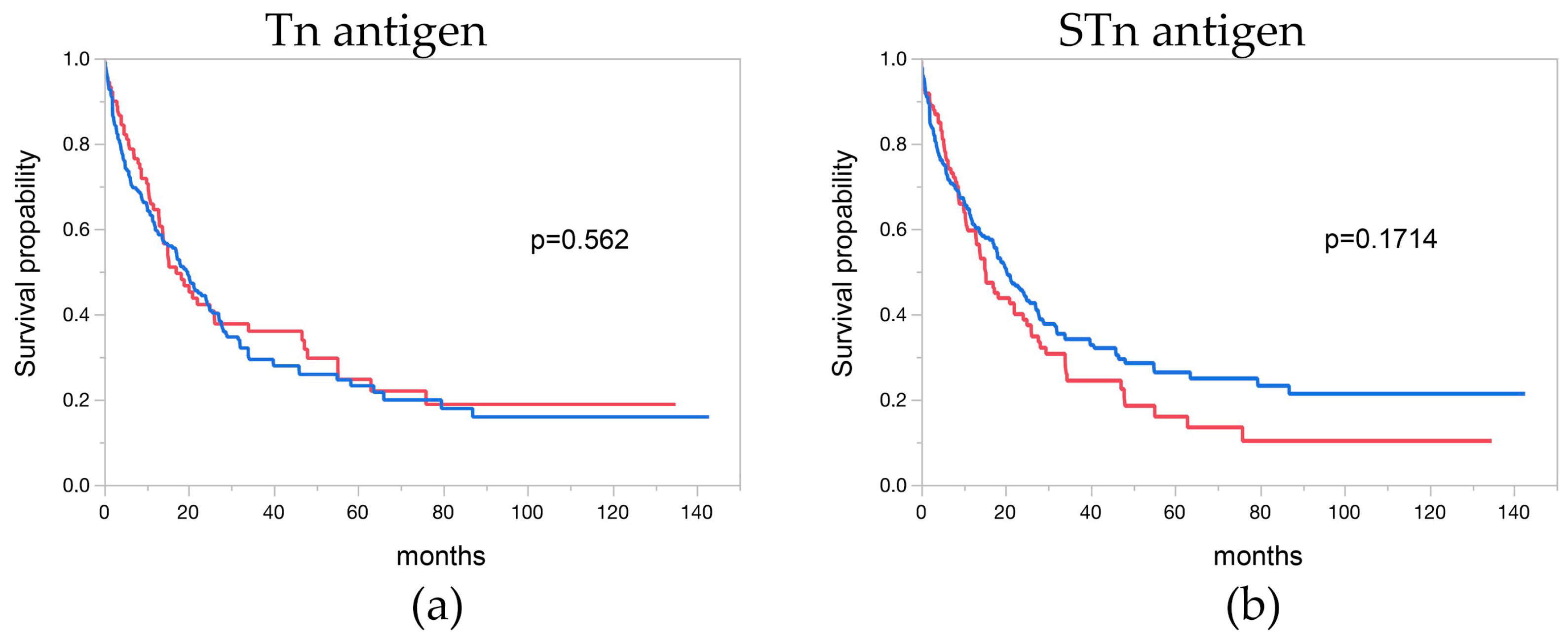
3.7. Tn and STn Antigen and Overall Survival

Follow-up data were accessible for 311 and 353 EAC patients with informative Tn and STn antigen data, respectively. Notably, Tn and STn antigen immunostaining exhibited no significant association with the overall survival of EAC patients (p = 0.562 and p = 0.171). The correlation between Tn and STn antigen immunostaining and the clinical outcome of the patients is depicted in Figure 3a,b.

4. Discussion

In this study, a large cohort of esophageal adenocarcinoma was stained against Tn and STn antigens using immunohistochemistry. Both tumor-associated O-glycans were expressed at 71% (Tn antigen) and 70.5% (STn antigen), across all tumor stages and grades. The survival analysis conducted in this study did not identify a statistically significant correlation between the expression of Tn and STn antigens and the overall survival of the patients. This finding implies that these antigens may have limitations when utilized as independent prognostic indicators and may be more appropriately regarded as general tumor markers in the context of EAC. Our retrospective study aims to serve as an initial exploration, paving the way for further analyses of Tn and STn antigens within the framework of clinicopathological correlation. Subsequent international research efforts may contribute to mitigating potential general biases and enriching our understanding of these antigens in EAC. The use of commercially available antibodies in this study guarantees the reproducibility of analyses across diverse cohorts, thereby advancing our comprehension of Tn and STn antigen expression in esophageal adenocarcinoma (EAC) and other tumor pathologies. It is essential to acknowledge, however, that monoclonal antibodies (mAbs) specific to Tn and STn antigens capture only a fraction of the tumors’ complexity. Consequently, future investigations should contemplate employing a spectrum of antibodies targeting various glyco-epitopes, along with the potential inclusion of well-characterized plant lectins or recombinant glycan-binding receptors. Such a multifaceted approach holds promise for a more comprehensive exploration of the intricacies within tumor biology.
To decipher the composition of the O-glycome, the COSMC gene was selectively deactivated in numerous human cancer cell lines through gene editing techniques, inducing the expression of the Tn antigen. Subsequently, protein and glycopeptide isolation was achieved using the Tn antigen-affine plant lectin Vicia villosa agglutinin (VVL), followed by identification through mass spectrometric proteome analysis [35]. Notably, the discernment emerged that there exists no discernible overlap in the identified O-glycoproteins, even when comparing cell lines within the same tumor entity. This revelation underscores the considerable heterogeneity inherent in the O-glycoproteome, introducing complexity into cell biological processes that defy straightforward predictions. Importantly, there remains a notable absence of an O-glycoproteome analysis for EAC to date.
The analysis of mRNA expression for pivotal genes involved in the initial step of O-glycosylation reveals a substantial downregulation of C1GALT1C1 (COSMC) in the carcinomas. In contrast, there is a modest yet significant upregulation observed in C1GALT1 (T-Synthase). The enzymatic role played by T-Synthase is crucial for the development of truncated O-glycans within tumors. T-Synthase requires its chaperone, COSMC, to convert the Tn antigen into the core 1 structure, which reflects the initial step in complex O-glycosylation. COSMC facilitates proper oligomerization and localization in the endoplasmic reticulum (ER). If COSMC undergoes mutation or depletion, it leads to the degradation of T-Synthase and the expression of Tn antigens [36].
Cummings et al. devised an assay for assessing T-Synthase activity in vitro, enabling the quantification of core 1 O-glycan synthesis in cell lines [37]. In the evaluation of T-Synthase activity in formalin-fixed, paraffin-embedded (FFPE) tissues, a challenge arises from the fact that the immunohistochemical detection of T-Synthase and COSMC offers only partial insights into protein detection, lacking information about their actual functionality. The same limitation applies to mRNA expression data, which currently does not facilitate the genotype–glycotype correlation.
It has been documented that the elevated expression of C1GALT1 contributes to the increased O-glycosylation of Mucin-1 (MUC1) in patients with esophageal squamous cell carcinoma [38] and enhances the invasive and metastatic phenotype by modifying O-glycans on integrin β1 in hepatocellular carcinoma [39]. Additionally, a complete gene knockout of C1galt1c1 in a pancreatic acinar mouse model resulted in the elevated expression of C1galt1 [40]. In Tn4 cells (Epstein–Barr virus (EBV)-transformed B lymphocytes from a male individual whose leukocytes express Tn antigens), characterized by silenced COSMC, methylome analysis unequivocally confirmed the hypermethylation of the COSMC core promoter, with no such observation noted for T-Synthase [41]. The transcriptional regulation of COSMC and T-Synthase, along with their widespread and coordinated expression patterns, requires further clarification in the context of EAC [42]. Additional investigation into glycan-related pathways and cell-to-cell interactions within the tumor microenvironment is essential to elucidate the involvement of T-Synthase and its associated molecules in the progression of tumors.
In a recent study, the presence of truncated O-glycans was assessed across various epithelial and non-epithelial cancers using antibodies targeting the Tn antigen (5F4), the STn antigen (3F1), and the T antigen (3C9). In the context of gastrointestinal tumors, the expression of Tn and STn antigens was detected in pancreatic adenocarcinoma (PDAC) (53%/56%) and colorectal adenocarcinoma (CRC) (51%/80%). Interestingly, the authors noted that Tn and STn antigens were predominantly expressed in tumor tissue and sporadically in healthy tissues [12]. Our data similarly revealed that the Tn antigen antibody yielded positive staining in only one case (n = 30), while the STn antigen antibody displayed positive staining in healthy tissue in 5 out of 30 cases. This suggests the transferability of the antibody specificities and an overarching robustness of the presented results.
In the case of PDAC, it was demonstrated that the aberrant expression of truncated O-glycans in PDAC enhances tumor aggressiveness by inducing epithelial-mesenchymal transition (EMT) and stemness properties [43,44].
For CRC, minimal intracellular Tn staining was observed in normal mucosa, while a significantly higher expression was noted in both peritumoral mucosa and adenocarcinoma. This staining pattern was reflected to a lesser extent by STn expression in these tissue types [45].
The glycocode not only governs immune cell lectin recognition, steering the immune response toward cancer-tolerant phenotypes [46], it also exerts influence within the tumor microenvironment. This environment promotes glycophenotypes that facilitate disease progression and dissemination, although the precise molecular and clinical drivers of glycosylation changes remain elusive in many instances [47]. The rapid translation of communication between the tumor microenvironment and tumor cells occurs through dynamic alterations in cell surface glycosylation, contributing to stress adaptation or active evasion [48]. Another pivotal consideration is the intricate interplay between different O-glycans within both the tumor and immune system cells. This dynamic interaction is critical in the context of glycoprotein modifications on the cell surface, influencing recognition by glycan-binding receptors such as C-type lectins, selectins, siglecs, and galectins. As glycans have evolved into essential components within homeostatic circuits, functioning as precise modulators of immunological responses, they emerge as potential molecular targets for intentionally modulating immune tolerance and activation across a spectrum of pathological scenarios. The recent review article emphasizes the significant role of Tn and STn antigen expression in gastrointestinal tumors in contributing to immunosuppression [49]. The focal point remains on innate immune system cells, specifically those expressing glycan-binding receptors like CD301 (MGL) [50], Siglec-4, -5, -14, and -15 [51], and potentially other receptors selectively interacting with tumor O-glycans.
To sum up, glycans serve as integral mediators of both external and internal signals, resulting in phenotypic changes on the cell surface and actively contributing to malignancy, or serving as surrogate markers for cellular adaptation. Consequently, glycans emerge as highly promising targets for theragnostic applications.
The Tn and STn antigens, recognized as truncated O-glycans, are well-defined cancer-associated glycans expressed across a spectrum of epithelial cancers, albeit to varying degrees. Consequently, therapeutic approaches targeting truncated O-glycans have surfaced, suggesting potential relevance in the future treatment of esophageal adenocarcinoma. A range of promising active compounds falls within different categories, encompassing antibodies [52], antibody-drug conjugates [53], CAR T cells [54,55,56], and sialidases [57].

5. Conclusions

Our study revealed a pronounced expression of Tn and STn antigens in esophageal adenocarcinoma compared to corresponding healthy tissues. Immunohistochemistry analysis demonstrated prominent surface expressions of both antigens in a substantial fraction of EAC specimens, distinguishing them as specific markers for tumor tissues. Notably, a limited or absent expression of these antigens was observed in healthy esophageal tissues.
To decipher the mechanisms underlying truncated O-glycan formation in EAC, we assessed the expression of T-Synthase (C1GALT1) and its chaperone COSMC (C1GALT1C1). Tumor tissues exhibited a statistically significant upregulation in T-Synthase expression, while C1GALT1C1 expression was notably reduced, compared to normal esophagus tissues, probably causing T-Synthase to malfunction.
Tn antigen expression demonstrated a significant association with the depth of infiltration, suggesting its potential role in tumor invasiveness. However, no significant correlation was observed with lymphatic or distant metastasis, tumor differentiation, UICC staging, or resection margin status. Similarly, STn antigen expression did not show a significant correlation with various histopathological parameters.
Analysis of overall survival in EAC patients with Tn and STn antigen immunostaining revealed no significant association, indicating that the expression of these antigens alone may not serve as independent prognostic indicators.
In summary, our comprehensive investigation establishes Tn and STn antigens as distinctive markers of esophageal adenocarcinoma. While Tn antigen expression correlates with the depth of infiltration, their association with overall survival suggests a complex interplay with other factors influencing the clinical outcome of EAC patients. These findings contribute valuable insights into the molecular landscape of esophageal adenocarcinoma, potentially informing future diagnostic and therapeutic strategies.

Author Contributions

Conceptualization, B.M., K.-F.K. and G.W.-E.; methodology, B.M., K.-F.K. and G.W.-E.; validation, B.M., K.-F.K., A.D.G., J.M. and G.W.-E.; formal analysis, K.-F.K., J.M., V.B. and G.W.-E.; investigation, B.M., K.-F.K., J.L., P.S., V.B., C.G. and G.W.-E.; resources, M.B., J.R.I., T.H. and N.M.; data curation, B.M., A.D.G. and G.W.-E.; writing—original draft preparation, B.M. and G.W.-E.; writing—review and editing, B.M., A.D.G. and G.W.-E.; visualization, B.M., K.-F.K., A.D.G., J.M. and G.W.-E.; supervision, G.W.-E.; project administration, B.M. and G.W.-E.; funding acquisition, G.W-E. All authors have read and agreed to the published version of the manuscript.

Funding

This research was funded by German Research Foundation, grant number 275533756.

Institutional Review Board Statement

All subjects gave their informed consent for inclusion before they participated in the study. The study was conducted in accordance with the Declaration of Helsinki, and the protocol was approved by the Ethics Committee of Hamburg (PV3548). The general usage of archived diagnostic leftover tissues for manufacturing of tissue microarrays (TMAs) and their analysis for research purposes as well as patient data analysis has been approved by the Ethics Committee of Hamburg (WF-049/09 and PV3652).

Informed Consent Statement

Informed consent was obtained from all subjects involved in the bio-banking study. Informed consent has not been collected specifically for the patient samples included in generating the TMA. The usage of routinely archived, fixed leftover patient tissue samples for research purposes by the attending physician is approved under local laws and does not require written consent (HmbKHG. §12.1).

Data Availability Statement

The data that support the findings of this study are available from the corresponding author upon reasonable request.

Acknowledgments

We are grateful to Antje Heinecke and Petra Schröder for their technical support in this project and all group members for their input and feedback to this manuscript.

Conflicts of Interest

The authors declare no conflicts of interest.

References

  1. Joseph, A.; Raja, S.; Kamath, S.; Jang, S.; Allende, D.; McNamara, M.; Videtic, G.; Murthy, S.; Bhatt, A. Esophageal adenocarcinoma: A dire need for early detection and treatment. Cleve Clin. J. Med. 2022, 89, 269–279. [Google Scholar] [CrossRef] [PubMed]
  2. Shaheen, N.J.; Richter, J.E. Barrett’s oesophagus. Lancet 2009, 373, 850–861. [Google Scholar] [CrossRef] [PubMed]
  3. Domper Arnal, M.J.; Ferrandez Arenas, A.; Lanas Arbeloa, A. Esophageal cancer: Risk factors, screening and endoscopic treatment in Western and Eastern countries. World J. Gastroenterol. 2015, 21, 7933–7943. [Google Scholar] [CrossRef] [PubMed]
  4. Liu, C.Q.; Ma, Y.L.; Qin, Q.; Wang, P.H.; Luo, Y.; Xu, P.F.; Cui, Y. Epidemiology of esophageal cancer in 2020 and projections to 2030 and 2040. Thorac. Cancer 2023, 14, 3–11. [Google Scholar] [CrossRef] [PubMed]
  5. Obermannova, R.; Alsina, M.; Cervantes, A.; Leong, T.; Lordick, F.; Nilsson, M.; van Grieken, N.C.T.; Vogel, A.; Smyth, E.C.; ESMO Guidelines Committee. Oesophageal cancer: ESMO Clinical Practice Guideline for diagnosis, treatment and follow-up. Ann. Oncol. 2022, 33, 992–1004. [Google Scholar] [CrossRef]
  6. Springer, G.F. Immunoreactive T and Tn epitopes in cancer diagnosis, prognosis, and immunotherapy. J. Mol. Med. 1997, 75, 594–602. [Google Scholar] [CrossRef]
  7. Bagdonaite, I.; Pallesen, E.M.H.; Nielsen, M.I.; Bennett, E.P.; Wandall, H.H. Mucin-Type O-GalNAc Glycosylation in Health and Disease. Adv. Exp. Med. Biol. 2021, 1325, 25–60. [Google Scholar] [CrossRef]
  8. Pinho, S.S.; Reis, C.A. Glycosylation in cancer: Mechanisms and clinical implications. Nat. Rev. Cancer 2015, 15, 540–555. [Google Scholar] [CrossRef]
  9. Ju, T.; Otto, V.I.; Cummings, R.D. The Tn antigen-structural simplicity and biological complexity. Angew. Chem. Int. Ed. Engl. 2011, 50, 1770–1791. [Google Scholar] [CrossRef]
  10. Sewell, R.; Backstrom, M.; Dalziel, M.; Gschmeissner, S.; Karlsson, H.; Noll, T.; Gatgens, J.; Clausen, H.; Hansson, G.C.; Burchell, J.; et al. The ST6GalNAc-I sialyltransferase localizes throughout the Golgi and is responsible for the synthesis of the tumor-associated sialyl-Tn O-glycan in human breast cancer. J. Biol. Chem. 2006, 281, 3586–3594. [Google Scholar] [CrossRef]
  11. Munkley, J. The Role of Sialyl-Tn in Cancer. Int. J. Mol. Sci. 2016, 17, 275. [Google Scholar] [CrossRef] [PubMed]
  12. Romer, T.B.; Aasted, M.K.M.; Dabelsteen, S.; Groen, A.; Schnabel, J.; Tan, E.; Pedersen, J.W.; Haue, A.D.; Wandall, H.H. Mapping of truncated O-glycans in cancers of epithelial and non-epithelial origin. Br. J. Cancer 2021, 125, 1239–1250. [Google Scholar] [CrossRef]
  13. Fu, C.; Zhao, H.; Wang, Y.; Cai, H.; Xiao, Y.; Zeng, Y.; Chen, H. Tumor-associated antigens: Tn antigen, sTn antigen, and T antigen. HLA 2016, 88, 275–286. [Google Scholar] [CrossRef]
  14. Flucke, U.; Zirbes, T.K.; Schroder, W.; Monig, S.P.; Koch, V.; Schmitz, K.; Thiele, J.; Dienes, H.P.; Holscher, A.H.; Baldus, S.E. Expression of mucin-associated carbohydrate core antigens in esophageal squamous cell carcinomas. Anticancer Res. 2001, 21, 2189–2193. [Google Scholar] [PubMed]
  15. Radhakrishnan, P.; Dabelsteen, S.; Madsen, F.B.; Francavilla, C.; Kopp, K.L.; Steentoft, C.; Vakhrushev, S.Y.; Olsen, J.V.; Hansen, L.; Bennett, E.P.; et al. Immature truncated O-glycophenotype of cancer directly induces oncogenic features. Proc. Natl. Acad. Sci. USA 2014, 111, E4066–E4075. [Google Scholar] [CrossRef] [PubMed]
  16. Hofmann, B.T.; Picksak, A.S.; Kwiatkowski, M.; Grupp, K.; Jucker, M.; Bachmann, K.; Mercanoglu, B.; Izbicki, J.R.; Kahlert, C.; Bockhorn, M.; et al. Truncated O-GalNAc glycans impact on fundamental signaling pathways in pancreatic cancer. Glycobiology 2021, cwab088. [Google Scholar] [CrossRef]
  17. Werther, J.L.; Rivera-MacMurray, S.; Bruckner, H.; Tatematsu, M.; Itzkowitz, S.H. Mucin-associated sialosyl-Tn antigen expression in gastric cancer correlates with an adverse outcome. Br. J. Cancer 1994, 69, 613–616. [Google Scholar] [CrossRef]
  18. Yamada, T.; Watanabe, A.; Yamada, Y.; Shino, Y.; Tanase, M.; Yamashita, J.; Miwa, T.; Nakano, H. Sialosyl Tn antigen expression is associated with the prognosis of patients with advanced gastric cancer. Cancer 1995, 76, 1529–1536. [Google Scholar] [CrossRef]
  19. Cotton, S.; Azevedo, R.; Gaiteiro, C.; Ferreira, D.; Lima, L.; Peixoto, A.; Fernandes, E.; Neves, M.; Neves, D.; Amaro, T.; et al. Targeted O-glycoproteomics explored increased sialylation and identified MUC16 as a poor prognosis biomarker in advanced-stage bladder tumours. Mol. Oncol. 2017, 11, 895–912. [Google Scholar] [CrossRef]
  20. Akita, K.; Yoshida, S.; Ikehara, Y.; Shirakawa, S.; Toda, M.; Inoue, M.; Kitawaki, J.; Nakanishi, H.; Narimatsu, H.; Nakada, H. Different levels of sialyl-Tn antigen expressed on MUC16 in patients with endometriosis and ovarian cancer. Int. J. Gynecol. Cancer 2012, 22, 531–538. [Google Scholar] [CrossRef]
  21. Hashiguchi, Y.; Kasai, M.; Fukuda, T.; Ichimura, T.; Yasui, T.; Sumi, T. Serum Sialyl-Tn (STN) as a Tumor Marker in Patients with Endometrial Cancer. Pathol. Oncol. Res. 2016, 22, 501–504. [Google Scholar] [CrossRef] [PubMed]
  22. Inoue, M.; Ogawa, H.; Nakanishi, K.; Tanizawa, O.; Karino, K.; Endo, J. Clinical value of sialyl Tn antigen in patients with gynecologic tumors. Obstet Gynecol. 1990, 75, 1032–1036. [Google Scholar]
  23. Kobayashi, H.; Terao, T.; Kawashima, Y. Sialyl Tn as a prognostic marker in epithelial ovarian cancer. Br. J. Cancer 1992, 66, 984–985. [Google Scholar] [CrossRef] [PubMed]
  24. Nanashima, A.; Yamaguchi, H.; Nakagoe, T.; Matsuo, S.; Sumida, Y.; Tsuji, T.; Sawai, T.; Yamaguchi, E.; Yasutake, T.; Ayabe, H. High serum concentrations of sialyl Tn antigen in carcinomas of the biliary tract and pancreas. J. Hepatobiliary Pancreat Surg. 1999, 6, 391–395. [Google Scholar] [CrossRef] [PubMed]
  25. Sato, T.; Nishimura, G.; Nonomura, A.; Miwa, K.; Miyazaki, I. Serological studies on CEA, CA 19-9, STn and SLX in colorectal cancer. Hepatogastroenterology 1999, 46, 914–919. [Google Scholar]
  26. Nakagoe, T.; Sawai, T.; Tsuji, T.; Jibiki, M.A.; Nanashima, A.; Yamaguchi, H.; Yasutake, T.; Ayabe, H.; Arisawa, K.; Ishikawa, H. Predictive factors for preoperative serum levels of sialy Lewis(x), sialyl Lewis(a) and sialyl Tn antigens in gastric cancer patients. Anticancer Res. 2002, 22, 451–458. [Google Scholar]
  27. Imada, T.; Rino, Y.; Takahashi, M.; Hatori, S.; Shiozawa, M.; Amano, T.; Kondo, J. Serum CA 19-9, SLX, STN and CEA levels of the peripheral and the draining venous blood in gastric cancer. Hepatogastroenterology 1999, 46, 2086–2090. [Google Scholar]
  28. Genega, E.M.; Hutchinson, B.; Reuter, V.E.; Gaudin, P.B. Immunophenotype of high-grade prostatic adenocarcinoma and urothelial carcinoma. Mod. Pathol. 2000, 13, 1186–1191. [Google Scholar] [CrossRef]
  29. Arai, T.; Fujita, K.; Fujime, M.; Irimura, T. Expression of sialylated MUC1 in prostate cancer: Relationship to clinical stage and prognosis. Int. J. Urol. 2005, 12, 654–661. [Google Scholar] [CrossRef]
  30. Reichelt, U.; Duesedau, P.; Tsourlakis, M.; Quaas, A.; Link, B.C.; Schurr, P.G.; Kaifi, J.T.; Gros, S.J.; Yekebas, E.F.; Marx, A.; et al. Frequent homogeneous HER-2 amplification in primary and metastatic adenocarcinoma of the esophagus. Mod. Pathol. 2007, 20, 120–129. [Google Scholar] [CrossRef]
  31. Grupp, K.; Uzunoglu, F.G.; Melling, N.; Hofmann, B.; El Gammal, A.T.; Groteluschen, R.; Heumann, A.; Bellon, E.; Reeh, M.; Wolters-Eisfeld, G.; et al. FOXO1 overexpression and loss of pSerine256-FOXO1 expression predicts clinical outcome in esophageal adenocarcinomas. Sci. Rep. 2018, 8, 17370. [Google Scholar] [CrossRef] [PubMed]
  32. Dancau, A.M.; Simon, R.; Mirlacher, M.; Sauter, G. Tissue Microarrays. Methods Mol. Biol. 2016, 1381, 53–65. [Google Scholar] [CrossRef] [PubMed]
  33. Bosman, F.T.; World Health Organization; International Agency for Research on Cancer. WHO Classification of Tumours of the Digestive System, 4th ed.; International Agency for Research on Cancer: Lyon, France, 2010; p. 417. [Google Scholar]
  34. Sobin, L.H.; Gospodarowicz, M.K.; Wittekind, C.; International Union against Cancer. TNM Classification of Malignant Tumours, 7th ed.; Wiley-Blackwell: Chichester, UK; Hoboken, NJ, USA, 2010; p. 66. [Google Scholar]
  35. Steentoft, C.; Vakhrushev, S.Y.; Joshi, H.J.; Kong, Y.; Vester-Christensen, M.B.; Schjoldager, K.T.; Lavrsen, K.; Dabelsteen, S.; Pedersen, N.B.; Marcos-Silva, L.; et al. Precision mapping of the human O-GalNAc glycoproteome through SimpleCell technology. EMBO J. 2013, 32, 1478–1488. [Google Scholar] [CrossRef] [PubMed]
  36. Aryal, R.P.; Ju, T.; Cummings, R.D. Tight complex formation between Cosmc chaperone and its specific client non-native T-synthase leads to enzyme activity and client-driven dissociation. J. Biol. Chem. 2012, 287, 15317–15329. [Google Scholar] [CrossRef]
  37. Ju, T.; Xia, B.; Aryal, R.P.; Wang, W.; Wang, Y.; Ding, X.; Mi, R.; He, M.; Cummings, R.D. A novel fluorescent assay for T-synthase activity. Glycobiology 2011, 21, 352–362. [Google Scholar] [CrossRef]
  38. Wang, Y.; Liao, X.; Ye, Q.; Huang, L. Clinic implication of MUC1 O-glycosylation and C1GALT1 in esophagus squamous cell carcinoma. Sci. China Life Sci. 2018, 61, 1389–1395. [Google Scholar] [CrossRef] [PubMed]
  39. Liu, C.H.; Hu, R.H.; Huang, M.J.; Lai, I.R.; Chen, C.H.; Lai, H.S.; Wu, Y.M.; Huang, M.C. C1GALT1 promotes invasive phenotypes of hepatocellular carcinoma cells by modulating integrin beta1 glycosylation and activity. PLoS ONE 2014, 9, e94995. [Google Scholar] [CrossRef]
  40. Wolters-Eisfeld, G.; Mercanoglu, B.; Hofmann, B.T.; Wolpers, T.; Schnabel, C.; Harder, S.; Steffen, P.; Bachmann, K.; Steglich, B.; Schrader, J.; et al. Loss of complex O-glycosylation impairs exocrine pancreatic function and induces MODY8-like diabetes in mice. Exp. Mol. Med. 2018, 50, 1–13. [Google Scholar] [CrossRef]
  41. Mi, R.; Song, L.; Wang, Y.; Ding, X.; Zeng, J.; Lehoux, S.; Aryal, R.P.; Wang, J.; Crew, V.K.; van Die, I.; et al. Epigenetic silencing of the chaperone Cosmc in human leukocytes expressing tn antigen. J. Biol. Chem. 2012, 287, 41523–41533. [Google Scholar] [CrossRef]
  42. Zeng, J.; Mi, R.; Wang, Y.; Li, Y.; Lin, L.; Yao, B.; Song, L.; van Die, I.; Chapman, A.B.; Cummings, R.D.; et al. Promoters of Human Cosmc and T-synthase Genes Are Similar in Structure, Yet Different in Epigenetic Regulation. J. Biol. Chem. 2015, 290, 19018–19033. [Google Scholar] [CrossRef]
  43. Thomas, D.; Sagar, S.; Caffrey, T.; Grandgenett, P.M.; Radhakrishnan, P. Truncated O-glycans promote epithelial-to-mesenchymal transition and stemness properties of pancreatic cancer cells. J. Cell. Mol. Med. 2019, 23, 6885–6896. [Google Scholar] [CrossRef] [PubMed]
  44. Hofmann, B.T.; Schluter, L.; Lange, P.; Mercanoglu, B.; Ewald, F.; Folster, A.; Picksak, A.S.; Harder, S.; El Gammal, A.T.; Grupp, K.; et al. COSMC knockdown mediated aberrant O-glycosylation promotes oncogenic properties in pancreatic cancer. Mol. Cancer 2015, 14, 109. [Google Scholar] [CrossRef] [PubMed]
  45. Dombek, G.E.; Ore, A.S.; Cheng, J.; Matsumoto, Y.; Glickman, J.N.; Fleishman, A.; Heimburg-Molinaro, J.; Poylin, V.Y.; Fabrizio, A.; Cataldo, T.; et al. Immunohistochemical analysis of Tn antigen expression in colorectal adenocarcinoma and precursor lesions. BMC Cancer 2022, 22, 1281. [Google Scholar] [CrossRef] [PubMed]
  46. RodrIguez, E.; Schetters, S.T.T.; van Kooyk, Y. The tumour glyco-code as a novel immune checkpoint for immunotherapy. Nat. Rev. Immunol. 2018, 18, 204–211. [Google Scholar] [CrossRef] [PubMed]
  47. Stowell, S.R.; Ju, T.; Cummings, R.D. Protein glycosylation in cancer. Annu. Rev. Pathol. 2015, 10, 473–510. [Google Scholar] [CrossRef]
  48. Peixoto, A.; Relvas-Santos, M.; Azevedo, R.; Santos, L.L.; Ferreira, J.A. Protein Glycosylation and Tumor Microenvironment Alterations Driving Cancer Hallmarks. Front. Oncol. 2019, 9, 380. [Google Scholar] [CrossRef] [PubMed]
  49. Rajesh, C.; Radhakrishnan, P. The (Sialyl) Tn antigen: Contributions to immunosuppression in gastrointestinal cancers. Front. Oncol. 2022, 12, 1093496. [Google Scholar] [CrossRef]
  50. Dusoswa, S.A.; Verhoeff, J.; Abels, E.; Mendez-Huergo, S.P.; Croci, D.O.; Kuijper, L.H.; de Miguel, E.; Wouters, V.; Best, M.G.; Rodriguez, E.; et al. Glioblastomas exploit truncated O-linked glycans for local and distant immune modulation via the macrophage galactose-type lectin. Proc. Natl. Acad. Sci. USA 2020, 117, 3693–3703. [Google Scholar] [CrossRef]
  51. Gonzalez-Gil, A.; Schnaar, R.L. Siglec Ligands. Cells 2021, 10, 1260. [Google Scholar] [CrossRef]
  52. Matsumoto, Y.; Jia, N.; Heimburg-Molinaro, J.; Cummings, R.D. Targeting Tn-positive tumors with an afucosylated recombinant anti-Tn IgG. Sci. Rep. 2023, 13, 5027. [Google Scholar] [CrossRef]
  53. Prendergast, J.M.; Galvao da Silva, A.P.; Eavarone, D.A.; Ghaderi, D.; Zhang, M.; Brady, D.; Wicks, J.; DeSander, J.; Behrens, J.; Rueda, B.R. Novel anti-Sialyl-Tn monoclonal antibodies and antibody-drug conjugates demonstrate tumor specificity and anti-tumor activity. MAbs 2017, 9, 615–627. [Google Scholar] [CrossRef] [PubMed]
  54. Posey, A.D., Jr.; Schwab, R.D.; Boesteanu, A.C.; Steentoft, C.; Mandel, U.; Engels, B.; Stone, J.D.; Madsen, T.D.; Schreiber, K.; Haines, K.M.; et al. Engineered CAR T Cells Targeting the Cancer-Associated Tn-Glycoform of the Membrane Mucin MUC1 Control Adenocarcinoma. Immunity 2016, 44, 1444–1454. [Google Scholar] [CrossRef] [PubMed]
  55. Lodewijk, I.; Duenas, M.; Paramio, J.M.; Rubio, C. CD44v6, STn & O-GD2: Promising tumor associated antigens paving the way for new targeted cancer therapies. Front. Immunol. 2023, 14, 1272681. [Google Scholar] [CrossRef] [PubMed]
  56. Raglow, Z.; McKenna, M.K.; Bonifant, C.L.; Wang, W.; Pasca di Magliano, M.; Stadlmann, J.; Penninger, J.M.; Cummings, R.D.; Brenner, M.K.; Markovitz, D.M. Targeting glycans for CAR therapy: The advent of sweet CARs. Mol. Ther. 2022, 30, 2881–2890. [Google Scholar] [CrossRef]
  57. Munkley, J. Aberrant Sialylation in Cancer: Therapeutic Opportunities. Cancers 2022, 14, 4248. [Google Scholar] [CrossRef]
Figure 1. Truncated O-glycans Tn and STn antigens are highly expressed in EAC. (a,b) Representative microscopic images at ×100 magnification featuring immunohistochemical staining with monoclonal antibodies targeting Tn or STn antigens on sections of EAC (weak and strong) or healthy esophageal tissue. (c,d) Sores of Tn and STn antigen IHC staining reflecting antibody binding in normal and EAC tissue. (e) Analysis and quantification of T-Synthase (C1GALT1) mRNA and (f) COSMC (C1GALT1C1) expression levels in healthy and EAC tissues. * equals p ≤ 0.05 and *** equals p ≤ 0.001.
Cancers 16 00240 g001
Figure 2. Microscopic images representing EAC TMA cores at ×100 magnification were obtained, featuring immunohistochemical (IHC) staining with monoclonal antibodies targeting Tn or STn antigens. The images capture varying degrees of Tn and STn antigen expression in EACs, ranging from negative to weak and strong.
Cancers 16 00240 g002
Figure 3. Overall survival of patients with esophageal adenocarcinoma based on Tn and STn antigen staining. (a) The Kaplan–Meier plot depicts EAC Tn antigen staining, and (b) STn antigen staining. The overall survival p-values indicate no significant difference between the analyzed groups, as determined by the LogRank test for Tn (p = 0.1714) and STn antigen (p = 0.562). The red line represents negative cases, while the blue line represents positive cases.
Cancers 16 00240 g003
Table 1. Histopathological and clinical features of the arrayed EAC specimens.
Parameter EACs (n = 501)
Sexmale425
female76
Tumor stagepT1108
pT287
pT3278
pT428
Lymph node metastasispN0135
pN1172
pN295
pN399
UICCI110
II87
III229
IV75
Distant MetastasisM0421
M180
GradingG114
G2318
G3169
Table 2. Association of Tn antigen (CD175) IHC results and clinicopathological features of EACs.
Tn Antigen (CD175)
Parametern EvaluableNegative (%)Positive (%)p Value
All cancers31128.971.1
Tumor stage
pT15131.468.60.0266
pT25839.760.3
pT318226.473.6
pT4175.994.1
Lymph node metastasis
pN07632.967.10.2877
pN110728.072.0
pN25633.966.1
pN36020.0080.00
UICC
I5530.969.10.0732
II5141.258.8
III15726.173.9
IV4017.5082.50
Distant metastasis
M025929.770.30.2579
M14621.778.3
Resection margin
R021929.770.30.7256
R17225.0075.00
R2825.0075.00
Grading
G11020.0080.000.2463
G210834.365.7
G318325.774.3
Table 3. Association of STn antigen (CD175s) IHC results and clinicopathological features of EACs.
STn Antigen (CD175s)
Parametern EvaluableNegative (%)Positive (%)p Value
All cancers35329.570.5
Tumor stage
pT15925.474.60.1218
pT26339.760.3
pT320828.871.2
pT42015.085.0
Lymph node metastasis
pN08626.773.30.8868
pN111730.869.2
pN26232.367.7
pN37628.971.1
UICC
I6225.874.20.581
II5635.764.3
III17830.369.7
IV4825.075.0
Distant metastasis
M029330.469.90.5062
M15425.974.1
Resection margin
R024830.269.80.7611
R18527.172.9
R2837.562.5
Grading
G11225.075.00.8482
G212027.572.5
G321030.070.0
Disclaimer/Publisher’s Note: The statements, opinions and data contained in all publications are solely those of the individual author(s) and contributor(s) and not of MDPI and/or the editor(s). MDPI and/or the editor(s) disclaim responsibility for any injury to people or property resulting from any ideas, methods, instructions or products referred to in the content.

