Article

Co-Supported CeO2Nanoparticles for CO Catalytic Oxidation: Effects of Different Synthesis Methods on Catalytic Performance

College of Chemistry and Chemical Engineering, Mudanjiang Normal University, Mudanjiang 157000, China
*
Author to whom correspondence should be addressed.
Submission received: 5 February 2020 / Revised: 14 February 2020 / Accepted: 16 February 2020 / Published: 18 February 2020

Abstract

:
Hydrothermal and co-precipitation methods were studied as two different methods for the synthesis of CeO2nanocatalysts. Co/CeO2 catalysts supported by 2, 4, 6, or 8wt% Co were further synthesized through impregnation and the performance of the catalytic oxidation of CO has been investigated. The highest specific surface area and the best catalytic performance was obtained by the catalyst 4wt% Co/CeO2 with the CeO2 support synthesized by the hydrothermal method (4% Co/CeO2-h), which yielded 100% CO conversion at 130 °C. The formation of CeO2 nanoparticles was confirmed by TEM analysis. XRD and SEM-EDX mapping analyses indicated that CoOx is highly dispersed on the 4% Co/CeO2-h catalyst surface. H2-TPR and O2-TPD results showed that 4% Co/CeO2-h possesses the best redox properties and the highest amount of chemically adsorbed oxygen on its surface among all tested catalysts. Raman and XPS spectra showed strong interactions between highly dispersed Co2+ active sites and exposed Ce3+ on the surface of the CeO2 support, resulting in the formation of the strong redox cycle Ce4+ + Co2+↔ Ce3+ + Co3+.This may explain that 4% Co/CeO2-h exhibited the best catalytic activity among all tested catalysts.

Graphical Abstract

1. Introduction

In the last decades, the application of metal oxide nanoparticles in the field of catalysis has experienced an unprecedented growth because of their significant contributions to environmental protection and energy utilization [1,2,3]. Many catalytic materials have also been prepared on the nanoscale by various synthesis methods and extensively studied to fully understand their complex surface reaction processes. Ceria (CeO2), famous for its excellent oxygen transport capacity, has been well used in industry as the main carrier of three-way catalysts (TWCs). In addition, CeO2 performs many important functions on the nanoscale. Metal oxides, as active components on CeO2 supports, show better reactivity and stability in catalytic processes, benefitting from oxygen transfer and other interactions between CeO2 support and active components. CeO2 can also keep metal oxides well dispersed and protect them from sintering at high temperature [3,4,5,6,7]. These profound changes in the catalytic performance are mainly attributed to differences in the nanoparticles’ size and morphology. Trovarelli and Llorca [3] reviewed the recent development and applications of CeO2 nanomaterials. Growth law and influencing factors of different CeO2 crystal faces have been reported, showing that shape (as well as size) and surface/face reconstruction of CeO2 can be controlled at the nanoscale. Based on these parameters, catalyst activity and stability can be effectively governed in catalytic reactions. Zhang et al. [5] developed a lattice oxygen distortion method to enhanceoxygen activation by quenching Ce–Zr oxide nanoparticles formed at extremely hightemperature, which has been applied in the oxidation of carbon monoxide (CO). Ce–Zr oxides can provide about 19 times more oxygenvacancies than traditional oxides in CO atmosphere. Based on the above factors, nanoparticles synthesized by different methods can exhibit different catalytic activities and exploring their different reactivities is of high scientific interest [8].
CO is one of the most common toxic gases and harmful to environment and human health. The elimination of CO and especially the oxidation of CO, a simple and typical reaction in heterogeneous catalysis, has attracted wide attention in recent years due to its academic value and possible applications [9,10,11,12,13]. Progress in nanotechnology provides a precious tool for the study of CO oxidation at the nanoscale. In particular, nano-ceria particles play an important role as support in catalytic reactions. Nobel metals supported on CeO2 show an excellent performance as catalysts for the oxidation of CO [14,15,16,17]. For example, Chen et al. [18] loaded Pd on the specific crystal plane of CeO2 nanoparticles and studied the electronic metal-support interactions (EMSI) in CO oxidation. They showed that interactions on well-defined interfaces improved the catalytic performance. Tanikawa and Egawa [19] found that the addition of Ba ions to palladium catalysts on different ZrO2 or Al2O3-modified CeO2 supports affected the adsorption strength of CO on the catalysts. However, noble-metal catalysts also exhibit obvious disadvantages. They are difficult to obtain and expensive, which limits their applications. Therefore, increasing research is performed on cheap and easily available alternatives. In recent years, transition-metal oxides have become popular as non-noble metal catalysts [20,21,22,23,24,25,26], and their CO oxidation performance has been reported to be strongly influenced by the catalysts’ structure, size, and morphology [27]. Chen et al. [28] synthesized Co3O4-CeO2 core–shell catalysts and proved that the synergistic effect between Co3O4 and CeO2 is responsible for their enhanced catalytic activity compared with that of regular catalysts. Narayana et al. [29] prepared spherical CeO2 nanoparticles with Mn-ion substitution and observed the highest CO oxidation rate for the structure with the highest amounts of Mn2+ as well as oxygen vacancies. Obviously, nanoscale catalysts exhibit excellent catalytic performances due to the size effect and their special morphology. In addition, among noble-metal or transition-metal oxide catalysts, CeO2 has a prominent role as one of the most widely used supports in catalytic oxidation technology.
Based on the above analysis, it can be inferred that different synthesis methods have great influence on the morphology and properties of CeO2 nanoparticles, which may affect the catalytic performance. In this paper, nano-ceria with different sizes were prepared by hydrothermal and co-precipitation methods. The impregnation method was further used to load a specific amount of CoOx onto the CeO2 surface. The catalysts’ CO oxidation performances were studied, and their physical and chemical properties were characterized by XRD, BET, Raman spectroscopy, H2-TPR, O2-TPD, and XPS. The results indicated that CeO2 nanoparticles prepared by the hydrothermal method with a loading of 4wt% Co possess the best CO oxidation performance.

2. Results and Discussion

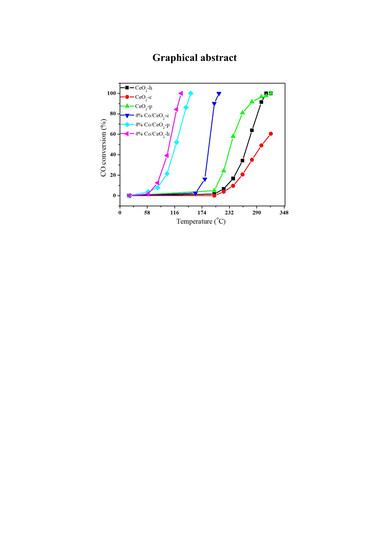
2.1. Catalytic Behavior

The catalytic performances of CeO2 catalysts with different Co loadings synthesized by the hydrothermal method are shown in Figure 1. The mixed oxides revealed superior performances compared with pure CeO2 catalyst, and the temperature of 100% conversion remarkably decreased after Co loading. However, the catalytic activity does not change linearly with increasing Co loading. The highest activity was observed for 4% Co/CeO2, and the maximum CO conversion rate of 100% was obtained at 130 °C.
CO conversions of pure supports and catalysts with 4% Co loading, prepared by different methods, are shown in Figure 2. The catalytic activity of CeO2 support prepared by the hydrothermal or co-precipitation method is higher than that of commercial CeO2. For pure CeO2-p, CeO2-h, and CeO2-c supports, CO conversion (10% efficiency) starts at 217, 232, and 244 °C, respectively. The CO conversion of CeO2-p and CeO2-h reaches nearly 100% at 320 °C, while the efficiency of CeO2-c is only 60% at this temperature. Interestingly, Co loading significantly improves the catalytic activity. For 4% Co/CeO2-h and 4% Co/CeO2-p catalysts, CO conversion (10% efficiency) starts at about 80 °C, and 4% Co/CeO2-c reaches the same initial conversion rate at 174 °C. In addition, 4% Co/CeO2-h displays the highest CO conversion rate among the three catalysts, yielding 100% CO conversion at 130 °C. In contrast, the second highest rate is detected for 4% Co/CeO2-p and 4% Co/CeO2-c, yielding 100% CO conversion at 150 and 210 °C, respectively. It can be inferred that Co is the main active component in the reaction, and different preparation methods exert significant influence on the catalytic performance. Different preparation methods may affect the catalysts structure, surface morphology, and grain size, which may change the catalytic performance. In addition, the performance is affected by the interaction between active components and supports. Therefore, we further explored the changes in the catalytic performance caused by different preparation methods.
The activity test of CO oxidation under different gas hourly space velocities (GHSV) is presented in Figure 3. Generally, GHSV reflects the residence time of reaction gas in the catalyst bed. Higher GHSV are correlated with shorter residence times, while lower reaction depths are correlated with larger treatment capacities. For GHSV = 12, 000 h−1 and GHSV = 24, 000 h−1, the CO conversion rates of 4% Co/CeO2-h are almost identical. However, when the GHSV reaches 30, 000 h−1, the catalytic activity decreases slightly, but it is still larger than 90% at 136 °C. For GHSV = 48, 000 h−1, the CO conversion decreases obviously, and complete CO conversion is reached at 190 °C. This temperature point is shifted back about 60 °C, and the CO conversion reaches 90% at about 166 °C. In general, when the space velocity increases to more than 24, 000 h−1, the catalytic activity decreases, but complete CO conversion is still reached before 190 °C.

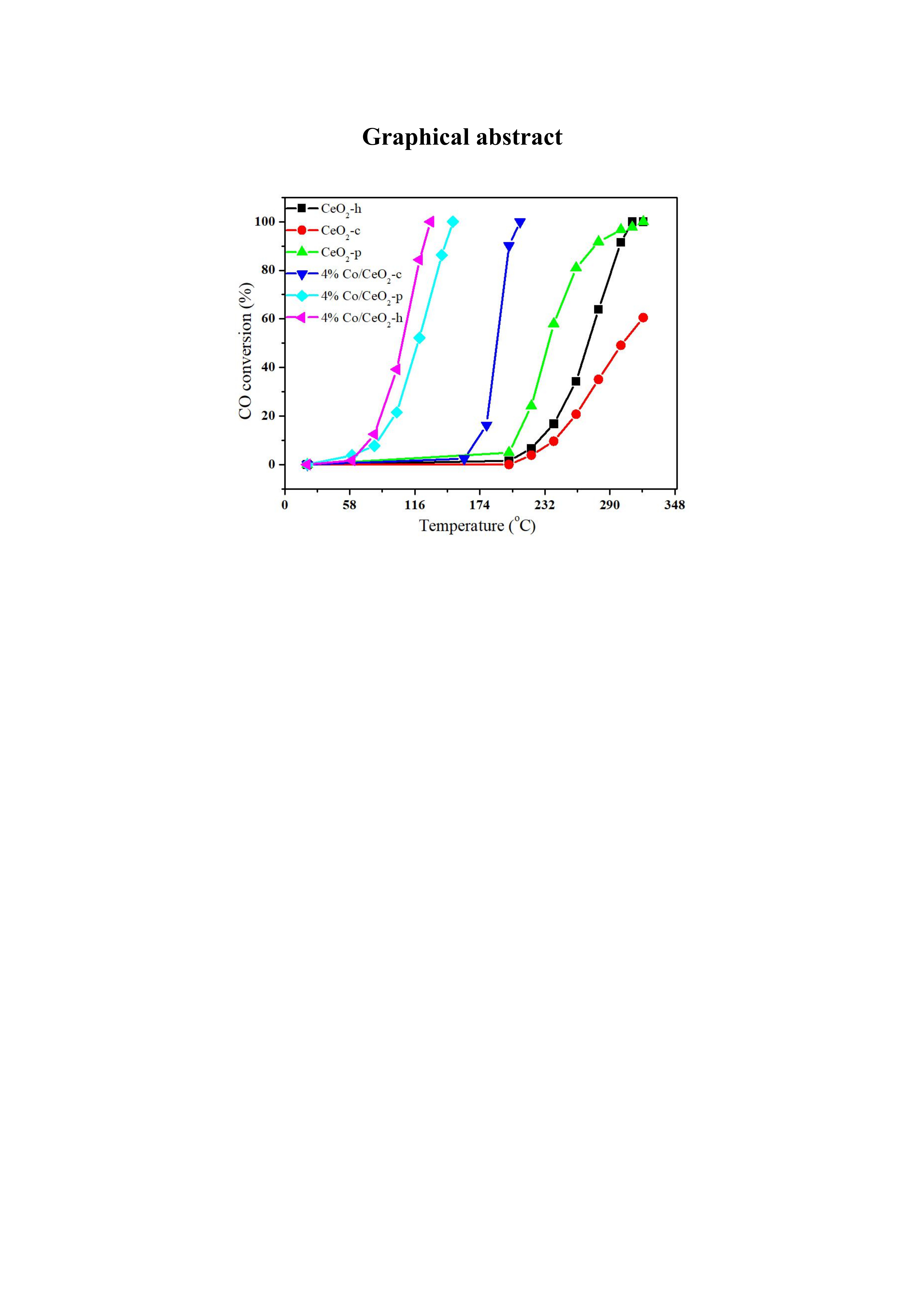
2.2. XRD and BET Analysis

The crystal structures of CeO2 supports and Co/CeO2 catalysts have been investigated by XRD, as presented in Figure 4. The XRD patterns of all pure CeO2 samples display identical peaks, and the diffraction peaks at 28.5, 33.0, 47.5, 56.3, 59.1, 69.4, 76.8, and 79.1° could be ascribed to the (111), (200), (220), (311), (222), (400), (331), and (420) crystal planes of the face-centered cubic structure of CeO2 (JCPD 43–1002), respectively. The CeO2-csupport exhibits sharper diffraction peaks than CeO2-h and CeO2-p, which showed both very weak diffraction peaks. The intensity of the XRD diffraction peaks in the pure CeO2 phase is related to the grain size [30]. Generally, the sharper the diffraction peak, the narrower its half-peak width and the larger the grain size. Therefore, the CeO2 particles are smaller in CeO2-h than those in CeO2-c and CeO2-p, and smaller grain size may be one of the reasons for the observed high activity of the Co/CeO2-h catalyst [31]. Co-doping does not significantly affect the diffraction peak positions of pure CeO2, and its original cubic fluorite structure is not obviously altered. In contrast, Co-supported catalysts exhibit diffraction peaks with higher intensity. Meanwhile, no peaks corresponding to crystalline Co oxide (CoOx) are observed in any sample, indicating that Co is highly dispersed on the CeO2 supports. It has been reported that high dispersion of CoOx nanoparticles is beneficial to increase the crystallinity of CeO2, which may be related to the strong interaction between CoOx and CeO2 supports. The grain size is calculated from the lowest angle diffraction peak in the XRD pattern, as shown in Table 1. This reveals that the smallest particle size is obtained for CeO2 prepared by the hydrothermal method. In general, a smaller particle size is correlated with a higher specific surface area, which contributes to the higher dispersion of CoOx on the surface and is beneficial to improve the catalytic activity [26,30]. This conclusion is confirmed by BET analysis.
BET surface areas of CeO2 supports and Co/CeO2 catalysts are listed in Table 1, revealing that CeO2-h has a significantly higher BET surface area than CeO2-p and CeO2-c. For CeO2-h and CeO2-p, the BET surface areas decrease with increasing CoOx loading. Interestingly, the BET surface area of 4% Co/CeO2-c increases only slightly. However, compared with Co/CeO2-p and Co/CeO2-c, Co/CeO2-h still has the highest BET surface area, and the surface area decreases in the order of Co/CeO2-h > Co/CeO2-p > Co/CeO2-c. The high surface area of Co/CeO2-h may affect the CO oxidation activity, which depends on the number of reaction sites and thus on the specific surface area.

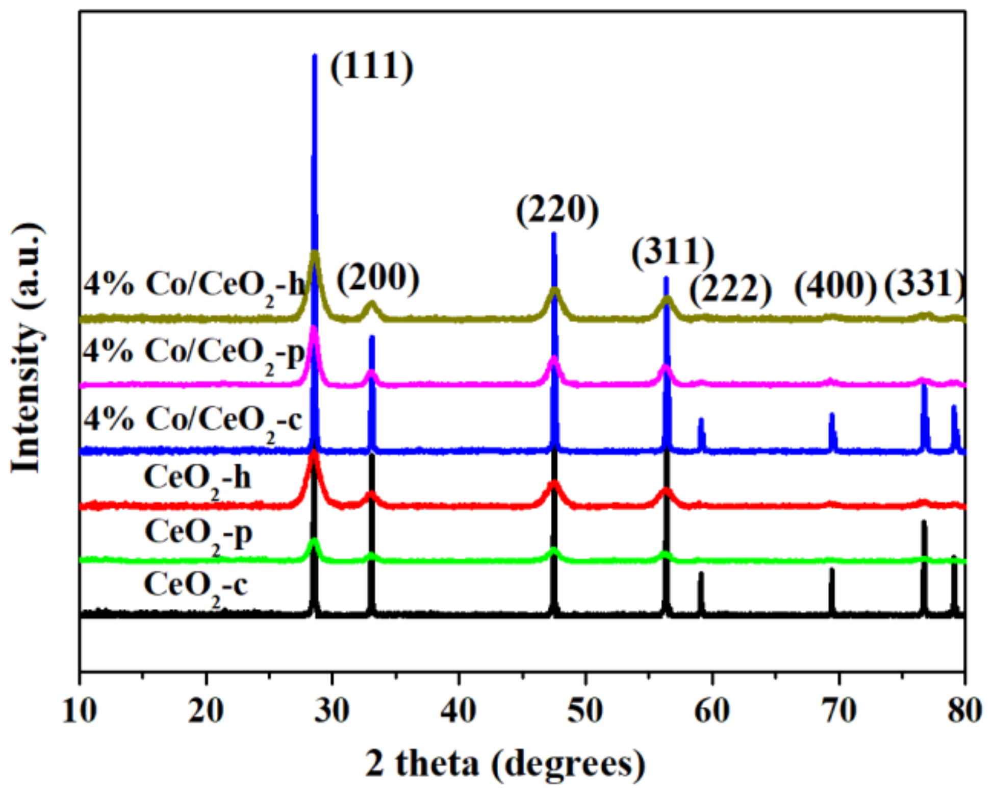
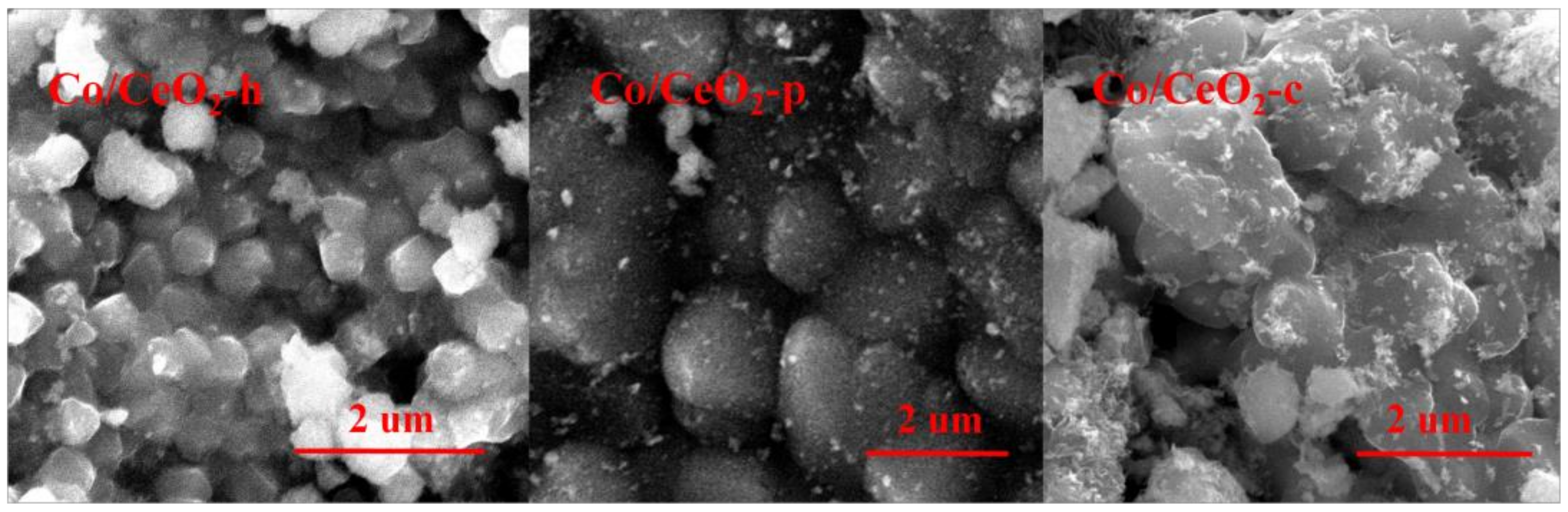
2.3. SEM and TEM Analysis

The SEM images of 4% Co/CeO2 catalysts are shown in Figure 5, revealing obvious differences in the particle sizes of these catalysts. The catalyst 4% Co/CeO2-h forms regular cubes and spheres with a relatively uniform size of <400 nm diameter.The catalysts 4%Co/CeO2-c and 4% Co/CeO2-p form larger particles of <1.5 μm diameter. As 4% Co/CeO2-h exhibits the smallest grain size as well as the best CO catalytic activity, a relationship between grain size and CO catalytic reactivity may be postulated. Although the grain size is not the only factor affecting the conversion efficiency, smaller grain sizes improve the CO catalytic activity. It should be noted that larger CeO2 particles observed in SEM may also result from the agglomeration of smaller particles. However, the determination of the particle size is limited by the instrumental magnification, and TEM analysis is required to precisely determine smaller grain sizes, as described in the following.
The catalysts 4% Co/CeO2-h, 4% Co/CeO2-p, and 4% Co/CeO2-c were studied by SEM-EDX elemental mapping, as shown in Figure 6. Among them, Figure 6b,e,h shows the elemental distribution of Ce, and Figure 6c,f,i shows the elemental distribution of Co of the three catalysts. The analysis reveals that the distribution of Co is more dispersed than that of Ce on Co/CeO2-p and especially on Co/CeO2-h. This higher dispersion of Co was theoretically expected, as Co was impregnated on the surface of CeO2. However, the Ce and Co distributions on CeO2-c are similar, which suggests some agglomeration of CoOx. Furthermore, the consistency of the support materials (CeO2) indicates that CeO2 support prepared by the hydrothermal method is more conducive to the dispersion of Co on the surface, and the Co dispersion on the support surface may be related to the interaction between Co and CeO2 with different particle sizes and specific surface areas. EDX analysis shows, in combination with the activity test results, that the excellent Co dispersion promotes the catalytic activity.
TEM was performed to determine the particle sizes of Co/CeO2-h, Co/CeO2-p, and Co/CeO2-c catalysts, as demonstrated in Figure 7. The images reveal the presence of CeO2 nanoparticles in all catalysts Co/CeO2-h (Figure 7a), Co/CeO2-p (Figure 7d), and Co/CeO2-c (Figure 7g). Co/CeO2-h showed an average particle size of about 8–10 nm (Figure 7a), Co/CeO2-p showed a larger average particle size of about 15–20 nm (Figure 7d), and Co/CeO2-c exhibited the largest particle sizes of more than 20 nm and more regular particles than the particles of the other two catalysts (Figure 7g). These differences in the grain sizes are consistent with previous test results. No lattice fringes corresponding to Co oxide were found, which explains, in combination with the SEM elemental mapping results, the high Co dispersion on the CeO2 surface. Furthermore, the HRTEM images reveal lattice distances of 0.308, 0.309, and 0.311 nm for the three catalysts, which are assigned to the (111) lattice plane of CeO2. These results fully prove the formation of CeO2 nanoparticles.

2.4. Raman Spectroscopy

The structural properties of the catalysts are very important for the analysis of active sites in solid–gas reactions. Raman spectroscopy, as a structural characterization technique, has been widely used for this purpose in different catalytic reaction systems [32,33,34,35]. The Raman spectra of all CeO2 and Co/CeO2 catalysts are shown in Figure 8. The distinct F2g symmetry mode of the CeO2 phase centered between 428 and 454 cm−1. Two weak bands around 1158 cm−1 are due to the defect-induced (D) mode and the second-order longitudinal (2LO) mode [36]. CeO2-h exhibits a much broader F2g peak than CeO2-c and CeO2-p, which is a size-dependent phenomenon observed for ceria nanoparticles and can be explained by inhomogeneous strain broadening associated with particle size dispersion and phonon confinement [36,37,38]. This is consistent with XRD measurements, which revealed for CeO2-h the smallest average particle size among all catalysts. Furthermore, the D/F2g peak area ratio can be estimated to gain insight into the relative population of oxygen vacancies, as shown in the Table 2. Compared with CeO2-c and CeO2-p, Co/CeO2-h reveals more oxygen vacancies. Therefore, application of high temperature and high pressure in the hydrothermal process promote the formation of oxygen vacancies, which could improve the catalytic performance.
The Raman spectra of 4% Co/CeO2 are similar to those of the CeO2 supports. No peaks corresponding to crystalline CoOx were detected, which further indicates that CoOx is highly dispersed on the support surface. The decrease of the peak intensity may be caused by the change of the relative Ce-O content. Meanwhile, Raman spectra of CeO2 support and Co/CeO2 catalysts prepared by different synthetic methods exhibit obvious peak shifts. The loading with cobalt oxide species slightly red-shifts the F2g characteristic peaks of Co/CeO2-p and Co/CeO2-c. This phenomenon is usually related to the change in lattice spacing and bond length caused by lattice constriction upon Co2+-doping [37,38], as it is possible that Co ions enter the interior of the CeO2 lattice. In contrast, the Co/CeO2-h catalyst shows a blue shift of the F2g characteristic peak. This further suggests a special interaction between CoOx and CeO2-h supports. CoOx is mainly dispersed on the surface of the Co/CeO2-h catalyst and well-dispersed cobalt oxide species contribute to the high catalytic activity of the mixed metal oxide catalysts [33,36].

2.5. H2-TPR and O2-TPD Analysis

Temperature-programmed reduction (H2-TPR) analysis has been conducted to investigate the reduction behavior of CeO2 supports and Co/CeO2 catalysts, as shown in Figure 9. The centers of the reduction peaks of pure CeO2 supports appear at about 200–550 °C and 800 °C, which can be assigned to the reduction of surface oxygen and lattice oxygen, respectively [39]. For CeO2-h, the centers of the reduction peaks appear at 325, 510, and 780 °C, while CeO2-p exhibits the centers of its reduction peaks at 426, 520, and 780 °C. However, CeO2-c shows only one reduction peak at 824 °C. These results indicate that more oxygen is adsorbed on the surfaces of CeO2-h and CeO2-p, which improves the catalytic activity. In addition, the initial reduction temperature of CeO2-h at about 225 °C is lower than that of the other two supports, and the reduction peak area is larger. It can be inferred that the low temperature reduction performance of CeO2-h is better than those of the other two catalysts. Compared with pure CeO2 supports, the reduction peak of the Co/CeO2 catalysts is shifted to low temperature. This indicates that Co-doping improves the redox capacity. Moreover, for Co/CeO2 catalysts, the reduction peaks α appear between 200 and 300 °C and are mainly related to the reduction of the Co3O4 phase, as this Co-species is easier to reduce than Ce4+. Similarly, all the samples display β peaks at about 800 °C due to the reduction of bulk CeO2, and it can be observed that the temperature of the reduction peak decreases gradually in the order 4%Co/CeO2-h< 4%Co/CeO2-p < 4%Co/CeO2-c. This further confirms that 4%Co/CeO2-h exhibits the best reduction performance among all catalysts and that strong interactions between the active component CoOx and CeO2 supports are present.
Various studies [40,41,42] have shown that the reduction of Co3O4 is a two-step process, involving the reductions of Co3O4 to CoO and CoO to Co. These results clearly show that the reduction behavior of Co3O4 strongly depends on the CeO2 synthesis method, dispersion state of CoOx, and the interaction between CoOx and CeO2, which is consistent with conclusions of previous reports [26,43]. For the Co/CeO2 catalysts, the reduction peaks can be classified as follows: (1) Range A (<240°C): reduction of surface-adsorbed oxygen species; (2) Range B (250–310°C): reduction of Co3+ to Co2+ on the interface between Co3O4 and CeO2; (3) Range C (310–350°C): reduction of independent Co3O4 weakly interacting with CeO2 to form Co; (4) Range D (350–500°C): reduction of Co2+ to Co [41,42]. Among these catalysts, Co/CeO2-h displayed the lowest reduction temperature, which indicates that Co/CeO2-h exhibits the best redox performance. Position and intensity of the reduction peaks often reflect differences in the interactions between active components and supports. The Co/CeO2-h catalysts exhibited only very small peaks compared with pure Co3O4, suggesting high component dispersion in these catalysts and implying that almost all present Co-species share a homogeneous interaction with the CeO2 phase. A rather large and sharp reduction peak appears for Co2+ interacting with CeO2, which may present further evidence for this homogeneous interaction. The CoOx dispersion in different catalysts decreases in the order Co/CeO2-h > Co/CeO2-p > Co/CeO2-c, which is in good agreement with the XRD results. Thus, it can be inferred that the different preparation methods significantly influence the component dispersion. Furthermore, the hydrogen consumption of all catalysts was calculated from the H2-TPR measurement summarized in Table 1. H2 consumption in the range of 50–600 °C was 2.01, 1.58, 1.11, 1.19, 0.88, and 0.07 mmol/g for Co/CeO2-h, Co/CeO2-p, Co/CeO2-c, CeO2-h, CeO2-p, and CeO2-c, respectively. The homogeneous and profound interaction provides the largest Co3O4–CeO2 interface area. Therefore, the Co/CeO2-h catalyst is expected to contain the largest amount of adsorbed surface oxygen, as indicated by the largest H2 consumption, resulting in an enhanced CO catalytic activity.
Since oxygen vacancies play a crucial role in CO oxidation over Co/CeO2 catalysts, O2-TPD measurements have been performed, as presented in Figure 10. One oxygen desorption peak at nearly 450 °C can be observed for the CeO2-h and CeO2-p supports, while CeO2-c only exhibits a very small peak around 800 °C. This proves that CeO2-h and CeO2-p supports possess better redox performance. Furthermore, Co/CeO2 catalysts show more chemically adsorbed oxygen species and lower desorption temperatures than pure CeO2 supports. Except for Co/CeO2-c, two oxygen desorption peaks can be observed for the Co/CeO2 catalysts, while Co/CeO2-c shows only one peak near 750°C. For Co/CeO2-p and Co/CeO2-h, the other wide peak α appears in the range of 150 to 680 °C. Generally, the peak β centered below 750 °C can be attributed to the oxygen desorption from Co and CeO2 sites, and the high-temperature peak at T > 750 °C corresponds to the thermal decomposition of Co3O4, which is consistent with previous reports [44,45,46]. The peak at T < 750 °C corresponds to the amount of oxygen desorption that has been listed in Table 1. The change rule of the oxygen desorption is consistent with activity test and H2-TPR results. It can be seen that the 4% Co/CeO2-h catalyst desorbs the largest amount of chemically adsorbed oxygen, indicating that this catalyst exhibits the largest oxygen amount on its surface among all catalysts, which explains its high catalytic activity. In addition, according to the literature, good performance for adsorption and activation of oxygen on the catalyst surface contributes significantly to the catalysts’ activity for CO oxidation [47,48,49]. This is one of the most important reasons that the Co/CeO2-h catalyst has the best CO catalytic oxidation performance. In conclusion, also these results confirmed that different synthetic methods have a significant effect on the amount of active oxygen on the catalyst surface.

2.6. XPS Analysis

XPS analysis was carried out to study the valence states of the surface elements on different catalysts. The recorded spectra are shown in Figure 11, and the relative concentrations of the surface atoms Ce and Co are shown in Table 2, revealing that curve shape and positions are typical for Ce 3d and Co 2p. The sub-bands labeled uʹ and vʹ represent the 3d104f1 state, corresponding to Ce3+ cations, whereas u, u″, u‴, v, v″, and v‴ represent the 3d104f0 state of Ce4+ cations, as shown in Figure 11A [50,51]. Meanwhile, the main peaks (with satellite peaks) are detected at 775–790 eV and correspond to the binding energy of Co 2p3/2. The Co 2p3/2 spectra of the catalysts could be fitted to two peaks with satellite peaks. Binding energies of 780.5 ± 0.6 and 779 ± 0.6 eV are assigned to Co2+ and Co3+ species, respectively, as shown in Figure 11B [52]. The relative concentrations of surface atoms on the catalysts prepared by three different methods are discrepant. For Co/CeO2-h, Co/CeO2-p, and Co/CeO2-c, the atomic ratios of Ce3+/Ce are 16.58%, 13.50%, and 11.89%, respectively, and the atomic ratios of Co3+/Co are 52.84%, 48.53%, and 45.72%, respectively shown in Table 3. It can be seen that Ce3+/Ce and Co3+/Co have the highest atomic ratios on the surface of Co/CeO2-h, which also shows the best CO conversion among all tested catalysts according to the activity test results. According to the literature [53,54], the presence of Ce3+ may result in a charge imbalance, which leads to oxygen vacancies and unsaturated chemical bonds. This situation will generate additional chemisorbed oxygen or weakly adsorbed oxygen species on the catalyst surface. Meanwhile, the presence of Co3+ also enhances the redox properties of Co-based catalysts [55], as more active sites are provided by a larger amount of exposed Ce3+ and Co3+ on the catalyst surface. The Co/CeO2-h catalyst promotes the transformations of Ce4+ to Ce3+ and Co2+ to Co3+. Moreover, based on the charge balance, the presence of Ce3+ and Co3+ can enable the redox cycle Ce4+ + Co2+↔ Ce3+ + Co3+, with Ce4+ and Co2+ being formed in the process of the catalytic reaction. The adsorption–desorption efficiency of CO on the catalyst surface is promoted, and the electron transfer process accelerated by this redox cycle regenerates active sites, which enhances the CO conversion.

3. Materials and Methods

3.1. Synthesis of CeO2Nanoparticles

CeO2-p was prepared by a co-precipitation method. Briefly, 10 g Ce(NO3)3·6H2O was completely dissolved in 30 mL distilled water and the resulting solution was stirred adequately. Then, ammonia solution was added to the above solution to reach pH 9, and the solution was stirred for 3 h before being filtrated and washed. This sample was dried at 80 °C overnight. Finally, the obtained product was calcined in a muffle furnace at 400 °C for 4 h.
CeO2-h was prepared by a hydrothermal method. In a typical experiment, 1 g Ce(NO3)3·6H2O was dissolved in 1 mL distilled water, 1 mL ethyl acetate, and 29 mL glycol, and the resulting solution was stirred at room temperature for 10 min. Then, 2 g urea was added dropwise to the above solution under vigorous stirring. After stirring for 30 min, the mixture was transferred into a 50 mL Teflon-lined autoclave and heated at 180 °C for 4 h. The resultant solid products were collected by filtration, washed with water, dried at 80 °C, and calcined at 400 °C for 4 h under static air conditions. The obtained CeO2 product was designated as CeO2-h.

3.2. Synthesis of Co/CeO2Nanoparticles

Co/CeO2 was synthesized by a wet impregnation method. Therefore, 0.5 g CeO2 synthesized by the hydrothermal method was dispersed in 50 mL of the appropriate amount of Co(NO3)2·6H2O. The amount of Co(NO3)2·6H2O was calculated to provide an amount of Co corresponding to 4wt% CeO2. The obtained mixture was stirred at room temperature for 3 h, and excess water was evaporated completely. The product was further dried at 80 °C for 12 h and then calcined in air at 400 °C for 3 h. The resulting sample was named 4wt% Co/CeO2-h. To study the effect of different Co loadings during CO oxidation, 2, 6, and 8wt% Co/CeO2, commercial CeO2 with 4%Co loading (named 4wt% Co/CeO2-c), and CeO2 synthesized by the co-precipitation method with 4%Co loading (named 4wt% Co/CeO2-p) were also prepared by a similar process.

3.3. Measurement of CO Oxidation Activity

As-prepared Co/CeO2 catalyst (100 mg; 40–60 mesh) was placed into a fixed-bed quartz tubular micro-reactor (diameter φ = 4 mm). The CO oxidation reaction was performed under a total gas flow rate of 20 mL min−1 (gas flow rates of 40, 50, and 80 mL min−1 were used to adjust the hourly space velocity of the gas). The typical reactant gas composition contains 1% CO and 5% O2 in 99% Ar. The temperature ramp was set at 10 °C min−1. The catalytic activity was examined from the decrease in the characteristic m/z 28 signal corrected for the CO2 cracking pattern in the temperature range of 100–300 °C.

3.4. Characterization of Materials

XRD patterns were recorded on a Rigaku D/MAX-3B Auto X-ray diffractometer(Herbin, CHN, China) with Cu Kα radiation (λ = 1.5416 Å, 40 kV, 200 mA). The diffraction patterns were taken in the 2θ range of 10–80° at a scan speed of 10° min−1. The Brunauer–Emmett–Teller (BET) specific surface area and the corresponding Barrett–Joyner–Halenda (BJH) pore-size distribution were measured on a micrometrics ASAP 2010M instrument(Herbin, CHN, China). Raman spectra were measured on the Raman spectrophotometer JY-HR800 operating at 458 nm(Herbin, CHN, China). H2-TPR and O2-TPD were measured on a multipurpose analytical system (TP-5080, company of Tianjin Xianquan). For H2-TPR, 0.2 g of the sample was first heated to 250 °C and kept in He at 30 mL min−1 for 60 min. After cooling to room temperature, the sample was again heated to 800 °C at a rate of 10 °C min−1 under 5 vol% H2/N2. For O2-TPD, 0.2 g of the sample was first heated to 250°C and kept in He at 30 mL min−1 for 60 min. After cooling to room temperature, the sample was exposed to pure oxygen for 60 min to achieve sufficient oxygen absorption. Then, the sample was heated to 800 °C at a heating rate of 10 °C min−1 under pure He at a flow rate of 30 mL min−1. X-ray photoelectron spectra (XPS) were recorded on a Physical Electronics Kratos Amicus spectrometer (Herbin, CHN, China)with Al Ka radiation source. All binding energies were referenced to the C1s line at 284.8 eV. Transmission electron microscopy (TEM) measurements were taken on a JEM-2100 electron microscope operating at 200 kV(ChangChun, CHN, China). EDX elemental mapping images were recorded on a FeiNa phenom prox electron microscope operating.

4. Conclusions

The 4wt% Co/CeO2 catalyst containing CeO2 support synthesized by the hydrothermal method yielded nearly 100% CO conversion at 130 °C. Furthermore, CeO2-h showed the best redox performance resulting from higher amounts of exposed Ce3+ (caused by oxygen vacancies) and Co3+ active sites (provided by highly dispersed CoOx on the support surface), which are present on the catalyst surface. In addition, a strong interaction between Co and CeO2 supports enhances the formation of chemically adsorbed oxygen and promotes the redox cycle: Ce4+ + Co2+ ↔ Ce3+ + Co3+. This redox cycle is charge balanced and further accelerates the electron transfer between CO and the active sites. Thus, the CO oxidation reaction is promoted, which may be the main reason for the excellent catalytic activity of the 4% Co/CeO2-h catalyst.

Author Contributions

C.S. designed and performed the experiments; L.X. and X.C. analyzed the data; Y.W., Q.Z. and M.L. contributed reagents/analysis tools; C.S. finally wrote the paper. All authors have read and agreed to the published version of the manuscript.

Funding

This research was funded by Scientific Research Fund of Heilongjiang Education Department (No. 1353ZD002), scientific research projects of MudanjiangNormal University (No.GP2019002), Mu Danjiang Normal University Teaching reform project (No.19-XJ21008), the Scientific Research Fund of Heilongjiang Education Department (No. 1353MSYQN003), University Nursing Program for Young Scholars with Creative Talents in Heilongjiang Province (No. UNPYSCT-2016108).

Conflicts of Interest

The authors declare no conflict of interest.

References

  1. Guan-Hung, L.; Lak, J.H.; Tsai, D.-H. Hydrogen Production via Low-Temperature Steam–Methane Reforming Using Ni–CeO2–Al2O3 Hybrid Nanoparticle Clusters as Catalysts. ACS Appl. Energy Mater. 2019, 2, 7963–7971. [Google Scholar]
  2. Bruix, A.; Neyman, K. Modeling Ceria-Based nanomaterials for catalysis and related applications. Catal. Lett. 2016, 146, 2053–2080. [Google Scholar] [CrossRef]
  3. Alessandro, T.; Jordi, L. Ceria Catalysts at Nanoscale: How Do Crystal Shapes Shape Catalysis? ACSCatal. 2017, 7, 4716–4735. [Google Scholar]
  4. Divins, N.J.; Angurell, I.; Escudero, C.; Dieste, V.; Liorca, J. Nanomaterials Influence of the support on surface rearrangements of bimetallic nanoparticles in real catalysts. Science 2014, 346, 620–623. [Google Scholar] [CrossRef] [Green Version]
  5. Zhe, Z.; Jiafeng, Y.; Jixin, Z.; Qingjie, G.; Hengyong, X.; Felix, D.; Roland, D.; Jian, S. Tailored metastable Ce–Zr oxides with highly distorted lattice oxygen for accelerating redox cycles. Chem. Sci. 2018, 9, 3386–3394. [Google Scholar]
  6. Dvořák, F.; Farnesi, C.M.; Tovt, A.; Tran, N.D.; Negreiros, F.R.; Vorokhta, M. Creating single-atom Pt-ceria catalysts by surface step decoration. Nat. Commun. 2016, 7, 10801. [Google Scholar] [CrossRef]
  7. Jones, J.; Xiong, H.; Delariva, A.; Peterson, E.; Pham, H.; Challa, S.; Qi, G.; Oh, S.; Wiebenga, M.; Hernandez, X.; et al. Thermally stable single-atom platinum-on-ceriacatalysts via atomtrapping. Science 2016, 353, 150–154. [Google Scholar] [CrossRef] [Green Version]
  8. Devaiah, D.; Reddy, L.; Park, S.; Reddy, B. Ceria-zirconia mixed oxides: Synthetic methods and applications. Catal. Rev. 2018, 60, 177–277. [Google Scholar] [CrossRef]
  9. Lykaki, M.; Stefa, S.; Carabineiro, S.A.C.; Pandis, P.K.; Stathopoulos, V.N.; Konsolakis, M. Facet-Dependent Reactivity of Fe2O3/CeO2 Nanocomposites: Effect of CeriaMorphology on CO Oxidation. Catalysts 2019, 9, 371. [Google Scholar] [CrossRef] [Green Version]
  10. Lykaki, M.; Pachatouridou, E.; Carabineiro, S.A.C.; Iliopoulou, E.; Andriopoulou, C.; Kallithrakas-Kontos, N.; Boghosian, S.; Konsolakis, M. Ceria nanoparticles shape effffects on the structural defects and surface chemistry: Implications in CO oxidation by Cu/CeO2 catalysts. Appl.Catal. B 2018, 230, 8–28. [Google Scholar] [CrossRef]
  11. Kalasin, S.; Browne, E.P.; Arcaro, K.F.; Santore, M.M. Surfaces that Adhesively Discriminate Breast Epithelial Cell Lines and Lymphocytes in Buffffer and Human Breast Milk. ACS Appl. Mater. Interfaces 2019, 11, 17035–17049. [Google Scholar] [CrossRef] [PubMed]
  12. Puigdollers, A.R.; Pacchioni, G. CO Oxidation on Au Nanoparticles Supported on ZrO2: Role of Metal/Oxide Interface and Oxide Reducibility. ChemCatChem 2017, 9, 1119–1127. [Google Scholar] [CrossRef]
  13. Shen, W.; Mao, D.; Luo, Z.; Yu, J. CO oxidation on mesoporous SBA-15 supported CuO–CeO2 catalyst prepared by a surfactant-assisted impregnation method. RSC Adv. 2017, 7, 27689–27698. [Google Scholar] [CrossRef] [Green Version]
  14. Junhao, L.; Zhongqi, L.; David, A.C.; Wenhui, H.; Jier, H.; Libo, Y.; Zhenmeng, P.; Peilin, L.; Ruigang, W. Distribution and Valence State of Ru Species on CeO2 Supports: Support Shape Effect and Its Influence on CO Oxidation. ACS Catal. 2019, 9, 11088–11103. [Google Scholar]
  15. Dong, G.; Wang, J.; Gao, Y.; Chen, S. A Novel Catalyst for CO Oxidation at Low Temperature. Catal. Lett. 1999, 58, 37–41. [Google Scholar] [CrossRef]
  16. Spezzati, G.; Su, Y.; Hofmann, J.; Benavibez, A.; Delariva, A.; Mccabe, J.; Datye, A.; Hensen, E. Atomically dispersed Pd-O species on CeO2(111) as highly active sites for low-temperature CO oxidation. ACS Catal. 2017, 7, 6887–6891. [Google Scholar] [CrossRef] [Green Version]
  17. Gulyaev, R.; Slavinskaya, E.; Novopashin, S.; Smovzh, D.; Zaikovsdii, A.; Osadchii, D.; Bulavchenkoa, O.; Koreven, S.; Boronin, A. Highly active PdCeOx composite catalysts for low-temperature CO oxidation, prepared by plasma-arc synthesis. Appl. Catal. B 2014, 147, 132–143. [Google Scholar] [CrossRef]
  18. Yarxin, C.; Junxiao, C.; Weiye, Q.; George, C.; Aouine, M.; Vernous, P.; Xingfu, T. Well-Defined Palladium-Ceria Interfacial Electronic Effects Trigger CO Oxidation. Chem. Commun. 2018, 54, 10140–10143. [Google Scholar]
  19. Xin, J.; Yang, D.; Dapeng, L.; Xilan, F.; Wang, L.; Zheng, Z.; Yu, Z. CO Oxidation Catalyzed by Two-Dimensional Co3O4/CeO2 Nanosheets. ACS Appl. Nano Mater. 2019, 2, 5769–5778. [Google Scholar]
  20. Liang, J.; Yang, X.; Wang, A.; Zhang, T.; Li, J. Theoretical investigations of non-noble metal single-atom catalysis: Ni1/FeOx for CO oxidation. Catal. Sci. Technol. 2016, 6, 1017–1036. [Google Scholar] [CrossRef]
  21. Zhou, L.; Li, X.; Yao, Z.; Chen, Z.; Hong, M.; Zhu, R.; Liang, Y.; Zhao, J. Transition-Metal Doped Ceria Microspheres with Nanoporous Structures for CO Oxidation. Sci. Rep. 2016, 6, 23900. [Google Scholar] [CrossRef] [PubMed] [Green Version]
  22. Liu, Z.; Wu, Z.; Peng, X.; Binder, A.; Chai, S.; Dai, S. Origin of active oxygen in a ternary CuOx/Co3O4-CeO2 catalyst for CO oxidation. J. Phys. Chem. C 2014, 118, 27870–27877. [Google Scholar] [CrossRef]
  23. Thomas, K.; Manos, M. Transition Metal Atoms Embedded in Graphene: How Nitrogen Doping Increases CO Oxidation Activity. ACS Catal. 2019, 9, 6864–6868. [Google Scholar]
  24. Cui, L.; Di, Z.; Yang, Y.; Wang, Y.; Zhang, X. Synthesis of highly efficient α-Fe2O3 catalysts for CO oxidation derived from MIL-100(Fe). J. Solid State Chem. 2017, 247, 168–172. [Google Scholar] [CrossRef]
  25. Kaplin, I.; Lokteva, S.; Golubina, E.; Maslakov, K.; Strokova, N.; Chernyak, S.; Lunin, V. Sawdust as an effective biotemplate for the synthesis of Ce0.8Zr0.2O2 and CuO–Ce0.8Zr0.2O2 catalysts for total COE oxidation. RSC Adv. 2017, 7, 51359–51372. [Google Scholar] [CrossRef] [Green Version]
  26. Jampaiah, D.; Venkataswamy, P.; Coyle, V.E.; Reddy, B.M.; Bhargava, S.K. Low-temperature CO oxidation over manganese, cobalt, and nickel doped CeO2 nanorods. RSC Adv. 2016, 6, 80541–80548. [Google Scholar] [CrossRef]
  27. Lee, J.; An, K. Catalytic CO Oxidation on Nanocatalysts. Top. Catal. 2018, 61, 986–1001. [Google Scholar] [CrossRef]
  28. Chen, G.; Xu, Q.; Wang, Y.; Song, G.; Li, C.; Zhao, W.; Fan, W. Solubility Product Difference-Guided Synthesis of Co3O4-CeO2 Core-Shell Catalysts for CO Oxidation. Catal. Sci. Technol. 2016, 6, 7273–7279. [Google Scholar] [CrossRef]
  29. Narayana, B.; Mukri, B.; Ghosal, P.; Subrahmanyam, C. Mn Ion substituted CeO2 Nano spheres for Low Temperature CO Oxidation: The Promoting Effect of Mn Ions. Chem. Sel. 2016, 1, 3150–3158. [Google Scholar]
  30. Gao, Y.; Wang, W.; Chang, S.; Huang, W. Morphology Effect of CeO2 Support in the Preparatport Interaction, and Catalytic Performance of Pt/CeO2 Catalysts. ChemCatChem 2013, 5, 3610–3620. [Google Scholar] [CrossRef]
  31. Junemin, B.; Dongjae, S.; Hojin, J.; Beom-Sik, K.; Jeong Woo, H.; Hyunjoo, L. Highly Water-Resistant La-Doped Co3O4 Catalyst for CO Oxidation. ACS Catal. 2019, 9, 10093–10100. [Google Scholar]
  32. Lei, L.; Qilei, Y.; Changyu, Z.; Jinlong, Y.; Yue, P.; Junhua, L. Hollow-Structural Ag/Co3O4Nanocatalyst for CO Oxidation: Interfacial Synergistic Effect. ACS Appl. Nano Mater. 2019, 2, 3480–3489. [Google Scholar] [CrossRef]
  33. Rashad, M.; Rüsing, M.; Berth, G.; Lischka, K.; Pawlis, A. CuO and Co3O4 Nanoparticles: Synthesis, Characterizations, and Raman Spectroscopy. J.Nanomater. 2013, 2013, 714853. [Google Scholar] [CrossRef] [Green Version]
  34. Gao, X.; Qin, X. Effect of cobalt oxide on surface structure of alumina supported molybdena catalysts studied by in situ Ramanspectroscopy. Catal. Lett. 1993, 18, 409–418. [Google Scholar] [CrossRef]
  35. Singhania, A.; Gupta, S. CeO2-xNx Solid Solutions: Synthesis, Characterization, Electronic Structure and Catalytic Study for CO Oxidation. Catal. Lett. 2018, 148, 2001–2007. [Google Scholar] [CrossRef]
  36. Niu, G.; Hildebrandt, E.; Schubert, M.; Boscherini, F.; Zoellner, M.; Alff, L.; Walczyk, D.; Zaumseil, P.; Costina, L.; Wilkens, H.; et al. Oxygen Vacancy Induced Room Temperature Ferromagnetism in Pr-Doped CeO2 Thin Films on Silicon. ACS Appl. Mater. Interfaces 2014, 6, 17496–17505. [Google Scholar] [CrossRef]
  37. LEE, Y.; He, G.; Akey, A.J.; Si, R.; Stephanopuulos, M. Raman analysis of mode softening in nanoparticle CeO(2-δ) and Au-CeO(2-δ) during CO oxidation. J. Am. Chem. Soc. 2011, 133, 12952–12955. [Google Scholar] [CrossRef]
  38. Parvulescu, V.; Tiseanu, C. Local structure in CeO2 and CeO2-ZrO2 nanoparticles probed by Eu luminescence. Catal. Today 2015, 253, 33–39. [Google Scholar] [CrossRef]
  39. Luo, J.; Meng, M.; Yao, J.; Li, J.; Zha, Y.; Wang, X.; Zhang, T. One-step synthesis of nanostructured Pd-doped mixed oxides MOx-CeO2 (M=Mn, Fe, Co, Ni, Cu) for efficient CO and C3H8 total oxidation. Appl.Catal. B 2009, 87, 92–103. [Google Scholar] [CrossRef]
  40. Yafeng, C.; Jia, X.; Yun, G.; Jingyue, L. Ultrathin, Polycrystalline, Two-Dimensional Co3O4 for Low-Temperature CO Oxidation. ACS Catal. 2019, 9, 2558–2567. [Google Scholar]
  41. Cronauer, D.; Kropf, A.; Marshall, C.; Gao, P.; Hopps, S.; Jacobs, G.; Davis, B. Fischer–Tropsch Synthesis: Preconditioning Effects Upon Co-Containing Promoted and Unpromoted Catalysts. Catal. Lett. 2012, 142, 698–713. [Google Scholar] [CrossRef]
  42. Reddy, B.; Rao, K.; Bharali, P. Copper Promoted Cobalt and Nickel Catalysts Supported on Ceria−Alumina Mixed Oxide: Structural Characterization and CO Oxidation Activity. Ind. Eng. Chem. Res. 2009, 48, 8478–8486. [Google Scholar] [CrossRef]
  43. Hou, X.; Wang, Y.; Zhao, Y. Effect of CeO2 Doping on Structure and Catalytic Performance of Co3O4 Catalyst for Low-Temperature CO Oxidation. Catal. Lett. 2008, 123, 321–326. [Google Scholar] [CrossRef]
  44. Qin, H.; Qian, X.; Tao, M.; Lin, Y.; Ma, Z. Pt/MOx/SiO2, Pt/MOx/TiO2, and Pt/MOx/Al2O3 Catalysts for CO Oxidation. Catal. 2015, 5, 606–633. [Google Scholar] [CrossRef]
  45. Jie, L.; Lu, G.; Wu, G.; Mao, D.; Wang, Y.; Guo, Y. Promotional role of ceria on cobaltosic oxide catalyst for low-temperature CO oxidation. Catal. Sci. Technol. 2012, 2, 1865–1871. [Google Scholar]
  46. Kaidong, W.; Can, W.; Feng, W.; Nan, J.; Guoqiang, J. Co/Co3O4 Nanoparticles Coupled with Hollow Nanoporous Carbon Polyhedrons for the Enhanced Electrochemical Sensing of Acetaminophen. ACS Sustain. Chem. Eng. 2019, 7, 18582–18592. [Google Scholar]
  47. Lou, Y.; Cao, X.; Lan, J.; Wang, L.; Dai, Q.; Guo, Y.; Ma, J.; Zhao, Z.; Guo, Y.; Hu, P.; et al. Ultralow-temperature CO oxidation on an In2O3-Co3O4catalyst: A strategy to tune CO adsorption strength and oxygen activation simultaneously. Chem.Commun. 2014, 50, 6835–6838. [Google Scholar] [CrossRef]
  48. Kezhi, L.; Jianjun, C.; Yue, P.; Weichen, L.; Tao, Y.; Junhua, L. The relationship between surface open cells ofa-MnO2 and CO oxidation ability from a surface point of view. J. Mater. Chem. A 2017, 5, 20911–20921. [Google Scholar]
  49. Hyunwoo, H.; Sinmyung, Y.; Kwangjin, A.; Hyun, Y.K. Catalytic CO Oxidation over Au Nanoparticles Supported on CeO2 Nanocrystals: Effect of the Au–CeO2 Interface. ACS Catal. 2018, 8, 11491–11501. [Google Scholar]
  50. Shan, W.; Liu, F.; He, H.; Shi, X.; Zhang, C. A superior Ce-W-Ti mixed oxide catalyst for the selective catalytic reduction of NOx with NH3. Appl. Catal. B 2012, 115, 100–106. [Google Scholar] [CrossRef]
  51. Peng, Y.; Wang, C.; Li, J. Structure–activity relationship of VOx/CeO2 nanorod for NO removal with ammonia. Appl. Catal. B 2014, 144, 538–546. [Google Scholar] [CrossRef]
  52. Li, J.; Lu, G.; Wu, G.; Mao, D.; Guo, Y.; Wang, Y.; Guo, Y. Effect of TiO2 crystal structure on the catalytic performance of Co3O4/TiO2 catalyst forlow-temperature CO oxidation. Catal. Sci. Technol. 2014, 4, 1268–1275. [Google Scholar] [CrossRef]
  53. Chuanyi, J.; Xijun, W.; Wenhui, Z.; Zhunzhun, W.; Oleg, V.P.; Yi, L.; Jun, J. Catalytic Chemistry Predicted by a Charge Polarization Descriptor: Synergistic O2 Activation and CO Oxidation by Au–Cu Bimetallic Clusters on TiO2(101). ACS Appl. Mater. Interfaces 2019, 11, 9629–9640. [Google Scholar]
  54. Li, H.; Wu, C.; Li, Y.; Zhang, J. Superior activity of MnOx-CeO2/TiO2 catalyst for catalytic oxidation of elemental mercury at low flue gas temperatures. Appl. Catal. B 2012, 111, 381–388. [Google Scholar] [CrossRef]
  55. Liu, J.; Li, X.; Zhao, Q.; Ke, J.; Xiao, H.; Lv, X.; Liu, S.; Tade, M.; Wang, S. Mwchanistic investigation of the enhanced NH3-SCR on cobalt-decorated Ce-Ti mixed oxide: In situ FTIR analysis for structure-activity aorrelation. Appl. Catal. B 2017, 300, 297–308. [Google Scholar] [CrossRef]
Figure 1. CO conversion as a function of Co loading for the catalysts produced by the hydrothermal method.
Catalysts 10 00243 g001
Figure 2. CO conversions of pure supports and catalysts with 4% Co loading prepared by different methods.
Catalysts 10 00243 g002
Figure 3. CO conversions of 4% Co/CeO2-h under different gas hourly space velocity.
Catalysts 10 00243 g003
Figure 4. XRD patterns of CeO2 supports and Co/CeO2 catalysts.
Catalysts 10 00243 g004
Figure 5. SEM images of 4% Co/CeO2 catalysts.
Catalysts 10 00243 g005
Figure 6. SEM elemental mapping for Co/CeO2-h(ac), Co/CeO2-p (df) and Co/CeO2-c(gi).
Catalysts 10 00243 g006
Figure 7. TEM profiles of Co/CeO2-h(ac), Co/CeO2-p(df) and Co/CeO2-c(gi).
Catalysts 10 00243 g007
Figure 8. Raman spectra of CeO2 supports and 4% Co/CeO2 catalysts.
Catalysts 10 00243 g008
Figure 9. H2-TPR profiles of different CeO2 supports and 4%Co/CeO2 catalysts.
Catalysts 10 00243 g009
Figure 10. O2-TPD profiles of CeO2 supports and 4% Co/CeO2 catalysts.
Catalysts 10 00243 g010
Figure 11. (A) Ce3d and (B) Co2p XPS spectrum of 4% Co/CeO2 catalysts.
Catalysts 10 00243 g011
Table 1. Physical property of the CeO2 supports and 4% Co/CeO2 catalysts.
CatalystsParticle Size
a(nm)
BET Surface Area (m2/g)O2 Desorption b (mmol/g)H2Consumption c (mmol/g)H/Htheoretical d
CeO2-h10.0128.10.821.190.59
CeO2-p14.665.10.500.880.57
CeO2-c>10040.600.070.24
4%Co/CeO2-h8.996.41.082.010.92
4%Co/CeO2-p12.556.10.941.580.91
4%Co/CeO2-c>10043.801.110.74
a Calculated by the XRD data. b Calculated by O2-TPD in the range of 20–750°C. c and d Calculated by H2-TPR in the range of 50–600 °C.
Table 2. Relative population of oxygen vacancies.
CeO2-cCeO2-pCeO2-hCo/CeO2-cCo/CeO2-pCo/CeO2-h
D/F2g (%)77.9682.4697.4274.6878.980.74
Table 3. The relative concentrations of different valence elements surface atomic Ce, Co.
CatalystsAtomic Ratio (%)
Ce3+/CeCo3+/Co
4% Co/CeO2-h16.5852.84
4% Co/CeO2-p13.5048.53
4% Co/CeO2-c11.8945.72

