Defective TiO2 Core-Shell Magnetic Photocatalyst Modified with Plasmonic Nanoparticles for Visible Light-Induced Photocatalytic Activity

 , ,
, ,  and
and
Abstract
:1. Introduction
2. Results
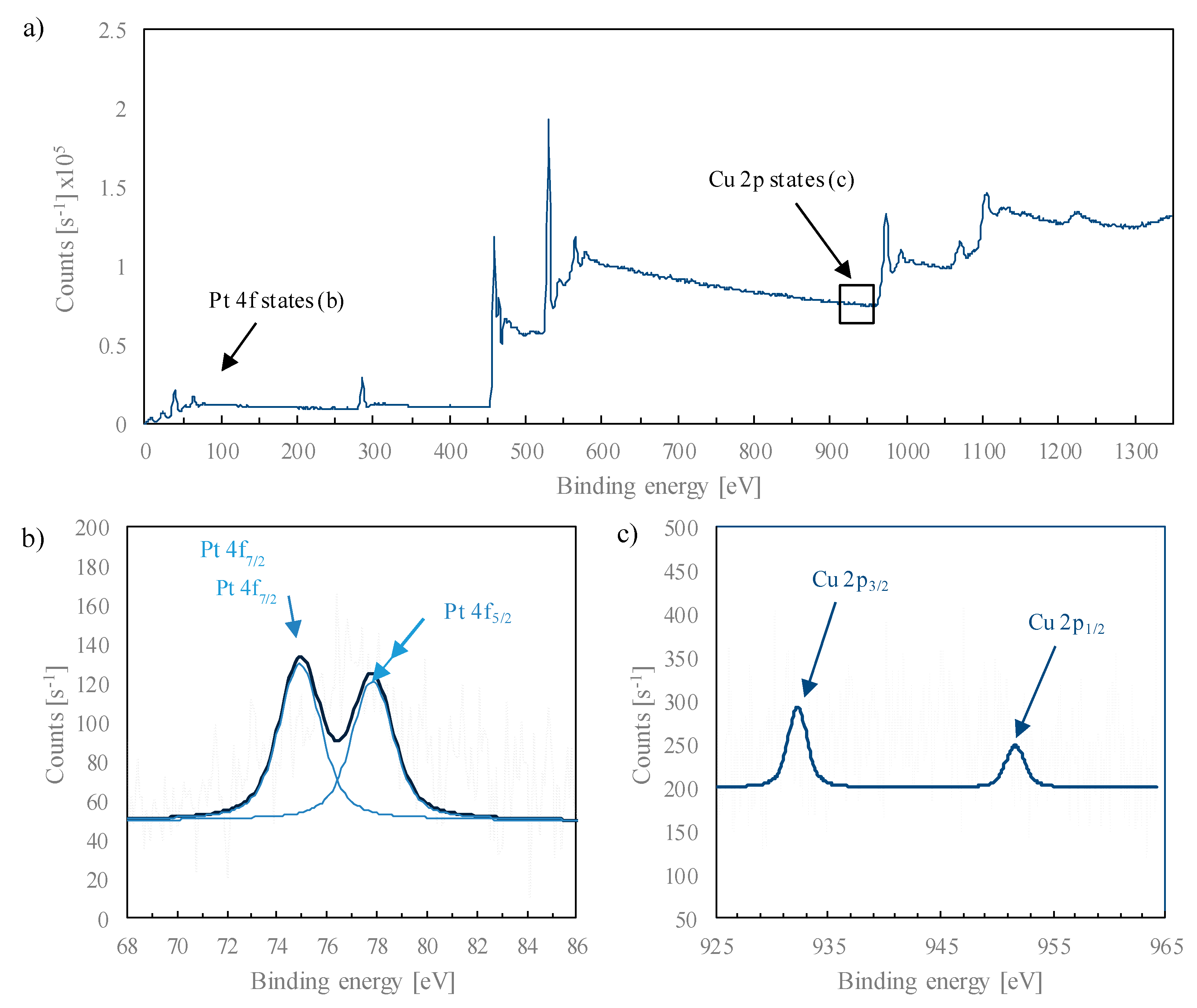
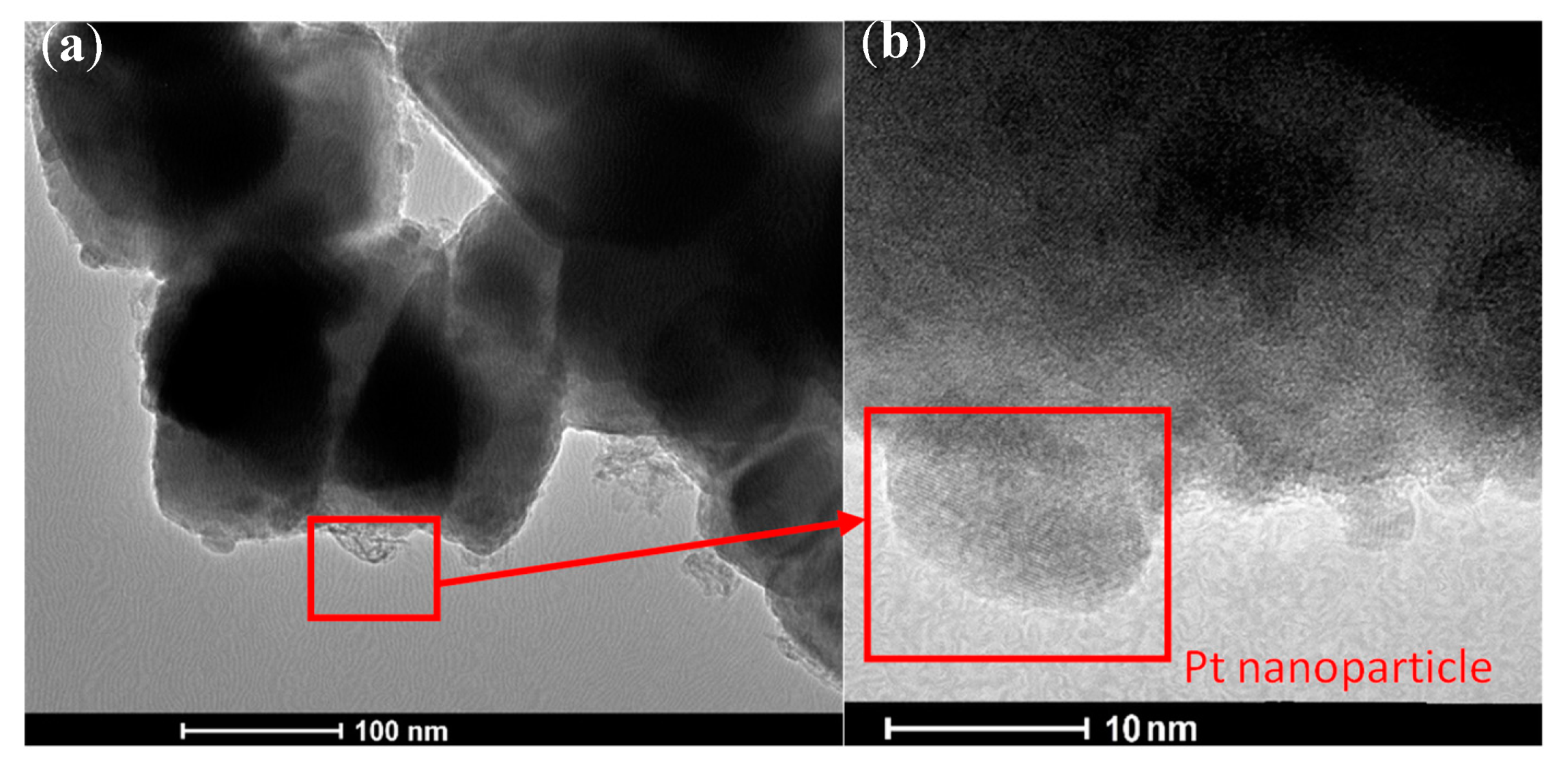
2.1. Physicochemical Characterization of d-TiO2-Pt/Cu and Magnetic Fe3O4@SiO2/d-TiO2-Pt/Cu Photocatalysts
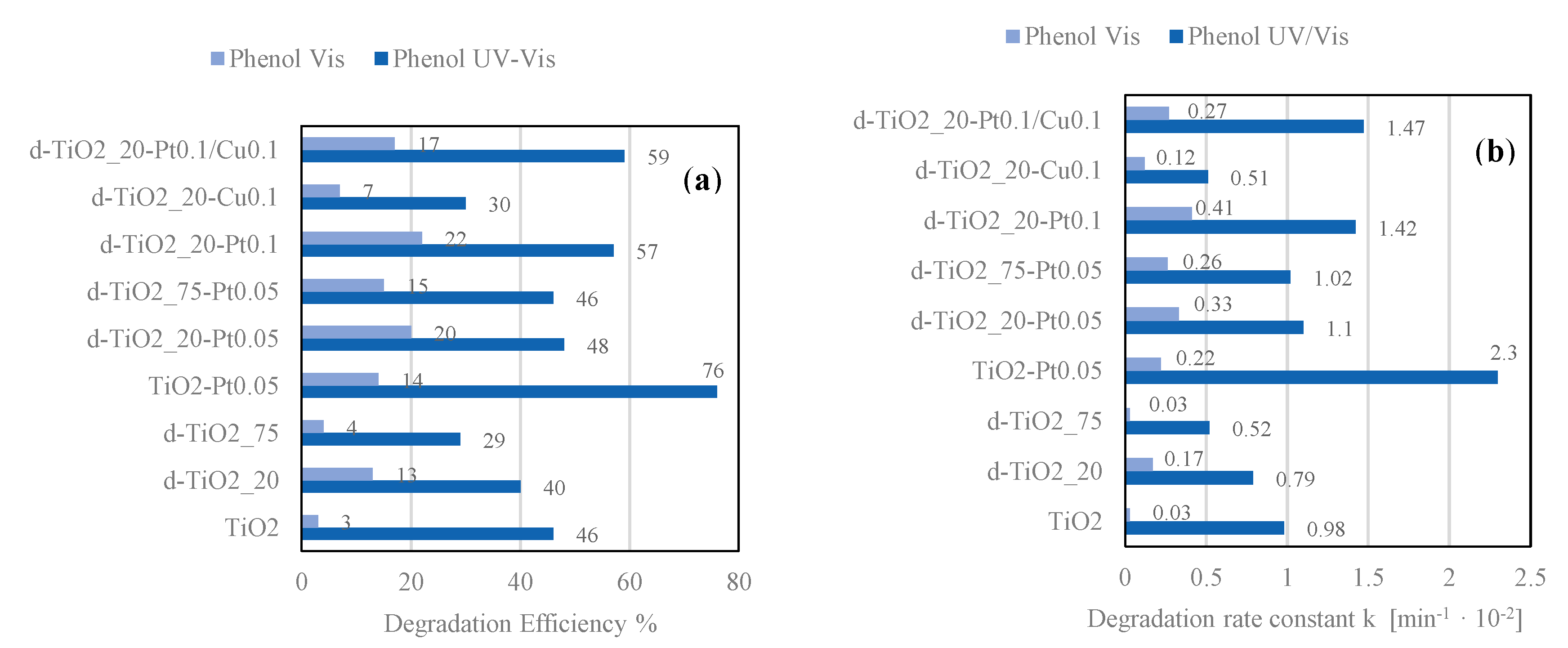
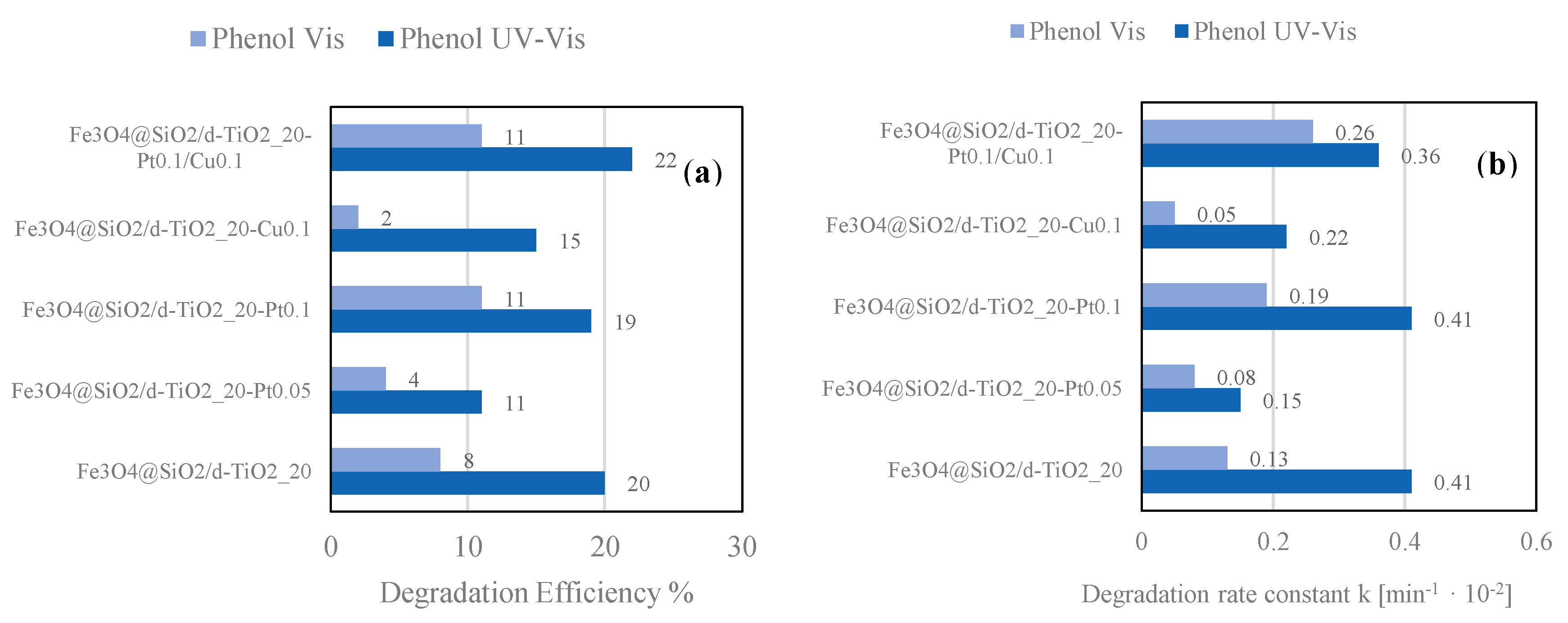
2.2. Photocatalytic Activity of d-TiO2-Pt/Cu and Fe3O4@SiO2/d-TiO2-Pt/Cu Photocatalysts
3. Materials and Methods
3.1. Preparation of Defective TiO2-Pt/Cu Photocatalysts
3.2. Preparation of Magnetic Fe3O4@SiO2/d-TiO2-Pt/Cu Nanocomposites
3.3. Characterization of the Obtained Magnetic Photocatalysts
3.4. Photocatalytic Activity Analysis
3.5. Electrochemical Measurements
4. Conclusions
Author Contributions
Funding
Acknowledgments
Conflicts of Interest
References
- Al-Mamun, M.R.; Kader, S.; Islam, M.S.; Khan, M.Z.H. Photocatalytic activity improvement and application of UV-TiO2 photocatalysis in textile wastewater treatment: A review. J. Environ. Chem. Eng. 2019, 7, 103248. [Google Scholar] [CrossRef]
- Zhu, D.; Zhou, Q. Action and mechanism of semiconductor photocatalysis on degradation of organic pollutants in water treatment: A review. Environ. Nanotechnol. Monit. Manag. 2019, 12, 100255. [Google Scholar] [CrossRef]
- Ahmad, K.; Ghatak, H.R.; Ahuja, S.M. Photocatalytic Technology: A review of environmental protection and renewable energy application for sustainable development. Environ. Technol. Innov. 2020, 19, 100893. [Google Scholar] [CrossRef]
- Wang, K.; Janczarek, M.; Wei, Z.; Raja-Mogan, T.; Endo-Kimura, M.; Khedr, T.M.; Ohtani, B.; Kowalska, E. Morphology- and Crystalline Composition-Governed Activity of Titania-Based Photocatalysts: Overview and Perspective. Catalysts 2019, 9, 1054. [Google Scholar] [CrossRef] [Green Version]
- Koe, W.S.; Lee, J.W.; Chong, W.C.; Pang, Y.L.; Sim, L.C. An overview of photocatalytic degradation: Photocatalysts, mechanisms, and development of photocatalytic membrane. Environ. Sci. Pollut. Res. 2020, 27, 2522–2565. [Google Scholar] [CrossRef] [PubMed]
- Loeb, S.K.; Alvarez, P.J.J.; Brame, J.A.; Cates, E.L.; Choi, W.; Crittenden, J.; Dionysiou, D.D.; Li, Q.; Li-Puma, G.; Quan, X.; et al. The Technology Horizon for Photocatalytic Water Treatment: Sunrise or Sunset? Environ. Sci. Technol. 2019, 53, 2937–2947. [Google Scholar] [CrossRef] [PubMed]
- Serpone, N. Is the Band Gap of Pristine TiO2 Narrowed by Anion- and Cation-Doping of Titanium Dioxide in Second-Generation Photocatalysts? J. Phys. Chem. B 2006, 110, 24287–24293. [Google Scholar] [CrossRef] [PubMed]
- Dozzi, M.V.; Selli, E. Doping TiO2 with p-block elements: Effects on photocatalytic activity. J. Photochem. Photobiol. C Photochem. Rev. 2013, 14, 13–28. [Google Scholar] [CrossRef]
- Diaz-Angulo, J.; Gomez-Bonilla, I.; Jimenez-Tohapanta, C.; Mueses, M.; Pinzon, M.; Machuca-Martinez, F. Visible-light activation of TiO2 by dye-sensitization for degradation of pharmaceutical compounds. Photochem. Photobiol. Sci. 2019, 18, 897–904. [Google Scholar] [CrossRef] [Green Version]
- Endo-Kimura, M.; Janczarek, M.; Bielan, Z.; Zhang, D.; Wang, K.; Markowska-Szczupak, A.; Kowalska, E. Photocatalytic and Antimicrobial Properties of Ag2O/TiO2 Heterojunction. ChemEngineering 2019, 3, 3. [Google Scholar] [CrossRef] [Green Version]
- Wysocka, I.; Kowalska, E.; Ryl, J.; Nowaczyk, G.; Zielińska-Jurek, A. Morphology, Photocatalytic and Antimicrobial Properties of TiO2 Modified with Mono- and Bimetallic Copper, Platinum and Silver Nanoparticles. Nanomaterials 2019, 9, 1129. [Google Scholar] [CrossRef] [Green Version]
- Klein, M.; Grabowska, E.; Zaleska, A. Noble metal modified TiO2 for photocatalytic air purification. Physicochem. Probl. Miner. Process. 2015, 51, 49–57. [Google Scholar]
- Janczarek, M.; Wei, Z.; Endo, M.; Ohtani, B.; Kowalska, E. Silver- and copper-modified decahedral anatase titania particles as visible light-responsive plasmonic photocatalyst. J. Photonics Energy 2016, 7, 12008. [Google Scholar] [CrossRef] [Green Version]
- Wei, Z.; Janczarek, M.; Endo, M.; Wang, K.; Balcytis, A.; Nitta, A.; Mendez-Medrano, M.G.; Colbeau-Justin, C.; Juodkazis, S.; Ohtani, B.; et al. Noble Metal-Modified Faceted Anatase Titania Photocatalysts: Octahedron versus Decahedron. Appl. Catal. B Environ. 2018, 237, 574–587. [Google Scholar] [CrossRef] [PubMed]
- Wu, Q.; Huang, F.; Zhao, M.; Xu, J.; Zhou, J.; Wang, Y. Ultra-small yellow defective TiO2 nanoparticles for co-catalyst free photocatalytic hydrogen production. Nano Energy 2016, 24, 63–71. [Google Scholar] [CrossRef]
- Liriano-Jorge, C.F.; Sohmen, U.; Özkan, A.; Gulyas, H.; Otterpohl, R. TiO2 Photocatalyst Nanoparticle Separation: Flocculation in Different Matrices and Use of Powdered Activated Carbon as a Precoat in Low-Cost Fabric Filtration. Adv. Mater. Sci. Eng. 2014, 2014, 1–12. [Google Scholar] [CrossRef] [Green Version]
- Lee, S.-A.; Choo, K.-H.; Lee, C.-H.; Lee, H.-I.; Hyeon, T.; Choi, W.; Kwon, H.-H. Use of Ultrafiltration Membranes for the Separation of TiO2 Photocatalysts in Drinking Water Treatment. Ind. Eng. Chem. Res. 2001, 40, 1712–1719. [Google Scholar] [CrossRef]
- Ray, S.; Lalman, J.A. Fabrication and characterization of an immobilized titanium dioxide (TiO2) nanofiber photocatalyst. Mater. Today Proc. 2016, 3, 1582–1591. [Google Scholar] [CrossRef]
- Zielińska-Jurek, A.; Klein, M.; Hupka, J. Enhanced visible light photocatalytic activity of Pt/I-TiO2in a slurry system and supported on glass packing. Sep. Purif. Technol. 2017, 189, 246–252. [Google Scholar] [CrossRef]
- Wei, J.H.; Leng, C.J.; Zhang, X.Z.; Li, W.H.; Liu, Z.Y.; Shi, J. Synthesis and magnetorheological effect of Fe3O4-TiO2 nanocomposite. J. Phys. Conf. Ser. 2009, 149, 25–29. [Google Scholar] [CrossRef] [Green Version]
- Zhang, L.; Wu, Z.; Chen, L.; Zhang, L.; Li, X.; Xu, H.; Wang, H.; Zhu, G. Preparation of magnetic Fe3O4/TiO2/Ag composite microspheres with enhanced photocatalytic activity. Solid State Sci. 2016, 52, 42–48. [Google Scholar] [CrossRef]
- Abbas, M.; Rao, B.P.; Reddy, V.; Kim, C. Fe3O4/TiO2 core/shell nanocubes: Single-batch surfactantless synthesis, characterization and efficient catalysts for methylene blue degradation. Ceram. Int. 2014, 40, 11177–11186. [Google Scholar] [CrossRef]
- Sathishkumar, P.; Viswanathan, R.V.; Anandan, S.; Ashokkumar, M. CoFe2O4/TiO2 nanocatalysts for the photocatalytic degradation of Reactive Red 120 in aqueous solutions in the presence and absence of electron acceptors. Chem. Eng. J. 2013, 220, 302–310. [Google Scholar] [CrossRef]
- Jia, Y.; Liu, J.; Cha, S.; Choi, S.; Chang, Y.C.; Liu, C. Magnetically separable Au-TiO2/nanocube ZnFe2O4 composite for chlortetracycline removal in wastewater under visible light. J. Ind. Eng. Chem. 2017, 47, 303–314. [Google Scholar] [CrossRef]
- Fu, W.; Yang, H.; Li, M.; Chang, L.; Yu, Q.; Xu, J.; Zou, G. Preparation and photocatalytic characteristics of core-shell structure TiO2/BaFe12O19 nanoparticles. Mater. Lett. 2006, 60, 2723–2727. [Google Scholar] [CrossRef]
- Zielińska-Jurek, A.; Bielan, Z.; Dudziak, S.; Wolak, I.; Sobczak, Z.; Klimczuk, T.; Nowaczyk, G.; Hupka, J. Design and Application of Magnetic Photocatalysts for Water Treatment. The Effect of Particle Charge on Surface Functionality. Catalysts 2017, 7, 360. [Google Scholar] [CrossRef] [Green Version]
- Zielińska-Jurek, A.; Bielan, Z.; Wysocka, I.; Strychalska, J.; Janczarek, M.; Klimczuk, T. Magnetic semiconductor photocatalysts for the degradation of recalcitrant chemicals from flow back water. J. Environ. Manage. 2017, 195, 157–165. [Google Scholar] [CrossRef]
- Wysocka, I.; Kowalska, E.; Trzciński, K.; Łapiński, M.; Nowaczyk, G.; Zielińska-Jurek, A. UV-Vis-Induced Degradation of Phenol over Magnetic Photocatalysts Modified with Pt, Pd, Cu and Au Nanoparticles. Nanomaterials 2018, 8, 28. [Google Scholar] [CrossRef] [Green Version]
- Mrotek, E.; Dudziak, S.; Malinowska, I.; Pelczarski, D.; Ryżyńska, Z.; Zielińska-Jurek, A. Improved degradation of etodolac in the presence of core-shell ZnFe2O4/SiO2/TiO2 magnetic photocatalyst. Sci. Total Environ. 2020, 724, 138167. [Google Scholar] [CrossRef]
- Gad-Allah, T.A.; Fujimura, K.; Kato, S.; Satokawa, S.; Kojima, T. Preparation and characterization of magnetically separable photocatalyst (TiO2/SiO2/Fe3O4): Effect of carbon coating and calcination temperature. J. Hazard. Mater. 2008, 154, 572–577. [Google Scholar] [CrossRef]
- Fan, Y.; Ma, C.; Li, W.; Yin, Y. Synthesis and properties of Fe3O4/SiO2/TiO2 nanocomposites by hydrothermal synthetic method. Mater. Sci. Semicond. Process. 2012, 15, 582–585. [Google Scholar] [CrossRef]
- Shi, F.; Li, Y.; Zhang, Q.; Wang, H. Synthesis of Fe3O4/C/TiO2 magnetic photocatalyst via vapor phase hydrolysis. Int. J. Photoenergy 2012, 2012, 1–8. [Google Scholar] [CrossRef] [Green Version]
- Zielińska-Jurek, A.; Wei, Z.; Janczarek, M.; Wysocka, I.; Kowalska, E. Size-Controlled Synthesis of Pt Particles on TiO2 Surface: Physicochemical Characteristic and Photocatalytic Activity. Catalysts 2019, 9, 940. [Google Scholar] [CrossRef] [Green Version]
- Bielan, Z.; Kowalska, E.; Dudziak, S.; Wang, K.; Ohtani, B.; Zielińska-Jurek, A. Mono- and bimetallic (Pt/Cu) titanium(IV) oxide core-shell photocatalysts with UV/Vis light activity and magnetic separability. Catal. Today 2020, in press. [Google Scholar] [CrossRef]
- Gamboa, J.A.; Pasquevich, D.M. Effect of Chlorine Atmosphere on the Anatase-Rutile Transformation. J. Am. Chem. Soc. 1992, 75, 2934–2938. [Google Scholar] [CrossRef]
- Byrne, C.; Fagan, R.; Hinder, S.; McCormack, D.E.; Pillai, S.C. New Approach of Modifying the Anatase to Rutile Transition Temperature in TiO2 Photocatalysts. RSC Adv. 2016, 6, 95232–95238. [Google Scholar] [CrossRef]
- Ricci, P.C.; Carbonaro, C.M.; Stagi, L.; Salis, M.; Casu, A.; Enzo, S.; Delogu, F. Anatase-To-Rutile Phase Transition In Nanoparticles Irradiated By Visible Light. J. Phys. Chem. C 2013, 117, 785–7857. [Google Scholar] [CrossRef]
- Liu, H.; Jia, Z.; Ji, S.; Zheng, Y.; Li, M.; Yang, H. Synthesis of TiO2/SiO2@Fe3O4 magnetic microspheres and their properties of photocatalytic degradation dyestuff. Catal. Today 2011, 175, 293–298. [Google Scholar] [CrossRef]
- Chi, Y.; Yuan, Q.; Li, Y.; Zhao, L.; Li, N.; Li, X.; Yan, W. Magnetically separable Fe3O4@SiO2@TiO2-Ag microspheres with well-designed nanostructure and enhanced photocatalytic activity. J. Hazard. Mater. 2013, 262, 404–411. [Google Scholar] [CrossRef] [PubMed]
- Zielińska-Jurek, A.; Hupka, J. Preparation and characterization of Pt/Pd-modified titanium dioxide nanoparticles for visible light irradiation. Catal. Today 2013, 230, 181–187. [Google Scholar] [CrossRef]
- Bielan, Z.; Kowalska, E.; Dudziak, S.; Wang, K.; Ohtani, B.; Zielinska-Jurek, A. Mono- and bimetallic (Pt/Cu) titanium(IV) oxide photocatalysts. Physicochemical and photocatalytic data for magnetic nanocomposites’ shell. Data Brief 2020, 31, 105814. [Google Scholar] [CrossRef]
- Chan, G.H.; Zhao, J.; Hicks, E.M.; Schatz, G.C.; Van Duyne, R.P. Plasmonic Properties of Copper Nanoparticles Fabricated by Nanosphere Lithography. Nano Lett. 2007, 7, 1947–1952. [Google Scholar] [CrossRef]
- Ghodselahi, T.; Vesaghi, M.A.; Shafiekhani, A.; Baghizadeh, A.; Lameii, M. XPS study of the Cu@Cu2O core-shell nanoparticles. Appl. Surf. Sci. 2008, 225, 2730–2734. [Google Scholar] [CrossRef]
- Li, B.; Luo, X.; Zhu, Y.; Wang, X. Immobilization of Cu(II) in KIT-6 Supported Co3O4 and Catalytic Performance for Epoxidation of Styrene. Appl. Surf. Sci. 2015, 359, 609–620. [Google Scholar] [CrossRef]
- Zielińska-Jurek, A.; Wei, Z.; Wysocka, I.; Szweda, P.; Kowalska, E. The effect of nanoparticles size on photocatalytic and antimicrobial properties of Ag-Pt/TiO2 photocatalysts. Appl. Surf. Sci. 2015, 353, 317–325. [Google Scholar] [CrossRef] [Green Version]
- Monga, A.; Rather, R.A.; Pal, B. Enhanced co-catalytic effect of Cu-Ag bimetallic core-shell nanocomposites imparted to TiO2 under visible light illumination. Sol. Energy Mater Sol. Cells 2017, 172, 285–292. [Google Scholar] [CrossRef]
- Devi, L.G.; Kavitha, R. A review on plasmonic metal—TiO2 composite for generation, trapping, storing and dynamic vectorial transfer of photogenerated electrons across the Schottky junction in a photocatalytic system. Appl. Surf. Sci. 2016, 360, 601–622. [Google Scholar] [CrossRef]
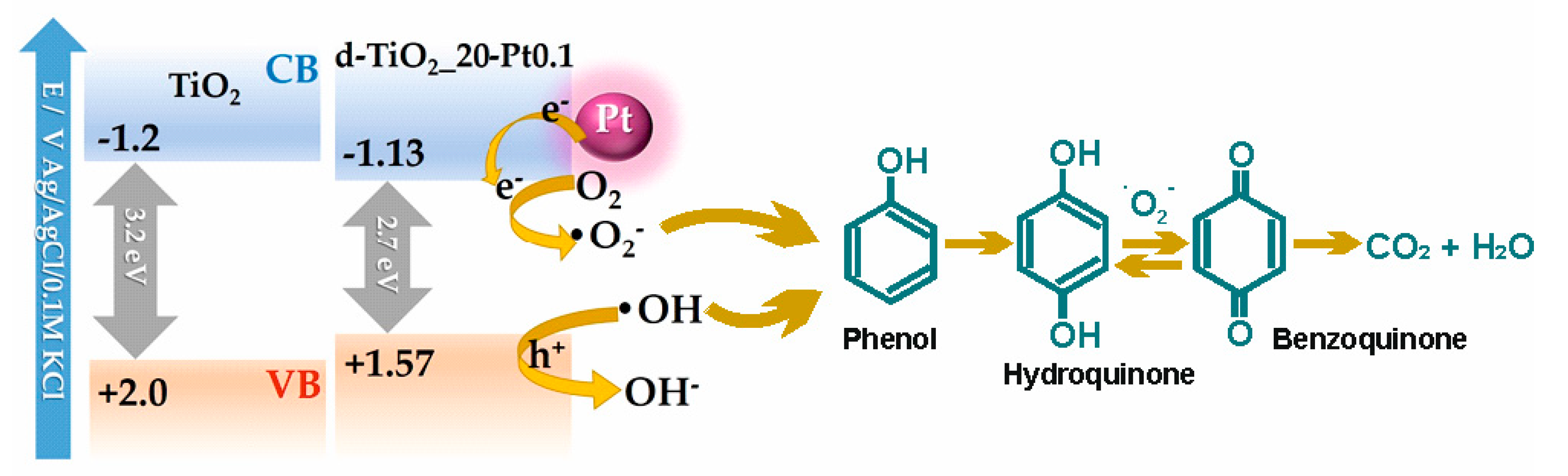
- Dang, T.T.T.; Le, S.T.T.; Channei, D.; Khanitchaidecha, W.; Nakaruk, A. Photodegradation mechanisms of phenol in the photocatalytic process. Res. Chem. Intermed. 2016, 42, 5961–5974. [Google Scholar] [CrossRef]
- Esplugas, S.; Gimenez, J.; Contreras, S.; Pascual, E.; Rodriguez, M. Comparison of different advanced oxidation processes for phenol degradation. Water Res. 2002, 36, 1034–1042. [Google Scholar] [CrossRef]
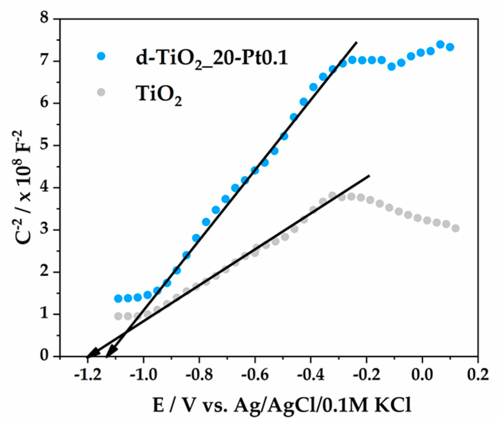
- Beranek, R. (Photo)electrochemical methods for the determination of the band edge positions of TiO2-based nanomaterials. Adv. Phys. Chem. 2011, 2011, 80–83. [Google Scholar] [CrossRef] [Green Version]


| Sample | BET (m2·g−1) | V Pores (cm3·g−1) | Eg (eV) | Rate Constant k (min−1)·10−2 UV-Vis | Phenol Removal (%) UV-Vis | Rate Constant k (min−1)·10−2 Vis | Phenol Removal (%) Vis |
|---|---|---|---|---|---|---|---|
| TiO2 | 169 | 0.0836 | 3.2 | 0.98 | 46 | 0.03 | 3 |
| d-TiO2_20 | 172 | 0.0847 | 2.7 | 0.79 | 40 | 0.17 | 13 |
| d-TiO2_75 | 167 | 0.0826 | 2.9 | 0.52 | 29 | 0.03 | 4 |
| TiO2-Pt0.05 | 166 | 0.0819 | 3.2 | 2.30 | 76 | 0.22 | 14 |
| d-TiO2_20-Pt0.05 | 148 | 0.0727 | 2.85 | 1.10 | 48 | 0.33 | 20 |
| d-TiO2_75-Pt0.05 | 164 | 0.0808 | 2.9 | 1.02 | 46 | 0.26 | 15 |
| d-TiO2_20-Pt0.1 | 152 | 0.0747 | 2.7 | 1.42 | 57 | 0.41 | 22 |
| d-TiO2_20-Cu0.1 | 101 | 0.0499 | 2.9 | 0.51 | 30 | 0.12 | 7 |
| d-TiO2_20-Pt0.1/Cu0.1 | 152 | 0.0744 | 2.75 | 1.47 | 59 | 0.27 | 17 |
| Sample | BET (m2·g−1) | V Pores (cm3·g−1) | Rate Constant k (min−1)·10−2 UV-Vis | Phenol Removal (%) UV-Vis | Rate Constant k (min−1)·10−2 Vis | Phenol Removal (%) Vis |
|---|---|---|---|---|---|---|
| Fe3O4@SiO2/d-TiO2_20 | 117 | 0.0578 | 0.41 | 20 | 0.13 | 8 |
| Fe3O4@SiO2/d-TiO2_20-Pt0.05 | 115 | 0.0568 | 0.15 | 11 | 0.08 | 4 |
| Fe3O4@SiO2/d-TiO2_20-Pt0.1 | 122 | 0.0602 | 0.41 | 19 | 0.19 | 11 |
| Fe3O4@SiO2/d-TiO2_20-Cu0.1 | 117 | 0.0579 | 0.22 | 15 | 0.05 | 2 |
| Fe3O4@SiO2/d-TiO2_20-Pt0.1/Cu0.1 | 117 | 0.0580 | 0.36 | 22 | 0.26 | 11 |
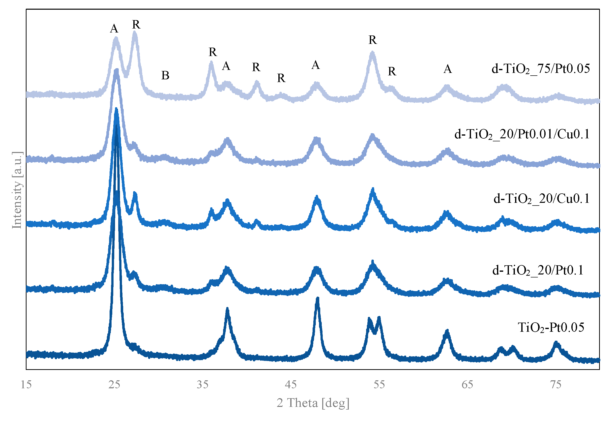
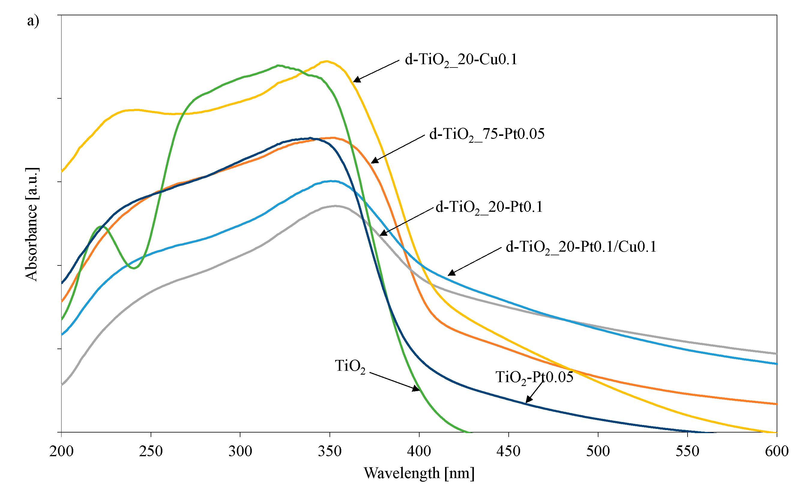
| Sample | Crystalline Size and Phase Content | |||||
|---|---|---|---|---|---|---|
| Anatase | Rutile | Brookite | ||||
| Size (nm) | Phase Content (wt.%) | Size (nm) | Phase Content (wt.%) | Size (nm) | Phase Content (wt.%) | |
| TiO2 | 5.97 ± 0.04 | 95.5 ± 1 | - | - | 6.1 ± 0.3 | 4.5 ± 1 |
| d-TiO2_20 | 5.14 ± 0.03 | 96 ± 1.0 | - | - | 4.0 ± 0.6 | 3.5 ± 0.5 |
| d-TiO2_75 | 5.67 ± 0.05 | 21 ± 3.5 | 6.57 ± 0.09 | 80 ± 2 | - | - |
| TiO2-Pt0.05 | 5.71 ± 0.06 | 91 ± 0.5 | - | - | 5.7 ± 0.6 | 9 ± 1 |
| d-TiO2_20-Pt0.05 | 5.66 ± 0.03 | 85 ± 8 | 9.8 ± 0.7 | 9 ± 1.0 | 4.9 ± 0.3 | 6 ± 0.5 |
| d-TiO2_75-Pt0.05 | 5.49 ± 0.04 | 48 ± 2 | 7.53 ± 0.12 | 52 ± 2 | - | - |
| d-TiO2_20-Pt0.1 | 5.58 ± 0.03 | 81 ± 8 | 7.6 ± 0.8 | 6 ± 1 | 4.8 ± 0.3 | 13 ± 1 |
| d-TiO2_20-Cu0.1 | 6.62 ± 0.04 | 72 ± 11 | 11.7 ± 0.4 | 9 ± 2 | 1.52 ± 0.08 | 8.5 ± 1 |
| d-TiO2_20-Pt0.1/Cu0.1 | 5.52 ± 0.03 | 83 ± 12 | 9.5 ± 0.6 | 8 ± 2 | 5.1 ± 0.3 | 8.5 ± 1 |
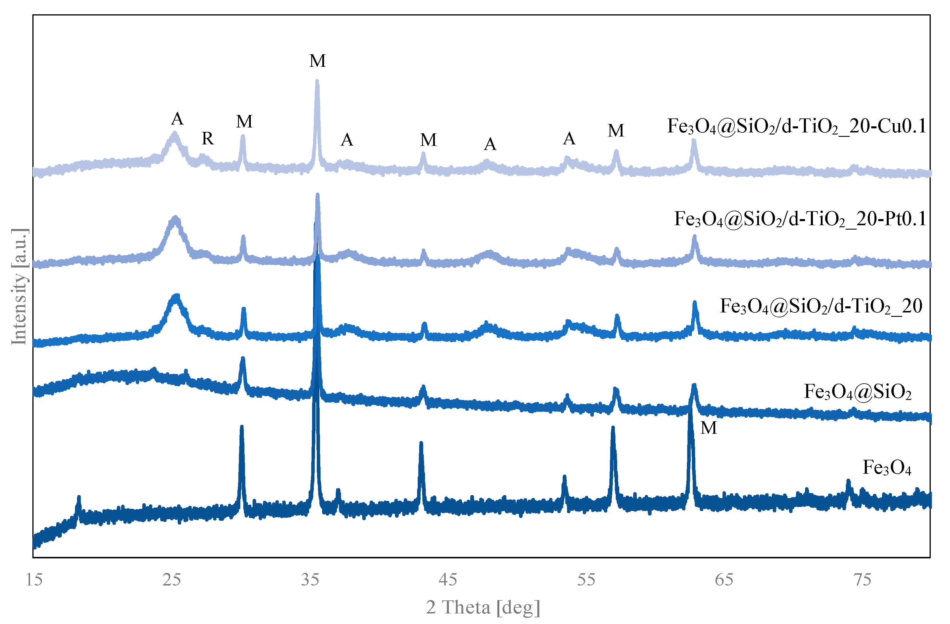
| Sample | Crystalline Size and Phase Content | |||||
|---|---|---|---|---|---|---|
| Anatase | Rutile | Magnetite | ||||
| Size (nm) | Phase Content (wt.%) | Size (nm) | Phase Content (wt.%) | Size (nm) | Phase Content (wt.%) | |
| Fe3O4@SiO2/d-TiO2_20 | 5.19 ± 0.05 | 71 ± 1.5 | 8.6 ± 0.5 | 8 ± 0.5 | 46.1 ± 1.1 | 21 ± 0.5 |
| Fe3O4@SiO2/d-TiO2_20-Pt0.05 | 5.60 ± 0.05 | 71 ± 1.5 | 8.9 ± 0.5 | 7 ± 0.5 | 45.7 ± 1.4 | 21 ± 0.5 |
| Fe3O4@SiO2/d-TiO2_20-Pt0.1 | 5.49 ± 0.05 | 68 ± 2 | 9.1 ± 0.6 | 7 ± 1 | 47.2 ± 4.0 | 24 ± 0.5 |
| Fe3O4@SiO2/d-TiO2_20-Cu0.1 | 7.81 ± 0.17 | 57 ± 2 | 13.3 ± 1.4 | 5 ± 1 | 37.1 ± 1.8 | 28 ± 1.5 |
| Fe3O4@SiO2/d-TiO2_20-Pt0.1/Cu0.1 | 5.48 ± 0.05 | 69 ± 1 | 7.9 ± 0.4 | 8 ± 0.5 | 42.6 ± 3.3 | 22 ± 1 |
| Sample | Oxidant Concentration [mol%] | Mass of Added Oxidant [g] |
|---|---|---|
| TBT | 0 | 0 |
| d-TiO2_20 | 20 | 1.032 |
| d-TiO2_75 | 75 | 3.869 |
© 2020 by the authors. Licensee MDPI, Basel, Switzerland. This article is an open access article distributed under the terms and conditions of the Creative Commons Attribution (CC BY) license (http://creativecommons.org/licenses/by/4.0/).
Share and Cite
Bielan, Z.; Sulowska, A.; Dudziak, S.; Siuzdak, K.; Ryl, J.; Zielińska-Jurek, A. Defective TiO2 Core-Shell Magnetic Photocatalyst Modified with Plasmonic Nanoparticles for Visible Light-Induced Photocatalytic Activity. Catalysts 2020, 10, 672. https://0-doi-org.brum.beds.ac.uk/10.3390/catal10060672
Bielan Z, Sulowska A, Dudziak S, Siuzdak K, Ryl J, Zielińska-Jurek A. Defective TiO2 Core-Shell Magnetic Photocatalyst Modified with Plasmonic Nanoparticles for Visible Light-Induced Photocatalytic Activity. Catalysts. 2020; 10(6):672. https://0-doi-org.brum.beds.ac.uk/10.3390/catal10060672
Chicago/Turabian StyleBielan, Zuzanna, Agnieszka Sulowska, Szymon Dudziak, Katarzyna Siuzdak, Jacek Ryl, and Anna Zielińska-Jurek. 2020. "Defective TiO2 Core-Shell Magnetic Photocatalyst Modified with Plasmonic Nanoparticles for Visible Light-Induced Photocatalytic Activity" Catalysts 10, no. 6: 672. https://0-doi-org.brum.beds.ac.uk/10.3390/catal10060672