
Biological Synthesis of Nanocatalysts and Their Applications

 , , ,
, , ,  , , ,
, , ,  ,
,  ,
,  and
and
Abstract
:1. Introduction
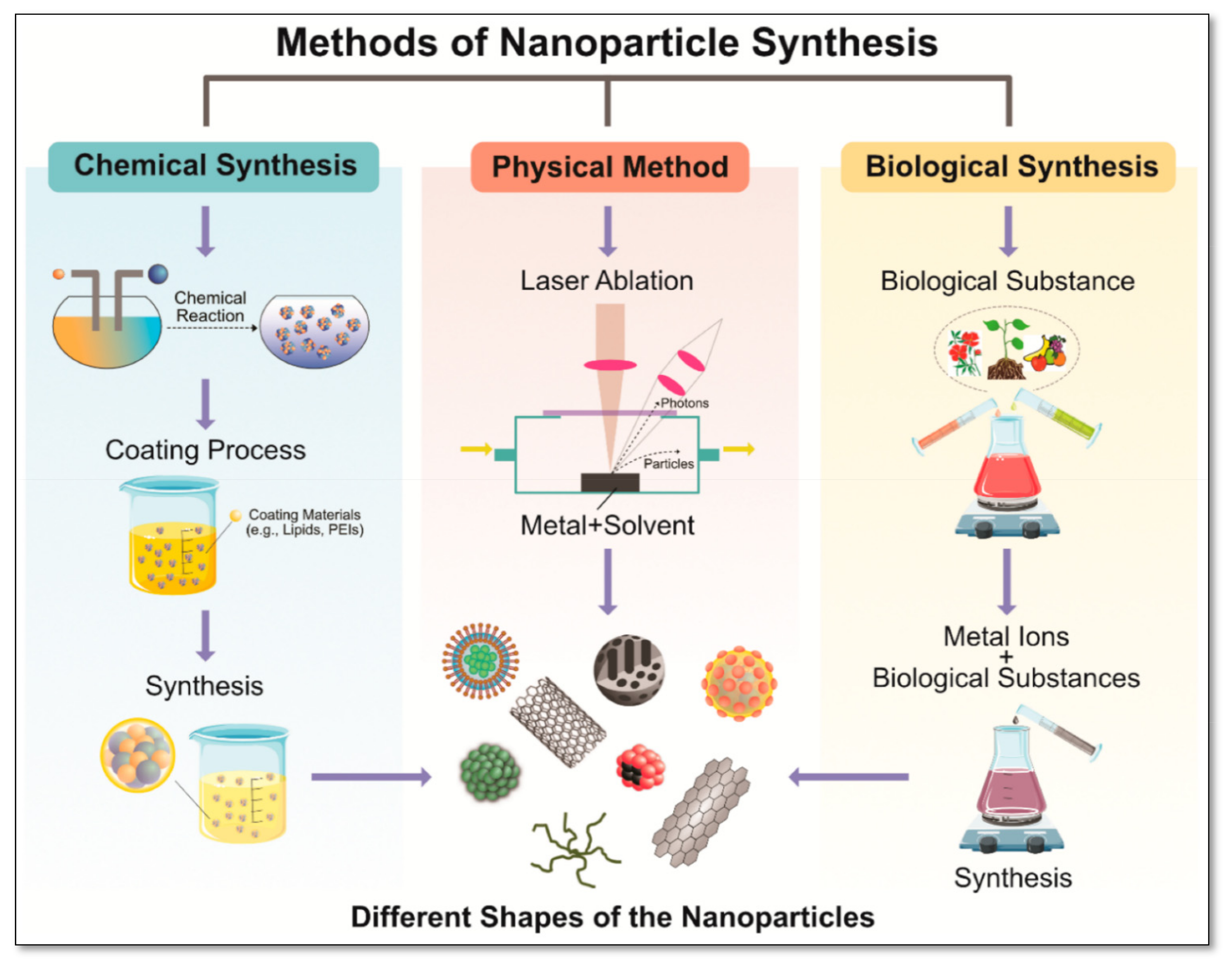
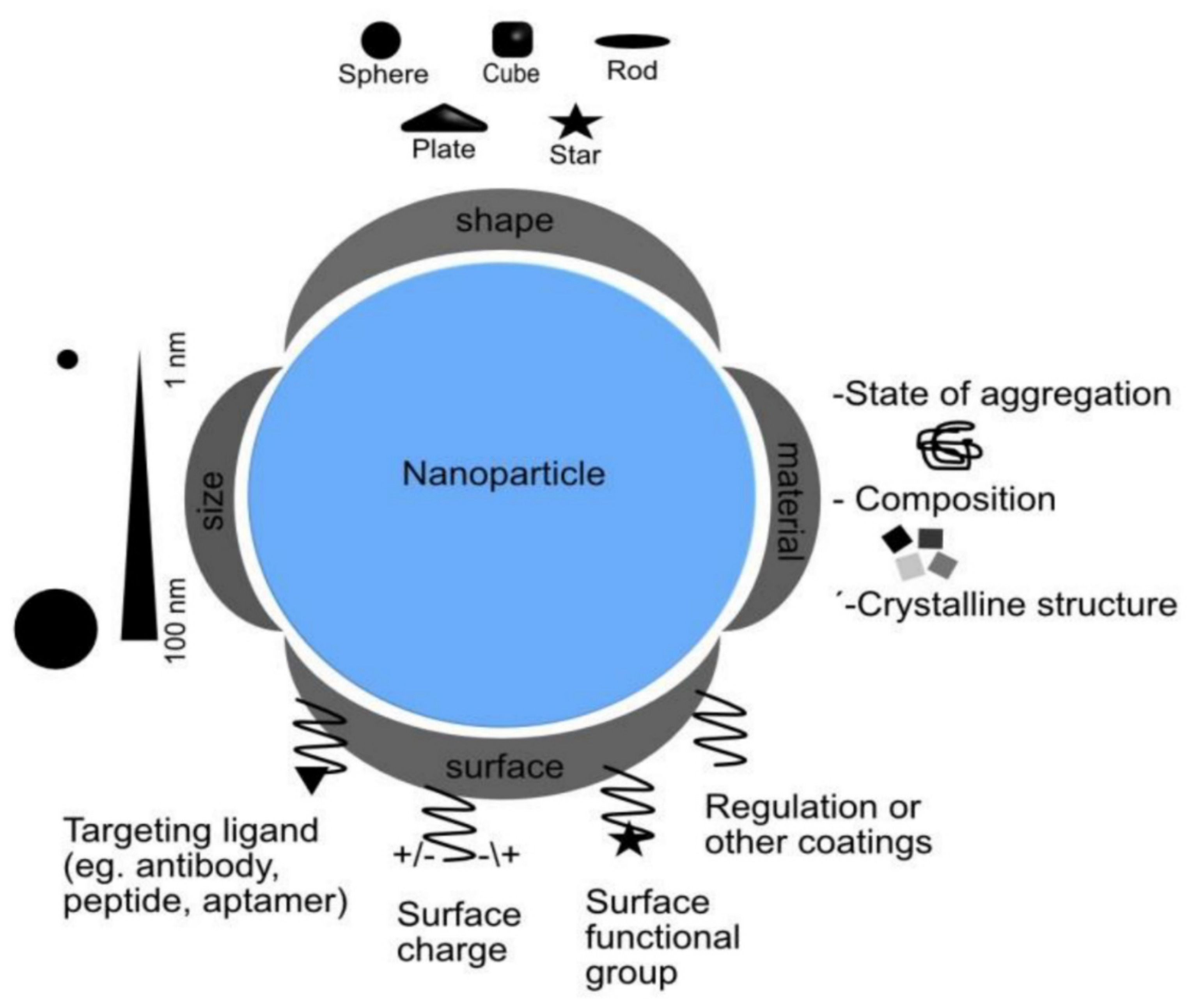
2. Different Methods of Nanocatalyst Synthesis
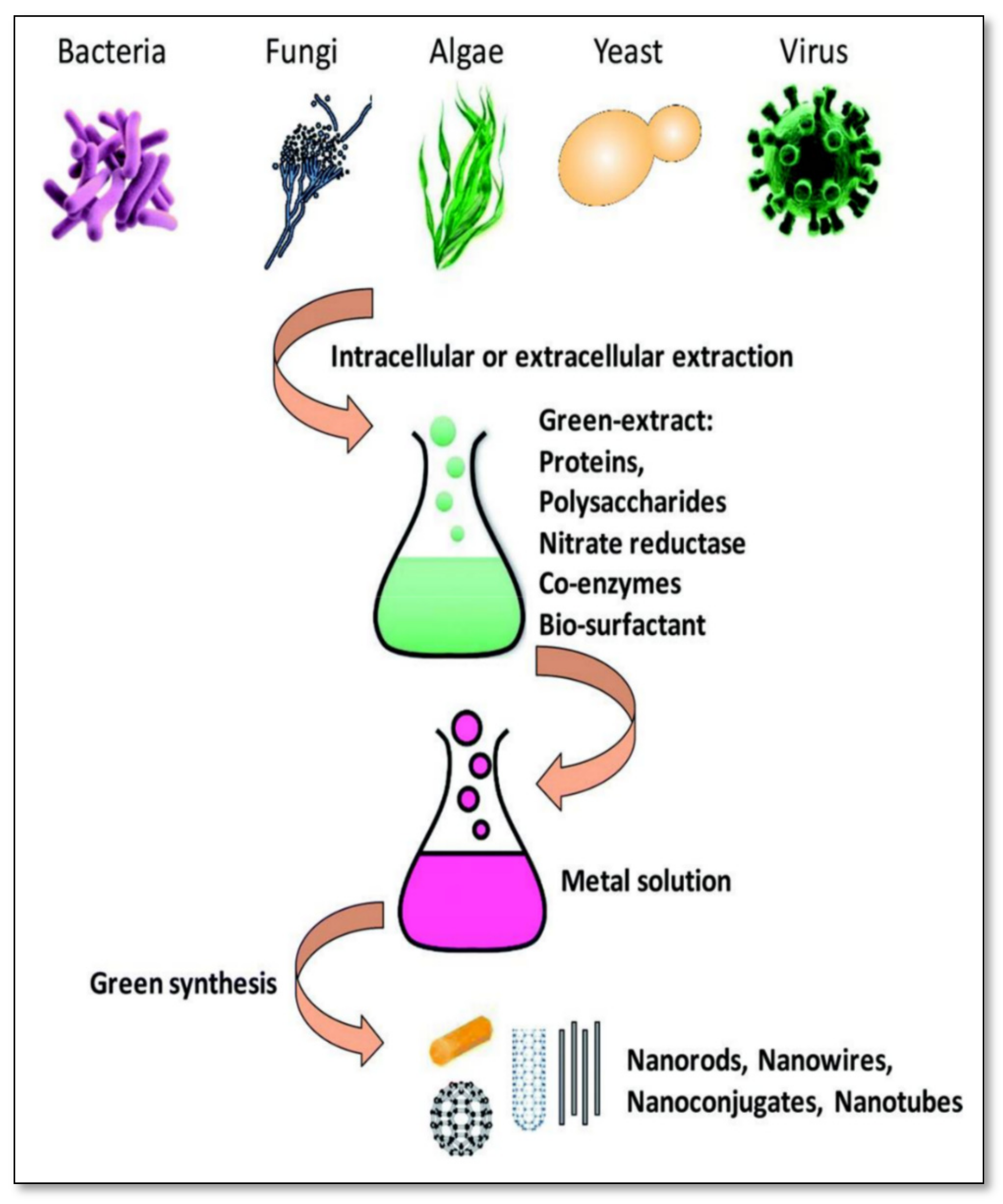
3. Biological Approach for Nanocatalyst/Nanoparticle Synthesis


3.1. Bacterial Synthesis
3.2. Fungal Synthesis
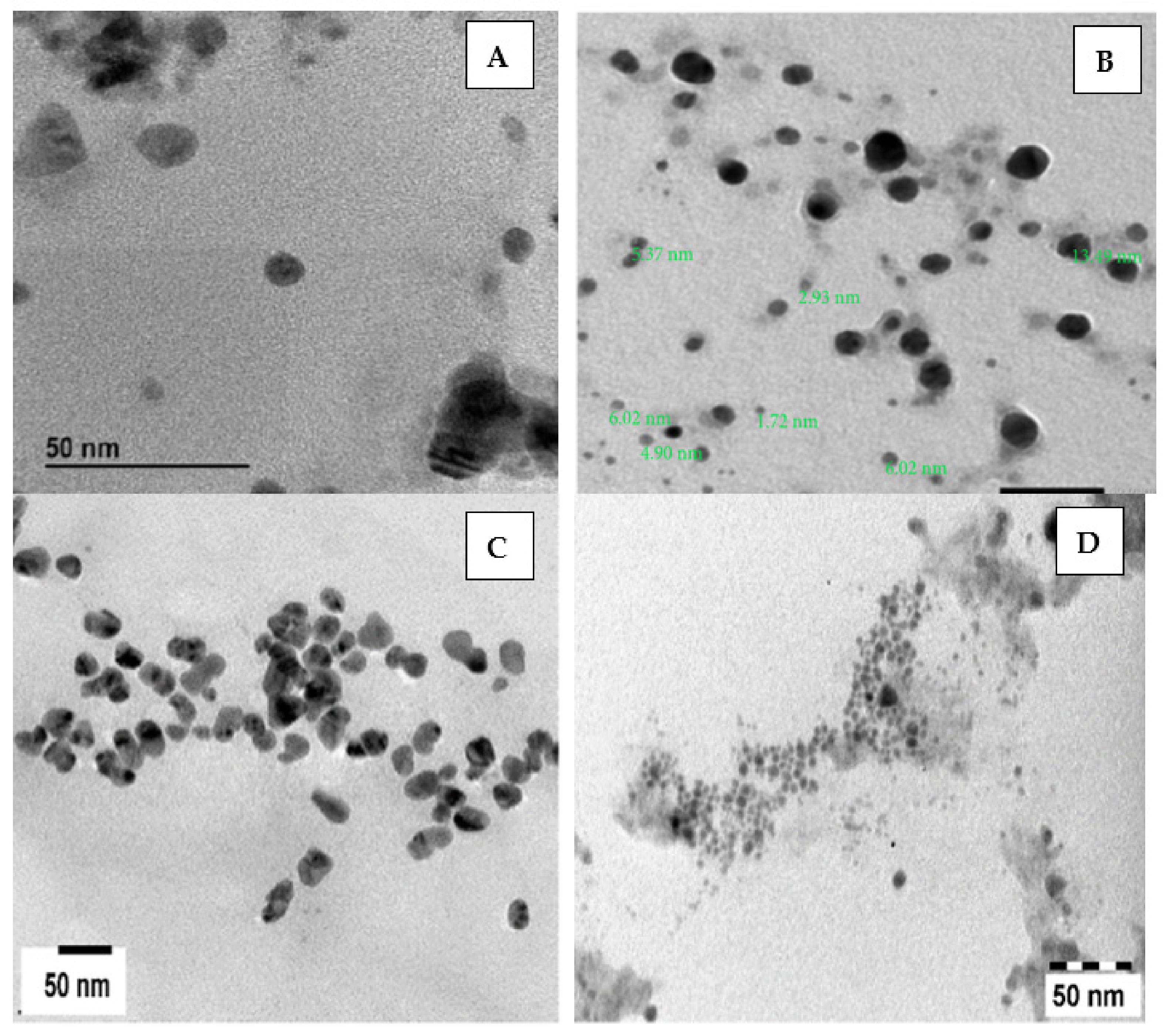
3.3. Algal Synthesis

| Source | Metal Involved | Size | Shape | References |
|---|---|---|---|---|
| Sargassum spp. | G | - | Hexagonal, triangular | [121] |
| Sargassum wightii | G | 8–12 | Thin planar structures | [109] |
| Sargassum Wightii | S | 8–27 | Spherical | [122] |
| Gelidiella acerosa | S | 22 | Spherical | [123] |
| Stoechospermum marginatum | G | 18.7–93.7 | Spherical | [124] |
| Ulva fasciata | S | 28–41 | Spherical | [124] |
| Sargassum myriocystum | G | 10–23 | Triangular and spherical | [125] |
| Ulva reticulata | S | 40–50 | Spherical | [126] |
| Chaetomorpha linum | S | 3–44 | - | [127] |
| Gracilaria corticate | G | 45–57 | - | [128] |
| Bifurcaria bifurcate | CO | 5–45 | Spherical | [129] |
| Enteromorpha flexuosa | S | 2–32 | Circular | [130] |
| Prasiola crispa | G | 5–25 | Cubic | [131] |
| Sargassum Alga | P | 5–10 | Octahedral | [132] |
| Caulerpa racemose | S | 5–25 | Spherical | [133] |
| Acanthophora specifera | S | 33–81 | Cubic | [134] |
| Isochrysis sp. | S | 98.1–193 | Spherical | [135] |
| Laurencia papillosa | S | - | Cubic | [136] |
| Spirulina platensis | S | 5–50 | Spherical | [137] |
| Caulerpa serrulate | S | 10 | Spherical | [138] |
| Botryococcus braunii | S | 40–100 | Cubical, spherical, and truncated triangular | [139] |
| Padina pavonia | S | 49.58–86.37 | Spherical, triangular, rectangle, polyhedral, and hexagonal | [140] |
| Gelidium amansii | S | 27–54 | Spherical | [141] |
| Padina sp. | S | 25–60 | Spherical | [142] |
| Gelidium corneum | S | 20–50 | Spherical | [143] |

4. Applications of Nanomaterials/Nanocatalysts
4.1. Nanocatalysts in Biological Applications
4.2. Nanocatalysts in Dye Degradation
| Dye | Nanoparticle Used | Synthesis of Nanoparticles Source | % Degradation | References |
|---|---|---|---|---|
| Malachite green | Silver | Bacillus paralicheniformis | 90 | [163] |
| Reactive black 5 | Palladium | Pseudomonas putida | 100 | [164] |
| Methyl orange | Palladium | Clostrodium sp. | 90 | [165] |
| Amaranth | Iron | Shewanella decolorotionis | 90.5 | [166] |
| Methyl orange | Tin(iv) oxide | Erwinia herbicola | 94 | [167] |
| Congo red | Silver | Pleurotus sajor caju | 78 | [168] |
| Malachite green | Silver | Acremonium kiliense | 95.4 | [169] |
| Methylene Blue | Silver | Saccharomyces cerevisiae | 80 | [170] |
| Congo red | Silver | Pestalotiopsis versicolor | 91.56 | [171] |
| Direct blue 71 | Palladium | Saccharomyces cerevisiae | 98 | [172] |
| Naphthol Green B | Iron–sulfur | Pseudoalteromonas sp. CF10-13 | 19.46 | [173] |
| Bismarck brown | Zinc-oxide | Aspergillus niger | 89 | [174] |
| Methyl orange | Platinum | Fusarium oxysporum | - | [175] |
| Acid Brilliant Scarlet GR | Gold | Trichoderma sp. | 94.7 | [176] |
| Rhodamine B | Gold | Cladosporium oxysporum AJP03 | - | [177] |
| Malachite green | Copper | Escherichia sp. SINT7 | 90.55 | [178] |
| Rhodamine B | Gold | Turbinaria conoides | - | [179] |
| Malachite green | Silver | Gracilaria corticata | - | [180] |
| Methylene blue, rhodamine B, and methyl orange | Gold and silver | Sargassum serratifolium | - | [181] |

4.3. Nanocatalysts in Heavy Metal Remediation
5. Conclusions
Author Contributions
Funding
Institutional Review Board Statement
Informed Consent Statement
Data Availability Statement
Acknowledgments
Conflicts of Interest
References
- Roy, A.; Bharadvaja, N. Silver nanoparticle synthesis from Plumbago zeylanica and its dye degradation activity. Bioinspired Biomim. Nanobiomaterials 2019, 8, 130–140. [Google Scholar] [CrossRef]
- Ahmed, H.M.; Roy, A.; Wahab, M.; Ahmed, M.; Othman-Qadir, G.; Elesawy, B.H.; Khandaker, M.U.; Islam, M.N.; Emran, T.B. Applications of Nanomaterials in Agrifood and Pharmaceutical Industry. J. Nanomater. 2021, 2021, 1472096. [Google Scholar] [CrossRef]
- Roy, A. Plant Derived Silver Nanoparticles and their Therapeutic Applications. Curr. Pharm. Biotechnol. 2021, 22, 1834–1847. [Google Scholar] [CrossRef] [PubMed]
- Thakkar, K.N.; Mhatre, S.S.; Parikh, R.Y. Biological synthesis of metallic nanoparticles. Nanomed. Nanotechnol. Biol. Med. 2010, 6, 257–262. [Google Scholar] [CrossRef]
- Jeevanandam, J.; Barhoum, A.; Chan, Y.S.; Dufresne, A.; Danquah, M.K. Review on nanoparticles and nanostructured materials: History, sources, toxicity and regulations. Beilstein J. Nanotechnol. 2018, 9, 1050–1074. [Google Scholar] [CrossRef] [Green Version]
- Remya, V.R.; Abitha, V.K.; Rajput, P.S.; Rane, A.V.; Dutta, A. Silver nanoparticles green synthesis: A mini review. Chem. Int. 2017, 3, 165–171. [Google Scholar]
- Iravani, S.; Korbekandi, H.; Mirmohammadi, S.V.; Zolfaghari, B. Synthesis of silver nanoparticles: Chemical, physical and biological methods. Res. Pharm. Sci. 2014, 9, 385. [Google Scholar]
- Raina, S.; Roy, A.; Bharadvaja, N. Degradation of dyes using biologically synthesized silver and copper nanoparticles. Environ. Nanotechnol. Monit. Manag. 2020, 13, 100278. [Google Scholar] [CrossRef]
- Nagore, P.; Ghotekar, S.; Mane, K.; Ghoti, A.; Bilal, M.; Roy, A. Structural Properties and Antimicrobial Activities of Polyalthia longifolia Leaf Extract-Mediated CuO Nanoparticles. BioNanoScience 2021, 11, 579–589. [Google Scholar] [CrossRef]
- Shah, M.; Fawcett, D.; Sharma, S.; Tripathy, S.K.; Poinern, G.E.J. Green synthesis of metallic nanoparticles via biological entities. Materials 2015, 8, 7278–7308. [Google Scholar] [CrossRef] [PubMed] [Green Version]
- Loomba, L.; Scarabelli, T. Metallic nanoparticles and their medicinal potential. Part II: Aluminosilicates, nanobiomagnets, quantum dots and cochleates. Ther. Deliv. 2013, 4, 1179–1196. [Google Scholar] [CrossRef] [PubMed]
- Tajzad, I.; Ghasali, E. Production methods of CNT-reinforced Al matrix composites: A review. J. Compos. Compd. 2020, 2, 1–9. [Google Scholar]
- Puente Santiago, A.R.; Fernandez-Delgado, O.; Gomez, A.; Ahsan, M.A.; Echegoyen, L. Fullerenes as Key Components for Low-Dimensional (Photo) Electrocatalytic Nanohybrid Materials. Angew. Chem. 2021, 133, 124–143. [Google Scholar] [CrossRef]
- C Thomas, S.; Kumar Mishra, P.; Talegaonkar, S. Ceramic nanoparticles: Fabrication methods and applications in drug delivery. Curr. Pharm. Des. 2015, 21, 6165–6188. [Google Scholar] [CrossRef] [PubMed]
- Chaudhary, R.G.; Bhusari, G.S.; Tiple, A.D.; Rai, A.R.; Somkuvar, S.R.; Potbhare, A.K.; Abdala, A.A. Metal/metal oxide nanoparticles: Toxicity, applications, and future prospects. Curr. Pharm. Des. 2019, 25, 4013–4029. [Google Scholar] [CrossRef] [PubMed]
- Suresh, S. Semiconductor nanomaterials, methods and applications: A review. Nanosci. Nanotechnol. 2013, 3, 62–74. [Google Scholar]
- Crucho, C.I.; Barros, M.T. Polymeric nanoparticles: A study on the preparation variables and characterization methods. Mater. Sci. Eng. C 2017, 80, 771–784. [Google Scholar] [CrossRef]
- Khan, I.; Saeed, K.; Khan, I. Nanoparticles: Properties, applications and toxicities. Arab. J. Chem. 2019, 12, 908–931. [Google Scholar] [CrossRef]
- Roy, A.; Bharadvaja, N. Qualitative analysis of phytocompounds and synthesis of silver nanoparticles from Centella asiatica. Innov. Tech. Agric. 2017, 1, 88–95. [Google Scholar]
- Nath, D.; Banerjee, P. Green nanotechnology—A new hope for medical biology. Environ. Toxicol. Pharmacol. 2013, 36, 997–1014. [Google Scholar] [CrossRef]
- Jeyaraj, M.; Gurunathan, S.; Qasim, M.; Kang, M.H.; Kim, J.H. A comprehensive review on the synthesis, characterization, and biomedical application of platinum nanoparticles. Nanomaterials 2019, 9, 1719. [Google Scholar] [CrossRef] [PubMed] [Green Version]
- Roy, A.; Bharadvaja, N. Silver nanoparticles synthesis from a pharmaceutically important medicinal plant Plumbago zeylanica. MOJ Bioequivalence Bioavailab. 2017, 3, 00046. [Google Scholar]
- Liang, X.J.; Kumar, A.; Shi, D.; Cui, D. Nanostructures for medicine and pharmaceuticals. J. Nanomater. 2012, 2012, 921897. [Google Scholar] [CrossRef]
- Auría-Soro, C.; Nesma, T.; Juanes-Velasco, P.; Landeira-Viñuela, A.; Fidalgo-Gomez, H.; Acebes-Fernandez, V.; Gongora, R.; Almendral Parra, M.J.; Manzano-Roman, R.; Fuentes, M. Interactions of nanoparticles and biosystems: Microenvironment of nanoparticles and biomolecules in nanomedicine. Nanomaterials 2019, 9, 1365. [Google Scholar] [CrossRef] [PubMed] [Green Version]
- Guilger-Casagrande, M.; de Lima, R. Synthesis of Silver Nanoparticles Mediated by Fungi: A Review. Front. Bioeng. Biotechnol. 2019, 7, 287. [Google Scholar] [CrossRef] [PubMed] [Green Version]
- Huynh, K.H.; Pham, X.H.; Kim, J.; Lee, S.H.; Chang, H.; Rho, W.Y.; Jun, B.H. Synthesis, properties, and biological applications of metallic alloy nanoparticles. Int. J. Mol. Sci. 2020, 21, 5174. [Google Scholar] [CrossRef] [PubMed]
- Nair, B.; Pradeep, T. Coalescence of nanoclusters and formation of submicron crystallites assisted by Lactobacillus strains. Cryst. Growth Des. 2002, 2, 293–298. [Google Scholar] [CrossRef]
- Ahmad, A.; Senapati, S.; Khan, M.I.; Kumar, R.; Sastry, M. Extracellular biosynthesis of monodisperse gold nanoparticles by a novel extremophilic actinomycete, Thermomonospora sp. Langmuir 2003, 19, 3550–3553. [Google Scholar] [CrossRef]
- Konishi, Y.; Ohno, K.; Saitoh, N.; Nomura, T.; Nagamine, S. Microbial synthesis of gold nanoparticles by metal reducing bacterium. Trans. Mater. Res. Soc. Jpn. 2004, 29, 2341–2343. [Google Scholar]
- He, S.; Guo, Z.; Zhang, Y.; Zhang, S.; Wang, J.; Gu, N. Biosynthesis of gold nanoparticles using the bacteria Rhodopseudomonas capsulata. Mater. Lett. 2007, 61, 3984–3987. [Google Scholar] [CrossRef]
- Parikh, R.Y.; Singh, S.; Prasad, B.L.V.; Patole, M.S.; Sastry, M.; Shouche, Y.S. Extracellular synthesis of crystalline silver nanoparticles and molecular evidence of silver resistance from Morganella sp.: Towards understanding biochemical synthesis mechanism. ChemBioChem 2008, 9, 1415–1422. [Google Scholar] [CrossRef] [PubMed]
- Reddy, A.S.; Chen, C.Y.; Chen, C.C.; Jean, J.S.; Chen, H.R.; Tseng, M.J.; Fan, C.-W.; Wang, J.C. Biological synthesis of gold and silver nanoparticles mediated by the bacteria Bacillus subtilis. J. Nanosci. Nanotechnol. 2010, 10, 6567–6574. [Google Scholar] [CrossRef] [PubMed]
- Bruna, N.; Collao, B.; Tello, A.; Caravantes, P.; Díaz-Silva, N.; Monrás, J.P.; Órdenes-Aenishanslins, N.; Flores, M.; Espinoza-Gonzalez, R.; Bravo, D.; et al. Synthesis of salt-stable fluorescent nanoparticles (quantum dots) by polyextremophile halophilic bacteria. Sci. Rep. 2019, 9, 1953. [Google Scholar] [CrossRef] [PubMed]
- Bharde, A.; Wani, A.; Shouche, Y.; Joy, P.A.; Prasad, B.L.; Sastry, M. Bacterial aerobic synthesis of nanocrystalline magnetite. J. Am. Chem. Soc. 2005, 127, 9326–9327. [Google Scholar] [CrossRef]
- Gericke, M.; Pinches, A. Microbial production of gold nanoparticles. Gold Bull. 2006, 39, 22–28. [Google Scholar] [CrossRef] [Green Version]
- Baesman, S.M.; Bullen, T.D.; Dewald, J.; Zhang, D.; Curran, S.; Islam, F.S.; Beveridge, T.J.; Oremland, R.S. Formation of tellurium nanocrystals during anaerobic growth of bacteria that use Te oxyanions as respiratory electron acceptors. Appl. Environ. Microbiol. 2007, 73, 2135–2143. [Google Scholar] [CrossRef] [Green Version]
- Du, L.; Jiang, H.; Liu, X.; Wang, E. Biosynthesis of gold nanoparticles assisted by Escherichia coli DH5α and its application on direct electrochemistry of hemoglobin. Electrochem. Commun. 2007, 9, 1165–1170. [Google Scholar] [CrossRef]
- Shahverdi, A.R.; Fakhimi, A.; Shahverdi, H.R.; Minaian, S. Synthesis and effect of silver nanoparticles on the antibacterial activity of different antibiotics against Staphylococcus aureus and Escherichia coli. Nanomed. Nanotechnol. Biol. Med. 2007, 3, 168–171. [Google Scholar] [CrossRef]
- Kalishwaralal, K.; Deepak, V.; Ramkumarpandian, S.; Nellaiah, H.; Sangiliyandi, G. Extracellular biosynthesis of silver nanoparticles by the culture supernatant of Bacillus licheniformis. Mater. Lett. 2008, 62, 4411–4413. [Google Scholar] [CrossRef]
- Nangia, Y.; Wangoo, N.; Goyal, N.; Shekhawat, G.; Suri, C.R. A novel bacterial isolate Stenotrophomonas maltophilia as living factory for synthesis of gold nanoparticles. Microb. Cell Factories 2009, 8, 39. [Google Scholar] [CrossRef] [Green Version]
- Pugazhenthiran, N.; Anandan, S.; Kathiravan, G.; Prakash, N.K.U.; Crawford, S.; Ashokkumar, M. Microbial synthesis of silver nanoparticles by Bacillus sp. J. Nanoparticle Res. 2009, 11, 1811. [Google Scholar] [CrossRef]
- Wen, L.; Lin, Z.; Gu, P.; Zhou, J.; Yao, B.; Chen, G.; Fu, J. Extracellular biosynthesis of monodispersed gold nanoparticles by a SAM capping route. J. Nanoparticle Res. 2009, 11, 279–288. [Google Scholar] [CrossRef] [Green Version]
- Ogi, T.; Saitoh, N.; Nomura, T.; Konishi, Y. Room-temperature synthesis of gold nanoparticles and nanoplates using Shewanella algae cell extract. J. Nanoparticle Res. 2010, 12, 2531–2539. [Google Scholar] [CrossRef]
- Fayaz, M.; Tiwary, C.S.; Kalaichelvan, P.T.; Venkatesan, R. Blue orange light emission from biogenic synthesized silver nanoparticles using Trichoderma viride. Colloids Surf. B Biointerfaces 2010, 75, 175–178. [Google Scholar] [CrossRef]
- Zonooz, N.F.; Salouti, M. Extracellular biosynthesis of silver nanoparticles using cell filtrate of Streptomyces sp. ERI-3. Sci. Iran. 2011, 18, 1631–1635. [Google Scholar] [CrossRef] [Green Version]
- Prakash, A.; Sharma, S.; Ahmad, N.; Ghosh, A.; Sinha, P. Synthesis of AgNps By Bacillus cereus bacteria and their antimicrobial potential. J. Biomater. Nanobiotechnol. 2011, 2, 155. [Google Scholar] [CrossRef] [Green Version]
- Kumar, C.G.; Mamidyala, S.K. Extracellular synthesis of silver nanoparticles using culture supernatant of Pseudomonas aeruginosa. Colloids Surf. B Biointerfaces 2011, 84, 462–466. [Google Scholar] [CrossRef] [PubMed]
- Seshadri, S.; Prakash, A.; Kowshik, M. Biosynthesis of silver nanoparticles by marine bacterium, Idiomarina sp. PR58-8. Bull. Mater. Sci. 2012, 35, 1201–1205. [Google Scholar] [CrossRef] [Green Version]
- Rajasree, S.R.; Suman, T.Y. Extracellular biosynthesis of gold nanoparticles using a gram negative bacterium Pseudomonas fluorescens. Asian Pac. J. Trop. Dis. 2012, 2, S796–S799. [Google Scholar] [CrossRef]
- Rajeshkumar, S.; Malarkodi, C.; Paulkumar, K.; Vanaja, M.; Gnanajobitha, G.; Annadurai, G. Intracellular and extracellular biosynthesis of silver nanoparticles by using marine bacteria Vibrio alginolyticus. J. Nanosci. Nanotechnol. 2013, 3, 21–25. [Google Scholar]
- Kupryashina, M.A.; Vetchinkina, E.P.; Burov, A.M.; Ponomareva, E.G.; Nikitina, V.E. Biosynthesis of gold nanoparticles by Azospirillum brasilense. Microbiology 2013, 82, 833–840. [Google Scholar] [CrossRef]
- Malarkodi, C.; Chitra, K.; Rajeshkumar, S.; Gnanajobitha, G.; Paulkumar, K.; Vanaja, M.; Annadurai, G. Novel eco-friendly synthesis of titanium oxide nanoparticles by using Planomicrobium sp. and its antimicrobial evaluation. Der Pharm. Sin. 2013, 4, 59–66. [Google Scholar]
- Ghorbani, H.R. Biosynthesis of silver nanoparticles using Salmonella typhirium. J. Nanostructure Chem. 2013, 3, 29. [Google Scholar] [CrossRef] [Green Version]
- Correa-Llantén, D.N.; Muñoz-Ibacache, S.A.; Castro, M.E.; Muñoz, P.A.; Blamey, J.M. Gold nanoparticles synthesized by Geobacillus sp. strain ID17 a thermophilic bacterium isolated from Deception Island, Antarctica. Microb. Cell Factories 2013, 12, 75. [Google Scholar] [CrossRef] [PubMed] [Green Version]
- Ibrahem, K.H.; Salman, J.A.S.; Ali, F.A. Effect of Titanium Nanoparticles Biosynthesis by Lactobacillus crispatus on Urease, Hemolysin & Biofilm Forming by Some Bacteria Causing Recurrent UTI in Iraqi Women. Eur. Sci. J. 2014, 10, 324–338. [Google Scholar]
- Das, V.L.; Thomas, R.; Varghese, R.T.; Soniya, E.V.; Mathew, J.; Radhakrishnan, E.K. Extracellular synthesis of silver nanoparticles by the Bacillus strain CS 11 isolated from industrialized area. 3 Biotech 2014, 4, 121–126. [Google Scholar] [CrossRef] [PubMed] [Green Version]
- Shantkriti, S.; Rani, P. Biological synthesis of copper nanoparticles using Pseudomonas fluorescens. Int. J. Curr. Microbiol. Appl. Sci. 2014, 3, 374–383. [Google Scholar]
- Cuevas, R.; Durán, N.; Diez, M.C.; Tortella, G.R.; Rubilar, O. Extracellular biosynthesis of copper and copper oxide nanoparticles by Stereum hirsutum, a native white-rot fungus from chilean forests. J. Nanomater. 2015, 2015, 57. [Google Scholar] [CrossRef] [Green Version]
- Ghorbani, H.R.; Mehr, F.P.; Poor, A.K. Extracellular synthesis of copper nanoparticles using culture supernatants of Salmonella typhimurium. Orient. J. Chem. 2015, 31, 527–529. [Google Scholar] [CrossRef] [Green Version]
- Singh, P.; Kim, Y.J.; Singh, H.; Mathiyalagan, R.; Wang, C.; Yang, D.C. Biosynthesis of anisotropic silver nanoparticles by Bhargavaea indica and their synergistic effect with antibiotics against pathogenic microorganisms. J. Nanomater. 2015, 2015, 4. [Google Scholar] [CrossRef] [Green Version]
- Padman, A.J.; Henderson, J.; Hodgson, S.; Rahman, P.K. Biomediated synthesis of silver nanoparticles using Exiguobacterium mexicanum. Biotechnol. Lett. 2014, 36, 2079–2084. [Google Scholar] [CrossRef]
- Li, J.; Li, Q.; Ma, X.; Tian, B.; Li, T.; Yu, J.; Dai, S.; Weng, Y.; Hua, Y. Biosynthesis of gold nanoparticles by the extreme bacterium Deinococcus radiodurans and an evaluation of their antibacterial properties. Int. J. Nanomed. 2016, 11, 5931. [Google Scholar] [CrossRef] [PubMed] [Green Version]
- Singh, P.; Singh, H.; Kim, Y.J.; Mathiyalagan, R.; Wang, C.; Yang, D.C. Extracellular synthesis of silver and gold nanoparticles by Sporosarcina koreensis DC4 and their biological applications. Enzym. Microb. Technol. 2016, 86, 75–83. [Google Scholar] [CrossRef]
- Saravanan, M.; Barik, S.K.; MubarakAli, D.; Prakash, P.; Pugazhendhi, A. Synthesis of silver nanoparticles from Bacillus brevis (NCIM 2533) and their antibacterial activity against pathogenic bacteria. Microb. Pathog. 2018, 116, 221–226. [Google Scholar] [CrossRef] [PubMed]
- Divya, M.; Kiran, G.S.; Hassan, S.; Selvin, J. Biogenic synthesis and effect of silver nanoparticles (AgNPs) to combat catheter-related urinary tract infections. Biocatal. Agric. Biotechnol. 2019, 18, 101037. [Google Scholar] [CrossRef]
- Gupta, R.; Padmanabhan, P. Biogenic synthesis and characterization of gold nanoparticles by a novel marine bacteria marinobacter algicola: Progression from nanospheres to various geometrical shapes. J. Microbiol. Biotechnol. Food Sci. 2019, 2019, 732–737. [Google Scholar] [CrossRef] [Green Version]
- Patil, M.P.; Kang, M.J.; Niyonizigiye, I.; Singh, A.; Kim, J.O.; Seo, Y.B.; Kim, G.D. Extracellular synthesis of gold nanoparticles using the marine bacterium Paracoccus haeundaensis BC74171T and evaluation of their antioxidant activity and antiproliferative effect on normal and cancer cell lines. Colloids Surf. B: Biointerfaces 2019, 183, 110455. [Google Scholar] [CrossRef]
- Almalki, M.A.; Khalifa, A.Y. Silver nanoparticles synthesis from Bacillus sp. KFU36 and its anticancer effect in breast cancer MCF-7 cells via induction of apoptotic mechanism. J. Photochem. Photobiol. B Biol. 2020, 204, 111786. [Google Scholar] [CrossRef]
- Dorcheh, S.K.; Vahabi, K. Biosynthesis of nanoparticles by fungi: Large-scale production. In Fungal Metabolites; Springer International Publishing: Cham, Switzerland, 2016; pp. 1–20. [Google Scholar]
- Molnár, Z.; Bódai, V.; Szakacs, G.; Erdélyi, B.; Fogarassy, Z.; Sáfrán, G.; Lagzi, I. Green synthesis of gold nanoparticles by thermophilic filamentous fungi. Sci. Rep. 2018, 8, 3943. [Google Scholar] [CrossRef] [PubMed]
- Gudikandula, K.; Vadapally, P.; Charya, M.S. Biogenic synthesis of silver nanoparticles from white rot fungi: Their characterization and antibacterial studies. OpenNano 2017, 2, 64–78. [Google Scholar] [CrossRef]
- Mukherjee, P.; Ahmad, A.; Mandal, D.; Senapati, S.; Sainkar, S.R.; Khan, M.I.; Parishcha, R.; Ajaykumar, P.V.; Alam, M.; Kumar, R.; et al. Fungus-mediated synthesis of silver nanoparticles and their immobilization in the mycelial matrix: A novel biological approach to nanoparticle synthesis. Nano Lett. 2001, 1, 515–519. [Google Scholar] [CrossRef]
- Bhainsa, K.C.; D’souza, S.F. Extracellular biosynthesis of silver nanoparticles using the fungus Aspergillus fumigatus. Colloids Surf. B Biointerfaces 2006, 47, 160–164. [Google Scholar] [CrossRef] [PubMed]
- Riddin, T.L.; Gericke, M.; Whiteley, C.G. Analysis of the inter-and extracellular formation of platinum nanoparticles by Fusarium oxysporum f. sp. lycopersici using response surface methodology. Nanotechnology 2006, 17, 3482. [Google Scholar] [CrossRef] [PubMed]
- Rai, M.; Yadav, A.; Gade, A. Silver nanoparticles as a new generation of antimicrobials. Biotechnol. Adv. 2009, 27, 76–83. [Google Scholar] [CrossRef] [PubMed]
- Sanghi, R.; Verma, P. A facile green extracellular biosynthesis of CdS nanoparticles by immobilized fungus. Chem. Eng. J. 2009, 155, 886–891. [Google Scholar] [CrossRef]
- Vahabi, K.; Mansoori, G.A.; Karimi, S. Biosynthesis of silver nanoparticles by fungus Trichoderma reesei (a route for large-scale production of AgNPs. Insciences J. 2011, 1, 65–79. [Google Scholar] [CrossRef]
- Castro-Longoria, E.; Vilchis-Nestor, A.R.; Avalos-Borja, M. Biosynthesis of silver, gold and bimetallic nanoparticles using the filamentous fungus Neurospora crassa. Colloids Surf. B Biointerfaces 2011, 83, 42–48. [Google Scholar] [CrossRef]
- Arun, G.; Eyini, M.; Gunasekaran, P. Green synthesis of silver nanoparticles using the mushroom fungus Schizophyllum commune and its biomedical applications. Biotechnol. Bioprocess Eng. 2014, 19, 1083–1090. [Google Scholar] [CrossRef]
- Akther, T.; Mathipi, V.; Kumar, N.S.; Davoodbasha, M.; Srinivasan, H. Fungal-mediated synthesis of pharmaceutically active silver nanoparticles and anticancer property against A549 cells through apoptosis. Environ. Sci. Pollut. Res. 2019, 26, 13649–13657. [Google Scholar] [CrossRef] [PubMed]
- Mukherjee, P.; Senapati, S.; Mandal, D.; Ahmad, A.; Khan, M.I.; Kumar, R.; Sastry, M. Extracellular synthesis of gold nanoparticles by the fungus Fusarium oxysporum. ChemBioChem 2002, 3, 461–463. [Google Scholar] [CrossRef]
- Chen, J.C.; Lin, Z.H.; Ma, X.X. Evidence of the production of silver nanoparticles via pretreatment of Phoma sp. 3.2883 with silver nitrate. Lett. Appl. Microbiol. 2003, 37, 105–108. [Google Scholar] [CrossRef] [PubMed] [Green Version]
- Bansal, V.; Rautaray, D.; Ahmad, A.; Sastry, M. Biosynthesis of zirconia nanoparticles using the fungus Fusarium oxysporum. J. Mater. Chem. 2004, 14, 3303–3305. [Google Scholar] [CrossRef]
- Ahmad, A.; Senapati, S.; Khan, M.I.; Kumar, R.; Sastry, M. Extra-/intracellular biosynthesis of gold nanoparticles by an alkalotolerant fungus, Trichothecium sp. J. Biomed. Nanotechnol. 2005, 1, 47–53. [Google Scholar] [CrossRef]
- Bharde, A.; Rautaray, D.; Bansal, V.; Ahmad, A.; Sarkar, I.; Yusuf, S.M.; Sanyal, M.; Sastry, M. Extracellular biosynthesis of magnetite using fungi. Small 2006, 2, 135–141. [Google Scholar] [CrossRef]
- Vigneshwaran, N.; Kathe, A.A.; Varadarajan, P.V.; Nachane, R.P.; Balasubramanya, R.H. Biomimetics of silver nanoparticles by white rot fungus, Phaenerochaete chrysosporium. Colloids Surf. B Biointerfaces 2006, 53, 55–59. [Google Scholar] [CrossRef]
- Durán, N.; Marcato, P.D.; De Souza, G.I.; Alves, O.L.; Esposito, E. Antibacterial effect of silver nanoparticles produced by fungal process on textile fabrics and their effluent treatment. J. Biomed. Nanotechnol. 2007, 3, 203–208. [Google Scholar] [CrossRef] [Green Version]
- Mukherjee, P.; Roy, M.; Mandal, B.P.; Dey, G.K.; Mukherjee, P.K.; Ghatak, J.; Tyagi, A.K.; Kale, S.P. Green synthesis of highly stabilized nanocrystalline silver particles by a non-pathogenic and agriculturally important fungus T. asperellum. Nanotechnology 2008, 19, 075103. [Google Scholar] [CrossRef]
- Ingle, A.; Gade, A.; Pierrat, S.; Sonnichsen, C.; Rai, M. Mycosynthesis of silver nanoparticles using the fungus Fusarium acuminatum and its activity against some human pathogenic bacteria. Curr. Nanosci. 2008, 4, 141–144. [Google Scholar] [CrossRef]
- Das, S.K.; Das, A.R.; Guha, A.K. Gold nanoparticles: Microbial synthesis and application in water hygiene management. Langmuir 2009, 25, 8192–8199. [Google Scholar] [CrossRef]
- Verma, V.C.; Kharwar, R.N.; Gange, A.C. Biosynthesis of antimicrobial silver nanoparticles by the endophytic fungus Aspergillus clavatus. Nanomedicine 2010, 5, 33–40. [Google Scholar] [CrossRef]
- Verma, V.C.; Singh, S.K.; Solanki, R.; Prakash, S. Biofabrication of anisotropic gold nanotriangles using extract of endophytic Aspergillus clavatus as a dual functional reductant and stabilizer. Nanoscale Res. Lett. 2011, 6, 16. [Google Scholar] [CrossRef] [PubMed] [Green Version]
- Banu, A.; Rathod, V. Synthesis and characterization of silver nanoparticles by Rhizopus stolonier. Int. J. Biomed. Adv. Res. 2011, 2, 148–158. [Google Scholar] [CrossRef]
- Li, G.; He, D.; Qian, Y.; Guan, B.; Gao, S.; Cui, Y.; Yokoyama, K.; Wang, L. Fungus-mediated green synthesis of silver nanoparticles using Aspergillus terreus. Int. J. Mol. Sci. 2012, 13, 466–476. [Google Scholar] [CrossRef] [PubMed] [Green Version]
- Raliya, R.; Tarafdar, J.C. ZnO nanoparticle biosynthesis and its effect on phosphorous-mobilizing enzyme secretion and gum contents in Clusterbean (Cyamopsis tetragonoloba L.). Agric. Res. 2013, 2, 48–57. [Google Scholar] [CrossRef] [Green Version]
- Chowdhury, S.; Basu, A.; Kundu, S. Green synthesis of protein capped silver nanoparticles from phytopathogenic fungus Macrophomina phaseolina (Tassi) Goid with antimicrobial properties against multidrug-resistant bacteria. Nanoscale Res. Lett. 2014, 9, 365. [Google Scholar] [CrossRef] [PubMed] [Green Version]
- Pereira, L.; Dias, N.; Carvalho, J.; Fernandes, S.; Santos, C.; Lima, N. Synthesis, characterization and antifungal activity of chemically and fungal-produced silver nanoparticles against Trichophyton rubrum. J. Appl. Microbiol. 2014, 117, 1601–1613. [Google Scholar] [CrossRef] [Green Version]
- Maliszewska, I.; Juraszek, A.; Bielska, K. Green synthesis and characterization of silver nanoparticles using ascomycota fungi Penicillium nalgiovense AJ12. J. Clust. Sci. 2014, 25, 989–1004. [Google Scholar] [CrossRef]
- Raliya, R.; Biswas, P.; Tarafdar, J.C. TiO2 nanoparticle biosynthesis and its physiological effect on mung bean (Vigna radiata L.). Biotechnol. Rep. 2015, 5, 22–26. [Google Scholar] [CrossRef] [PubMed] [Green Version]
- Elgorban, A.M.; Al-Rahmah, A.N.; Sayed, S.R.; Hirad, A.; Mostafa, A.A.F.; Bahkali, A.H. Antimicrobial activity and green synthesis of silver nanoparticles using Trichoderma viride. Biotechnol. Biotechnol. Equip. 2016, 30, 299–304. [Google Scholar] [CrossRef] [Green Version]
- Shende, S.; Gade, A.; Rai, M. Large-scale synthesis and antibacterial activity of fungal-derived silver nanoparticles. Environ. Chem. Lett. 2017, 15, 427–434. [Google Scholar] [CrossRef]
- Saravanan, M.; Arokiyaraj, S.; Lakshmi, T.; Pugazhendhi, A. Synthesis of silver nanoparticles from Phenerochaete chrysosporium (MTCC-787) and their antibacterial activity against human pathogenic bacteria. Microb. Pathog. 2018, 117, 68–72. [Google Scholar] [CrossRef]
- Tyagi, S.; Tyagi, P.K.; Gola, D.; Chauhan, N.; Bharti, R.K. Extracellular synthesis of silver nanoparticles using entomopathogenic fungus: Characterization and antibacterial potential. SN Appl. Sci. 2019, 1, 1545. [Google Scholar] [CrossRef] [Green Version]
- Hulikere, M.M.; Joshi, C.G. Characterization, antioxidant and antimicrobial activity of silver nanoparticles synthesized using marine endophytic fungus-Cladosporium cladosporioides. Process Biochem. 2019, 82, 199–204. [Google Scholar] [CrossRef]
- Gond, S.K.; Mishra, A.; Verma, S.K.; Sharma, V.K.; Kharwar, R.N. Synthesis and Characterization of Antimicrobial Silver Nanoparticles by an Endophytic Fungus Isolated from Nyctanthes arbortristis. Proc. Natl. Acad. Sci. India Sect. B Biol. Sci. 2019, 90, 641–645. [Google Scholar] [CrossRef]
- Clarance, P.; Luvankar, B.; Sales, J.; Khusro, A.; Agastian, P.; Tack, J.C.; Al Khulaifi, M.M.; Al-Shwaiman, H.A.; Elgorban, A.M.; Syed, A.; et al. Green synthesis and characterization of gold nanoparticles using endophytic fungi Fusarium solani and its in-vitro anticancer and biomedical applications. Saudi J. Biol. Sci. 2020, 27, 706–712. [Google Scholar] [CrossRef] [PubMed]
- Chatterjee, S.; Mahanty, S.; Das, P.; Chaudhuri, P.; Das, S. Biofabrication of iron oxide nanoparticles using manglicolous fungus Aspergillus niger BSC-1 and removal of Cr (VI) from aqueous solution. Chem. Eng. J. 2020, 385, 123790. [Google Scholar] [CrossRef]
- LewisOscar, F.; Vismaya, S.; Arunkumar, M.; Thajuddin, N.; Dhanasekaran, D.; Nithya, C. Algal nanoparticles: Synthesis and biotechnological potentials. Algae–Org. Imminent Biotechnol. 2016, 7, 157–182. [Google Scholar]
- Singaravelu, G.; Arockiamary, J.S.; Kumar, V.G.; Govindaraju, K. A novel extracellular synthesis of monodisperse gold nanoparticles using marine alga, Sargassum wightii Greville. Colloids Surf. B Biointerfaces 2007, 57, 97–101. [Google Scholar] [CrossRef]
- Xie, J.; Lee, J.Y.; Wang, D.I.; Ting, Y.P. Identification of active biomolecules in the high-yield synthesis of single-crystalline gold nanoplates in algal solutions. Small 2007, 3, 672–682. [Google Scholar] [CrossRef] [PubMed]
- Mata, Y.N.; Torres, E.; Blazquez, M.L.; Ballester, A.; González, F.M.J.A.; Munoz, J.A. Gold (III) biosorption and bioreduction with the brown alga Fucus vesiculosus. J. Hazard. Mater. 2009, 166, 612–618. [Google Scholar] [CrossRef]
- Shakibaie, M.; Forootanfar, H.; Mollazadeh-Moghaddam, K.; Bagherzadeh, Z.; Nafissi-Varcheh, N.; Shahverdi, A.R.; Faramarzi, M.A. Green synthesis of gold nanoparticles by the marine microalga Tetraselmis suecica. Biotechnol. Appl. Biochem. 2010, 57, 71–75. [Google Scholar] [CrossRef] [PubMed] [Green Version]
- Barwal, I.; Ranjan, P.; Kateriya, S.; Yadav, S.C. Cellular oxido-reductive proteins of Chlamydomonas reinhardtii control the biosynthesis of silver nanoparticles. J. Nanobiotechnol. 2011, 9, 56. [Google Scholar] [CrossRef] [PubMed] [Green Version]
- Mahdieh, M.; Zolanvari, A.; Azimee, A.S. Green biosynthesis of silver nanoparticles by Spirulina platensis. Sci. Iran. 2012, 19, 926–929. [Google Scholar] [CrossRef] [Green Version]
- Senapati, S.; Syed, A.; Moeez, S.; Kumar, A.; Ahmad, A. Intracellular synthesis of gold nanoparticles using alga Tetraselmis kochinensis. Mater. Lett. 2012, 79, 116–118. [Google Scholar] [CrossRef]
- Roychoudhury, P.; Pal, R. Spirogyra submaxima—A green alga for nanogold production. J. Algal Biomass Util. 2014, 5, 15–19. [Google Scholar]
- Suganya, K.U.; Govindaraju, K.; Kumar, V.G.; Dhas, T.S.; Karthick, V.; Singaravelu, G.; Elanchezhiyan, M. Blue green alga mediated synthesis of gold nanoparticles and its antibacterial efficacy against Gram positive organisms. Mater. Sci. Eng. C 2015, 47, 351–356. [Google Scholar] [CrossRef]
- Arsiya, F.; Sayadi, M.H.; Sobhani, S. Green synthesis of palladium nanoparticles using Chlorella vulgaris. Mater. Lett. 2017, 186, 113–115. [Google Scholar] [CrossRef]
- Sanaeimehr, Z.; Javadi, I.; Namvar, F. Antiangiogenic and antiapoptotic effects of green-synthesized zinc oxide nanoparticles using Sargassum muticum algae extraction. Cancer Nanotechnol. 2018, 9, 3. [Google Scholar] [CrossRef] [PubMed]
- Fatima, R.; Priya, M.; Indurthi, L.; Radhakrishnan, V.; Sudhakaran, R. Biosynthesis of silver nanoparticles using red algae Portieria hornemannii and its antibacterial activity against fish pathogens. Microb. Pathog. 2020, 138, 103780. [Google Scholar] [CrossRef]
- Liu, B.; Xie, J.; Lee, J.Y.; Ting, Y.P.; Chen, J.P. Optimization of high-yield biological synthesis of single-crystalline gold nanoplates. J. Phys. Chem. B 2005, 109, 15256–15263. [Google Scholar] [CrossRef]
- Govindaraju, K.; Kiruthiga, V.; Kumar, V.G.; Singaravelu, G. Extracellular synthesis of silver nanoparticles by a marine alga, Sargassum wightii Grevilli and their antibacterial effects. J. Nanosci. Nanotechnol. 2009, 9, 5497–5501. [Google Scholar] [CrossRef] [PubMed]
- Vivek, M.; Kumar, P.S.; Steffi, S.; Sudha, S. Biogenic silver nanoparticles by Gelidiella acerosa extract and their antifungal effects. Avicenna J. Med. Biotechnol. 2011, 3, 143. [Google Scholar] [PubMed]
- Rajathi, F.A.A.; Parthiban, C.; Kumar, V.G.; Anantharaman, P. Biosynthesis of antibacterial gold nanoparticles using brown alga, Stoechospermum marginatum (kützing). Spectrochim. Acta Part A Mol. Biomol. Spectrosc. 2012, 99, 166–173. [Google Scholar] [CrossRef] [PubMed]
- Dhas, T.S.; Kumar, V.G.; Abraham, L.S.; Karthick, V.; Govindaraju, K. Sargassum myriocystum mediated biosynthesis of gold nanoparticles. Spectrochim. Acta Part A Mol. Biomol. Spectrosc. 2012, 99, 97–101. [Google Scholar] [CrossRef] [PubMed]
- Dhanalakshmi, P.K.; Azeez, R.; Rekha, R.; Poonkodi, S.; Nallamuthu, T. Synthesis of silver nanoparticles using green and brown seaweeds. Phykos 2012, 42, 39–45. [Google Scholar]
- Kannan, R.R.R.; Arumugam, R.; Ramya, D.; Manivannan, K.; Anantharaman, P. Green synthesis of silver nanoparticles using marine macroalga Chaetomorpha linum. Appl. Nanosci. 2013, 3, 229–233. [Google Scholar] [CrossRef] [Green Version]
- Naveena, B.E.; Prakash, S. Biological synthesis of gold nanoparticles using marine algae Gracilaria corticata and its application as a potent antimicrobial and antioxidant agent. Asian J. Pharm. Clin. Res. 2013, 6, 179–182. [Google Scholar]
- Abboud, Y.; Saffaj, T.; Chagraoui, A.; El Bouari, A.; Brouzi, K.; Tanane, O.; Ihssane, B. Biosynthesis, characterization and antimicrobial activity of copper oxide nanoparticles (CONPs) produced using brown alga extract (Bifurcaria bifurcate). Appl. Nanosci. 2014, 4, 571–576. [Google Scholar] [CrossRef] [Green Version]
- Yousefzadi, M.; Rahimi, Z.; Ghafori, V. The green synthesis, characterization and antimicrobial activities of silver nanoparticles synthesized from green alga Enteromorpha flexuosa (wulfen) J. Agardh. Mater. Lett. 2014, 137, 1–4. [Google Scholar] [CrossRef]
- Sharma, B.; Purkayastha, D.D.; Hazra, S.; Gogoi, L.; Bhattacharjee, C.R.; Ghosh, N.N.; Rout, J. Biosynthesis of gold nanoparticles using a freshwater green alga, Prasiola crispa. Mater. Lett. 2014, 116, 94–97. [Google Scholar] [CrossRef]
- Momeni, S.; Nabipour, I. A simple green synthesis of palladium nanoparticles with Sargassum alga and their electrocatalytic activities towards hydrogen peroxide. Appl. Biochem. Biotechnol. 2015, 176, 1937–1949. [Google Scholar] [CrossRef] [Green Version]
- Kathiraven, T.; Sundaramanickam, A.; Shanmugam, N.; Balasubramanian, T. Green synthesis of silver nanoparticles using marine algae Caulerpa racemosa and their antibacterial activity against some human pathogens. Appl. Nanosci. 2015, 5, 499–504. [Google Scholar] [CrossRef] [Green Version]
- Ibraheem, I.B.M.; Abd-Elaziz, B.E.E.; Saad, W.F.; Fathy, W.A. Green biosynthesis of silver nanoparticles using marine Red Algae Acanthophora specifera and its antimicrobial activity. J. Nanomed. Nanotechnol. 2016, 7, 1–4. [Google Scholar]
- Tevan, R.; Jayakumara, S.; Sahimia, N.H.A.; Iqbala, N.F.A.; Zapria, I.; Govindana, N.; Ichwan, S.J.A.; Ichwanb, G.P.M. Biosynthesis of silver nanoparticles using marine microalgae Isochrysis sp. J. Chem. Eng. Ind. Biotechnol. 2017, 1, 12. [Google Scholar]
- Omar, H.H.; Bahabri, F.S.; El-Gendy, A.M. Biopotential application of synthesis nanoparticles as antimicrobial agents by using Laurencia papillosa. Int. J. Pharmacol. 2017, 13, 303–312. [Google Scholar] [CrossRef]
- Muthusamy, G.; Thangasamy, S.; Raja, M.; Chinnappan, S.; Kandasamy, S. Biosynthesis of silver nanoparticles from Spirulina microalgae and its antibacterial activity. Environ. Sci. Pollut. Res. 2017, 24, 19459–19464. [Google Scholar] [CrossRef]
- Aboelfetoh, E.F.; El-Shenody, R.A.; Ghobara, M.M. Eco-friendly synthesis of silver nanoparticles using green algae (Caulerpa serrulata): Reaction optimization, catalytic and antibacterial activities. Environ. Monit. Assess. 2017, 189, 349. [Google Scholar] [CrossRef]
- Arya, A.; Gupta, K.; Chundawat, T.S.; Vaya, D. Biogenic synthesis of copper and silver nanoparticles using green alga Botryococcus braunii and its antimicrobial activity. Bioinorg. Chem. Appl. 2018, 2018, 7879403. [Google Scholar] [CrossRef] [Green Version]
- Abdel-Raouf, N.; Al-Enazi, N.M.; Ibraheem, I.B.M.; Alharbi, R.M.; Alkhulaifi, M.M. Biosynthesis of silver nanoparticles by using of the marine brown alga Padina pavonia and their characterization. Saudi J. Biol. Sci. 2019, 26, 1207–1215. [Google Scholar] [CrossRef]
- Pugazhendhi, A.; Prabakar, D.; Jacob, J.M.; Karuppusamy, I.; Saratale, R.G. Synthesis and characterization of silver nanoparticles using Gelidium amansii and its antimicrobial property against various pathogenic bacteria. Microb. Pathog. 2018, 114, 41–45. [Google Scholar] [CrossRef] [PubMed]
- Bhuyar, P.; Rahim, M.H.A.; Sundararaju, S.; Ramaraj, R.; Maniam, G.P.; Govindan, N. Synthesis of silver nanoparticles using marine macroalgae Padina sp. and its antibacterial activity towards pathogenic bacteria. Beni-Suef Univ. J. Basic Appl. Sci. 2020, 9, 1–15. [Google Scholar] [CrossRef] [Green Version]
- Öztürk, B.Y.; Gürsu, B.Y.; Dağ, İ. Antibiofilm and antimicrobial activities of green synthesized silver nanoparticles using marine red algae Gelidium corneum. Process Biochem. 2020, 89, 208–219. [Google Scholar] [CrossRef]
- Huq, M. Green synthesis of silver nanoparticles using Pseudoduganella eburnea MAHUQ-39 and their antimicrobial mechanisms investigation against drug resistant human pathogens. Int. J. Mol. Sci. 2020, 21, 1510. [Google Scholar] [CrossRef] [Green Version]
- Bukhari, S.I.; Hamed, M.M.; Al-Agamy, M.H.; Gazwi, H.S.; Radwan, H.H.; Youssif, A.M. Biosynthesis of copper oxide nanoparticles using Streptomyces MHM38 and its biological applications. J. Nanomater. 2021, 2021, 6693302. [Google Scholar] [CrossRef]
- Mmola, M.; Roes-Hill, M.L.; Durrell, K.; Bolton, J.J.; Sibuyi, N.; Meyer, M.E.; Beukes, D.R.; Antunes, E. Enhanced antimicrobial and anticancer activity of silver and gold nanoparticles synthesised using Sargassum incisifolium aqueous extracts. Molecules 2016, 21, 1633. [Google Scholar] [CrossRef] [Green Version]
- Pal, S.; Tak, Y.K.; Song, J.M. Does the antibacterial activity of silver nanoparticles depend on the shape of the nanoparticle? A study of the gram-negative bacterium Escherichia coli. Appl. Environ. Microbiol. 2007, 73, 1712–1720. [Google Scholar] [CrossRef] [PubMed] [Green Version]
- Mishra, A.; Tripathy, S.K.; Wahab, R.; Jeong, S.H.; Hwang, I.; Yang, Y.B.; Kim, Y.-S.; Shin, H.-S.; Yun, S.I. Microbial synthesis of gold nanoparticles using the fungus Penicillium brevicompactum and their cytotoxic effects against mouse mayo blast cancer C 2 C 12 cells. Appl. Microbiol. Biotechnol. 2011, 92, 617–630. [Google Scholar] [CrossRef]
- Chauhan, A.; Zubair, S.; Tufail, S.; Sherwani, A.; Sajid, M.; Raman, S.C.; Azam, A.; Owais, M. Fungus-mediated biological synthesis of gold nanoparticles: Potential in detection of liver cancer. Int. J. Nanomed. 2011, 6, 2305. [Google Scholar]
- Soni, N.; Prakash, S. Synthesis of gold nanoparticles by the fungus Aspergillus niger and its efficacy against mosquito larvae. Rep. Parasitol. 2012, 2, 1–7. [Google Scholar]
- Sunkar, S.; Nachiyar, C.V. Biogenesis of antibacterial silver nanoparticles using the endophytic bacterium Bacillus cereus isolated from Garcinia xanthochymus. Asian Pac. J. Trop. Biomed. 2012, 2, 953–959. [Google Scholar] [CrossRef] [Green Version]
- Abdeen, S.; Isaac, R.R.; Geo, S.; Sornalekshmi, S.; Rose, A.; Praseetha, P.K. Evaluation of Antimicrobial Activity of Biosynthesized Iron and Silver Nanoparticles Using the Fungi Fusarium oxysporum and Actinomycetes sp. on Human Pathogens. Nano Biomed. Eng. 2013, 5, 39–45. [Google Scholar] [CrossRef] [Green Version]
- Murugan, K.; Samidoss, C.M.; Panneerselvam, C.; Higuchi, A.; Roni, M.; Suresh, U.; Chandramohan, B.; Subramaniam, J.; Madhiyazhagan, P.; Dinesh, D.; et al. Seaweed-synthesized silver nanoparticles: An eco-friendly tool in the fight against Plasmodium falciparum and its vector Anopheles stephensi? Parasitol. Res. 2015, 114, 4087–4097. [Google Scholar] [CrossRef] [PubMed]
- Ahmad, M.S.; Yasser, M.M.; Sholkamy, E.N.; Ali, A.M.; Mehanni, M.M. Anticancer activity of biostabilized selenium nanorods synthesized by Streptomyces bikiniensis strain Ess_amA-1. Int. J. Nanomed. 2015, 10, 3389. [Google Scholar]
- Borse, V.; Kaler, A.; Banerjee, U.C. Microbial synthesis of platinum nanoparticles and evaluation of their anticancer activity. Int. Conf. Recent Trends Eng. Technol. 2015, 11, 26–31. [Google Scholar]
- Mohamed, Y.M.; Azzam, A.M.; Amin, B.H.; Safwat, N.A. Mycosynthesis of iron nanoparticles by Alternaria alternata and its antibacterial activity. Afr. J. Biotechnol. 2015, 14, 1234–1241. [Google Scholar] [CrossRef] [Green Version]
- El-Batal, A.I.; Mona, S.; Al-Tamie, M. Biosynthesis of gold nanoparticles using marine Streptomyces cyaneus and their antimicrobial, antioxidant and antitumor (in vitro) activities. J. Chem. Pharm. Res. 2015, 7, 1020–1036. [Google Scholar]
- Składanowski, M.; Wypij, M.; Laskowski, D.; Golińska, P.; Dahm, H.; Rai, M. Silver and gold nanoparticles synthesized from Streptomyces sp. isolated from acid forest soil with special reference to its antibacterial activity against pathogens. J. Clust. Sci. 2017, 28, 59–79. [Google Scholar] [CrossRef] [Green Version]
- Husain, S.; Afreen, S.; Yasin, D.; Afzal, B.; Fatma, T. Cyanobacteria as a bioreactor for synthesis of silver nanoparticles—An effect of different reaction conditions on the size of nanoparticles and their dye decolorization ability. J. Ofmmicrobiological Methods 2019, 162, 77–82. [Google Scholar] [CrossRef]
- Dananjaya, S.H.S.; Thao, N.T.; Wijerathna, H.M.S.M.; Lee, J.; Edussuriya, M.; Choi, D.; Kumar, R.S. In vitro and in vivo anticandidal efficacy of green synthesized gold nanoparticles using Spirulina maxima polysaccharide. Process Biochem. 2020, 92, 138–148. [Google Scholar] [CrossRef]
- Noman, M.; Shahid, M.; Ahmed, T.; Niazi, M.B.K.; Hussain, S.; Song, F.; Manzoor, I. Use of biogenic copper nanoparticles synthesized from a native Escherichia sp. as photocatalysts for azo dye degradation and treatment of textile effluents. Environ. Pollut. 2020, 257, 113514. [Google Scholar] [CrossRef]
- Rónavári, A.; Igaz, N.; Adamecz, D.I.; Szerencsés, B.; Molnar, C.; Kónya, Z.; Pfeiffer, I.; Kiricsi, M. Green silver and gold nanoparticles: Biological synthesis approaches and potentials for biomedical applications. Molecules 2021, 26, 844. [Google Scholar] [CrossRef]
- Allam, N.G.; Ismail, G.A.; El-Gemizy, W.M.; Salem, M.A. Biosynthesis of silver nanoparticles by cell-free extracts from some bacteria species for dye removal from wastewater. Biotechnol. Lett. 2019, 41, 379–389. [Google Scholar] [CrossRef] [PubMed]
- Batool, S.; Akib, S.; Ahmad, M.; Balkhair, K.S.; Ashraf, M.A. Study of modern nano enhanced techniques for removal of dyes and metals. J. Nanomater. 2014, 2014, 1–20. [Google Scholar] [CrossRef] [Green Version]
- Johnson, A.; Merilis, G.; Hastings, J.; Palmer, M.E.; Fitts, J.; Chidambaram, D. Nanotechnology and microbial electrochemistry for environmental remediation. ECS Trans. 2011, 33, 103. [Google Scholar] [CrossRef]
- Fang, Y.; Xu, M.; Wu, W.M.; Chen, X.; Sun, G.; Guo, J.; Liu, X. Characterization of the enhancement of zero valent iron on microbial azo reduction. BMC Microbiol. 2015, 15, 85. [Google Scholar] [CrossRef] [PubMed] [Green Version]
- Srivastava, N.; Mukhopadhyay, M. Biosynthesis of SnO2 nanoparticles using bacterium Erwinia herbicola and their photocatalytic activity for degradation of dyes. Ind. Eng. Chem. Res. 2014, 53, 13971–13979. [Google Scholar] [CrossRef]
- Nithya, R.; Ragunathan, R. Decolorization of the dye congo red by pleurotus sajor caju silver nanoparticle. In Proceedings of the International Conference on Food Engineering and Biotechnology, Bangkok, Thailand, 7–9 May 2011; Volume 9, pp. 12–15. [Google Scholar]
- Youssef, A.S.; El-Sherif, M.F.; El-Assar, S.A. Studies on the decolorization of malachite green by the local isolate Acremonium kiliense. Biotechnology 2008, 7, 213–223. [Google Scholar] [CrossRef] [Green Version]
- Roy, K.; Sarkar, C.K.; Ghosh, C.K. Photocatalytic activity of biogenic silver nanoparticles synthesized using yeast (Saccharomyces cerevisiae) extract. Appl. Nanosci. 2015, 5, 953–959. [Google Scholar] [CrossRef]
- Kavish, R.; Shruti, A.; Jyoti, S.; Agrawal, P.K. Mycosynthesis of silver nanoparticles using endophytic fungus Pestalotiopsis versicolor and investigation of its antibacterial and azo dye degradation efficacy. Kavaka 2018, 49, 65–71. [Google Scholar]
- Sriramulu, M.; Sumathi, S. Biosynthesis of palladium nanoparticles using Saccharomyces cerevisiae extract and its photocatalytic degradation behaviour. Adv. Nat. Sci. Nanosci. Nanotechnol. 2018, 9, 025018. [Google Scholar] [CrossRef]
- Cheng, S.; Li, N.; Jiang, L.; Li, Y.; Xu, B.; Zhou, W. Biodegradation of metal complex Naphthol Green B and formation of iron–sulfur nanoparticles by marine bacterium Pseudoalteromonas sp. CF10-13. Bioresour. Technol. 2019, 273, 49–55. [Google Scholar] [CrossRef] [PubMed]
- Kalpana, V.N.; Kataru, B.A.S.; Sravani, N.; Vigneshwari, T.; Panneerselvam, A.; Rajeswari, V.D. Biosynthesis of zinc oxide nanoparticles using culture filtrates of Aspergillus niger: Antimicrobial textiles and dye degradation studies. OpenNano 2018, 3, 48–55. [Google Scholar] [CrossRef]
- Gupta, K.; Chundawat, T.S. Bio-inspired synthesis of platinum nanoparticles from fungus Fusarium oxysporum: Its characteristics, potential antimicrobial, antioxidant and photocatalytic activities. Mater. Res. Express 2019, 6, 1050d6. [Google Scholar] [CrossRef]
- Qu, Y.; Shen, W.; Pei, X.; Ma, F.; You, S.; Li, S.; Wang, J.; Zhou, J. Biosynthesis of gold nanoparticles by Trichoderma sp. WL-Go for azo dyes decolorization. J. Environ. Sci. 2017, 56, 79–86. [Google Scholar] [CrossRef]
- Bhargava, A.; Jain, N.; Khan, M.A.; Pareek, V.; Dilip, R.V.; Panwar, J. Utilizing metal tolerance potential of soil fungus for efficient synthesis of gold nanoparticles with superior catalytic activity for degradation of rhodamine B. J. Environ. Manag. 2016, 183, 22–32. [Google Scholar] [CrossRef]
- Mandeep, P.S. Microbial nanotechnology for bioremediation of industrial wastewater. Front. Microbiol. 2020, 11, 590631. [Google Scholar] [CrossRef] [PubMed]
- Ramakrishna, M.; Babu, D.R.; Gengan, R.M.; Chandra, S.; Rao, G.N. Green synthesis of gold nanoparticles using marine algae and evaluation of their catalytic activity. J. Nanostructure Chem. 2016, 6, 1–13. [Google Scholar] [CrossRef] [Green Version]
- Poornima, S.; Valivittan, K. Degradation of malachite green (dye) by using photo-catalytic biogenic silver nanoparticles synthesized using red algae (Gracilaria corticata) aqueous extract. Int. J. Curr. Microbiol. Appl. Sci. 2017, 6, 62–70. [Google Scholar] [CrossRef]
- Kim, B.; Song, W.C.; Park, S.Y.; Park, G. Green Synthesis of Silver and Gold Nanoparticles via Sargassum serratifolium Extract for Catalytic Reduction of Organic Dyes. Catalysts 2021, 11, 347. [Google Scholar] [CrossRef]
- Kaur, S.; Roy, A. Bioremediation of heavy metals from wastewater using nanomaterials. Environ. Dev. Sustain. 2021, 23, 9617–9640. [Google Scholar] [CrossRef]
- Roy, A.; Bharadvaja, N. Removal of toxic pollutants using microbial fuel cells. In Removal of Toxic Pollutants through Microbiological and Tertiary Treatment; Elsevier: Amsterdam, The Netherlands, 2020; pp. 153–177. [Google Scholar]
- Roy, A.; Bharadvaja, N. Efficient removal of heavy metals from artificial wastewater using biochar. Environ. Nanotechnol. Monit. Manag. 2021, 16, 100602. [Google Scholar] [CrossRef]
- Ha, C.; Zhu, N.; Shang, R.; Shi, C.; Cui, J.; Sohoo, I.; Wu, P.; Cao, Y. Biorecovery of palladium as nanoparticles by Enterococcus faecalis and its catalysis for chromate reduction. Chem. Eng. J. 2016, 288, 246–254. [Google Scholar] [CrossRef]
- Mahanty, S.; Chatterjee, S.; Ghosh, S.; Tudu, P.; Gaine, T.; Bakshi, M.; Das, S.; Das, P.; Bhattacharyya, S.; Bandyopadhyay, S.; et al. Synergistic approach towards the sustainable management of heavy metals in wastewater using mycosynthesized iron oxide nanoparticles: Biofabrication, adsorptive dynamics and chemometric modeling study. J. Water Process Eng. 2020, 37, 101426. [Google Scholar] [CrossRef]
- Mensah, M.B.; Lewis, D.J.; Boadi, N.O.; Awudza, J.A. Heavy metal pollution and the role of inorganic nanomaterials in environmental remediation. R. Soc. Open Sci. 2021, 8, 201485. [Google Scholar] [CrossRef] [PubMed]


| Source | Metal | Size (nm) | Shape | Location | References |
|---|---|---|---|---|---|
| Actinobacter spp. | M | 10–40 (24 h) 50–150 (48 h) | Quasi-spherical cubic | E | [34] |
| Verticillium luteoalbum | G | 100 | Spherical, Triangular, Hexagonal | I and E | [35] |
| Bacillus selenitireducens | T | ~10 | Nanorods | E | [36] |
| Escherichia coli DH5a | G | 25 ± 8 | Spherical | I | [37] |
| Klebsiella pneumoniae | S | 5–32 | - | E | [38] |
| Bacillus licheniformis | S | 50 | - | E | [39] |
| Stenotrophomonas maltophilia | G | ~40 | - | I | [40] |
| Bacillus sp. | S | 5–15 | - | I | [41] |
| Bacillus megatherium D01 | G | 1.9 ± 0.8 | Spherical | E | [42] |
| Shewanella algae | G | 9.6 | Spherical | E | [43] |
| Trichoderma viride | S | 2–4 | Spherical | E | [44] |
| Streptomyces sp. | S | 10–100 | Spherical | E | [45] |
| Bacillus cereus | S | 10–30 | Spherical | E | [46] |
| Pseudomonas aeruginosa | S | 13 | Spherical | E | [47] |
| Idiomarina sp. PR58-8 | S | 26 | - | I | [48] |
| Pseudomonas fluorescens | G | 50–70 | Spherical | E | [49] |
| Vibrio alginolyticus | S | 50–100 | Spherical | I and E | [50] |
| Azospirillum brasilense | G | 5–50 | Nanospheres | E | [51] |
| Planomicrobium sp. | TO | 8.89 | Spherical | E | [52] |
| Salmonella typhirium | S | 50–150 | - | E | [53] |
| Geobacillus sp. | G | 5–50 | Quasi-hexagonal | I | [54] |
| Lactobacillus crispatus | T | 70.98 | - | E | [55] |
| Bacillus strain CS 11 | S | 42–94 | Spherical | I and E | [56] |
| Pseudomonas fluorescens | C | 49 | Spherical, Hexagonal | E | [57] |
| Stereum hirsutum | C/CO | 5–20 | Spherical | E | [58] |
| Salmonella typhimurium | C | 40–60 | - | E | [59] |
| Bhargavaea indica | S | 30–100 | Pentagon, spherical, icosahedron, nanobar, hexagonal, truncated triangle, and triangular | E | [60] |
| Exiguobacterium mexicanum | S | 5–40 | - | I and E | [61] |
| Deinococcus radiodurans | G | 43.75 | Spherical, triangular, and irregular | I and E | [62] |
| Sporosarcina koreensis | G and S | - | Spherical | E | [63] |
| Bacillus brevis | S | 41–68 | Spherical | E | [64] |
| Alcaligenes sp. | S | 30–50 | Spherical | - | [65] |
| Marinobacter algicola | G | 4–168 | Spherical, triangular, pentagonal, and hexagonal | I | [66] |
| Paracoccus haeundaensis | G | 20.93 ± 3.46 | Spherical | E | [67] |
| Bacillus sp. | S | 5–15 | Spherical | - | [68] |
| Source | Metal | Size (nm) | Shape | Location | Reference |
|---|---|---|---|---|---|
| Fusarium oxysporum | G | 20–40 | Spherical; triangular | E | [81] |
| Phoma sp. 3.2883 | S | 71.06–74.46 | - | E | [82] |
| Fusarium oxysporum | Z | 3–11 | Regular | E | [83] |
| Trichothecium sp. | G | 10–25 | Hexagonal, triangular | E | [84] |
| Fusarium oxysporum | M | 20–50 | Quasi-spherical | E | [85] |
| Phaenerochaete chrysosporium | S | 50–200 | Pyramid | E | [86] |
| Fusarium oxysporum | S | 1.6 | Spherical | E | [87] |
| Trichoderma asperellum | S | 13–18 | - | E | [88] |
| Fusarium acuminatum | S | 5–40 | Spherical | E | [89] |
| Rhizopus oryzae | G | 10 | Spherical | - | [90] |
| Aspergillus clavatus | S | 10–25 | Spherical, hexagonal | E | [91] |
| Aspergillus clavatus | G | 20–35 | nanotriangle | I | [92] |
| Rhizopus stolonier | S | 5–50 | Spherical | E | [93] |
| Aspergillus terreus | S | 1–20 | Spherical | E | [94] |
| Aspergillus fumigatus | ZO | 1.2–6.8 | Spherical | E | [95] |
| Macrophomina phaseolina | S | 5–40 | Spherical | E | [96] |
| Penicillium chrysogenum | S | 19–60 | Spherical | E | [97] |
| Penicillium nalgiovense | S | 15.2 ± 2.6 | Spherical | E | [98] |
| Aspergillus flavus | TO | 12–15 | - | E | [99] |
| Trichoderma viride | S | 1–50 | Globular | - | [100] |
| Phoma exigua | S | 22 | Spherical | E | [101] |
| Phenerochaete chrysosporium | S | 34–90 | Spherical, oval | E | [102] |
| Beauveria bassiana | S | 10–50 | Circular, triangular, hexagonal | E | [103] |
| Cladosporium cladosporioides | S | 30–60 | Spherical | E | [104] |
| Phomopsis helianthin | S | 5–60 | Spherical, hexagonal | E | [105] |
| Fusarium solani | G | 40–45 | Needle, flower-like | - | [106] |
| Aspergillus niger | IO | 20–40 | Flake | E | [107] |
Publisher’s Note: MDPI stays neutral with regard to jurisdictional claims in published maps and institutional affiliations. |
© 2021 by the authors. Licensee MDPI, Basel, Switzerland. This article is an open access article distributed under the terms and conditions of the Creative Commons Attribution (CC BY) license (https://creativecommons.org/licenses/by/4.0/).
Share and Cite
Roy, A.; Elzaki, A.; Tirth, V.; Kajoak, S.; Osman, H.; Algahtani, A.; Islam, S.; Faizo, N.L.; Khandaker, M.U.; Islam, M.N.; et al. Biological Synthesis of Nanocatalysts and Their Applications. Catalysts 2021, 11, 1494. https://0-doi-org.brum.beds.ac.uk/10.3390/catal11121494
Roy A, Elzaki A, Tirth V, Kajoak S, Osman H, Algahtani A, Islam S, Faizo NL, Khandaker MU, Islam MN, et al. Biological Synthesis of Nanocatalysts and Their Applications. Catalysts. 2021; 11(12):1494. https://0-doi-org.brum.beds.ac.uk/10.3390/catal11121494
Chicago/Turabian StyleRoy, Arpita, Amin Elzaki, Vineet Tirth, Samih Kajoak, Hamid Osman, Ali Algahtani, Saiful Islam, Nahla L. Faizo, Mayeen Uddin Khandaker, Mohammad Nazmul Islam, and et al. 2021. "Biological Synthesis of Nanocatalysts and Their Applications" Catalysts 11, no. 12: 1494. https://0-doi-org.brum.beds.ac.uk/10.3390/catal11121494