Article

Manipulating the Structure and Characterization of Sr1−xLaxTiO3 Nanocubes toward the Photodegradation of 2-Naphthol under Artificial Solar Light

1
Faculty of Chemical Engineering, Ho Chi Minh City University of Technology, Ho Chi Minh City 700000, Vietnam
2
Vietnam National University Ho Chi Minh City, Ho Chi Minh City 700000, Vietnam
3
Faculty of Applied Sciences, Ton Duc Thang University, Ho Chi Minh City 700000, Vietnam
4
Faculty of Environmental and Chemical Engineering, Duy Tan University, Danang 550000, Vietnam
5
Department of Technical Physics, Institute of Applied Materials Science, Vietnam Academy of Science and Technology Ho Chi Minh City, Ho Chi Minh City 700000, Vietnam
6
Department of Chemical and Materials Engineering, National Kaohsiung University of Science and Technology, No. 415, Jiangong Road, Kaohsiung 80778, Taiwan
7
Photo-SMART (Photo-Sensitive Material Advanced Research and Technology) Center, National Kaohsiung University of Science and Technology, Kaohsiung 80778, Taiwan
8
Faculty of Biotechnology, Binh Duong University, Thu Dau Mot 820000, Vietnam
*
Authors to whom correspondence should be addressed.
Submission received: 15 March 2021 / Revised: 25 April 2021 / Accepted: 26 April 2021 / Published: 28 April 2021

Abstract

:
Effective La-doped SrTiO3 (Sr1−xLaxTiO3, x = 0–0.1 mol.% La-doped) nanocubes were successfully synthesized by a hydrothermal method. The influence of different La dopant concentrations on the physicochemical properties of the host structure of SrTiO3 was fully characterized. X-ray photoelectron spectroscopy (XPS) and X-ray diffraction (XRD) revealed that the Sr2+ in the crystal lattice of SrTiO3 was substituted by La3+. As a result, the absorption region of the Sr1−xLaxTiO3 could be extended to visible light. Scanning electron microscopy (SEM) images confirmed that their morphologies are associated with an increased surface area and an increased La-doping concentration. The decrease in the photoluminescence (PL) intensity of the dopant samples showed more defect levels created by the dopant La+3 cations in the SrTiO3 structure. The photocatalytic activities of Sr1−xLaxTiO3 were evaluated with regard to the degradation of 2-naphthol at typical conditions under artificial solar light. Among the candidates, Sr0.95La0.05TiO3 exhibited the highest photocatalytic performance for the degradation of 2-naphthol, which reached 92% degradation efficiency, corresponding to a 0.0196 min−1 degradation rate constant, within 180 minutes of irradiation. Manipulating the structure of Sr1−xLaxTiO3 nanocubes could produce a more effective and stable degradation efficiency than their parent compound, SrTiO3. The parameters remarkably influence the Sr1−xLaxTiO3 nanocubes’ structure, and their degradation efficiencies were also studied. Undoubtedly, substantial breakthroughs of Sr1−xLaxTiO3 nanocube photocatalysts toward the treatment of organic contaminants from industrial wastewater are expected shortly.

1. Introduction

To date, 2-naphthol, or β-naphthol, is one of the most important industrial chemicals, and it is used extensively in dyestuff manufacturing, pharmaceutical production, and some biogeochemical processes [1]. This compound is preserved in industrial wastewater and is quite slowly and incompletely biodegradable, which may have a negative effect on human health and ecological balance. In recent years, advanced oxidation processes, particularly photocatalysis, have been found to be effective in the degradation of organic pollutants [2,3,4,5,6]. Photocatalytic processes could completely degrade organic compounds into the final products of CO2 and H2O or less harmful secondary chemicals under mild reaction conditions [7,8,9,10,11,12,13,14,15,16,17,18,19,20,21,22,23,24,25,26,27,28,29,30]. Several studies have attempted to degrade 2-naphthol using TiO2 [31], SrTiO3 [32], and ZnO [33] as photocatalytic catalysts due to their suitable band structures, which able to act as photocatalysts [32]. Titanium dioxide (TiO2) is the most broadly utilized to address many serious environmental problems and challenges from pollution due to its abundance, nontoxicity, low cost, and corrosion stability [34]. However, TiO2 photocatalysts suffer from wide bandgap energy of 3.2 eV, which is a response to ultraviolet (UV) light. Therefore, it remains a significant challenge to explore a solar light-driven photocatalyst for efficient organic compound degradation.
Perovskite titanate is considered an ideal building block for photocatalyst assembly [35]. Strontium titanate (SrTiO3) displays more impressive photocatalytic activity than the well-known TiO2 due to its intense catalytic activity, high chemical stability, high thermal stability, good biological compatibility, and long lifetime of electron–hole pair recombination [32,36,37,38,39]. It has been investigated as a photocatalyst in hazardous organic degradation [40] and photocatalytic hydrogen evolution [41]. However, the wide bandgap energy of SrTiO3 has limited its practicality because it only absorbs UV region, which is 5% of solar irradiation [32].
Numerous studies have attempted to extend the photosensitivity of SrTiO3 towards the visible light region in order to use solar illumination as the light source. In order to utilize solar energy in the photocatalytic process, it is necessary to narrow down the bandgap energy by substituting the semiconductor with foreign elements, which may lead to the enhanced efficiency of the photocatalytic system and the achievement of visible light-sensitive photocatalysts. Substituting the transition metal ion into the SrTiO3 lattice is a common and effective method to reduce the bandgap energy of the host material and hinder the recombination of the generated electron-hole, increasing the overall photoactivity [42]. To date, various strategies have been employed to enhance the photocatalytic activity of SrTiO3 under visible light irradiation by doping with non-metal ions [43] and metal ions, such as Ag [44], Cu [45], Mn [33], Cr [46], Rh [47], La [48,49,50], and Cr–La [32,51,52], Ni–Er [53], and Ni–La [54]. Among the candidates for the modification of SrTiO3 materials, lanthanum (La) has received much attention. Numerous findings have shown the advantages of La-doped SrTiO3, such as the closer ionic radius of La3+ (1.03.2 Å) to Sr4+ (1.18 Å), the same coordination number of six, preventing considerable lattice strain [32,49,51,52,54,55]. In addition, La-doped SrTiO3 exhibits a transition of the absorption band from the UV towards the visible region as the doping concentration increases [56,57]. Moreover, La3+ doping also increases the surface area, pore-volume, and adsorption capacity for dye pollution, resulting in the enhancement of the photocatalytic performance of SrTiO3 in the photocatalytic degradation of organic contaminants [51,55]. In particular, Park et al. provided a practical method for surface doping by La ions on SrTiO3 nanocubes, which could be used to synthesize novel nanostructures [57]. Yang et al. found that a 5 mol.% La-doped SrTiO3 photocatalyst reduced 84% Cr(VI), which was higher than that of the undoped sample at only 54% [58]. Taking the advantages of the hydrothermal method to prepare novel nanostructures into consideration [57], we modified the starting materials and synthesis procedure to prepare La-doped SrTiO3 nanocubes.
In this study, we focused on exploring the influences of the dopant amount of La on the photocatalytic activity of La-doped SrTiO3 nanocubes synthesized via an autoclave hydrothermal method. Various analytical techniques characterized the synthesized photocatalysts. Firstly, a series of La-doped SrTiO3 (Sr1−xLaxTiO3) photocatalysts with different doping concentrations was successfully prepared, and their activity for 2-naphthol degradation was investigated under various conditions. To the best of our knowledge, no application of Sr1−xLaxTiO3 nanocubes for the degradation of 2-naphthol has been reported. Thus, it is useful to explore how Sr1−xLaxTiO3 materials technologically enhance environmental remediation. Furthermore, the reusability of photocatalysts has been investigated in terms of the stability of photoactivity and X-ray diffraction. Sr1−xLaxTiO3 exhibited promising photocatalytic performance for the visible-light–driven degradation of 2-naphthol.

2. Results and Discussion

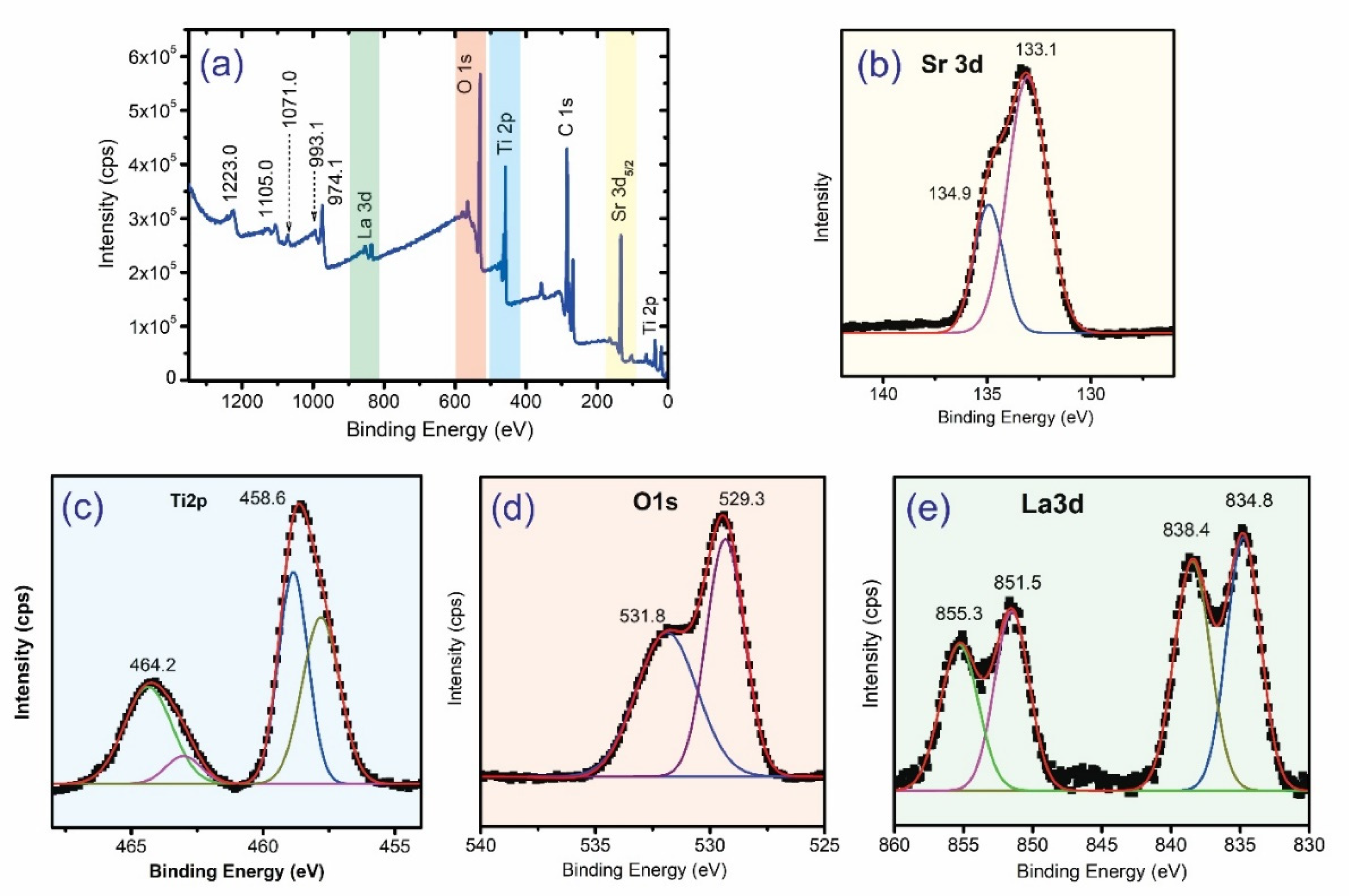
2.1. Catalyst Characterizations

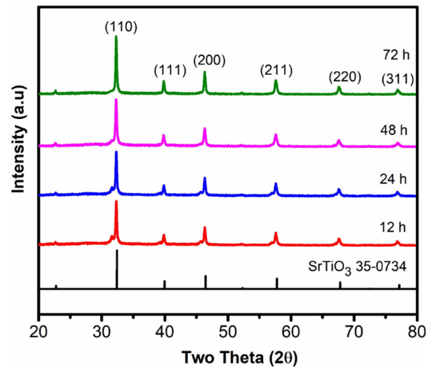
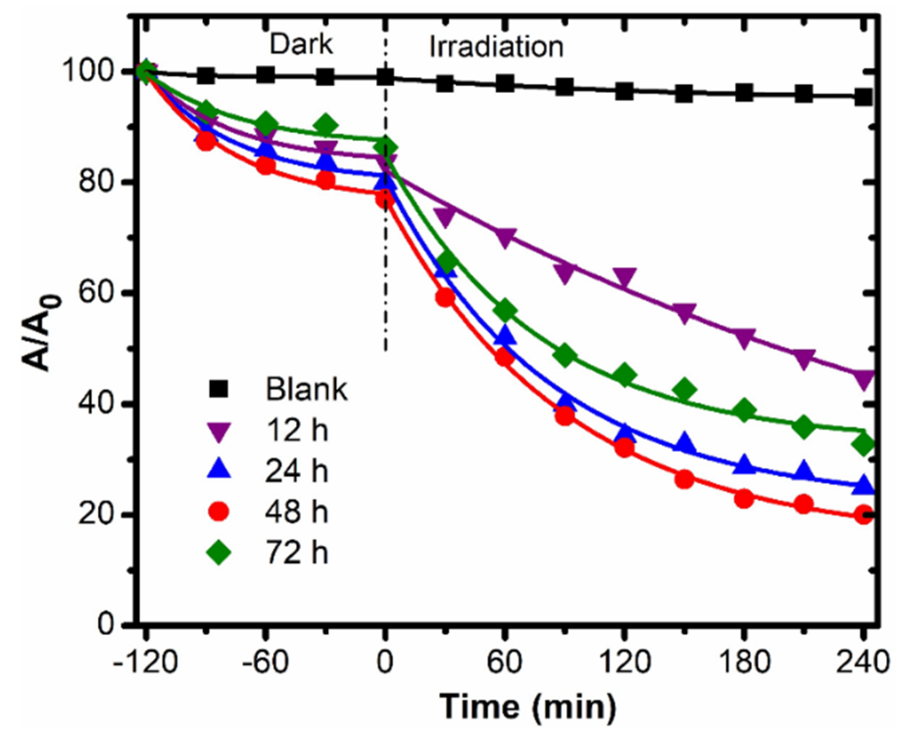
2.1.1. The Influence of Heating Durations

The powder X-ray diffraction patterns of SrTiO3 samples at different heating durations (12, 24, 48, and 72 h) are shown in Figure 1. The characteristic peaks of the SrTiO3 samples appear at 32.0, 40.0, 46.5, 57.5, 68.0, and 77.0° and are ascribed to the (100), (111), (200), (211), (220), and (311) planes of cubic symmetry of SrTiO3, which match well with the standard card for SrTiO3 (JCPDS Card No. 35-0734). The obtained results reveal that a cubic phase of SrTiO3 could be synthesized by a hydrothermal method at 130 °C. As shown in Figure 1, as the hydrothermal duration increases, the intensities of the corresponding samples also increase, implying an increase in the rate of crystallization. This phenomenon could be attributed to the Ostwald ripening process, which is due to smaller particles being less stable, and the growth of larger particles being energetically favourable [59].
The crystal sizes of SrTiO3 NPs synthesized at various hydrothermal durations were calculated using the Scherrer formula, D ( 100 ) = K B   c o s , according to the full width at half maximum (FWHM) of the (110) crystal plane. A small value of FWHM, as shown in Table 1, implies a high sample crystallinity. The calculated average crystal sizes of the SrTiO3 samples suggest that the crystallinity of SrTiO3 increases with an increasing hydrothermal duration.
The specific surface area was measured using the Brunauer-Emmett-Teller (BET) method, as presented in Table 1. The specific surface area decreases with an increasing hydrothermal duration from 12 h to 48 h. This result suggests that the increase in the hydrothermal duration fosters the tendency of agglomeration. Moreover, the specific surface area of all of the SrTiO3 synthesized in this research by the hydrothermal method is higher than that of samples synthesized by the solid-state reaction (i.e., 1.6 m2/g) [60,61] or even the sol-gel hydrothermal procedure (i.e., 17.1 m2/g) [62], which are appropriate hydrothermal methods for the preparation of SrTiO3 for photocatalytic applications.

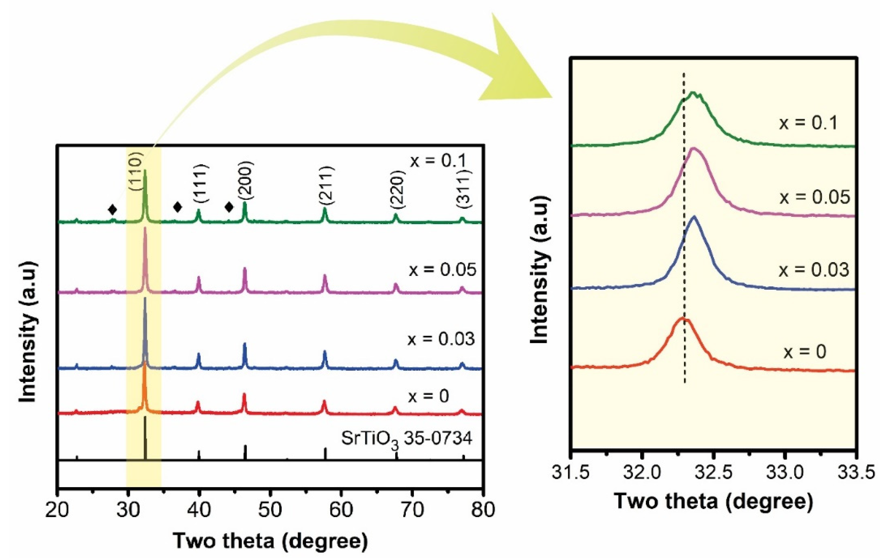
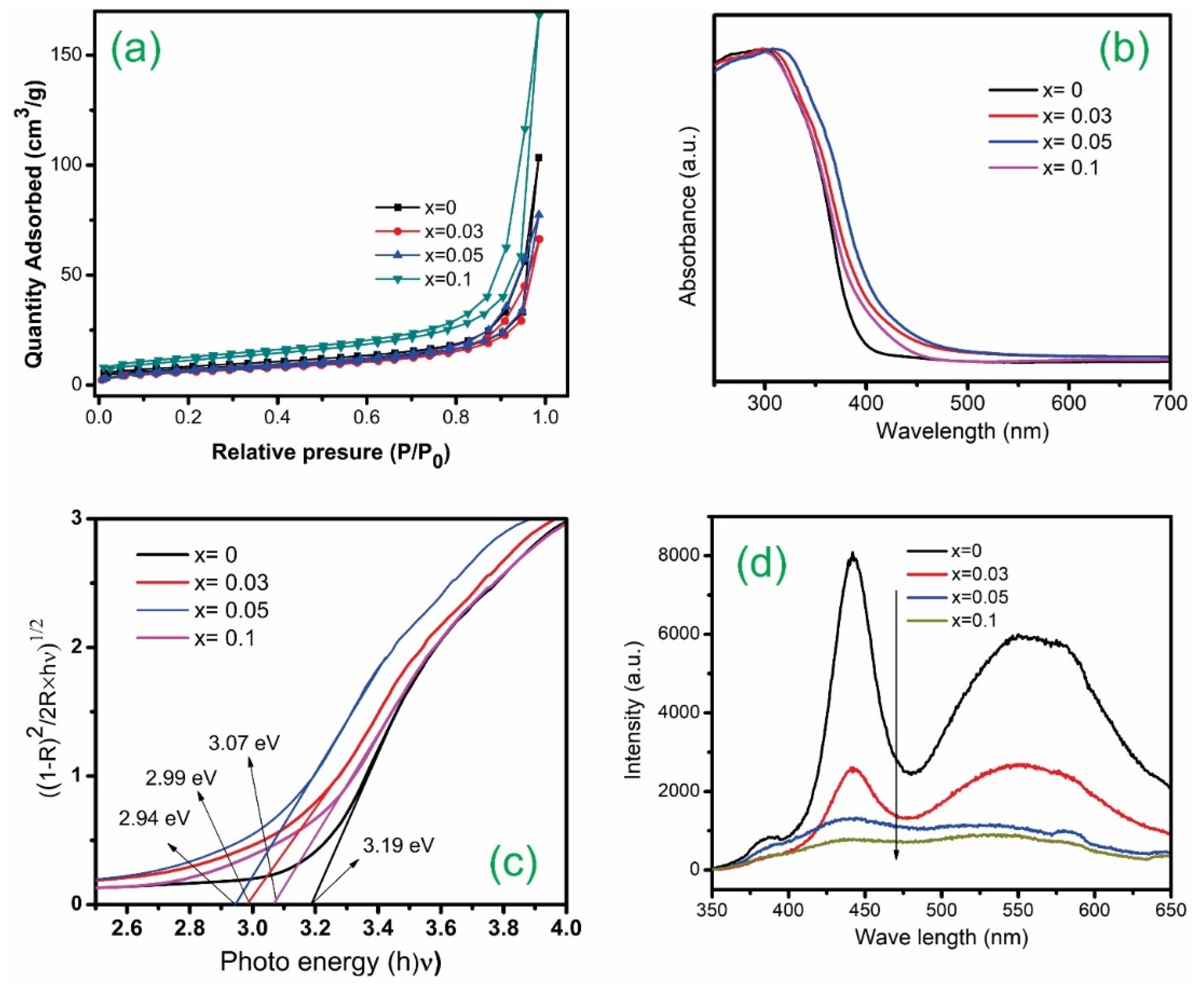
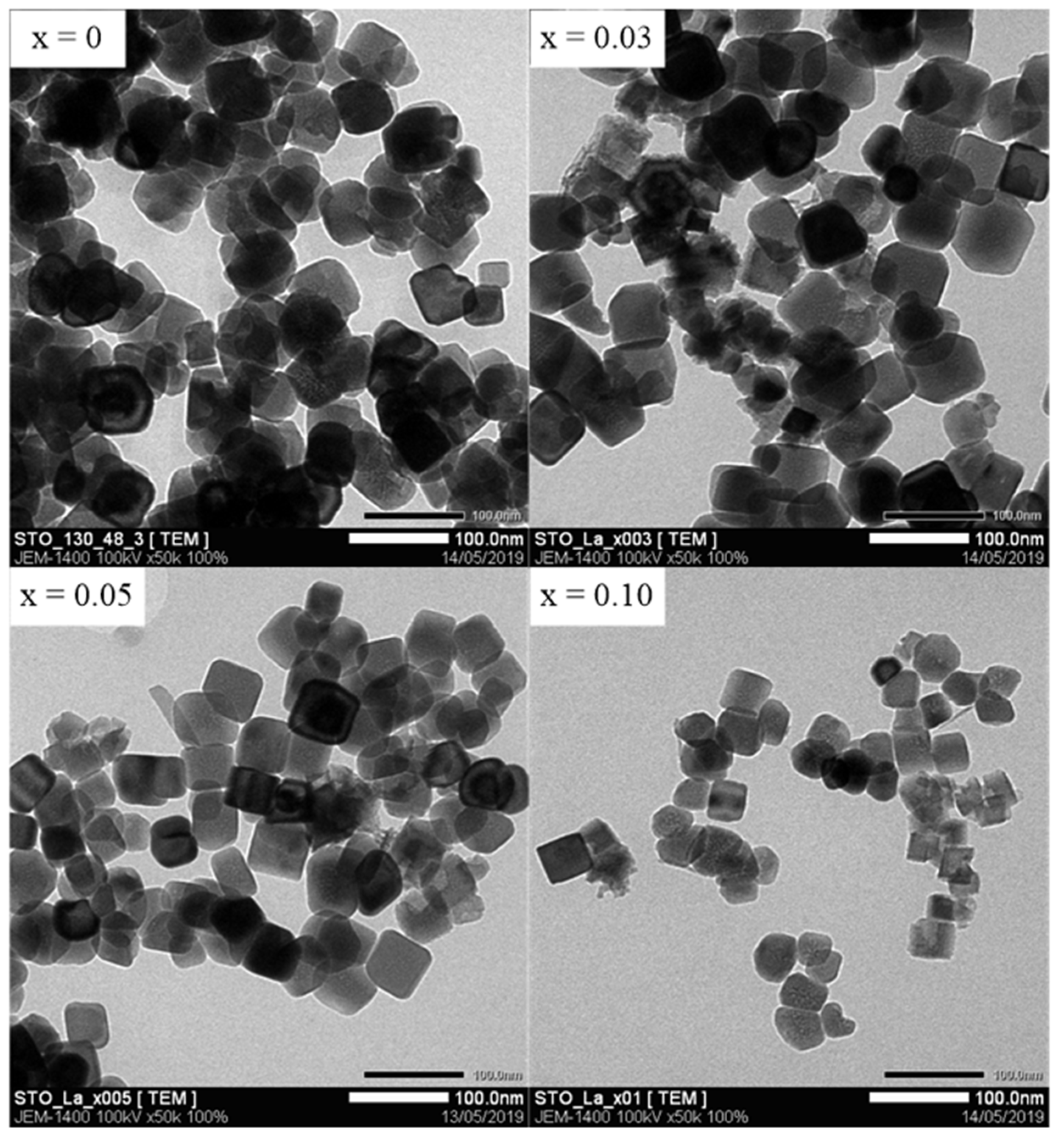
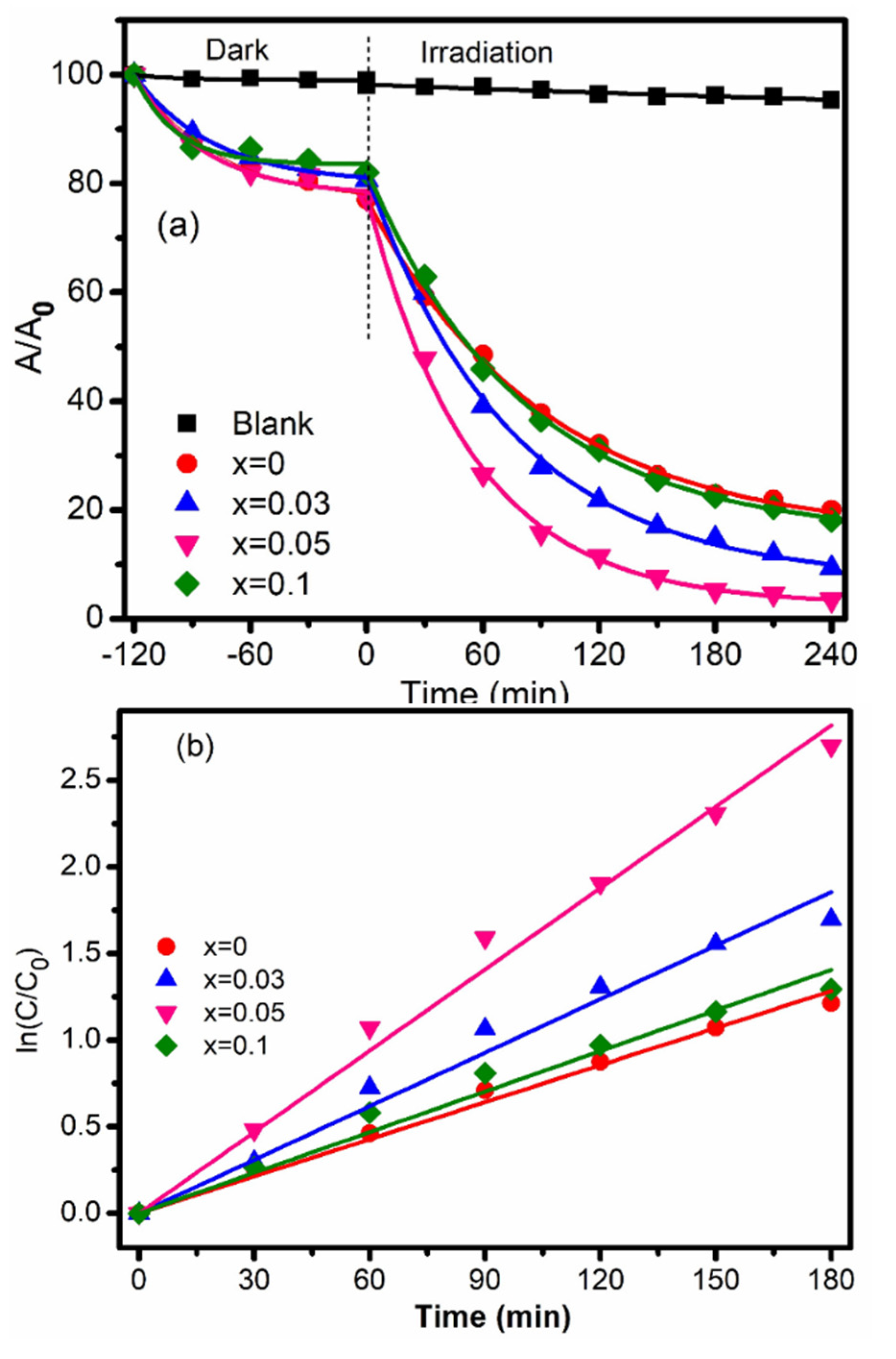
2.1.2. The Influence of La Doping Concentrations

Figure 2 shows the XRD diffraction pattern of La-doped SrTiO3 (Sr1−xLaxTiO3) samples with different La-dopant concentrations synthesized at 130 °C for 48 h. We found that the diffraction peaks of the samples (at 22.84, 32.0, 40.0, 46.5, 57.5, 68.0, and 77.0°) are indexed well to the cubic lattice of perovskite SrTiO3 (JCPDS 35-0734), suggesting that the crystal structure of SrTiO3 does not influence the substitution of La [63]. However, the relatively low-intensity peaks of La-doped samples observed at 27.74°, 36.56° and 44.12° are due to the formation of TiO2 (JCPDS files 21-1272 and 21-1276). The reduced intensity of the Sr0.9La0.1TiO3 (x = 0.1) sample is also recognized, which may be due to the formation of a higher concentration of impurity phases.
The estimated average crystal sizes of the Sr1−xLaxTiO3 (x = 0, 0.03, 0.05, 0.1) samples were calculated based on the Scherrer formula and tabulated in Table 2. The obtained results show that the crystal size of Sr1−xLaxTiO3 is smaller than that of their parent SrTiO3 crystals, confirming that La doping can inhibit the growth of SrTiO3 crystals. Moreover, at high La doping concentrations (x = 0.1), the intensity of the sample was significantly reduced, which was associated with an increasing amount of impurity, providing an unnecessary amount of impurity, which can cause some adverse effects. Besides this, with an increasing La doping amount, the (100) diffraction peak shifts to a higher 2θ value, and the crystallinity decreases. These results indicate that crystals with a high doping concentration are distorted and contain an internal strain, or maybe the different valences of La3+ and Sr2+ ions, which can cause lattice defects in SrTiO3 when La3+ is substituted for Sr2+ [64].
The lattice parameter of Sr1−xLaxTiO3 as a function of the dopant concentration was calculated from the powder data using the program UnITCell [65]; the results are presented in Table 2. The calculated unit-cell parameter of SrTiO3-48 is 3.988 Å, which is slightly higher than that of JCPDS–card no. 35-0734 (3.905 Å). The calculated values of lattice parameter (a = b = c) are 3.988 Å (x = 0), 3.984 Å (x = 0.03), 3.982 Å (x = 0.05), and 3.983 Å (x = 0.1). The lattice parameter continuously decreases with an increasing dopant concentration (from x = 0 to x = 0.05), which can be explained by the replacement of the larger-sized Sr2+ ions (118 pm) in the A-sites of the perovskite structure with smaller La3+ ions (103.2 pm) [49].
As shown in Table 2, the surface area was found to increase from 21.7–39.8 m2/g as the La content increases in Sr1−xLaxTiO3, which is due to the decrease in particle size. These results imply that La is substituted into the SrTiO3 structure. The porous structure of Sr1−xLaxTiO3 was explored by nitrogen adsorption–desorption isotherms, as shown in Figure 3a, where all of the samples show type IV adsorption isotherms. The adsorption isotherm in a relative pressure range of 0 < P/P0 < 0.8 indicates a low affinity for the nitrogen adsorbate and an intrinsically low specific surface area, implying that the sample also possesses some large macropores. The adsorption isotherms of the samples have a vertical rise at a high relative pressure P/P0 > 0.9), with a type H3 hysteresis loop, thus supporting the presence of micropores in the catalysts [66]. The corresponding pore size is shown in Table 2, which was found in the range 2.38–2.46 nm, revealing that the Sr1−xLaxTiO3 nanocubes possess mesopores. The value of the pore radius varies slightly, which is associated with a slight increase in the specific surface area.
The light-harvesting ability of Sr1−xLaxTiO3 photocatalysts was determined from their UV-visible absorption spectra, as shown in Figure 3b. The Kubelka–Munk function was used to calculate the bandgap energy of different photocatalysts by plotting [F(R)-hν]1/2 with the energy of light (hν) (Figure 3c). The Eg values were estimated from the x-axis intercept obtained by the linear fit of the spectra shown in Figure 3c. In general, a progressive redshift of the absorption edge in the visible region is observed with an increasing La doping concentration. In other words, the La doping concentration accordingly affected the bandgap energy of the parent SrTiO3 material, which is the lower bandgap energy of pristine SrTiO3 samples. The bandgap energies decrease from 3.19 eV to 2.94 eV (Table 2) when the La doping concentration is increased from 0 to 5 mol.%. The bandgap energy of the Sr1−xLaxTiO3 photocatalysts decreased with an increasing La doping level. In previous studies, Surendar et al. and Wu et al. reported that La-doping would introduce the electron donor levels in the lattice structure of SrTiO3, leading to the shifting of the Fermi level into the conduction band [67,68]. As a result, the bandgap energy of SrTiO3 would be reduced and could be activated under visible light irradiation. However, at a higher La doping concentration, x = 0.1, the bandgap energy of the Sr0.90La0.1TiO3 sample increases (3.07 eV), which may be due to the presence of impurity phases, as shown in Figure 2. Finally, the substitution of La dopant in the mesoporous SrTiO3 nanocrystal photocatalyst dramatically contributes to the increase in light-harvesting within the visible light region (λ > 400 nm).
As shown in Figure 3d, the PL spectra of SrTiO3 contain three characteristic peaks, including a weak peak at 383 nm, a strong peak at 440 nm, and a broad peak in the range of 460–650 nm [69,70]. Furthermore, the broad green band emission in the PL spectra of Sr1−xLaxTiO3 photocatalysts demonstrates the successful doping of La+3 cations in the parent lattice of SrTiO3. The lower PL emission intensity at a higher La doping concentration indicates that more defect levels are created by the increasing La doping concentration [66], according to Equation (1). This emission is due to the O 2p valance band, which corresponds to the defect level created by the oxygen deficiency. Moreover, related studies on SrTiO3 have reported the influence of doping on photocatalytic properties, which suggests that the dopants create more impurity defects and quench the recombination process of exciting holes and electrons [66,71,72], resulting in the improvement of the overall photocatalytic activity.
O O x   V O + 2   e + 1 2 O 2 ( g )  
TEM was employed in order to study the morphology and particle size of samples with various La doping concentrations that were hydrothermally synthesized at 130 °C for 48 h. As illustrated in Figure 4, all of the samples exhibited cubic-like morphology. Moreover, the TEM images of doped samples such as Sr0.95La0.05TiO3 and Sr0.90La0.1TiO3 show slightly sharp and uniform distributions under the same synthesis conditions as the non-doped sample, which has also been reported elsewhere [42,44]. Besides this, the results clearly show decreasing particle sizes with increasing La doping concentrations, which enhances the surface area of doped SrTiO3. The decrease in the particle size plays a vital role in decreasing the recombination of photogenerated electrons and holes, and enhances the photocatalytic activities of the catalysts. Consequently, Sr1−xLaxTiO3 may perform well in the photocatalysis process under visible light conditions. We note that the particle sizes observed from the TEM were consistently more significant than those calculated from the Scherrer equation. Based on the Scherrer equation, the particle sizes are determined from line broadening. Because both compositional and geometric inhomogeneity, i.e., lattice strain, are affected and dependent on the line broadening [73], the above discrepancy might be attributed to both the compositional and geometric inhomogeneity of Sr1−xLaxTiO3.
The chemical compositions of the Sr0.95La0.05TiO3 samples were elucidated by XPS analysis (Figure 5). Figure 5a shows the presence of C, O, Ti, Sr, and La elements in the sample, indicating that La has been introduced into the SrTiO3 structure. The presence of C 1s with a sharp peak at approximately 284 eV corresponds to the C–C or C–H bond, which is ascribed to the adventitious carbon-based contaminant in the apparatus itself [48,74]. The high-resolution Sr 3d curve in Figure 5b is fitted into two peaks at the binding energy positions of 133.1 and 134.9 eV, belonging to Sr 3d5/2 and Sr 3d3/2 [48], respectively. The binding energy positions of the Ti 2d-doublet lines at 458.6 and 464.2 eV (Figure 5c)—based on Gaussian curve fitting—can be assigned to the 2p3/2 and 2p1/2 orbitals of Ti4+ [48,75], respectively, which correspond to Ti with an oxidation state of 4+. As shown in Figure 5d, two observable peaks at 529.3 and 531.8 eV of the high-resolution O 1s spectrum are assigned to the Ti–O or Sr–O, and O–H bands, respectively [76]. As shown in the La 3d spectra (Figure 5e), there are two doublet peaks near 837 and 853 eV. The peaks at 834.8 and 851.5 eV are assigned to La 3d5/2 and La 3d3/2, respectively, which correspond to La3+ [77], indicating that La3+ is doped in SrTiO3. However, peaks were found at 838.4 and 855.3 eV, which correspond to the shake-up satellite peaks of La 3d5/2 and La 3d3/2 [77], respectively. It can be concluded that La3+ had been successfully doped into SrTiO3 by substituting La3+ for Sr2+.

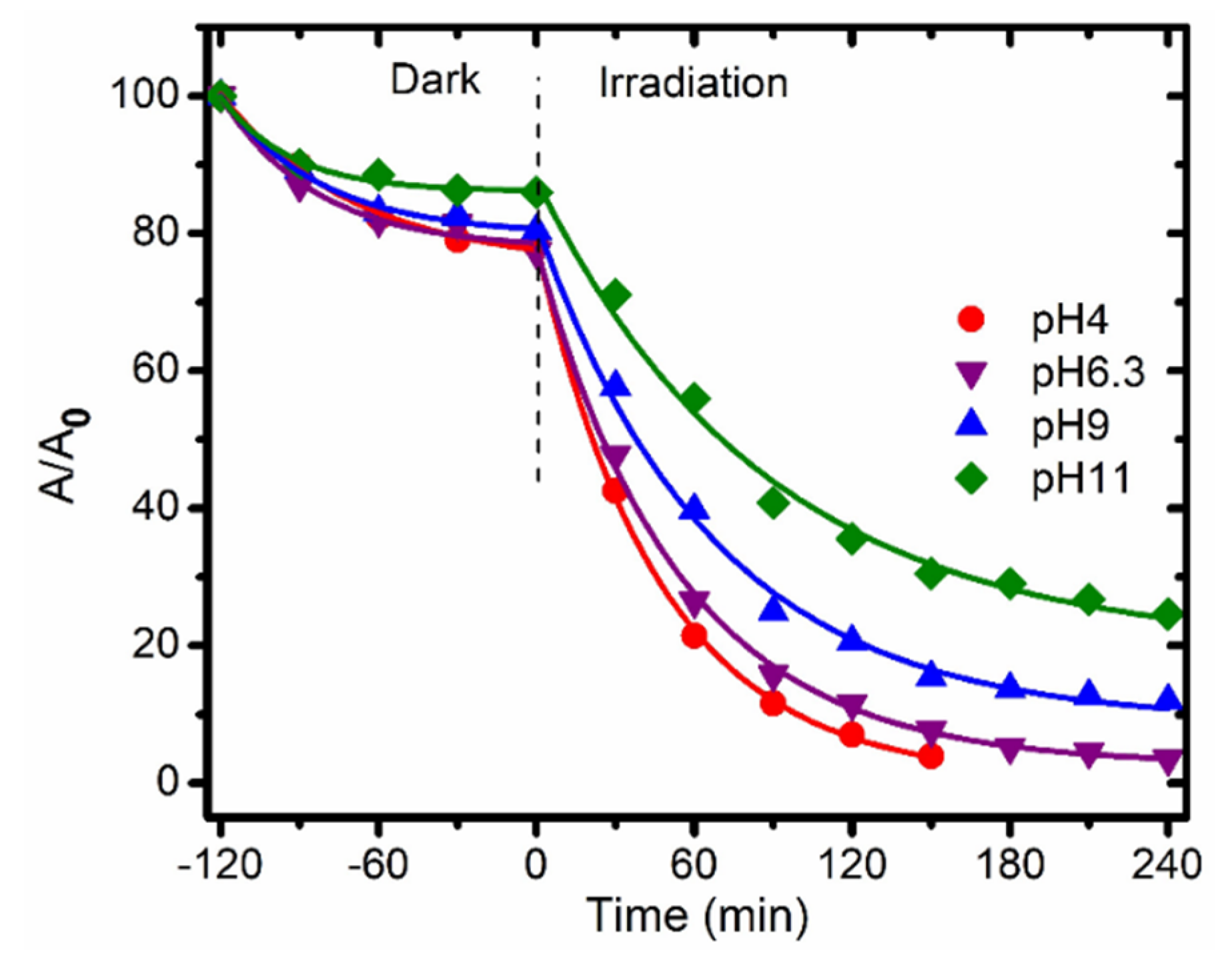
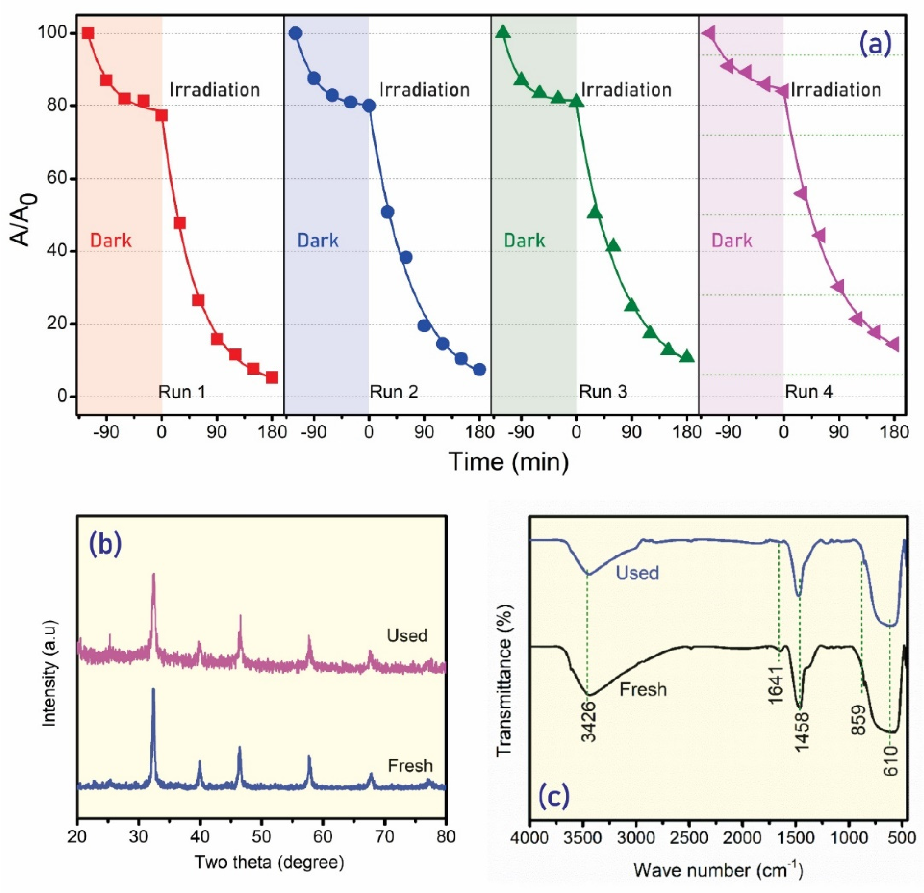
2.2. Photocatalytic Activities

The photocatalytic degradation process of 2-naphthol over photocatalysts synthesized at 130 °C was obtained by measuring the residual 2-naphthol concentration in the solution over time during the irradiation. Generally, the dependence of the 2-naphthol concentration on the irradiation time can be quantitatively estimated by the kinetics of the photodegradation reaction, which is described by the kinetic equation below:
r = d C d t = k C ln C o C = k t
where k is the apparent first-order kinetic constant that represents the reaction rate, and Co and C are the concentrations of 2-naphthol before and after irradiation, respectively.
Figure 6 shows the photocatalytic activity of SrTiO3 at various synthetic durations for 2-naphthol degradation under simulated natural light irradiation. The samples exhibit a different adsorption capability, which increases with the increasing BET of the sample, as shown in Table 1. Consequently, these samples exhibit quite different photocatalytic activities under simulated natural light irradiation (Table 3). The sample calcined for 48 h degraded 2-naphthol up to 83% within 240 h of irradiation, exhibiting the highest photocatalytic activity. Moreover, the blank test reveals the stability of 2-naphthol under photolysis conditions, which is degraded by only 2.68% within 240 min of irradiation. This information confirms the outstanding photocatalytic activities of cubic SrTiO3 photocatalysts.
Figure 7 presents the photocatalytic activity of Sr1−xLaxTiO3 photocatalysts as a function of the La doping concentration over the irradiation time. All of the Sr1−xLaxTiO3 photocatalysts enhance photocatalytic activities and exhibit much higher photocatalytic activities for 2-naphthol degradation than the parent SrTiO3 catalyst (Figure 7a). The substitution of higher-valance La3+ can explain the improved photocatalytic activities of Sr1−xLaxTiO3 by Sr2+ in the parent structure of SrTiO3, which can act as sites to capture electrons, resulting in the inhibition of the recombination of photogenerated charge carriers and the acceleration of the photocatalytic reaction [48]. With the presence of a small amount of lanthanum (x = 0.03), the photoactivity of Sr0.97La0.03TiO3 was obviously enhanced. Furthermore, the photoactivity of Sr0.95La0.05TiO3 showed the highest catalytic activity, which was reduced by merely 92% within 180 min, which is higher than that of the parent SrTiO3 catalyst (83%). The bandgap of Sr0.95La0.05TiO3 is the narrowest (Table 2), which enhances its ability to absorb simulated natural light and thus improves its photocatalytic activity.
According to the corresponding kinetic study, the kinetic curves of the samples for the degradation of 2-naphthol are shown in Figure 7b, which agree with the pseudo-first-order reaction, and the best-fit parameters are listed in Table 4. The rate constant of Sr0.95La0.05TiO3 (k = 0.0196 min−1) is the highest among the samples, and decreases in the order of Sr0.95La0.05TiO3 > Sr0.97La0.03TiO3 > Sr0.9La0.1TiO3 > SrTiO3. At higher La contents (x = 0.1, Sr0.9La0.1TiO3), the photocatalytic activity decreases. The decrease in photocatalytic activity can be attributed to the enhanced concentration of the impurity phase, which can become the recombination center of electrons and holes [78], thereby reducing the efficiency of the charge separation and resulting in reduced photocatalytic activity. Besides this, the ability of Sr0.95La0.05TiO3 nanocubes to degrade 2-naphthol is much better than that of other photocatalysts reported in the literature [79], which was 0.0126 min−1. It could be concluded that Sr0.95La0.05TiO3 enhanced the highest photocatalytic activities, which is mainly due to their higher light absorption capability, narrower bandgap, and lower recombination rate of photogenerated electron–hole pairs.
The zero-point charge (pHzpc) strongly affects the adsorption capability/behaviour of organic compounds on the photocatalyst’s surface during a photocatalytic process [80]. The ΔpH = pHfinal − pHinitial was measured for different pH values (from 4 to 10) in order to understand the behaviour of the surface charge of the photocatalysts in aqueous solution. As seen in Table 2, the pHzpc values of the Sr1−xLaxTiO3 photocatalysts were measured to be approximately 7.78, 7.42, 7.32, and 7.61, respectively, which reveals that the charge on the surface of SrTiO3 has been modified by La doping. Moreover, the reduction in pHzpc was due to the increase in oxygen vacancies, increasing the surface −OH groups owing to La doping.
The photocatalytic degradation of 2-naphthol by the photocatalyst Sr0.95La0.05TiO3 in aqueous suspensions prepared at different pH values (pH = 4.1–11.3) was investigated. Figure 8 shows the effect of the initial pH on the 2-naphthol degradation efficiency. The 2-naphthol degradation decreased when the initial pH shifted from 4.1 to 11.3. The degradation efficiencies were recorded to be 95, 93, 85, and 74% after 150 min of irradiation at pH 4.1, 6.3, 9.1, and 11.3, respectively. The change in the overall photocatalytic degradation can be explained by the surface charge of the photocatalyst and the dissociation behaviour of 2-naphthol. With the pHzpc for Sr0.95La0.05TiO3 being near 7.32, the surface of the Sr0.95La0.05TiO3 photocatalyst is predominantly positively charged at pH values below pHzpc, but higher pH values promote the formation of a negative charge on the NPs [81]. However, 2-naphthol is a weak acid with a dissociation constant (pKa) of 9.5 [82]. It exists in neutral forms at pH < pKa and becomes anionic when the solution pH > pKa. Therefore, it is more favourable to adsorb on the surface of the catalyst at pH < 9.5. The higher efficiencies of Sr0.95La0.05TiO3 at pH 4.1 and 6.3 may be attributed to the adsorption of neutral 2-naphthol on the positive charge on photocatalyst surfaces resulting in photocatalytic degradation. On the other hand, at higher pH (> 9.5), the surface of Sr1−xLaxTiO3 becomes negatively charged and 2-naphthol becomes an anionic species, leading to higher electrostatic repulsion between the catalyst and 2-naphthol [83]. Finally, it can be concluded that the degradation rate was higher in acid media than in base media. Similar behaviour was observed by several studies reported in the literature [75,84].
In order to evaluate the stability of the Sr0.95La0.05TiO3 nanocube photocatalytic activities, four photocatalytic experimental runs were carried out by adding recycled Sr0.95La0.05TiO3 photocatalyst to fresh 2-naphthol solutions (10 ppm) with the same catalyst dosage (Figure 9). As demonstrated in Figure 9a, the photoactivity of the photocatalyst did not show a significant decrease after four cycles of degradation. The yield of 2-naphthol degradation was maintained at nearly 86% after 180 min of irradiation up to the fourth cycle, which is 95% of the original photocatalytic degradation efficiency. Additionally, the degradation rate was observed to be stable after four cycles. The XRD patterns and FTIR spectra that compare the crystal structure and surface absorption bands of the fresh and used Sr0.95La0.05TiO3 photocatalyst are illustrated in Figure 9b,c. Clearly, the crystal structure of the used Sr0.95La0.05TiO3 photocatalyst did not change after the photocatalytic process. As shown in Figure 9c, the normal characteristic IR peaks below 1000 cm−1 feature the possibilities of Sr–O/Ti–O/Cr–O bonds and functional groups. The two characteristic absorption bands, located at about 457 and 610 cm−1, are ascribed to the vibrations of the metal-oxygen bonds Sr–O/Ti–O/La–O [32,85,86]. The broad and low-intensity absorption bands observed in all of the samples in the range 2850–3440 cm−1, 1458 cm−1, and 1618 cm−1 correspond to the stretching and bending vibrations of O–H groups in the physically adsorbed H2O molecules, respectively [32]. In all of the samples, the shoulder around 860 cm−1 represents the SrTiO3 crystal lattice vibrations [42]. The similarity of the FTIR spectra of the fresh and used photocatalysts is notable. These results further confirm that the synthesized Sr0.95La0.05TiO3 nanoparticles could be regenerated easily and reused with good reusability for organic degradation.
Although the conditions for conducting the experiments are different, it is worth comparing the recent studies on the photodegradation of 2-naphthol in terms of degradation efficiency. Table 5 shows several achievements of the photodegradation of 2-naphthol in the literature [31,87,88,89,90,91,92]. As we can see in the table, photocatalysis could effectively remove 2-naphthol with attractive performance. As an example, MnOx-modified (Ce0.73Bi0.27)O2-δ performed an excellent photocatalytic activity at 50 °C, which reached 98% degradation efficiency [88]. In this study, it is achieved 86.6% degradation efficiency at room-temperature conditions over Sr0.95La0.05TiO3 photocatalyst.

3. Materials and Methods

3.1. Materials and Reagents

The titanium dioxide (P25), 2-naphthol (99.0%), lanthanum nitrate hexahydrate (La(NO3)3.6H2O, ≥ 99%), and sodium hydroxide (NaOH, ≥ 99%) were purchased from Sigma Aldrich (St. Louis, MO, USA). The strontium chloride hexahydrate (SrCl2.6H2O, extra pure) was purchased from Acros Organic (Geel, Belgium). All of the chemical reagents were used as received without further purification.

3.2. Synthesis of Sr1−xLaxTiO3 Nanocubes

The Sr1−xLaxTiO3 nanocubes were synthesized by a facile hydrothermal method. For the parent SrTiO3, 3.9990 g (0.015 mol), SrCl2.6H2O was dissolved in 40 mL 3 M NaOH at room temperature under vigorous magnetic stirring for 1 h. Next, 1.1985 g TiO2 (molar ratio Sr:Ti = 1:1) was dispersed into the above solution and stirred for another 1 h before being transferred to the autoclave. The mixture was transferred to a 200 mL Teflon-lined autoclave vessel, sealed, and heated at 130 °C for various durations (from 12 h to 72 h). After the hydrothermal process, the system was cooled to room temperature, and the solid precipitate was removed by centrifugation and washed several times with absolute ethyl alcohol and deionized water in order to remove the sodium hydroxide and residual chemicals. The washing process was repeated three times. Finally, the obtained powder was dried at 80 °C overnight. The samples that were hydrothermally processed at 130 °C for 12, 24, 48, and 72 h were designated SrTiO3-12, SrTiO3-24, SrTiO3-48, and SrTiO3-72.
For the La-doped SrTiO3 (Sr1−xLaxTiO3, x = 0.03–0.1), the nanocubes were synthesized using the same procedure as that used for SrTiO3. In typically, an appropriate amount of La(NO3)3.6H2O was dissolved together with the strontium precursor, and the hydrothermal conditions were set to 130 °C for 48 h.

3.3. Characterizations

X-ray diffraction (XRD) studies were recorded with a D8 Advance-Brucker diffractometer (Bruker, USA) generating Cu Kα radiation (λ = 0.15418 nm) at a scanning rate of 0.03 °/s and in the scanning range of 20 to 80°, with a step size of 0.02°. FTIR spectroscopy was measured at 4 cm−1 spectral resolution between the 400 to 4000 cm−1 range using a Perkin Elmer Frontier 1600 series spectroscope. The transfer behaviour of the photogenerated electrons and holes between various samples was studied using photoluminescence (PL) spectra (HORIBA Jobin YVON iHR 320, (Kyoto, Japan)) with a wavelength range of 350–620 nm. Scanning electron microscopy (SEM) images were taken using a JSM-6500F, JEOL (Tokyo, Japan). The morphology of the obtained nanocubes was observed by transmission electron microscopy (TEM, JEOL-JEM1010, Tokyo, Japan). The bandgap energy of each sample was calculated from diffuse reflectance spectroscopy (DSR), ranging from 850 to 220 nm, with a scanning step of 2 nm at a rate of 400 nm/min using solid UV-vis JASCO V-550 (Tokyo, Japan) equipment. The N2 adsorption–desorption isotherms of SrTiO3 were obtained by using a nitrogen adsorption–desorption apparatus (Quantachrome NOVA 1000e Surface Area and Pore Size Analyzer (Boynton Beach, FL, U.S.A.) at a liquid nitrogen temperature of −196 °C. The multipoint Brunauer–Emmett–Teller (BET) equation was applied using the data of the relative pressure (P/P0) to determine the specific surface area. The porosity of the samples was also investigated using DA (Dubinin-Astakhov) models. X-ray photoelectron spectroscopy (XPS), was recorded on ESCALAB 250Xi (Thermo Fisher Scientific, Waltham, MA, USA) to determine the oxidation states of the elements.
The zero point charges (pHzpc) of the photocatalysts were measured in 30 mL portions of NaCl aqueous solution (0.05 mol/L) adjusted to the different pH values by either 0.1 M HCl or 0.1 M NaOH solution. N2 gas was bubbled through the NaCl solutions in order to remove the dissolved CO2 until the initial pH was stabilized. After the determination of the initial pH value, the solutions were mixed with 0.090 g of the catalyst sample for 24 h, and the final pH of the solutions was then measured.

3.4. Photocatalytic Activity

The photocatalytic process was conducted in a water-jacketed Pyrex photoreactor, which was circulated with water to maintain a temperature of approximately 30 °C. The photoreactor was placed in a black chamber in order to prevent the impact of any external radiation sources. Photocatalytic 2-naphthol degradation was conducted by dispersing 0.10 g photocatalyst powder in 200 mL 2-naphthol solution with an initial concentration of 10 ppm (natural pH 6.3) under Exo Terra Natural Light irradiation (Repti Glo 2.0 Comp Fluor 26 W PT2191-220, Illuminance: 15350 Lux, 400–700 nm) and a stirring speed of 400 rpm. In order to avoid systematic errors, the aforementioned 2-naphthol solution was diluted from the stock 1000 ppm solution. Prior to the visible light illumination, the suspension was magnetically stirred for 2 h in the dark in order to establish adsorption–desorption equilibrium between the photocatalyst and 2-naphthol. The mixture was then exposed to illumination, and at a given irradiation time interval of 30 min, 5 mL of the solution was extracted using a syringe and then filtered through a 0.22 μm cellulose acetate membrane (Millipore Corp. Bedford, MA). A model U-2910 Spectrophotometer from Hitachi, Tokyo, Japan monitored the residual 2-naphthol concentration with the characteristic absorption of 2-naphthol at a wavelength of 223 nm. The photocatalytic degradation efficiency (De) of 2-naphthol was calculated by the expression of De = (1 − At/Ao) × 100%, where A0 and At are the absorbance at 464 nm of the characteristic wavelength of the 2-naphthol solution before and after irradiation at time t, respectively. The blank test was also carried out with the same photocatalytic process procedure for comparison purposes, but without the presence of Sr1−xLaxTiO3 photocatalysts. The photocatalytic stability was also investigated by evaluating the removal of 2-naphthol as a function of the experimental cycle. At the end of each cycle, the photocatalyst was collected by centrifugation, rinsed with deionized water, and dried at 300 °C for 2 h to remove the 2-naphthol contaminant before its use in the next cycle.

4. Conclusions

In this study, Sr1−xLaxTiO3 nanocubes with different La dopant concentrations were successfully synthesized by a hydrothermal method, and their physical properties were investigated. The XRD patterns showed a cubic structure, and the TEM images presented a nanocube morphology with a size range of 40–50 nm. The higher photocatalytic activity of the doped samples compared to pristine SrTiO3 was attributed to the fact that La-doped SrTiO3 can harvest irradiation light because of its relatively narrow bandgap energy, create more impurity defects, and quench the recombination process of exciting holes and electrons. The Sr0.95La0.05TiO3 nanocubes exhibited the best photocatalytic performance for 2-naphthol degradation under artificial solar light with a 95% degradation efficiency after 180 min and a pseudo-first-order degradation rate constant of 0.0196 min−1, which are higher than those of pure SrTiO3. Importantly, the photocatalytic activity over Sr0.95La0.05TiO3 maintained stable efficiency after four cycles. These observations demonstrated that Sr1−xLaxTiO3 nanocubes could be widely applied in wastewater treatment.

Author Contributions

M.-V.L.: Supervision, Conceptualization, Methodology, Investigation, Writing—Original Draft Preparation, Reviewing and Editing; N.-Q.-D.V.: Conceptualization, Methodology, Investigation, Writing—Original Draft Preparation; Q.-C.L.: Conceptualization, Methodology, Investigation; V.A.T.: Conceptualization, Methodology, Investigation; T.-Q.-P.P.: Methodology, Investigation; C.-W.H.: Conceptualization, Methodology, Writing—Original Draft Preparation, Reviewing and Editing; V.-H.N.: Conceptualization, Methodology, Writing—Original Draft Preparation, Reviewing and Editing. All authors have read and agreed to the published version of the manuscript.

Funding

This research was funded by Ho Chi Minh City University of Technology–VNU-HCM, under grant number To-KTHH-2019-02.

Data Availability Statement

Not applicable.

Acknowledgments

This research is funded by Ho Chi Minh City University of Technology–VNU-HCM, under grant number To-KTHH-2019-02.

Conflicts of Interest

The authors declare no conflict of interest.

References

  1. Ma, T.; Wu, J.; Mi, Y.; Chen, Q.; Ma, D.; Chai, C. Novel Z-Scheme g-C3N4/C@Bi2MoO6 composite with enhanced visible-light photocatalytic activity for β-naphthol degradation. Sep. Purif. Technol. 2017, 183, 54–65. [Google Scholar] [CrossRef]
  2. Kumar, A.; Raizada, P.; Singh, P.; Saini, R.V.; Saini, A.K.; Hosseini-Bandegharaei, A. Perspective and status of polymeric graphitic carbon nitride based Z-scheme photocatalytic systems for sustainable photocatalytic water purification. Chem. Eng. J. 2020, 391, 123496. [Google Scholar] [CrossRef]
  3. Raizada, P.; Sudhaik, A.; Singh, P.; Shandilya, P.; Gupta, V.K.; Hosseini-Bandegharaei, A.; Agrawal, S. Ag3PO4 modified phosphorus and sulphur co-doped graphitic carbon nitride as a direct Z-scheme photocatalyst for 2, 4-dimethyl phenol degradation. J. Photochem. Photobiol. A Chem. 2019, 374, 22–35. [Google Scholar] [CrossRef]
  4. Singh, P.; Shandilya, P.; Raizada, P.; Sudhaik, A.; Rahmani-Sani, A.; Hosseini-Bandegharaei, A. Review on various strategies for enhancing photocatalytic activity of graphene based nanocomposites for water purification. Arab. J. Chem. 2020, 13, 3498–3520. [Google Scholar] [CrossRef]
  5. Raizada, P.; Kumari, J.; Shandilya, P.; Singh, P. Kinetics of photocatalytic mineralization of oxytetracycline and ampicillin using activated carbon supported ZnO/ZnWO. Desalination Water Treat. 2017, 79, 204–213. [Google Scholar] [CrossRef]
  6. Dutta, V.; Sharma, S.; Raizada, P.; Hosseini-Bandegharaei, A.; Kumar Gupta, V.; Singh, P. Review on augmentation in photocatalytic activity of CoFe2O4 via heterojunction formation for photocatalysis of organic pollutants in water. J. Saudi Chem. Soc. 2019, 23, 1119–1136. [Google Scholar] [CrossRef]
  7. Hasija, V.; Sudhaik, A.; Raizada, P.; Hosseini-Bandegharaei, A.; Singh, P. Carbon quantum dots supported AgI /ZnO/phosphorus doped graphitic carbon nitride as Z-scheme photocatalyst for efficient photodegradation of 2, 4-dinitrophenol. J. Environ. Chem. Eng. 2019, 7, 103272. [Google Scholar] [CrossRef]
  8. Raizada, P.; Sudhaik, A.; Singh, P.; Hosseini-Bandegharaei, A.; Thakur, P. Converting type II AgBr/VO into ternary Z scheme photocatalyst via coupling with phosphorus doped g-C3N4 for enhanced photocatalytic activity. Sep. Purif. Technol. 2019, 227, 115692. [Google Scholar] [CrossRef]
  9. Wu, Y.-T.; Yu, Y.-H.; Nguyen, V.-H.; Lu, K.-T.; Wu, J.C.-S.; Chang, L.-M.; Kuo, C.-W. Enhanced xylene removal by photocatalytic oxidation using fiber-illuminated honeycomb reactor at ppb level. J. Hazard. Mater. 2013, 262, 717–725. [Google Scholar] [CrossRef]
  10. Lu, K.-T.; Nguyen, V.-H.; Yu, Y.-H.; Yu, C.-C.; Wu, J.C.S.; Chang, L.-M.; Lin, A.Y.-C. An internal-illuminated monolith photoreactor towards efficient photocatalytic degradation of ppb-level isopropyl alcohol. Chem. Eng. J. 2016, 296, 11–18. [Google Scholar] [CrossRef]
  11. Li, D.; Yu, J.C.-C.; Nguyen, V.-H.; Wu, J.C.S.; Wang, X. A dual-function photocatalytic system for simultaneous separating hydrogen from water splitting and photocatalytic degradation of phenol in a twin-reactor. Appl. Catal. B Environ. 2018, 239, 268–279. [Google Scholar] [CrossRef]
  12. Nguyen, V.-H.; Smith, S.M.; Wantala, K.; Kajitvichyanukul, P. Photocatalytic remediation of persistent organic pollutants (POPs): A review. Arab. J. Chem. 2020, 13, 8309–8337. [Google Scholar] [CrossRef]
  13. Lam, S.S.; Nguyen, V.-H.; Nguyen Dinh, M.T.; Khieu, D.Q.; La, D.D.; Nguyen, H.T.; Vo, D.V.N.; Xia, C.; Varma, R.S.; Shokouhimehr, M.; et al. Mainstream avenues for boosting graphitic carbon nitride efficiency: Towards enhanced solar light-driven photocatalytic hydrogen production and environmental remediation. J. Mater. Chem. A 2020, 8, 10571–10603. [Google Scholar] [CrossRef]
  14. Nguyen, V.-H.; Phan Thi, L.-A.; Van Le, Q.; Singh, P.; Raizada, P.; Kajitvichyanukul, P. Tailored photocatalysts and revealed reaction pathways for photodegradation of polycyclic aromatic hydrocarbons (PAHs) in water, soil and other sources. Chemosphere 2020, 260, 127529. [Google Scholar] [CrossRef]
  15. Thakur, A.; Kumar, A.; Kumar, P.; Nguyen, V.-H.; Vo, D.-V.N.; Singh, H.; Pham, T.-D.; Thi Thanh Truc, N.; Sharma, A.; Kumar, D. Novel synthesis of advanced Cu capped Cu2O nanoparticles and their photo-catalytic activity for mineralization of aqueous dye molecules. Mater. Lett. 2020, 276, 128294. [Google Scholar] [CrossRef]
  16. Nguyen, V.-H.; Tran, Q.B.; Nguyen, X.C.; Hai, L.T.; Ho, T.T.T.; Shokouhimehr, M.; Vo, D.-V.N.; Lam, S.S.; Nguyen, H.P.; Hoang, C.T.; et al. Submerged photocatalytic membrane reactor with suspended and immobilized N-doped TiO2 under visible irradiation for diclofenac removal from wastewater. Process Saf. Environ. Prot. 2020, 142, 229–237. [Google Scholar] [CrossRef]
  17. Nguyen, T.D.; Nguyen, V.-H.; Nanda, S.; Vo, D.-V.N.; Nguyen, V.H.; Van Tran, T.; Nong, L.X.; Nguyen, T.T.; Bach, L.-G.; Abdullah, B.; et al. BiVO4 photocatalysis design and applications to oxygen production and degradation of organic compounds: A review. Environ. Chem. Lett. 2020, 18, 1779–1801. [Google Scholar] [CrossRef]
  18. Raizada, P.; Sudhaik, A.; Patial, S.; Hasija, V.; Parwaz Khan, A.A.; Singh, P.; Gautam, S.; Kaur, M.; Nguyen, V.-H. Engineering nanostructures of CuO-based photocatalysts for water treatment: Current progress and future challenges. Arab. J. Chem. 2020, 13, 8424–8457. [Google Scholar] [CrossRef]
  19. Sharma, S.; Dutta, V.; Raizada, P.; Hosseini-Bandegharaei, A.; Singh, P.; Nguyen, V.-H. Tailoring cadmium sulfide-based photocatalytic nanomaterials for water decontamination: A review. Environ. Chem. Lett. 2020. [Google Scholar] [CrossRef]
  20. Nguyen, V.-H.; Mousavi, M.; Ghasemi, J.B.; Delbari, S.A.; Le, Q.V.; Sabahi Namini, A.; Shahedi Asl, M.; Shokouhimehr, M.; Azizian-Kalandaragh, Y.; Mohammadi, M. Z-scheme g-C3N4 nanosheet/MgBi2O6 systems with the visible light response for impressive photocatalytic organic contaminants degradation. J. Photochem. Photobiol. A Chem. 2021, 406, 113023. [Google Scholar] [CrossRef]
  21. Vu, C.M.; Nguyen, V.-H.; Thi, H.V.; Jin, C.W.; Nguyen, D.D. Hybrid material based on TiO2, CuBTC, and magnetic particles as a novel photocatalyst for MB removal. J. Chem. Technol. Biotechnol. 2020, 95, 2648–2655. [Google Scholar] [CrossRef]
  22. Nguyen, L.T.; Nguyen, H.T.; Pham, T.-D.; Tran, T.D.; Chu, H.T.; Dang, H.T.; Nguyen, V.-H.; Nguyen, K.M.; Pham, T.T.; Van der Bruggen, B. UV–Visible Light Driven Photocatalytic Degradation of Ciprofloxacin by N,S Co-doped TiO2: The Effect of Operational Parameters. Top. Catal. 2020, 63, 985–995. [Google Scholar] [CrossRef]
  23. Bhuvaneswari, K.; Nguyen, B.-S.; Nguyen, V.-H.; Nguyen, V.-Q.; Nguyen, Q.-H.; Palanisamy, G.; Sivashanmugan, K.; Pazhanivel, T. Enhanced photocatalytic activity of ethylenediamine-assisted tin oxide (SnO2) nanorods for methylene blue dye degradation. Mater. Lett. 2020, 276, 128173. [Google Scholar] [CrossRef]
  24. Sudhaik, A.; Raizada, P.; Thakur, S.; Saini, R.V.; Saini, A.K.; Singh, P.; Kumar Thakur, V.; Nguyen, V.-H.; Khan, A.A.P.; Asiri, A.M. Synergistic photocatalytic mitigation of imidacloprid pesticide and antibacterial activity using carbon nanotube decorated phosphorus doped graphitic carbon nitride photocatalyst. J. Taiwan Inst. Chem. Eng. 2020, 113, 142–154. [Google Scholar] [CrossRef]
  25. Hasija, V.; Raizada, P.; Hosseini-Bandegharaei, A.; Singh, P.; Nguyen, V.-H. Synthesis and Photocatalytic Activity of Ni–Fe Layered Double Hydroxide Modified Sulphur Doped Graphitic Carbon Nitride (SGCN/Ni–Fe LDH) Photocatalyst for 2,4-Dinitrophenol Degradation. Top. Catal. 2020, 63, 1030–1045. [Google Scholar] [CrossRef]
  26. Dutta, V.; Sharma, S.; Raizada, P.; Kumar, R.; Thakur, V.K.; Nguyen, V.-H.; Asiri, A.M.; Khan, A.A.P.; Singh, P. Recent progress on bismuth-based Z-scheme semiconductor photocatalysts for energy and environmental applications. J. Environ. Chem. Eng. 2020, 8, 104505. [Google Scholar] [CrossRef]
  27. Palanisamy, G.; Nguyen, B.-S.; Nguyen, V.-Q.; Nguyen, V.-H.; Bhuvaneswari, K.; Sivashanmugan, K.; Pazhanivel, T. Novel biomolecule-capped CdTe nanoparticles for highly efficient photodegradation of methyl orange dye under visible-light irradiation. Mater. Lett. 2020, 275, 128167. [Google Scholar] [CrossRef]
  28. Soni, V.; Raizada, P.; Kumar, A.; Hasija, V.; Singal, S.; Singh, P.; Hosseini-Bandegharaei, A.; Thakur, V.K.; Nguyen, V.-H. Indium sulfide-based photocatalysts for hydrogen production and water cleaning: A review. Environ. Chem. Lett. 2021. [Google Scholar] [CrossRef]
  29. Kumar, A.; Raizada, P.; Hosseini-Bandegharaei, A.; Thakur, V.K.; Nguyen, V.-H.; Singh, P. C-, N-Vacancy defect engineered polymeric carbon nitride towards photocatalysis: Viewpoints and challenges. J. Mater. Chem. A 2021, 9, 111–153. [Google Scholar] [CrossRef]
  30. Patial, S.; Raizada, P.; Hasija, V.; Singh, P.; Thakur, V.K.; Nguyen, V.H. Recent advances in photocatalytic multivariate metal organic frameworks-based nanostructures toward renewable energy and the removal of environmental pollutants. Mater. Today Energy 2021, 19, 100589. [Google Scholar] [CrossRef]
  31. Vo, N.-Q.-D.; Huynh, N.-D.-T.; Le, M.-V.; Vo, K.-D.; Vo, D.-V.N. Fabrication of Ag-photodeposited TiO2/cordierite honeycomb monolith photoreactors for 2-naphthol degradation. J. Chem. Technol. Biotechnol. 2020, 95, 2628–2637. [Google Scholar] [CrossRef]
  32. Tonda, S.; Kumar, S.; Anjaneyulu, O.; Shanker, V. Synthesis of Cr and La-codoped SrTiO3 nanoparticles for enhanced photocatalytic performance under sunlight irradiation. Phys. Chem. Chem. Phys. 2014, 16, 23819–23828. [Google Scholar] [CrossRef]
  33. Wu, G.; Li, P.; Xu, D.; Luo, B.; Hong, Y.; Shi, W.; Liu, C. Hydrothermal synthesis and visible-light-driven photocatalytic degradation for tetracycline of Mn-doped SrTiO3 nanocubes. Appl. Surf. Sci. 2015, 333, 39–47. [Google Scholar] [CrossRef]
  34. Balyanov, A.; Kutnyakova, J.; Amirkhanova, N.A.; Stolyarov, V.V.; Valiev, R.Z.; Liao, X.Z.; Zhao, Y.H.; Jiang, Y.B.; Xu, H.F.; Lowe, T.C.; et al. Corrosion resistance of ultra fine-grained Ti. Scr. Mater. 2004, 51, 225–229. [Google Scholar] [CrossRef]
  35. Guo, Y.; Qiu, X.; Dong, H.; Zhou, X. Trends in non-metal doping of the SrTiO3 surface: A hybrid density functional study. Phys. Chem. Chem. Phys. 2015, 17, 21611–21621. [Google Scholar] [CrossRef]
  36. Fu, Q.; He, T.; Li, J.L.; Yang, G.W. Band-engineered SrTiO3 nanowires for visible light photocatalysis. J. Appl. Phys. 2012, 112, 104322. [Google Scholar] [CrossRef]
  37. Ouyang, S.; Tong, H.; Umezawa, N.; Cao, J.; Li, P.; Bi, Y.; Zhang, Y.; Ye, J. Surface-Alkalinization-Induced Enhancement of Photocatalytic H2 Evolution over SrTiO3-Based Photocatalysts. J. Am. Chem. Soc. 2012, 134, 1974–1977. [Google Scholar] [CrossRef]
  38. Kuang, Q.; Yang, S. Template Synthesis of Single-Crystal-Like Porous SrTiO3 Nanocube Assemblies and Their Enhanced Photocatalytic Hydrogen Evolution. Acs Appl. Mater. Interfaces 2013, 5, 3683–3690. [Google Scholar] [CrossRef]
  39. Reunchan, P.; Ouyang, S.; Umezawa, N.; Xu, H.; Zhang, Y.; Ye, J. Theoretical design of highly active SrTiO3-based photocatalysts by a codoping scheme towards solar energy utilization for hydrogen production. J. Mater. Chem. A 2013, 1, 4221–4227. [Google Scholar] [CrossRef] [Green Version]
  40. He, H.Y. Comparison Study of Photocatalytic Properties of SrTiO3 and TiO2 Powders in Decomposition of Methyl Orange. Int. J. Environ. Res. 2009, 3, 57–60. [Google Scholar] [CrossRef]
  41. Liu, Y.; Xie, L.; Li, Y.; Yang, R.; Qu, J.; Li, Y.; Li, X. Synthesis and high photocatalytic hydrogen production of SrTiO3 nanoparticles from water splitting under UV irradiation. J. Power Sources 2008, 183, 701–707. [Google Scholar] [CrossRef]
  42. Jia, A.; Liang, X.; Su, Z.; Zhu, T.; Liu, S. Synthesis and the effect of calcination temperature on the physical–chemical properties and photocatalytic activities of Ni,La codoped SrTiO3. J. Hazard. Mater. 2010, 178, 233–242. [Google Scholar] [CrossRef] [PubMed]
  43. Chang, C.-W.; Hu, C. Graphene oxide-derived carbon-doped SrTiO3 for highly efficient photocatalytic degradation of organic pollutants under visible light irradiation. Chem. Eng. J. 2020, 383, 123116. [Google Scholar] [CrossRef]
  44. Rizwan, M.; Ali, A.; Usman, Z.; Khalid, N.R.; Jin, H.B.; Cao, C.B. Structural, electronic and optical properties of copper-doped SrTiO3 perovskite: A DFT study. Phys. B Condens. Matter 2019, 552, 52–57. [Google Scholar] [CrossRef]
  45. Kiss, B.; Manning, T.D.; Hesp, D.; Didier, C.; Taylor, A.; Pickup, D.M.; Chadwick, A.V.; Allison, H.E.; Dhanak, V.R.; Claridge, J.B.; et al. Nano-structured rhodium doped SrTiO3–Visible light activated photocatalyst for water decontamination. Appl. Catal. B Environ. 2017, 206, 547–555. [Google Scholar] [CrossRef]
  46. Yang, D.; Zhao, X.; Zou, X.; Zhou, Z.; Jiang, Z. Removing Cr (VI) in water via visible-light photocatalytic reduction over Cr-doped SrTiO3 nanoplates. Chemosphere 2019, 215, 586–595. [Google Scholar] [CrossRef]
  47. Zhang, Y.; Li, Y.; Ni, D.; Chen, Z.; Wang, X.; Bu, Y.; Ao, J.-P. Improvement of BiVO4 Photoanode Performance During Water Photo-Oxidation Using Rh-Doped SrTiO3 Perovskite as a Co-Catalyst. Adv. Funct. Mater. 2019, 29, 1902101. [Google Scholar] [CrossRef]
  48. Yang, D.; Zou, X.; Sun, Y.; Tong, Z.; Jiang, Z. Fabrication of three-dimensional porous La-doped SrTiO3 microspheres with enhanced visible light catalytic activity for Cr(VI) reduction. Front. Chem. Sci. Eng. 2018, 12, 440–449. [Google Scholar] [CrossRef]
  49. Park, K.; Son, J.S.; Woo, S.I.; Shin, K.; Oh, M.-W.; Park, S.-D.; Hyeon, T. Colloidal synthesis and thermoelectric properties of La-doped SrTiO3 nanoparticles. J. Mater. Chem. A 2014, 2, 4217–4224. [Google Scholar] [CrossRef]
  50. Guo, X.; Pu, Y.; Wang, W.; Zhang, L.; Ji, J.; Shi, R.; Shi, Y.; Yang, M.; Li, J. High Insulation Resistivity and Ultralow Dielectric Loss in La-Doped SrTiO3 Colossal Permittivity Ceramics through Defect Chemistry Optimization. Acs Sustain. Chem. Eng. 2019, 7, 13041–13052. [Google Scholar] [CrossRef]
  51. Yi, F.; Li, H.; Chen, H.; Zhao, R.; Jiang, X. Preparation and characterization of La and Cr co-doped SrTiO3 materials for SOFC anode. Ceram. Int. 2013, 39, 347–352. [Google Scholar] [CrossRef]
  52. Jiang, J.; Jia, Y.; Wang, Y.; Chong, R.; Xu, L.; Liu, X. Insight into efficient photocatalytic elimination of tetracycline over SrTiO3(La,Cr) under visible-light irradiation: The relationship of doping and performance. Appl. Surf. Sci. 2019, 486, 93–101. [Google Scholar] [CrossRef]
  53. Matuszewska, C.; Elzbieciak-Piecka, K.; Marciniak, L. Transition Metal Ion-Based Nanocrystalline Luminescent Thermometry in SrTiO3:Ni2+,Er3+ Nanocrystals Operating in the Second Optical Window of Biological Tissues. J. Phys. Chem. C 2019, 123, 18646–18653. [Google Scholar] [CrossRef]
  54. Jia, A.; Zhang, X.; Li, F.; Wang, Y. Facile fabrication of sponge-like hierarchically porous Ni,La–SrTiO3 templated by in situ generated carbon deposits and the enhanced visible-light photocatalytic activity. New J. Chem. 2019, 43, 7409–7418. [Google Scholar] [CrossRef]
  55. Ruzimuradov, O.; Hojamberdiev, M.; Fasel, C.; Riedel, R. Fabrication of lanthanum and nitrogen—Co-doped SrTiO3—TiO2 heterostructured macroporous monolithic materials for photocatalytic degradation of organic dyes under visible light. J. Alloys Compd. 2017, 699, 144–150. [Google Scholar] [CrossRef]
  56. Lucas, T.T.A.; Melo, M.A.; Freitas, A.L.M.; Souza, F.L.; Gonçalves, R.V. Enhancing the solar water splitting activity of TiO2 nanotube-array photoanode by surface coating with La-doped SrTiO3. Sol. Energy Mater. Sol. Cells 2020, 208, 110428. [Google Scholar] [CrossRef]
  57. Park, N.-H.; Dang, F.; Wan, C.; Seo, W.-S.; Koumoto, K. Self-originating two-step synthesis of core–shell structured La-doped SrTiO3 nanocubes. J. Asian Ceram. Soc. 2013, 1, 35–40. [Google Scholar] [CrossRef] [Green Version]
  58. Yang, D.; Sun, Y.; Tong, Z.; Nan, Y.; Jiang, Z. Fabrication of bimodal-pore SrTiO3 microspheres with excellent photocatalytic performance for Cr(VI) reduction under simulated sunlight. J. Hazard. Mater. 2016, 312, 45–54. [Google Scholar] [CrossRef]
  59. Huang, S.-T.; Lee, W.W.; Chang, J.-L.; Huang, W.-S.; Chou, S.-Y.; Chen, C.-C. Hydrothermal synthesis of SrTiO3 nanocubes: Characterization, photocatalytic activities, and degradation pathway. J. Taiwan Inst. Chem. Eng. 2014, 45, 1927–1936. [Google Scholar] [CrossRef]
  60. Shen, H.; Lu, Y.; Wang, Y.; Pan, Z.; Cao, G.; Yan, X.; Fang, G. Low temperature hydrothermal synthesis of SrTiO3 nanoparticles without alkali and their effective photocatalytic activity. J. Adv. Ceram. 2016, 5, 298–307. [Google Scholar] [CrossRef] [Green Version]
  61. Yu, H.; Ouyang, S.; Yan, S.; Li, Z.; Yu, T.; Zou, Z. Sol–gel hydrothermal synthesis of visible-light-driven Cr-doped SrTiO3 for efficient hydrogen production. J. Mater. Chem. 2011, 21, 11347–11351. [Google Scholar] [CrossRef]
  62. Ouyang, S.; Li, P.; Xu, H.; Tong, H.; Liu, L.; Ye, J. Bifunctional-Nanotemplate Assisted Synthesis of Nanoporous SrTiO3 Photocatalysts toward Efficient Degradation of Organic Pollutant. ACS Appl. Mater. Interfaces 2014, 6, 22726–22732. [Google Scholar] [CrossRef] [PubMed]
  63. Miyauchi, M.; Takashio, M.; Tobimatsu, H. Photocatalytic Activity of SrTiO3 Codoped with Nitrogen and Lanthanum under Visible Light Illumination. Langmuir 2004, 20, 232–236. [Google Scholar] [CrossRef] [PubMed]
  64. Marina, O.A.; Canfield, N.L.; Stevenson, J.W. Thermal, electrical, and electrocatalytical properties of lanthanum-doped strontium titanate. Solid State Ion. 2002, 149, 21–28. [Google Scholar] [CrossRef]
  65. Holland, T.J.B.; Redfern, S.A.T. Unit cell refinement from powder diffraction data: The use of regression diagnostics. Mineral. Mag. 2018, 61, 65–77. [Google Scholar] [CrossRef]
  66. Hoang, V.-Q.-T.; Phan, T.-Q.-P.; Senthilkumar, V.; Doan, V.-T.; Kim, Y.S.; Le, M.-V. Enhanced photocatalytic activities of vandium and molybdenum co-doped strontium titanate under visible light. Int. J. Appl. Ceram. Technol. 2019, 16, 1651–1658. [Google Scholar] [CrossRef]
  67. Surendar, T.; Kumar, S.; Shanker, V. Influence of La-doping on phase transformation and photocatalytic properties of ZnTiO3 nanoparticles synthesized via modified sol–gel method. Phys. Chem. Chem. Phys. 2014, 16, 728–735. [Google Scholar] [CrossRef]
  68. Wu, H.-H.; Deng, L.-X.; Wang, S.-R.; Zhu, B.-L.; Huang, W.-P.; Wu, S.-H.; Zhang, S.-M. The Preparation and Characterization of La Doped TiO2 Nanotubes and Their Photocatalytic Activity. J. Dispers. Sci. Technol. 2010, 31, 1311–1316. [Google Scholar] [CrossRef]
  69. Hanzig, J.; Abendroth, B.; Hanzig, F.; Stöcker, H.; Strohmeyer, R.; Meyer, D.C.; Lindner, S.; Grobosch, M.; Knupfer, M.; Himcinschi, C.; et al. Single crystal strontium titanate surface and bulk modifications due to vacuum annealing. J. Appl. Phys. 2011, 110, 064107. [Google Scholar] [CrossRef]
  70. Longo, V.M.; Figueiredo, A.T.d.; Lázaro, S.d.; Gurgel, M.F.; Costa, M.G.S.; Paiva-Santos, C.O.; Varela, J.A.; Longo, E.; Mastelaro, V.R.; Vicente, F.S.D.; et al. Structural conditions that leads to photoluminescence emission in SrTiO3: An experimental and theoretical approach. J. Appl. Phys. 2008, 104, 023515. [Google Scholar] [CrossRef]
  71. Cong, Y.; Zhang, J.; Chen, F.; Anpo, M. Synthesis and Characterization of Nitrogen-Doped TiO2 Nanophotocatalyst with High Visible Light Activity. J. Phys. Chem. C 2007, 111, 6976–6982. [Google Scholar] [CrossRef]
  72. Chen, S.W.; Lee, J.M.; Lu, K.T.; Pao, C.W.; Lee, J.F.; Chan, T.S.; Chen, J.M. Band-gap narrowing of TiO2 doped with Ce probed with x-ray absorption spectroscopy. Appl. Phys. Lett. 2010, 97, 012104. [Google Scholar] [CrossRef]
  73. Yao, M.H.; Baird, R.J.; Kunz, F.W.; Hoost, T.E. An XRD and TEM Investigation of the Structure of Alumina-Supported Ceria–Zirconia. J. Catal. 1997, 166, 67–74. [Google Scholar] [CrossRef]
  74. Zhang, Y.; Zhao, Z.; Chen, J.; Cheng, L.; Chang, J.; Sheng, W.; Hu, C.; Cao, S. C-doped hollow TiO2 spheres: In situ synthesis, controlled shell thickness, and superior visible-light photocatalytic activity. Appl. Catal. B Environ. 2015, 165, 715–722. [Google Scholar] [CrossRef]
  75. Yang, C.; Dong, W.; Cui, G.; Zhao, Y.; Shi, X.; Xia, X.; Tang, B.; Wang, W. Highly-efficient photocatalytic degradation of methylene blue by PoPD-modified TiO2 nanocomposites due to photosensitization-synergetic effect of TiO2 with PoPD. Sci. Rep. 2017, 7, 3973. [Google Scholar] [CrossRef] [Green Version]
  76. Ng, J.; Xu, S.; Zhang, X.; Yang, H.Y.; Sun, D.D. Hybridized Nanowires and Cubes: A Novel Architecture of a Heterojunctioned TiO2/SrTiO3 Thin Film for Efficient Water Splitting. Adv. Funct. Mater. 2010, 20, 4287–4294. [Google Scholar] [CrossRef]
  77. Zhang, J.; Zhao, Z.; Wang, X.; Yu, T.; Guan, J.; Yu, Z.; Li, Z.; Zou, Z. Increasing the Oxygen Vacancy Density on the TiO2 Surface by La-Doping for Dye-Sensitized Solar Cells. J. Phys. Chem. C 2010, 114, 18396–18400. [Google Scholar] [CrossRef]
  78. Ma, S.S.K.; Maeda, K.; Hisatomi, T.; Tabata, M.; Kudo, A.; Domen, K. A Redox-Mediator-Free Solar-Driven Z-Scheme Water-Splitting System Consisting of Modified Ta3N5 as an Oxygen-Evolution Photocatalyst. Chem. A Eur. J. 2013, 19, 7480–7486. [Google Scholar] [CrossRef] [PubMed]
  79. Kong, L.; Wang, C.; Wan, F.; Zheng, H.; Zhang, X. Synergistic effect of surface self-doping and Fe species-grafting for enhanced photocatalytic activity of TiO2 under visible-light. Appl. Surf. Sci. 2017, 396, 26–35. [Google Scholar] [CrossRef]
  80. Malika, M.; Rao, C.V.; Das, R.K.; Giri, A.S.; Golder, A.K. Evaluation of bimetal doped TiO2 in dye fragmentation and its comparison to mono-metal doped and bare catalysts. Appl. Surf. Sci. 2016, 368, 316–324. [Google Scholar] [CrossRef]
  81. Azeez, F.; Al-Hetlani, E.; Arafa, M.; Abdelmonem, Y.; Nazeer, A.A.; Amin, M.O.; Madkour, M. The effect of surface charge on photocatalytic degradation of methylene blue dye using chargeable titania nanoparticles. Sci. Rep. 2018, 8, 7104. [Google Scholar] [CrossRef]
  82. Yang, S.; Gao, M.; Luo, Z. Adsorption of 2-Naphthol on the organo-montmorillonites modified by Gemini surfactants with different spacers. Chem. Eng. J. 2014, 256, 39–50. [Google Scholar] [CrossRef]
  83. Hassan, S.M.; Ahmed, A.I.; Mannaa, M.A. Structural, photocatalytic, biological and catalytic properties of SnO2/TiO2 nanoparticles. Ceram. Int. 2018, 44, 6201–6211. [Google Scholar] [CrossRef]
  84. Dalhatou, S.; Pétrier, C.; Laminsi, S.; Baup, S. Sonochemical removal of naphthol blue black azo dye: Influence of parameters and effect of mineral ions. Int. J. Environ. Sci. Technol. 2015, 12, 35–44. [Google Scholar] [CrossRef] [Green Version]
  85. Muralidharan, M.; Anbarasu, V.; Elaya Perumal, A.; Sivakumar, K. Carrier mediated ferromagnetism in Cr doped SrTiO3 compounds. J. Mater. Sci. Mater. Electron. 2015, 26, 6352–6365. [Google Scholar] [CrossRef]
  86. Wu, Z.; Zhang, Y.; Wang, X.; Zou, Z. Ag@SrTiO3 nanocomposite for super photocatalytic degradation of organic dye and catalytic reduction of 4-nitrophenol. New J. Chem. 2017, 41, 5678–5687. [Google Scholar] [CrossRef]
  87. Shiohara, M.; Isobe, T.; Matsushita, S.; Nakajima, A. Decomposition of 2-naphthol in water by TiO2 modified with MnOx and CeOy. Mater. Chem. Phys. 2016, 183, 37–43. [Google Scholar] [CrossRef]
  88. Otsuka, N.; Isobe, T.; Matsushita, S.; Nakajima, A. Preparation and decomposition activity of MnOx-modified (Ce0.73, Bi0.27)O2-δ on 2-naphthol in water in the dark or under visible light. Mater. Chem. Phys. 2019, 233, 346–352. [Google Scholar] [CrossRef]
  89. Tanaka, D.; Isobe, T.; Matsushita, S.; Nakajima, A. Decomposition of 2-naphthol in water by TiO2 modified with SnOx or (Mn, Sn)Ox and MnOx. J. Ceram. Soc. Jpn. 2018, 126, 122–127. [Google Scholar] [CrossRef] [Green Version]
  90. González, L.T.; Leyva-Porras, C.; Sánchez-Domínguez, M.; Maza, I.J.; Longoria Rodríguez, F.E. Comparative Photocatalytic Performance on the Degradation of 2-Naphthol Under Simulated Solar Light Using α-Bi4V2O11 Synthesized by Solid-State and Co-precipitation Methods. Waterairsoil Pollut. 2017, 228, 75. [Google Scholar] [CrossRef]
  91. Naya, S.-i.; Tada, H. Dependence of the plasmonic activity of Au/TiO2 for the decomposition of 2-naphthol on the crystal form of TiO2 and Au particle size. J. Catal. 2018, 364, 328–333. [Google Scholar] [CrossRef]
  92. Lan, Y.; Li, Z.; Li, D.; Yan, G.; Yang, Z.; Guo, S. Graphitic carbon nitride synthesized at different temperatures for enhanced visible-light photodegradation of 2-naphthol. Appl. Surf. Sci. 2019, 467–468, 411–422. [Google Scholar] [CrossRef]
Figure 1. XRD pattern of SrTiO3 photocatalysts at hydrothermal temperatures of 130 °C.
Catalysts 11 00564 g001
Figure 2. XRD pattern of Sr1−xLaxTiO3 (x = 0–0.1) photocatalysts and their zoomed section between 31.5° and 33.5° showing a peak-shift with the increased La-doping concentration.
Catalysts 11 00564 g002
Figure 3. The characterizations of the Sr1−x LaxTiO3 samples with the optional doping rate (x = 0, 0.03, 0.05, 0.1): (a) nitrogen adsorption–desorption isotherms; (b) UV-Vis diffuse reflectance spectra; (c) calculated bandgap; and (d) PL spectrum.
Catalysts 11 00564 g003
Figure 4. TEM images of Sr1−x LaxTiO3 samples with the optional doping rate (x = 0, 0.03, 0.05, 0.1).
Catalysts 11 00564 g004
Figure 5. XPS spectra of Sr0.95La0.05TiO3: (a) full spectra, (b) Sr 3d, (c) Ti 2p, (d) O and (e) La 3d spectra.
Catalysts 11 00564 g005
Figure 6. Photocatalytic degradation of 2-naphthol over SrTiO3 calcined at various hydrothermal durations (hydrothermal temperature 130 °C). Conditions: catalyst, 0.1 g; 2-naphthol (10 ppm, 200 mL); pH 6.3; 30 °C; Exo Terra Natural Light (Repti Glo 2.0 Comp Fluor 26W PT2191-220, Illuminance: 15350 Lux, 400–700 nm).
Catalysts 11 00564 g006
Figure 7. (a) Photocatalytic activities for the degradation of 10 ppm 2-naphthol and (b) pseudo-first-order kinetics cure of Sr1−xLaxTiO3 photocatalysts. Conditions: catalyst, 0.1 g; 2-naphthol (10 ppm, 200 mL); pH 6.3; 30 °C; Exo Terra Natural Light (Repti Glo 2.0 Comp Fluor 26W PT2191-220, Illuminance: 15350 Lux, 400–700 nm).
Catalysts 11 00564 g007
Figure 8. Degradation ratio of 2-naphthol of Sr0.95La0.05TiO3 powder at different 2-naphthol solution pH values. Conditions: catalyst, 0.1 g; 2-naphthol (10 ppm, 200 mL); pH 4–11; 30 °C; Exo Terra Natural Light (Repti Glo 2.0 Comp Fluor 26W PT2191-220, Illuminance: 15350 Lux, 400–700 nm).
Catalysts 11 00564 g008
Figure 9. (a) Recycling test, (b) XRD patterns and (c) FTIR spectra of Sr0.95La0.05TiO3 powder after the photocatalytic recycling test. Conditions: catalyst, 0.1 g; 2-naphthol (10 ppm, 200 mL); pH 6.3; 30 °C; Exo Terra Natural Light (Repti Glo 2.0 Comp Fluor 26W PT2191-220, Illuminance: 15350 Lux, 400–700 nm).
Catalysts 11 00564 g009
Table 1. Physical parameters of the samples prepared at different hydrothermal durations.
SampleFWHMCrystal Sizes (nm)Specific Surface Area (m2/g)
SrTiO3-120.23133.523.9
SrTiO3-240.23135.921.4
SrTiO3-480.24735.821.7
SrTiO3-720.21838.019.6
Table 2. Physical parameters of the Sr1−xLaxTiO3 photocatalysts.
SampleSrTiO3
(x = 0)
Sr0.97La0.03TiO3 (x = 0.03)Sr0.95La0.05TiO3 (x = 0.05)Sr0.9La0.1TiO3 (x = 0.1)
Crystallite size (nm)38.934.533.929.8
Unit cell parameter, a = b = c (Å)3.9883.9843.9823.983
Specific surface area (m2/g)21.723.025.939.8
Pore volume (cm3/g)0.0300.0260.0280.049
Pore diameter (nm)2.382.402.422.46
Eg (eV)3.193.072.942.99
Wavelength (nm)395405415390
pHzpc7.787.427.327.61
Table 3. Photodegradation data of R2 values, rate constant (k), and degradation percentage for different La dopant concentrations
SamplePhotodegradation (%)The First Reaction
Rate Constants, k (min−1)
R2
Blank2.7 ± 0.4--
SrTiO3-1260.3 ± 2.50.01690.998
SrTiO3-2478.4 ± 2.60.01790.975
SrTiO3-4883.2 ± 3.00.01830.989
SrTiO3-7269.4 ± 3.60.01730.965
Table 4. The degradation efficiency and pseudo-first-order rate constant of Sr1−xLaxTiO3.
Catalysts
Sr1−xLaxTiO3
The Degradation Efficiency, (%)k
(min−1)
R2
Blank2.7 ± 0.4
x = 083.2 ± 3.00.01830.989
x = 0.0387.1 ± 2.90.01880.978
x = 0.0592.0 ± 2.60.01960.987
x = 0.185.1 ± 1.90.01860.980
Table 5. Recent achievements of the photodegradation of 2-naphthol.
No.Photocatalytic
Materials
Reaction ConditionsDegradation
Efficiency, %
Ref.
1TiO2-Mn3-Ce1; 2 g/L2-naphthol (4.0 × 10⁻⁵ mol/L), 1% acetonitrile; pH 6; 50 °C; 360 min; LED light ((LPWI-1007II; Hayashi Watch Works, Tokyo, Japan) with a Y-44 filter (Asahi Glass Co. Ltd., Japan, λ > 440 nm)): 6.87 × 10³ lux;60%[87]
2MnOx-modified (Ce0.73 Bi0.27)O2-δ; 2 g/L2-naphthol (4.0 × 10⁻⁵ mol/L), 1% acetonitrile; 50 °C; 60 min; LED light ((LPWI-1007II; Hayashi Watch Works, Tokyo, Japan) with a Y-44 filter (Asahi Glass Co. Ltd., Japan, λ > 440 nm)): 6.87 × 10³ lux;98%[88]
3TiO2-(Mn,Sn)2-Mn3; 2 g/L 2-naphthol (4.0 × 10⁻⁵ mol/L), 1% acetonitrile; pH 6; 50 °C; 360 min; LED light ((LPWI-1007II; Hayashi Watch Works, Tokyo, Japan) with a Y-44 filter (Asahi Glass Co. Ltd., Japan, λ > 440 nm)): 6.87 × 10³ lux;50%[89]
4α-Bi4V2O11 (co-precipitation); 3 g/L2-naphthol (6 mg/L); pH 12; 240 min; simulated solar light lamp (Xenon, 10,000 K and 2100 lm)79%[90]
5Ag-TiO2/cordierite honeycomb monoliths2-naphthol (10 ppm); pH 6; 30 ± 2 °C; 240 min; LED light (12 W, 395 nm)91.0%[31]
6Au/TiO22-naphthol (10 μM, 200 mL), 1% acetonitrile; 300 W Xe lamp (HX-500, Wacom, λ > 430 nm)90%[91]
7g-C3N4 (600 °C), 0.02 g2-naphthol (100 mg/L, 200 mL); 60 min; visible light (λ ≥ 420 nm)86.6%[92]
8Sr0.95La0.05TiO3, 0.1 g2-naphthol (10 ppm, 200 mL); pH 6.3; 30 °C; 240 min; Exo Terra Natural Light (Repti Glo 2.0 Comp Fluor 26 W PT2191-220, Illuminance: 15350 Lux, 400–700 nm)92%This study
Publisher’s Note: MDPI stays neutral with regard to jurisdictional claims in published maps and institutional affiliations.

